Page 1
Metabolomics and Camphor Biosynthetic Pathway Analysis of Ocimum
kilimandscharicum
THESIS Submitted to
Academy of Scientific & Innovative Research for the award of the degree of Doctor of Philosophy
In BIOLOGICAL SCIENCES
By
Ms. Priyanka Singh Registration Number: 10BB11J26110
Under the guidance of
Dr. Ashok P. Giri (Research Supervisor) Dr. H. V. Thulasiram (Research Co-supervisor)
CSIR-National Chemical Laboratory,
Pune -411008, India August 2016
Page 2
Dedicated to My Family
The Love of a Family Is Life’s Greatest Blessing!
Page 5
Acknowledgements
AcSIR-NCL | Priyanka Singh
ACKNOWLEDGEMENTS The task of devoting oneself to a journey in pursuit of unanswered
questions is not an easy one. This journey, called Ph.D., cannot be
travelled in solitude. My Ph.D. is dedicated to all those who believed in
my dreams and through their constant support, acumen, appreciation
and constructive critisism made this journey one of the most challenging
and enriching experiences of my life. The satisfaction of solving the most
difficult puzzles, the pride in self belief, the art to never give up, the
ability to take calculated risks and to imagine what lies beyond was all,
but a small part of my scientific research. However, it would not have
been possible without the painstaking efforts of several people.
Firstly, I would like to express my heartfelt grattitude to my guide, Dr.
Ashok P. Giri, for his unparalled contributions in mentoring me as a
“student of science” and shaping my Ph.D. and career. His enthusiasm for
research was both contagious as well as truly inspiring. His
encouragement for testing even the weirdest of ideas set him apart. He
imbibed in me the ability to think out of the box, to take ownership of my
work, to seek multiple evidences to test one idea and spend substantial
amount of time in analysing the results from one experiment. He taught
me to be patient with work and with people. His confidence in me was my
biggest moral support even when I was treading the roughtest roads in
my academic life. Thanks Ashok for the endless guidance, insightful
discussions, morale boosting sessions and mood uplifting lab celebrations.
Indeed I am honoured to have a guide and mentor like you.
I would like to thank Dr. H.V. Thulasiram, my coguide for providing me
extremely valuable inputs in form of his ideas, time and funds. He
Page 6
Acknowledgements
AcSIR-NCL | Priyanka Singh
expanded my knowledge and understanding in the field of chemical
sciences. Indeed, the discussions with him were mind provoking and
extremely useful in understanding the chemistry behind all the biology.
My association with Dr. Thulasiram was truly enlightening and helped in
giving the right direction to my research. I would like to thank Dr.
Mahesh J. Kulkarni for helping me in my work related to diabetes. He
greatly aided in the smooth functioning of all experiments and analysis of
the results. He helped me in formulating and testing new hypothesis
relevant to my research and reviewed my work periodically. I would also
like to thank Dr. Sachin Agawane and Dr. Ramanamoorthy Boppanna
for helping me with the mouse model studies in my diabetes research.
Their enthusiasm and willingness to work was admirable. I take this
opportunity to thank my doctoral advisory committee members Dr. V.
Ravi Kumar and Dr. Asish Kumar Bhattacharya for evaluating my
work in depth and providing valuable feedback enabling fine refinement
of my research goals. I would like to thank Dr. Vidya Gupta for being an
outstanding woman scientist and a source of motivation to all the
budding women scientists. Her critical analysis of my work from time to
time during presentations and seminars helped me in strengthening faith
in my research and boosting my confidence. A very special thanks to
Prof. Asaph Aharoni for hosting me at the Weizmann Institute of Science,
Israel and providing me with an opportunity of a lifetime. His outlook
towards science was awe-inspiring, one of its kind and of instrumental in
remoulding my research attitude. I am greatly indebted to both Ashok
and Asaph for making it possible for me to experience the work culture on
an international platform and working with people from 14 different
nationalities. It was not only an unforgettable academic experience but
also a very rewarding personal experience!
Page 7
Acknowledgements
AcSIR-NCL | Priyanka Singh
I take this opportunity to thank Dr. Oren Tzfaldia, Dr. Sachin Punekar,
Dr. Arvind Korwar, Mr. Mahmud shaikh, Mr. Garikapatti Swamy, Mr.
Anurag Shukla, Ms. Priya Sarate, Dr. Rakesh Joshi, Mr. H. J. Ramesha
and Dr. Raviraj Kalunke who helped me in planning experiments and
analysing data. I would like to thank all labmates especially Mr. Amey
Bhide, Mr. Atul Anand, Mr. Rahul Tanpure, Dr. Yojana Chikate, Dr.
Neha Mahajan, Dr. Neha Khandelwal, Dr. Meena Pandey for their
cherished friendships, helping with daily lab work and providing moral
and emotional support throughout my PhD.
I take this opportunity to thank Director CSIR-NCL for providing me an
opportunity to work in one of the most esteemed institutes in India and
providing extensive infrastructure. I also wish to thank the chair and all
faculty members of Division of Biochemical Sciences, CSIR-NCL, for their
constant support and cooperation and CSIR for research fellowship.
At the end I would like to thank my parents without whom none of this
would have been possible. Their blessings and good wishes have always
been like a protective viel surrounding me. This PhD is dedicated to my
mother Mrs. Rekha Singh, who stands at the tallest pedestal in my heart;
my father, Mr. Anil Kumar Singh, who is an epitome of love and sacrifice
for me; my brother, Dr. Aditya Kumar Singh, who is a symbol of
hardwork and perseverance; my sister-in-law, Dr. Shikha Singh, who
stands tall against all odds; my grandparents who taught me to smile in
the face fear and rejection, my husband and soulmate Mr. Rohan Johri,
who holds my hand till eternity for all the good and bad; and finally God,
who brought such wonderful family, friends, collegeues and mentors in
my life.
Priyanka Singh
Page 8
List of Tables
AcSIR-NCL | Priyanka Singh
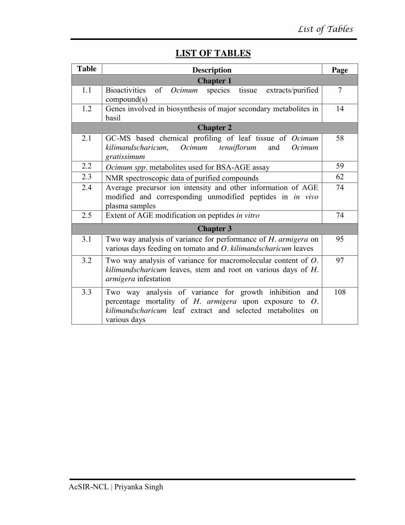
LIST OF TABLES
Table Description Page Chapter 1
1.1 Bioactivities of Ocimum species tissue extracts/purified compound(s)
7
1.2 Genes involved in biosynthesis of major secondary metabolites in basil
14
Chapter 2 2.1 GC-MS based chemical profiling of leaf tissue of Ocimum
kilimandscharicum, Ocimum tenuiflorum and Ocimum gratissimum
58
2.2 Ocimum spp. metabolites used for BSA-AGE assay 59 2.3 NMR spectroscopic data of purified compounds 62 2.4 Average precursor ion intensity and other information of AGE
modified and corresponding unmodified peptides in in vivo plasma samples
74
2.5 Extent of AGE modification on peptides in vitro 74
Chapter 3 3.1 Two way analysis of variance for performance of H. armigera on
various days feeding on tomato and O. kilimandscharicum leaves 95
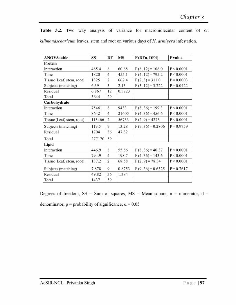
3.2 Two way analysis of variance for macromolecular content of O. kilimandscharicum leaves, stem and root on various days of H. armigera infestation
97
3.3 Two way analysis of variance for growth inhibition and percentage mortality of H. armigera upon exposure to O. kilimandscharicum leaf extract and selected metabolites on various days
108
Page 9
List of Figures
AcSIR-NCL | Priyanka Singh
LIST OF FIGURES
Figure Description Page Chapter 1
1.1 Representative examples of structurally diverse classes of secondary metabolites viz. monoterpenes, sesquiterpenes and phenylpropanoids found across genus Ocimum
10
1.2 Overview of diversity across Ocimum species 11 1.3 Factors responsible for chemical diversity; and terpenoid and
phenylpropanoid pathway diversification in Ocimum species 17
1.4 Major regulatory checkpoints in phenylpropanoid and terpenoid pathways
18
Chapter 2 2.1 In vitro BSA-AGE inhibition assay 60 2.2 Glycation inhibition assays by aminoguanidine and eugenol 61 2.3 In silico analysis of interactions between eugenol and MSA 64 2.4 Glycation modifications depicting surface exposed lysine
residues 64
2.5 Biophysical analysis of BSA and eugenol interaction 65 2.6 Kinetic studies of alpha-glucosidase inhibition by eugenol 67 2.7 Inhibition of α-glucosidase by eugenol slows carbohydrate
metabolism resulting in decrease in blood glucose 68
2.8 Analysis of blood biochemical parameters 69 2.9 Histopathological examination of pancreas, kidney, liver and
brain tissues 70
2.10 Proteomic analysis of in vitro and in vivo samples for AGE formation
72
2.11 MS/MS spectra annotation of AGE modified peptides of MSA 75 2.12 MS/MS spectra annotation of AGE modified peptides of BSA 76 2.13 Schematic presentation of proposed potential dual role of
eugenol in inhibiting AGEs 78
Chapter 3 3.1 Performance of H. armigera feeding on tomato and O.
kilimandscharicum leaves 94
3.2 Protein, carbohydrate and lipid content of O. kilimandscharicum leaves following H. armigera feeding
98
3.3 Changes in protein, carbohydrate and lipid content in local versus systemic leaves of O. kilimandscharicum following H. armigera feeding
99
Page 10
List of Figures
AcSIR-NCL | Priyanka Singh
3.4 Digestive enzymes of H. armigera larvae fed on O. kilimandscharicum leaves
102
3.5 Metabolic changes in leaves of O. kilimandscharicum following H. armigera infestation
104
3.6 Metabolic changes in stems and roots of O. kilimandscharicum following H. armigera infestation
105
3.7 Antibiosis to H. armigera following exposure to O. kilimandscharicum leaf extract and selected compounds
107
Chapter 4 4.1 Tissue- specific metabolite partitioning observed in young leaves,
mature leaves, inflorescence (Infl.), flower and root of Ocimum species
126
4.2 Differential expression of genes involved in phenylpropanoid and terpenoid pathway
128
4.3 LC- Orbitrap based global untargeted metabolomics 130 4.4 LC-Orbitrap based trichome metabolomics 131 4.5 Putative camphor biosynthetic pathway in O. kilimandscharicum 133 4.6 A. tumefaciens- mediated gene silencing (RNAi) of geranyl
diphosphate synthase (gpps) and borneol dehydrogenase (bdh) in O. kilimandscharicum
135
4.7 A. tumefaciens- mediated gene overexpression (OE) of geranyl diphosphate synthase (gpps) and borneol dehydrogenase (bdh) in O. kilimandscharicum
136
4.8 Eugenol biosynthetic pathway from Ocimum species: Enzymes and metabolites/intermediates detected in NGS and global metabolomic analysis
138
4.9 Gene co-expression network of EGS1 and 4CL4 using CoExpNetViz
140
4.10 Diagrammatic representation of mechanism underlying metabolite partitioning in O. kilimandscharicum
142
Chapter 5 5.1 Factors responsible for chemical diversity; and terpenoid and
phenylpropanoid pathway diversification in Ocimum species 150
5.2 Schematic presentation of proposed potential dual role of eugenol in inhibiting AGEs
151
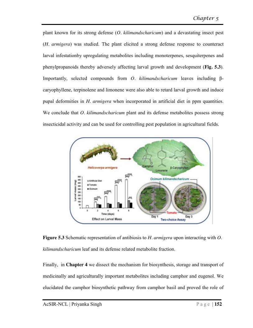
5.3 Schematic representation of antibiosis to H. armigera upon interacting with O. kilimandscharicum leaf and its defense related metabolite fraction
152
5.4 Mechanism underlying metabolite partitioning in O. kilimandscharicum
153
Page 11
Abbreviations
AcSIR-NCL | Priyanka Singh
LIST OF ABBREVIATIONS
°C degree centigrade
µL Microlitre
µM Micromolar
4CL 4-Coumarate-CoA Ligase
AAT Alcohol Acyl Transferase
ABC ATP- Binding Cassette
ADT Aerogenate Dehydratase
AGE Advanced Glycation End Products
AGT Anthocyanidin 3-O-Glucoside 5-O-Glucosyltransferase
ANNOVA Analysis Of Variance
ANS Anthocyanidin Synthase
ATP Adenosine Triphosphate
BCS β-Caryophyllene Synthase
bdh Borneol Dehydrogenase
BERS Bergamotene Synthase
BIS Bisabolol Synthase
bppd Bornyl Diphosphate Diphosphatase
bpps Bornyl Diphosphate Synthase
BSA Bovine Serum Albumin
C3H p-Coumarate 3-Hydroxylase
C4H Cinnamate-4-Hydroxylase
CAAT Coniferyl Alcohol Acetyl Transferase
Page 12
Abbreviations
AcSIR-NCL | Priyanka Singh
CAD Cinnamyl Alcohol Dehydrogenase
CDS Cadinene Synthase
CCMT Cinnamate Carboxyl Methyltransferase
CCR Cinnamoyl-CoA Reductase
CEL Carboxyethyl Lysine
CHI Chalcone Isomerase
CHS Chalcone Synthase
CM Chorismate Mutase
CML Carboxy Methyl Lysine
CO2 Carbon Dioxide
COMT Caffeoyl O-Methyl Transferase
CPS 9-Epi-Caryophyllene Synthase
CS Chorismate Synthase
CVOMT Chavicol O-Methyl Transferase
Cyp Cytochrome p450 Oxidoreductase
DCM Dichloromethane
DFR Dihydroflavonol 4-Reductase
DHQD 3-Dehydroquinate Dehydratase
DMAPP Dimethyl Allyl Pyrophosphate
DMSO Dimethyl Sulphoxide
DTPs Diterpene synthases
DXR 1-Deoxy-D-Xylulose-5-Phosphate Reductoisomerase
DXS 1-Deoxy-D-Xylulose 5-Phosphate Synthase
Page 13
Abbreviations
AcSIR-NCL | Priyanka Singh
EF1α Elongation Factor-1-alpha
EGS Eugenol Synthase
EME Eugenol Methyl Ether
EOMT Eugenol-O-Methyltransferase
ERF Ethylene Responsive Factor
HESI Heated Electrospray Ionization
F3H Flavanone 3-Hydroxylase
FA Fatty Acids
FAR Farnesene Synthase
FES Fenchol Synthase
FPP Farnesyl Diphosphate
FPPS Farnesyl Diphosphate Synthase
GAS Germacrene-A Synthase
GC Gas Chromatography
GDS Germacrene- D synthase
GES Geraniol Synthase
GGPPS Geranylgeranyl Diphosphate Synthase
GPP Geranyl Diphosphate
GPPS Geranyl Diphosphate Synthase
GPPS.LSU Geranyl Diphsophate Synthase Large Subunit
GPPS.SPS Solanesyl Diphhosphate Synthase
GPPS.SSU Geranyl Diphsophate Synthase Small Subunit
GSAR Glycation Sensitive Amino acid Residues
Page 14
Abbreviations
AcSIR-NCL | Priyanka Singh
GST Glutathione S-Transferase
GTS Gamma-Terpinene Synthase
HbA1c Glycated Hemoglobin
HCA Heirarchial Cluster Analysis
HCT Hydroxycinnamoyl Transferase
HDS 4-hydroxy-3-methylbut-2-enyl Diphosphate Synthase
HMGR 3-Hydroxy-3-Methylglutaryl-CoenzymeA Reductase
HMGS 3-Hydroxy-3-Methylglutaryl-CoenzymeA Synthase
HPPD 4-Hydroxyphenylpyruvate Dioxygenase
HRP Horse Radish Peroxidase
IPI Isopentenyl Diphosphate Isomerase
IPP Isopentenyl Pyrophosphate
KS Ent-Kaurene Synthase
LC Liquid chromatography
LIM Limonene Synthase
LIS Linalool Synthase
LMS Limonene-Myrcene Synthase
m/z Mass by Charge
MATE Multi-Antimicrobial Extrusion Protein
MCT 2-C-Methyl-D-Erythritol 4-Phosphate cytidylyltransferase
MDS 2-C-Methyl-D-Erythritol 2,4-Cyclodiphosphate Synthase
MeJa Methyl Jasmonate
MEK 4-Diphosphocytidyl-2-C-Methyl-D-Erythritol Kinase
Page 15
Abbreviations
AcSIR-NCL | Priyanka Singh
MEP Methylerythritol Phosphate
MeV Multiexperiment Viewer
MS Mass Spectrometry
MSA Mouse Serum Albumin
MTPs Monoterpene Synthases
MVA Mevalonic Acid
MVK Mevalonate Kinase
MYS Myrcene Synthase
NADH Reduced Nicotinamide Adenine Dinucleuotide
NES/LIS Nerolidol/Linalool Synthase
NES Nerolidol Synthase
NGS Next Generation Sequencing
NMR Nuclear Magnetic Resonance
Ob4CL Ocimum basilicum 4-Coumarate-CoA Ligase
OE Gene Overexpression
OE_BDH bdh overexpression
OE_GPPS gpps overexpression
PAGE Polyacrylamide Gel Electrophoresis
PAL Phenylalanine Ammonia-Lyase
PAT Prephanate Aminotransferase
PCR Polymerase Chain Reaction
PHD Plant Homeo Domain
PIN Pinene Synthase
Page 16
Abbreviations
AcSIR-NCL | Priyanka Singh
PNPG para-Nitrophenyl-α-D-Glucopyranoside
PPO Polyphenol Oxidase
PUR Pulegone Reductase
PVDF Polyvinylidene Difluoride
RAGE Receptor for Advanced Glycation End Product
RAS Rosmarinic Acid Synthase
RNA Ribonucleic acid
RNAi Gene silencing
RNAi_BDH bdh silencing
RNAi_GPPS gpps silencing
RPKM Reads Per Kilobase per Million
SAM S-Adenosyl-L-Methionine
SCS 1,8-Cineole Synthase
SDS Sodium Dodecyl Sulphate
SDSS Sequence Detection System software
SES Selinene Synthase
STPS Sesquiterpene Synthases
STZ Streptozotocin
TBS Tris Buffered Saline
TES Terpinolene Synthase
TLC Thin Layer Chromatography
UV-A Ultraviolet-A
ZIS Zingiberene Synthase
Page 17
Table of Contents
AcSIR-NCL | Priyanka Singh
Table of contents
Preface……………………………………………………………………………….... 1-2
CHAPTER 1
General Review of Metabolite Diversity and Complex Chemical Evolution in
Genus Ocimum………………………………………………………………………... 3-46
1.1 Introduction……………………………………………………………………… 4
1.1.1 Importance of studying genus Ocimum……………………………….. 5
1.1.2 Overview of extensive diversity within genus Ocimum………………. 6
1.2 Potential evolutionary events influencing metabolite diversity via pathway
modulation……………………………………………………………………………... 12
1.3 Factors regulating secondary metabolite flux and chemical diversity in Ocimum
species…………………………………………………………………………………. 16
1.3.1 Differential gene expression of enzymes in phenylpropanoid and
terpenoid pathways…………………………………………………………….. 17
1.3.2 Enzyme promiscuity…………………………………………………. 19
1.3.3 Transcription factors…………………………………………………. 20
1.3.4 Post-translational modifications………………………………………. 21
1.3.5 Presence of isozymes…………………………………………………. 22
1.3.6 External factors……………………………………………………….. 22
1.3.7 Developmental and tissue specific regulation………………………… 23
1.3.8 MicroRNA mediated regulation……………………………………… 24
1.4 Future Applications……………………………………………………………… 24
Page 18
Table of Contents
AcSIR-NCL | Priyanka Singh
1.5 Objectives of the work…………………………………………………………... 26
1.6 References……………………………………………………………………….. 27
CHAPTER 2
Potential dual role of eugenol in inhibiting advanced glycation end products
(AGEs) in diabetes: Proteomic and mechanistic insights………………………….. 47-84
2.1 Introduction……………………………………………………………………… 48
2.2 Materials and method……………………………………………………………. 50
2.2.1 Chemicals and plant material………………………………………….. 50
2.2.2 Gas chromatography-mass spectrometry (GC-MS) analysis of
Ocimum plant tissues…………………………………………………………… 50
2.2.3 Purification and NMR characterization of major metabolites from
Ocimum species………………………………………………………………… 51
2.2.4 BSA-AGE fluorescence assay………………………………………… 51
2.2.5 Blind docking and probability analysis ……………………………….. 52
2.2.6 Intrinsic fluorescence assay…………………………………………… 52
2.2.7 Circular dichorism analysis of BSA and BSA-eugenol complexes…… 52
2.2.8 Animal experiments…………………………………………………… 53
2.2.9 Estimation of blood glucose and HbA1c levels……………………….. 53
2.2.10 α-glucosidase inhibition assay and kinetics…………………………… 54
2.2.11 Plasma collection and insulin measurement ………………………….. 54
2.2.12 Tissue processing for histopathology …………………………………. 54
2.2.13. Western blotting ……………………………………………………… 55
2.2.14 In-gel trypsin digestion and LC-MS analysis of in vitro samples…….. 55
Page 19
Table of Contents
AcSIR-NCL | Priyanka Singh
2.2.15 In-solution trypsin digestion and LC-MS/MS analysis of plasma
proteins………………………………………………………………………….. 56
2.2.16 Statistical Analysis…………………………………………………….. 57
2.3 Results and discussion…………………………………………………………… 57
2.3.1 Chemical profiling unravels terpene and phenylpropanoid abundance
in Ocimum species……………………………………………………………… 57
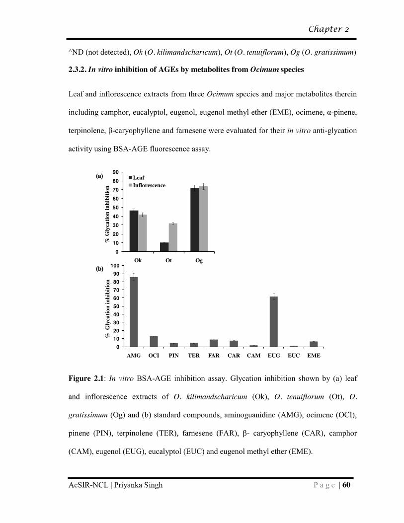
2.3.2 In vitro inhibition of AGEs by metabolites from Ocimum species……. 60
2.3.3 Eugenol shows increased binding affinity for surface lysine residues
on mouse serum albumin but does not alter the protein secondary structure…... 63
2.3.4 Eugenol administration affects blood biochemical parameters……….. 66
2.3.5 Mixed inhibition of α-glucosidase by eugenol might lead to decrease
in blood glucose………………………………………………………………… 69
2.3.6 Eugenol treated mice display significantly less histopathological
lesions…………………………………………………………………………… 70
2.3.7 Western blot analysis shows in vitro and in vivo inhibition of AGEs
by Eugenol …………………………………………………………………….. 71
2.3.8 LC-MS analysis reveals lesser extent of AGE modification on
peptides upon eugenol treatment……………………………………………….. 73
2.4 Conclusions…………………………………………………………………….. 77
2.5 References……………………………………………………………………… 79
CHAPTER 3
Insecticidal potential of defense metabolites from O. kilimandscharicum against
Helicoverpa armigera…………………………………………………………………. 85-114
Page 20
Table of Contents
AcSIR-NCL | Priyanka Singh
3.1 Introduction ……………………………………………………………………. 86
3.2 Materials and methods………………………………………………………….. 88
3.2.1 Insect culture ………………………………………………………….. 88
3.2.2 Plant maintenance……………………………………………………... 88
3.2.3 Feeding-choice assay …………………………………………………. 89
3.2.4 Growth and mortality data…………………………………………….. 89
3.2.5 Biochemical and metabolite study…………………………………….. 89
3.2.6 Estimation of carbohydrates, proteins, and lipids from plant tissues….. 90
3.2.7 H. armigera enzyme activity assays ………………………………….. 90
3.2.8 Extraction and analysis of metabolites………………………………… 91
3.2.9 H. armigera larvae fed on specific compounds ………………………. 92
3.2.10 Statistical analysis……………………………………………………... 9 92
3.3 Results and discussion………………………………………………………….. 93
3.3.1 O. kilimandscharicum defense compounds deter larvae from feeding,
adversely affecting their growth and development……………………………... 93
3.3.2 Changes in protein, carbohydrate and lipid content in O.
kilimandscharicum upon insect attack………………………………………….. 96
3.3.4 Compounds associated with secondary metabolism are central to O.
kilimandscharicum defense…………………………………………………….. 103
3.3.5 O. kilimandscharicum metabolites cause severe pupal deformities in
H. armigera……………………………………………………………………... 106
3.4 Conclusion………………………………………………………………………. 108
3.5 References………………………………………………………………………. 109
Page 21
Table of Contents
AcSIR-NCL | Priyanka Singh
CHAPTER 4
Integrating transcriptomics with metabolomics reveals tissue- specific metabolic
partitioning in O. kilimandscharicum……………………………………………….. 115-148
4.1 Introduction……………………………………………………………………. 116
4.2 Materials and methods…………………………………………………………. 119
4.2.1 Chemicals and reagents………………………………………………... 119
4.2.2 Plant growth conditions……………………………………………….. 119
4.2.3 Gas Chromatography – Mass Spectrometry…………………………... 119
4.2.4 Transcriptome profiling……………………………………………….. 120
4.2.5 Global untargeted metabolomics using LC-Orbitrap………………….. 120
4.2.5.1 Metabolite extraction……………………………………… 120
4.2.5.2 Liquid Chromatography-Orbitrap instrument set up……… 121
4.2.5.3 LC-MS data preprocessing and analysis…………………... 121
4.2.6 Heatmap and HCA analysis…………………………………………… 122
4.2.7 Real time analysis for gpps and bdh…………………………………... 122
4.2.8 A. tumefaciens mediated in planta transient silencing and
overexpression…………………………………………………………………... 123
4.2.8.1 Vector construction and agroinfiltration…………………... 123
4.2.8.2 Analysis of transient transgenics local and systemic leaves. 124
4.2.9 Co-expression Analysis………………………………………………... 125
4.3 Results and Discussion…………………………………………………………. 125
4.3.1 Ocimum species display stringent metabolite partitioning between
aerial and root tissue……………………………………………………………. 125
Page 22
Table of Contents
AcSIR-NCL | Priyanka Singh
4.3.2 Transcriptomic data reveals tissue-specific expression of terpenoid
and phenylpropanoid pathway genes in O. kilimandscharicum………………… 127
4.3.3 Global untargeted metabolomics reveals tissue- specific accumulation
of metabolites in O. kilimandscharicum………………………………………... 129
4.3.4 Camphor biosynthesis: Differential expression of both gpps and bdh is
putatively responsible for partitioning of camphor……………………………... 132
4.3.5 Transient gene silencing and overexpression studies reveal role of
gpps and bdh in camphor biosynthesis…………………………………………. 134
4.3.6 Eugenol biosynthesis and partitioning: Transport from source (leaves)
to sink (root)…………………………………………………………………….. 137
4.3.7 Co-expression analysis suggests that transporter(s) might be involved
in partitioning of eugenol in roots………………………………………………. 139
4.4 Conclusion……………………………………………………………………… 141
4.5 References……………………………………………………………………… 143
CHAPTER 5
Summary and Conclusion……………………………………………………………. 149
List of Publications…………………………………………………………………… 155
List of Patents………………………………………………………………………… 157
Curriculum Vitae……………………………………………………………………... 158
Page 23
Preface
AcSIR-NCL | Priyanka Singh P a g e | 1
Preface
Ocimum species present a wide array of diverse metabolites possessing immense
medicinal and economic value. The importance of this genus is undisputable and
exemplified in the ancient science of Chinese and Indian (Ayurveda) traditional medicine.
The key to this medicinal potential of genus Ocimum might lie in the vast array of
secondary metabolites and phytochemicals including terpenoids, phenylpropanoids,
flavonoids, phenolic compounds etc. present in various plant parts. Unlike several other
plant species of Artemisia, Salvia, Catharanthus, Taxus, Mentha, etc. that are largely
exploited, detailed characterization and identification of important metabolites from
Ocimum species remained unexplored. Also, molecular pathways leading to the
production, storage, transport and metabolism of these compounds are poorly understood.
We believe that a better understanding of the multi-level regulation of biosynthesis of
intermediates and metabolites, coupled with an understanding of their bioactivity, will
help us harness the inherent diversity of Ocimum species optimally.The present study aims
to explore Ocimum metabolome for important metabolites and study their biosynthesis,
storage and transport in plant. Chapter 1 includes a detailed survey of literature related to Ocimum species and their
importance in the plant kingdom. It also discusses our present understanding of the
metabolite diversity present across different species and factors responsible for the
complex chemical evolution. Based on our comprehensive understanding of the genus and
Page 24
Preface
AcSIR-NCL | Priyanka Singh P a g e | 2
available metabolomic and genetic resources, the chapter concludes with the objectives of
the work.
Chapter 2 comprises exploring medicinal applications of Ocimum metabolites. The study
revealed eugenol as an effective inhibitor of Advanced Glycation End products (AGEs)
using biophysical, biochemical, proteomic and in vivo mice model studies. Eugenol was
found to have a dual mode of action in combating diabetes; it lowered blood glucose by
inhibiting α-glucosidase and prevented AGE formation by binding to ε-amine group on
lysine, protecting it from glycation, offering potential use in diabetic management.
Chapter 3 deals with exploring agro- based application of Ocimum metabolites. Defense
metabolites including camphor, limonene and β-caryophyllene were found to be effective
bio-pesticides, using feeding- choice assays, analysis of insect growth and mortatlity data
as well as studying changes in tuning of primary and secondary metabolism in plant.
Chapter 4 deals with understanding the biosynthesis, transport and storage of important
Ocimum metabolites revealed in our previous studies. Integrating transcriptomics with
metabolomics not only helped us in dissecting the camphor and eugenol biosynthetic
pathway from camphor basil, it also revealed stringent metabolite partitioning betweeen
aerial and root tissues. The molecular mechanism underlying metabolite partitioning and
its probable biological relevance to the plant was further explored.
Chapter 5 gives a comprehensive summary, highliting key findings from the work done
during the Ph.D. tenure, finally culminating into major conclusions and future prospects.
Page 25
Chapter 1
AcSIR-NCL | Priyanka Singh P a g e | 3
CHAPTER 1
_________________________________ General Review of Metabolite
Diversity and Complex Chemical Evolution in Genus Ocimum
________________________________
Page 26
Chapter 1
AcSIR-NCL | Priyanka Singh P a g e | 4
1.1 Introduction
Genus Ocimum belonging to family Lamiaceae comprises between 50 to150 species.1 The
difference in the estimates of species number is partly attributed to reasons like taxonomic
revisions and generic description of the genus amongst others. It was first described by
Linnaeus in 1753 in the book Species Plantarum.2 The name Ocimum basilicum was
derived from the Greek word Okimon (smell) and basilikon (royal), referring to its royal
fragnance. While in India the Ocimum plant is considered sacred and worshipped, in other
parts of the world it is hailed as the ―queen of herbs‖ because of its strong aromatic appeal
and culinary usage. With the establishment of ancient medicinal practises in India
(Ayurveda) and China (Traditional Chinese Medicine), Ocimum was recognised as a
medicinal herb with great healing powers. Ocimum kilimandscharicum or camphor basil
is a commercially and medicinally important specie which grows in rich moist well
drained soils and ambient temperature.
Main centres of diversity for Ocimum include tropical and subtropical regions of Africa,
India and South America.3 With the exception of O. tenuiflorum and O. gratissimum that
are indegenous to India, most species are native to Africa including O. kilimandscharicum
and found in wild population.4 Although Ocimum species are known to abound in
medicinally important metabolites, only few species have been thoroughly profiled. Our
knowledge about most other species remains limited. All species are identifiable by the
presence of a large amount of signature metabolite(s) along with several other metabolites
in relatively minute quantities. The diversity of metabolites produced by Ocimum plants is
indeed enormous. Specific functions and/or necessity for production of such diverse and
complex chemical compounds by the plant remain elusive. Interestingly, what we know is
Page 27
Chapter 1
AcSIR-NCL | Priyanka Singh P a g e | 5
certain Ocimum species are either ―terpenoid-rich‖ or ―phenylpropanoid-rich‖. However,
factors determining the direction of flux are largely unknown. Terpenoids are formed from
the mevalonic acid (MVA) pathway in the cytoplasm and the methylerythritol phosphate
(MEP) pathway in the plastid.5 Phenylpropanoid pathway starts with the amino acid
phenylalanine and eventually results in the formation of phenylpropenes such as eugenol,
chavicol, anethole etc., along with intermediates for biosynthesis of lignin, rosmarinic
acid, anthocyanins etc. These pathways have been well characterized in related genera
including Salvia, Mentha and Lavandula 6-12 but not in such details in any Ocimum
species. However, with the influx of next generation sequencing data13, 14 along with
metabolomics, proteomics and phylogeny studies,15-19 now it seems possible to gain a
deeper insight into the perplexing diversity. The present review aims at providing a
comprehensive overview of the evolutionary, environmental and internal factors that may
have resulted in pathway diversification and extensive chemical evolution across Ocimum
species.
1.1.1 Importance of studying genus Ocimum
The unequivocal importance of genus Ocimum was established more than 5,000 years
back with the advent of ancient traditional medicinal practises in India and China.
Thereafter, there have been several reports of important bioactivities of Ocimum species;
tissue extracts and metabolites there in (Table 1.1).20-81 Although most species in this
genus are associated with some or the other bioactivity, the exact compound or group of
compounds, responsible for the said bioactivity remains elusive in most cases (Table 1.1).
Basil also finds extensive application in the food, flavor, and fragnance industry, and the
essential oil serves as a major source of economic wealth to the country. The plant is easy
Page 28
Chapter 1
AcSIR-NCL | Priyanka Singh P a g e | 6
to grow and propagate, and adapts well to extreme environmental conditions including
high precipitation, long dry spells and high temperature. Some species are capable of
vegetative propagation through stem cuttings like O. kilimandscharicum, which makes
commercial cultivation less tedious and more cost effective. Several Ocimum species grow
as wild plants in various parts of the world. Since there has been no significant
domestication of this wild medicinal plant, its genetic diversity has been preserved in
nature, making the system more interesting to explore. Furthermore, presence of different
basil types/cultivars rich in diverse metabolites provides a unique system for studying
secondary metabolic pathways. In addition, glandular trichomes accord the opportunity to
study the biosynthesis and regulation of these pathways at the level of a single cell.
Ocimum thus presents an attractive system to explore, particularly from the point of view
of secondary metabolism.
1.1.2 Overview of extensive diversity within genus Ocimum
Although genus Ocimum boasts of 50 – 150 species, metabolite data for very few species
is available. For example, O. obovatum and O. labiatum are well tested for therapeutic
properties (Table 1.1); however, their chemical composition remains unknown. Ocimum
species abound in diverse secondary metabolites including terpenoids, phenylpropanoids,
rosmarinic acid, flavonoids and phenolics. Figure 1.1 shows representative examples of
structurally diverse classes of secondary metabolites found across genus Ocimum. These
mainly include monoterpenes (example, camphor, eucalyptol, α-pinene, β-ocimene,
terpinolene), sesquiterpenes (example, farnesene, β-caryophyllene, germacrene D) and
phenylpropanoids (example, eugenol, eugenol methyl ether, chavicol, methyl chavicol,
methyl cinnamate). Few metabolites like germacrene D and β-caryophyllene are
Page 29
Chapter 1
AcSIR-NCL | Priyanka Singh P a g e | 7
commonly found across most species in the genus; however, others like camphor and
eugenol have a specie- specific distribution. Higher terpenes (C20 and above) and
alkaloids have not been well charaterized from any Ocimum species.
Table 1.1: Bioactivities of Ocimum species tissue extracts/purified compound(s)
sp. Bioactivity Extract/Compound
Ok Free radical scavanging20 Leaf essential oil, camphor, mixture of 1,8-cineole and limonene
Anticancer20, 21 Leaf essential oil;20 50% alcoholic aqueous leaf extract21
Anti-inflamatory20 Leaf essential oil, camphor, mixture of 1,8-cineole and limonene
Insecticidal22 DCM leaf extract, camphor, limonene and β-caryophyllene
Antidiarrhoel23 Aqueous leaf extract Antimicrobial24 Essential oil, borneol, bornyl acetate,
camphor, caryophyllene oxide, 1,8-cineole, limonene, linalool, α-pinene, β-pinene, spathulenol
Antiplasmodial25 DCM plant extract
Antioxidant26,27 Methanolic extracts of leaves,26, 27 and callus26
Radioprotective21 50% alcoholic aqueous leaf extract Mosquito repellent28 Plant essential oil, dry plant material Olb Antioxidant29 Ethanolic leaf extract, labdane (isolated
diterpenoid) Anti-inflammatory29 Ethanolic leaf extract, labdane diterpenoid Ola Antimicrobial24,30,31 Essential oil extract; Ethanolic extract of
various plant parts; methanol, aqueous and n-hexane extracts
Mosquito-repellent32 Volatiles from fresh, dried and smoking dried leaves
Antioxidant27,33 Plant essential oils and methanolic extracts27
Anti-inflammatory34 Aqueous and ethanolic leaf extracts Hepatoprotective35 Aqueous and methanolic leaf extracts
Page 30
Chapter 1
AcSIR-NCL | Priyanka Singh P a g e | 8
Analgesic36 Aqueous and ethanolic plant extracts Oo Anitmicrobial37 Leaf essential oil Ot Antidiabetic38 60% ethanolic leaf extract Anti-hyperlipidemic38 60% ethanolic leaf extract Anti-oral toxicity effect38 Hydroalcoholic leaf extract
Antioxidant39 Methanolic extracts of leaf, inflorescence, stem and callus
DNA damage protective40 Anthocyanin extracts Antibacterial41,42 Essential oil Anticancer43 Aqueous and ethanolic leaf extracts Antiglycation44 Methanolic and water extracts and their
fractions (DCM, ethyl-acetate, n-butanol, water)
Antistress45 OciBest (whole plant extract in gelatin capsules)
α-amylase inhibitory46 Isopropanol extract Mosquito repellent47 Plant essential oil Antiherpes48 Methanol and DCM extracts Ameliorative potential49 Methanol extracts, Saponin- rich fraction Oa Free Radical Scavenging50 Ethanol, butanol and ethyl-acetate extracts
from leaves Anti-inflammatory Activity51 Essential oil, linalool, 1,8-cineole Anti-herpes48 Methanol and DCM extracts Antimircobial52 Plant essential oil Oba Antiherpes48 Methanol and DCM extracts Anti-inflammatory 53,54 Whole plants;53 Ethanol-water (25%) extract
of leaves54
Antiplasmodial55 Plant ethanolic extracts (leaf, stem, root,flower)
Antioxidant and Antimicrobial56-60
Essential oil extracted via hydrodistillation;56 plant extracts prepared using ethanol, butanol, chloroform,water, ethyl acetate;58 essential oil, linalool, eugenol59 acetone and ethanol extracts60
Antimalarial61 Leaf essential oil Anticancer62,63 Plant methanolic extract;62 petroleum ether
soluble and insoluble methanolic extracts, ursolic acid
Larvicidal activity64,65 Leaf essential oil64
Page 31
Chapter 1
AcSIR-NCL | Priyanka Singh P a g e | 9
Antituberculosis66 Methanolic extract of leaves, fruits and flowers; bacilicin
Preventing ischemia, reperfusion-induced cerebral damage and motor dysfunctions67
Ethyl-acetate extract of leaves
Antihypertensive effects68 Aqueous plant extract Vasorelaxant and anti-platelet
aggregation69 Aqueous plant extract
Antigiardial activity70 Plant essential oil, linalool, eugenol Antiviral71 Aqueous and ethanolic plant extracts,
apigenin, linalool, ursolic acid Og Protection of liver from
oxidative stress and fibrosis72 Polyphenol extract
Antioxidant and Antimutagenic73
Leaf aqueous extract
Antitrypanosomal and antiplasmodial74
Crude ethanol extract, essential oil and pure compounds
Free radical scavenging and antioxidant75
Aqueous extract, methanol extract and eugenol
Prevention against Liver Fibrosis76
Aqueous leaf extract
Antimicrobial77,78,79 Plant essential oil;77,78 eugenol, methyl eugenol;77 hexane and methanol extracts alone and in combination with aminoglycosides79
Corrosion Inhibition80 Seed extract
Cerebroprotection81 Ethanolic plant extract
*Ok (O. kilimandscharicum), Olb (O. labiatum), Ola (O. lamiifolium), Oo (O. Obovatum),
Ot (O. tenuiflorum), Oa (O. americanum), Oba (O. basilicum), Og (O. gratissimum),
DCM (dichlorormethane)
Since most Ocimum species have not been profiled for their metabolites, the possibility
that the genus represents much more diversity than what we percieve now is realistic. As
mentioned previously, each species is characterized by a distinct metabolic fingerprint and
Page 32
Chapter 1
AcSIR-NCL | Priyanka Singh P a g e | 10
presence of a signature compound(s) as the major fraction. Although metabolite profiling
via conventional techniques such as gas chromatography (GC) has been routinely
employed, advanced analytical techniques including liquid chromatography (LC), mass
spectrometry (MS) and nuclear magnectic resonance (NMR) have not been reported,
which help in gaining a better understanding of the global distribution of metabolites and
pathway intermediates.
Figure 1.1: Representative examples of structurally diverse classes of secondary
metabolites viz. monoterpenes, sesquiterpenes and phenylpropanoids found across genus
Ocimum
Till now only 12 Ocimum species have been analysed for their essential oil composition
(Fig. 1.2).
Page 33
Chapter 1
AcSIR-NCL | Priyanka Singh P a g e | 11
Overview of diversity across Ocimum species
Rela
tive
abun
danc
e (%
)
Figure 1.2: Overview of diversity across Ocimum species. Numbers in parenthesis
indicates number of compounds; numbers outside parenthesis indicate percentage of
metabolite in total volatile fraction. (Ot, O. tenuiflorum; Os, O. selloi; Og, O. gratissimum;
Ok, O. kilimandscharicum; Om, O. minimum; Oba, O.basilicum; Oa, O. americanum;
Oci x Cit, Ocimum x Citriodorum; Ola, O. lamiifolium; Oc, O. campechianum; Omi,
O. micranthum; Oca, O. canum)
Overall, they can be classified as having (i) high phenylpropanoid content, (ii) high
terpenoid content, and (iii) similar/comparable amounts of phenylpropnoids and
terpenoids. High phenylpropanoid content group contains about 60 to 90%
phenylpropanoids and includes O. gratissimum,82 O. tenuiflorum83 and O. selloi.84 High
terpenoid containing group, includes O kilimandscharicum,85 O. minimum,86 O.
basilicum,87 O. americanum,88 Ocimum × citriodorum89 and O. lamiifolium90 contain
approximately 40 to 75% terpenoids. The third group includes O. campechianum,91 O.
Page 34
Chapter 1
AcSIR-NCL | Priyanka Singh P a g e | 12
micranthum92 and O. canum93 which show approximately equal amount of
phenylpropanoids and terpenoids. Interestingly, terpenoids unlike phenylpropanoids, show
a universal presence in varying amount in all Ocimum species.
Signature compounds known in Ocimum species are as follows: camphor in O.
kilimandscharicum (56%), citral in O. americanum (47%), eugenol in O. gratissimum
(82%) and O. micranthum (47%), eugenol methyl ether in O. tenuiflorum (62%), linalool
in O. basilicum (48%), methyl chavicol in O. selloi (93%) and O. canum (53%), geranyl
acetate in O. minimum (70%), sabinene in O. lamiifolium (33%) and geranial in Ocimum ×
citriodorum (43%) (Fig. 1.2). In plant kingdom, metabolite diversity is commonly found
at the level of family or genus, but such vivid diversity at the level of species and subtypes
(within species) makes genus Ocimum occupy a special niche in nature.
1.2 Potential evolutionary events influencing metabolite diversity via
pathway modulation
Ocimum genome has evolved as a result of dramatic series of events including polyploidy,
aneuploidy, chromosomal duplications/translocations/deletions etc., 16, 94, 95 which led to
unprecedented diversification of species in Africa, India and South America. The ability to
cross-pollinate and hybridize further led to the emergence of subtypes within species,
which were capable of interbreeding and producing hybrids. For example,
Ocimum×citriodorum is a hybrid between O. americanum and O. basilicum and has a
strong lemony scent.18 O. americanum originated from O. canum and O. basilicum.96 The
African blue basil subtype (O. kilimandscharicum) is evolved as a hybrid between O.
kilimandscharicum and O. basilicum and abounds in camphor, linalool and eucalyptol.
Page 35
Chapter 1
AcSIR-NCL | Priyanka Singh P a g e | 13
Interestingly, the hybrids display significantly different metabolite profile than their
parents including new metabolites that are not found in the parents, indicating co-
dominance, epistatis or interaction of genes.89 As reported in several other plant genera,
ploidy levels also affect essential oil production, resulting in a greater accumulation of
essential oils in polyploids than that in diploids.95, 97-99 All these events taken together
might have led to greater genetic diversity and continuous expansion of gene pool,
yielding new species/subtypes/varieties over a short period.
During the course of evolution, there may have been events that led to terpenoid and
phenylpropanoid pathway diversification across different Ocimum species. It is interesting
to note that species abounding in phenylpropanoids also have an active terpenoid pathway
and vice versa. This suggests that all species evolved from an ancestor, which harbored
active genes for both the pathways. However, differential expression and regulation of
pathway genes determined the final chemical composition in each species.5, 100 Other
factors like plant habit may also have influenced the selection of one pathway over the
other. For example, it has been suggested that the sanctum group has evolved to produce
phenolic compounds because of its perennial woody habit, whereas the basilicum group
has evolved to produce terpenoid-rich compounds owing to its annual herbaceous habit.4
Evolution of gene coding regions also had a profound impact on the diversity of Ocimum
species metabolites. For example, O. basilicum fenchol synthase and myrcene synthase,
and geraniol synthase and linalool synthase are 95% and 81% similar, respectively;
however, they catalyse the formation of very different products. These genes most
probably evolved as a result of gene duplication events and acquired mutations leading to
functional differentiation, 100 eventually contributing to metabolite diversity. Few pathway
Page 36
Chapter 1
AcSIR-NCL | Priyanka Singh P a g e | 14
genes involved in the biosynthesis of selected metabolites have been reported and
characterized from Ocimum and few other genera (Table 1.2).
Table 1.2: Genes involved in biosynthesis of major secondary metabolites in basil
Compound Gene (abb.) (org.) Reaction catalysed Eugenol Eugenol synthase (EGS) (O.
basilicum and F. annanasa) coniferyl acetate to eugenol
Coumaryl CoA Ligase (4CL) (O. tenuiflorum)
hydroxycinnamic acids to Coenzyme A (CoA) esters
R2R3-MYB transcription factor (EOBII) (F. annanasa)
transcription factor regulating structural genes in phenylpropanoid pathway
Eugenol methyl ether
Eugenol O-methyl transferase (EOMT) (O. basilicum)
eugenol to eugenol methyl ether
Methyl chavicol
Chavicol O-methyl transferase (CVOMT) (O. basilicum)
chavicol to methyl chavicol
Camphor bornyl diphosphate synthase (BPPS) (S. officinalis)
geranyl diphosphate to bornyl diphosphate
Borneol dehydrogenase (BDH) (S. officinalis and L. intermedia)
borneol to camphor
Eucalyptol (1,8- cineole)
1,8-cineole synthetase (S. officinalis)
neryl diphosphate to 1,8-cineole
Linalool Linalool synthase (LIS) (O. basilicum)
GPP to linalool
Terpinolene Terpinolene synthase (TES) (O. basilicum)
GPP to terpinolene (as major product) and α-pinene and limonene (as side products)
Fenchol Fenchol synthase (FES) (O. basilicum)
GPP to fenchol (as major product) and α-pinene and limonene (as side products)
Myrcene Myrcene synthase (MES) (O. basilicum)
GPP to myrcene
Cadinene Cadinene synthase (CDS) (O. basilicum)
FPP to γ-cadinene (as major product) and Muurola 3,5-diene (as side product)
Page 37
Chapter 1
AcSIR-NCL | Priyanka Singh P a g e | 15
Selinene Selinene synthase (SES) (O. basilicum)
FPP to α & β- selinene (major product); β-elemene and nerolidol (side product)
Zingiberene Zingiberene synthase (ZIS) (O. basilicum)
FPP to α-zingiberene (major product); α-bergamotene, nerolidol, β-farnesene and β-bisabolene (side product)
Germacrene-D Germacrene-D synthase (GDS) (O. basilicum)
FPP to Germacrene-D
Geraniol Geraniol synthase (GES) (O. basilicum)
GPP to geraniol
Amyrin (triterpene)
2,3-oxidosqualene cyclase (AS1 and AS2) (O. basilicum)
2,3-epoxy-2,3-dihydrosqualene to α/β- amyrin
General phenyl propanoid pathway
Production of anthocyanin pigment 1 (PAP1) (A. thaliana)
transcriptional regulator of floral scent
p-coumaroyl shikimate 3'-hydroxylase (CS3'H) (O. tenuiflorum)
p-coumaroyl 5-O- shikimate to caffeoyl 5-O- shikimate
Caffeic acid O-methyl transferase (COMT) (O. basilicum)
caffeate to ferrulate
Caffeic acid O-methyl transferase (COMT) (O. tenuiflorum)
caffeate to ferrulate
Cinnamyl alcohol dehydrogenase (CAD) (O. tenuiflorum)
cinnamyl alcohol to cinnamyldehyde
Cinnamyl alcohol dehydrogenase (CAD) (O. basilicum)
cinnamyl alcohol to cinnamyldehyde
Cinnamate-4-hydroxylase (C4H) (O. tenuiflorum)
cinnamic acid to 4-coumaric acid
Cinnamate-4-hydroxylase (C4H) (O. basilicum)
cinnamic acid to 4-coumaric acid
Genes like eugenol synthase involved in catalysing the final step of eugenol production
has been well charaterized (Table 1.2). However, most genes present upstream in the
eugenol biosynthetic pathway remain functionally uncharacterized despite availability of
Page 38
Chapter 1
AcSIR-NCL | Priyanka Singh P a g e | 16
huge transcriptomic databases. Genes from camphor biosynthesis pathway have been well
characterized from related genera like Salvia and Lavandula, however, there are no reports
from genus Ocimum (Table 1.2). Modifying enzymes like chavicol and eugenol O-
methyltransferases also have been well characterized (Table 1.2). Information about
transcription factors responsible for controlling biosynthesis of these metabolites and the
transporter proteins responsible for long distance transport from source to sink tissue in
Ocimum species also remains scarce. Genes reported from yet another important category
of compounds, flavones and flavonoids, have been listed in Table 1.2. Overall,
information about the biosynthesis, transport and storage of these metabolites, at the
genetic level is very scarce and need to be further probed. Several other factors during
species diversification and naturalization in other parts of the world have been discussed
briefly, which help us in explaining the mystery behind the complex chemical evolution
and pathway diversification.
1.3 Factors regulating secondary metabolite flux and chemical
diversity in Ocimum species
Metabolite diversity observed at the level of species in genus Ocimum is dependent on
several internal and external factors (Fig. 1.3). Some of the known factors responsible for
regulating terpenoid and phenylpropanoid pathways are discussed.
Page 39
Chapter 1
AcSIR-NCL | Priyanka Singh P a g e | 17
External factors•Sunlight•Colour shading•Seasonal variation •Geographical location•Climatic condition•Rainfall/ precipitation
Internal factors•Differential gene expression•Transcription factors/ miRNAs•Post- translational modifications•Presence of isozymes•Tissue specific regulation•Enzyme promiscuity
Evolution & Speciation•Aneuploidy, polyploidy•Chromosomal aberrations•Cross pollination, hybridisation•Habit (annual/perennial)
Terpenoid-richspecies
Phenylpropanoid-rich species
Terpenoid and phenylpropanoid rich species
Pathway diversification
Figure 1.3: Factors responsible for chemical diversity; and terpenoid and phenylpropanoid
pathway diversification in Ocimum species
1.3.1 Differential gene expression of enzymes in phenylpropanoid and terpenoid
pathways
Gene expression plays an important role in diverting metabolic flux toward either the
terpenoid or the phenylpropanoid pathway.5, 13,100 In particular increased expression of
terminal enzymes in the terpenoid pathway and reduced expression of phenylpropanoid
entry point enzymes such as phenylalanine ammonia-lyase (PAL) has been observed in O.
basilicum var. SD, rich in terpenoids. In another variety, O. basilicum var. EMX,
however, the expression level of general phenylpropanoid pathway enzymes, PAL and 4-
coumarate-CoA ligase (4CL) was found to be significantly higher corresponding to
Page 40
Chapter 1
AcSIR-NCL | Priyanka Singh P a g e | 18
Phenylpropanoid biosynthesis
Shikimate
Phenylalanine
Cinnamic acid
4-Coumaric acid
4-Coumaryl CoA
Lignins
Phenylpropenes
Rosmarinic acidAnthocyanins
DAHPS
PAL
C4H
4CL
GPP
Route 1Route 2Route 3Route 4
Sucrose PEP Pyruvate Acetyl CoA
DXSDXP
MEPDXR
G3PAcetoacetyl CoA
MVA
MEP pathway MVA pathway
Terpenoid biosynthesis
IPP DMAPP
Monoterpenes
Triterpenes
SesquiterpenesDiterpenes
MTPSSTPSDTPSTTPS
Glycolysis
Figure 1.4: Major regulatory checkpoints in phenylpropanoid and terpenoid pathways.
Enzymes potentially governing the direction of flux have been marked in red. (PEP,
Phosphoenol pyruvate; G3P, Glyceraldehyde-3-phosphate; DXP, 1-deoxy-D-xylulose 5-
phosphate; MEP, Methylerythritol phosphate; MVA, Mevalonic acid; IPP, Isopentenyl
pyrophosphate; DMAPP, Dimethylallyl pyrophosphate; GPP, General phenylpropanoid
pathway; DAHPS, 3-Deoxy-D-arabinoheptulosonate 7-phosphate synthase; PAL,
Phenylalanine ammonia-lyase; C4H, Cinnamate-4-hydroxylase; 4CL, 4-Coumarate-CoA
ligase; DXS, 1-deoxy-D-xylulose 5-phosphate synthase; DXR, 1-deoxy-D-xylulose 5-
phosphate reductoisomerase; MTPS, Monoterpene synthases; STPS, Sesquiterpene
synthases; DTPS, diterpene synthases; TTPS; Triterpene synthases)
Page 41
Chapter 1
AcSIR-NCL | Priyanka Singh P a g e | 19
higher phenylpropanoid content.5 These results were supported by next generation
sequencing data of O. tenuiflorum and O. basilicum.14 O. tenuiflorum rich in
phenylpropanoids, shows much higher expression of general phenylpropanoid pathway
enzymes including PAL, cinnamate-4-hydroxylase (C4H) and 4CL, reads per kilobase per
million (RPKM) = 91.47, 34.53 and 9.52 respectively; compared to O. basilicum rich in
terpenoids, RPKM = 11.3, 11.83 and 5.65 respectively. In O. basilicum, however, the
entry point enzymes of the MEP pathway, representing the cytosolic pathway for
terpenoid synthesis, including 1-deoxy-D-xylulose 5-phosphate reductoisomerase (DXR)
was more (RPKM = 50.58) compared to O. tenuiflorum (RPKM = 15.69).14 Thus,
overexpressing the entry point enzymes at major metabolic branching points also helps in
directing the flux towards either phenylpropanoid or terpenoid pathway.122-124 Evidently,
differential expression of enzymes strategically present at pathway branch points might
play a crucial role in determining flux regulation (Fig. 1.4).
1.3.2 Enzyme promiscuity
One of the major reasons for metabolite diversity observed in Ocimum species is the
promiscuity of terpene synthases. These enzymes are capable of accepting a substrate and
yielding a major product as well as multiple side products. For instance, Iijima et al
characterized eight terpene synthases from three cultivars of O. basilicum.100, 114 In vitro
recombinant protein assays using geranyl diphosphate (GPP) as substrate for putative
monoterpene synthases and farnesyl diphosphate (FPP) as substrate for putative
sesquiterpene synthases was performed. Terpinolene synthase gave terpinolene as the
major product and α-pinene, limonene and an unidentified monoterpene as the side
products. Fenchol synthase produced fenchol and limonene as major products and α-
Page 42
Chapter 1
AcSIR-NCL | Priyanka Singh P a g e | 20
pinene and an unidentified monoterpene as the side products. Cadinene synthase produced
γ-cadinene as the major product and muurola 3, 5-diene as the side product. Selinene
synthase produced selinene as the major product and β-elemene and nerolidol as side
products. In contrast, myrcene synthase and geraniol synthase exclusively produced
myrcene and geraniol as end products.100 In another study by Major et al, using bornyl
diphosphate synthase (producing camphene as the side product), it was proven that
electrostatically guided dynamics determined end product formation.110 Current evidence
suggests that enzyme promiscuity may play an important role in contributing to the
diversity across Ocimum species.
1.3.3 Transcription factors
Transcriptional regulation of secondary metabolism in plants for flavonoids (particularly
anthocyanins), alkaloids (including nicotine, indole alkaloids and benzylisoquinolines)
and terpenoids has been widely reported.125-127 Recently, PAP1 transcription factor was
shown to enhance the production of both terpenoids and phenylpropanoids in rose plant.116
Deep sequencing of O. tenuiflorum and O. basilicum revealed the presence of 40
transcription factor families including MYB, WRKY, bHLH, HB, NAC, bZIP etc. which
are known regulators of secondary metabolism in plants.14 A recent study performed using
the red and green forma of O. tenuiflorum suggested light-mediated regulation of
anthocyanin accumulation.128 It was observed that when red forma seedlings grown under
natural lighting conditions, were transferred to a special greenhouse which cuts off the
UV-A and UV-B radiation, the leaves turned green within 20 days. Further investigation
revealed the role of transcription factors, bHLH and WD40, in downregulating the
terminal enzymes of anthocyanin biosynthesis including flavonone-3′-hydroxylase,
Page 43
Chapter 1
AcSIR-NCL | Priyanka Singh P a g e | 21
leucoanthocyanidine dioxygenase and dihydroflavonol reductase, responsible for red
coloration. In another study by Misra et al, transcription factors belonging to
APETALA2/Ethylene responsive factor (ERF), WRKY, plant homeo domain (PHD) and
zinc finger families were upregulated in methyl jasmonate (MeJa)-treated O. basilicum
plants, suggesting their possible role in regulating secondary metabolism in Ocimum
species.115 Thus, available data suggests transcription factors are also key regulators of
terpenoid and phenylpropanoid pathway in Ocimum species and provide a more stringent
control over the direction of flux.
1.3.4 Post-translational modifications
Post-translational modifications including phosphorylation, ubiquitination and arginine
monomethylation of phenylpropanoid and terpenoid pathway enzymes such as
phosphoglucomutase, glucose-6-phosphate isomerase, phosphoglycerate mutase, PAL and
chavicol O-methyl transferase (CVOMT) were observed in basil glandular trichomes. Post
translation modifications help in explaining situations where the mRNA level does not
match with the metabolite or protein level. For example, the enzyme CVOMT is
responsible for methylating chavicol. O. basilicum var. SD produces negligible amount of
methylchavicol. However, the mRNA and protein levels for this enzyme were found to be
very high. In contrast, very little enzyme activity and metabolites were detected. It was
observed that this enzyme was ubiquinated providing a valid explanation for the
discrepencies in mRNA, protein, enzyme activity and metabolite level. Ubiquitination
leads to a rapid degradation of CVOMT post translation,5 resulting in decreased formation
of methyl chavicol. In another example, PAL, catalyzing the first committed step in
phenylpropanoid biosynthesis, is phosphorylated in O. basilicum var. SD, rich in
Page 44
Chapter 1
AcSIR-NCL | Priyanka Singh P a g e | 22
monoterpenes; however, other basil varieties (SW, MC, and EMX-1), rich in
phenylpropanoids, lack PAL phosphorylation.5 It has been reported earlier that
phosphorylation results in the reduction of PAL activity.129,130 Above examples suggest
that post translation modifications provide an additional regulatory step in determining the
expression of key enzyme activities in secondary metabolic pathways in Ocimum species.
1.3.5 Presence of isozymes
Phenylpropanoid pathway produces substrates for synthesis of several important
secondary metabolites. PAL, C4H and 4CL catalyse the initial few steps leading to the
formation of coumaryl CoA. Latter represents a branching point, from which different end
products including phenylpropenes, lignins, flavonoids and rosmarinic acid can be
synthesized. Thus, 4CL represents a crucial step in pathway regulation and diversification.
In recent work by Rastogi et al, it was reported that O. basilicum 4CL has 5 different
isoforms.103 RNAi experiments involving the silencing of a specific isoform, Oba4CL, led
to a reduced production of phenylpropanoids without affecting lignin and sinapic acid
content. Thus, only one of the isoforms of 4CL was involved in the synthesis of
phenylpropenes. This also represents the commitment of a specific isoform of an enzyme
to a specific biosynthetic pathway at a very initial step. Presence of such pathway-
committed isoforms keeps the pathway finely tuned and delicately balanced in basil.
1.3.6 External factors
Being species native to the tropics, Ocimum plants are always subjected to severe
environmental conditions including excessive heat, rainfall, humidity, dryness etc.
Adaptability, thus, is the key to survival. It has been reported that external environmental
Page 45
Chapter 1
AcSIR-NCL | Priyanka Singh P a g e | 23
factors, including the type of light, radiation, season, geographic conditions etc., influence
essential oil composition. Same Ocimum specie show altered metabolic profile under
different environmental factors. Red and blue shading conditions in O. selloi showed
decline in level of phenylpropanoids and elevated level of in comparison with plants
grown in full light.84 Plants grown under blue shading had more number of metabolites
than plants subjected to full light and red shading. Decreased accumulation of methyl
chavicol was observed in plants cultured under colored netting, accompanied by an
increase in α-copaene, germacrene D and bicyclogermacrene content.84 This suggests a
chemical defense strategy of plants against less favorable growth conditions. Similar kind
of study was performed with O. basilicum cultivated in soil covered by colored mulches
which demonstrated that size and aroma of leaves as well as the concentration of soluble
phenols greatly improved.131 Seasonal variation of essential oil composition was observed
in O. basilicum and O. tenuiflorum.83 To show the effect of geographic conditions on
essential oil composition, O. gratissimum and O. campechianum were grown in Chocó
Department (Columbia) and Ecuador region that resulted in different chemical
composition.91 Similarly, O. basilicum and O. gratissimum grown in Benin, Cameroon,
Congo and Gabon vary in chemical composition.90 O.gratissimum plants grown in
Columbia showed altered metabolite profile as compared with those grown in
Europe.91,132-135 This data indicates external factors including climatic conditions and
geographical variations might be influencing the chemical profile of Ocimum species.
1.3.7 Developmental and tissue specific regulation
During cinnamic acid and methylcinnamate (MC) formation from phenylalanine, activity
of two enzymes, PAL and S-adenosyl-L-methionine: cinnamate carboxyl
Page 46
Chapter 1
AcSIR-NCL | Priyanka Singh P a g e | 24
methyltransferase (SAM:CCMT) shows an important regulatory control point.136 In
different developemental stages of O. basilicum, the relation between MC content, PAL
and SAM:CCMT activity was examined. SAM: CCMT activity showed correlation with
MC content in young leaves.136 Likewise, eugenol-O-methyltransferase (EOMT) is
responsible for methylation of eugenol to form methyleugenol in one of the final steps of
phenylpropanoid pathway. The expression pattern of EOMT positively correlated with the
amount of eugenol/isoeugenol and methyleugenol in different developmental stages of all
the analyzed chemotypes.137 Along with development-specific regulation of metabolite
accumulation, some metabolites in Ocimum species also show tissue-specific regulation.
For example, analysis of trichome, leaf, stem and root shows a strong association between
eugenol content and Ob4CL expression in O. basilicum.103
1.3.8 MicroRNA mediated regulation
Based on O. basilicum EST data set, the function of miRNAs and their targets was
predicted using in silico approach.138 Four miRNA families miR164c, miR5658, miR414
and miR5021 were evaluated for their potential targets. These miRNA families showed
regulatory role during stress-metabolite response. Although this study was based upon
computational evaluation, further in planta experimentation is required to determine the
critical role of miRNAs during secondary metabolism in Ocimum species.138
1.4 Future Applications
Ocimum acts as a reservoir of several important secondary metabolites found in nature,
thereby making it a very attractive system to explore. Although the genome of Ocimum
has not yet been sequenced, the recent influx of next generation sequencing data of
various tissues such as trichomes and leaves, has helped us in understanding various
Page 47
Chapter 1
AcSIR-NCL | Priyanka Singh P a g e | 25
factors that are responsible for regulating the formation of phenylpropanoids and
terpenoids in Ocimum species. Using the current information, we can genetically engineer
Ocimum species to overexpress the desired metabolites by redirecting the metabolite
flux.139-142 This knowledge can also be used for breeding new chemotypes producing
interesting spectra of essential metabolites. Since these metabolites impart flavor and
aroma, and possess medicinal properties, they can be heterologously expressed in plants,
which are routinely used raw in our diet, such as tomato, thereby increasing their flavor
and nutritive value. The expression of O. basilicum α-zingiberene synthase under the
control of polygalacturonase promotor led to the unexpected accumulation of 15
sesquiterpenes and 10 monoterpenes, which were not present in the non-transformed
fruit.143 In a separate study, the expression of O. basilicum geraniol synthase under the
same promoter led to the accumulation of geraniol and its derivatives, which had profound
impact on tomato flavor as well as aroma.144 Moreover, expressing terpene synthase genes
from Ocimum in food crops will impart greater resistance against pathogens and pests. Till
date, it is not well established whether there is a cross talk between the phenylpropanoid
and terpenoid pathways. The glandular trichomes present in several Ocimum plants
provide a very exciting isolated single-celled system to unravel the exchange, if any, of
upstream intermediates between these two pathways. Thus, Ocimum species find useful
applications in industrial, culinary, medicinal as well as scientific research areas, asserting
their important position in the plant kingdom.
Page 48
Chapter 1
AcSIR-NCL | Priyanka Singh P a g e | 26
1.5 Objectives of the work
The experimental work in this thesis revolves around exploring the Ocimum metabolome
for metabolites of medicinal, agricultural and commercial importance and understanding
their biosynthesis, transport and storage in planta. The major objectives of the work are as
follows:
¾ Understanding metabolite diversity present across genus Ocimum and factors
responsible for complex chemical evolution
¾ Exploring medicinal applications of Ocimum metabolites using biophysical,
biochemical, proteomic and in vivo mice model studies
¾ Elucidating agro- based application of Ocimum metabolites (defense
metabolites as effective bio-pesticides)
¾ Next generation sequencing (using Illumina platform) and global untargeted
metabolomics (using LC- Orbitrap) of different tissues of camphor basil to
understand putative genes and metabolites involved in synthesis of
commercially and medicinally important metabolites like camphor and
eugenol.
¾ Dissecting camphor biosynthetic pathway from O. kilimandscharicum by
cloning and in planta functional characterization (gene silencing and
overexpression) of pathway genes including geranyl diphosphate synthase and
borneol dehydrogenase.
¾ Understanding the mechanism underlying metabolite partitioning of eugenol
and camphor in camphor basil to understand their synthesis, transport and
storage in plant.
Page 49
Chapter 1
AcSIR-NCL | Priyanka Singh P a g e | 27
1.6 References
(1) J.E. Simon, J. Quinn and R.G. Murray, in Advances in new crops, ed. J. Janick and
J.E. Simon, Timber Press, Portland, 1990, Basil: A source of essential oils, 484.
(2) C. Linnaeus, in Species Plantarum, Laurentii Salvii, Holmiae (Stockholm), 1753,
Ocimum, 597.
(3) P.D. Cantino, R.M. Harley and S.J. Wagstaff, in Advances in Labiate Science, ed. R.
M. Harley and T. Reynolds, Royal Botanic Gardens, Kew, 1992, Genera of Labiatae:
Status and Classification, 502.
(4) M. K. Khosla, Study of inter-relationship, phylogeny and evolutionary tendencies in
genus Ocimum. Ind J. Genet., 1995, 55, 71.
(5) Z. Xie, J. Kapteyn and D.R. Gang, A systems biology investigation of the
MEP/terpenoid and shikimate/phenylpropanoid pathways points to multiple levels of
metabolic control in sweet basil glandular trichomes. Plant J., 2008, 54, 349.
(6) D. Yang, X. Du, X. Liang, R. Han, Z. Liang, Y. Liu, F. Liu and J. Zhao, Different roles
of the mevalonate and methylerythritol phosphate pathways in cell growth and tanshinone
production of Salvia miltiorrhiza hairy roots. PLoSOne, 2012, 7, e46797.
(7) A. Marchev, C. Haas, S. Schulz, V. Georgiev, J. Steingroewer, T. Bley and A. Pavlov,
Sage in vitro cultures: a promising tool for the production of bioactive terpenes and
phenolic substances. Biotechnol. Lett, 2014, 36, 211.
(8) G.W. Turner and R. Croteau, Organization of monoterpene biosynthesis in Mentha.
Immunocytochemical localizations of geranyl diphosphate synthase, limonene-6-
hydroxylase, isopiperitenol dehydrogenase, and pulegone reductase. Plant Physiol, 2004,
136, 4215.
Page 50
Chapter 1
AcSIR-NCL | Priyanka Singh P a g e | 28
(9) R. B. Croteau, E. M. Davis, K. L. Ringer and M. R. Wildung, (−)-Menthol biosynthesis
and molecular genetics. Naturwissenschaften, 2005, 92, 562.
(10) R. Rios-Estepa, G. W. Turner, J. M. Lee, R. B. Croteau and B. M. Lange, A systems
biology approach identifies the biochemical mechanisms regulating monoterpenoid
essential oil composition in peppermint. PNAS, 2008, 105, 2818.
(11) A. Lane, A. Boecklemann, G. N. Woronuk, L. Sarker and S. S. Mahmoud, A genomics
resource for investigating regulation of essential oil production in Lavandula angustifolia.
Planta, 2010, 231, 835.
(12) G. Woronuk, Z. Demissie, M. Rheault and S. Mahmoud, Biosynthesis and therapeutic
properties of Lavandula essential oil constituents. Planta Med., 2011, 77, 7.
(13) D. R. Gang, J. Wang, N. Dudareva, K. H. Nam, J. E. Simon, E. Lewinsohn and E.
Pichersky, An investigation of the storage and biosynthesis of phenylpropenes in sweet
basil. Plant Physiol., 2001, 125, 539.
(14) S. Rastogi, S. Meena, A. Bhattacharya, S. Ghosh, R. K.Shukla, N.S. Sangwan, R. K.
Lal, M. M. Gupta, U. C. Lavania, V. Gupta, D. A. Nagegowda and A. K. Shasany, De novo
sequencing and comparative analysis of holy and sweet basil transcriptomes. BMC
Genomics, 2014, 15, 588.
(15) F. Bast, P. Rani and D. Meena, Chloroplast DNA phylogeography of holy basil
(Ocimum tenuiflorum) in Indian subcontinent. Scientific World J., 2014, 2014,
doi:10.1155/2014/847482
(16) K. Carović-Stanko, Z. Liber, V. Besendorfer, B. Javornik, B. Bohanec, I. Kolak and Z.
Satovic, Genetic relations among basil taxa (Ocimum L.) based on molecular markers,
nuclear DNA content, and chromosome number. Plant Syst. Evol., 2010, 285, 13.
Page 51
Chapter 1
AcSIR-NCL | Priyanka Singh P a g e | 29
(17) A. J. Paton, D. Springate, S. Suddee, D. Otieno, R. J. Grayer, M. M. Harley, F. Willis,
M. S. J. Simmonds, M. P. Powell and V. Savolainen, Phylogeny and evolution of basils and
allies (Ocimeae, Labiatae) based on three plastid DNA regions. Mol. Phylogenet. Evol.,
2004, 31, 277.
(18) A. Paton and E. Putievsky, Taxonomic problems and cytotaxonomic relationships
between and within varieties of Ocimum basilicum and related species (Labiatae). Kew
Bull., 1996, 51, 509.
(19) A. P. Singh, S. Dwivedi, S. Bharti, S. Srivastava, V. Singh and S. P. S. Khanuja,
Phylogenetic relationships as in Ocimum revealed by RAPD markers. Euphytica, 2004,
136, 11.
(20) V. T. de Lima, M. C. Vieira, C. A. Kassuya, C. A. Cardoso, J. M. Alves, M. A. Foglio,
J. E. de Carvalho and A. S. Formagio, Chemical composition and free radical-scavenging,
anticancer and anti-inflammatory activities of the essential oil from Ocimum
kilimandscharicum. Phytomedicine, 2014, 21, 1298.
(21) J. Monga, M. Sharma, N. Tailor and N. Ganesh, Antimelanoma and radioprotective
activity of alcoholic aqueous extract of different species of Ocimum in C57BL mice.
Pharm. Biol., 2011, 49, 428.
(22) P. Singh, R. H. Jayaramaiah, P. Sarate, H. V. Thulasiram, M. J. Kulkarni and A. P.
Giri, Insecticidal potential of defense metabolites from Ocimum kilimandscharicum against
Helicoverpa armigera. PloS One, 2014, 9, e104377.
(23) R. V. Sarin, S. Narwal and P. A. Bafna, Anti-diarrhoeal activity of aqueous extract of
Ocimum kilimandscharicum. J. Ethnopharmacol., 2013, 148, 223.
Page 52
Chapter 1
AcSIR-NCL | Priyanka Singh P a g e | 30
(24) D. Runyoro, O. Ngassapa, K. Vagionas, N. Aligiannis, K. Graikou and I. Chinou,
Chemical composition and antimicrobial activity of the essential oils of four Ocimum
species growing in Tanzania. Food Chem., 2010, 119, 311.
(25) B. O. Owuor, J. O. Ochanda, J. O. Kokwaro, A. C. Cheruiyot, R. A. Yeda, C. A.
Okudo and H. M. Akala, In vitro antiplasmodial activity of selected Luo and Kuria
medicinal plants. J. Ethnopharmacol., 2012, 144, 779.
(26) H. Song, P. Kumar, G. Arivazhagan, S. I. Lee, H. M. Yoon, I. H. Kim, H. J. Kwon, J.
M. Kim and F. L. Hakkim, Antioxidant property of leaves and calluses extracts of in-vitro
grown 5 different Ocimum species. J. Plant Biotechnol, 2012, 39, 146.
(27) F. L. Hakkim, G. Arivazhagan and R. Boopathy, Antioxidant property of selected
Ocimum species and their secondary metabolite content. J. Med. Plant. Res, 2013, 2, 250.
(28) E. J. Kweka, F. W. Mosha, A. Lowassa, A. M. Mahande, M. J. Mahande, C. P.
Massenga, F. Tenu, E. E. Lyatuu, M. A. Mboya and E. A. Temu, Longitudinal evaluation
of Ocimum and other plants effects on the feeding behavioral response of mosquitoes
(Diptera: Culicidae) in the field in Tanzania. Parasit. Vectors, 2008, 1, 1.
(29) P. Kapewangolo, J. J. Omolo, R. Bruwer, P. Fonteh and D. Meyer, Antioxidant and
anti-inflammatory activity of Ocimum labiatum extract and isolated labdane diterpenoid. J.
Inflamm., 2015, 12, 1.
(30) E. Lulekal, J. Rondevaldova, E. Bernaskova, J. Cepkova, Z. Asfaw, E. Kelbessa, L.
Kokoska and P. Van Damme, Antimicrobial activity of traditional medicinal plants from
ankober district, north shewa zone, amhara region, Ethiopia. Pharm. Biol., 2014, 52, 614.
Page 53
Chapter 1
AcSIR-NCL | Priyanka Singh P a g e | 31
(31) D. Damtie and Y. Mekonnen, In vitro evaluation of the antibacterial activities of the
methanol, aqueous and n-hexane extracts of Ocimum lamiifolium from Ethiopia. Afr. J. of
Pharm. Pharmacol, 2015, 9, 91.
(32) F. F. Dube, K. Tadesse,G. Birgersson, E. Seyoum, H. Tekie, R. Ignell and S. R. Hill,
Fresh, dried or smoked? Repellent properties of volatiles emitted from ethnomedicinal
plant leaves against malaria and yellow fever vectors in Ethiopia. Malar. J., 2011, 10, 1.
(33) M. J. Mukazayire, J. C. Tomani, C. Stevigny, J. C. Chalchat, F. Conforti, F. Menichini
and P. Duez, Essential oils of four Rwandese hepatoprotective herbs: Gas chromatography–
mass spectrometry analysis and antioxidant activities. Food Chem., 2011, 129, 753.
(34) W. Mequanint, E. Makonnen and K. Urga, In vivo anti-inflammatory activities of leaf
extracts of Ocimum lamiifolium in mice model. J. Ethnopharmacol., 2011, 134, 32.
(35) M. J. Mukazayire, V. Allaeys, P. B. Calderon, C. Stévigny, M. J. Bigendako and P.
Duez, Evaluation of the hepatotoxic and hepatoprotective effect of Rwandese herbal drugs
on in vivo (guinea pigs barbiturate-induced sleeping time) and in vitro (rat precision-cut
liver slices, PCLS) models. Exp. Toxicol. Pathol., 2010, 62, 289.
(36) E. Makonnen, A. Debella, D. Abebe and F. Teka, Analgesic properties of some
Ethiopian medicinal plants in different models of nociception in mice. Phytother. Res.,
2003, 17, 1108.
(37) Y. Naidoo, C. T. Sadashiva, N. Kasim, A. Nicholas and G. Naidoo, Chemical
Composition and Antimicrobial Activity of the Essential Oil of Ocimum obovatum E. Mey.
Ex Benth. (Lamiaceae). J. Essent. Oil Bear. Pl., 2014, 17, 142.
(38) S. Parasuraman, S. Balamurugan, P. V. Christapher, R. R. Petchi, W. Y. Yeng, J.
Sujithra and C. Vijaya, Evaluation of Antidiabetic and Antihyperlipidemic Effects of
Page 54
Chapter 1
AcSIR-NCL | Priyanka Singh P a g e | 32
Hydroalcoholic Extract of Leaves of Ocimum tenuiflorum (Lamiaceae) and Prediction of
Biological Activity of its Phytoconstituents. Pharmacogn. Res., 2015, 7, 156.
(39) F. L. Hakkim, C. G. Shankar and S. Girija, Chemical composition and antioxidant
property of holy basil (Ocimum sanctum L.) leaves, stems, and inflorescence and their in
vitro callus cultures. J. Agric.Food Chem., 2007, 55, 9109.
(40) B. Sarkar, D. Kumar, D. Sasmal and K. Mukhopadhyay, Antioxidant and DNA
damage protective properties of anthocyanin-rich extracts from Hibiscus and Ocimum: a
comparative study. Nat. Prod. Res., 2014, 28, 1393.
(41) P. Phanthong, P. Lomarat, M. T. Chomnawang and N. Bunyapraphatsara,
Antibacterial activity of essential oils and their active components from Thai spices against
foodborne pathogens. Science Asia, 2013, 39, 472.
(42) M. Stefan, M. M. Zamfirache, C. Padurariu, E. Trută and I. Gostin, The composition
and antibacterial activity of essential oils in three Ocimum species growing in Romania.
Open Life Sci., 2013, 8, 600.
(43) K. Karthikeyan, P. Gunasekaran, N. Ramamurthy and S. Govindasamy, Anticancer
activity of Ocimum sanctum. Pharm. Biol., 1999, 37, 285.
(44) F. M. Mahomoodally, A. H. Subratty, A. Gurib-Fakim and M. I. Choudhary,
Antioxidant, antiglycation and cytotoxicity evaluation of selected medicinal plants of the
Mascarene Islands. BMC Complement. Altern. Med., 2012, 12, 1.
(45) R. C. Saxena, R. Singh, P. Kumar, M. P. S. Negi, V. S. Saxena, P. Geetharani, J. J.
Allan and K. Venkateshwarlu, Efficacy of an extract of Ocimum tenuiflorum (OciBest) in
the management of general stress: A double-blind, placebo-controlled study. Evid. Based
Complement. Alternat. Med., 2011, 2012, 1.
Page 55
Chapter 1
AcSIR-NCL | Priyanka Singh P a g e | 33
(46) P. Sudha, S. S. Zinjarde, S.Y. Bhargava and A.R. Kumar, Potent α-amylase inhibitory
activity of Indian Ayurvedic medicinal plants. BMC Complement. Altern. Med., 2011, 11,
1.
(47) O. Chokechaijaroenporn, N. Bunyapraphatsora and S. Kongchuensin, Mosquito
repellent activities of Ocimum volatile oils. Phytomedicine, 1994, 1, 135.
(48) R. Yucharoen, S. Anuchapreeda and Y. Tragoolpua, Anti-herpes simplex virus activity
of extracts from the culinary herbs Ocimum sanctum L., Ocimum basilicum L. and Ocimum
americanum L. Afr. J. Biotechnol., 2013, 10, 860.
(49) G. Kaur, A. Bali, N. Singh and A. S. Jaggi, Ameliorative potential of Ocimum sanctum
in chronic constriction injury-induced neuropathic pain in rats. An. Acad. Bras. Ciênc.,
2015, 87, 417.
(50) B. T. Aluko, O. I. Oloyede and A. J. Afolayan, Polyphenolic contents and free radical
scavenging potential of extracts from leaves of Ocimum americanum L. Pak. J. Biol. Sci.,
2013, 16, 22.
(51) A. N. Yamada, R. Grespan, A. T. Yamada, E. L. Silva, S. E. Silva-Filho, M. J.
Damião, M. M. de Oliveira Dalalio, C. A. Bersani-Amado and R. K. N. Cuman, Anti-
inflammatory activity of Ocimum americanum L. essential oil in experimental model of
zymosan-induced arthritis. Am. J. Chin. Med., 2013, 41, 913.
(52) S. Thaweboon and B. Thaweboon, In vitro antimicrobial activity of Ocimum
americanum L. essential oil against oral microorganisms. Southeast Asian J. Trop. Med.
Public Health, 2009, 40, 1025.
Page 56
Chapter 1
AcSIR-NCL | Priyanka Singh P a g e | 34
(53) U. Szymanowska, U. Złotek, M. Karas and B. Baraniak, Anti-inflammatory and
antioxidative activity of anthocyanins from purple basil leaves induced by selected abiotic
elicitors. Food Chem., 2015, 172, 71.
(54) N. P. Yadav, R. Khatri, D. U. Bawankule, A. Pal, K. Shanker, P. Srivastava, A. K.
Gupta and D. Chanda, Topical anti-inflammatory effects of Ocimum basilicum leaf extract
in the phorbol-12, 13-dibutyrate model of mouse ear inflammation. Planta Med., 75, PA72
(55) S. J. Inbaneson, R. Sundaram and P. Suganthi, In vitro antiplasmodial effect of
ethanolic extracts of traditional medicinal plant Ocimum species against Plasmodium
falciparum. Asian Pac. J.Trop. Med., 2012, 5, 103.
(56) D. Beatović, D. Krstić-Milošević, S. Trifunović, J. Šiljegović, J. Glamočlija, M. Ristić,
and S. Jelačić, Chemical Composition, antioxidant and antimicrobial activities of the
essential oils of twelve Ocimum basilicum L. cultivars grown in Serbia. Rec. Nat. Prod.,
2015, 9, 62.
(57) L. Vlase, D. Benedec, D. Hanganu, G. Damian, I. Csillag, B. Sevastre, A. C. Mot, R.
Silaghi-Dumitrescu and I. Tilea, Evaluation of antioxidant and antimicrobial activities and
phenolic profile for Hyssopus officinalis, Ocimum basilicum and Teucrium chamaedrys.
Molecules, 2014, 19, 5490.
(58) B. Kaurinovic, M. Popovic, S. Vlaisavljevic and S. Trivic, Antioxidant capacity of
Ocimum basilicum L. and Origanum vulgare L. extracts. Molecules, 2011, 16, 7401.
(59) I. H. N. Bassolé, A. Lamien-Meda, B. Bayala, S. Tirogo, C. Franz, J. Novak, R. C.
Nebié and M. H. Dicko, Composition and antimicrobial activities of Lippia multiflora
Moldenke, Mentha x piperita L. and Ocimum basilicum L. essential oils and their major
monoterpene alcohols alone and in combination. Molecules, 2010, 15, 7825.
Page 57
Chapter 1
AcSIR-NCL | Priyanka Singh P a g e | 35
(60) K. R. Durga, S. Karthikumar and K. Jegatheesan, Isolation of potential antibacterial
and antioxidant compounds from Acalypha indica and Ocimum basilicum. Afr. J. Plant
Sci., 2010, 4, 160.
(61) P. A. Ntonga, N. Baldovini, E. Mouray, L. Mambu, P. Belong and P. Grellier, Activity
of Ocimum basilicum, Ocimum canum, and Cymbopogon citratus essential oils against
Plasmodium falciparum and mature-stage larvae of Anopheles funestus ss. Parasite, 2014,
21, 1.
(62) K. H. Al-Ali, H. A. El-Beshbishy, A. A. El-Badry and M. Alkhalaf, Cytotoxic activity
of methanolic extract of Mentha longifolia and Ocimum basilicum against human breast
cancer. Pak. J. Biol. Sci., 2013, 16, 1744.
(63) A. K. Qamar, A. Dar, B. S. Siddiqui, N. Kabir, H. Aslam, S. Ahmed, S. Erum, S.
Habib and S. Begum, Anticancer activity of Ocimum basilicum and the effect of ursolic
acid on the cytoskeleton of MCF-7 human breast cancer cells. Lett. Drug Des. Discov.,
2010, 7, 726.
(64) M. Govindarajan, R. Sivakumar, M. Rajeswary and K. Yogalakshmi, Chemical
composition and larvicidal activity of essential oil from Ocimum basilicum (L.) against
Culex tritaeniorhynchus, Aedes albopictus and Anopheles subpictus (Diptera: Culicidae).
Exp. Parasitol., 2013, 134, 7.
(65) K. Murugan, P. Murugan and A. Noortheen, Larvicidal and repellent potential of
Albizzia amara Boivin and Ocimum basilicum Linn against dengue vector, Aedes aegypti
(Insecta: Diptera: Culicidae). Bioresour. Technol., 2007, 98, 198.
Page 58
Chapter 1
AcSIR-NCL | Priyanka Singh P a g e | 36
(66) B. S. Siddiqui, H. A. Bhatti, S. Begum and S. Perwaiz, Evaluation of the
antimycobacterium activity of the constituents from Ocimum basilicum against
Mycobacterium tuberculosis. J. Ethnopharmacol., 2012, 144, 220.
(67) K. S. Bora, S. Arora and R. Shri, Role of Ocimum basilicum L. in prevention of
ischemia and reperfusion-induced cerebral damage, and motor dysfunctions in mice brain.
J. Ethnopharmacol., 2011, 137, 1360.
(68) A. Umar, G. Imam, W. Yimin, P. Kerim, I. Tohti, B. Berké and N. Moore,
Antihypertensive effects of Ocimum basilicum L. (OBL) on blood pressure in renovascular
hypertensive rats. Hypertension Res., 2010, 33, 727.
(69) S. Amrani, H. Harnafi, D. Gadi, H. Mekhfi, A. Legssyer, M. Aziz, F. Martin- Nizard
and L. Bosca, Vasorelaxant and anti-platelet aggregation effects of aqueous Ocimum
basilicum extract. J. Ethnopharmacol., 2009, 125, 157.
(70) I. de Almeida, D. S. Alviano, D. P. Vieira, P. B. Alves, A. F. Blank, A. H. C. Lopes,
C. S. Alviano and S. R. Maria do Socorro, Antigiardial activity of Ocimum basilicum
essential oil. Parasitol. Res., 2007, 101, 443.
(71) L. C. Chiang, L. T. Ng, P. W. Cheng, W. Chiang and C. C. Lin, Antiviral activities of
extracts and selected pure constituents of Ocimum basilicum. Clin. Exp. Pharmacol.
Physiol., 2005, 32, 811.
(72) Y. H. Chen, Y. W. Chiu, J. C. Shyu, C. C. Tsai, H. H. Lee, C. C. Hung, J. M. Hwang,
J. Y. Liu and W. H. Wang, Protective Effects of Ocimum gratissimum Polyphenol Extract
on Carbon Tetrachloride-Induced Liver Fibrosis in Rats. Chinese J. Physiol., 2015, 58, 55.
Page 59
Chapter 1
AcSIR-NCL | Priyanka Singh P a g e | 37
(73) D. C. Gontijo, L. C. Fietto and J. P. V. Leite, Phytochemical Assessment And
Antioxidant, Antimutagenic And Toxicological Activity Of Ocimum gratissimum L. Leaf
aqueous extract. Rev. Bras. Plantas Med., 2014, 16, 874.
(74) G. H. Bénédicta, K. Kpoviessia, S. D. S. Kpoviessia, E. Y. Ladekan, F. Gbaguidi, M.
Frédérichd, M. Moudachiroub, J. Quetin-Leclercqc, J.C. Accrombessia and J. Beroc, In
vitro antitrypanosomal and antiplasmodial activities of crude extracts and essential oils of
Ocimum gratissimum Linn from Benin and influence of vegetative stage. J.
Ethnopharmacol., 2014, 155, 1417.
(75) S. K. Mahapatra and S. Roy, Phytopharmacological approach of free radical
scavenging and anti-oxidative potential of eugenol and Ocimum gratissimum Linn. Asian
Pac. J.Trop. Med., 2014, 7, S391.
(76) Y. W. Chiu, P. Y. Chao, C. C. Tsai, H. L. Chiou, Y. C. Liu, C. C. Hung, H. C. Shih, T.
J. Lai and J. Y. Liu, Ocimum gratissimum is effective in prevention against liver fibrosis in
vivo and in vitro. Am. J. Chin. Med., 2014, 42, 833.
(77) R. K. Joshi, Chemical composition, in vitro antimicrobial and antioxidant activities of
the essential oils of Ocimum gratissimum, O. sanctum and their major constituents. Indian
J. Pharm. Sci., 2013, 75, 457.
(78) A. Katara, C. K. Pradhan, P. Singh, V. Singh and M. Ali, Volatile Constituents and
Antimicrobial Activity of Aerial parts of Ocimum gratissimum Linn. J. Essent. Oil Bear.
Pl., 2013, 16, 283.
(79) E. F. F. Matias, F. A. V. Santos, J. M. F. Silva, C. E. S. Souza, S. R. Tintino, G. M. M.
Guedes, C. R. Medeiros, M. F. B. Braga, T. S. Almeida, J. G. M. Costa, I. R. A. Menezes
Page 60
Chapter 1
AcSIR-NCL | Priyanka Singh P a g e | 38
and H. D. M. Coutinho, Screening the in vitro modulation of antibiotic activity of the
extracts and fractions of Ocimum gratissimum L. Afr. J. Microbiol. Res., 2012, 6, 1902.
(80) M. O. Nkiko and J. T. Bamgbose, Corrosion inhibitive effect of Ocimum gratissimum
extract on zinc-aluminium alloy in hydrochloric acid. Portugaliae Electrochimica Acta,
2011, 29, 419.
(81) K. S. Bora, R. Shri and J. Monga, Cerebroprotective effect of Ocimum gratissimum
against focal ischemia and reperfusion-induced cerebral injury. Pharm. Biol., 2011, 49,
175.
(82) A. C. M. de Santana, G. S.Pereira, C. M. Boaventura, A. P. T. Uetenabaro, L. C. D. B.
Costa and R. A. de Oliveira, Rupture of glandular trichomes in Ocimum gratissimum leaves
influences the content of essential oil during the drying method. Revista Brasileira de
Farmacognosia., 2014, 24, 524.
(83) S. R. Vani, S. F. Cheng and C. H. Chuah, Comparative study of volatile compounds
from genus Ocimum. Am. J. Appl. Sci., 2009, 6, 523.
(84) L. C. Costa, J. E. Pinto, E. M. Castro, E. Alves, L. F. Rosal, S. K. Bertolucci, P. B.
Alves and T. S. Evangelino, Yield and composition of the essential oil of Ocimum selloi
Benth. cultivated under colored netting. J.Essent. Oil Res., 2010, 22, 34.
(85) S. K.Singh, A. Anand, S. K. Verma, M. A. Siddiqui, A. Mathur and S. Saklani,
Analysis of phytochemical and antioxidant potential of Ocimum kilimandscharicum Linn.
Int. J. curr. Pharm. Res., 2010 3, 40.
(86) M. Özcan and J. C. Chalchat, Essential oil composition of Ocimum basilicum L. and
Ocimum minimum L. in Turkey. Czech J. Food Sci., 2002, 20, 223.
Page 61
Chapter 1
AcSIR-NCL | Priyanka Singh P a g e | 39
(87) D. Ioannidis, B. Lynda and C. B. Johnson, UV‐ B is required for normal development
of oil glands in Ocimum basilicum L. (Sweet Basil). Ann. Botany, 2002, 90, 453.
(88) R. Parida, S. I. Sandeep, B. K. Sethy, S.Sahoo and S. Mohanty, Chemical composition,
antioxidant and antimicrobial activities of essential oil from lime basil (Ocimum
americanum): a potent source for natural antioxidant. Int J Pharm Pharm Sci, 2014, 6, 481.
(89) A. S. D. Costa, M. D. F. Arrigoni-Blank, M. A. A. P. D. Silva, M. F. Alves, D. D. A.
Santos, P. B. Alves and A. F. Blank, The impact of hybridization on the volatile and
sensorial profile of Ocimum basilicum L. Scientific World J., 2014, 2014, 1.
(90) F. Tchoumbougnang, P. A. Zollo, F. Avlessi, G. A. Alitonou, D. K. Sohounhloue, J.
M. Ouamba and C. Menut, Variability in the chemical compositions of the essential oils of
five Ocimum species from tropical African area. J. Essent. Oil Res., 2006, 18, 194.
(91) N. P. Benitez, E. M. M. León and E. E. Stashenko, Eugenol and methyl eugenol
chemotypes of essential oil of species Ocimum gratissimum L. and Ocimum campechianum
Mill. from Colombia. J. Chromatogr. Sci., 2009, 47, 800.
(92) G. Sacchetti, A. Medici, S. Maietti, M. Radice, M. Muzzoli, S. Manfredini, E.
Braccioli and R.Bruni, Composition and functional properties of the essential oil of
Amazonian basil, Ocimum micranthum Willd., Labiatae in comparison with commercial
essential oils. J. Agric. Food Chem., 2004, 52, 3486.
(93) J. C. Nascimento, L.C. Barbosa, V. F. Paula, J. M. David, R. Fontana, L. A. Silva and
R. S. França, Chemical composition and antimicrobial activity of essential oils of Ocimum
canum Sims. and Ocimum selloi Benth. Anais da Academia Brasileira de Ciências, 2011,
83, 787.
Page 62
Chapter 1
AcSIR-NCL | Priyanka Singh P a g e | 40
(94) O. U. Edet and P. O. Aikpokpodion, Karyotype Analysis of Ocimum basilicum in
South-Eastern Nigeria. Am. J. Plant Sci., 2014, 5, 126.
(95) R. B. Bose and J. K. Choudhury, A Comparative Study of the Cytotaxonomy,
Pallynology, Physiology of ‗Diploid‘and ‗Polyploid‘Plants of Ocimum kilimandscharicum
Guerke and their Yield of Raw Material and Volatile Contents. Caryologia, 1962, 15, 435.
(96) P. Pushpangadan and S. N. Sobti, Cytogenetical studies in the genus Ocimum. Origin
of O. americanum, cytotaxonomical and experimental proof. Cytologia, 1982, 47, 575.
(97) T. Zderkiewicz, Content of oil in different stages of ripe fruits of diploid and tetraploid
cumin (Carum carvi L.). Acta Agrobot., 1971, 24, 121.
(98) H. Dijkstra and G. J. Speckmann, Autotetraploidy in caraway (Carum carvi L.) for the
increase of the aetheric oil content of the seed. Euphytica, 1980, 29, 89.
(99) E. K. Janaki Ammal and S. N. Sobti, The origin of the Jammu mint. Curr. Sci., 1962,
31, 387.
(100) Y. Iijima, R. Davidovich-Rikanati, E. Fridman, D. R. Gang, E. Bar, E. Lewinsohn
and E. Pichersky, The biochemical and molecular basis for the divergent patterns in the
biosynthesis of terpenes and phenylpropenes in the peltate glands of three cultivars of basil.
Plant Physiol., 2004, 136, 3724.
(101) G. V. Louie, T. J. Baiga, M. E. Bowman, T. Koeduka, J. H. Taylor, S. M. Spassova,
E. Pichersky and J. P. Noel, Structure and reaction mechanism of basil eugenol synthase.
PloS One, 2007, 2, e993.
(102) I. Aragüez, S. Osorio, T. Hoffmann, J. L. Rambla, N. Medina-Escobar, A. Granell,
M. A. Botella, W. Schwab and V. Valpuesta, Eugenol production in achenes and
Page 63
Chapter 1
AcSIR-NCL | Priyanka Singh P a g e | 41
receptacles of strawberry fruits is catalyzed by synthases exhibiting distinct kinetics. Plant
Physiol., 2013, 163, 946.
(103) S. Rastogi, R. Kumar, C. S. Chanotiya, K. Shanker, M. M. Gupta, D. A. Nagegowda
and A. K. Shasany, 4-Coumarate: CoA ligase partitions metabolites for eugenol
biosynthesis. Plant Cell Physiol., 2013, pct073.
(104) L. Medina-Puche, F. J. Molina-Hidalgo, M. Boersma, R. C. Schuurink, I. López-
Vidriero, R. Solano, J. M. Franco-Zorrilla, J. L. Caballero, R. Blanco-Portales and J.
Muñoz-Blanco, An R2R3-MYB Transcription Factor Regulates Eugenol Production in
Ripe Strawberry Fruit Receptacles. Plant Physiol., 2015, 168, 598.
(105) D. R. Gang, N. Lavid, C. Zubieta, F. Chen, T. Beuerle, E. Lewinsohn, J. P. Noel and
E. Pichersky, Characterization of phenylpropene O-methyltransferases from sweet basil
facile change of substrate specificity and convergent evolution within a plant O-
methyltransferase family. Plant Cell, 2002,14, 505.
(106) E. Lewinsohn, I. Ziv-Raz, N. Dudai, Y. Tadmor, E. Lastochkin, O. Larkov, D.
Chaimovitsh, U. Ravid, E. Putievsky, E. Pichersky and Y. Shoham, Biosynthesis of
estragole and methyl-eugenol in sweet basil (Ocimum basilicum L). Developmental and
chemotypic association of allylphenol O-methyltransferase activities. Plant Sci., 2000, 160,
27.
(107) R. Croteau and F. Karp, Biosynthesis of monoterpenes: preliminary characterization
of bornyl pyrophosphate synthetase from sage (Salvia officinalis) and demonstration that
geranyl pyrophosphate is the preferred substrate for cyclization. Arch. Biochem. Biophys.,
1979, 198, 512.
Page 64
Chapter 1
AcSIR-NCL | Priyanka Singh P a g e | 42
(108) M. L. Wise, T. J. Savage, E. Katahira and R. Croteau, Monoterpene synthases from
common sage (Salvia officinalis) cDNA isolation, characterization, and functional
expression of (+)-sabinene synthase, 1, 8-cineole synthase, and (+)-bornyl diphosphate
synthase. J. Biol. Chem., 1998, 273, 14891.
(109) D. A. Whittington, M. L. Wise, M. Urbansky, R. M. Coates, R. B. Croteau and D. W.
Christianson, Bornyl diphosphate synthase: structure and strategy for carbocation
manipulation by a terpenoid cyclase. Proc. Natl. Acad. Sci. USA, 2002, 99, 15375.
(110) D.T. Major and M. Weitman, Electrostatically Guided Dynamics The Root of
Fidelity in a Promiscuous Terpene Synthase?.J. Am. Chem. Soc., 2012, 134, 19454.
(111) R. Croteau, C. L. Hooper and M. Felton, Biosynthesis of monoterpenes: Partial
purification and characterization of a bicyclic monoterpenol dehydrogenase from sage
(Salvia officinalis). Arch. Biochem. Biophys., 1978, 188, 182.
(112) L. S. Sarker, M. Galata, Z. A. Demissie and S. S. Mahmoud, Molecular cloning and
functional characterization of borneol dehydrogenase from the glandular trichomes of
Lavandula x intermedia. Arch. Biochem. Biophys., 2012, 528, 163.
(113) R. Croteau and F. Karp, Biosynthesis of monoterpenes: partial purification and
characterization of 1, 8-cineole synthetase from Salvia officinalis. Arch. Biochem. Biophys.,
1977, 179, 257.
(114) Y. Iijima, D. R. Gang, E. Fridman, E. Lewinsohn and E. Pichersky, Characterization
of geraniol synthase from the peltate glands of sweet basil. Plant Physiol., 2004, 134, 370.
(115) R. C. Misra, P. Maiti, C. S. Chanotiya, K. Shanker and S. Ghosh, Methyl jasmonate-
elicited transcriptional responses and pentacyclic triterpene biosynthesis in sweet basil.
Plant Physiol., 2014, 164, 1028.
Page 65
Chapter 1
AcSIR-NCL | Priyanka Singh P a g e | 43
(116) M. M. B. Zvi, E. Shklarman, T. Masci, H. Kalev, T. Debener, S. Shafir, M. Ovadis
and A. Vainstein, PAP1 transcription factor enhances production of phenylpropanoid and
terpenoid scent compounds in rose flowers. New Phytol., 2012, 195, 335.
(117) A. K. Upadhyay, A. R. Chacko, A. Gandhimathi, P. Ghosh, K. Harini, A. P. Joseph,
et al., Genome sequencing of herb Tulsi (Ocimum tenuiflorum) unravels key genes behind
its strong medicinal properties. BMC Plant Biol., 2015, 15, 212.
(118) A. Berim, J. Park and D. R. Gang, Unexpected roles for ancient proteins: flavone
8‐ hydroxylase in sweet basil trichomes is a Rieske‐ type, PAO‐ family oxygenase. Plant J.,
2014, 80, 385.
(119) A. Berim and D. R. Gang, Characterization of two candidate flavone 8-O-
methyltransferases suggests the existence of two potential routes to nevadensin in sweet
basil. Phytochem., 2013, 92, 33.
(120) A. Berim and D. R. Gang, The roles of a flavone-6-hydroxylase and 7-O-
demethylation in the flavone biosynthetic network of sweet basil. J. Biol. Chem., 2013,
288, 1795.
(121) A., Berim, D. C. Hyatt and D. R. Gang, A set of regioselective O-methyltransferases
gives rise to the complex pattern of methoxylated flavones in sweet basil. Plant Physiol.,
2012, 160, 1052.
(122) N. J. Bate, J. Orr, W. T. Ni, A. Meromi, T. Nadlerhassar, P. W. Doerner, R. A.
Dixon, C. J. Lamb and Y. Elkind, Quantitative relationship between phenylalanine
ammonia-lyase levels and phenylpropanoid accumulation in transgenic tobacco identifies a
rate-determining step in natural product synthesis. Proc. Natl Acad. Sci., 1994, 91, 7608.
Page 66
Chapter 1
AcSIR-NCL | Priyanka Singh P a g e | 44
(123) L. Carretero-Paulet, I. Ahumada, N. Cunillera, M. Rodriguez-Concepcion, A. Ferrer,
A. Boronat and N. Campos, Expression and Molecular Analysis of the ArabidopsisDXR
Gene Encoding 1-Deoxy-d-Xylulose 5-Phosphate Reductoisomerase, the First Committed
Enzyme of the 2-C-Methyl-d-Erythritol 4-Phosphate Pathway. Plant Physiol., 2002, 129,
1581.
(124) M. Rodriguez-Concepcion, Early steps in isoprenoid biosynthesis: multilevel
regulation of the supply of common precursors in plant cells. Phytochem. Rev., 2006, 5, 1.
(125) Z. X. Yu, J. X. Li, C. Q. Yang, W. L. Hu, L. J. Wang and X. Y. Chen, The
jasmonate-responsive AP2/ERF transcription factors AaERF1 and AaERF2 positively
regulate artemisinin biosynthesis in Artemisia annua L. Mol. Plant, 2012, 5, 353.
(126) S. Mishra, V. Triptahi, S. Singh, U. J. Phukan, M. M. Gupta, K. Shanker and R. K.
Shukla, Wound induced tanscriptional regulation of benzylisoquinoline pathway and
characterization of wound inducible PsWRKY transcription factor from Papaver
somniferum. PLoS ONE, 2013, 8, e52784
(127) M. A. Naoumkina, X. He and R. A. Dixon, Elicitor-induced transcription factors for
metabolic reprogramming of secondary metabolism in Medicago truncatula. BMC Plant
Biol, 2008, 8, 1.
(128) P. Vyas, I. Haque, M. Kumar and M. Mukhopadhyay, Photocontrol of differential
gene expression and alterations in foliar anthocyanin accumulation: a comparative study
using red and green forma Ocimum tenuiflorum. Acta Physiol. Plant., 2014, 36, 2091.
(129) E. G. Allwood, D. R. Davies, C. Gerrish, B. E. Ellis and G. P. Bolwell,
Phosphorylation of phenylalanine ammonia‐ lyase: evidence for a novel protein kinase and
identification of the phosphorylated residue. FEBS Lett., 1999, 457, 47.
Page 67
Chapter 1
AcSIR-NCL | Priyanka Singh P a g e | 45
(130) G. P. Bolwell, A role for phosphorylation in the down-regulation of phenylalanine
ammonia-lyase in suspension-cultured cells of French bean. Phytochemistry, 1992, 31,
4081.
(131) J. H. Loughrin and M. J. Kasperbauer, Light reflected from colored mulches affects
aroma and phenol content of sweet basil (Ocimum basilicum L.) leaves. J. Agr. Food
Chem., 2001, 49, 1331.
(132) M. G. de Vasconcelos, A. A. Craveiro, F. J. Abreu, M. I. L. Machado and J.W.
Alencar, Chemical variation during daytime of constituents of the essential oil of Ocimum
gratissimum leaves. Fitoterapia, 1999, 70, 32.
(133) C. M. M. Freire, M. O. M. Marques and M. Costa. Effects of seasonal variation on
the central nervous system activity of Ocimum gratissimum L. essential oil. J.
Ethnopharmacol., 2006, 105, 161.
(134) E. Guenther, in The Essential oils., van Nostrand Co. Inc., New York, USA, Volume
III, V, 1995, 427.
(135) R. Hiltunen and Y. Holm, Basil the genus Ocimum, Harwood Academic Publishers,
1999.
(136) Z. Hao, Biosynthesis of methylcinnamate in Ocimum basilicum L., Purdue
University, 1998.
(137) I. K. Renu, I. Haque, M. Kumar, R. Poddar, R. Bandopadhyay, A. Rai and K.
Mukhopadhyay, Characterization and functional analysis of eugenol O-methyltransferase
gene reveal metabolite shifts, chemotype specific differential expression and
developmental regulation in Ocimum tenuiflorum L. Mol. Biol. Rep., 2014, 41, 1857.
Page 68
Chapter 1
AcSIR-NCL | Priyanka Singh P a g e | 46
(138) N. Singh and A. Sharma, In-silico identification of miRNAs and their regulating
target functions in Ocimum basilicum. Gene, 2014, 552, 277.
(139) B. M. Lange and A. Ahkami, Metabolic engineering of plant monoterpenes,
sesquiterpenes and diterpenes—current status and future opportunities. Plant Biotech. J.,
2013, 11, 169.
(140) N. Dudareva, A. Klempien, J. K. Muhlemann and I. Kaplan, Biosynthesis, function
and metabolic engineering of plant volatile organic compounds. New Phytol., 2013, 198,
16.
(141) M. J. Fischer, S. Meyer, P. Claudel, M. Bergdol and F. Karst, Metabolic engineering
of monoterpene synthesis in yeast. Biotechnol. Bioeng., 2011, 108, 1883.
(142) N. Misawa, Pathway engineering for functional isoprenoids. Curr. Opin. Biotechnol.,
2011, 22, 627.
(143) R. Davidovich‐Rikanati, E. Lewinsohn, E. Bar, Y. Iijima, E. Pichersky and Y. Sitrit,
Overexpression of the lemon basil α‐ zingiberene synthase gene increases both mono‐ and
sesquiterpene contents in tomato fruit. Plant J., 2008, 56, 228.
(144) R. Davidovich-Rikanati, Y. Sitrit, Y. Tadmor, Y. Iijima, N. Bilenko, E. Bar, B.
Carmona, E. Fallik, N. Dudai, J. E. Simon, E. Pichersky and E. Lewinsohn, Enrichment of
tomato flavor by diversion of the early plastidial terpenoid pathway. Nature Biotechnol.,
2007, 25, 899.
Page 69
Chapter 2
AcSIR-NCL | Priyanka Singh P a g e | 47
CHAPTER 2
_________________________________ Potential dual role of eugenol in
inhibiting advanced glycation end products (AGEs) in diabetes:
Proteomic and mechanistic insights _________________________________
Page 70
Chapter 2
AcSIR-NCL | Priyanka Singh P a g e | 48
2.1 Introduction
Diabetes mellitus (DM) is a metabolic disorder of multiple etiologies characterized by
elevated levels of blood glucose resulting from defects in insulin production, insulin
action, or both. Pharmacological treatment of diabetes includes the use of oral anti-
diabetic agents that aid in controlling hyperglycemia. These drugs either promote insulin
secretion, insulin sensitivity; decrease the hepatic glucose output or aid in absorption of
glucose. An important class of drug molecules, effective in management of diabetes is α-
glucosidase inhibitors. These regulate blood glucose level by inhibiting digestion of
oligosaccharides/ carbohydrates like maltose, maltotriose, dextrins, sucrose etc. into
glucose.1 Examples of α-glucosidase inhibitors includes acarbose,2 miglitol,3 voglibose4
etc.
Apart from these drugs, inhibition of advanced glycation end products (AGEs) is
considered as a useful therapeutic strategy in management of diabetes. AGEs are formed
by a series of non-enzymatic reactions between reducing sugars and amine group of
proteins.5, 6 Upon glycation, proteins tend to lose their structure and function7. AGEs bind
to receptor for AGEs (RAGE), spawn reactive oxygen species, and downstream signaling
contributes basically in elicitation of pro-inflammatory response.8
AGEs and AGE-RAGE axis has been implicated in various disease pathophysiologies
including vascular and diabetic complications.5, 6 Growing evidences on involvement of
AGEs in disease has made them attractive therapeutic targets. Thus molecule(s) that
inhibit the formation of AGEs are effective in management of diabetes. Consequently
extensive research effort has been devoted to develop anti-AGE therapeutics. These
Page 71
Chapter 2
AcSIR-NCL | Priyanka Singh P a g e | 49
includes (i) preventing the formation of AGEs, example, ascorbic acid,9 aspirin, 10
metformin,11 etc. (ii) de-glycation of Schiff bases/Amadori products involving
transglycation approach using drugs like hydralazine,12 (iii) reversal of AGE induced
modifications like cross links, example, phenacylthiazolium bromide (PTB),13 and (iv)
preventing the body from ill effects of AGE formation, example, resveratrol and
curcumin.14,15 Combination of oral drugs, and oral drugs with insulin has been used for
better control of diabetes and diabetic vascular complications. For example, combination
therapy with repaglinide and rosiglitazone has been verified to be safe and effective in
diabetes treatment.16 Similarly, combination of voglibiose with glibenclamide or gliclazide
17; or miglitol with metformin18 offer better glycemic control.
Therapeutically important genus Ocimum, comprising herbaceous members belonging to
family Lamiaceae, is considered a boon for medicinal chemists.19 Leaf extracts of several
species have a long and successful history of being used in ancient folk medicine; having
antioxidant,20 antistress,21 anticancer,22 radiation protection,23 antifungal,24 insecticidal25
and several other bioactivities. The key to this medicinal potential of genus Ocimum might
lie in the vast array of secondary metabolites and phytochemicals including terpenoids,
phenylpropanoids, flavonoids, phenolic compounds etc. present in various plant parts.
Ocimum tenuiflorum (former O. sanctum) leaf extracts have been shown to have
hypoglycemic effects by induction of insulin secretion from perfused pancreas, isolated
islets and clonal pancreatic β-cells; 26, 27 however, the principle compound and its
mechanism of action are poorly understood. In view of this, we demonstrate the anti-
diabetic activity of eugenol from O. gratissimum using in vitro and in vivo approaches.
Here we have shown, eugenol isolated from O. gratissimum to have a potential dual
Page 72
Chapter 2
AcSIR-NCL | Priyanka Singh P a g e | 50
effector role in diabetes control; it acts as an effective α-glucosidase inhibitor as well as a
glycation inhibitor, mimicking the effect of combination therapy. Although both synthetic
and natural anti-diabetic therapeutics are available, latter seems to be the obvious choice
owing to its low toxicity and lesser side effects. Thus, identifying anti-AGE lead
molecules of natural origin would provide a significant thrust to diabetes research in
future.
2.2 Materials and method
2.2.1. Chemicals and plant material
All chemicals were procured from Sigma-Aldrich (St Louis, MO, USA), otherwise
mentioned. Yeast α-glucosidase and p-nitrophenyl-α-D-glucopyranoside were procured
from SRL and Himedia (Mumbai, MS, India), respectively. Three species namely O.
kilimandscharicum, O. tenuiflorum and O. gratissimum were grown under the following
greenhouse conditions: temperature, 28 to 30°C; humidity, 35 to 40%; light conditions,
16h light, 8h dark. After harvesting, tissue was immediately subjected to further extraction
procedure.
2.2.2. Gas chromatography-mass spectrometry (GC-MS) analysis of Ocimum plant
tissues
Extractions were performed as described earlier.25 Leaf tissue (1g) was mixed in 10mL
dichloromethane (DCM) and kept for 18 to 24h at 28ºC. The extract was filtered and
incubated for 2h at -20°C to allow lipid precipitation. DCM extract was filtered again,
concentrated under vacuum on a rotary evaporator and subjected to GC and GC-MS
analyses as reported previously.25
Page 73
Chapter 2
AcSIR-NCL | Priyanka Singh P a g e | 51
2.2.3. Purification and NMR characterization of major metabolites from Ocimum
species
Large-scale metabolite extraction was performed using 10g leaves as mentioned earlier.25
Thin-layer chromatography (TLC) was performed on silica gel G-coated plates (0.25mm
for analytical) developed three times in 5% petroleum ether in ethyl acetate. Compounds
were visualized under UV light (254nm) or by spraying with a solution of 3%
anisaldehyde, 2.8% H2SO4, 2% acetic acid in ethanol followed by heating for 1 to 2 min.
Purification of major compounds was performed by flash chromatography using 240-400
mesh silica gel columns and petroleum ether-ethyl acetate gradient mixture as the eluent.
NMR (1H and 13C) for purified compounds was carried out on Bruker DRX-500
(500MHz), Bruker AC-200 (200MHz) spectrometers in CDCl3. Chemical shifts were
reported in parts per million, with respect to tetramethylsilane as the internal standard.
2.2.4. BSA-AGE fluorescence assay
Stock solutions (100mM) of aminoguanidine hydrochloride, ocimene, α-pinene,
terpinolene, farnesene, β-caryophyllene, camphor, eugenol, eugenol methyl ether (EME)
and eucalyptol were prepared in 30% DMSO and vortexed for 15 min for uniform mixing.
For extract preparation, dichloromethane extract was concentrated to dryness under
vacuum on a rotary evaporator, re-dissolved in 30% DMSO and vortexed for 15 min.
The reaction was set up as described earlier.28 BSA glycation reaction was carried out by
incubating 1mL of 50mg mL-1 BSA in 0.1M phosphate buffer (pH 7.4) and 0.5M dextrose
monohydrate containing 5mM sodium azide as bacteriostat at 37°C for 7 days with
extracts and 15mM of above mentioned compounds. DMSO (30%) and aminoguanidine
were used as solvent and positive control, respectively. The BSA glycation was monitored
Page 74
Chapter 2
AcSIR-NCL | Priyanka Singh P a g e | 52
at 370/440nm by using Varioskan Flash 4.00.53 spectrofluorometer (Thermo Scientific,
Waltham, MA, USA). Percent inhibition of glycation was calculated by using the
formulae; (C-T)/C×100 where C is the relative fluorescence intensity of glycated BSA in
absence of an inhibitor and T is the relative fluorescence intensity of glycated BSA in
presence of an inhibitor.
2.2.5. Blind docking and probability analysis
Blind docking and probability analysis of eugenol with mouse serum albumin (MSA) was
performed as described earlier.29
2.2.6. Intrinsic fluorescence assay
BSA (50mg mL-1) in phosphate buffer (50mM, pH 7.4) was incubated at 37oC for 2h with
different concentrations of eugenol (1-50mM) dissolved in 30% DMSO. Intrinsic
fluorescence was monitored using spectrofluorometer (excitation: 280nm, emission: 300-
450nm).
2.2.7. Circular dichorism analysis of BSA and BSA-eugenol complexes
BSA (50mg mL-1) in phosphate buffer (50mM, pH 7.4) was incubated at 37oC for 2h with
varying concentrations of eugenol (5, 10 and 25mM) dissolved in 30% DMSO. 0.02mg
mL-1 concentration of protein was used to measure the CD spectra. All the CD spectra
were recorded at room temperature (24oC) using JASCO J-815 CD spectropolarimeter
(Jasco Inc., Easton, MD, USA) over wavelength ranging from 250-190nm.
Page 75
Chapter 2
AcSIR-NCL | Priyanka Singh P a g e | 53
2.2.8. Animal experiments
Ethics statement: All animal experiments were approved by Institutional Animal Ethics
Committee of National Centre for Cell Sciences, Pune, MS, India. The experimental
protocols were carried out in accordance with the guidelines of Committee for the Purpose
of Control and Supervision of Experiments on Animals, India
Twenty healthy male balb/c mice, 6 to 8 weeks old, weighing 20 to 25g were used for
experiments. Mice were maintained in standard polyvinyl cages under the following
conditions: temperature, 24 to 26°C; humidity, 35 to 40%; light conditions, 16h light, 8h
dark and fed on pellet diet and water ad libitum. Streptozotocin (STZ; 45 mg/kg body
weight, 100 µL) was administered intraperitoneally in citrate buffer (50mM, pH 4.5) to
mice for 5 consecutive days to induce diabetes. Mice were monitored for a period of 15
days for establishment of stable hyperglycemic condition. During this time parameters like
weight, water intake, physical appearance, animal behavior, urine output and blood
glucose were measured routinely. Mice which displayed stable hyperglycemic condition
were chosen for experiments and divided in three groups: Group I, STZ control; Group II,
Vehicle control; Group III, eugenol- treated mice. 3 mice per control group and 9 mice per
treated group were taken. 100µL intraperitoneal injection of eugenol (100mg/kg body
weight) in vehicle (Ethanol: Tween80: Saline = 1:1:18) twice a week for two weeks was
administered. Time between consecutive injections was 3 days.
2.2.9. Estimation of blood glucose and HbA1c levels
Page 76
Chapter 2
AcSIR-NCL | Priyanka Singh P a g e | 54
Blood glucose and HbA1c levels were measured using Bayer‘s CONTOUR blood glucose
meter and Bayer A1C Now Kit (Bayer, Leverkusen, NRW, Germany), respectively
according to the manufacturer's instructions.
2.2.10. α-glucosidase inhibition assay and kinetics
α-glucosidase inhibition assay was performed as described earlier.30 100µL eugenol in
varying concentration (2.5 to 12.5mM; prepared in 30% DMSO), 50µL of 5mM p-
nitrophenyl- α-D-glucopyranoside (PNPG) and 50µL yeast α-glucosidase (0.25U/mL)
were mixed and incubated at 37oC for 30 min. Reaction was terminated by addition of
2mL of 200mM Na2CO3. Amount of p-nitrophenol released was measured using a
spectrophotometer at 405nm. Mode of inhibition of yeast α-glucosidase by eugenol was
determined by measuring enzyme activity with increasing concentration of PNPG in the
presence and absence of eugenol at different concentration.30 Type of enzyme inhibition
was determined using Lineweaver-burke plot analysis using Michelis-Menten kinetics.
2.2.11. Plasma collection and insulin measurement
Plasma was collected on the last day of experiment (day 45) and stored at -80°C until
further use. Plasma insulin measurements were performed at Department of Biochemistry,
King Edward Memorial (KEM) Hospital, Pune, MS, India.
2.2.12. Tissue processing for histopathology
Mice were sacrificed on the last day of experiment (day 45) by cervical dislocation. Part of
spleen, liver, heart, lungs, kidney, pancreas and brain were fixed in 10% formalin for
histopathological analysis. Tissues were processed in a Leica TP 1020 tissue processor
Page 77
Chapter 2
AcSIR-NCL | Priyanka Singh P a g e | 55
and embedded in paraffin blocks using Leica EG 1160 paraffin embedder. The paraffin
blocks were cut into sections of 4mm using a Microm HM 360 microtome. The slides
were stained with hemotoxylin and eosin using a Microm HMS-70 stainer. Permanent
slides were made and evaluated for histopathological changes under Olympus BX51
microscope.
2.2.13. Western blotting
Western blotting was performed in biological duplicates and technical triplicates for
plasma samples. In vitro BSA-AGE assay was performed in duplicate and technical
triplicates of each sample. Protein (5µg), in each case, was resolved on 12% SDS-PAGE,
transferred onto polyvinylidene difluoride membrane (PVDF) membranes and blocked
overnight at 4°C with 5% membrane blocking agent prepared in TBS. The membranes
were incubated with primary antibody Anti-AGE (Abcam) at a dilution of 1:2000 for 1h,
followed by 1:5000 secondary antibody (Goat Anti-Rabbit IgG) conjugated with
horseradish peroxidase (HRP) for 30 min. Protein bands were visualized using
WesternBright ECL HRP substrate (Advansta, Menlo Park, CA, USA) and documented
by using Syngene DYVERSITY gel doc system (Syngene, Cambridge, UK).
2.2.14. In-gel trypsin digestion and LC-MS analysis of in vitro samples
In gel trypsin digestion was performed a described earlier.31 Tryptic peptides were
analyzed by nano LC-MSE (MS at elevated energy) using a Nano Acquity UPLC system
(Waters Corporation, Milford, MA) online coupled to a Q-TOF, SYNAPT-HDMS
(Waters Corporation) as described by Cheng et al.32 LC-MSE data were processed using
Protein Lynx Global Server 2.4 (PLGS; Waters Corporation). Search was performed
Page 78
Chapter 2
AcSIR-NCL | Priyanka Singh P a g e | 56
against UniProt-P02769 (BSA) sequence database. Glycation modifications of lysine,
amadori (162.05Da), CML (+58.0Da) and CEL (+72.02Da) were considered as additional
variable modifications. Glycation modifications identified by PLGS were manually
validated as described by Bhonsle et al.33, 34
2.2.15. In-solution trypsin digestion and LC-MS/MS analysis of plasma proteins
In solution trypsin digestion was performed as described earlier.31 Peptides were desalted
using Zip tip C18 (Millipore, Billerica, MA, USA), concentrated by vacuum centrifuge
and stored at -20°C until further use. Peptides (5µl injections containing 3.5µg of
peptides) were loaded on Eskigent C18 reverse phase column (100*0.3mm, 3µm, 120Å)
with 97% of mobile phase A (100% water, 0.1% formic acid) and 3% of mobile phase B
(100% acetonitrile, 0.1% formic acid) at 8µl/min flow rate. The peptides were separated at
8µl/min flow rate for 100 min linear gradient of 3% to 50% mobile phase B. After 100
min the gradient was raised to 90% B for 9 min and the column was re-equilibrated to 3%
mobile phase B for 11 min. All samples were analyzed on Triple TOF 5600 mass
spectrometer (Sciex; Concord, ON, Canada) as described by Jones et al.35 The samples
were acquired in positive and high-sensitivity mode using Electrospray ionization (ESI)
method. The acquired MS dataset was processed using the Proteome Discoverer software
(Version 1.4.1.14, Thermo Fisher Scientific, Bremen, Germany). SEQUEST HT search
engine was used for peptide identification. Data was searched against UniProt P07724
(mouse serum albumin) sequence database. Ion search parameters used included peptide
precursor and fragment mass tolerance- 10ppm and 0.5Da respectively with 2 missed
cleavages and 1% FDR. Glycation modifications of lysine, Amadori (162.05Da), CML
(+58.0Da) and CEL (+72.02Da) were considered as additional variable modifications.
Page 79
Chapter 2
AcSIR-NCL | Priyanka Singh P a g e | 57
2.2.16. Statistical Analysis
GC-MS analyses of leaf tissue, in vitro BSA-AGE assay/s for extracts and compounds and
IC50 assays for eugenol and aminoguanidine were performed in triplicates and values were
represented as mean ± standard deviation. Unpaired t-test was performed for blood
glucose, plasma insulin and HbA1c measurements. Western blotting for plasma samples
was performed in biological duplicates and technical triplicates. One-way ANOVA
followed by unpaired t-test was performed for blot density analysis. Unpaired t-test
suggested significant differences between data at p<0.0001 (indicated as ‗****‘), p<0.001
(indicated as ‗***‘), p<0.01 (indicated as ‗**‘) and p<0.05 (indicated as ‗*‘). NS
represents non-significant difference in data.
2.3. Results and discussion
2.3.1. Chemical profiling unravels terpene and phenylpropanoid abundance in
Ocimum species
GC-MS based chemical profiling of leaf tissue of three Ocimum spp. revealed that each
species was rich in a specific set of compounds representing a distinct metabolic
fingerprint. Results indicated predominance of monoterpenes, sesquiterpenes and
phenylpropanoids. Hydrocarbons including dodecane, dodecene, heptene, octane
derivatives etc. were detected in minor quantities. The entire list of compounds identified
is provided in Table 2.1. Table 2.2 provides a selected subset of major metabolites
screened for antiglycation activity using BSA-AGE assay.
Page 80
Chapter 2
AcSIR-NCL | Priyanka Singh P a g e | 58
Table 2.1: GC-MS-based chemical profiling of leaf tissue of Ocimum kilimandscharicum, Ocimum tenuiflorum and Ocimum gratissimum Compound name O. kilimandscharicum O. tenuiflorum O. gratissimum Monoterpenes
Pinene 0.92 ± 0.2 ND ND β- Ocimene ND ND 4.94 ± 0.3 Borneal 0.74 ± 0.003 ND ND Borneol ND 1.2 ± 0.06 ND Camphene 3.59 ± 0.01 ND ND Camphor 47.33 ± 0.3 ND ND Eucalyptol 19.85 ± 0.16 ND ND Limonene 4.97 ± 0.01 ND ND Myrtenol 0.99 ± 0.15 ND ND Terpineal 0.28 ± 0.002 ND ND Terpineol 0.25 ± 0.03 ND ND Terpinolene 0.41 ± 0.01 ND ND Thujanol 2.78 ± 0.01 ND 0.5 Cis-thujene ND ND 0.24 Sesquiterpenes
α- Caryophyllene 0.45 ± 0.02 ND ND α- Copaene 0.56 ± 0.01 3.33 ± 0.12 1.54 α- Humulene ND 0.88 ± 0.04 ND β- Bourbonene ND 1 ± 0.05 0.39 β- Caryophyllene 3.68 ± 0.02 14.5 ± 0.09 2.87 ± 0.03 β- Cubebene 0.39 ± 0.004 2.33 ± 0.01 0.66 ± 0.05 β- Elemene 0.25 ± 0.002 0.89 ± 0.06 0.36 ± 0.05 δ- Cadinene 0.18 ± 0.01 3.11 ± 0.15 0.36 ± 0.001 Elemol ND 1.72 ± 0.08 ND Farnesene 0.69 ± 0.23 ND ND Germacrene D 5.19 ± 0.05 5.83 ± 0.35 9.52 ± 0.15 Germacrene-D-al 0.09 ± 0.01 ND 0.21 Murrolene ND ND 0.10 ± 0.01 Others
Eugenol ND ND 78.25 ± 0.4 Eugenol Methyl Ether ND 60.41 ± 0.75 ND Dodecane 0.19 ± 0.002 0.37± 0.02 ND Dodecene 0.18 ± 0.002 0.43 ± 0.01 ND Heptene 0.33 ± 0.02 ND ND Octane derivative 0.10 ± 0.03 0.46 ± 0.12 ND Tetradecane ND 0.23 ± 0.01 ND
^ND (not detected)
Page 81
Chapter 2
AcSIR-NCL | Priyanka Singh P a g e | 59
Table 2.2: Ocimum spp. metabolites used for BSA-AGE assay
Class of metabolite Name Structure % composition
Ok Ot Og
A. Monoterpene Camphor
47.33 ± 0.3
ND ND
Eucalyptol
19.85 ± 0.16
ND ND
α-Pinene
0.92 ±0.20
ND ND
β-Ocimene
ND ND 4.94 ± 0.3
Terpinolene
0.41 ± 0.01
ND ND
B. Sesquiterpene Farnesene
0.69 ± 0.23
ND ND
β-caryophyllene
3.68 ± 0.02
14.5 ± 0.09
2.87 ± 0.03
C. Phenylpropanoid Eugenol
ND ND 78.25 ± 0.4
Eugenol methyl ether
ND 60.41 ± 0.75
ND
Page 82
Chapter 2
AcSIR-NCL | Priyanka Singh P a g e | 60
^ND (not detected), Ok (O. kilimandscharicum), Ot (O. tenuiflorum), Og (O. gratissimum)
2.3.2. In vitro inhibition of AGEs by metabolites from Ocimum species
Leaf and inflorescence extracts from three Ocimum species and major metabolites therein
including camphor, eucalyptol, eugenol, eugenol methyl ether (EME), ocimene, α-pinene,
terpinolene, β-caryophyllene and farnesene were evaluated for their in vitro anti-glycation
activity using BSA-AGE fluorescence assay.
(a)
(b)
0102030405060708090
Ok Ot Og
% G
lyca
tion
inhi
bitio
n
Leaf Inflorescence
0102030405060708090
100
AMG OCI PIN TER FAR CAR CAM EUG EUC EME
% G
lyca
tion
inhi
bitio
n
Figure 2.1: In vitro BSA-AGE inhibition assay. Glycation inhibition shown by (a) leaf
and inflorescence extracts of O. kilimandscharicum (Ok), O. tenuiflorum (Ot), O.
gratissimum (Og) and (b) standard compounds, aminoguanidine (AMG), ocimene (OCI),
pinene (PIN), terpinolene (TER), farnesene (FAR), β- caryophyllene (CAR), camphor
(CAM), eugenol (EUG), eucalyptol (EUC) and eugenol methyl ether (EME).
Page 83
Chapter 2
AcSIR-NCL | Priyanka Singh P a g e | 61
Maximum inhibition of glycation was observed with O. gratissimum extract, which is rich
in eugenol. Inflorescence and leaf extracts of O. gratissimum inhibited the formation of
AGEs by 74% and 72%, respectively (Fig. 2.1a). O. tenuiflorum leaf extracts rich in EME
showed least (10%) inhibition of glycation (Fig. 2.1a). O. kilimandscharicum leaf and
inflorescence extracts, rich in camphor and eucalyptol (Table 2.1) displayed significant
inhibition of AGE formation, 46% and 42%, resp. (Fig. 2.1a). Of all the metabolites
assessed, eugenol displayed highest, 58% inhibition of glycation (Fig. 2.1b). Other
metabolites did not inhibit AGE formation significantly. Based on these studies, eugenol,
the major metabolite present in O. gratissimum was considered for in vivo studies.
Inhibitory concentration required to inhibit 50% AGE formation (IC50) for eugenol was
10mM (Fig. 2.2b) while for aminoguanidine hydrochloride was 1mM (Fig. 2.2a).
0102030405060708090
100
0 10 20 30
% G
lyca
tion
inh
ibit
ion
Aminoguanidine (mM)
0102030405060708090
0 5 10 15 20 25 30Eugenol (mM)
(b)(a)
Figure 2.2: Glycation inhibition assays by aminoguanidine and eugenol. IC50 values for
AGE inhibition by (a) Aminoguanidine and (b) Eugenol. Values represent mean±standard
deviation (n=3).
Since anti-glycation activity of extracts was significantly higher than that of individual
metabolites, it was hypothesized that either of the following possibilities might exist: (i)
Page 84
Chapter 2
AcSIR-NCL | Priyanka Singh P a g e | 62
anti-glycation activity of tissue extracts maybe due to synergistic action of a group of
metabolites rather than a single metabolite or (ii) the metabolites present in planta are
structurally modified, imparting them enhanced anti-glycation potential. To test this
hypothesis, major metabolites (eugenol, EME and camphor) were purified and
characterized by NMR analysis.
Table 2.3: NMR spectroscopic data of purified compounds
Compound (Mol. Wt.)
Purity (%)
1H NMR 13C NMR
Eugenol
(164 g/mol)
99.9 (200 MHz, CDCl3); δ (ppm):3.30–3.33(d, 2H, H-7), 3.87 (s, 3H, H-10), 5.03 (br s, 1H, H-9), 5.08 -5.12 (m, 1H, H-9), 5.5 (br s, 1H, 1-OH), 5.85-6.05 (m, 1H, H-8), 6.69 (m, 2H, H-3, 5), 6.83-6.87 (m, 1H, H-6).
(50 MHz, CDCl3); δ(ppm): 39.86 (C-7), 52.82 (C-10), 111.05 (C-3), 114.20 (C-6), 115.49 (C-9), 121.14 (C-5), 131.89 (C-4), 137.79 (C-8), 143.86 (C-1), 146.39 (C-2)
Eugenol Methyl Ether
(178 g/mol)
98.2 (200 MHz, CDCl 3); δ(ppm):3.85 (s, 3H, H-10), 3.86 (s, 3H, H-11), 3.31-3.34 (d, 2H, H-7), 5.03 (br s, 1H, H-9), 5.08 (m, 1H, H-9), 5.85-6.05 (m, 1H, H-8), 6.70 (br s, 1H, H-3), 6.73-6.74 (m, 1H, H-5), 6.78-6.82 (m, 1H, H-6).
(50 MHz, CDCl3); δ(ppm): 39.77 (C-7), 55.75 (C-10), 55.89 (C-11), 111.18 (C-3), 111.79 (C-6), 115.57 (C-9), 120.35 (C-5), 132.59 (C-4), 137.66 (C-8), 147.32 (C-1), 148.84 (C-2).
Camphor
(152 g/mol)
98 (200 MHz, CDCl3); δ(ppm):2.29-2.42 (m, 1H, H-3), 0.84 (s, 3H, H-8), 0.91 (s, 3H, H-9), 0.96 (s, 3H, H-10), 1.25-1.47 (m, 2H, H-4), 1.65-1.80 (m, 2H, H-5), 1.89-2.09 (m, 2H, H-2).
(50 MHz, CDCl3); δ(ppm): 9.22 (C-10), 19.12 (C-8), 19.76 (C-9), 27.02 (C-4), 29.89 (C-5), 43.02 (C-3), 43.28 (C-2), 46.77 (C-7), 57.69 (C-6), 200.98 (C-1).
Results indicated that there were no structural difference between metabolites present in
planta, in comparison with standard compounds procured from Sigma-Aldrich (Table
2.3). Since these metabolites present in planta were not structurally modified, it can be
Page 85
Chapter 2
AcSIR-NCL | Priyanka Singh P a g e | 63
suggested that the increased antiglycation activity of Ocimum tissue extracts is probably
due to synergistic effect of a group of metabolites. However, further investigation is
required to acertain the hypothesis.
2.3.3. Eugenol shows increased binding affinity for surface lysine residues on mouse
serum albumin but does not alter the protein secondary structure
Based on in vitro BSA-AGE assay, eugenol was observed to be a potent inhibitor of AGEs
compared to EME. Structurally, the difference between eugenol and EME is that, in
eugenol a hydroxyl group is present at the para position (4‘-OH); however, in EME this
group is masked by the presence of a methyl group. We speculate the anti-glycation
activity of eugenol might be due to the presence of free 4‘-OH group. Previous studies
support the fact that, presence and position of hydroxyl group determines the activity of
flavonoids.36 We imply that masking of active hydroxyl group in EME is responsible for
decline in antiglycation activity of the compound. To gain more insight into eugenol-
protein interaction, blind docking study of eugenol with mouse serum albumin (MSA) was
performed. Out of many surface-exposed lysine, blind docking results indicate that
eugenol preferentially binds to Lys-236 and Lys-375. In Fig. 2.3, surface exposed lysine
residues have been marked in red, and binding of multiple eugenol molecules to single
MSA molecule is depicted.
Page 86
Chapter 2
AcSIR-NCL | Priyanka Singh P a g e | 64
Figure 2.3: In silico analysis of interactions between eugenol and MSA. Blind docking
and probablity analysis of eugenol with MSA. Surface exposed lysine have been marked
in red. Inset depicts binding of several eugenol molecules to surface lysines on MSA.
Eugenol shows stronger binding (average binding energy, 6 Kcal/mol) with surface
exposed lysines as compared to AMG (average binding energy, 4.3 Kcal/mol).
(A) (B)
Figure 2.4: Glycation modifications depicting surface exposed lysine residues (A)
glycated BSA with AMG (B) glycated BSA with EME
Page 87
Chapter 2
AcSIR-NCL | Priyanka Singh P a g e | 65
EME also showed strong binding affinity for MSA (Fig. 2.4), however, it did not display
good inhibition of glycation in vitro. Thus, preliminary evidence suggests that the 4‘-OH
group of eugenol is potentially capable of binding to the amine group of lysine residues on
protein molecule and competitively inhibiting the binding of sugar.
Intrinsic fluorescence assay and circular dichorism (CD) were performed to understand the
nature of interaction between eugenol and BSA.
(b)
0102030405060708090
100
Native BSA 5mM Eugenol 10mM Eugenol 25mM Eugenol
Perc
enta
ge (%
)
α-Helix (%) β-sheet (%) β-turn (%) Unordered structure (%)
0
200
400
600
800
1000
1200
300 310 320 330 340 350 360 370 380 390 400 410 420 430 440 450
Flur
osce
nce
(au)
Emission wavelength (nm)
Native BSABSA+ 30% DMSO1 mM5 mM10 mM20 mM30 mM40 mM50 mM
(a)
Figure 2.5: Biophysical analysis of BSA and eugenol interaction (a) Fluorescence
quenching of BSA by eugenol (b) CDPro analysis of native BSA and BSA treated with 5,
10 and 25mM eugenol.
Page 88
Chapter 2
AcSIR-NCL | Priyanka Singh P a g e | 66
In the intrinsic fluorescence assay, it was evident that eugenol binds to BSA in a
concentration dependent manner (Fig. 2.5a).As the concentration of eugenol was
increased, a consistent decrease in intrinsic fluorescence intensity was documented. CD of
BSA incubated with eugenol was performed to understand the effect of binding of eugenol
on the secondary structure of BSA. However, the secondary structure of BSA (Fig. 2.5b)
remained unchanged (84.3 ± 1.9 %, α-helix; 4.85 ± 0.2%, β-sheet; 4.45 ± 1.9%, β-turn; 7.4
± 0.2% unordered). The interaction did not induce any transition from α-helix to β-sheet or
vice versa. in the structure of protein. Hence we conclude that eugenol binds to BSA but
the binding does not cause any significant change in its secondary structure.
2.3.4. Eugenol administration affects blood biochemical parameters
The effect of administration of eugenol on blood glucose, HbA1c and insulin was
investigated. Since hyperglycemia is known to be the foremost cause of diabetic
complications, molecule(s) that lower blood glucose are frontrunners for management of
diabetes. Mice belonging to Group I (STZ control) and group II (vehicle control) did not
show any significant decrease in blood glucose (396 mg/dL and 353 mg/dL, respectively).
Mice belonging to Group III (eugenol- treated mice) exhibited a 38% decrease in blood
glucose, with levels dropping from 420 mg/dL to 262mg/dL, on an average (two-tailed p-
value, 0.0042). Lowering blood glucose level helps subsequently in lowering formation of
AGEs. Administration of eugenol showed significant decrease in blood glucose level (Fig.
2.6a). Although the blood glucose levels decreased, it did not lead to a severe
hypoglycemic condition. Decrease in blood glucose may be attributed to the inhibition of
α-glucosidase activity (Fig. 2.7), as discussed in subsequent section. Furthermore, the
blood glucose levels increased when the treatment was withdrawn.
Page 89
Chapter 2
AcSIR-NCL | Priyanka Singh P a g e | 67
0102030405060708090
100
2.5 5 7.5 10 12.5
% In
hibi
tion
of α
-glu
cosid
ase
Eugenol (mM)
NS NS **
0
100
200
300
400
500
600
-1.5 -1 -0.5 0 0.5 1 1.5
1/V
(mM
/min
)-1
1/S (mM)-1
0 mM
15 mM
20 mM
25 mM0
0.001
0.002
0.003
0.004
0.005
0.006
0.007
0 1 2 3 4 5
Velo
city (
mM
/min
)
S (mM)
0 mM
15 mM
20 mM
25 mM
(a) (b)
(c) (d)
0
100
200
300
400
500
600
STZ control vehicle control eugenol
Bloo
d gl
ucos
e (m
g/dL
)
Figure 2.6: Kinetic studies of alpha-glucosidase inhibition by eugenol (a) Blood glucose
measurement after intraperitoneal administration of eugenol (n=8) in STZ- induced balb/c
mice. Unpaired t-test suggested significant differences between data at p<0.01(indicated
as ‗**‘) and p<0.05 (indicated as ‗*‘). NS represents non-significant difference in data. (b)
α-glucosidase inhibition assay. Inhibition kinetics depicted via (c) Lineweaver-burke plot
and (d) Michaelis- Menten plot showing mixed inhibition of α-glucosidase by eugenol.
Page 90
Chapter 2
AcSIR-NCL | Priyanka Singh P a g e | 68
Eugenol
Disaccharide + enzyme Inhibitor + enzyme
Complex Carbohydrates
Blood Glucose
Hydrolysis and absorption of Glucose
Inhibition of hydrolysis and absorption of Glucose
Blood Glucose
α-glucosidase
Figure 2.7: Inhibition of α-glucosidase by eugenol slows carbohydrate metabolism
resulting in decrease in blood glucose
HbA1c essentially serves as a marker for monitoring glycemic status over a period of
three months. We did not notice any significant change in HbA1c levels between different
groups of mice: Group I (9.9), Group II (10.7), Group III (8.9), values in the parenthesis
represent average for each group of treatment (Fig. 2.8a). The possible reason behind no
significant change in HbA1c level may be due to the short duration of study (45 days).
HbA1c levels are known to changes over a period of 3 to 4 months. Since our study was
restricted to a short period of 45 days, no drastic changes in HbA1c levels were observed.
Page 91
Chapter 2
AcSIR-NCL | Priyanka Singh P a g e | 69
No significant change in the insulin level was evident between the groups (Fig. 2.8b)
suggesting that eugenol lowers the blood glucose by inhibiting glucosidase activity.
(a)
0
2
4
6
8
10
12
14
STZ control Vehicle control
Eugenol
% H
bA1c
(b)
00.20.40.60.8
11.21.41.6
STZ Vehicle control
Eugenol
Insu
lin (u
U/m
L)
NSNSNS
NS
Figure 2.8: Analysis of blood biochemical parameters. Measurement of (a) HbA1c, (b)
plasma insulin. Unpaired t-test suggested significant differences between data at p<0.05
(indicated as ‗*‘). NS represents non-significant difference in data.
2.3.5. Mixed inhibition of α-glucosidase by eugenol might lead to decrease in blood
glucose
We speculate that, the decrease in blood glucose may be due to inhibition of intestinal α-
glucosidase. α-Glucosidase is localized in brush border epithelium of small intestine and
catalyzes the conversion of oligosaccharides/carbohydrates like maltose, maltotriose,
dextrins, sucrose etc. into glucose. α-Glucosidase inhibitors are known to lower blood
glucose level by slowing carbohydrate metabolism.2,3,4 Eugenol inhibited yeast α-
glucosidase in a concentration dependent manner (Fig. 2.6b) with IC50 value around 5mM.
Lineweaver-Burke plot analysis revealed mixed type of inhibition; thus, eugenol can bind
to both enzyme and enzyme-substrate complex (Fig. 2.6c & d). Fig. 2.7 shows schematic
Page 92
Chapter 2
AcSIR-NCL | Priyanka Singh P a g e | 70
representation of inhibition of α-glucosidase by eugenol. Hence, eugenol, an α-glucosidase
inhibitor can be a potential candidate in the treatment and management of diabetes.
2.3.6. Eugenol treated mice display significantly less histopathological lesions
Histopathology was performed to understand the gross and microscopic effect of eugenol
on different tissues (spleen, liver, heart, lungs, kidney, pancreas and brain). Spleen, heart
and lung tissues of mice belonging to all three groups did not show significant abnormal
lesions. However, brain, pancreas, kidney and liver tissue displayed moderate to severe
histopathological lesions depending upon the treatment administered.
Pancreas Kidney Liver Brain
Mononuclear CellsInfiltration
Hemorrhages Necrosis of tubularepithelial cells
Hemorrhages
Mononuclear CellsInfiltration Severe Hemorrhages
Necrosis of hepatic cells
Mononuclear CellsInfiltration
Hemorrhages
Hemorrhages
Congestion of blood vessel
Mononuclear Cells Infiltration
Destruction of acinar cells
(a)
(b)
(c)
Figure 2.9: Histopathological examination of pancreas, kidney, liver and brain tissues of
(a) STZ control (b) vehicle control (c) eugenol treated mice.
Mice belonging to Group I revealed most severe lesions (Fig. 2.9a) followed by those
belonging to Group II (Fig. 2.9b). In both these groups, brain tissue revealed mononuclear
Page 93
Chapter 2
AcSIR-NCL | Priyanka Singh P a g e | 71
cell proliferation and focal hemorrhages. Pancreas exhibited focal destruction of acinar
cells, hemorrhages and mononuclear cell infiltration. Kidney sectioning exposed necrosis
of tubular epithelial cells, focal hemorrhages and mononuclear cell infiltration. Liver
reflected severe hemorrhages and focal necrosis of hepatic cells. Occurrence of these
severe lesions may be due to oxidative stress related cell injury caused by hyperglycemia
in these groups. Occurrence of aforementioned lesions was significantly less in mice
belonging to Group III (Fig. 2.9c), however, mild degeneration was observed in
hepatocytes. Results indicate that eugenol treated mice displayed healthier histopathology
than diabetic mice emphasizing therapeutic effect of eugenol against hyperglycemia.
2.3.7. Western blot analysis shows in vitro and in vivo inhibition of AGEs by eugenol
Western blot analysis of in vitro BSA-AGE assay samples and in vivo plasma samples
showed significant differences in eugenol-treated and un-treated glycated BSA (Fig. 2.10a
& c) as well as eugenol-treated and un-treated STZ mice plasma (Fig. 2.10b & d)
following statistical analysis of blot density data. The results clearly indicate significant
reduction in binding of glucose in presence of eugenol, showing western blot relative
density, 0.46 for eugenol-treated glycated BSA compared to control with p=0.0008
(p<0.001 indicated by ***) in BSA-AGE samples (Fig. 2.10c) and 0.64 for eugenol-
treated STZ mice with p=0.0011 (p<0.01 indicated by **) in in vivo plasma samples (Fig.
2.10d).
Page 94
Chapter 2
AcSIR-NCL | Priyanka Singh P a g e | 72
(b)(a)
CON DIAB EUGBSA GBSA EUG
(e) (f)
DIABCONEUG
K 58
8
R 16
8
K 54
9
R 45
2
00.5
11.5
CON DIA EUG
***NS
**
00.5
11.5
BSA BSA+G EUG
*******
***(c) (d)
BSA GBSA EUG CON DIAB EUG
Figure 9
BSAGBSAGBSA+AMGGBSA+EUG
K 36
K 88
K 16
0
K 18
4
K 26
3
K 43
8K
548
Figure 2.10: Proteomic analysis of in vitro and in vivo samples for AGE formation.
Western blot using anti-AGE antibody & blot density analysis of in vitro BSA-AGE assay
samples (a) & (c) and in vivo plasma samples (b) & (d) for probing AGE formation. One
way ANOVA followed by unpaired t-test suggested significant differences between data
at p<0.01 (indicated as ‗**‘), p<0.001 (indicated as ‗***‘) and p<0.0001 (indicated as
‗****‘). NS represents non-significant difference in data. Heat map showing extent of
AGE induced modifications on specific lysine and arginine residues in (e) in vitro BSA-
AGE assay and (f) plasma protein, identified by LC-MSE. Heat map generated using Multi
Experiment Viewer (MEV) software. (GBSA, glycated BSA; AMG, aminoguanidine;
EUG, eugenol-treated sample; CON, control healthy mice; DIAB, STZ control plasma)
Page 95
Chapter 2
AcSIR-NCL | Priyanka Singh P a g e | 73
One of the reasons for lesser accumulation of AGEs in plasma could be because of
decreased level of blood glucose, which can be attributed to α-glucosidase inhibitory
activity of eugenol. Further, eugenol can also inhibit formation of AGEs by competitively
inhibiting the binding of sugar to proteins both in vivo and in vitro, as discussed
previously. Thus, both these factors result in overall lesser accumulation of AGEs.
2.3.8. LC-MS analysis reveals lesser extent of AGE modification on peptides upon
eugenol treatment
LC-MS analysis was performed to understand the sites of AGE modification for both in
vitro BSA-AGE assay and in vivo plasma protein especially mouse serum albumin (MSA).
Heatmap analysis showing extent of AGE modification on glycated peptides, including
glycation sensitive amino acid residues (GSAR) containing peptides of MSA is depicted
in Fig. 2.10f. It was noticed that AGE modified peptides were found to decrease in
response to eugenol treatment and were found to be highest in plasma of STZ treated
diabetic mice. The MS/MS spectra of AGE modified peptides are provided in Table 2.4
and Fig. 2.11. Glycation sensitive sites R168 and R452, 12, 37 which are suggested to be
markers for type II diabetes37 significantly showed lesser extent of glycation in response to
eugenol treatment. The fact that extent of AGE-modification is less on peptides containing
GSAR residues, makes eugenol a convincing candidate for early inhibition of glycation.
Similarly, eugenol showed significantly lesser AGE modification in in vitro glycated BSA
sites K36, K88, K160, K184, K263, K438 and K548 in comparison with glycated BSA
and positive control AMG (Fig. 2.10e). The MS/MS spectra for these sites are provided
in Table 2.5 and Fig. 2.12). Thus, LC-MS analysis of glycated peptides reveals that
Page 96
Chapter 2
AcSIR-NCL | Priyanka Singh P a g e | 74
eugenol, contributes significantly in reducing the extent of glycation, both in vitro and in
vivo eventually resulting in lesser accumulation of AGEs.
Table 2.4: Average precursor ion intensity and other information of AGE modified and
corresponding unmodified peptides in in vivo plasma samples.
Mod Site
Peptide Start-end
Peptide sequence
Peptide m/z Da
Peptide MH+ Da
PCS
Avg. XCorr
STZ-control(APII)
Vehicle control (APII)
Eug(APII)
1 588 585-602
AADK*DTC*FSTEGPNLVTR
715.3 2143.97 +3 4.44 2.29e4 1.60e4 2.24e4
2 UM 585-602
AADKDTCFSTEGPNLVTR
661.3 1981.92 +3 6.39 2.44e4 5.81e5 4.41e5
3 168 153-168
ENPTTFMGHYLHEVAR*
688.6 2063.95 +3 2.10 2.06e4 2.20e4 7.88e3
4 UM 153-168
ENPTTFMGHYLHEVAR
634.6 1901.90 +3 5.16 2.16e4 3.10e5 9.20e4
5 549 549-558
K*QTALAELVK
421.5 1262.71 +3 3.71 1.20e5 8.41e4 5.78e4
6 UM 549-558
KQTALAELVK
367.5 1100.66 +3 4.72 3.51e5 7.05e5 7.78e5
7 452 439-452
APQVSTPTLVEAAR*
801.4 1601.83 +2 1.03 1.49e4 8.56e4 8.92e4
8 UM 439-452
APQVSTPTLVEAAR
720.4 1439.79 +2 4.46 1.5e4 9.64e4 1.50e5
^ UM, Unmodified; * Indicates modified amino acid side chain; PCS, Peptide Charge
State; APII, Average Precursor Ion Intensity.
Table 2.5: Extent of AGE modification on peptides in vitro. Values in the table represent
the average cumulative intensity ratio (CIR) of AGE modified peptides to their
unmodified form
Site K36 K88 K160 K184 K263 K438 K490 K548BSA 0 0 0 0 0 0 0 0
glycated BSA
11.91 ±02.56
14.52 ±02.32
47.09 ±10.78
16.36 ±04.40
12.70 ±01.61
184.28 ±46.10
09.44±02.47
199.82 ±47.55
glycated BSA+AMG
04.91 ±00.83
06.93±00.52
16.46 ±03.63
06.07 ±01.08
05.46±00.56
68.79 ± 14.04
03.47 ±00.43
30.61 ±03.44
glycated BSA+EUG
02.03 ±00.11
03.43±00.69
05.90 ±00.96
04.08±00.23
06.19±01.32
24.66 ± 06.62
05.47 ±01.08
36.52 ±03.20
^ Bovine Serum Albumin, BSA; Aminoguanidine hydrochloride, AMG; Eugenol, EUG;
lysine, K; Arginine, R.
Page 97
Chapter 2
AcSIR-NCL | Priyanka Singh P a g e | 75
.
Figure 2.11: MS/MS spectra annotation of AGE modified peptides of MSA at (A) K588
(B) R168 (C) K549 (D) R452; K, Lysine; R, Arginine.
AADKDTCFSTEGPNLVTR Amadori K(4), Carbamidomethyl C(7)
ENPTTFMGHYLHEVAR Amadori R(16)
(A)
(B)
KQTALAELVK Amadori K(1)
APQVSTPTLVEAAR Amadori R (14)
(C)
(D)
Page 98
Chapter 2
AcSIR-NCL | Priyanka Singh P a g e | 76
(R)FKDLGEEHFKGLVLIAFSQYLQQCPFDEHVK(L) CEL (2), CEL (10), Carbamidomethyl C (24)
(K)TCVADESHAGCEKSLHTLFGDELCKVASLR(E) Carbamidomethyl C (2), Carbamidomethyl C (11), CML (13), Carbamidomethyl C (24), AMADORI (25)
(A)
(B)
(K)FWGKYLYEIAR(R) CEL (4)
(R)RHPYFYAPELLYYANKYNGVFQECCQAEDK(G) CEL (16), Carbamidomethyl C (24), Carbamidomethyl C (25), CEL (30)
(C)
(D)
Page 99
Chapter 2
AcSIR-NCL | Priyanka Singh P a g e | 77
(K)LVTDLTKVHKECCHGDLLECADDR(A) CML (7), CML (10), Carbamidomethyl C (12), Carbamidomethyl C (13), Carbamidomethyl C (20)
(R)KVPQVSTPTLVEVSRSLGK(V) CEL (1), AMADORI (19)
(E)
(F)
(K)KQTALVELLKHKPK(A) CEL (10), CML (12)
(R)MPCTEDYLSLILNRLCVLHEKTPVSEK(V) Carbamidomethyl C (3), Carbamidomethyl C (16), FL2H2O (21)
(G)
(H)
Figure 2.12: MS/MS spectra annotation of AGE modified peptides of BSA at (A) K36
(B) K88 (C) K160 (D) K184 (E) K263 (F) K438 (G) K548
2.4 Conclusions
We identified eugenol, a phenylpropanoid, as a potent inhibitor of AGEs both by in vitro
and in vivo studies.
(G)
Page 100
Chapter 2
AcSIR-NCL | Priyanka Singh P a g e | 78
Figure 2.13: Schematic presentation of proposed potential dual role of eugenol in
inhibiting AGEs. Leaf photograph courtesy, R.H.J. (co-author).
We propose that eugenol exerts potential dual mode of action in combating AGEs (Fig.
2.13). It might inhibit intestinal α-glucosidase and block the conversion of complex
carbohydrates to glucose, resulting in lower blood glucose level and subsequent reduction
in AGE formation. Also, eugenol competitively inhibits the binding of sugar to serum
albumin by binding to amine group of surface exposed lysine residues via its reactive 4‘-
OH group. The above observations were strongly supported by biophysical, biochemical,
proteomic and histopathological studies. Thus, here we report eugenol, isolated from O.
gratissimum, as a natural, FDA-approved non-toxic potent inhibitor of AGEs that can be
used in management of diabetes.
Page 101
Chapter 2
AcSIR-NCL | Priyanka Singh P a g e | 79
2.5 References
(1) F. A. Van De Laar, P.L. Lucassen, R.P. Akkermans, E. H. Van De Lisdonk, G. E.
Rutten and C. Van Weel, α-Glucosidase inhibitors for patients with type 2 diabetes:
results from a Cochrane systematic review and meta-analysis. 2005, Diabetes Care, 28,
154.
(2) A. J. Scheen, Clinical efficacy of acarbose in diabetes mellitus: a critical review of
controlled trials. Diabetes Metab., 1998, 24, 311.
(3) N. Fukaya, K. Mochizuki, Y. Tanaka,T. Kumazawa, Z. Jiuxin, M. Fuchigami and T.
Goda, The alpha-glucosidase inhibitor miglitol delays the development of diabetes and
dysfunctional insulin secretion in pancreatic beta-cells in OLETF rats. Eur. J.
Pharmacol., 2009, 624, 51.
(4) K. Matsumoto, M. Yano, S. Miyake, Y. Ueki, Y. Yamaguchi, S. Akazawa and Y.
Tominaga, Effects of voglibose on glycemic excursions, insulin secretion, and insulin
sensitivity in non-insulin-treated NIDDM patients. Diabetes Care, 1998, 21, 256.
(5) M. Brownlee, Advanced protein glycosylation in diabetes and aging. Annu. Rev.
Med., 1995, 46, 223.
(6) M. Brownlee, The pathobiology of diabetic complications. Diabetes, 2005, 54, 1615.
(7) M. Luthra and D. Balasubramanian, Nonenzymic glycation alters protein structure
and stability. J. Biol. Chem., 1993, 288, 18119.
(8) M. J. Kulkarni, A. M. Korwar, S. Mary, H. S. Bhonsle and A. P. Giri, Glycated
proteome: From reaction to intervention. Proteomics Clin. Appl., 2013, 7, 155.
Page 102
Chapter 2
AcSIR-NCL | Priyanka Singh P a g e | 80
(9) J. A. Vinson and T. B. Howard, Inhibition of protein glycation and advanced
glycation end products by ascorbic acid and other vitamins and nutrients. J. Nutr.
Biochem., 1996, 7, 659.
(10) P. Urios, A. M. Grigorova-Borsos and M. Sternberg, Aspirin inhibits the formation
of pentosidine, a cross-linking advanced glycation end product, in collagen. Diabetes
Res. Clin. Pract., 2007, 77, 337.
(11) D. Ruggiero-Lopez, M. Lecomte, G. Moinet, G. Patereau, M. Lagarde and N.
Wiernsperger, Reaction of metformin with dicarbonyl compounds. Possible implication
in the inhibition of advanced glycation end product formation. Biochem. Pharmacol.,
1999, 58, 1765.
(12) S. K. Kesavan, S. Bhat, S. B. Golegaonkar, M.G. Jagadeeshaprasad, A. B.
Deshmukh, H. S. Patil et al., Proteome wide reduction in AGE modification in
streptozotocin induced diabetic mice by hydralazine mediated transglycation. Sci. Rep.,
2013, 3, 2941.
(13) S. Vasan, X. Zhang, X. Zhang, A. Kapurniotu, J. Bernhagen, S. Teichberg, An agent
cleaving glucose derived protein crosslinks in vitro and in vivo. Nature, 1996, 382, 275.
(14) K. Mizutani, K. Ikeda and Y. Yamori, Resveratrol inhibits AGEs- induced
proliferation and collagen synthesis activity in vascular smooth muscle cells from
stroke-prone spontaneously hypertensive rats. Biochem. Biophys. Res. Commun., 2000,
274, 61.
(15) Y. Tang, Curcumin eliminates the effect of advanced glycation end-products (AGEs)
on the divergent regulation of gene expression of receptors of AGEs by interrupting
leptin signaling. Lab. Invest., 2014, 94, 503.
Page 103
Chapter 2
AcSIR-NCL | Priyanka Singh P a g e | 81
(16) P. Raskin, J. McGill, M. F. Saad, J. M. Cappleman, W. Kaye, N. Khutoryansky and
P. M. Hale, Combination therapy for Type 2 diabetes: repaglinide plus rosiglitazone.
Diabetic Med., 2004, 21, 329.
(17) K. Matsumoto, Y. Sera, Y. Abe, T. Tominaga, Y. Ueki and S. Miyake, Combination
therapy of alpha-glucosidase inhibitor and a sulfonylurea compound prolongs the
duration of good glycemic control. Metabolism, 2002, 51, 1548.
(18) J. L. Chiasson and L. Naditch, The synergistic effect of miglitol plus metformin
combination therapy in the treatment of type 2 diabetes. Diabetes Care, 2001, 24, 989.
(19) P. Prakash and N. Gupta, Therapeutic uses of Ocimum sanctum Linn (Tulsi) with a
note on eugenol and its pharmacological actions: a short review. Indian J. Physiol.
Pharmacol., 2005, 49, 125.
(20) F. L. Hakkim, G. Arivazhagan and R. Boopathy, Antioxidant property of selected
Ocimum species and their secondary metabolite content. J. Med. Plants Res., 2008, 2,
250.
(21) K. P. Bhargava and N. Singh, Anti-stress activity of Ocimum sanctum Linn. Indian
J. Med. Res., 1981, 73, 443.
(22) K. Karthikeyan, P. Gunasekaran, N. Ramamurthy and S. Govindasamy, Anticancer
activity of Ocimum sanctum. Pharmac. Biol., 1999, 37, 285.
(23) P. Uma Devi, A. Ganasoundari, B. Vrinda, K. K. Srinivasan and M. K.
Unnikrishnan, Radiation protection by the Ocimum flavanoids orientin and vicenin:
mechanisms of action. Radiat. Res., 2005, 154, 455.
Page 104
Chapter 2
AcSIR-NCL | Priyanka Singh P a g e | 82
(24) A. Khan, A. Ahmad, N. Manzoor and L. A. Khan, Antifungal Activities of Ocimum
sanctum essential oil and its lead molecules. Nat. Prod. Commun., 2010, 5, 345.
(25) P. Singh, R. H. Jayaramaiah, P. Sarate, H. V. Thulasiram, M. J. Kulkarni and A. P.
Giri, Insecticidal Potential of Defense Metabolites from Ocimum kilimandscharicum
against Helicoverpa armigera. PLoS ONE, 2014, 9, e104377.
(26) R. R. Chattopadhyay, Hypoglycemic effect of Ocimum sanctum leaf extract in
normal and streptozotocin diabetic rats. Indian J. Exp. Biol., 1993, 31, 891.
(27) J. M. A. Hannan, L. Marenah, L. Ali, B. Rokeya, P. R. Flatt and Y. H. A. Abdel-
Wahab, Ocimum sanctum leaf extracts stimulate insulin secretion from perfusd pancreas,
isolated islets and clonal pancreatic beta-cells. J. Endocrinol., 2006, 189, 127.
(28) A. M. Korwar, G. Vannuruswamy, M. G. Jagadeeshaprasad, R. H. Jayaramaiah, S.
Bhat, B. Regin et al, Development of diagnostic fragment ion library for glycated
peptides of human serum albumin: Targeted quantification in prediabetic, diabetic and
microalbuminuria plasma by parallel reaction monitoring, SWATH and MSE. Mol. Cell
Proteomics, 2015, 14, 2150.
(29) K. A. Jones, P. D. Kim, B. B. Patel, S. G. Kelsen, A. Braverman, D. J. Swinton et al,
Immunodepletion plasma proteomics by tripleTOF 5600 and Orbitrap elite/LTQ-
Orbitrap Velos/Q exactive mass spectrometers. J. Proteome Res., 2013, 12, 4351.
(30) W. Bors and C. Michel, Chemistry of the antioxidant activity of polyphenols. Ann. N
Y Acad. Sci., 2002, 957, 57.
(31) M. Zhang, W. Xu and Y. A. Deng, New Strategy for Early Diagnosis of Type 2
Diabetes Mellitus by standard- free, label- free LC- MS/MS quantification of glycated
peptides. Diabetes, 2013, 62, 3936.
Page 105
Chapter 2
AcSIR-NCL | Priyanka Singh P a g e | 83
(32) K. Yanagisawa, Z. Makita, K. Shiroshita, T. Ueda, T. Fusegawa, S. Kuwajima et al,
Specific fluorescence assay for advanced glycation end products in blood and urine of
diabetic patients. Metabolism, 1998, 47, 1348.
(33) Y. M. Kolekar, G. Vannuruswamy, S. B. Bansode, B. Santhakumari, H. V.
Thulasiram and M. J. Kulkarni, Investigation of antiglycation activity of isoprenaline.
RSC Adv., 2015, 5, 25051.
(34) B. Elya, K. Basah, A. Mun'im, W. Yuliastuti, A. Bangun and E. K. Septiana,
Screening of α-glucosidase inhibitory activity from some plants of apocynaceae,
clusiaceae, euphorbiaceae, and rubiaceae. J. Biomed. Biotechnol., 2011, 2012, 1.
(35) H. S. Bhonsle, A. M. Korwar, S. S. Kote, S. B. Golegaonkar, A. D. Chougale, M. L.
Shaik et al, Low plasma albumin levels are associated with increased plasma protein
glycation and HbA1c in diabetes. J. Proteome Res., 2012, 11, 1391.
(36) F. Y. Cheng, K. Blackburn, Y. M. Lin, M. B. Goshe and J. D. Williamson, Absolute
protein quantification by LC/MS (E) for global analysis of salicylic acid-induced plant
protein secretion responses. J. Proteome Res., 2009, 8, 82.
(37) H. S. Bhonsle, A. M. Korwar, K. K. Suresh, S. D. Bhosale, S. B. Bansode and M. J.
Kulkarni, "Zoom-ln"-A targeted database search for identification of glycation
modifications analyzed by untargeted tandem mass spectrometry. Eur. J. Mass
Spectrom., 2012, 18, 475.
Page 106
Chapter 2
AcSIR-NCL | Priyanka Singh P a g e | 84
Page 107
Chapter 3
AcSIR-NCL | Priyanka Singh P a g e | 85
CHAPTER 3
_________________________________ Insecticidal potential of defense
metabolites from O. kilimandscharicum against Helicoverpa armigera
________________________________
Page 108
Chapter 3
AcSIR-NCL | Priyanka Singh P a g e | 86
3.1 Introduction
Members of genus Ocimum have a unique blend of secondary metabolites which imparts
them great medicinal properties as well as a peculiar flavor and taste.1 Several members of
Ocimum are known to possess antioxidant,2 antistress,3 anticancer,4 radiation protection,5
antifungal,6 antidiabetic,7 insecticidal,8 properties and other important bioactivities.
Ocimum species abound in a diversity of secondary metabolites including terpenes,
phenylpropanoids, phenolics etc., some of which may be involved in defensive roles.
However, defense metabolites from these species have not been characterized, although,
the insecticidal activity of the plant leaves against storage pests is reported.8 Different
species of Ocimum greatly differ in the composition of their secondary metabolites and
may offer variable levels of resistance to specific insect pests. Ocimum
kilimandscharicum, also known as camphor basil, is a relatively unexplored tropical plant
species widely distributed in East Africa, India and Thailand. The species possesses a rich
reservoir of secondary metabolites such as camphor, eucalyptol, limonene, geramacrene D
and β-caryophyllene. These metabolites are reported to have insecticidal properties.9, 10, 11
Thus, O. kilimandscharicum is an attractive system for studying potential insecticidal
molecules.
Usually, insect infestation results in the reprogramming of both primary and secondary
metabolism in plants. The roles of secondary metabolites in plant defense have been
extensively studied and well documented.12 However, the changes in primary metabolism
that occur during infestation are equally important. Primary metabolites provide building
blocks and energy molecules, all of which are required for defense pathways to function.
Primary metabolites such as carbohydrates, proteins and lipids are also affected
Page 109
Chapter 3
AcSIR-NCL | Priyanka Singh P a g e | 87
significantly during insect infestation. For example, the deposition of a plant
polysaccharide callose is crucial for induced plant defense in rice and Arabidopsis.13, 14
Similarly, large amount of callose deposition is evident in O. basilicum after phloem
injury.15 Plant proteins such as chitinases, enzyme inhibitors, and lectins have been well
characterized and are known to aid in defense by repelling insects, inhibiting their feeding,
or impairing their digestive or neural systems.16, 17 Lipids or fatty acids (FAs) have direct
and indirect roles in plant defense and function to provide biosynthetic precursors for
cuticular components and jasmonic acid.18 The fuel for producing secondary metabolites
is derived from primary metabolites in the form of isopentenyl pyrophosphate (IPP),
adenosine triphosphate (ATP), reduced nicotinamide adenine dinucleotide (NADH), etc.
Plant secondary metabolites are involved in several defense-related and other functions
such as (i) prevention of herbivore and pathogen attack, (ii) attraction of pollinators and
symbionts,19 and (iii) plant-plant communication.20 The diverse pool of secondary
metabolites in genus Ocimum probably offers great resistance to biotic stresses. Unlike
synthetic insecticides, plant-based bio-insecticides provide an organic, low-risk,
environmentally friendly approach toward the management of insects in agriculture.
Moreover, most of the terpenes and phenylpropanoids are ingredients of several medicinal
formulations, and therefore their toxicity for mammals could be minimal.21 The basil plant
contains many useful secondary metabolites, which may prove to be important for the
formulation of cost-effective bio-insecticides.
Helicoverpa armigera (Lepidoptera: Noctuidae) is a devastating insect pest that feeds on
several economically important crop plants such as cotton, tomato, maize, chickpea,
pigeon pea, etc.22, 23. O. kilimandscharicum is a non-host plant for H. armigera. Our
Page 110
Chapter 3
AcSIR-NCL | Priyanka Singh P a g e | 88
earlier studies revealed the developmental and digestive flexibility in H. armigera fed on
various diets.24, 25 H. armigera regulates its enzyme levels to obtain better nourishment
from its diet and avoid toxicity due to nutritional imbalance. Previous studies showed that
ethyl acetate extracts of O. canum flowers and acetone extracts of O. tenuiflorum
(previously O. sanctum) possess antifeedent and larvicidal characteristics, enabling them
to act against H. armigera.26 However, our knowledge of the interactions between O.
kilimandscharicum and H. armigera is limited. The current study documents the changes
in levels of primary and secondary metabolites in O. kilimandscharicum after H. armigera
infestation. Furthermore, we have analyzed the responses of H. armigera larvae after
feeding on O. kilimandscharicum metabolites.
3.2 Materials and methods
3.2.1 Insect culture
H. armigera larvae were maintained on chickpea flour-based artificial diet under
laboratory conditions (28 ± 2 ºC and 75% relative humidity). The composition of the
artificial diet was as follows: (A) 50 g chickpea flour, 5 g wheat germ, 12 g yeast extract,
3.5 g casein, 0.5 g sorbic acid, and 1 g methyl paraben in 150 mL distilled water, (B) 0.35
g choline chloride, 0.02 streptomycin sulphate, 2 g ascorbic acid, 0.15 g cholesterol,
becadexamin multivitamin multi–mineral capsule (GlaxoSmithKline Pharmaceuticals
Limited), 200 mg vitamin E, 1mL formaldehyde, 0.3 g bavistin, 30 mL distilled water; and
(C) 6.5 g agar in 180 mL distilled water. ‗A‘ and ‗B‘ were mixed together and molten agar
‗C‘ was added to that mixture .27
3.2.2 Plant maintenance
Page 111
Chapter 3
AcSIR-NCL | Priyanka Singh P a g e | 89
O. kilimandscharicum and tomato plants (var. Abhinav) were grown in the greenhouse.
The conditions in the greenhouse were as follows: temperature, 28 to 30 °C; humidity, 35
to 40%; light conditions, 16 h light, 8 h dark.
3.2.3 Feeding-choice assay
One gram each of O. kilimandscharicum and tomato leaves were arranged in plastic Petri
plates (15 cm diameter) opposite each other on moist filter paper. Second-instar H.
armigera larvae were randomly transferred to the Petri plates (6 larvae/plate; n=5). The
amount of tissue remaining was noted each day at the same time for four days. The
insects‘ preference for a particular tissue type was proportional to the amount of tissue
consumed. Greater consumption indicated greater preference in the choice assay (Fig.
3.1A).
3.2.4 Growth and mortality data
H. armigera second- instar larvae were allowed to feed on artificial diet, tomato and O.
kilimandscharicum plants individually. Five larvae per plant and 10 plants each of O.
kilimandscharicum and tomato were infested with the larvae. Plants were covered with
polythene bags, which were pierced with holes to allow respiration and maintained under
the following greenhouse conditions: temperature, 28 to 30 °C; humidity, 35 to 40%; light
conditions, 16 h light, 8 h dark. For feeding on artificial diet, 50 larvae were maintained in
vials (1 larvae/vial) containing equal amount of artificial diet. Percentage larval mortality
and average increase in body mass were recorded every alternate day for 8 days.
3.2.5 Biochemical and metabolite study
Second-instar H. armigera larvae were allowed to feed on O. kilimandscharicum plants (6
larvae/plant), 12 plants, for 6 days. Controls plants with no insects were also maintained.
Page 112
Chapter 3
AcSIR-NCL | Priyanka Singh P a g e | 90
Control and test plants were covered with polythene bags, which were pierced with holes
to allow respiration and maintained under the following greenhouse conditions:
temperature, 28 to 30 °C; humidity, 35- 40%; light conditions, 16 h light, 8 h dark. Tissues
were collected from the plants (local and systemic leaves, stem and roots) and larvae
(whole larvae) after 12h, 24 h, day 3 and day 6 and stored at -80oC till further use. The
plant extracts for gas chromatography- mass spectrometry (GC-MS) were prepared using
freshly harvested tissue that is described in further section.
3.2.6 Estimation of carbohydrates, proteins, and lipids from plant tissues
The plant tissues (local and systemic leaves, stem and roots) collected at different time
intervals (12h, 24 h, day 3 and day 6) were analyzed for carbohydrates, proteins, and
lipids. Total protein content was determined by the Kjeldahl method.28 The phenol sulfuric
acid method29 was used to estimate total carbohydrate content using glucose as a standard.
Total lipid content was measured using the sulpho–phospho–vanillin method.30 All the
assays were performed in duplicate and repeated thrice.
3.2.7 H. armigera enzyme activity assays
Whole larval tissues (100 mg) were homogenized in 300 μL of 0.02 M sodium-phosphate
buffer (pH 6.8) containing 10 mM NaCl for 2 h at 4 ºC. The homogenate was then
centrifuged at 13,000× g for 30 min at 4 ºC. The supernatant was collected, stored at -20
ºC and used as crude enzyme source.
Total protease activity from H. armigera larvae fed on O. kilimandscharicum plants was
assayed using azocasein as substrate.31 Trypsin activity assays were performed as
Page 113
Chapter 3
AcSIR-NCL | Priyanka Singh P a g e | 91
described by Tamhane et al.32 One protease unit was defined as the amount of enzyme in
the assay that causes an increase in absorbance by one optical density under the given
assay conditions. Amylase activity from the gut of H. armigera larvae was analyzed by
the dinitrosalycylic acid method,33 as described by Kotkar et al.24 One amylase unit was
defined as the amount of enzyme required to release 1 μM maltose/minute at 37 °C under
the given assay conditions. Lipase activity from gut homogenates was estimated using the
p164 nitrophenyl palmitate assay.34 One unit of lipase activity was defined as the amount
of enzyme that causes an increase of one optical density under the given assay conditions.
All the assays were performed in duplicate and repeated thrice.
3.2.8 Extraction and analysis of metabolites
Plant tissue (1 g) (local and systemic leaves, stem and roots) was mixed in 10 mL
dichloromethane (DCM) and kept for 18 to 24 h at 28 ºC. The extract was filtered and
incubated for 2 h at -20 °C to allow lipid precipitation. DCM extract was filtered again,
concentrated under vacuum on a rotary evaporator and subjected to GC and GC-MS
analysis.
GC analyses were carried out on an Agilent 7890A instrument equipped with a hydrogen
flame ionization detector and an HP-5 capillary column (30 m X 0.32 mm X 0.25 µm, J
and W Scientific). Nitrogen was used as the carrier gas at a flow rate of 1 mL/min. The
column temperature was raised from 70 °C to 110 °C at 2 °C min-1, then raised to 180 °C
at 3 °Cmin-1 and finally to a temperature of 220 °C with a 10 °C min-1 rise; here it was
held for 2 min. Injector and detector temperatures were 230 °C and 250 °C, respectively.
GC-MS was performed on a HP 5975C mass selective detector interfaced with a HP
7890A gas chromatograph. GC-MS analyses were performed under similar conditions
Page 114
Chapter 3
AcSIR-NCL | Priyanka Singh P a g e | 92
using an HP-5 MS capillary column (30 m X 0.32 mm X 0.25 µm, J and W Scientific)
with helium as the carrier gas. Compounds were identified by comparing the retention
time and mass fragmentation pattern of the standards of major constituents and also by
comparing acquired mass spectra and retention indices with NIST/NBS and the Wiley
mass spectral library (software version 2.0, Dec. 2005).
3.2.9 H. armigera larvae fed on specific compounds
O. kilimandscharicum leaf extract and candidate compounds (camphor, limonene, β-
caryophyllene, procured from Sigma, St. Louis, MO, USA) were dissolved in 30%
dimethyl sulfoxide (DMSO) and incorporated in artificial diet at final concentration of 10,
100 and 1000 ppm. Diet prepared with equivalent amount of 30% DMSO was used as
control. Larvae (20 per diet) were maintained individually in vials. . Percentage larval
mortality and average larval body mass were recorded every alternate day up to pupation.
Pupal deformities were also recorded.
3.2.10 Statistical analysis
Significant differences between diet treatments were determined using two way
ANOVA followed by Tukey's multiple comparison test for figure 3.1B, 3.2A, 3.2B, 3.2C,
3.7A, 3.7B. One way ANOVA followed by Tukey's multiple comparison test was
performed to analyze the statistical differences between data in figure 3.4. Unpaired T test
was used to compare data from two treatments i. e. tomato and O. kilimandscharicum in
figure 3.1C and to compare metabolic changes in local and systemic tissue in
supplementary figure 3.3A, 3.3B and 3.3C. One way ANOVA and Unpaired t-test data
was considered to be significantly different within the treatments if the F-value obtained
was higher than the critical F-value at p<0.001, p<0.01, p<0.05. Small letters are used to
Page 115
Chapter 3
AcSIR-NCL | Priyanka Singh P a g e | 93
indicate statistically different groups of treatments.. NS represents non-significant
difference within the treatments and/or in the respective day.
3.3 Results and discussion
3.3.1 O. kilimandscharicum defense compounds deter larvae from feeding, adversely
affecting their growth and development
Feeding- choice assays showed that H. armigera larvae consumed significantly less O.
kilimandscharicum leaf tissue than tomato (Fig. 3.1A). By the end of the fourth day,
larvae had consumed all the tomato leaves and showed lower preference for O.
kilimandscharicum leaves. Such resistance to feeding on O. kilimandscharicum leaves by
H. armigera larvae clearly indicates the presence of defense compounds, which strongly
deter larval feeding. It was also observed that larvae fed on O. kilimandscharicum plants
showed significant growth impairment as well as an increase in mortality.
Page 116
Chapter 3
AcSIR-NCL | Priyanka Singh P a g e | 94
Figure 3.1. Performance of H. armigera feeding on tomato and O. kilimandscharicum
leaves. A. Feeding assay showing feeding preference of H. armigera second-instar larvae
for tomato over O. kilimandscharicum B. average larval mass and C. percentage mortality
of larvae fed on artificial diet, tomato and O. kilimandscharicum. Two way ANOVA
followed by Tukey's multiple comparisons test (Figure 3.1B) and Unpaired T test (Figure
3.1C) suggested significant difference between the data at p<0.0001(indicated as ****),
p<0.001(indicated as ‗***‘), p<0.05(indicated as ‗*‘).
These results are statistically supported by two way ANOVA followed by Tukey‘s
multiple comparison test and Unpaired T test respectively (Fig. 3.1B and 3.1C). Results
of the two way ANOVA show a statistically significant interaction between treatments
and larval mass at various days for a total variance of 28.27% at p <0.0001 (Table 3.1 and
Fig. 3.1B).
Page 117
Chapter 3
AcSIR-NCL | Priyanka Singh P a g e | 95
Table 3.1. Two way analysis of variance for performance of H. armigera on various days
feeding on tomato and O. kilimandscharicum leaves.
ANOVA table SS DF MS F (DFn, DFd) P value
Interaction 195388 8 24424 F (8, 24) = 113.6 P < 0.0001
Time 201904 4 50476 F (4, 24) = 234.8 P < 0.0001
Diets (AD, TO, OC) 290117 2 145058 F (2, 6) = 2400 P < 0.0001Subjects (matching) 362.7 6 60.45 F (6, 24) = 0.2812 P = 0.9402Residual 5160 24 215Total 692931 44
DF = Degrees of freedom, SS = Sum of squares, MS = Mean square, n = numerator, d =
denominator, p = probability of significance, α = 0.05
The average body mass of larvae fed on O. kilimandscharicum was consistently lower on
day 2, 4 and 6 than larvae fed on tomato but on day 8 it was significantly lower (at
p<0.001 ‗***‘). As expected, control (artificial) diet fed larvae showed significantly
higher body mass (at p<0.0001 ‗****‘) as compared to O. kilimandscharicum and tomato
fed larvae on all days (Fig. 3.1B). No significant difference was observed in the mortality
of O. kilimandscharicum and tomato fed larvae on day 2, 4 and 6. Although tomato is a
host plant for H. armigera, it is known that larvae prefer to feed on the tomato fruit.
Mortality of larvae fed on tomato leaves may be attributed to the presence of defense
proteinaceous molecules like proteinase inhibitors or secondary metabolites. Results
clearly indicate that the insects were unable to counteract the action of potential defense
metabolites (Fig. 3.1C). Overall growth in H. armigera larvae fed on O.
kilimandscharicum was slowed, possibly owing to the presence of defense metabolites.
Page 118
Chapter 3
AcSIR-NCL | Priyanka Singh P a g e | 96
3.3.2 Changes in protein, carbohydrate and lipid content in O. kilimandscharicum
upon insect attack
Different parts of O. kilimandscharicum plant were analyzed after larval infestation over a
period of six days to estimate the changes in total carbohydrate, protein and lipid content.
Two way ANOVA followed by Tukey‘s multiple comparisons test showed significant
interaction for the total varience of 13.32%, 27.23% and 31.11% at p<0.0001 between the
up and down regulation of primary metabolites (i.e. protein, carbohydrate and lipase
respectively) in different tissue (leaf, stem and root respectively) and the days of
infestation (Table 3.2 and Fig. 3.2). Protein content in O. kilimandscharicum leaves
increased significantly during 12 and 24 h following insect infestation (Fig. 3.2A).
Moreover, protein content increased in systemic leaves compared to in local leaves (Fig.
3.2A). However, the protein content decreased progressively as time increased. A similar
trend was observed in stem and root tissues. The early increase in protein content might be
a part of induced plant defense. A similar trend in lipid content was observed in all tissues.
The carbohydrate content in O. kilimandscharicum plants increased two-fold in the first 24
h following infestation as compared to the carbohydrate content in uninfested plants (Fig.
3.2B), and subsequently remained the same pattern. The sudden increase in carbohydrate
content confirms previous reports, which state that sugars play an important role in
induced- defense by acting as important signaling molecules.35, 36
Page 119
Chapter 3
AcSIR-NCL | Priyanka Singh P a g e | 97
Table 3.2. Two way analysis of variance for macromolecular content of O.
kilimandscharicum leaves, stem and root on various days of H. armigera infestation.
Degrees of freedom, SS = Sum of squares, MS = Mean square, n = numerator, d =
denominator, p = probability of significance, α = 0.05
ANOVA table SS DF MS F (DFn, DFd) P valueProteinInteraction 485.4 8 60.68 F (8, 12) = 106.0 P < 0.0001Time 1820 4 455.1 F (4, 12) = 795.2 P < 0.0001Tissue (Leaf, stem, root) 1325 2 662.4 F (2, 3) = 311.0 P = 0.0003Subjects (matching) 6.39 3 2.13 F (3, 12) = 3.722 P = 0.0422Residual 6.867 12 0.5723Total 3644 29CarbohydrateInteraction 75461 8 9433 F (8, 36) = 199.3 P < 0.0001Time 86421 4 21605 F (4, 36) = 456.6 P < 0.0001Tissue (Leaf, stem, root) 113466 2 56733 F (2, 9) = 4273 P < 0.0001Subjects (matching) 119.5 9 13.28 F (9, 36) = 0.2806 P = 0.9759Residual 1704 36 47.32
Total 277170 59LipidInteraction 446.9 8 55.86 F (8, 36) = 40.37 P < 0.0001Time 794.9 4 198.7 F (4, 36) = 143.6 P < 0.0001Tissue (Leaf, stem, root) 137.2 2 68.58 F (2, 9) = 78.34 P < 0.0001
Subjects (matching) 7.878 9 0.8753 F (9, 36) = 0.6325 P = 0.7617Residual 49.82 36 1.384Total 1437 59
Page 120
Chapter 3
AcSIR-NCL | Priyanka Singh P a g e | 98
Figure 3.2. Protein, carbohydrate and lipid content of O. kilimandscharicum leaves
following H. armigera feeding. Changes in the levels of A. total proteins B. total
carbohydrates C. total lipids in leaves, stems and roots of tomato and O.
kilimandscharicum at 12h, 24h, day 3, and day 6 post-infestation by H. armigera second-
instar larvae. Two way ANOVA followed by Tukey's multiple comparisons test suggested
significant difference between the data at. p<0.001(indicated as ‗***‘), p<0.01 (indicated
as ‗**‘), p<0.05(indicated as ‗*‘). One color represents data from respective day. NS
Page 121
Chapter 3
AcSIR-NCL | Priyanka Singh P a g e | 99
represents group with non-significant difference in that particular day. Error bars represent
Mean ± SD of 4 independent sets of tissue samples.
Figure 3.3: Changes in protein,
carbohydrate and lipid content in local
versus systemic leaves of O.
kilimandscharicum following H. armigera
feeding. Changes in levels of A. total
proteins B. total carbohydrates C. total
lipids in local (L) versus systemic (S) leaf
tissue in O. kilimandscharicum at 12 and
24h post-infestation by H. armigera second-
instar larvae. Unpaired t test suggested
significant difference between the local and
systemic tissue analysis data at.
p<0.001(indicated as ‗***‘),
p<0.05(indicated as ‗*‘). Error bars
represent Mean ± SD of 4 independent sets
of tissue samples.
However, with a decrease in aerial tissues, the carbohydrates might relocate to the roots;
this could explain the significant increase in the carbohydrate content of the root tissue on
Page 122
Chapter 3
AcSIR-NCL | Priyanka Singh P a g e | 100
the sixth day (Fig. 3.2B). Schwachtje et al. 37 reported that Nicotiana attenuata plants
divert their resources to less vulnerable tissues within the plant such as roots as a part of
their defense strategy. We observed significantly more carbohydrates accumulation in
systemic leaf tissue than in local tissue (Fig. 3.3B). This could be the plant‘s way to
protect its non-damaged plant parts by mobilizing resources and defense compounds. It
was previously demonstrated that after a plant is injured or wounded by herbivore attack,
local tissues signal systemic tissues to increase the plant's defense activity.38, 39 From these
observations, it can be hypothesized that O. kilimandscharicum adopts a carbohydrate-
mediated defense strategy to combat insect infestation, a strategy that exists at the level of
primary metabolism. The lipid content of O. kilimandscharicum leaves increased
significantly during 12 and 24 h following infestation and then gradually declined (Fig.
3.2C). Furthermore, insect infestation was found to be responsible for the accumulation
more lipids in systemic leaves as compared to local leaves (Fig. 3.3C). According to
earlier reports, both 16- and 18-carbon fatty acids are known to modulate basal, effector-
triggered and systemic immunity in plants. A sudden increase of lipid content in leaves of
O. kilimandscharicum indicated the onset of secondary metabolite formation as a part of
plant defense. Although basil is rich in secondary metabolites, no such details are
available for the fatty-acid derived plant defense in O. kilimandscharicum.
3.3.3 H. armigera regulates its digestive enzymes after feeding on O.
kilimandscharicum
One way ANOVA followed by Tukey's multiple comparisons test suggested significant
difference between the expression of protease, amylase and lipase in insect gut on 12h,
24h, day 3 and 6. The total protease activity of larvae fed on O. kilimandscharicum was
Page 123
Chapter 3
AcSIR-NCL | Priyanka Singh P a g e | 101
measured at various time intervals. Initially, protease activity was found to decrease
beginning at 12 h after feeding and continuing to the third day of feeding (Fig. 3.4A);
however, protease activity increased dramatically on the sixth day of feeding. The initial
decrease in protease activity can be attributed to the increased expression of inhibitory
proteins in O. kilimandscharicum. The digestive track of insect is enriched with cocktail of
proteases to utilize plant proteins and obtain amino acids for nutrition from plants.
Moreover, plant defensive proteins also play a significant role in modulating the
expression of insect proteases. Therefore, the higher protease activity observed on the
sixth day after feeding might be indicative of the attempts of H. armigera larvae to obtain
more nutrition from the ingested plant food. The plants produced antifeedent and
antinutritive compounds that might be responsible for significant differences in amylase,
protease and lipase activities in H. armigera larvae fed on O. kilimandscharicum. Amylase
activity was examined during all the feeding assays (Fig. 3.4B). The amylase activity
found in larvae correlated with the carbohydrate content of O. kilimandscharicum, which
remained significantly high. Possibly, H. armigera maintains its amylase activity to better
utilize the higher amount of carbohydrates from O. kilimandscharicum.24, 25
Page 124
Chapter 3
AcSIR-NCL | Priyanka Singh P a g e | 102
Figure 3.4. Digestive enzymes of H. armigera larvae fed on O. kilimandscharicum leaves.
Changes in the levels of A. protease B. amylase C. lipase activity of H. armigera second-
instar larvae fed on O. kilimandscharicum plants at 12h, 24h, day 3 and day 6. One way
ANOVA followed by Tukey's multiple comparisons test suggested significant difference
between the data at. p<0.001(indicated as ‗***‘), p<0.01(indicated as ‗**‘),
p<0.05(indicated as ‗*‘). Results are an average of three independent experiments
conducted in duplicate. Error bars represent Mean ± SD.
Page 125
Chapter 3
AcSIR-NCL | Priyanka Singh P a g e | 103
A gradual decline in lipase activity was observed in larvae fed on O. kilimandscharicum
from 12 h after feeding to the third day (Fig.3.4C). Lipase activity measured in H.
armigera larvae fed on O. kilimandscharicum was correlated with lipid content in the
plant. The current study is consistent with our previous findings which revealed the
differential expression of proteases, amylases and lipases in H. armigera in response to
different diets.25
3.3.4 Compounds associated with secondary metabolism are central to O.
kilimandscharicum defense
Consistently increasing accumulations of monoterpenes, sesquiterpenes, phenylpropanoids
and hydrocarbons were evident in the leaves of O. kilimandscharicum from 12 h to day 3
after insect infestation (Fig. 3.5). The maximum defense response was elicited on the third
day, when levels of all the metabolites were higher. However, the accumulation of
metabolites decreased progressively towards day 6. When a few leaves are left, plants
mobilize their resources in the direction of their stems and roots. However, metabolite
accumulation in systemic leaf tissues was higher than in local tissues in 12 and 24 h after
insect attack (Fig. 3.5).
Changes in the levels of metabolites observed in the stem follow a pattern similar to that
in leaves (Fig. 3.6). The defense response was high on the third day, so the plant
mobilized all its reserves in the roots, and hence fewer metabolites were detected in the
stem on the sixth day. Generally, the stem contains fewer metabolites than leaves. Our
results suggest that the stem seems to play the role of translocator. The stem transports
metabolites from roots to leaves during the initial defense response and channels
Page 126
Chapter 3
AcSIR-NCL | Priyanka Singh P a g e | 104
metabolite reserves to the roots during later stages of infestation when the aerial tissues
are consumed.
Figure 3.5. Metabolic changes in leaves of O. kilimandscharicum following H. armigera
infestation. Heat map representing relative expression of a sub-set of volatiles elicited in
leaf tissue during O. kilimandscharicum-H. armigera interaction; comparison between
metabolite profiles of local (L) and systemic (S) leaf tissue in O. kilimandscharicum, 12h
Page 127
Chapter 3
AcSIR-NCL | Priyanka Singh P a g e | 105
and 24h after feeding by H. armigera, and also on days 3 (D3) and 6 (D6), compared to
control (C) plants
Figure 3.6. Metabolic changes in stems and roots of O. kilimandscharicum following H.
armigera infestation. Heat map representing relative expression of a sub-set of volatiles
elicited in A. stems and B. roots during O. kilimandscharicum- H. armigera interaction at
12h, 24h, and on days 3 (D3) and 6 (D6) as compared to control (C) plants
Few compounds were detected in roots, such as camphor, eucalyptol, limonene, eugenol,
geramacrene D and humulene. Levels of these metabolites decreased initially (12 h post-
infestation) and were minimal at 24 h (Fig. 3.6B). This probably happened because
defense metabolites present in the roots were mobilized to the leaves, which need to be
protected from the insect feeding and damage. However, the metabolite concentration in
Page 128
Chapter 3
AcSIR-NCL | Priyanka Singh P a g e | 106
roots gradually increased between days 3 and 6. The metabolic pool might be channeled
back to the roots if the aerial parts are destroyed.
3.3.5 O. kilimandscharicum metabolites cause severe pupal deformities in H.
armigera
To measure the insecticidal performance of individual defense metabolites from O.
kilimandscharicum, feeding assays were carried out with H. armigera second instar
larvae. Results of two way ANOVA show a statistically significant interaction for the
total variance of 9.95% at p<0.0001 between the days of infestation and growth of larvae
fed on leaf extract, camphor, limonene, β-caryophyllene, artificial diet (Fig. 3.7A, Table
3.3) and also for mortality (for the total variance of 5.96 % at p=0.0009) (Fig. 3.7B, Table
3.3). Growth was retarded in all larvae fed on the diet supplemented with selected
metabolites on all days. Artificial diet fed larvae showed significantly more larval mass as
compared to larvae fed on the other diets at day 4, 6, and 8 (at p<0.0001) (Fig. 3.7A).
Larvae fed on the selected metabolites exhibited different percentage mortality. Larvae fed
on limonene-based diet showed significantly more mortality (forming separate group ‗b‘)
as compared to other three diets on day 2 and 4 (at p<0.01, p<0.0001) whereas β-
caryophyllene showed significantly more mortality compared to other diets (forming
separate group ‗c‘) on day 6 and 8 (at p<0.05, p<0.01, p<0.0001). Significantly less
growth and high mortality in four diets (leaf extract, camphor, limonene, β-caryophyllene)
fed larvae indicates gradual effect of these compounds on insect survival (Fig. 3.7B).
Additionally, pupal deformities were evident in the insects fed on camphor and O.
kilimandscharicum leaf extract (Fig. 3.7C).
Page 129
Chapter 3
AcSIR-NCL | Priyanka Singh P a g e | 107
Figure 3.7. Antibiosis to H. armigera following exposure to O. kilimandscharicum leaf
extract and selected compounds. A. Average larval mass and B. percentage of mortality of
H. armigera second-instar larvae fed on artificial diet supplemented with O.
kilimandscharicum leaf extract, camphor, limonene, β-caryophyllene at 100 ppm (day 0 to
8) and C. pupal deformities and death for larvae fed on diet supplemented with O.
kilimandscharicum extract and camphor. Two way ANOVA followed by Tukey's multiple
comparisons test between the treatment and larval mass (A) at different time points
suggested significant interaction at p<0.0001. Significant difference in data at p<0.0001
(indicated by ****), at p<0.001 (indicated by ***), at p<0.01 (indicated by **), at p<0.05
(indicated by *). Small letters in (B) represents results for Tukey‘s post hoc test. Similar
alphabets in column represent group of diets showing non-significant (NS) difference in
Page 130
Chapter 3
AcSIR-NCL | Priyanka Singh P a g e | 108
mortality while different alphabets represents diets exhibiting statistically different
mortality in that particular day. NS represents non-significant difference in mortality.
Table 3.3. Two way analysis of variance for growth inhibition and percentage mortality of
H. armigera upon exposure to O. kilimandscharicum leaf extract and selected metabolites
on various days
ANOVA table SS DF MS F (DFn, DFd) P valueGrowth inhibitionInteraction 140388 16 8774 F (16, 40) = 32.79 P < 0.0001Time 1.04E+06 4 259175 F (4, 40) = 968.6 P < 0.0001Treatments 210262 4 52566 F (4, 10) = 41.55 P < 0.0001Subjects (matching) 12651 10 1265 F (10, 40) = 4.728 P = 0.0002Residual 10704 40 267.6Total 1.41E+06 74% MortalityInteraction 1246 9 138.4 F (9, 24) = 4.838 P = 0.0009Time 16021 3 5340 F (3, 24) = 186.6 P < 0.0001Treatments 2732 3 910.5 F (3, 8) = 33.80 P < 0.0001Subjects (matching) 215.5 8 26.94 F (8, 24) = 0.9415 P = 0.5019Residual 686.7 24 28.61Total 20900 47
DF = Degrees of freedom, SS = Sum of squares, MS = Mean square, n = numerator, d =
denominator, p = probability of significance, α = 0.05
Our results clearly show that these metabolites can directly affect insect growth, survival
and pupation, and hence can be used as potent insecticides.
3.4 Conclusion
O. kilimandscharicum elicited a strong defense response to counteract H. armigera larval
infestation. The defense-associated metabolites such as monoterpenes, sesquiterpenes and
Page 131
Chapter 3
AcSIR-NCL | Priyanka Singh P a g e | 109
phenylpropanoids were upregulated. The growth and development of H. armigera larvae
was significantly retarded when they fed on O. kilimandscharicum leaves as compared to
tomato leaves. Initially, primary metabolism in O. kilimandscharicum was drastically
affected by insect infestation as was evident from the increased concentration of
carbohydrates. Moreover, metabolites such as camphor, β-caryophyllene, terpinolene and
limonene increased greatly during infestation. This increase might be attributed to the
plant‘s strong insecticidal properties.40, 41 Importantly, selected compounds from O.
kilimandscharicum leaves were also able to retard larval growth and induce pupal
deformities in H. armigera. We conclude that defense metabolites from O.
kilimandscharicum possess strong insecticidal activity even at lower concentrations
revealed by present study and corroborated by earlier reports. 40, 41
3.5 References
(1) P. Prakash and N. Gupta, Therapeutic uses of Ocimum sanctum Linn (Tulsi) with a
note on eugenol and its pharmacological actions: a short review. Indian J. Physiol.
Pharmacol., 2005, 49, 125.
(2) F. L. Hakkim, G. Arivazhagan and R. Boopathy, Antioxidant property of selected
Ocimum species and their secondary metabolite content. J. Med. Plants Res., 2008, 2,
250.
(3) K. P. Bhargava and N. Singh N, Anti-stress activity of Ocimum sanctum Linn. Indian
J. Med. Res., 1981, 73, 443.
(4) K. Karthikeyan, P. Gunasekaran, N. Ramamurthy and S. Govindasamy, Anticancer
activity of Ocimum sanctum. Pharmaceutical Biology, 1999, 37, 285.
Page 132
Chapter 3
AcSIR-NCL | Priyanka Singh P a g e | 110
(5) P. Uma Devi, A. Ganasoundari, B. Vrinda, K. K. Srinivasan and M. K. Unnikrishnan,
Radiation protection by the Ocimum flavanoids orientin and vicenin: mechanisms of
action. Radiat. Res., 2005, 154, 455.
(6) A. Khan, A. Ahmad, N. Manzoor and L. A. Khan, Antifungal Activities of Ocimum
sanctum essential oil and its lead molecules. Nat. Prod. Commun., 2010, 5, 345.
(7) M. R. I. Khan, M. A. Islam, M. S. Hossain, M. Asadujjaman, M. I. I. Wahed, B. M.
Rahman et al., Antidiabetic effects of the different fractions of ethanolic extracts of
Ocimum sanctum in normal and alloxan induced diabetic rats. J. Sci. Res., 2010, 2, 158.
(8) J. O. Ogendo, M. Kostyukovsky, U. Ravid, J. C. Matasyoh, A. L. Deng, E. O. Omolo
et al, Bioactivity of Ocimum gratissimum L oil and two constituents against five insect
pests attacking stored food products. J. Stored Prod. Res., 2008, 44, 328.
(9) V. Rozman, I. Kalinovic and Z. Korunic, Toxicity of naturally occurring compounds
of Lamiaceae and Lauraceae to three stored- product insects. J. Stored Prod. Res., 2006,
43, 349.
(10) A. Liska, V. Rozman, I. Kalinovic, M. Ivecic and R. Balicevic, Contact and
fumigant activity of 1, 8-cineole, eugenol and camphor against Tribolium castaneum
(Herbst). Julius-Kühn-Archiv, 2010, 425, 716.
(11) L. Qiantai and S. Yongcheng, Studies on effect of several plant materials against
stored grain insects. Proceedings of the Seventh International Conference on Stored-
Product Protection, 1998, 1, 836.
(12) R. N. Bennett and R. M. Wallsgrove, Secondary metabolites in plant defence
mechanisms. New Phytol., 1994, 127, 617.
Page 133
Chapter 3
AcSIR-NCL | Priyanka Singh P a g e | 111
(13) P. Hao, C. Liu, Y. Wang, R. Chen, M. Tang, B. Du et al. Herbivore-induced callose
deposition on the sieve plates of rice: An important mechanism for host resistance. Plant
Physiol., 2008, 146, 1810.
(14) R. Maor and K. Shirasu, The arms race continues: battle strategies between plants
and fungal pathogens. Curr. Opin. Microbiol., 2005, 8, 399.
(15) A. C. U. Furch, J. B. Hafke and A. J. E. van Bel, Plant and stimulus-specific
variations in remote-controlled sieve-tube occlusion. Plant Signal. Behav., 2008, 3, 858.
(16) J. A. Zavala, A. P. Patankar, K. Gase, D. Hui, I. T. Baldwin, Manipulation of
endogenous trypsin proteinase inhibitor production in Nicotiana attenuate demonstrates
their function as anti-herbivore defences. Plant Physiol., 2004, 134, 1181.
(17) H. Chen, C. G. Wilkerson, J. A. Kuchar, B. S. Phinney and G. A. Howe, Jasmonate-
inducible plant enzymes degrade essential amino acids in the herbivore midgut. Proc.
Natl. Acad. Sci. USA, 2005, 102, 19237.
(18) A. Kachroo, J. Shanklin, E. Whittle, L. Lapchyk, D. Hildebrand, P. Kachroo, The
Arabidopsis stearoyl-acyl carrier protein-desaturase family and the contribution of leaf
isoforms to oleic acid synthesis. Plant Mol. Biol., 2006, 63, 257.
(19) J. B. Harborne, Secondary Metabolites: Attracting Pollinators: els., 2001.
(20) J. K. Holopainen, Multiple functions of inducible plant volatiles. Trends Plant Sci.,
2004, 9, 529.
(21) M. B. Isman, Plant essential oils for pest and disease management. Crop Prot., 2000,
19, 603.
(22) G. P. Fitt, The ecology of Heliothis species in relation to agroecosystems. Ann. Rev.
Entomol., 1989, 34, 17.
Page 134
Chapter 3
AcSIR-NCL | Priyanka Singh P a g e | 112
(23) C. N. K. Rajapakse and G. H. Walter, Polyphagy and primary host plants:
oviposition preference versus larval performance in the lepidopteran pest Helicoverpa
armigera. Arthropod–Plant Interact., 2007, 1, 17.
(24) H. M. Kotkar, P. J. Sarate, V. A. Tamhane, V. S. Gupta and A. P. Giri, Responses of
midgut amylases of Helicoverpa armigera to feeding on various host plants. J. Insect
Physiol., 2009, 55, 663.
(25) P. J. Sarate, V. A. Tamhane, H. M. Kotkar, N. Ratnakaran, N. Susane, V. S. Gupta et
al. Developmental and digestive flexibilities in the midgut of a polyphagous pest, the
cotton bollworm, Helicoverpa armigera. J. Insect Sci., 2012, 42, 1.
(26) C. Kamaraj, A. Abdul Rahuman and A. Bagavan, Screening for antifeedant and
larvicidal activity of plant extracts against Helicoverpa armigera (Hübner), Sylepta
derogate (F.) and Anopheles stephensi (Liston). Parasitol. Res., 2008, 103, 1361.
(27) S. Nagarkatti and A. Prakash, Rearing Heliothis armigera (Hubn) on artificial diet.
Commonwealth Institute of Biological Control, Bangalore, Technical Bulletin, 1974, 17,
169.
(28) J. Kjeldahl, A new method for the estimation of nitrogen in organic compounds. Z
Anal. Chem., 1883, 22, 366.
(29) M. DuBois, K. Gilles, J. Hamilton, P. Rebers and F. Smith, Colorimetric method for
determination of sugars and related substances. Anal. Chem., 1956, 28, 350.
(30) N. Zöllner and K. Kirsch K, über die quantitative Bestimmung von Lipoiden
(Mikromethode) mittels der vielen natürlichen Lipoiden (allen bekannten
Plasmalipoiden) gemeinsamen Sulfophosphovanilin- Reaktion. Z Ges. Exp. Med., 1962,
135, 545.
Page 135
Chapter 3
AcSIR-NCL | Priyanka Singh P a g e | 113
(31) F. M. Brock, C. W. Frosberg and J. G. Buchanan-Smith, Proteolytic activity of
rumen microorganisms and effect of proteinase inhibitors. Appl. Environ. Microbiol.,
1982, 44, 561.
(32) V. A. Tamhane, N. P. Chougule, A. P. Giri, A. R. Dixit and M. N. Sainani, In vivo
and in vitro effect of Capsicum annum proteinase inhibitors on Helicoverpa armigera gut
proteinases. Biochimica. Et. Biophysica. Acta., 2005, 1722, 156.
(33) P. Bernfeld, Amylases, α and β. Methods in Enzymology, 1955, 1, 149.
(34) U. K. Winkler and M. Stuckman, Polysaccharide enhancement of exolipase
formation by S. Marcescens. J. Bacteriol., 1979, 138, 663.
(35) F. Rolland, E. Baena-Gonzalez and J. Sheen, Sugar sensing and signaling in plants:
conserved and novel mechanisms. Annu. Rev. Plant Biol., 2006, 57, 675.
(36) T. Roitsch and M. C. González, Function and regulation of plant invertases: sweet
sensations. Trends Plant Sci., 2004, 9, 606.
(37) J. Schwachtje, P. E. H. Minchin, S. Jahnke, J. T. van Dongen, U. Schittko, I. T.
Baldwin, SNF1-related kinases allow plants to tolerate herbivory by allocating carbon to
roots. Proc Natl Acad Sci USA, 2006, 103, 12935.
(38) J. León, E. Rojo and J. J. Sánchez‐Serrano, Wound signaling in plants. J. Exp. Bot.,
2001, 52, 1.
(39) S. Dinant and P. Suárez-López, Multitude of signal molecules acting long-distance
via Phloem. Signaling and Communication in Plants, Springer series, 2012, Chapter 6,
89.
Page 136
Chapter 3
AcSIR-NCL | Priyanka Singh P a g e | 114
(40) A. J. Bekele, D. O. Ofori and A. Hassanali, Products derived from the leaves of
ocimum kilimandscharicum (Labiatae) as post- harvest grain protectants against the
infestation of three major stored product insect pests. Bull. Entomol. Res., 1995, 85, 361.
(41) J. Bekele and A. Hassanali, Blend effects in the toxicity of the essential oil
constituents of Ocimum kilimandscharicum and Ocimum kenyense (Labiateae) on two
post-harvest insect pests. Phytochemistry., 2001, 57, 385.
Page 137
Chapter 4
AcSIR-NCL | Priyanka Singh P a g e | 115
CHAPTER 4
_________________________________Integrating transcriptomics with
metabolomics reveals tissue- specific metabolic partitioning in O.
kilimandscharicum _________________________________
Page 138
Chapter 4
AcSIR-NCL | Priyanka Singh P a g e | 116
4.1 Introduction
Metabolite partitioning refers to the vectorial transport and accumulation of primary
and/or secondary metabolites in an organ-, tissue-, cell- or organelle-specific manner.
Plants are known to habor several intricately interwoven metabolic pathways, sharing
substrates, intermediates and products among themselves. In view of this complexity, the
strategy of partitioning and compartmentation may be to circumvent metabolic
interference between diverse classes of metabolites leading to their optimal
biosynthesis.1-5 Partitioning of metabolites is also important to strike a compromise
between allocation of resources towards growth and/or defense.6-9 Partitioning defense-
related metabolites to floral organs, roots or trichomes helps to enhance their defense
potential and enables them to ward off predators effectively. Metabolite partitioning or
redistribution may take place under normal as well as stress conditions, enabling the plant
to achieve a steady state as postulated in the theory of functional balance.10 Under normal
growth conditions, partitioning of sulphur between biosynthesis of amino acids and
glucosinolates for defense is controlled by the activity of adenosine-5‘-phosphosulfate-
3‘-phosphokinase (APS kinase).11 Partitioning of carbohydrates between source (leaves)
and sink (roots) tissue has been extensively studied in tomato.12-16 Upon experiencing
drought, partitioning and reallocation of carbon was confirmed between shoot and root
system in maize plants.8 High atmospheric CO2 levels resulted in increased partitioning
of carbon to photosynthetic and phenylpropanoid pathway in oil palms.17 Hence,
partitioning of metabolites is crucial for optimal and prompt reverberation of growth,
development, defense and stress-related simuli.
Page 139
Chapter 4
AcSIR-NCL | Priyanka Singh P a g e | 117
Broadly, metabolite partitioning can occur via two major underlying mechanisms; (1)
differential or tissue- specific expression of pathway genes or (2) transport of metabolite
from source to sink tissue. Tissues accumulating more amount of a metabolite mostly
show a greater expression of pathway genes and thus, a more active pathway for
production of that metabolite. In several cases, the expression of pathway entry point,
rate-limiting or committed step catalysing enzymes, plays a critical role in manipulating
the metabolic flux. For example, overexpression of myo-inositol oxygenase, the entry
point enzyme for ascorbate biosynthesis led to 2 to 3- fold increase in ascorbate levels in
Arabidopsis leaves.18 The bean pal2 gene, encoding phenylalanine ammonia lyase,
catalyzing the first step in phenylpropanoid synthesis was overexpressed in transgenic
tobacco, which resulted in significant increase in hydroxycinnamic esters in leaves.
However, only a small increase in lignin content in stem and no significant change in leaf
rutin (flavonoid) content was observed.19 In another example, enhancement of artemisinin
biosynthesis was achieved by (1) overexpression of entry point enzyme of general
terpenoid pathway (dxr) as well as by (2) co-overexpression of artemisinin pathway-
specific genes (cyp71av and cpr) in the whole plant.20 On the other hand some tissues
tend to accumulate certain metabolites in enormous quantities, however, they lack the
pathway enzymes for its biosynthesis. In such cases, the metabolite is synthesized in
source tissue and transported to the sink tissue via proteins called transporters. An
interesting and extensively studied example is that of long distance transport of sucrose
via phloem. Sucrose is further unloaded in the sink organs and utilized as a source of
energy and carbon.21 Long distance transport of nicotine alkaloids, synthesized in roots
and transported via xylem to the vacoules of the leaf mesophyll cells has been deeply
Page 140
Chapter 4
AcSIR-NCL | Priyanka Singh P a g e | 118
investigated and involves jasmonate- mediated signalling.22 Several ABC (ATP- Binding
Cassette) transpoters have been implicated to be involved in transportation of secondary
metabolites including alkaloids, terpenoids, phenolics etc.23 In order to understand the
phenomena of partitioning, one needs to understand changes taking place at the level of
genes as well as metabolites in that pathway. Integrating metabolomics with
transcriptomics helps us gain a holistic view into pathway genes, intermediates,
transporters, transcription factors etc. that may be involved in synthesis, transport or
storage of the said metabolite.
Ocimum species contain numerous secondary metabolites including terpenes,
phenylpropanoids, flavonoids, phenolics etc.24 These metabolites are distributed in a
highly tissue- specific manner in the plant. Ocimum species thus provide, an attractive
model system for studying the mechanism of metabolite partitioning and its underlying
biological significance. The biosynthesis, transport and storage of most of these essential
metabolites remains unknown. However, our understanding of this genus has increased in
the past few years due to the availability of genomic, transcriptomic and metabolomic
data sets on different species.25-27 Here, we consider the case of O. kilimandscharicum,
also called camphor basil, as a representaive member of genus Ocimum to study
metabolite partitioning. The present study investigates the biosynthesis of two major
metabolites, camphor and eugenol, in Ocimum kilimandscharicum. Further we
understand the mechanism and biological significance of stringent partitioning of these
metabolites between the aerial and underground system.
Page 141
Chapter 4
AcSIR-NCL | Priyanka Singh P a g e | 119
4.2 Materials and methods
4.2.1 Chemicals and reagents: All chemicals and reagents were procured from Sigma
Aldrich unless mentioned otherwise.
4.2.2 Plant growth conditions: O. kilimandscharicum plants were grown in the
greenhouse under the following conditions: temperature, 28 to 30 °C; humidity, 35 to
40%; light conditions, 16 h light, 8 h dark.
4.2.3 Gas Chromatography – Mass Spectrometry: GC-MS was performed as described
earlier.28 Briefly, plant tissue (1 g) was mixed in 10 mL dichloromethane (DCM) and
kept for 18 to 24 h at 28 ºC. The extract was filtered and incubated for 2 h at -20 °C to
allow lipid precipitation. DCM extract was filtered again, concentrated under vacuum on
a rotary evaporator and subjected to GC and GC-MS analysis. GC analyses were carried
out on an Agilent 7890A instrument equipped with a hydrogen flame ionization detector
and an HP-5 capillary column (30 m X 0.32 mm X 0.25 µm, J and W Scientific).
Nitrogen was used as the carrier gas at a flow rate of 1 mL/min. The column temperature
was raised from 70 °C to 110 °C at 2 °C min-1, then raised to 180 °C at 3 °Cmin-1 and
finally to a temperature of 220 °C with a 10 °C min-1 rise; here it was held for 2 min.
Injector and detector temperatures were 230 °C and 250 °C, respectively. GC-MS was
performed on a HP 5975C mass selective detector interfaced with a HP 7890A gas
chromatograph. GC-MS analyses were performed under similar conditions using an HP-5
MS capillary column (30 m X 0.32 mm X 0.25 µm, J and W Scientific) with helium as
the carrier gas. Compounds were identified by comparing the retention time and mass
fragmentation pattern of the standards of major constituents and also by comparing
Page 142
Chapter 4
AcSIR-NCL | Priyanka Singh P a g e | 120
acquired mass spectra and retention indices with NIST/NBS and the Wiley mass spectral
library (software version 2.0, Dec. 2005).
4.2.4 Transcriptome profiling: Tissues (flowers, young leaves, roots and trichomes
from inflorescence) were collected from O. kilimandscharicum plants growing in green
house at CSIR-National Chemical Laboratory. Sigma Spectrum Plant RNA Isolation kit
was used for RNA isolation. Quality of RNA was checked on Nanodrop. Transcriptome
sequencing using Illumina Next Generation Sequencing (NGS), de novo assembly,
sequence clustering, sequence annotation, gene ontology assignment and transcript
abundance measurement was performed at Genotypic Technology, Bangalore, India.
4.2.5 Global untargeted metabolomics using LC-Orbitrap
4.2.5.1 Metabolite extraction: Extractions were performed as described earlier.29
Briefly, 100 mg tissue [Young leaves, mature leaves, stem, seeds, buds, flower, petal,
sepal, anther and pistil (A+P), trichomes from young leaves, trichomes from stem and
trichomes from inflorescence] was ground to a fine powder using liquid nitrogen. 600 µL
ice cold extraction buffer (98.875% methanol with 0.125% formic acid) was added and
vortexed immediately for 10 s. Sonication was performed at 40 mHz for 15 mins in a
sonicating water bath and centrifuged at 20,000g for 10 mins. The supernatent was
transferred to a fresh 1.5 mL eppendorf and pellet was re-extracted using 400 µL
extraction buffer. Supernatent was pooled and filtered through a 0.2 µM PVDF filter and
stored at -80°C until further use.
Page 143
Chapter 4
AcSIR-NCL | Priyanka Singh P a g e | 121
4.2.5.2 Liquid Chromatography-Orbitrap instrument set up: All samples were run in
triplicates and analyzed using thermoscientific hybrid quadrapole orbitrap mass
spectrometer as described earlier.29 All samples were acquired in negative mode using
Heated Electrospray Ionization (HESI) method. Samples (injection volume, 10 µL) were
loaded on the C18 reverse phase column (150 mm × 4.6 mm; particle size, 3µM). Data
was acquired in full scan mode with m/z window 66.7 to 1000 Da. HRMS was set at:
resolution, 70,000; AGC target, 1e6; maximum inject time, 120; sheath gas flow rate, 45;
auxillary gas flow rate, 10; sweap gas flow rate, 2; spray voltage, 2.80 kV; spray current,
24.70 µA; capillary temperature, 300°C; S- lens RF level, 45; auxillary gas heater
temperature, 390°C. Chromatographic runs of 30 mins including washing and re-
conditioning of column were employed with a mobile phase flow rate of 1mLmin-1.Two
eluents were used as mobile phase; eluent A was 0.1% formic acid in ultrapure water and
eluent B was 0.1% formic acid in acetonitrile. The inlet file was programmed according
to the following gradient settings:
Time (min) %A %B
0 95 5
20 25 75
25 25 75
26 95 5
30 95 5
4.2.5.3 LC-MS data preprocessing and analysis: Xcaliber raw files (.raw) were
converted to .mzXML format using default parameters in proteowizard.30 Analysis was
Page 144
Chapter 4
AcSIR-NCL | Priyanka Singh P a g e | 122
performed with XCMS using R program, 31 and Maven.32 For performing analysis using
XCMS, R program was launched and XCMS package was loaded. The directory was
changed to select the folder containing the .mzXML files. Data files (Young leaves,
mature leaves, stem, seeds, buds, flower, petal, sepal, an ther and pistil were uploaded,
peaks were grouped, retention time was corrected, data was re-grouped after retention
time correction and the report was generated as an excel sheet (as per the published and
optimized protocol for orbitrap acquired data). Statistical analysis was performed and
mass/charge‘s (m/z‘s) detected in all three replicates were only considered for further
analysis. Differentially accumulated m/z‘s were sorted and hierarchial clustering analysis
(HCA) was performed. Trichomes from young leaves, trichomes from stem and
trichomes from inflorescence were analysed separately using the same steps as above and
differentially accumulated m/z‘s were subjected to heatmap analysis. Maven was used for
analysing the intermediates from eugenol biosynthesis pathway using default parameters.
4.2.6 Heatmap and HCA analysis
The HCA analysis for 710 differentially accumulated m/z‘s in (Young leaves, mature
leaves, stem, seeds, buds, flower, petal, sepal, anther and pistil) was performed using
Multiexperiment Viewer software (MeV). All heatmap analyses were also pereformed
using Multiexperiment Viewer (MeV) software.
4.2.7 Real time analysis for gpps and bdh
Total RNA was extracted from young leaves, flowers, trichomes of inflorescence and
root of O. kilimandscharicum using Sigma Spectrum Plant RNA Isolation Kit. 2 µg RNA
was used to synthesis cDNA using the Superscript III first strand cDNA synthesis kit
from Invitrogen. Expression of gpps and bdh was analyzed across all tissues using qPCR
Page 145
Chapter 4
AcSIR-NCL | Priyanka Singh P a g e | 123
(7900 HT Applied Biosystems, USA) and SYBR Green PCR Master Mix (2X) (Roche).
The PCR (reaction volume, 10 µL) was set up by mixing 5 µL SYBR Green Master mix,
gene specific forward and reverse primers and 25ng cDNA as template.Elongation factor
1 alpha (EF1a) was used as the reference gene. The thermal cycling conditions were as
follows: initial hold (50°C, 2 min); initial denaturation (95°C, 10 min); 40 amplification
cycles (95°C, 15 s; 60°C, 1 min) and finally followed by additional stage (60°C, 15 s;
95°C, 15 s and 37°C, 2 min). Data was analyzed using SDS software (version 4.0) and
relative quantitation of gene expression was performed using ΔΔCT method.
4.2.8 A. tumefaciens mediated in planta transient silencing and overexpression:
4.2.8.1 Vector construction and agroinfiltration
For A. tumefaciens- mediated transient overexpression (OE) and silencing (RNAi) of bdh
and gpps, the genes were amplified from O. kilimandscharicum cDNA (from young
leaves) using specific forward and reverse primers in PCR. The amplified PCR fragment
was cloned in pGEMT Easy and gene sequence was confirmed by nucleotide sequencing.
For making overexpression constructs, the amplicon was cloned into binary vector
pRI101AN using Xba I and Sac I- mediated restriction digestion. For making silencing
constructs, sense and antisense fragments were cloned sequentially on either side of the
wheat starch branching intron between SalI/KpnI and BamHI/SacI restriction sites of
binary vector pRI101AN, respectively. Correct orientation of amplicons was confirmed
by nucleotide sequencing. Overexpression and silencing constructs were transformed in
A. tumefaciens strain GV3101 and agroinfiltration was performed using a 5 mL syringe
barrel on the abaxial surface of O. kilimandscharicum leaves. Normal untreated plants
Page 146
Chapter 4
AcSIR-NCL | Priyanka Singh P a g e | 124
(control or uninduced) and plants infiltrated with A. tumefaciens GV3101 harboring
empty pRI101AN vetor (Empty vector) acted as controls. Plants were maintained in the
greenhouse under the following conditions: temperature, 16 to 18 °C; humidity, 35 to
40%; light conditions, 16 h light, 8 h dark. For overexpression analysis, local leaf tissue
was collected on day 4 for real time and metabolite analysis. For silencing analysis, local
as well as upper systemic leaf tissue was collected on day 8 for semi-quantitative PCR
and metabolite analysis.
4.2.8.2 Analysis of transient transgenics local and systemic leaves
Plants transiently overexpressing gpps (OE_GPPS) and bdh (OE_BDH) were analyzed
for the extent of gene overexpression using real time PCR (decribed above). The effect of
gene overexpression on metabolite profile was studied using GC-MS (described above)
of infiltrated local leaf tissue compared to control treatments (control and empty vector
plants). For silencing studies, hairpin formation was checked in local leaf tissue as well as
tissues of control treatments using intron- specific primers in gpps (RNAi_GPPS) and
bdh (RNAi_BDH) transient transgenics. The effect of hairpin formation on gene
expression was analysed by semiquantitative PCR using gene specific primers. GPPS-
and BDH- local as well as systemic leaves and control leaves (control and empty vector
plants) were used as template. To study the effect of gene silencing on metabolite profile
GC-MS of infiltrated local and control leave tissues was performed and compared as
described above.
Page 147
Chapter 4
AcSIR-NCL | Priyanka Singh P a g e | 125
4.2.9 Co-expression Analysis
Co-expression analysis was performed using egs1 and 4cl4 as bait genes using
CoExpNetviz.33 Gene correlation matrix was prepared using RPKM values from the NGS
data. The network was vizualized and edited in cytoscape (v3.5.1).
4.3 Results and Discussion
4.3.1 Ocimum species display stringent metabolite partitioning between aerial and root tissue
GC-MS analysis of tissues across all examined Ocimum species revealed strict
partitioning of metabolites between the aerial shoot system including young leaves,
mature leaves, inflorescence and flowers; and the underground root system (Fig. 4.1). In
O. kilimandscharicum (Ok), camphor, a monoterpene, accumulated predominantly in the
aerial tissues (Young leaf, 54.6%; Mature leaf, 51.03%; Inflorescence, 58.04%; Flower,
60.5%) while eugenol, a phenylpropanoid, was detected between 0 to 0.1% in these
tissues (Fig. 4.1). In contrast, roots largely accumulated eugenol (56.6%) as compared to
camphor (16%). On the other hand, in O. gratissimum (Og) eugenol accumulated in aerial
tissues (Young leaf, 71.2%; Mature leaf, 80.1%; Inflorescence, 63.7%; Flower, 74.8%)
and roots accumulated borneol (57%), a monoterpene, as the most abundant metabolite
(Fig. 4.1). Thus, tissue specificity in accumulation of metabolites was independent of
their chemical nature (terpene or phenylpropanoid). Chemotypes belonging to the same
Ocimum specie also show partitioning of metabolites. Both O. basilicum chemotype I
(Oba I) and O. basilicum chemotype II (Oba II) accumulate methyl cinnamate in their
aerial parts (40 to 63%). However, O. basilicum chemotype I accumulates large
quantities of estragole (32.1%) (Fig. 4.1) and O. basilicum chemotype II accumulates
Page 148
Chapter 4
AcSIR-NCL | Priyanka Singh P a g e | 126
eugenol (44.4%) in their roots (Fig. 4.1). Thus, metabolite partitioning also occurs at the
sub- specie level. Interestingly, in the above example, eugenol, estragole and methyl
cinnamate are all phenylpropanoids, and despite belonging to the same chemical class of
compounds are still stringently partitioined. In another example, O. tenuiflorum (Ot), also
called purple ruffles, partitions eugenol methyl ether, a
0
20
40
60
80
100CamphorEugenol
0
20
40
60
80
100 EMEGermacrene A
0
20
40
60
80
100 EugenolBorneol
0
20
40
60
80
100 Methyl cinnamateEstragole
0
20
40
60
80
100
Young Leaf
Mature Leaf
Infl. Flower Root
Methyl cinnamateEugenol
0
20
40
60
80
100
Young Leaf
Mature Leaf
Infl. Flower Root
CamphorEugenol
Rela
tive a
bund
ance
Ok
OaOba II
Oba IOg
Ot
Figure 4.1: Tissue- specific metabolite partitioning observed in young leaves, mature
leaves, inflorescence (Infl.), flower and root of O. kilimandscharicum (Ok), O.
gratissimum (Og), O. basilicum chemotype I (ObaI), O. tenuiflorum (Ot), O. basilicum
chemotype II (Oba II) and O. americanum (Oa)
Page 149
Chapter 4
AcSIR-NCL | Priyanka Singh P a g e | 127
phenylpropanoid in the aerial tissues (Young leaf, 55.6%; Mature leaf, 47.4%;
Inflorescence, 34.7%, Flower, 21.9%) and germacrene A, a sesquiterpene in the roots
(44%) (Fig. 4.1). Like O. kilimandscahricum, O. americanum (Oa) also partitions
camphor to the aerial tissues (Young leaf, 46.6; Mature leaf, 44.6; Inflorescence, 26.3%,
Flower, 24.3%) and eugenol to the roots (38.9%) (Fig. 4.1). Thus, the phenomenon of
metabolite partitioning was found to be active across several species in genus Ocimum,
and was independent of the chemical nature of metabolite or the type of plant tissue.
4.3.2 Transcriptomic data reveals tissue-specific expression of terpenoid and phenylpropanoid pathway genes in O. kilimandscharicum
Among all the Ocimum species examined, O. kilimandscharicum was further studied as a
representative example to understand the basic mechanism underlying metabolite
partitioining and its biological relevance to the plant. Next generation sequencing of
flowers, young leaves, trichomes of inflorescence and roots was performed. Analysis
including transcript assembly statistics, transcript annotation, gene ontology and
metabolic pathway prediction revealed significant diversity among the tissue
transcriptomes. Differential expression of general phenylpropanoid pathway enzymes and
their isoforms including phenylalanine ammonia lyase (PAL), cinnamate-4- hydroxylase
(C4H) and coumarate-CoA ligase (4CL) was evident (Fig. 4.2A). These enzymes play a
crucial role in diverting the metabolic flux towards synthesis of phenylpropanoids and
related compounds. Although O. kilimandscharicum roots primarily accumulate eugenol
to a large extent, eugenol synthase (egs), the gene for the enzyme catalysing the final step
in eugenol biosynthesis, converting coniferyl acetate to eugenol, was conspicuous by its
absence. Okegs was infact expressed mainly in the young leaves (Reads per kilobase per
Page 150
Chapter 4
AcSIR-NCL | Priyanka Singh P a g e | 128
million, RPKM = 89.3). The absence of eugenol synthase in roots leads to the speculation
that eugenol is produced in the young leaves or their trichomes (source tissue) and is
subsequently transported to the roots (sink tissue).
Flower Leaf Trichome RootDHQDCSCMPATADT3HPPDTATTAT2TAT3HPPRHPPR2HPPR3RASPALPAL1PAL2C4HC4H24CL14CL24CL44CL54CL64CL74CL84CL9CCMT1C3H1COMT1CCRCADCAD1AAT1AAT2AAT3CAAT1EGS1CHSCHS-ACHIF3H1DFRANSANS1ANS2AGT1AGT2PPOHCT2HCT-DHCT-H
Flower Leaf Trichome RootGGPPS
GPPS.SSUII.2
GGPPS2GPPS.SSUGPPS.SSUIGPPS.LSUGPPS SPS1GPPS SPS2FPPSDXRHMGRHMGR1HMGR3ISPE IPKIPIMCTHMGSHDSDXS1DXS2DXS3DXS4ISPS1MDSMKPMKMYSLIMLMSNES/LIS-1NES/LIS-2NES1LISSCSTESFESGESBISPINBGDSGASGDSBSCBESZISSESCDSFARKSKS1KS2CPSCPS1CPS3PUR
(A) P
heny
lpro
pano
idpa
thw
ay
(B) T
erpe
noid
path
way
Dig
ital G
ene
Expr
essi
on (R
PKM
)
Figure 4.2: Differential expression of genes involved in (A) Phenylpropanoid pathway
and (B) terpenoid pathway in flowers, trichomes of inflorescence, roots and young leaves
in O. kilimandscharicum.
Page 151
Chapter 4
AcSIR-NCL | Priyanka Singh P a g e | 129
Genes involved in terpenoid biosynthesis revealed a similar pattern (Fig. 4.2B). Aerial
tissues (young leaf, inflorescence, flower) showed higher expression of geranyl
diphosphate synthase (entry point enzyme for monoterpene biosynthesis) as well as
downstream monoterpene synthases (MTPSs). In contrast, roots showed higher
expression of geranylgeranyl diphosphate synthase (entry point enzyme for diterpene
biosynthesis) as well as downstream diterpene synthases (DTPSs). Thus, preliminary
evidence from NGS data suggests that partitioning of metabolites between aerial and root
tissue in O. kilimandscharicum can be attributed to both; differential expression of
pathway genes as well as transport from source to sink tissue.
4.3.3 Global untargeted metabolomics reveals tissue- specific accumulation of metabolites in O. kilimandscharicum
In order to get a comprehensive overview of tissue- specific distribution of metabolites
and pathway intermediates, global untargeted metabolomics of aerial tissues was
performed (Fig. 4.3A). The number of putative metabolites detected in each tissue are as
follows: Young leaf (2238), mature leaf (2248), stem (2213), bud (2295), flower (2367),
anther and pistil (2235), petal (2287), sepal (2307), seed (2311). Maximum number of
putative metabolites were detected in flower followed by seed and sepal, and least in
stem. Overall, a total of 2589 putative metabolites were detected, out of which 1878
were uniformly present across all nine tissues (Fig 4.3B). Over 700 putative metabolites
were variably distributed, that is, detected in one to eight tissues. Interestingly, 110
putative metabolites showed highly tissue specific accumulation, that is, they were
uniquely detected in only one tissue. Of these 59 putative metabolites were detected in
seed, 15 in anther and pistil, 11 in stem, 9 in petal, 5 in young leaf, 3 each in sepal, bud,
Page 152
Chapter 4
AcSIR-NCL | Priyanka Singh P a g e | 130
mature leaf and 2 in flower (Fig. 4.3C). HCA analysis for the 710 differentially detected
putative metabolites shows clustering of tissues according to their metabolite profiles
(Fig. 4.3D). It was observed that all tissues are not only rich in metabolites but also have
their own unique metabolic fingerprint. However, whether these metabolites are
synthesized in the tissue or transported from a source tissue needs to be further
understood.
(D)
Young Leaf
Stem
AntherPistil
SepalPetal
SeedBud
(A)
(C)Anther + Pistil (15)
Bud (3)
Mature Leaf (3)Petal (9)
Seed (59)
Stem (11)Young Leaf (5)
Sepal (3) Flower (2)
Mature Leaf
(B) 25881878
710110
Total Uniform Variable Unique
No.
of m
/z’s
Min Average Max
Figure 4.3: LC- Orbitrap based global untargeted metabolomics. (A) tissues used for
metabolomic analysis (B) Bar graph representing total number, uniformly-, variably- and
uniquely- occuring putative metabolites (C) Pie chart representing distribution of highly
Page 153
Chapter 4
AcSIR-NCL | Priyanka Singh P a g e | 131
tissue-specific (unique) putative metabolites (D) Heirarchial clustering analysis (HCA) of
710 variably occuring putative metabolites
In a separate analysis using trichomes from inflorescence (Infl.), stem and young leaves,
we observed that trichomes accumulated much lesser putative metabolites (619) than
other aerial parts suggesting functional specialization of metabolism (Fig. 4.4A).
619
579 581
599
550560570580590600610620630
TOT TI TS TL
Num
ber o
f m/z
's d
etec
ted
Trichome(Infl.)
6
Trichome(Leaf)
4
Trichome(Stem)
3
551
12
134
(A)
(B)
(C)
Total Trichome (Infl.)
Trichome (Stem)
Trichome (Leaf)
No.
of m
/z’s
Trichome(Infl.)
Min Average Max
133.06198.06204.10229.15259.10242.02114.02111.0490.520272.13372.31188.13283.1070.070282.28221.10228.58208.07251.13190.05229.07188.12146.09190.09176.11136.07309.97325.11191.11214.18167.01181.09151.01206.02296.97255.16208.04159.04247.05172.96198.01154.99
Trichome(Leaf)
Trichome(Stem)
Figure 4.4: LC-Orbitrap based trichome metabolomics. (A) Bar graph representing
number of putative metabolites in trichomes of inflorescence, leaves and stem (B) Venn
diagram showing distribution of uniformly occuring and unique or tissue-specific
Page 154
Chapter 4
AcSIR-NCL | Priyanka Singh P a g e | 132
putative metabolites (C) Heatmap showing distribution of 42 tissue-specific putative
metabolites
Number of putative metabolites detected in each tissue are as follows: Trichomes of
inflorescence (579), trichomes of leaves (599) and trichomes of stem (581). Of these 551
were uniformly present in all three trichome types (Fig. 4.4B). Trichomes of different
parts also exhibited differential metabolite profile (Fig. 4.4C). Thus, global untargeted
metabolomics helps us gain a deeper insight into the perplexing complexity and unique
metabolite fingerprints of O. kilimandscharicum. Eugenol and camphor pathway- specific
metabolites and intermediates have been discussed below in revelant sub-sections.
4.3.4 Camphor biosynthesis: Differential expression of both gpps and bdh is putatively responsible for partitioning of camphor
The putative camphor biosynthesis pathway starting from IPP/DMAPP comprised of 4
steps catalysed by 4 different enzymes: geranyl diphosphate synthase (gpps), bornyl
diphosphate synthase (bpps), bornyl diphosphate diphosphatase (bppd) and borneol
dehydrogenase (bdh) (Fig. 4.5A).34-37 GPPS, catalysing the first step in the pathway, that
is, conversion of IPP/DMAPP into GPP; and BDH, catalyzing the final step, that is,
conversion from borneol to camphor were cloned from O. kilimandscharicum. GC-MS
analysis revealed that aerial tissues including young leaves, mature leaves, inflorescence
(Infl.), flower and stem accumulated large quantities of camphor (Fig. 4.5B). However,
borneol, the precursor for camphor was not detected. This leads to the hypothesis that
maybe in aerial tissues the efficency of conversion from borneol to camphor was high
and hence no borneol was detected. This can be attributed to the high expression of bdh
in aerial tissues. In roots, however, the conversion from borneol to camphor was not as
Page 155
Chapter 4
AcSIR-NCL | Priyanka Singh P a g e | 133
efficient, which was evident by the presence of significant amounts of both the precursor
(borneol) as well as the product (camphor) (Fig. 4.5B).
Borneol
Camphor
YoungLeaf
MatureLeaf Infl. Flower Stem Root
Min Avg Max0
5
10
15
20
25
Flo YL Root T(Infl)
Rela
tive
expr
essi
on
gppsbdh
(A)bdh CamphorGPP Borneolbpps bppd
IPP
DMAPP
BPPgpps
(B) (C)
Flower Leaf Root Trichome
Figure 4.5: (A) Schematic representation of putative camphor biosynthetic pathway in O.
kilimandscharicum. Genes marked in red are cloned and characterized to show their
involvement in camphor biosynthesis [Isopentenyl diphosphate (IPP), dimethylallyl
diphosphate (DMAPP), geranyl diphosphate synthase (gpps), bornyl diphosphate
synthase (bpps), bornyl diphosphate diphosphatase (bppd), borneol dehydrogenase (bdh)]
(B) GC-MS based tissue- specific profiling of pathway metabolites (borneol and
camphor) in young leaves, mature leaves, inflorescence, stem and roots in O.
kilimandscharicum.(C) Bar graph representing real time analysis for gpps and bdh across
young leaves, flower, trichomes and roots
Higher expression of gpps was uniformly apparent in all aerial tissues including flower,
young leaf and trichome as compared to roots. While the expression of bdh was
significantly higher only in flower compared to other tissues (Fig. 4.5C). Thus, roots
Page 156
Chapter 4
AcSIR-NCL | Priyanka Singh P a g e | 134
show lower expression levels of both the pathway entry point enzyme (gpps) as well as
the terminal enzyme (bdh). Although young leaves also show relatively lower expression
level of bdh, the expression of gpps was significantly higher, which may be responsible
for diverting the metabolic flux towards camphor production in leaves. Thus, the
partitioning of camphor to aerial tissues can be attributed to differential expression of
putative pathway genes (gpps and bdh).
4.3.5 Transient gene silencing and overexpression studies reveal role of gpps and bdh in camphor biosynthesis
To ascertain the role of gpps and bdh in camphor biosynthesis, both genes were
functionally characterized via Agrobacterium mediated transient gene silencing (RNAi)
and overexpression (OE) studies. The constructs designed for RNAi are depicted in Fig.
4.6A. For gene silencing, formation of hairpin structure is essential. Hairpin formation in
the RNAi_GPPS and RNAi_BDH plants (in local leaf tissue) was confirmed by semi-
quantitative PCR using intron-specific primers (Fig. 4.6B). Absence of intron in
uninduced (control) and empty vector infiltrated plants acted as negative control (Fig.
4.6B). Following hairpin formation, gpps and bdh were successfully silenced, in both the
local as well as systemic tissue of RNAi_GPPS and RNAi_BDH plants respectively as
revealed by expression analysis (Fig 4.6C). For both gpps and bdh, the gene expression
in uninduced (control) and empty vector infiltrated plants remained unaltered. To
understand the effect of gene silencing on metabolite levels, GC-MS of infiltrated local
leaf tissue was performed. In the RNAi_BDH plants, the relative abundance of camphor
decreased significantly from 58.4% to 35.6% (Fig. 4.6D). Similarly, in the RNAi_GPPS
Page 157
Chapter 4
AcSIR-NCL | Priyanka Singh P a g e | 135
plants, the relative abundance of most monoterpenes including camphor, eucalyptol,
terpinolene, thujanol, borneol, terpineol and myrtenol decreased significantly (Fig. 4.6E).
584bp 512bp 584bp
Introns BDH as BDH
Introns GPPS as GPPS
427bp 427bp
SalI KpnI BamHI SacI(A) (B)
(C)
(D)
ControlEmptyvector
RNAi_GPPS
RNAi_GPPS
RNAi_BDH
Local SystemicControlEmptyvector
RNAi_BDH
010203040506070
Camphor
Rela
tive
abun
danc
e (%
)
ControlRNAi_BDH
020406080
Eucalyptol Camphor
00.20.40.60.8
11.21.41.61.8
Rela
tive
abun
danc
e (%
) ControlRNAi_GPPS
**
***
*
***
******
(E)
****
Figure 4.6: A. tumefaciens- mediated gene silencing (RNAi) of geranyl diphosphate
synthase (gpps) and borneol dehydrogenase (bdh) in O. kilimandscharicum (A)
construction of RNAi vectors using gene- specific sense (s) and antisense (as) sequences;
(B) semi-quantitative PCR for detecting hairpin formation in uninduced leaves (control),
leaves infiltrated with empty vector and local leaf tissue of RNAi_GPPS and RNAi_BDH
plants; (C) semi-quantitative PCR showing RNAi- mediated gene silencing in local and
systemic leaves of RNAi_GPPS and RNAi_BDH plants; GC-MS based volatile profile
for (D) RNAi_BDH, and (E) RNAi_GPPS plants. Unpaired t-test suggested significant
Page 158
Chapter 4
AcSIR-NCL | Priyanka Singh P a g e | 136
differences between data at p<0.0001 (indicated as ‗****‘), p<0.001 (indicated as ‗***‘),
p<0.01 (indicated as ‗**‘) and p<0.05 (indicated as ‗*‘).
Thus, gene silencing studies proved that gpps and bdh play key role in camphor
biosynthesis and silencing either gene positively affects the production of camphor in
plant.
Overexpression of gpps (OE_GPPS) and bdh (OE_BDH) was confirmed, 1.5 and 1.8 fold
resp., by gene expression analysis compared to uninduced (control) plants (Fig. 4.7A, B).
0
0.5
1
1.5
2
Control Empty vector
OE_BDH
Rela
tive
expr
essio
n
0
0.5
1
1.5
2
Control Empty vector
OE_GPPS
Rela
tive
expr
essio
n3.7×
3.2×1.8×
1.5×(A) (B)
(C)(D)
0
0.5
1
1.5
2
2.5
3
Rela
tive
abun
danc
e (%
)
ControlOE_BDH0
50
100
Eucalyptol Camphor
*
*
* 0
0.5
1
1.5
2
2.5
3
Rela
tive
abun
danc
e(%
) ControlOE_GPPS0
50
100
Eucalyptol Camphor
***
*
Figure 4.7: A. tumefaciens- mediated gene overexpression (OE) of geranyl diphosphate
synthase (gpps) and borneol dehydrogenase (bdh) in O. kilimandscharicum (A) Real time
PCR analysis for detecting gene expression in (A) OE_BDH and (B) OE_GPPS plants
Page 159
Chapter 4
AcSIR-NCL | Priyanka Singh P a g e | 137
compared to uninduced (control) and empty vector infiltrated leaves ; GC-MS based
volatile profile for (C) OE_BDH, and (D) OE_GPPS plants. Unpaired t-test suggested
significant differences between data at p<0.0001 (indicated as ‗****‘), p<0.001
(indicated as ‗***‘), p<0.01 (indicated as ‗**‘) and p<0.05 (indicated as ‗*‘).
Transient overexpression of BDH in leaf tissue resulted in significant increase in
camphor content from 59 to 68.4% (Fig. 4.7C). The amount of other monoterpenes also
increased, however, the increase was not significant. Further, transient overexpression of
gpps resulted in significant increase in amount of major monoterpenes including
camphor, eucalyptol and terpinolene (Fig. 4.7D). Although, the amount of other
monoterpenes also increased, the increase was not found to be significant. Thus, gene
overexpression studies support the findings of RNAi studies and confirm that indeed both
gpps and bdh are involved in camphor biosynthesis thereby validating the proposed
putative pathway. Thus, we dissected the camphor biosynthesis pathway from O.
kilimandscharicum and showed that partitioning of camphor to the aerial plant parts is
due to differential expression of pathway genes (gpps and bdh).
4.3.6 Eugenol biosynthesis and partitioning: Transport from source (leaves) to sink (root)
In O. kilimandscharicum plants, roots partition and accumulate large quantities of
eugenol (56%). In order to understand the mechanism underlying partitioning of eugenol
in roots, the genes and metabolites (pathway intermediates) involved in eugenol
biosynthesis were analysed (Fig. 4.8A).
Page 160
Chapter 4
AcSIR-NCL | Priyanka Singh P a g e | 138
A+ P Bud Petal Flower Sepal Leaf
Mature Stem Seed
Trichome Leaf StemInfl.Young
Phenylalanine Eugenol
Cinnamic acid
Coumaric acid
Caffeate
Ferrulate Feruloyl CoA
Coniferyl aldehyde
Coniferyl alcohol
Coniferyl acetate
EugenolBiosynthesis
PAL
C4H
C3H
COMT4CL
CCR
CAD
AAT
EGS
Metabolite m/zCinnamic acid 149.05954-Coumarate 165.0545Caffeate 181.0495Ferrulate 195.0654Coniferyl aldehyde 179.0702Coniferyl alcohol 181.0859Eugenol 165.0909
(A)
(B)Floral tissue
Figure 4.8: (A) Eugenol biosynthetic pathway from Ocimum species. Enzymes marked
in red and metabolites/intermediates marked in black were detected in NGS and global
metabolomic analysis respectively. Metabolites marked in blue could not be detected on
the LC-Orbitrap. (B) Heat map showing accumulation of eugenol pathway intermediates
and their corresponding mass/charge (m/z) ratio in aerial parts including anther and pistil
(A+P), buds, petals, flowers, sepals, young leaves, mature leaves, stem, seeds and,
trichomes of young leaves, inflorescence and stem
The eugenol biosynthetic pathway has been previously reported from Ocimum.26 The
pathway starts with the amino acid phenylalanine and through a sequential series of nine
enzyme catalyzed steps gets converted to eugenol. All genes involved in eugenol
Page 161
Chapter 4
AcSIR-NCL | Priyanka Singh P a g e | 139
biosynthesis reported previously were detected in O. kilimandscharicum. NGS analysis
revealed highest expression of egs in young leaves (RPKM, 89.3) followed by flowers
(RPKM, 5.08). However, egs expression was not detected in roots. This result positively
co-relates with the data from global untargeted metabolomics which shows greater
accumulation of pathway intermediates like cinnamic acid, 4-coumarate, caffeate,
ferrulate and coniferyl aldehyde in young leaves and organs like anther and pistil, bud,
petal, flower and sepals (Fig. 4.8B). Integrating data from transcriptomics and
metabolomics reveals that O. kilimandscharicum roots lack eugenol synthesis machinery.
Hence we propose that eugenol is synthesized in the aerial tissue including leaves and/or
their trichomes and transported to the roots via transporters.
4.3.7 Co-expression analysis suggests that transporter(s) might be involved in partitioning of eugenol in roots
In order to understand the entire machinery involved in biosynthesis, transport and
partitioning of eugenol in O. kilimandscharicum, co-expression analysis of eugenol
synthase (egs1) and coumarate-CoA ligase (4cl4) was performed. Co-expression analysis
revealed several candidate genes including pal1, 4cl5, 4cl6, 4cl1, c4h2, aat3, comt,
cad1and ccr to be putatively involved in eugenol biosynthesis (Fig. 4.9). Of these 4cl6,
comt, cad, c4h2, aat3 showed positive co-expression pattern with bait genes, while 4cl5,
4cl1, ccr and pal1 showed negative coexpression. Although eugenol biosynthesis has
been reported from other Ocimum species,26 co-expression analysis is essential to
understand the involvement of specific isoforms in eugenol biosynthesis. For example,
the enzyme coumarate Co-A ligase (4cl) has atleast ten isoforms in O.
kilimandscharicum. Previously, Rastogi et al have reported the involvement of a very
Page 162
Chapter 4
AcSIR-NCL | Priyanka Singh P a g e | 140
specific isoform, os4cl, in eugenol biosynthesis.38 Other isoforms of the same enzyme
participate in formation of lignins, flavonoids or other classes of phenolic compounds.
Co-expression analysis thus helps us understand the isoforms whose expression pattern
across several tissues is similar to the expression of genes involved in eugenol
biosynthesis.
Figure 4.9: Gene co-expression network of EGS1 and 4CL4 using CoExpNetViz. Co-
expression analysis depicting genes co-expresssed with EGS1 and 4CL4 (baits),
including eugenol biosynthesis- related genes (marked in blue) and putative transporters
(marked in red). Candidate genes positively co-expressing with bait genes are connected
by green lines while genes showing negative correlation in expression are connected by
red lines.
Several genes including those for ABC, MATE and sugar transporters were also co-
expressed with eugenol biosynthesis machinery indicating their possible involvement in
transport of eugenol towards the sink (Fig. 4.9). Different classes of ABC transporters
including ABCC, ABC2, ABCF, ABCG were co-expressed with EGS1 and 4CL4. Of
Page 163
Chapter 4
AcSIR-NCL | Priyanka Singh P a g e | 141
these while ABCC, ABCF and ABCG showed positive co-expression pattern to the bait
genes, ABC2 showed negative co-expression pattern. It is well known that ABC and
MATE transporters are associated with transport of secondary metabolites in plants.23,39,40
Also potentially long distance transport of phenylpropanoids is achieved by glycosylating
the phenolic intermediates first.41,42 The sugar transporter SWEET was positively co-
expressed along with other transporters. Other co-expressed transporters include V-
ATPases and P-ATPases, which may transport eugenol along the proton gradient.
However, these candidate genes need to be functionally characterized using silencing and
overexpression approaches to confirm their role in eugenol biosynthesis and transport.
Co-expression analyis thus helped us retrieve candidate genes that may be putatively
responsible for transport and metabolite partitioning in O. kilimandscharicum.
4.4 Conclusion
Metabolite partitioning was evident across several species of genus Ocimum. Figure 4.10
gives a diagrammatic representation of metabolite partitioning and its underlying
potential mechanisms. We attempted to understand mechanism of partitioining of
camphor and eugenol in O. kilimandscharicum by integrating trascriptomics, global
untargeted metabolomics and volatile profiling. We dissected the camphor biosynthetic
pathway and proved that partitioning of camphor to the aerial tissues was attributed to
tissue- specific expression of pathway entry point enzyme (GPPS) and terminal enzyme
(BDH). Interestingly, the partitioining of eugenol to roots was attributed to transport from
source (young leaves) to sink (roots) tissue in lieu of absence of egs expression in roots.
We believe that partitioning of metabolites by the plant may have a defense related role,
expecially because metabolites similar in chemical nature (example two
Page 164
Chapter 4
AcSIR-NCL | Priyanka Singh P a g e | 142
phenylpropanoids in O. basilicum) were also found to be partitioined in the same plant.
We hypothesize that eugenol stored in roots, forms the second
Figure 4.10: Diagrammatic representation of mechanism underlying metabolite
partitioning in O. kilimandscharicum. [Borneol (BOR), camphor (CAM), eugenol (EUG),
geranyl diphosphate synthase (gpps), borneol ehydrogenase (bdh), eugenol synthase
(egs); high (H), medium (M),and low (L) expression or accumulation]
line of defense in Ocimum. It helps in combating insects and pests in later days of attack
(example, day 3 onwards). It is slowly mobilized from roots to the aerial tissues upon
insect attack as reported by us in our previous studies.28 Since the composition of defense
metabolite fraction changes constantly in the aerial parts, it provides an advantage to the
plant to resist insect and pest attack. On the contrary, such mechanism offers less chance
Page 165
Chapter 4
AcSIR-NCL | Priyanka Singh P a g e | 143
to insect pests for adaptation to plant defenses. This may be the reason why Ocimum is not
susceptible to pest attack compared to other plants as it is not only rich in secondary
metabolites, but is also capable of changing the composition of defense fraction by
remobilizing metabolites from root thereby illiciting a more effective defense response.
Metabolite partitioning, however, may have other unforseeable consequences too.
4.5 References
(1) J. E. Lunn, Compartmentation in plant metabolism. J.Exp. Bot., 2007, 58, 35.
(2) A. Tiessen and D. Padilla-Chacon, Subcellular compartmentation of sugar signaling:
links among carbon cellular status, route of sucrolysis, sink-source allocation, and
metabolic partitioning. Front.Plant Sci., 2013, 3, 306.
(3) D. A. Nagegowda, Plant volatile terpenoid metabolism: biosynthetic genes,
transcriptional regulation and subcellular compartmentation. FEBS Lett., 2010, 584,
2965.
(4) E. Ikonen, Cellular cholesterol trafficking and compartmentalization. Nat. Rev. Mol.
Cell Biol., 2008, 9, 125.
(5) S. Gerdes, C. Lerma-Ortiz, O. Frelin, S. M. Seaver, C. S. Henry, V. de Crécy-Lagard
and A. D. Hanson, Plant B vitamin pathways and their compartmentation: a guide for the
perplexed. J. Exp. Bot., 2012, 63, 5379.
(6) P. D. Coley, J. P. Bryant and F. S. Chapin, Resource availability and plant
antiherbivore defense. Science, 1985, 230, 895.
(7) P. D. Coley, Effects of plant growth rate and leaf lifetime on the amount and type of
anti-herbivore defense. Oecologia, 1988, 74, 531.
Page 166
Chapter 4
AcSIR-NCL | Priyanka Singh P a g e | 144
(8) A. O. Lavinsky, P. C. Magalhães, R. G. Ávila, M. M. Diniz and T. C. de Souza,
Partitioning between primary and secondary metabolism of carbon allocated to roots in
four maize genotypes under water deficit and its effects on productivity. Crop J., 2015,
3, 379.
(9) N. E. Havko, I. T. Major, J. B. Jewell, E. Attaran and G. A. Howe, Control of carbon
assimilation and partitioning by jasmonate: An accounting of growth–defense tradeoffs.
Plants, 2016, 5, 7.
(10) R. Brouwer, Nutritive influences on the distribution of dry matter in the plant. Neth.
J. Agr. Sci., 1962, 10, 399.
(11) S. Kopriva, S. G. Mugford, P. Baraniecka, B. R. Lee, C. A. Matthewman and A.
Koprivova, Control of sulfur partitioning between primary and secondary metabolism in
Arabidopsis. Front. Plant Sci., 2012, 3, 163.
(12) S. Osorio, Y. L. Ruan and A. R.Fernie, An update on source-to-sink carbon
partitioning in tomato. Front. Plant Sci., 2014, 5, 516.
(13) L. C. Ho, The mechanism of assimilate partitioning and carbohydrate
compartmentation in fruit in relation to the quality and yield of tomato. J. Exp.Bot., 1996,
47, 1239.
(14) Z. Gao, M. Sagi and S. H. Lips, Carbohydrate metabolism in leaves and assimilate
partitioning in fruits of tomato (Lycopersicon esculentum L.) as affected by salinity. Plant
Sci., 1998, 135, 149.
(15) A. C. Worrell, J. M. Bruneau, K. Jummerfelt, M. Boersig and T. A. Voelker,
Expression of a maize sucrose phosphate synthase in tomato alters leaf carbohydrate
partitioning. Plant Cell, 1991, 3, 1121.
Page 167
Chapter 4
AcSIR-NCL | Priyanka Singh P a g e | 145
(16) C. C. De Groot, L. F. M. Marcelis, R. Van den Boogaard and H. Lambers, H.
Growth and dry‐mass partitioning in tomato as affected by phosphorus nutrition and
light. Plant Cell Environ., 2001, 24, 1309.
(17) M. H. Ibrahim and H. Z. Jaafar, Impact of elevated carbon dioxide on primary,
secondary metabolites and antioxidant responses of Eleais guineensis Jacq. (Oil Palm)
seedlings. Molecules, 2012, 17, 5195.
(18) A. Lorence, B. I. Chevone, P. Mendes and C. L. Nessler, Myo-Inositol oxygenase
offers a possible entry point into plant ascorbate biosynthesis. Plant Physiol., 2004, 134,
1200.
(19) P. A. Howles, V. J. Sewalt, N. L. Paiva, Y. Elkind, N. J. Bate, C. Lamb and R. A.
Dixon, Overexpression of L-phenylalanine ammonia-lyase in transgenic tobacco plants
reveals control points for flux into phenylpropanoid biosynthesis. Plant Physiol., 1996,
112, 1617.
(20) L. Xiang, L. Zeng, Y. Yuan, M. Chen, F. Wang, X. Liu et al, Enhancement of
artemisinin biosynthesis by overexpressing dxr, cyp71av1 and cpr in the plants of
Artemisia annua L. Plant Omics, 2012, 5, 503.
(21) F. Ludewig and U. I. Flügge, Role of metabolite transporters in source-sink carbon
allocation. Front. Plant Sci., 2013, 4, 231.
(22) T. Shoji, Y. Yamada and T. Hashimoto, Jasmonate induction of putrescine N-
methyltransferase genes in the root of Nicotiana sylvestris. Plant Cell Physiol., 2000, 41,
831.
(23) K. Yazaki, ABC transporters involved in the transport of plant secondary
metabolites. FEBS Lett., 2006, 580, 1183.
Page 168
Chapter 4
AcSIR-NCL | Priyanka Singh P a g e | 146
(24) P. Singh, R. M. Kalunke and A. P. Giri, Towards comprehension of complex
chemical evolution and diversification of terpene and phenylpropanoid pathways in
Ocimum species. RSC Adv., 2015, 5, 106886.
(25) A. K. Upadhyay, A. R. Chacko, A. Gandhimathi, P. Ghosh, K. Harini, A. P. Joseph
et al, Genome sequencing of herb Tulsi (Ocimum tenuiflorum) unravels key genes behind
its strong medicinal properties. BMC Plant Biol., 2015, 15, 1.
(26) D. R. Gang, J. Wang, N. Dudareva, K. H. Nam, J. E. Simon, E. Lewinsohn and E.
Pichersky, An investigation of the storage and biosynthesis of phenylpropenes in sweet
basil. Plant Physiol., 2001, 125, 539.
(27) S. Rastogi, S. Meena, A. Bhattacharya, S. Ghosh, R. K. Shukla, N. S. Sangwan et al,
De novo sequencing and comparative analysis of holy and sweet basil transcriptomes.
BMC genomics, 2014, 15, 1.
(28) P. Singh, R. H. Jayaramaiah, P. Sarate, H. V. Thulasiram, M. J. Kulkarni and A. P.
Giri, Insecticidal potential of defense metabolites from Ocimum kilimandscharicum
against Helicoverpa armigera. PloS one, 2014, 9, e104377.
(29) R. C. De Vos, S. Moco, A. Lommen, J. J. Keurentjes, R. J. Bino and R. D. Hall,
Untargeted large-scale plant metabolomics using liquid chromatography coupled to mass
spectrometry. Nat. Protoc., 2007, 2, 778.
(30) D. Kessner, M. Chambers, R. Burke, D. Agus and P. Mallick, ProteoWizard: open
source software for rapid proteomics tools development. Bioinformatics, 2008, 24, 2534.
(31) C. A. Smith, E. J. Want, G. O'Maille, R. Abagyan and G. Siuzdak, XCMS:
processing mass spectrometry data for metabolite profiling using nonlinear peak
alignment, matching, and identification. Anal. Chem., 2006, 78, 779.
Page 169
Chapter 4
AcSIR-NCL | Priyanka Singh P a g e | 147
(32) M. F. Clasquin, E. Melamud and J. D. Rabinowitz, LC‐MS data processing with
MAVEN: a metabolomic analysis and visualization engine. Curr. Protoc. Bioinform.,
2012, 37, 14.11.
(33) O. Tzfadia, T. Diels, S. De Meyer, K. Vandepoele, A. Aharoni and Y. Van de Peer,
CoExpNetViz: comparative co-expression networks construction and visualization tool.
Front. Plant Sci., 2015, 6, 1194.
(34) R. Croteau, C. L. Hooper and M. Felton, Biosynthesis of monoterpenes: Partial
purification and characterization of a bicyclic monoterpenol dehydrogenase from sage
(Salvia officinalis). Arch. Biochem. Biophysics, 1978, 188, 182.
(35) R. Croteau and F. Karp, Biosynthesis of monoterpenes: preliminary characterization
of bornyl pyrophosphate synthetase from sage (Salvia officinalis) and demonstration that
geranyl pyrophosphate is the preferred substrate for cyclization. Arch. Biochem. Biophys.,
1979, 198, 512.
(36) R. Croteau and F. Karp, Biosynthesis of monoterpenes: hydrolysis of bornyl
pyrophosphate, an essential step in camphor biosynthesis, and hydrolysis of geranyl
pyrophosphate, the acyclic precursor of camphor, by enzymes from sage (Salvia
officinalis). Arch. Biochem. Biophys., 1979, 198, 523.
(37) L. S. Sarker, M. Galata, Z. A. Demissie and S. S. Mahmoud, Molecular cloning and
functional characterization of borneol dehydrogenase from the glandular trichomes of
Lavandula x intermedia. Arch. Biochem. Biophys., 2012, 528, 163.
(38) S. Rastogi, R. Kumar, C. S. Chanotiya, K. Shanker, M. M. Gupta, D. A. Nagegowda
and A. K. Shasany, 4-Coumarate: CoA ligase partitions metabolites for eugenol
biosynthesis. Plant and cell physiology, 2013, pct073.
Page 170
Chapter 4
AcSIR-NCL | Priyanka Singh P a g e | 148
(39) J. Ehlting, N. Mattheus, D. S. Aeschliman, E. Li, B. Hamberger, I. F. Cullis et al,
Global transcript profiling of primary stems from Arabidopsis thaliana identifies
candidate genes for missing links in lignin biosynthesis and transcriptional regulators of
fiber differentiation. Plant J., 2005, 42, 618.
(40) C. J. Liu, Y. C. Miao and K. W. Zhang, Sequestration and transport of lignin
monomeric precursors. Molecules, 2011, 16, 710.
(41) J. Le Roy, B. Huss, A. Creach, S. Hawkins and G. Neutelings, Glycosylation is a
major regulator of phenylpropanoid availability and biological activity in plants. Front.
Plant Sci., 2016, 7, 735.
(42) T. Vogt, Phenylpropanoid biosynthesis. Mol. Plant, 2010, 3, 2.
Page 171
Chapter 5
AcSIR-NCL | Priyanka Singh P a g e | 149
CHAPTER 5
_________________________________
Summary and Conclusion
_________________________________
Page 172
Chapter 5
AcSIR-NCL | Priyanka Singh P a g e | 150
This chapter provides a comprehensive summary of the work done, with major
conclusions from each chapter, and future prospects of the work undertaken.
Chapter 1 presents the vast metabolic diversity existent in genus Ocimum. It provides
cumulative information about the potential reason/s (evolutionary, environmental and
molecular) for the complex chemical evolution across Ocimum species. We conclude that
genes for both terpenoid and phenylpropanoid biosynthesis are present in all Ocimum
species. However, several factors including genetic background, habit, ploidy levels,
hybridization, differential gene expression, transcriptional and post translational
modifications, isozymes etc. have played a major role in metabolic pathway
diversification making Ocimum species either terpene- or phenylpropanoid- rich (Fig.
5.1). Additionally, the presence of many terpene synthases in single species and each
one‘s ability to synthesize diverse metabolites from a single sustrate has further
complicated the chemical evolution process.
Figure 5.1 Factors responsible for chemical diversity; and terpenoid and phenylpropanoid
pathway diversification in Ocimum species
Page 173
Chapter 5
AcSIR-NCL | Priyanka Singh P a g e | 151
Chapter 2 dealt with exploring the Ocimum metabolome for medicinally important
metabolites. We identified eugenol, a phenylpropanoid, isolated from O. gratissimum, as a
natural, FDA-approved non-toxic potent AGE inhibitor. We propose that eugenol has a
potential dual mode of action (Fig. 5.2). It inhibits intestinal α-glucosidase and block
conversion of complex carbohydrates to glucose, resulting in lowering blood glucose
level. In addition, eugenol also competittively inhibits the binding of sugar to serum
albumin by binding to amine group of surface exposed lysine residues via its reactive 4‘ -
hydroxyl group. Thus, here we report eugenol, a potent inhibitor of AGEs that can be used
in management of diabetes.
Figure 5.2 Schematic presentation of proposed potential dual role of eugenol in inhibiting
AGEs.
In Chapter 3, we explored the Ocimum metabolome for potential insecticidal
metabolites that may have agricultural applications. The interaction between a non-host
Page 174
Chapter 5
AcSIR-NCL | Priyanka Singh P a g e | 152
plant known for its strong defense (O. kilimandscharicum) and a devastating insect pest
(H. armigera) was studied. The plant elicited a strong defense response to counteract
larval infestationby upregulating metabolites including monoterpenes, sesquiterpenes and
phenylpropanoids thereby adversely affecting larval growth and development (Fig. 5.3).
Importantly, selected compounds from O. kilimandscharicum leaves including β-
caryophyllene, terpinolene and limonene were also able to retard larval growth and induce
pupal deformities in H. armigera when incorporated in artificial diet in ppm quantities.
We conclude that O. kilimandscharicum plant and its defense metabolites possess strong
insecticidal activity and can be used for controlling pest population in agricultural fields.
Figure 5.3 Schematic representation of antibiosis to H. armigera upon interacting with O.
kilimandscharicum leaf and its defense related metabolite fraction.
Finally, in Chapter 4 we dissect the mechanism for biosynthesis, storage and transport of
medicinally and agriculturally important metabolites including camphor and eugenol. We
elucidated the camphor biosynthetic pathway from camphor basil and proved the role of
Page 175
Chapter 5
AcSIR-NCL | Priyanka Singh P a g e | 153
gpps and bdh in regulating tissue- specific camphor levels. By integrating information
from transcriptomics and metabolomics, we also studied how these metabolites are
partitioned between the aerial and root system in the plant (Fig. 5.4). While partitioning of
camphor in the aerial tissues was attributed to differential gene expression of the pathway
entry point (gpps) as well as terminal enzyme (bdh); partitioning of eugenol in roots was
mediated via long distance transport from leaves evident by absence of egs in roots. Co-
expression analysis revealed candidate transporter genes that may aid in eugenol
mobilization including ABC, MATE and SWEET transporters. We believe that metabolite
partitioning may have specific defense- related implications in the Ocimum plant.
Figure 5.4 Mechanism underlying metabolite partitioning in O. kilimandscharicum.
Page 176
Chapter 5
AcSIR-NCL | Priyanka Singh P a g e | 154
The present work lays a strong foundation for further studies on genus Ocimum. Ocimum
metabolome can be further tapped for other medicinally-, agriculturally- or commercially-
important metabolites. Understanding the mode of action of medicinally essential
metabolites, at molecular level, can help in management of several diseases. Also, these
metabolites can be chemically derivatized to enhance their bioactivity and improve their
potency. Understanding the in planta biosynthesis and transport of these metabolites via
functional characterization of candidate genes, like transporters, is also important.It would
further aid in engineering these pathways, in heterologous plant or bacterial systems,
enabling large scale production of important metabolites.
Page 177
List of Publications
AcSIR-NCL | Priyanka Singh P a g e | 155
LIST OF PUBLICATIONS
(1) M. Itkin, U. Heinig, O. Tzfadia, A. J. Bhide, B. Shinde, P. Cardenas, S. E. Bocobza, T.
Unger, S. Malitsky, R. Finkers, Y. Tikunov, A. Bovy, Y. Chikate, P. Singh, I. Rogachev,
J. Beekwilder, A. P. Giri and A. Aharoni, Biosynthesis of antinutritional alkaloids in
solanaceous crops is mediated by clustered genes. Science, 2013, 341, 175.
(2) P. Singh, R. H. Jayaramaiah, P. Sarate, H. V. Thulasiram, M. J. Kulkarni and A. P.
Giri, Insecticidal potential of defense metabolites from Ocimum kilimandscharicum
against Helicoverpa armigera. PloS One, 2014, e104377.
(3) P. Singh, R. M. Kalunke and A. P. Giri, Towards comprehension of complex chemical
evolution and diversification of terpene and phenylpropanoid pathways in Ocimum
species. RSC Adv., 2015, 5, 106886.
(4) P. Singh, R. H. Jayaramaiah, S. B. Agawane, G. Vannuruswamy, A. M. Korwar, A.
Anand, V. S. Dhaygude, M. L. Shaikh, R. S. Joshi, R. Boppana, M. J. Kulkarni and A. P.
Giri, Potential dual role of eugenol in inhibiting advanced glycation end products in
diabetes: Proteomic and mechanistic insights. Sci. Rep., 2016, 6, 18798.
(5) A. Anand, R. H. Jayaramaiah, S. D. Beedkar, P. Singh, R. S. Joshi, F. A. Mulani, B.
Dholakia, S. A. Punekar, W. N. Gade, H. V.Thulasiram and A. P. Giri, Comparative
functional characterization of eugenol synthase from four different Ocimum species:
implications on eugenol accumulation. BBA-Proteins and proteomics, 2016.
(6) P. Singh, R. M. Kalunke, O. Tzfaldia, A. Shukla, H. V. Thulasiram and A. P. Giri
Integrating transcriptomics with metabolomics reveals tissue- specific metabolite
partitioning involving long distance transport in Ocimum kilimandscharicum. (Manuscript
submitted)
Page 178
List of Publications
AcSIR-NCL | Priyanka Singh P a g e | 156
(7) V. D. Anumone, P. Singh, A. P. Giri., M. V. Badiger. Eugenol incorporated
polyurethane hydrogels as potent anti-diabetic polymers (Manuscript under preparation)
Page 179
List of Patents
AcSIR-NCL | Priyanka Singh P a g e | 157
LIST OF PATENTS
1. A method for inhibition of α-glucosidase and advanced glycation end products (AGEs)
in diabetes using eugenol (INV-2015-117)
Page 180
Curriculum Vitae
AcSIR-NCL | Priyanka Singh P a g e | 158
PRIYANKA SINGH E-Mail: [email protected] Mobile No.: +919881732285
~ Biotechnology, Genetic Engineering, Medicinal Chemistry, Drug Development, Transcriptomics, Metabolomics, Mass Spectrometry~
P R O F I L E S U M M A R Y
A research enthusiast holding Ph.D. in Biotechnology from CSIR- National Chemical Laboratory, Pune with 6 years of research and 2 years of teaching experience.
x Publications in high impact factor journals (Science, Nature Sci. Rep). x Demonstrated expertise in genetic engineering, molecular biology,
pathway engineering, drug development, transcriptomics, metabolomics, mass spectrometry, transgenic technology.
x Worked in international research institutes and displayed excellent spirit for team work and troubleshooting ability.
x Proficient in development & implementation of research plans and methodologies.
x Experience in mentoring research interns, trainees and students. x An effective communicator & team leader with strong analytical
and organizational abilities with flexible, detail oriented attitude.
A R E A S O F E X P E R T I S E
Biotechnology and Genetic Engineering
x Dissecting biosynthetic pathways of commercially/medicinally important metabolites x Cloning and bacterial expression of candidate genes x Developing transient transgenics by gene overexpression & silencing (RNAi) technique x Real time analysis of gene expression
Medicinal Chemistry and Drug Developement
x Identifying potent medicinal metabolites from plants using in vitro and in vivo assays x Isolation, purification and NMR characterization of potential drug molecules from
plants x Biosphysical characterization of interaction of drug with proteins x Enzyme inhibition and kinetics upon drug binding x Histopathologic examination for effect of drug administration on tissues x Proteomic analysis including western blotting and LC-MS analysis using Triple- TOF
and LC-Orbitrap to understand drug induced modifications at specific residues.
Transcriptomics
x Analysing data generated from next generation sequencing using Illumina platform. x Performing co-expression and digital gene expression analysis
Effective communication
capability
Excellent mentoring
ability
Productive networking
aptitude
Efficient team work & Trouble shooting skills
Outstanding interpersonal
skills
Soft Skills
Biotechnology
GeneticEngineering
MedicinalChemistry
Drug Development
Transcriptomics
Metabolomics
MassSpectrometry
Areas of Expertise
Page 181
Curriculum Vitae
AcSIR-NCL | Priyanka Singh P a g e | 159
Metabolomics
x Handling LC-Orbitrap for global untargeted metabolomics followed by data analysis using XCMS (R program), XCMS online, Maven, Metlin etc.
x Handling gas chromatography- mass spectrometry (GC-MS) for volatile analysis
Mass- spectrometry
x Handling LC-Orbitrap, HDMS-Synapt, Triple-TOF, MALDI-TOF and GC-MS for several analyses
R E S E A R C H A N D T E A C H I N G E X P E R I E N C E
Since July’10: CSIR- National Chemical Laboratory, Pune, as PhD Researcher
Growth Path:
Jul’10-Jul’12: CSIR- Junior Research Fellow
Since Jul’12: CSIR- Senior Research Fellow
PhD thesis title
Metabolomics and Camphor Biosynthetic Pathway Analysis of Ocimum kilimandscharicum (Camphor basil)
Highlights:
x Publications in high impact factor journals like Science and Nature Scientific Reports. x 4 full-length research papers published in peer-reviewed journals x 3 manuscripts under preparation x 1 patent filed x Handled and mentored 6 research interns towards dissertation thesis x Attended 3 international conferences x Organising member for international conference on GMO Food Safety at CSIR-NCL
Jul’12 – Dec’12: Weizmann Institute of Science, Rehovot, Israel as Invited Researcher
Worked as an Invited Researcher at the Weizmann Institute of Science, Rehovot, Israel in the lab of Prof. Asaph Aharoni, Dept. of Plant Sciences on “Elucidation of Biosynthetic pathway of Steroidal Glycoalkaloids in Solanaceous plants”.
Highlights:
x Collaborated with scientists from different institutes in Israel, India and Netherlands x Published work in the prestigious Science Magazine (Impact Factor 33.6)
Jun’08 – Nov’09: L.A.D. College, Nagpur, Maharashtra as Full- time Post Graduate Lecturer
Employed as a lecturer (for PG/M.Sc. Biotechnology) at Dept. of Biotechnology, L.A.D. College, Nagpur on full time contract basis.
Page 182
Curriculum Vitae
AcSIR-NCL | Priyanka Singh P a g e | 160
Highlights:
x Conducted lectures and biotechnology practicals for post- graduation students x Helped in forming the teaching timetable and conducting regular exams, seminars and presentations. x Active organising member & hands-on-instructor for “BIOtechnique” – a workshop on techniques in
Molecular Biology, Biophysical Chemistry and Immunology, organized by R.B. School of Biotechnology, L.A.D. College, Nagpur
x Participated (as faculty) in presentation of paper titled “Biotechnological approach for aroma production using agro-industrial waste by Ceratocyctis”, at NCOAT-NIRMITI-2009 (National conference)
x Participated in scientific deliberations/poster presentation at, “ Herbotech 2007 ”, a DST & ICMR sponsored National seminar on “Green treasure: An Interdisciplinary approach: Prospects and promises for human welfare” organized by Dharampeth M.P. Deo Memorial Science College , Nagpur
Jan’08 – Apr’08: University of Delhi, South Campus, New Delhi as Research Intern
x Worked as Research Intern at the University of Delhi, South Campus, New Delhi in the lab of Dr. Saurabh Raghvanshi, Dept. of Plant Molecular Biology (DPMB) on “Detailed analysis of argonaute proteins involved in microRNA biogenesis”
P U B L I C A T I O N S A N D P A T E N T S
x Itkin M., Heinig U., Tzfadia O., Bhide A. J., Shinde B., Cardenas P., Bocobza S. E., Unger T., Malitsky ., Finkers R., Tikunov Y., bovy A., Chikate Y., Singh P., Rogachev I., Beekwilder J., Giri A. P., Aharoni A. (2013) Biosynthesis of antinutritional alkaloids in solanaceous crops is mediated by clustered genes. Science, 341, 175-179. (Impact Factor - 33.6)
x Singh, P., Jayaramaiah, R. H., Sarate, P., Thulasiram, H. V., Kulkarni, M. J., Giri, A. P. (2014) Insecticidal Potential of Defense Metabolites from Ocimum kilimandscharicum against Helicoverpa armigera. PloS One, e104377. (Impact Factor - 3.2)
x Singh, P., Kalunke, R. M., & Giri, A. P. (2015) Towards comprehension of complex chemical evolution and diversification of terpene and phenylpropanoid pathways in Ocimum species. RSC Advances, 5, 106886-106904. (Impact Factor - 3.84)
x Singh, P., Jayaramaiah, R.H., Agawane, S.B., Vannuruswamy, G., Korwar, A.M., Anand, A., Dhaygude, V.S., Shaikh, M.L., Joshi, R.S., Boppana, R., Kulkarni, M.J., Giri A.P. (2016) Potential Dual Role of Eugenol in Inhibiting Advanced Glycation End Products in Diabetes: Proteomic and Mechanistic Insights. Scientific reports, 6, 18798. (Impact Factor - 5.6)
x Singh, P., Kalunke, R., Giri A.P. Integrating transcriptomics with metabolomics reveals tissue- specific metabolite partitioning in camphor basil. (Manuscript under preparation)
x Anand, A., Jayaramaiah, R.H., Bidkar, S., Singh, P., Joshi, R.S. , Gade, W. N., Thulasiram, H. V., Giri, A. P. Phenylpropanoid abundance of Ocimum spp. is regulated by upstream hydroxylase and acyltransferase.(Manuscript under preparation)
x Anumone V.D., Singh P. Eugenol incorporated polyurethane hydrogels as potent anti-diabetic polymers (Manuscript under preparation)
x A method for inhibition of α-glucosidase and advanced glycation end products (AGEs) in diabetes using eugenol (Patent filed)
E D U C A T I O N
x Xth from Bhartiya Vidya Bhavan, Nagpur CBSE in 2001 (88 %) x XII from Bhartiya Vidya Bhavan, Nagpur CBSE in 2003 (86 %) x B.Sc. (Biotechnology) from L.A.D. College, R.T.M. Nagpur University, Nagpur in 2006 (81 %) x M.Sc. (Biotechnology) from L.A.D. College, R.T.M. Nagpur University, Nagpur in 2008 (67 %) x Ph.D. (Biotechnology) from CSIR-National Chemical Laboratory, Pune in 2016 (Coursework CGPA 8.9)
Page 183
Curriculum Vitae
AcSIR-NCL | Priyanka Singh P a g e | 161
T R A I N I N G / C O U R S E S U N D E R T A K E N
x Special training on Plant Molecular Biology and Tissue Culture Techniques at Biotechnology section of Central Institute of Cotton Research (CICR), Nagpur, Maharashtra (M.S.)
x On-job training programme at Rainbow Medinova Diagonistic Services, Nagpur, Maharashtra (M.S.) x Certificate course in german language from R.T.M. University, Nagpur, Maharashtra (M.S.) x C and C++ computer programming courses from Aptech Computer Education, Nagpur, Maharashtra
(M.S.)
E X T R A C U R R I C U L A R A C T I V I T I E S
x Spreading diabetes awareness & importance of hydroponic method of crop cultivation among rural belts of India as a part of CSIR 800-linked AcSIR project
P E R S O N A L D E T A I L S
Date of Birth: 22nd February 1985
Languages Known: Hindi, English, Marathi, German
Permanent Address: “Shantiniketan”, 20-21, Mashruwala Marg, Dharampeth extension, Cement Road, Nagpur- 440010
Maharashtra