Leading Opinion Subtleties of biomineralisation revealed by manipulation of the eggshell membrane q Nan Li a,1 , Li-na Niu b,1 , Yi-pin Qi c,1 , Cynthia K.Y. Yiu d , Heonjune Ryou e , Dwayne D. Arola e , Ji-hua Chen b, ** , David H. Pashley f , Franklin R. Tay f, * a College of Osteopedics & Traumatology, Fujian University of Traditional Chinese Medicine, Fuzhou, China b Department of Prosthodontics, School of Stomatology, Fourth Military Medical University, Xi’an 710032, China c Guanghua School of Stomatology, Sun Yat-sen University, Guangzhou, China d Prince Philip Dental Hospital, The University of Hong Kong, Hong Kong SAR, China e Department of Mechanical Engineering, University of Maryland Baltimore County, Baltimore, MD, USA f School of Graduate Studies, Georgia Health Sciences University, Augusta, GA 30912-1129, USA article info Article history: Received 20 July 2011 Accepted 4 August 2011 Available online 23 August 2011 Keywords: Apatite Biomineralisation Silica Membrane abstract Biocalcification of collagen matrices with calcium phosphate and biosilicification of diatom frustules with amorphous silica are two discrete processes that have intrigued biologists and materials scientists for decades. Recent advancements in the understanding of the mechanisms involved in these two bio- mineralisation processes have resulted in the use of biomimetic strategies to replicate these processes separately using polyanionic, polycationic or zwitterionic analogues of extracellular matrix proteins to stabilise amorphous mineral precursor phases. To date, there is a lack of a universal model that enables the subtleties of these two apparently dissimilar biomineralisation processes to be studied together. Here, we utilise the eggshell membrane as a universal model for differential biomimetic calcification and silicification. By manipulating the eggshell membrane to render it permeable to stabilised mineral precursors, it is possible to introduce nanostructured calcium phosphate or silica into eggshell membrane fibre cores or mantles. We provide a model for infiltrating the two compartmental niches of a biopolymer membrane with different intrafibre minerals to obtain materials with potentially improved structure-property relationships. Ó 2011 Elsevier Ltd. All rights reserved. 1. Introduction Calcification of the eggshell is among the most rapid bio- mineralisation processes known, with precise spatiotemporal control of its sequence of events [1]. As the egg yolk traverses the oviduct, it acquires egg white in the magnum followed by deposi- tion of a fibrous eggshell membrane (ESM) in the isthmus. In the distal part of the isthmus, proteoglycan-rich mammillary knobs are secreted over the ESM to serve as sites for deposition of columnar calcite crystals that form the palisade layer of the eggshell [2]. The ESM is divided into an inner interlacing network of thinner fibres and an outer network of thicker fibres. Each fibre is traditionally conceived to be made up of a collagen-rich core and a glycoprotein- rich mantle [2,3]. Fibre cores from the outer ESM contain predom- inantly type I collagen while those from the inner ESM contain types I and V collagen [1]. Type X collagen has also been identified from both membrane layers and is postulated to function as a minerali- zation inhibitor to prevent the underlying egg white and yolk from being mineralized [4]. Despite immunohistochemical identification of these collagen variants, fibre cores from the ESM appear homo- geneously stained at the electron microscopical level and lack substructural fibrillar characteristics or the 67-nm cross striations seen in fibrillar collagen [5]. This may be due to masking of these avian collagens with a cysteine-rich eggshell membrane protein (CREMP) that contains multiple disulphide bonds [6]. Scientists find biomineralisation intriguing because amorphous and crystalline structures created through interactions between proteins and minerals are considerably more advanced than what may be achieved by contemporary materials engineering [7,8]. As q Editor’s Note: This paper is one of a newly instituted series of scientific articles that provide evidence-based scientific opinions on topical and important issues in biomaterials science. They have some features of an invited editorial but are based on scientific facts, and some features of a review paper, without attempting to be comprehensive. These papers have been commissioned by the Editor-in-Chief and reviewed for factual, scientific content by referees. * Corresponding author. Tel.: þ1 706 7212031. ** Corresponding author. Tel./fax: þ86 29 84776329. E-mail addresses: [email protected](J.-h. Chen), [email protected], [email protected](F.R. Tay). 1 These authors contributed equally to this work. Contents lists available at ScienceDirect Biomaterials journal homepage: www.elsevier.com/locate/biomaterials 0142-9612/$ e see front matter Ó 2011 Elsevier Ltd. All rights reserved. doi:10.1016/j.biomaterials.2011.08.007 Biomaterials 32 (2011) 8743e8752
Transcript
lable at ScienceDirect
Biomaterials 32 (2011) 8743e8752
Contents lists avai
Biomaterials
journal homepage: www.elsevier .com/locate/biomater ia ls
Leading Opinion
Subtleties of biomineralisation revealed by manipulation of the eggshellmembraneq
Nan Li a,1, Li-na Niu b,1, Yi-pin Qi c,1, Cynthia K.Y. Yiu d, Heonjune Ryou e, Dwayne D. Arola e,Ji-hua Chen b,**, David H. Pashley f, Franklin R. Tay f,*
aCollege of Osteopedics & Traumatology, Fujian University of Traditional Chinese Medicine, Fuzhou, ChinabDepartment of Prosthodontics, School of Stomatology, Fourth Military Medical University, Xi’an 710032, ChinacGuanghua School of Stomatology, Sun Yat-sen University, Guangzhou, Chinad Prince Philip Dental Hospital, The University of Hong Kong, Hong Kong SAR, ChinaeDepartment of Mechanical Engineering, University of Maryland Baltimore County, Baltimore, MD, USAf School of Graduate Studies, Georgia Health Sciences University, Augusta, GA 30912-1129, USA
a r t i c l e i n f o
Article history:Received 20 July 2011Accepted 4 August 2011Available online 23 August 2011
Keywords:ApatiteBiomineralisationSilicaMembrane
q Editor’s Note: This paper is one of a newly institutthat provide evidence-based scientific opinions on tobiomaterials science. They have some features of an inon scientific facts, and some features of a review papcomprehensive. These papers have been commissionreviewed for factual, scientific content by referees.* Corresponding author. Tel.: þ1 706 7212031.** Corresponding author. Tel./fax: þ86 29 84776329
0142-9612/$ e see front matter � 2011 Elsevier Ltd.doi:10.1016/j.biomaterials.2011.08.007
a b s t r a c t
Biocalcification of collagen matrices with calcium phosphate and biosilicification of diatom frustules withamorphous silica are two discrete processes that have intrigued biologists and materials scientists fordecades. Recent advancements in the understanding of the mechanisms involved in these two bio-mineralisation processes have resulted in the use of biomimetic strategies to replicate these processesseparately using polyanionic, polycationic or zwitterionic analogues of extracellular matrix proteins tostabilise amorphous mineral precursor phases. To date, there is a lack of a universal model that enablesthe subtleties of these two apparently dissimilar biomineralisation processes to be studied together.Here, we utilise the eggshell membrane as a universal model for differential biomimetic calcification andsilicification. By manipulating the eggshell membrane to render it permeable to stabilised mineralprecursors, it is possible to introduce nanostructured calcium phosphate or silica into eggshellmembrane fibre cores or mantles. We provide a model for infiltrating the two compartmental niches ofa biopolymer membrane with different intrafibre minerals to obtain materials with potentially improvedstructure-property relationships.
� 2011 Elsevier Ltd. All rights reserved.
1. Introduction
Calcification of the eggshell is among the most rapid bio-mineralisation processes known, with precise spatiotemporalcontrol of its sequence of events [1]. As the egg yolk traverses theoviduct, it acquires egg white in the magnum followed by deposi-tion of a fibrous eggshell membrane (ESM) in the isthmus. In thedistal part of the isthmus, proteoglycan-rich mammillary knobs aresecreted over the ESM to serve as sites for deposition of columnar
ed series of scientific articlespical and important issues invited editorial but are baseder, without attempting to beed by the Editor-in-Chief and
calcite crystals that form the palisade layer of the eggshell [2]. TheESM is divided into an inner interlacing network of thinner fibresand an outer network of thicker fibres. Each fibre is traditionallyconceived to bemade up of a collagen-rich core and a glycoprotein-rich mantle [2,3]. Fibre cores from the outer ESM contain predom-inantly type I collagenwhile those from the inner ESM contain typesI and V collagen [1]. Type X collagen has also been identified fromboth membrane layers and is postulated to function as a minerali-zation inhibitor to prevent the underlying egg white and yolk frombeing mineralized [4]. Despite immunohistochemical identificationof these collagen variants, fibre cores from the ESM appear homo-geneously stained at the electron microscopical level and lacksubstructural fibrillar characteristics or the 67-nm cross striationsseen in fibrillar collagen [5]. This may be due to masking of theseavian collagens with a cysteine-rich eggshell membrane protein(CREMP) that contains multiple disulphide bonds [6].
Scientists find biomineralisation intriguing because amorphousand crystalline structures created through interactions betweenproteins and minerals are considerably more advanced than whatmay be achieved by contemporary materials engineering [7,8]. As
N. Li et al. / Biomaterials 32 (2011) 8743e87528744
the ESM does not mineralise in-situ, it has been utilised as a bio-mineralisation model [1,9] or as a biological template for surfacemodification of crystal growth [10e12]. However, biomimeticmineralisation within the ESM matrix has not yet been achievedeven when pepsin is employed to remove its purported minerali-sation inhibition components [4]. Nevertheless, mineralisation ofpepsin pre-treated ESMs in the presence of a biomimetic analogueof matrix phosphoproteins resulted only in the deposition ofextrafibre apatite crystals on the ESM surface [8].
The recent discovery of the involvement of calcium phosphateprenucleation clusters has considerably advanced our under-standing of the biomineralisation of collagen [13]. Using poly-carboxylic acid analogues of extracellular matrix proteins tostabilise pre-nucleation clusters-derived amorphous calciumphosphate as plastic, liquid-like precursor phases [14], it is possibleto take advantage of the templating properties of type I collagen tointroduce intrafibrillar apatite crystallites into collagen fibrils [15].Likewise, biosilicification of diatom frustules is under the precisecontrol of highly-phosphorylated biomolecules and long-chainpolyamines that produce plastic protein-stabilised silica phases[16,17]. In this work, we utilised the eggshell membrane asa universal biomineralisation model to test the hypothesis that it ispossible to differentially introduce biominerals into the differentcompartmental niches of a biopolymer membrane (i.e. calciumphosphate in ESM fibre cores and silica in ESM fibre mantles) byusing biomimetic analogues to create stabilised amorphous phasesof the corresponding mineral.
2. Materials and methods
2.1. Retrieval of eggshell membranes
Eggshell membranes were obtained from commercial breeding lines of Gallusgallus. The outer membranes were carefully removed using forceps and washedwith Milli-Q water (18.2 mU-cm). The membranes were stored in water to avoiddehydration and used within 24 h after harvesting. The ESMs were cut whileimmersed in water into 1 cm � 1 cm specimens for the biomineralisationexperiments.
2.2. Biocalcification
Polyacrylic acid-stabilised amorphous calcium phosphate precursors wereprepared using a concentrated calcium phosphate mineralising medium containing10.5 mM CaCl2$2H2O and 6.3 mM K2HPO4 in HEPES buffer (pH 7.4). They wereprevented from spontaneous precipitation by incorporating 500 mg/mL polyacrylicacid (Mw 1800, Sigma-Aldrich, St. Louis, MO, USA) as an apatite nucleation-inhibiting agent. The concentration of polyacrylic acid used was based on theminimal amount required for the solution to remain stable and visibly clear for atleast 1 month. This was monitored with optical density measurements taken atdifferent time intervals with a 96-well plate reader at 650 nm.
Prior to biocalcification, ESM specimens were treated with 1.25 N3-mercaptopriprionic acid (MPA, Sigma-Aldrich) dissolved in 10% acetic acid for 3 h.After rinsing with Milli-Q water, they were incubated in a 5 wt% sodium tripoly-phosphate solution (Mw 367.9, Sigma-Aldrich) at room temperature for 1 h andfurther rinsed with Milli-Q water. The phosphorylated ESMs were then calcified byimmersing each ESM square in 1 mL of stabilised amorphous calcium phosphateprecursors at 37 �C for 14 or 28 days, with daily change of the calcifying medium.
2.3. Biosilicification
Choline-stabilised silicic acid precursors were prepared using a 3% silicic acidstock solution. The latter was prepared by mixing Silbond� 40 (40% hydrolysedtetraethyl orthosilicate; Silbond Corp., Weston, MI, USA), absolute ethanol, waterand 37% HCl in the molar ratios of 1.875 : 396.79 : 12.03 : 0.0218 (mass ratio 15 :182.8 : 2.167 : 0.008) for 1 h at room temperature to complete the hydrolysis oftetraethyl orthosilicate into orthosilicic acid and its oligomers. The 3% silicic acidsolution was then mixed with 0.07 M choline chloride (Mw 139.6 m, Sigma-Aldrch)in a 1:1 volume ratio (final pH ¼ 5) under vibration for 1 min. After centrifuging themixture at 3000 RPM, the supernatant containing choline-stabilised silicic acid wascollected for the biosilicification experiments. The MPA-treated ESMs wereimmersed in a 5 wt% sodium tripolyphosphate solution for 1 h and rinsed thor-oughly with Milli-Q water. Each ESM square was silicified in 1 mL of choline-stabilised silicic acid at 37 �C for 4 days with daily change of the silicifying solution.
2.4. Scanning electron microscopy (SEM)
To examine the surface morphology of ESMs before and after MPApre-treatment, the specimens were desiccated in anhydrous calcium sulphate,sputter-coated with gold/palladium and examined using a field emission-scanningelectron microscope (XL-30 FEG; Philips, Eindhoven, The Netherlands) at 10 kV.
2.5. Transmission electron microscopy (TEM)
Eggshell membranes before and after biomineralisation were fixed in 2%glutaraldehyde, post-fixed in 1% osmium tetroxide, dehydrated in an ascendingethanol series (50e100%), immersed in propylene oxide and embedded in epoxyresin. Ninety nanometre thick sections were prepared and examined using a JSM-1230 TEM (JEOL, Tokyo, Japan) at 110 kV. Intact or MPA pre-treated specimenswithout biomineralisation were examined after staining with 2% uranyl acetate andReynold’s lead citrate. Mineralised specimens were examined unstained. Selectedarea electron diffraction (SAED) was performed on the mineralised.
2.6. Attenuated total reflection e Fourier transform infrared spectroscopy (ATR-FTIR)
Each ESM specimen was desiccated with anhydrous calcium sulphate for 24 hprior to spectrum acquisition. A Nicolet 6700 FT-IR spectrophotometer (Thermo-Scientific, Waltham, MA, USA)with an ATR setup was used to collect infrared spectrabetween 4000 and 400 cm�1 at 4 cm�1 resolution using 32 scans.
2.7. Amino acid analyses
Amino acid analyses were performed on untreated ESMs as well as ESMs thatwere subjected to 3 h and 5 h of MPA pre-treatment to determine if there werecommon trends in the changes of amino acid profiles. A L8900 Analyser (Hitachi,Schaumburg, IL, USA) equipped with a Hitachi AAA Special Analysis Column(855-4516) was employed for the analyses. The 16 common amino acids togetherwith hydroxyproline, hydroxylysine and cyst(e)ine were analysed. The analyser wascalibrated using standard amino acid stock solution (AAS18, Sigma-Aldrich) andadditional amino acid standards: trans-4-hydroxy-L-proline, hydroxylysine, L-cysteicacid and pyridylethyl-L-cysteine. L-norleucine was used as the internal standard(Sigma-Aldrich).
The ESMs were hydrolysed with 6 N HCL/2% phenol at 110 �C for 22 h undervacuum into individual amino acid residues. A defined amount of the norleucineinternal standard was added to each sample prior to hydrolysis. Cyst(e)ine wasoxidised with H2O2 and formic acid (1:10 v/v) to cysteic acid prior to analysis. Theindividual amino acids were separated by ion-exchange chromatography withmeasurement of the ninhydrin chromophore. Data analysis was performed usingthe EZChrom Elite software (Version 3.1E; Scientific Software International Inc.,Lincolnwood, IL, USA). The data were normalised to the known concentration of theinternal standard.
2.8. Electron tomography, serial sectioning and 3-D reconstruction
Electron tomography was performed with 200 nm thick unstained epoxy resin-embedded sections using a Tecnai G2 STEM (FEI, Hillsboro, OR, USA) at 200 kV.Tomographic images were taken fromþ30� to�30� at 1� increment. Tilt series werecreated using the Gatan Digital Micrograph software. Image alignment was per-formed using Reconstruct Version 1.1.0.0. (http://synapses.clm.utexas.edu/tools/reconstruct/reconstruct.stm). Three-dimensional reconstruction, segmentationand visualisation of the 3D volume were performed using Amira 5.3.3. (VisageImaging Inc., Andover MA, USA). For serial sectioning, 120-130 sixty nm thicksections were prepared for TEM imaging, aligned using the Reconstruct softwareand reconstructed for visualisation using the Amira software programme.
2.9. Scanning transmission electron microscopy-energy dispersive X-ray analysis(STEM-EDX)
Elemental analysis of the mineralised ESMs was performed on the thin sectionsprepared previously for TEM using the FEI Tecnai G2 STEM at 200 kV. Spectrumacquisition andelementalmappingwere conductedusinganOxford Instruments INCAx-sight detector. Elemental mappings were acquired with the FEI TIA software usinga spot dwell time of 300 ms with drift correction performed after every 30 images.
2.10. Nanoindentation
Control (non-mineralised) and silicified ESMs were prepared by placing smallportions (3� 3 mm) of the membrane on a glass cover slip. The hydrated specimenswere coveredwithin droplets ofMilli-Qwater tominimisemoisture loss. Mechanicalproperties of the specimens were evaluated by quasi-static indentation using aninstrumented nanoindenter (Hysitron Tribinderter 900, Minneapolis, MN, USA)witha 100 mm radius cono-spherical diamond tip indenter. A standard trapezoidal profilewas used including a maximum load of 100 mN, indentation hold time of 5 s, and
N. Li et al. / Biomaterials 32 (2011) 8743e8752 8745
loading and unloading rates of 20 mN/s. An initial offset load of 10 mN was used foridentifying contact and initialise the indentation process. For each specimen, 10indentations were performed to characterise the mechanical behaviour. Theload-displacement curves generated for the individual indentations were correctedfor the offset force, and the unloading response was used to estimate the reducedmodulus and hardness according to the Oliver and Pharr approach [18]. The sphericaltip function utilised in estimating the properties was determined using a single-crystal aluminum calibration sample over the range in indentation depths experi-enced in evaluating the membrane specimens. As the data obtained for the reducedmodulus and hardness of the specimenswere not normally distributed, each data setwas statistically analysed with Mann Whitney rank sum test at a ¼ 0.05.
3. Results
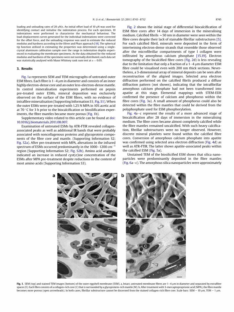
Fig. 1a represents SEM and TEMmicrographs of untreated outerESM fibres. Each fibre is 1e4 mm in diameter and consists of an innerhighlyelectron-dense core andanouter less-electron-densemantle.In control mineralisation experiments performed on pepsinpre-treated outer ESMs, mineral deposition was exclusivelyobserved on the surface of the ESM fibres, with no evidence ofintrafibremineralisation (Supporting InformationS1; Fig. S1).Whenthe outer ESMs were pre-treated with 1.25 NMPA in 10% acetic acidat 70 �C for 3 h prior to the biocalcification or biosilification exper-iments, the fibre mantles became more porous (Fig. 1b).
Supplementary video related to this article can be found at doi:10.1016/j.biomaterials.2011.08.007.
Examination of untreated ESMs by ATR-FTIR revealed collagen-associated peaks as well as additional IR bands that were probablyassociated with noncollagenous proteins and glycoprotein compo-nents of the fibre core and mantle. (Supporting Information S2;Fig. S2a). After pre-treatment with MPA, alterations in the infraredspectrum of ESMs occurred predominantly in the 1000e1200 cm�1
region (Supporting Information S2; Fig. S2b). Amino acid analysesindicated an increase in reduced cyst(e)ine concentration of theESMs after MPA pre-treatment despite reductions in the content ofmost amino acids (Supporting Information S3).
Fig. 1. SEM (top) and stained TEM images (bottom) of the outer eggshell membrane (ESM). aspaces (E). Eachfibre consists of a collagen-rich core (C) that is surroundedbya glycoprotein-ribecomes more porous (open arrowheads). In both cases, fibrillar substructure cannot be disc
Fig. 2 shows the initial stage of differential biocalcification ofESM fibre cores after 14 days of immersion in the mineralisingmedium. Calcified fibrilsw50 nm in diameter were seenwithin thefibre cores despite their lack of stainable fibrillar substructures [5].For each calcified fibril, minerals were deposited in the form ofintertwining electron-dense strands that resemble those observedafter the microfibrillar compartments of type I collagen wereinfiltrated by amorphous calcium phosphate [15,19]. Electrontomography of the bicalcified fibre cores (Fig. 2d) is less revealingdue to the limitation that only a fraction of a 1e4 mm diameter ESMfibre could be visualised even with 200 nm thick sections. Never-theless, a 3-dimensional array of mineral deposits can be seen afterreconstruction of the aligned images. Selected area electrondiffraction performed on the calcified fibrils produced a diffusediffraction pattern (not shown), indicating that the intrafibrillaramorphous calcium phosphate had not been transformed intoapatite at this stage. Elemental mappings with STEM-EDXconfirmed the presence of calcium and phosphorus within thefibre cores (Fig. 3a). A small amount of phosphorus could also bedetected within the fibre mantles that could be derived from thepolyphosphate used for ESM phosphorylation.
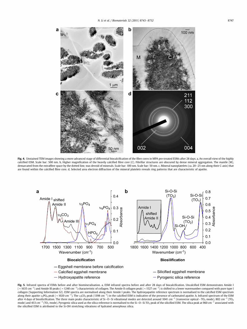
Fig. 4aec represent the results of a more advanced stage ofbiocalcification after 28 days of immersion in the mineralisingmedium. The fibre cores became almost completely calcified whilethe fibre mantles remained uncalcified. With such heavy calcifica-tion, fibrillar substructures were no longer observed. However,discrete mineral platelets were found within the calcified fibrecores. Conversion of amorphous calcium phosphate into apatitewas confirmed using selected area electron diffraction (Fig. 4d) aswell as ATR-FTIR. The latter shows apatite-associated peaks withinthe calcified ESM (Fig. 5a).
Unstained TEM of the biosilicified ESM shows that silica nano-particles were predominantly deposited in the fibre mantles(Fig. 6aec). The amorphous silica nanoparticleswere approximately
, Intact, untreated membrane fibres are 1e4 mm in diameter and separated by extrafibrechmantle (M). b, After treatmentwith 3-mercaptoproprionic acid (MPA), thefibremantleerned from the stained collagen-rich fibre core. Scale bars: SEM e 10 mm, TEM e 1 mm.
Fig. 2. Unstained TEM images showing the initial stage of differential biocalcification of the fibre cores in MPA pre-treated ESMs after 14 days. a, An overall view of the fibres withpartially-calcified cores (C) and uncalcified mantles (M). E: extrafibre space. Scale bar: 500 nm. b, Higher magnification of the w50 nm diameter calcified core fibrils. Scale bar:100 nm. c, Mineral deposition in the form of microfibrillar strands within the calcified fibrils. Scale bar: 50 nm. d, Three-dimensional reconstructed profile of the calcium phosphatedeposits (cyan) within the fibre core. The fibre mantle was completely devoid of mineral deposits.
N. Li et al. / Biomaterials 32 (2011) 8743e87528746
10 nm in diameter. Three-dimensional reconstruction of multipleelectron tomography images derived from a slice of the biosilicifiedESM is shown in Fig. 6d. As electron tomography is incapable ofshowing the full thickness of a 1e4 mm diameter ESM fibre, serial
Fig. 3. STEM-EDX mappings of elemental distributions within MPA pre-treated ESMs. a, Uppbe recognised within the fibre cores (C). The mineralised fibrils yield strong signals of calciPhosphorus is co-localised with calcium in the fibre cores but is also detected from the fibrstrong signals of oxygen and silicon can be detected within the fibre mantles (M). The fibr
sectioning was found to be a better alternative for depicting theextent of mantle silicification within the large, interconnectingmineralised fibres (Supporting Information S4, Fig. S4). A movieshowing the continuity of the silicified ESMmantles can be found in
er left in “a” after 14 days of biocalcification when individual mineralised fibrils can stillum and phosphorus. Fibre mantle (M) and extrafibre spaces (E) are devoid of calcium.e mantle (asterisk). Scale bar: 1 mm. b, Upper left in “b” after 4 days of biosilicification,e cores (C) are devoid of silicon. E: extrafibre space. Scale bar: 1 mm.
Fig. 4. Unstained TEM images showing a more advanced stage of differential biocalcification of the fibre cores in MPA pre-treated ESMs after 28 days. a, An overall view of the highlycalcified ESM. Scale bar: 500 nm. b, Higher magnification of the heavily calcified fibre core (C). Fibrillar structures are obscured by dense mineral aggregation. The mantle (M),demarcated from the extrafibre space by the dotted line, was devoid of minerals. Scale bar: 100 nm. Scale bar: 50 nm. c, Mineral nanoplatelets (ca. 20e25 nm along their C-axis) thatare found within the calcified fibre core. d, Selected area electron diffraction of the mineral platelets reveals ring patterns that are characteristic of apatite.
Fig. 5. Infrared spectra of ESMs before and after biomineralisation. a, ESM infrared spectra before and after 28 days of biocalcification. Uncalcified ESM demonstrates Amide I(w1635 cm�1) and Amide III peaks (w1240 cm�1) characteristic of collagen. The Amide II collagen peak (w1527 cm�1) is shifted to a lower wavenumber compared with pure type Icollagen (Supporting Information S2). ESM spectra are normalised along their Amide I peaks. The hydroxyapatite reference spectrum is normalised to the calcified ESM spectrumalong their apatite y3PO4 peak (w1020 cm�1). The y3CO3 peak (1398 cm�1) in the calcified ESM is indicative of the presence of carbonated apatite. b, Infrared spectrum of the ESMafter 4 days of biosilicification. The three main peaks characteristic of SieOeSi vibrational modes are detected around 1041 cm�1 (transverse optical - TO3 mode), 802 cm�1 (TO2
mode) and 453 cm�1 (TO1 mode). Pyrogenic silica used as the silica reference is normalised to the SieOeSi TO3 peak of the silicified ESM. The silica peak at 960 cm�1 associated withthe silicified ESM is attributed to the Si-OH stretching vibrations of hydrated amorphous silica.
N. Li et al. / Biomaterials 32 (2011) 8743e8752 8747
N. Li et al. / Biomaterials 32 (2011) 8743e87528748
Supporting Information S4 asmantle.mov. These silicified structuresresemble diatom frustules in that a hollow silica shell is formedaround a soft organic core [17]. Elemental analysis of the biosilicifiedESM by STEM-EDX indicates that silicon is localised within the fibremantle (Fig. 3b). The presence of silica-associated peaks within thebiosilicified ESM was further confirmed using ATR-FTIR (Fig. 5b).
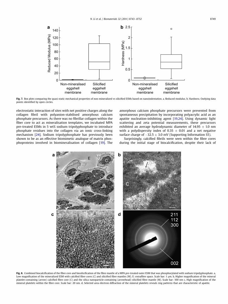
Quasi-static nanoindentation results of the non-mineralised andsilicified ESMs are shown in Fig. 7. The reduced modulus ofhydrated ESMs after biosilicification (72.84 � 66.56 MPa) wassignificantly higher (p < 0.05) than that of hydrated, non-mineralised ESMs (1.84 � 0.33 MPa). Likewise, the hardness ofhydrated ESMs after biosilicification (0.97 � 0.66 MPa) was signif-icantly higher (p < 0.05) than that of hydrated, non-mineralisedESMs (0.11 � 0.02 MPa).
4. Discussion
Eggshell membrane is an inutile hatchery by-product that isa rich source of collagen and other proteins. In its natural form, thisbiopolymer network provides a non-mineralised platform foroutward mineralisation of the eggshell while preventing inwardmineralisation of the egg white and yolk. Preponderance ofdisulphide-rich CREMPs [6] and covalent lysine-derived cross-links[20] may have caused the ESMs to remain impermeable to amor-phous mineral precursors. Reducing agents such as MPA [21],dithiothreitol [6] and thioglycolate [22] have been used forreducing disulphide linkages of ESMs. Here, we demonstrate thatby rendering the ESM permeable with MPA and by further treatingit with a polyphosphate analogue of matrix phosphoproteins, it is
Fig. 6. Unstained TEM images of differential biosilicification of the fibre mantles in MPA pextrafibrillar space (E) is also sparsely filled with silica nanoparticle clusters. C: fibre core. Scfibre core is devoid of silica. Scale bar: 200 nm c, Silica nanoparticles (ca. 10 nm in diameterreveals the amorphous nature of the silica nanoparticles. d, Three-dimensional reconstructedevoid of silica deposits. Gray: extrafibre spaces.
possible to introduce calcium phosphate or silica into ESM fibres atthe nanoscopical scale by using amorphous precursor phases of thecorresponding mineral. Based on the results of the amino acidanalyses, the increased permeability of MPA pre-treated ESMs isattributed to the combined results of protein extraction andcleavage of cystine disulphide bonds. We initially attempted toremove the mineralisation inhibiting components of the ESM byincubating MPA pre-treated membranes in pepsin for differenttime periods. However, MPA pre-treatment alone was found to besufficient for the ESMs to be mineralised. Moreover, combined MPApre-treatment and pepsin digestion adversely altered the handlingcharacteristics of the ESMs and resulted in aggressive dissolution ofthe collagen-rich fibre cores. Hence, all biomineralisation experi-ments were subsequently performed without pepsin digestion.
Biominerals are perfect examples of organic-inorganic hybridcomposites whose structures and properties are modified byextracellular matrix proteins. These hybrid composites are createdby non-classical crystallisation pathways that utilise amorphousmineral precursor phases in mesoscale bottom-up approaches [23].Calcium and phosphate ions from a calcifying medium self-assemble into stable particulate units known as pre-nucleationclusters. In the presence of polyanionic analogues of matrixproteins, these pre-nucleation clusters further condense into fluidicamorphous calcium phosphate precursors that are capable ofinfiltrating the intrafibrillar compartments of type I collagen [13].Contrary to previous beliefs that type I collagen provides a passivedepot for apatite deposition, there is a recent paradigm shift thatsupports an active role of type I collagen in templating intrafibrillarapatite nucleation and growth [15]. This is achieved via
re-treated ESMs after 4 days. a, Heavy silicification within the fibre mantle (M). Theale bar: 1 mm. b, Dense aggregation of silica nanoparticles within the fibre mantle. The) within the silicified mantle. Scale bar: 50 nm. Inset: SAED of the silicified fibre mantled profile of the silica deposits (yellow) within the fibre mantle. The fibre core (pink) is
Fig. 7. Box plots comparing the quasi-static mechanical properties of non-mineralized vs silicified ESMs based on nanoindentation. a. Reduced modulus. b. Hardness. Outlying datapoints identified by open circles.
N. Li et al. / Biomaterials 32 (2011) 8743e8752 8749
electrostatic interaction of sites with net positive charges along thecollagen fibril with polyanion-stabilised amorphous calciumphosphate precursors. As there was no fibrillar collagen within thefiber core to act as mineralisation templates, we incubated MPApre-treated ESMs in 5 wt% sodium tripolyphosphate to introducephosphate residues into the collagen via an ionic cross-linkingmechanism [24]. Sodium tripolyphosphate has previously beenshown to be as an effective biomimetic analogue of matrix phos-phoproteins involved in biomineralisation of collagen [19]. The
Fig. 8. Combined biocalcification of the fibre core and biosilicification of the fibre mantle of aLow magnification of the mineralized ESM with calcified fibre cores (C) and silicified fibre mplatelet-containing (arrow) calcified fibre core (C) and the silica nanoparticle-containing (amineral platelets within the fibre core. Scale bar: 20 nm. d, Selected area electron diffractio
amorphous calcium phosphate precursors were prevented fromspontaneous precipitation by incorporating polyacrylic acid as anapatite nucleation-inhibiting agent [19,24]. Using dynamic lightscattering and zeta potential measurements, these precursorsexhibited an average hydrodynamic diameter of 14.95 � 1.0 nmwith a polydispersity index of 0.35 � 0.01 and a net negativesurface charge of �32.5 � 3.0 mV (Supporting Information S5).
Surprisingly, calcified fibrils were seen within the fibre coresduring the initial stage of biocalcification, despite their lack of
MPA pre-treated outer ESM that was phosphorylated with sodium tripolyphosphate. a,antles (M). E: extrafibre space. Scale bar: 1 mm. b, Higher magnification of the mineralrrowhead) silicified fibre mantle (M). Scale bar: 100 nm c, High magnification of then of the mineral platelets reveals ring patterns that are characteristic of apatite.
N. Li et al. / Biomaterials 32 (2011) 8743e87528750
stainable fibrillar substructures. This suggests that fibrillar colla-gens are invariably present within the ESM fibre cores. Theirinability to be identified by staining may be due to the formation ofcomplex alloys with other nonfibrillar collagen or not-yet-identified collagen types such as the FACITs (fibril-associatedcollagen with interrupted triple helix) [25]. These additionalcollagen entities may interact with proteins containing thedisulphide-rich CREMP motifs [6] via their terminal non-helicaldomains. Pre-treatment of ESMs with MPA could have disruptedthese interactions, enabling amorphous calcium phosphateprecursors to infiltrate the water compartments of the fibrillarcollagen.
Biosilicification of diatom cell walls and sponge spiculesrepresents Nature’s ingenious mechanism for polymerisation ofnano-structured silica from silicic acid [8,26]. These biosilica
Fig. 9. STEM-EDX of ESMs after combined biocalcification and biosilicification. Elemental mBiocalcified fibre core; M: biosilicified fibre mantle; E: extrafibre space. Scale bar: 1 mm.
structures are composites containing zwitterionic proteins andlong-chain polyamines in addition to silica. Biogenesis of silica indiatoms is catalysed by silaffins that are characterized by thepresence of polyamines as well as phosphorylation, N-methyla-tion and hydroxylation of amino acid residues [17]. Attempts havebeen made to synthesise nanostructured silica materials usingbiomimetic catalysts such as polyamines or amine-terminateddendrimers [27,28]. Catechol (1,2-dihydroxybenzene)-stabilisedsilicic acid complex acts as a silica precursor for silicificationunder simulated biological conditions [29]. In our experiments,we employed choline-stabilised orthosilicic acid (ch-OSA)complex as the silica precursor. The positively-charged quaternaryammonium group in choline (2-hydroxy-N,N,N-trimethylethana-minium chloride) interacts with oxygen in silicic acid to forma relatively stable complex [30]. As a dietary supplement, ch-OSA
aps of the distribution of C, Ca, P, Si and O in the biocalcified and biosilicified ESM. C:
N. Li et al. / Biomaterials 32 (2011) 8743e8752 8751
reduces bone turnover in overiectomized rats [31] and in humanclinical trials [32].
We serendipitously discovered that the ESM mantle serves asa template for biocatalytic polymerisation of silicic acid intonano-silica. In biomimetic silicification, polycations such as poly-allylamine and phosphate are simultaneously required to createmicro-phase separations for silica to precipitate [17]. However, nopolyallylamine was utilised in the present work. Chitosan is aneffective biosilicification template in the presence of phosphateions because of the multiple hexosamines in its polymer chain [33].Glycoprotein extracts derived from ESMs also contain abundanthexosamines (N-acetylglucosamine and N-acetylgalatosamine) [3].This probably explains why the glycoprotein-rich fibre mantleserves as a biocatalyst for polymerisation of silicic acid into silicawithout using synthetic polyamine analogues. Absence of silicadeposition within the collagen-rich ESM fibre core may likewise berationalised.
As phosphorus is retained within the fibre mantle after bio-calcification of the fibre core, we further hypothesise that the ESMmay be used as a template for creating hybrid mineralised tissuesunseen in Nature by using a combined biocalcification and bio-silicification scheme. To test this hypothesis, we first calcified thefibre cores prior to silicifying the fibre mantles without additionaltripolyphosphate supplement. This resulted in heavily mineralisedESMs in which the fibre cores were differentially mineralised withapatite crystallites while the fibre mantles were differentiallymineralised with silica nanoparticles (Fig. 8). STEM-EDX of themineralised ESMs indicates that calcium was exclusively found inthe fibre core and siliconwas exclusively located in the fibremantle(Fig. 9). While this represents a proof-of-concept that hybrid min-eralised ESM structures may be constructed using a combinedbiomimetic scheme, the resulting product is too brittle to behandled without breakage.
Nanoindentation of ESMs revealed that their mechanical prop-erties vary with the orientation of the ESM [34]. In that study, theYoung’s modulus in the hemispherical direction was 5.50 �3.26 MPa, which is in the same order of magnitude as the resultsobtained fromnon-mineralised hydrated ESMs in the present study.Moreover, a similar high standard deviation of the mechanicalproperties was also observed. In its natural form, the ESM is a rathertoughmembranebut lacks stiffness andhardness due to the absenceof intrafibre minerals. This structure-property relation enables theESM to limit movement of the developing embryo and supportingnutrients towithin their spatial confines. A natural ESM that is bothtough and stiff is not necessary as protection of the developingembryo from physical injury is efficaciously handled by the calcite-protein component of the eggshell. Nevertheless, by incorporatingsilica in the fibre mantle, novel ESM-based membranous materialswith moderate increases in stiffness may be generated that havepotential bioengineering applications. Eggshell membranes withsilicified fibre mantles may be milled in liquid nitrogen into micro-leaflets and silanised for incorporation in resin composites asa biomimetic toughening agent [35].
5. Conclusion
In the grand scheme of things, Nature is replete with diversity inher biomineralisation designs. Among such creative solutions is themotif of using amorphous mineral precursors as starting materialsand the ability to precisely control where mineralisation isrequired. Despite many years of research, the exact compositions ofthe ESM fibre core andmantle have not been fully elucidated due totheir highly cross-linked nature and the presence of protein motifswith extensive disulphide cross-links. As commonplace and crypticas is the ESM, it exemplifies how potentially mineralisable soft
tissues can serve as a mineralisation barrier via a proteinaceouscoating that surrounds those tissues. Although Nature neverrequiresmore than onemineral in her design of a particular matrix-mediated mineralised framework, it is amazing that the ESM hasthe hidden potential to be mineralised by chemically dissimilarminerals in its different compartmental niches.
Acknowledgements
This work was supported by grant R21 DE019213 from NIDCR(PI. Franklin Tay) and the PSRP and ESA awards from the GeorgiaHealth Sciences University. We thank R. Smith (Electron Micros-copy Core Unit, Georgia Health Sciences University, USA) for per-forming electron diffractions, F. Chan (Electron Microscopy Unit,The University of Hong Kong, China) for performing STEM-EDX andelectron tomography and M. Burnside for secretarial support.
Appendix. Supplementary material
Supplementary data related to this article can be found online atdoi:10.1016/j.biomaterials.2011.08.007.
References
[1] Carrino DA, Dennis JE, Wu TM, Arias JL, Fernandez MS, Rodriguez JP, et al. Theavian eggshell extracellular matrix as a model for biomineralization. ConnectTissue Res 1996;35:325e8.
[2] Nys Y, Gautron J, Garcia-Ruiz JM, Hincke MT. Avian eggshell mineralization:biochemical and functional characterization of matrix proteins. ComptesRendus Palevol 2004;3:549e62.
[3] Picard J, Paul-Gardais A, Vedel M. Sulfated glycoproteins form egg shellmembranes and hen oviduct. Isolation and characterization of sulfatedglycopeptides. Biochim Biophys Acta (GAA) 1973;320:427e41.
[4] Arias JL, Nakamura O, Fernández MS, Wu JJ, Knigge P, Eyre DR, et al. Role oftype-X collagen on experimental mineralization of eggshell membranes.Connect Tissue Res 1997;36:21e31.
[5] Wong M, Hendrix MJC, Von der Mark K, Little C, Stern R. Collagen in theeggshell membranes of the hen. Dev Biol 1984;104:28e36.
[6] Kodali VK, Gannon SA, Paramasivam S, Raje S, Polenova T, Thorpe C. A noveldisulfide-rich protein motif from avian eggshell membranes. PLoS One 2011;6:e18187.
[7] Weiner S. Biomineralization: a structural perspective. J Struct Biol 2008;163:229e34.
[8] Kröger N, Poulsen N. Diatoms-from cell wall biogenesis to nanotechnology.Annu Rev Genet 2008;42:83e107.
[9] Xu Z, Neoh KG, Kishen A. A biomimetic strategy to form calcium phosphatecrystals on type I collagen substrate. Mat Sci Eng C-Mater 2010;30:822e6.
[10] Li J, Chiu KL, Kwong FL, Ng DHL, Chan SLI. Conversion of egg shell membraneto inorganic porous CexZr1-xO2 fibrous network. Curr Appl Phys 2009;9:1438e44.
[11] Su H, Song F, Dong Q, Li T, Zhang Z, Zhang D. Bio-inspired synthesis of ZnOpolyhedral single crystals under eggshell membrane direction. Appl Phy A2010;104:269e74.
[12] Zhang Y, Liu Y, Ji XB, Banks CE, Song JF. Flower-like agglomerates ofhydroxyapatite crystals formed on an egg-shell membrane. Colloid Surf B2011;82:490e6.
[13] Dey A, Bomans PH, Müller FA, Will J, Frederik PM, de With G, et al. The role ofprenucleation clusters in surface-induced calcium phosphate crystallization.Nat Mater 2010;9:1010e4.
[14] Gower LB. Biomimetic model systems for investigating the amorphousprecursor pathway and its role in biomineralization. Chem Rev 2008;108:4551e627.
[15] Nudelman F, Pieterse K, George A, Bomans PH, Friedrich H, Brylka LJ, et al. Therole of collagen in bone apatite formation in the presence of hydroxyapatitenucleation inhibitors. Nat Mater 2010;9:1004e9.
[16] Kröger N, Lorenz S, Brunner E, Sumper M. Self-assembly of highly phos-phorylated silaffins and their function in biosilica morphogenesis. Science2002;298:584e6.
[17] Sumper M, Brunner E. Learning from diatoms: Nature’s tools for theproduction of nanostructured silica. Adv Func Mater 2006;16:7e28.
[18] Oliver WC, Pharr GM. An improved technique for determining hardness andelastic modulus using load and displacement sensing indentation experi-ments. J Mater Res 1992;7:1564e83.
[19] Liu Y, Li N, Qi YP, Dai L, Bryan TE, Mao J, et al. Intrafibrillar collagen miner-alization produced by biomimetic hierarchical nanoapatite assembly. AdvMater 2011;23:975e80.
N. Li et al. / Biomaterials 32 (2011) 8743e87528752
[20] Crombie G, Snider R, Faris B, Franzblau C. Lysine-derived cross-links in the eggshell membrane. Biochim Biophys Acta 1981;640:365e7.
[21] Yi F, Guo ZX, Zhang LX, Yu J, Li Q. Soluble eggshell membrane protein:preparation, characterization and biocompatibility. Biomaterials 2004;25:4591e9.
[22] Yang T, Chen ML, Hu XW,Wang ZW,Wang JH, Dasgupta PK. Thiolated eggshellmembranes sorb and speciate inorganic selenium. Analyst 2011;136:83e9.
[23] Cölfen H. A crystal-clear view. Nat Mater 2010;9:960e1.[24] Liu Y, Kim YK, Dai L, Li N, Khan SO, Pashley DH, et al. Hierarchical and
non-hierarchical mineralisation of collagen. Biomaterials 2011;32:1291e300.[25] Ricard-Blum S, Ruggiero F. The collagen superfamily: from the extracellular
matrix to the cell membrane. Pathol Biol 2005;53:430e42.[26] Schlossmacher U, Wiens M, Schröder HC, Wang X, Jochum KP, Müller WE.
Silintaphin-1einteraction with silicatein during structure-guiding bio-silicaformation. FEBS J 2011;278:1145e55.
[27] Brutchey RL, Morse DE. Silicatein and the translation of its molecular mech-anism of biosilicification into low temperature nanomaterial synthesis. ChemRev 2008;108:4915e34.
[28] Ramanathan R, Campbell JL, Soni SK, Bhargava SK, Bansal V. Cationicamino acids specific biomimetic silicification in ionic liquid: a quest tounderstand the formation of 3-d structures in diatoms. PLoS One 2011;6:e17707.
[29] Belton DJ, Deschaume O, Patwardhan SV, Perry CC. A solution study of silicacondensation and speciation with relevance to in vitro investigations of bio-silicification. J Phys Chem B 2010;114:9947e55.
[30] Bronder SR. Stabilizing orthosilicic acid comprising preparation and biologicalpreparation. United States Patent Office 1999;5,922,360.
[31] Calomme M, Geusens P, Demeester N, Behets GJ, D’Haese P, Sindambiwe JB,et al. Partial prevention of long-term femoral bone loss in aged ovariecto-mized rats supplemented with choline-stabilized orthosilicic acid. CalcifTissue Int 2006;78:227e32.
[32] Spector TD, CalommeMR, Anderson SH, Clement G, Bevan L, Demeester N, et al.Choline-stabilized orthosilicic acid supplementation as an adjunct to calcium/vitamin D3 stimulates markers of bone formation in osteopenic females:a randomized, placebo-controlled trial. BMC Musculoskelet Disord 2008;9:85.
[33] Leng B, Shao Z, Bomans PH, Brylka LJ, Sommerdijk NA, de With G, et al.Cryogenic electron tomography reveals the template effect of chitosan inbiomimetic silicification. Chem Commun (Camb) 2010;46:1703e5.
[34] Torres FG, Troncoso OP, Piaggio F, Hijar A. Structure-property relationships ofa biopolymer network: the eggshell membrane. Acta Biomater 2010;6:3687e93.
[35] TuanW-H, Yu Y-J, Chin Y. L. From biomimetic concept to engineering reality e
a case study on the design of ceramic reinforcement. Adv Eng Mater 2011;13:351e5.