Nanomedicine: Nanotechnology, Biology, and Medicine12 (2016) 1053–1062
nanomedjournal.com
Subtumoral analysis of PRINT nanoparticle distribution reveals targetingvariation based on cellular and particle properties
Luke E. Roode, PhDa, Hailey Brighton, BSb, Tao Bo, MSc, Jillian L. Perry, PhDd,Matthew C. Parrott, PhDc,d,e, f, Farrell Kersey, PhDe, J. Chris Luft, PhDa,
James E. Bear, PhDc,d,b,g, Joseph M. DeSimone, PhDa,c,d,h, Ian J. Davis, MD, PhDd, i,⁎aDepartment of Pharmaceutical Sciences, Eshelman School of Pharmacy, University of North Carolina–Chapel Hill, Chapel Hill, NC, USA
bDepartment of Cell Biology and Physiology, School of Medicine, University of North Carolina–Chapel Hill, Chapel Hill, NC, USAcCarolina Center for Nanotechnology Excellence, University of North Carolina–Chapel Hill, Chapel Hill, NC, USA
dLineberger Comprehensive Cancer Center, University of North Carolina–Chapel Hill, Chapel Hill, NC, USAeDepartment of Radiology, School of Medicine, University of North Carolina–Chapel Hill, Chapel Hill, NC, USA
fBiomedical Research Imaging Center, University of North Carolina–Chapel Hill, Chapel Hill, NC, USAgHoward Hughes Medical Institute, University of North Carolina–Chapel Hill, Chapel Hill, NC, USA
hDepartment of Chemistry, College of Arts and Sciences, University of North Carolina–Chapel Hill, Chapel Hill, NC, USAiDepartments of Genetics and Pediatrics, School of Medicine, University of North Carolina–Chapel Hill, Chapel Hill, NC, USA
Key words: PRINT; Nanomedicine; Nanoparticle; Flow cytometry; Cancer
Funding Sources: This work is supported in part by LiquidiaTechnologies, an NIH Director's Pioneer Award (1DP1OD006432), theCarolina Center for Nanotechnology Excellence (U54CA151652), and theUNC University Cancer Research Fund. I.J.D. is supported by grants fromthe NIH (R01CA166447), the Corn-Hammond Fund for Pediatric CancerResearch, the Wide Open Charitable Foundation, and the V Foundation forCancer Research. The UNC Flow Cytometry Core Facility is supported inpart by P30 CA016086Cancer Center Core Support Grant to the UNCLineberger Comprehensive Cancer Center.
The authors declare the following competing financial interest(s): J.M.D. isa founder and maintains a financial interest in Liquidia Technologies.
⁎Corresponding author at: Lineberger Comprehensive Cancer Center,University of North Carolina at Chapel Hill, Chapel Hill, NC, USA.
Downloaded from ClinicalKey.com at University of North For personal use only. No other uses without permission. C
Appreciation of the Enhanced Permeation and Retention(EPR) effect for nanocarrier-mediated drug delivery in oncologyhas resulted in a focus on the accumulation of particles in wholetumors.1 A range of methods to determine the fraction of theinjected dose of the carrier or cargo that accumulates in a wholeorgan or tumor has driven the assessment of nanoparticletargeting to solid tumors.2–13 However, tumors are composed ofa variety of cell types, such as fibroblasts and endothelial cellsand macrophages and neutrophils, in addition to cancer cells.The relative distribution of these cell types varies betweentumors.14–17 Whole organ approaches are unable to discriminatebetween accumulation in the intended target, typically cancercells, and other cells or the extracellular space. For cargo with an
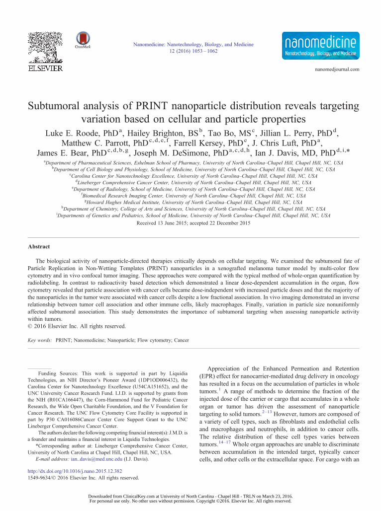
Figure 1. Particle accumulation measured by gamma radiation. (A) Radioactive emission was determined for each tissue 18 h after intravenous administration.Fractional association for all tissues examined is shown. (B) Calculated mass of nanoparticles contained within tumor organ. Error bar indicates SD of n = 4.
1054 L.E. Roode et al / Nanomedicine: Nanotechnology, Biology, and Medicine 12 (2016) 1053–1062
intracellular mechanism of action, such as nucleic acids andproteins, delivery to specific cell types is crucial to assessingnanoparticle efficacy and optimizing targeting.
Methods for the identification of subtumoral cellularcomponents include microscopy and flow cytometry. Confocalmicroscopy has been used to determine particle internalization invivo by analyzing multiple sections of an organ.18 However,meaningful quantification can be challenging. Flow cytometrypermits concurrent cellular identification and nanoparticlequantification. Previous studies that have used flow cytometryto examine nanoparticle targeting to organs have not exploredthe effects of particle characteristics (composition, shape, etc.) ordose on the accumulation in specific cell populations and do notcorrelate their findings with whole organ assessment.14,19–25
Studies that account for both nanocarrier properties as well asintra-organ or intra-tumor distribution have the potential to bestinform nanoparticle design and delivery.
PRINT is a top-down fabrication strategy that relies onprecisionmolds, offering the advantage of reproducible productionof monodisperse particles. This reproducibility eliminates largevariation in particle sizes (i.e., PDI) that could influence theassociation of a subset of the particleswith one cell population overanother confounding data interpretation. In addition, PRINT alsoaffords homogeneity in the composition of the particles andflexibility in the composition of the desired nanoparticle material.
Using flow cytometry, whole organ assessment and liveanimal in vivo confocal microscopy, we analyzed the celltype-specific distribution of PRINT nanoparticles. We identifiedwide variation in subtumoral cellular association and identifydose and particle properties that influence cellular targeting.
Downloaded from ClinicalKey.com at University of NorFor personal use only. No other uses without permission
pyridine, borate buffer (pH 8.6), acetic anhydride, and methanolwere obtained from Fisher Scientific. Conventional filters (2 μm)were purchased from Agilent and poly(vinyl alcohol) (Mw 2000)(PVOH) was purchased from Acros Organics. PRINT molds(80 nm × 80 nm × 320 nm) were obtained from Liquidia Tech-nologies. Tetraethylene glycolmonoacrylate (HP4A) was synthe-sized in-house as previously described. 26 Methoxy-PEG(5k)-succinimidyl carboxy methyl ester (mPEG5k-SCM)was purchased from Creative PEGWorks. Typsin, DPBS, andcell culture media were purchased from Gibco.
PRINT nanoparticle fabrication and characterization
The PRINT particle fabrication technique has been describedpreviously in detail.27,28 The pre-particle solution was preparedby dissolving 3.5 wt% of the various reactive monomers inmethanol. The pre-particle solution comprised 67.75 wt% HP4A,20 wt% AEM, 10 wt% PEG700DA, 1 wt% TPO and 1.25 wt%Dylight 488 maleimide. Stock particle concentrations weredetermined by thermogravimetric analysis (TGA) on both analiquot of the stock and a centrifuged sample of the stock, toaccount for any mass due to PVOH, using a TA InstrumentsQ5000. Particles were visualized by scanning electron micros-copy (SEM) using a Hitachi S-4700 SEM. Prior to imaging,SEM samples were coated with 3.5 nm of gold–palladium alloyusing a Cressington 108 auto sputter coater. Particle size and zetapotential were measured by dynamic light scattering (DLS) on aZetasizer Nano ZS (Malvern Instruments, Ltd.).
Particles were PEGylated and acetylated following a previouslydescribed method.27 Post-acetylation, particles were analyzed byTGA, DLS and SEM and stored at 4 °C. To radiolabel thenanoparticles, high specific activity 64Cu (14,000 ± 7600 Ci/mmolor 518 ± 28 TBq/mmol) was obtained from the WashingtonUniversity School of Medicine (St. Louis, MO, USA). 64Cu wasproduced on a CS-15 biomedical cyclotron by the 64Ni(p,n)64Cunuclear reaction using previously established methods,29 with ahalf-life of 12.7 h. Following PEGylation (described above),particles were characterized as described above by TGA andreacted in 0.1 M Na2CO3 buffer (pH 9) with 2-(4-isothiocyana-tobenzyl) 1,4,7,10-tetraazacyclododecane-1,4,7,10-tetraacetic acid
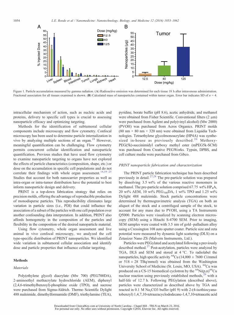
Figure 2. Flow cytometry gating strategy used to identify subtumoral compartments. To determine relative cell populations, dissociate tumor was stained withFixable Blue, CD45, CD31, Ly6G and F4/80. Fluorescence minus one controls (gray lines) are used to distinguish significant staining (black). Representativeplots are shown.
1055L.E. Roode et al / Nanomedicine: Nanotechnology, Biology, and Medicine 12 (2016) 1053–1062
(p-SCN-Bn-DOTA) at 5 mg/mL (2:1 DOTA:AEM molar ratio). Aconversion from positive to negative zeta potential indicated that thereaction went to completion. Particles were then incubated with64CuCl2 for 30 min at 65 °C in 0.1 Mammonium acetate, washed 3times by centrifugation with deionized water, and resuspended forinjection in 9.25% sucrose.
Cells, cell culture, and spheroid injections
Cells were cultured as previously described in high glucoseDMEM (Gibco) with 10% FBS (Gemini Bio-products) and 1%penicillin/streptomycin (Gibco).30 Hanging droplet spheroids
Downloaded from ClinicalKey.com at University of North For personal use only. No other uses without permission. C
were generated by trypsinization of the cells and resuspensionto 2 × 10⁶ cells/mL. Twenty microliters of the cell suspensionwas pipetted into wells of a 60-well minitray (Nunc, Thermo-fisher scientific). The minitray was inverted and placed in ahumidified 150 mm dish. Cells were incubated at 37 °C and 5%CO2 for 4-6 days. Individual spheroids were harvested andverified visually. Athymic nude mice were anesthetized byinhalation with 2% isoflurane. A mouse ear was affixed onto aconical tube with double-sided tape. The tumor spheroid wasinjected as described.30 Successful spheroid injection wasverified by epifluorescence macroscopic imaging (SupplementalFigure S1, A).
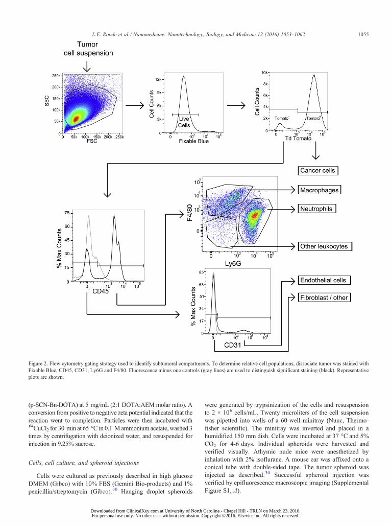
Figure 3. Fractional composition of LKB498 xenografted tumors. Twogroups of 16 animals were processed, with the data collected and analyzedseparately then aggregated for the analysis. Error bars represent SD (n = 32).
1056 L.E. Roode et al / Nanomedicine: Nanotechnology, Biology, and Medicine 12 (2016) 1053–1062
Mice and particle injections
All animals were handled according to the NIH Guide for theCare and Use of Laboratory Animals. All Procedures wereapproved by the University of North Carolina–Chapel HillInstitutional Animal Care and Use Committee (IACUC),protocol #11-154.0. 4-8-week-old, male Foxn1nu (athymic;C57BL/6J background) nude mice were purchased from theUNC Animal Services Core. Nanoparticles were suspended inisotonic 9.25% sucrose and injected intravenously via the tailvein at a maximum of 300 μl per animal. Tumors were of equalvolumes between groups and ranged from 100 to 300 mm3, asdetermined using caliper measurements using the formula V =(L/2) × (W/2) × (H/2) × (4/3 × π) (Supplemental Figure S1, A).Particles were resuspended at different concentrations (1.45,4.65, 14.65 mg/mL) to deliver similar injection volumes ofapproximately 300 μl.
Tumor dissociation and flow cytometry
Tumorswere harvested as describedwith slightmodifications.31
Briefly, tumors were dissociated for 3 h in 5 mL of DMEM with10% FBS containing 1500 U collagenase and 500 U hyaluron-idase (10× collagenase/hyaluronidase, StemCell Technologies).After centrifugation (600×g for 5 min), cell pellets wereresuspended in a 1:4 mixture of HEPES Buffered Salinecontaining 2% FBS (HF solution) and RBC Lysis Buffer (0.8%NH4Cl and 0.1 mM EDTA, Stem Cell Technologies), recen-trifuged (450×g for 5 min) and then resuspended in RBC lysisbuffer. After centrifugation, cells were resuspended in 0.05%Trypsin/EDTA solution (Gibco) and incubated 5 min at 37 °C.Cells were pelleted and resuspended in Dispase (1 U/mL) andDNase I (0.1 mg/mL). After 30-min incubation at 37 °C, 10 mLof HF solution was added, and cells were passed through a 40-μmcell strainer (Fisher), centrifuged and resuspended in HF solution.Cells were then washed and counted with a hemocytometer.Live-Dead Fixable Blue (Invitrogen) was then added at aconcentration of 1 μl/4 × 106 cells in 1 mL PBS and incubatedon ice for 15 min. Cells were washed with PBS and thenresuspended in 100 μl PBS. Fc Block (BD Biosciences) wasincubated with the sample for 5 min on ice followed by anantibody (Biolegend) mixture consisting of PE-Cy7 CD31 (clone390), APC F4/80 (clone BM8), Alexa 700 Ly6G (clone 1A8), andPacific Blue CD45 (clone 30-F11). After 1 h on ice, cells werewashedwith PBS and fixed in 500 μl of 4% paraformaldehyde for
Downloaded from ClinicalKey.com at University of NorFor personal use only. No other uses without permission
15 min at room temperature. Cells were washed twice with FACSbuffer (1% FBS in PBS), then resuspended in a final volume of500 μl of FACS Buffer and stored at 4 °C until data acquisition(LSRII, BD Biosciences). Data analysis of FCS3 files wasperformed using FACSDiva version 10.6 (BD Biosciences). LiveDead Blue (Invitrogen) was used to gate on the living cells. Allsurface markers were compared to their fluorescence minus one(FMO) controls to set appropriate gates. Particle association wasdetermined by comparison to control sucrose injected animals toestablish appropriate gating.
Two-photon microscopy
Tumor-bearing animals with tumors between 10 and 30 mm3
were imaged as previously described.30 Animals were anesthe-tized with isofluorane, and tumors were imaged before and afteradministration of particles. All imaging was performed at910 nm with an Olympus FV1000MPE mounted on an uprightBX-61WI microscope, using a 25×, 1.05 N.A. (2 mm W.D.)water immersion objective with optical imaging gel to captureimages. Olympus Fluoview software and microscope settingswere consistent for all acquired images (the laser power was at14% and each channel's PMT voltage was 580, 635, and 600,respectively). The laser unit (MaiTai DeepSee) is tunable from690 to 1040 nm with a pulse width b100 fs. Three channelnon-descan detectors were used: Ch1 (420-460 nm) BFP, Ch2(495-540 nm) GFP, and Ch3 (575-630 nm) RFP.
Statistics
All data, unless otherwise noted, were analyzed using aone-way ANOVA and Tukey's post-hoc analysis. Meanfluorescence data were analyzed using a two-way ANOVAand Tukey's post-hoc analysis.
Results
PRINT fabrication
To explore the influence of particle characteristics onnanoparticle association, we generated tumor spheroids fromLKB498 mouse melanoma cells that stably express the redfluorescent protein tdTomato.30 Tumor cell spheroids wereimplanted intradermally and allowed to grow to a size of~100 mm3 (Supplemental Figure S1, A-B). Nanoparticles werefabricated using the Particle Replication in Non-WettingTemplates (PRINT) technique.27,28,32 PRINT generates nano-particles of highly consistent and precise size, shape, andcomposition, eliminating variables which could influencecellular targeting. 80 nm × 80 nm × 320 nm PRINT nanoparti-cles were composed of a covalently cross-linked hydrogel matrixand were PEGylated as previously described.27 Depending onthe assay, fluorescent moieties were incorporated, or copper-64(64Cu) was chelated to the particles. Particles were characterizedby DLS and SEM and demonstrated a similar negative zetapotential and narrow size distribution (Supplemental FigureS1, C).
To determine nanoparticle accumulation using a whole-organbased approach, 64Cu radiolabeled PRINT nanoparticles were
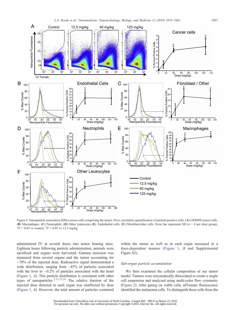
Figure 4. Nanoparticle association differs across cells comprising the tumor. Flow cytometric quantification of particle-positive cells. (A) LKB498 cancer cells.(B) Macrophages. (C) Neutrophils. (D) Other leukocytes (E). Endothelial cells. (F) Fibroblast/other cells. Error bar represents SD (n = 4 per dose group).*P b 0.05 vs control, #P b 0.05 vs 12.5 mg/kg.
1057L.E. Roode et al / Nanomedicine: Nanotechnology, Biology, and Medicine 12 (2016) 1053–1062
administered IV at several doses into tumor bearing mice.Eighteen hours following particle administration, animals weresacrificed and organs were harvested. Gamma emission wasmeasured from several organs and the tumor accounting for~70% of the injected dose. Radioactive signal demonstrated awide distribution, ranging from ~45% of particles associatedwith the liver to ~0.2% of particles associated with the heart(Figure 1, A). This particle distribution is consistent with othertypes of nanoparticles.2,12,13,18 The relative fraction of theinjected dose detected in each organ was unaffected by dose(Figure 1, A). However, the total amount of particles contained
Downloaded from ClinicalKey.com at University of North For personal use only. No other uses without permission. C
within the tumor as well as in each organ increased in adose-dependent manner (Figure 1, B and SupplementalFigure S2).
Sub-organ particle accumulation
We then examined the cellular composition of our tumormodel. Tumors were enzymatically dissociated to create a singlecell suspension and analyzed using multi-color flow cytometry(Figure 2). After gating on viable cells, tdTomato fluorescenceidentified the melanoma cells. To distinguish these cells from the
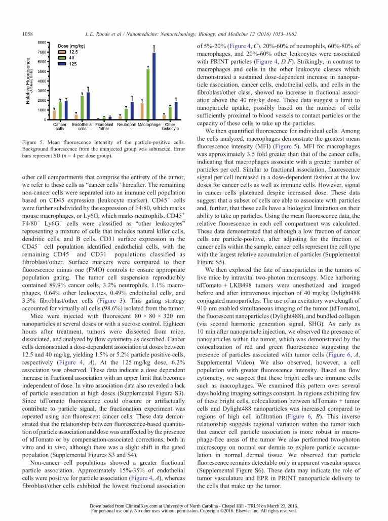
Figure 5. Mean fluorescence intensity of the particle-positive cells.Background fluorescence from the uninjected group was subtracted. Errorbars represent SD (n = 4 per dose group).
1058 L.E. Roode et al / Nanomedicine: Nanotechnology, Biology, and Medicine 12 (2016) 1053–1062
other cell compartments that comprise the entirety of the tumor,we refer to these cells as “cancer cells” hereafter. The remainingnon-cancer cells were separated into an immune cell populationbased on CD45 expression (leukocyte marker). CD45+ cellswere further subdivided by the expression of F4/80, which marksmouse macrophages, or Ly6G, which marks neutrophils. CD45+
F4/80− Ly6G− cells were classified as “other leukocytes”representing a mixture of cells that includes natural killer cells,dendritic cells, and B cells. CD31 surface expression in theCD45− cell population identified endothelial cells, with theremaining CD45− and CD31− populations classified asfibroblast/other. Surface markers were compared to theirfluorescence minus one (FMO) controls to ensure appropriatepopulation gating. The tumor cell suspension reproduciblycontained 89.9% cancer cells, 3.2% neutrophils, 1.1% macro-phages, 0.64% other leukocytes, 0.49% endothelial cells, and3.3% fibroblast/other cells (Figure 3). This gating strategyaccounted for virtually all cells (98.6%) isolated from the tumor.
Mice were injected with fluorescent 80 × 80 × 320 nmnanoparticles at several doses or with a sucrose control. Eighteenhours after treatment, tumors were dissected from mice,dissociated, and analyzed by flow cytometry as described. Cancercells demonstrated a dose-dependent association at doses between12.5 and 40 mg/kg, yielding 1.5% or 5.2% particle positive cells,respectively (Figure 4, A). At the 125 mg/kg dose, 6.2%association was observed. These data indicate a dose dependentincrease in fractional association with an upper limit that becomesindependent of dose. In vitro association data also revealed a lackof particle association at high doses (Supplemental Figure S3).Since tdTomato fluorescence could obscure or artifactuallycontribute to particle signal, the fractionation experiment wasrepeated using non-fluorescent cancer cells. These data demon-strated that the relationship between fluorescence-based quantita-tion of particle association and dosewas unaffected by the presenceof tdTomato or by compensation-associated corrections, both invitro and in vivo, although there was a slight shift in the gatedpopulation (Supplemental Figures S3 and S4).
Non-cancer cell populations showed a greater fractionalparticle association. Approximately 15%-35% of endothelialcells were positive for particle association (Figure 4, A), whereasfibroblast/other cells exhibited the lowest fractional association
Downloaded from ClinicalKey.com at University of NorFor personal use only. No other uses without permission
of 5%-20% (Figure 4, C). 20%-60% of neutrophils, 60%-80% ofmacrophages, and 20%-60% other leukocytes were associatedwith PRINT particles (Figure 4, D-F). Strikingly, in contrast tomacrophages and cells in the other leukocyte classes whichdemonstrated a sustained dose-dependent increase in nanopar-ticle association, cancer cells, endothelial cells, and cells in thefibroblast/other class, showed no increase in fractional associ-ation above the 40 mg/kg dose. These data suggest a limit tonanoparticle uptake, possibly based on the number of cellssufficiently proximal to blood vessels to contact particles or thecapacity of these cells to take up the particles.
We then quantified fluorescence for individual cells. Amongthe cells analyzed, macrophages demonstrate the greatest meanfluorescence intensity (MFI) (Figure 5). MFI for macrophageswas approximately 3.5 fold greater than that of the cancer cells,indicating that macrophages associate with a greater number ofparticles per cell. Similar to fractional association, fluorescencesignal per cell increased in a dose-dependent fashion at the lowdoses for cancer cells as well as immune cells. However, signalin cancer cells plateaued despite increased dose. These datasuggest that a subset of cells are able to associate with particlesand, further, that these cells have a biological limitation on theirability to take up particles. Using the mean fluorescence data, therelative fluorescence in each cell compartment was calculated.These data demonstrated that although a low fraction of cancercells are particle-positive, after adjusting for the fraction ofcancer cells within the sample, cancer cells represent the cell typewith the largest relative accumulation of particles (SupplementalFigure S5).
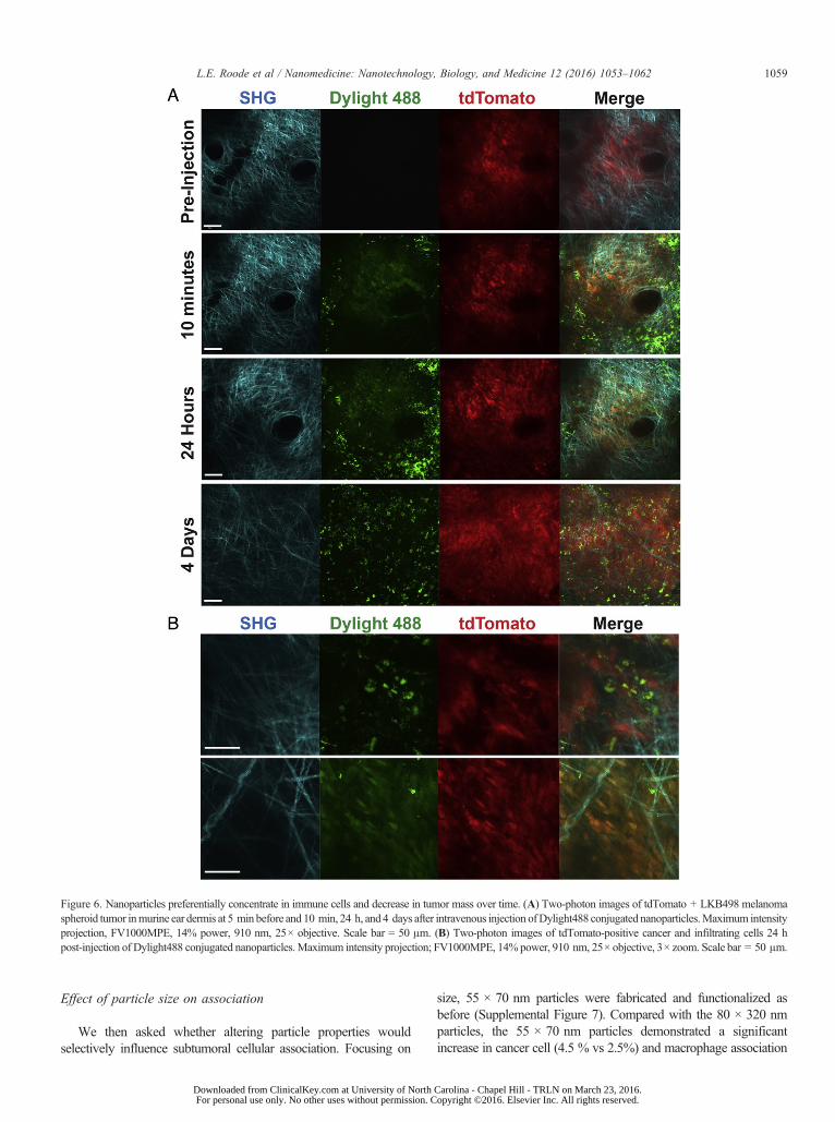
We then explored the fate of nanoparticles in the tumors oflive mice by intravital two-photon microscopy. Mice harboringtdTomato + LKB498 tumors were anesthetized and imagedbefore and after intravenous injection of 40 mg/kg Dylight488conjugated nanoparticles. The use of an excitatory wavelength of910 nm enabled simultaneous imaging of the tumor (tdTomato),the fluorescent nanoparticles (Dylight488), and bundled collagen(via second harmonic generation signal, SHG). As early as10 min after nanoparticle injection, we observed the presence ofnanoparticles within the tumor, which was demonstrated by thecolocalization of red and green fluorescence suggesting thepresence of particles associated with tumor cells (Figure 6, A,Supplemental Video). We also observed, however, a cellpopulation with greater fluorescence intensity. Based on flowcytometry, we suspect that these bright cells are immune cellssuch as macrophages. We examined this pattern over severaldays holding imaging settings constant. In regions exhibiting fewof these bright cells, colocalization between tdTomato + tumorcells and Dylight488 nanoparticles was increased compared toregions of high cell infiltration (Figure 6, B). This inverserelationship suggests regional variation within the tumor suchthat cancer cell particle association is more robust in macro-phage-free areas of the tumor We also performed two-photonmicroscopy on normal ear dermis to explore particle accumu-lation in normal dermal tissue. We observed that particlefluorescence remains detectable only in apparent vascular spaces(Supplemental Figure S6). These data may indicate the role oftumor vasculature and EPR in PRINT nanoparticle delivery tothe cells that make up the tumor.
Figure 6. Nanoparticles preferentially concentrate in immune cells and decrease in tumor mass over time. (A) Two-photon images of tdTomato + LKB498 melanomaspheroid tumor inmurine ear dermis at 5 min before and 10 min, 24 h, and 4 days after intravenous injection ofDylight488 conjugated nanoparticles.Maximum intensityprojection, FV1000MPE, 14% power, 910 nm, 25× objective. Scale bar = 50 μm. (B) Two-photon images of tdTomato-positive cancer and infiltrating cells 24 hpost-injection of Dylight488 conjugated nanoparticles.Maximum intensity projection; FV1000MPE, 14% power, 910 nm, 25× objective, 3× zoom. Scale bar = 50 μm.
1059L.E. Roode et al / Nanomedicine: Nanotechnology, Biology, and Medicine 12 (2016) 1053–1062
Effect of particle size on association
We then asked whether altering particle properties wouldselectively influence subtumoral cellular association. Focusing on
Downloaded from ClinicalKey.com at University of North For personal use only. No other uses without permission. C
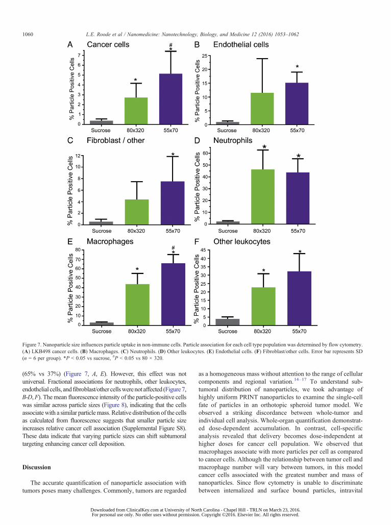
size, 55 × 70 nm particles were fabricated and functionalized asbefore (Supplemental Figure 7). Compared with the 80 × 320 nmparticles, the 55 × 70 nm particles demonstrated a significantincrease in cancer cell (4.5 % vs 2.5%) and macrophage association
Figure 7. Nanoparticle size influences particle uptake in non-immune cells. Particle association for each cell type population was determined by flow cytometry.(A) LKB498 cancer cells. (B) Macrophages. (C) Neutrophils. (D) Other leukocytes. (E) Endothelial cells. (F) Fibroblast/other cells. Error bar represents SD(n = 6 per group). *P b 0.05 vs sucrose, #P b 0.05 vs 80 × 320.
1060 L.E. Roode et al / Nanomedicine: Nanotechnology, Biology, and Medicine 12 (2016) 1053–1062
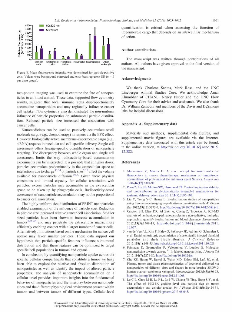
(65% vs 37%) (Figure 7, A, E). However, this effect was notuniversal. Fractional associations for neutrophils, other leukocytes,endothelial cells, and fibroblast/other cellswerenot affected (Figure7,B-D,F). Themean fluorescence intensity of the particle-positive cellswas similar across particle sizes (Figure 8), indicating that the cellsassociatewith a similar particlemass. Relative distribution of the cellsas calculated from fluorescence suggests that smaller particle sizeincreases relative cancer cell association (Supplemental Figure S8).These data indicate that varying particle sizes can shift subtumoraltargeting enhancing cancer cell deposition.
Discussion
The accurate quantification of nanoparticle association withtumors poses many challenges. Commonly, tumors are regarded
Downloaded from ClinicalKey.com at University of NorFor personal use only. No other uses without permission
as a homogeneous mass without attention to the range of cellularcomponents and regional variation.14–17 To understand sub-tumoral distribution of nanoparticles, we took advantage ofhighly uniform PRINT nanoparticles to examine the single-cellfate of particles in an orthotopic spheroid tumor model. Weobserved a striking discordance between whole-tumor andindividual cell analysis. Whole-organ quantification demonstrat-ed dose-dependent accumulation. In contrast, cell-specificanalysis revealed that delivery becomes dose-independent athigher doses for cancer cell population. We observed thatmacrophages associate with more particles per cell as comparedto cancer cells. Although the relationship between tumor cell andmacrophage number will vary between tumors, in this modelcancer cells associated with the greatest number and mass ofnanoparticles. Since flow cytometry is unable to discriminatebetween internalized and surface bound particles, intravital
Figure 8. Mean fluorescence intensity was determined for particle-positivecells. Values were background corrected and error bars represent SD (n = 6per dose group).
1061L.E. Roode et al / Nanomedicine: Nanotechnology, Biology, and Medicine 12 (2016) 1053–1062
two-photon imaging was used to examine the fate of nanopar-ticles in an intact animal. These data, supported flow cytometryresults, suggest that local immune cells disproportionatelyaccumulate nanoparticles and may regionally influence cancercell uptake. Flow cytometry also demonstrated the non-uniforminfluence of particle properties on subtumoral particle distribu-tion. Reduced particle size increased the association withcancer cells.
Nanomedicines can be used to passively accumulate smallmolecule cargo (e.g., chemotherapy) in tumors via the EPR effect.However, biologically active, membrane-impermeable cargo (e.g.,siRNA) requires intracellular and cell-specific delivery. Single-cellassessment offers lineage-specific quantification of nanoparticletargeting. The discrepancy between whole organ and single cellassessment limits the way radioactivity-based accumulationexperiments can be interpreted. It is possible that at higher doses,particles accumulate predominantly in the extracellular space asinteractions due to charge33,34 or particle size5,35 affect the volumeavailable for nanoparticle diffusion.36,37 Given these physicalconstraints and limited capacity for cellular association withparticles, excess particles may accumulate in the extracellularspace or be taken up by phagocytic cells. Radioactivity-basedassessment of nanoparticle accumulation may not be proportionalto cancer cell association.
The highly uniform size distribution of PRINT nanoparticlesenabled examination of the influence of particle size. Reductionin particle size increased relative cancer cell association. Smallersized particles have been shown to increase accumulation intumors5,35,38 and may penetrate the extracellular matrix moreefficiently enabling contact with a larger number of cancer cells.Alternatively, limitations based on the mechanism for cancer celluptake may favor smaller particles. These data support ourhypothesis that particle-specific features influence subtumoraldistribution and that these features can be optimized to targetspecific cell populations in tumors and organs.
In conclusion, by quantifying nanoparticle uptake across thespecific cellular compartments that constitute a tumor we havebeen able to explore the relative and absolute distribution ofnanoparticles as well as identify the impact of altered particleproperties. The analysis of nanoparticle accumulation on acellular level provides important insights into the fundamentalbehavior of nanoparticles and the interplay between nanomedi-cines and the different physiological environment present withintumors and between tumors of different types. Cellular-level
Downloaded from ClinicalKey.com at University of North For personal use only. No other uses without permission. C
quantification is critical when assessing the function ofimpermeable cargo that depends on an intracellular mechanismof action.
Author contributions
The manuscript was written through contributions of allauthors. All authors have given approval to the final version ofthe manuscript.
Acknowledgments
We thank Charlene Santos, Mark Ross, and the UNCLineberger Animal Studies Core. We acknowledge AmarKhumbhar of CHANL, Nancy Fisher and the UNC FlowCytometry Core for their advice and assistance. We also thankDr. William Zamboni and members of the Davis and DeSimonelabs for helpful discussions.
Appendix A. Supplementary data
Materials and methods, supplemental data figures, andsupplemental movie figures are available via the Internet.Supplementary data associated with this article can be found,in the online version, at http://dx.doi.org/10.1016/j.nano.2015.12.382.
References
1. Matsumura Y, Maeda H. A new concept for macromoleculartherapeutics in cancer chemotherapy: mechanism of tumoritropicaccumulation of proteins and the antitumor agent Smancs. Cancer Res1986;46(12):6387-92.
2. Poon Z, Lee JB, Morton SW, Hammond PT. Controlling in vivo stabilityand biodistribution in electrostatically assembled nanoparticles forsystemic delivery. Nano Lett 2011;11(5):2096-103.
3. Liu Y, Tseng Y-C, Huang L. Biodistribution studies of nanoparticlesusing fluorescence imaging: a qualitative or quantitative method? PharmRes 2012;29(12):3273-7, http://dx.doi.org/10.1007/s11095-012-0818-1.
4. Crayton SH, Elias DR, Al Zaki A, Cheng Z, Tsourkas A. ICP-MSanalysis of lanthanide-doped nanoparticles as a non-radiative, multiplexapproach to quantify biodistribution and blood clearance. Biomaterials2012;33(5):1509-19, http://dx.doi.org/10.1016/j.biomaterials.2011.10.077.
5. van de Ven AL, Kim P, Haley O, Fakhoury JR, Adriani G, Schmulen J,et al. Rapid tumoritropic accumulation of systemically injected plateloidpar t ic les and thei r b iodis t r ibut ion. J Control Release2012;158(1):148-55, http://dx.doi.org/10.1016/j.jconrel.2011.10.021.
6. Psimadas D, Georgoulias P, Valotassiou V, Loudos G. Molecularnanomedicine towards cancer: 111In-labeled nanoparticles. J Pharm Sci2012;101(7):2271-80, http://dx.doi.org/10.1002/jps.
7. Chu KS, Hasan W, Rawal S, Walsh MD, Enlow EM, Luft JC, et al.Plasma, tumor and tissue pharmacokinetics of docetaxel delivered viananoparticles of different sizes and shapes in mice bearing SKOV-3human ovarian carcinoma xenograft. Nanomedicine 2013;9(5):686-93,http://dx.doi.org/10.1016/j.nano.2012.11.008.
8. Lo C-L, Chou M-H, Lu P-L, Lo I-W, Chiang Yi-Ting, Hung S-Y, et al.The effect of PEG-5K grafting level and particle size on tumoraccumulation and cellular uptake. Int J Pharm 2013;456(2):424-31,http://dx.doi.org/10.1016/j.ijpharm.2013.08.045.
1062 L.E. Roode et al / Nanomedicine: Nanotechnology, Biology, and Medicine 12 (2016) 1053–1062
9. Ueno T, Dutta P, Keliher E, Leuschner F, Majmudar M, Marinelli B, et al.Nanoparticle PET-CT detects rejection and immunomodulation in cardiacallografts. Circ Cardiovasc Imaging 2013;6(4):568-73, http://dx.doi.org/10.1161/CIRCIMAGING.113.000481.
10. Clark DP, Ghaghada K, Moding EJ, Kirsch DG, Badea CT. In vivocharacterization of tumor vasculature using iodine and gold nanoparti-cles and dual energy micro-CT. Phys Med Biol 2013;58(6):1683-704,http://dx.doi.org/10.1088/0031-9155/58/6/1683.
11. Ernsting MJ, Murakami M, Roy A, Li SD. Factors controlling thepharmacokinetics, biodistribution and intratumoral penetration ofnanoparticles. J Control Release 2013;172(3):782-94, http://dx.doi.org/10.1016/j.jconrel.2013.09.013.
12. Ganesh S, Iyer AK, Gattacceca F, Morrissey DV, Amiji MM. In vivobiodistribution of siRNA and cisplatin administered using CD44-targeted hyaluronic acid nanoparticles. J Control Release2013;172(3):699-706, http://dx.doi.org/10.1016/j.jconrel.2013.10.016.
13. Toita R, Nakao K, Mahara A, Yamaoka T, Akashi M. Biodistribution ofvaccines comprised of hydrophobically-modified poly(γ-glutamic acid)nanoparticles and antigen proteins using fluorescence imaging. BioorgMed Chem 2013;21(21):6608-15, http://dx.doi.org/10.1016/j.bmc.2013.08.024.
14. Movahedi K, Laoui D, Gysemans C, Baeten M, Stangé G, Van denBossche J, et al. Different tumor microenvironments contain functionallydistinct subsets of macrophages derived from Ly6C(high) monocytes.Cancer Res 2010;70(14):5728-39, http://dx.doi.org/10.1158/0008-5472.CAN-09-4672.
15. Li R, Hu H, Ma H, Chen L, Zhou B, Liu Y, et al. The anti-tumor effect andincreased tregs infiltration mediated by rAAV-SLC vector. Mol Biol Rep2013;40(10):5615-23, http://dx.doi.org/10.1007/s11033-013-2663-7.
16. Zhao G, Rodriguez BL. Molecular targeting of liposomal nanoparticlesto tumor microenvironment. Int J Nanomedicine 2013;8:61-71, http://dx.doi.org/10.2147/IJN.S37859.
17. Junttila MR, de Sauvage FJ. Influence of tumour micro-environmentheterogeneity on therapeutic response. Nature 2013;501(7467):346-54,http://dx.doi.org/10.1038/nature12626.
18. Xiao K, Li Y, Luo J, Lee JS, Xiao W, Gonik AM, et al. The effect ofsurface charge on in vivo biodistribution of PEG-oligocholic acid basedmicellar nanoparticles. Biomaterials 2011;32(13):3435-46, http://dx.doi.org/10.1016/j.biomaterials.2011.01.021.
19. Almeida JPM, Lin AY, Langsner RJ, Eckles P, Foster AE, Drezek RA.In vivo immune cell distribution of gold nanoparticles in naïve and tumorbearing mice. Small 2014;10(4):812-9, http://dx.doi.org/10.1002/smll.201301998.
20. Blank F, Stumbles PA, Seydoux E, Holt PG, Fink A, Rothen-Rutishauser B, et al. Size-dependent uptake of particles by pulmonaryantigen-presenting cell populations and trafficking to regional lymphnodes. Am J Respir Cell Mol Biol 2013;49(1):67-77, http://dx.doi.org/10.1165/rcmb.2012-0387OC.
21. Kirpotin DB, Drummond DC, Shao Y, ShalabyMR, Hong K, Nielsen UB,et al. Antibody targeting of long-circulating lipidic nanoparticles does notincrease tumor localization but does increase internalization in animalmodels. Cancer Res 2006;66(13):6732-40, http://dx.doi.org/10.1158/0008-5472.CAN-05-4199.
22. Kourtis IC, Hirosue S, de Titta A, Kontos S, Stegmann T, Hubbel JA, et al.Peripherally administered nanoparticles target monocytic myeloid cells,secondary lymphoid organs and tumors in mice. PLoS One2013;8(4):e61646, http://dx.doi.org/10.1371/journal.pone.0061646.
23. Manolova V, Flace A, Bauer M, Schwarz K, Saudan P, Bachmann MF.Nanoparticles target distinct dendritic cell populations according to their
Downloaded from ClinicalKey.com at University of NorFor personal use only. No other uses without permission
24. VanHandel M, Alizadeh D, Zhang L, Kateb B, Bronikowski M,Manohara H, et al. Selective uptake of multi-walled carbon nanotubes bytumor macrophages in a murine glioma model. J Neuroimmunol2009;208(1–2):3-9, http://dx.doi.org/10.1016/j.jneuroim.2008.12.006.
25. Zheng M, Librizzi D, Kılıç A, Liu Y, Renz H, Merkel OM, et al.Enhancing in vivo circulation and siRNA delivery with biodegradablepolyethylenimine-graft-polycaprolactone-block-poly(ethylene glycol)copolymers. Biomaterials 2012;33(27):6551-8, http://dx.doi.org/10.1016/j.biomaterials.2012.05.055.
26. Guzmán J, Iglesias MT, Riande E, Compan V, Andrio A. Synthesis andpolymerization of acrylic monomers with hydrophilic long side groups.Oxygen transport through water swollen membranes prepared from thesepolymers. Polymer (Guildf) 1997;38(20):5227-32, http://dx.doi.org/10.1016/S0032-3861(97)00039-6.
27. Perry JL, Reuter KG, Kai MP, Herlihy KP, Jones SW, Luft JC, et al.PEGylated PRINT nanoparticles: the impact of PEG density on proteinbinding, macrophage association, biodistribution, and pharmacokinetics.Nano Lett 2012;12(10):5304-10, http://dx.doi.org/10.1021/nl302638g.
28. Enlow EM, Luft JC, Napier ME, DeSimone JM. Potent engineeredPLGA nanoparticles by virtue of exceptionally high chemotherapeuticloadings. Nano Lett 2011;11(2):808-13, http://dx.doi.org/10.1021/nl104117p.
29. Kume M, Carey PC, Gaehle G, Madrid E, Voller T, Margenau W, et al.A semi-automated system for the routine production of copper-64. ApplRadiat Isot 2012;70(8):1803-6, http://dx.doi.org/10.1016/j.apradiso.2012.03.009.
30. Chan KT, Jones SW, Brighton HE, Bo T, Cochran SD, Sharpless NE, et al.Intravital imaging of a spheroid-based orthotopicmodel ofmelanoma in themouse ear skin. Intravital 2013;2(2):1-8.
31. Sharpless NE. Preparation and immortalization of primary murine cells.In: Celis JE, editor. Cell Biology. A laboratory handbook. 3rd ed.London: Academic Press; 2006. p. 223-8.
32. ChuKS, SchorzmanAN, FinnissMC, BowermanCJ, Peng L, Luft JC, et al.Nanoparticle drug loading as a design parameter to improve docetaxelpharmacokinetics and efficacy. Biomaterials 2013;34(33):8424-9, http://dx.doi.org/10.1016/j.biomaterials.2013.07.038.
33. Dellian M, Yuan F, Trubetskoy VS, Torchilin VP, Jain RK. Vascularpermeability in a human tumour xenograft: molecular charge depen-dence. Br J Cancer 2000;82(9):1513-8, http://dx.doi.org/10.1054/bjoc.1999.1171.
34. Stylianopoulos T, Poh M-Z, Insin N, Bawendi MG, Fukumura D, MunnLL, et al. Diffusion of particles in the extracellular matrix: the effect ofrepulsive electrostatic interactions. Biophys J 2010;99(5):1342-9, http://dx.doi.org/10.1016/j.bpj.2010.06.016.
35. Smith BR, Kempen P, Bouley D, Xu A, Liu Z, Melosh N, et al. Shapematters: intravital microscopy reveals surprising geometrical dependencefor nanoparticles in tumor models of extravasation. Nano Lett2012;12(7):3369-77, http://dx.doi.org/10.1021/nl204175t.
36. Jain RK, Stylianopoulos T. Delivering nanomedicine to solid tumors. NatRev Clin Oncol 2010;7(11):653-64, http://dx.doi.org/10.1038/nrclinonc.2010.139.
37. Krol A, Maresca J, Dewhirst MW, Yuan F. Available volume fraction ofmacromolecules in the extravascular space of a fibrosarcoma: implica-tions for drug delivery. Cancer Res 1999;59(16):4136-41.
38. Huo S,Ma H, Huang K, Liu J, Wei T, Jin S, et al. Superior penetration andretention behavior of 50 nm gold nanoparticles in tumors. Cancer Res2013;73(1):319-30, http://dx.doi.org/10.1158/0008-5472.CAN-12-2071.