The Optimum Diagnostic Tool for Optimum Patient Care Since its creation in 1995, Suni Medical Imaging has been a leader in digital radiography, pioneering the development of digital sensor technology for dental clinical applications. Initially, Suni was best known for designing and manufacturing sensors for many of the early leaders in digital radiography. In 2002, however, the company added its own brand of high-quality sensors and intraoral cameras to its product portfolio. Today, at its ISO-certified facility in San Jose, CA, Suni manufactures two lines of digital x-ray sensors – Suni- Ray and Dr. Suni Plus – plus a broad line of OEM sensor products and components. In addition, the company distributes SuniCam II, a lightweight, ergonomic intra-oral camera. In 2009, Suni began distributing Cone Beam 3D Systems in the U.S. Market under the Suni3D brand name. This technologically advanced 3D system provides a small-field of view focus for general dental practitioners and specialists alike, allowing them to increase the productivity and efficiency of their practices. Suni’s culture is built on three basic principles – technological innovation, continuous improvement and exceptional customer service! Headquartered in Silicon Valley, the company’s team of design engineers is credited with a number of firsts, including the world’s thinnest intra-oral sensor and the “best overall value for price” in the sensor marketplace. Looking to the future, Suni will continue to focus relentlessly on developing products that simplify the lives of dental practitioners while providing the ultimate in clinical care for their patients. Suni - The Pioneer in Digital Radiography
Transcript
The Optimum Diagnostic Tool for Optimum Patient Care
Since its creation in 1995, Suni Medical Imaging has been a leader in digital radiography, pioneering the
development of digital sensor technology for dental clinical applications.
Initially, Suni was best known for designing and manufacturing sensors for many of the early leaders in digital
radiography. In 2002, however, the company added its own brand of high-quality sensors and intraoral
cameras to its product portfolio.
Today, at its ISO-certi�ed facility in San Jose, CA, Suni manufactures two lines of digital x-ray sensors – Suni-
Ray and Dr. Suni Plus – plus a broad line of OEM sensor products and components. In addition, the company
distributes SuniCam II, a lightweight, ergonomic intra-oral camera.
In 2009, Suni began distributing Cone Beam 3D Systems in the U.S. Market under the Suni3D brand name.
This technologically advanced 3D system provides a small-�eld of view focus for general dental practitioners
and specialists alike, allowing them to increase the productivity and e�ciency of their practices.
Suni’s culture is built on three basic principles – technological innovation, continuous improvement and
exceptional customer service! Headquartered in Silicon Valley, the company’s team of design engineers is
credited with a number of �rsts, including the world’s thinnest intra-oral sensor and the “best overall value for
price” in the sensor marketplace.
Looking to the future, Suni will continue to focus relentlessly on developing products that simplify the lives of
dental practitioners while providing the ultimate in clinical care for their patients.
Suni - The Pioneer in Digital Radiography
The Optimum SolutionFor All Diagnoses
High-resolution images with selectable voxel sizesfor consistent diagnosis everytime.
One-shot, brilliant quality cephalometric images for optimum orthodontic care.
Crystal-clear panoramic imaging with optional bite wing capture for general diagnostic needs.
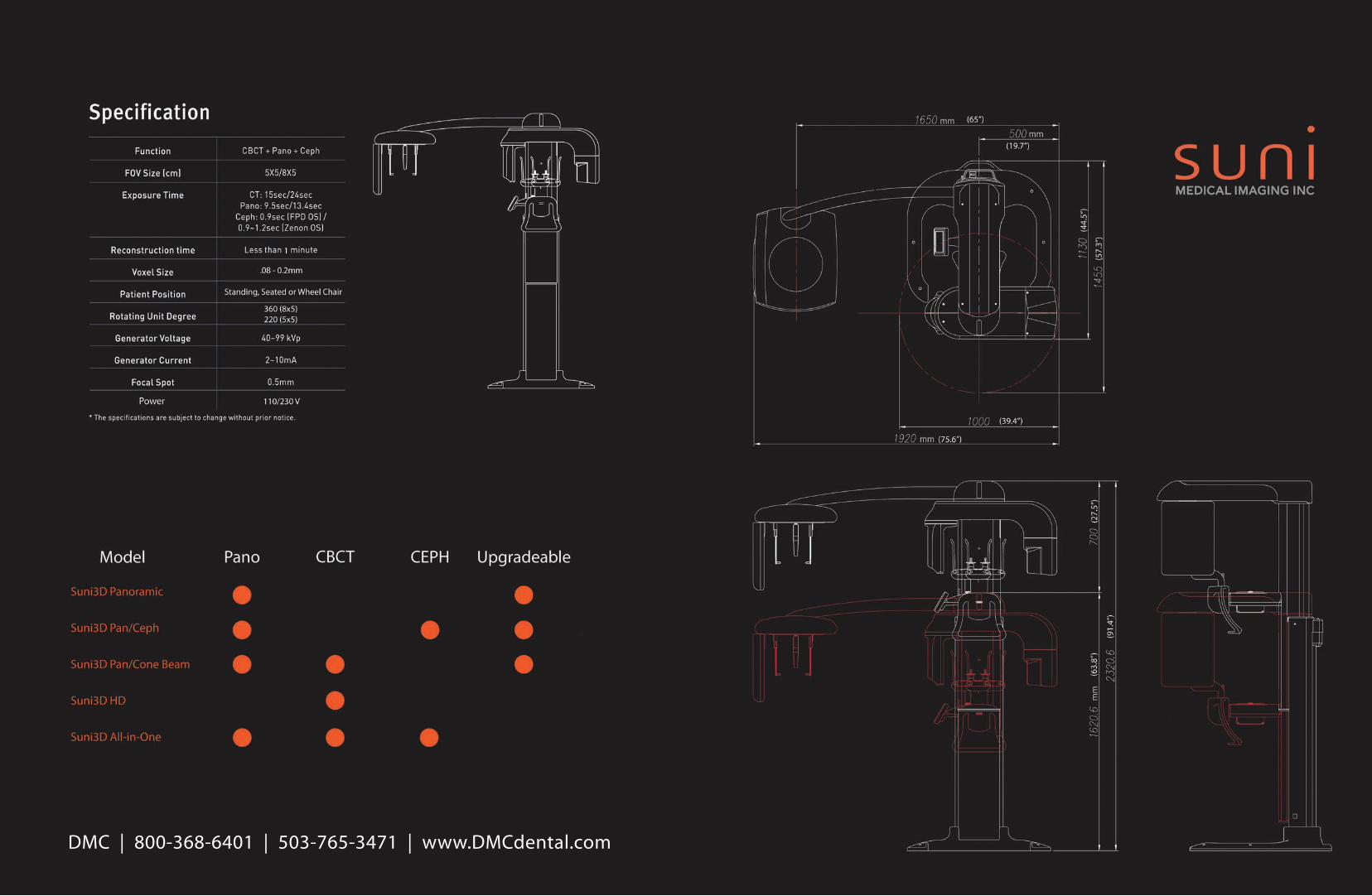
Upgradeable technology - start with panoramic capability and add cephalometric and 3D capability as your practice grows.
SUNI 3 D Values at a Glance:
With its just right FOV for 3D applications,and panoramic and cephalometric imagesthat are beyond compare, the Suni3DSystem is the optimum tool for all your specialty and diagnostic procedures.
CT Cone Beam Panoramic FPD OS CEPH
3D & Panoramic SystemAvailable as an easy to operate panoramic unit, or a dual function panoramic and 3D system, Suni3D o�ers the best of both worlds
Auto-Switching betweenPanoramic & CBCT Sensors
No manual sensor switching required. Simply choose the scan program you want and the sensors automatically switch into place.
With a FOV of 8x5, dentists can easily diagnose one dental quadrant at a time giving �exibility and data for single to multiple implant cases.
FOV 8x5
Perfect size for single implant cases and endodontic diagnosis.
FOV 5x5
Optional Panoramic bite wing software provides a low-dose alternative for capturing panoramic bite wings in anterior and posterior regions - an optimum alternative for patients with challenging anatomies.
Optional Bitewing Module with Auto Focus Technology
Suni3D Panoramic
Accurate and simple panoramic technology provides a series of automated programs for all your diagnostic needs --the Suni3D Panoramic system combines simplicity with state of the art technology to provided ease of use, superior image quality and extensive diagnostic capability.
There are nine anatomical programs including Standard, Hemi-Panoramic, Frontal Dentition, Sinus, TMJ Open and Closed, Incisor Clear, Orthogonal, Canal Clear and Maxillary Clear. The best part is that the Suni3D Panoramic System gives you the �exibility to upgrade to Ceph or 3D whenever you're needs require it.
Suni3D Cone Beam
Suni3D has an optimum 5x5 or 8x5 FOV that is just the right size for quadrant dentistry. The focal �eld of view allows you to see all the anatomical structures needed for optimum care. Suni3D is ideal for implantology, TMJ evaluation, and endodontic procedures.
With Suni3D’s outstanding diagnostic image quality, you can view coronal, sagittal, and axial images together with the panoramic image in one viewer. The exact measurements and implant simulation functions give you the security of accurate treatment planning.
FOV 5x5
FOV 8x5
The Optimum SolutionFor All Diagnoses
High-resolution images with selectable voxel sizesfor consistent diagnosis everytime.
One-shot, brilliant quality cephalometric images for optimum orthodontic care.
Crystal-clear panoramic imaging with optional bite wing capture for general diagnostic needs.
Upgradeable technology - start with panoramic capability and add cephalometric and 3D capability as your practice grows.
SUNI 3 D Values at a Glance:
With its just right FOV for 3D applications,and panoramic and cephalometric imagesthat are beyond compare, the Suni3DSystem is the optimum tool for all your specialty and diagnostic procedures.
CT Cone Beam Panoramic FPD OS CEPH
3D & Panoramic SystemAvailable as an easy to operate panoramic unit, or a dual function panoramic and 3D system, Suni3D o�ers the best of both worlds
Auto-Switching betweenPanoramic & CBCT Sensors
No manual sensor switching required. Simply choose the scan program you want and the sensors automatically switch into place.
With a FOV of 8x5, dentists can easily diagnose one dental quadrant at a time giving �exibility and data for single to multiple implant cases.
FOV 8x5
Perfect size for single implant cases and endodontic diagnosis.
FOV 5x5
The Suni3D Di�erence
Optional Panoramic bite wing software provides a low-dose alternative for capturing panoramic bite wings in anterior and posterior regions - an optimum alternative for patients with challenging anatomies.
Optional Bitewing Module with Auto Focus Technology
Suni3D Panoramic
Accurate and simple panoramic technology provides a series of automated programs for all your diagnostic needs --the Suni3D Panoramic system combines simplicity with state of the art technology to provided ease of use, superior image quality and extensive diagnostic capability.
There are nine anatomical programs including Standard, Hemi-Panoramic, Frontal Dentition, Sinus, TMJ Open and Closed, Incisor Clear, Orthogonal, Canal Clear and Maxillary Clear. The best part is that the Suni3D Panoramic System gives you the �exibility to upgrade to Ceph or 3D whenever you're needs require it.
Suni3D Cone Beam
Suni3D has an optimum 5x5 or 8x5 FOV that is just the right size for quadrant dentistry. The focal �eld of view allows you to see all the anatomical structures needed for optimum care. Suni3D is ideal for implantology, TMJ evaluation, and endodontic procedures.
With Suni3D’s outstanding diagnostic image quality, you can view coronal, sagittal, and axial images together with the panoramic image in one viewer. The exact measurements and implant simulation functions give you the security of accurate treatment planning.
FOV 5x5
FOV 8x5
FOV 5x5
FOV 8x5
One-Shot CephalometricState of the art cephalometric imaging with one-shot exposures delivers exceptional image quality for orthodontic, maxillofacial and multi-disciplinary practices
The Most Advanced System for Orthodontic SpecialistsState of the Art “One-Shot” Technology Revolutionary One Shot Exposure
With a 0.9 second exposure time, Suni3D ensures the best image quality and minimal retakes by eliminating image distortion that could result from patient move-ment. The less than 1 second exposure time reduces x-ray dose proving a safer diagnostic environment for your sta� and patients.
Suni3D’s highly sensitive �at panel x-ray detector ensures high-quality images never before seen. Orthodontic and multi-disciplinary practices can bene�t from Suni’s advanced technology. And, the Cephalometric module can be easily added at any time to the Suni3D Panoramic.
The slim and elegant design of the Cephalometric module allows for simple o�ce placement and excellent space e�ciency.
A broad range of image formats that suit any orthodontic tracing need round out the �exibility of the Cephalometric module.
A complete diagnostic cephalometric tool- Unparalleled image quality, covering the entire head- Signi�cant reduction in x-ray exposure dose- Multiple cephalometric formats- Upgradeable to localized 3D imaging
* Sample Images of FPD OS Cephalometric system
Optimal Capture Modes for Orthodontic Diagnosis
HD
Endodontics and Implantology in High De�nition
Small �eld of view and optimal voxel sizes that are ideal for endodontic and implant procedures
Suni3D HD’s 5x5cm Field of View is Ideal for Specialty Applications
And, with the added bene�t of a lower radiation dose, you can be assured that you are providing the safest and most e�ective care for you patients. With most endodontic and implant procedures involving 1 or 2 teeth, there is no need to obtain entire scans of the patient’s cranial anatomy. Suni3D HD gives you just the right amount of information to give your patients just the right amount of care without causing possible liability issues in the future for you and your practice.
Con�gurable up to .125mm, the imaging capabilities of the Suni3D HD outshine the competition. High-resolution 3D imaging gives you a new perspective when looking at dental structures and anatomies allowing you to see things that you simply cannot see with conventional 2D imaging systems. Or even some of the competitive 3D systems on the market today. The small voxel size provides images that are ideal for implantology and even for the most demanding endodontic cases.
Technology for the User & PatientTechnology for the User & PatientOne-Shot Cephalometric
Auto PositioningIn CBCT scan, the target tooth can be scannedexactly using this technology.
The Position is adjusted automatically accordingto the number of teeth. (scout view: only for FOV 5x5cm)
LCD GuidanceWhen scanning a patient, it displays the directionwith LCD guidance.
With voice and LCD guidance it enables the accurate positioning of the patient
Touch Panel LCDDuring operation, you can use not only the PC but also the touch panel.
Operate and perform positioning beside the patientwithout the inconvenience of moving every time.
Wheel-chair AccessibleDesign based on the convenience of patients.
0.9 second exposure time minimizes radiation exposure and possible image distortion from patient movement. The result, images with exceptional quality.
Auto Switching TechnologyThe Panoramic and CBCT sensors rotate automati-cally depending on the resultant image desired. No manual sensor switching required minimizes risk of accidental damage.
Customized SWUsers can customize various features such as the composition and position of the toolbar. You can use it intuitively by deleting the unnecessary features and creating your own menu. From now on, enjoy uniquely customized SW.
Intuitive, User-friendlyMenu barYou can �nd the function you want conveniently by using the menu bar or toolbar at the same time.
GuidanceGuidance is given through anatomical icons displaying the direction of image in a user-friendly interface.
Pro�le
Displays the bone density pro�le ensuring optimal implant placement.
Canal ManagerCanal size and color can be adjusted allowing accurate diagnosis and implant planning.
Implant SimulationThe Implant Simulation function reduces the risk during surgery and Ez3D allows simple and accurate planning without complicated processes seen in other software.
Houns�eld ScaleQuantitative scale for determining bone density to ensure optimal implant placement.
Various View ModesYou can diagnose using various view modes such as Cross-sectional View, Oblique View, and 3D Zoom.
Measurement
1:1 ratio allows for accurate measurement.
Technology for the User & PatientTechnology for the User & PatientOne-Shot Cephalometric
Auto PositioningIn CBCT scan, the target tooth can be scannedexactly using this technology.
The Position is adjusted automatically accordingto the number of teeth. (scout view: only for FOV 5x5cm)
LCD GuidanceWhen scanning a patient, it displays the directionwith LCD guidance.
With voice and LCD guidance it enables the accurate positioning of the patient
Touch Panel LCDDuring operation, you can use not only the PC but also the touch panel.
Operate and perform positioning beside the patientwithout the inconvenience of moving every time.
Wheel-chair AccessibleDesign based on the convenience of patients.
0.9 second exposure time minimizes radiation exposure and possible image distortion from patient movement. The result, images with exceptional quality.
Auto Switching TechnologyThe Panoramic and CBCT sensors rotate automati-cally depending on the resultant image desired. No manual sensor switching required minimizes risk of accidental damage.
Customized SWUsers can customize various features such as the composition and position of the toolbar. You can use it intuitively by deleting the unnecessary features and creating your own menu. From now on, enjoy uniquely customized SW.
Intuitive, User-friendlyMenu barYou can �nd the function you want conveniently by using the menu bar or toolbar at the same time.
GuidanceGuidance is given through anatomical icons displaying the direction of image in a user-friendly interface.
Pro�le
Displays the bone density pro�le ensuring optimal implant placement.
Canal ManagerCanal size and color can be adjusted allowing accurate diagnosis and implant planning.
Implant SimulationThe Implant Simulation function reduces the risk during surgery and Ez3D allows simple and accurate planning without complicated processes seen in other software.
Houns�eld ScaleQuantitative scale for determining bone density to ensure optimal implant placement.
Various View ModesYou can diagnose using various view modes such as Cross-sectional View, Oblique View, and 3D Zoom.