The EMBO Journal vol.12 no.3 pp.1067- 1075, 1993 Stably maintained microdomain of localized unrestrained supercoiling at a Drosophila heat shock gene locus Eldon R.Jupe, Richard R.Sinden1 and lain L.Cartwright2 Department of Molecular Genetics, Biochemistry and Microbiology, University of Cincinnati College of Medicine, 231 Bethesda Avenue, Cincinnati, OH 45267-0524, USA 'Present address: Institute of Biosciences and Technology, Texas A&M University, 2121 Holcombe, Houston, TX 77030, USA 2Corresponding author Communicated by A.Nordheim A psoralen crosslinking assay was utilized to detect localized, unrestrained DNA supercoiling (torsional tension) in vivo in Drosophila chromosomal regions subject to differential transcriptional activity. By comparing rates of crosslinking in intact cells with those in cells where potential tension in chromosomal domains was relaxed by DNA strand nicking, the contribution to psoralen accessibility caused by altered DNA-protein interactions (e.g. nucleosomal perturbations) was distinguished from that due to the presence of unrestrained supercoiling in a region of interest. The heat shock protein 70 (hsp7O) genes were wound with a significant level of superhelical tension that remained virtually unaltered whether or not the genes were transcriptionally activated by thermal elevation. Constitutively expressed 18S ribosomal RNA genes also exhibited unrestrained superhelical tension at a level comparable with that across hsp70. In contrast, flanking regions downstream of each of the divergent hsp70 genes at locus 87A7 exhibited substantially less tension. Thus the results point to the existence of stable, torsionally stressed topological domains within eukaryotic chromosomal DNA, suggesting that the relaxing action of topoisomerases is not ubiquitous throughout the nucleus but, in fact, is likely to be tightly regulated. Key words: chromatin domains/DNA supercoiling/ Drosophila hsp7O/psoralen/gene expression Introduction Packaging linear DNA into chromosomes requires compaction of 1000-fold in Escherichia coli and up to 10000-fold in human cells at metaphase (Dupraw, 1970). Such chromosomal organization leads to the partitioning of DNA into adjacent, independent loops of topologically closed domains (Benyajati and Worcel, 1976; Cook and Brazell, 1976; Igo-Kemenes and Zachau, 1977). Within these loops, supercoiling of the DNA may be differentially restrained by winding associations with chromosomal proteins. Release of supercoils from restraint can lead to the equilibration of supercoiling energy throughout the DNA helix as torsional tension, a condition of altered helical twist relative to that of relaxed DNA (Esposito and Sinden, 1989). Levels of torsional tension in a topological domain (or a portion thereof) may also be influenced by topoisomerase activity (Wang, 1985; Brill and Stemglanz, 1988) and by the helical tracking of replication or transcription complexes (Liu and Wang, 1987; Wang and Giaver, 1988; Wu et al., 1988). In prokaryotes, precise regulation of localized DNA tension levels clearly influences the expression of many genes (Drlica, 1984; Pruss and Drlica, 1989); however, in eukaryotes the issue of whether localized torsional tension occurs and its possible function has been controversial (reviewed in Eissenberg et al., 1985; Esposito and Sinden, 1989; Patient and Allan, 1989; Freeman and Garrard, 1992). A number of assays for detecting torsional tension in DNA have utilized various derivatives of psoralen. These are cell membrane-permeable molecules with a planar, aromatic structure that allows them to intercalate randomly into DNA (Cimino et al., 1985). Upon treatment with 360 nm light, intercalated psoralens mediate crosslinking of opposite DNA strands via formation of covalent bonds at each end of the molecule to adjacent thymines. Because of the particular geometry of the intercalated psoralen within the helix, the most favorable contacts for crosslink formation occur at 5'-TA dinucleotides (Cimino et al., 1985). It has been demonstrated that the rate of psoralen photobinding to DNA is linearly related to its level of negative superhelicity (Sinden et al., 1980). Detection of negative supercoiling in vivo can be accomplished by comparing rates of crosslinking in intact cells with those in cells where potential tension has been relaxed by DNA strand nicking (Sinden et al., 1980; Sinden and Ussery, 1992). This is possible because, by definition, unrestrained supercoiling will be relaxed as soon as a swivel (e.g. a strand break) is introduced in the helix. Such an approach also allows a distinction to be drawn between psoralen crosslinldng levels (and potential alterations thereof) affected by steric hindrance considerations (e.g. DNA -protein interactions) and those arising from a state of helical twist different from that of fully relaxed DNA (Sinden et al., 1980, 1982). In prokaryotes, measurements averaged globally across the E. coli genome have detected unrestrained supercoiling present at a superhelical density (a) of -0.03 to -0.05 (Sinden et al., 1980). In eukaryotes, similar global assays performed on either HeLa or Drosophila cells did not detect torsional tension (Sinden et al., 1980), probably because the majority of eukaryotic chromosomal DNA is wound in nucleosomes (Lacy and Axel, 1975; Garel and Axel, 1976), each of which effectively restrains one negative supercoil (Klug and Lutter, 1981). However, the sensitivity of this assay was not sufficient to detect tension that might be present in a small fraction of the genome, where perturbation of winding associations of DNA with chromosomal proteins in localized regions of chromatin could lead to partial loss of supercoil restraint. Various adaptations of the psoralen probing approach have been used to detect localized torsional tension in bacterial chromosomal DNA (Cook et al., 1989), chloroplast DNA (Thompson and Mosig, 1990; Davies et al., 1991), and constitutively expressed amplified human DNA (Ljungman 1067
Transcript
The EMBO Journal vol.12 no.3 pp.1067- 1075, 1993
Stably maintained microdomain of localized unrestrainedsupercoiling at a Drosophila heat shock gene locus
Eldon R.Jupe, Richard R.Sinden1 andlain L.Cartwright2Department of Molecular Genetics, Biochemistry and Microbiology,University of Cincinnati College of Medicine, 231 Bethesda Avenue,Cincinnati, OH 45267-0524, USA'Present address: Institute of Biosciences and Technology, Texas A&MUniversity, 2121 Holcombe, Houston, TX 77030, USA2Corresponding author
Communicated by A.Nordheim
A psoralen crosslinking assay was utilized to detectlocalized, unrestrained DNA supercoiling (torsionaltension) in vivo in Drosophila chromosomal regionssubject to differential transcriptional activity. Bycomparing rates of crosslinking in intact cells with thosein cells where potential tension in chromosomal domainswas relaxed by DNA strand nicking, the contribution topsoralen accessibility caused by altered DNA-proteininteractions (e.g. nucleosomal perturbations) wasdistinguished from that due to the presence ofunrestrained supercoiling in a region of interest. The heatshock protein 70 (hsp7O) genes were wound with asignificant level of superhelical tension that remainedvirtually unaltered whether or not the genes weretranscriptionally activated by thermal elevation.Constitutively expressed 18S ribosomal RNA genes alsoexhibited unrestrained superhelical tension at a levelcomparable with that across hsp70. In contrast, flankingregions downstream of each of the divergent hsp70 genesat locus 87A7 exhibited substantially less tension. Thusthe results point to the existence of stable, torsionallystressed topological domains within eukaryoticchromosomal DNA, suggesting that the relaxing actionof topoisomerases is not ubiquitous throughout thenucleus but, in fact, is likely to be tightly regulated.Key words: chromatin domains/DNA supercoiling/Drosophila hsp7O/psoralen/gene expression
IntroductionPackaging linear DNA into chromosomes requirescompaction of 1000-fold in Escherichia coli and up to10000-fold in human cells at metaphase (Dupraw, 1970).Such chromosomal organization leads to the partitioning ofDNA into adjacent, independent loops of topologically closeddomains (Benyajati and Worcel, 1976; Cook and Brazell,1976; Igo-Kemenes and Zachau, 1977). Within these loops,supercoiling of the DNA may be differentially restrained bywinding associations with chromosomal proteins. Releaseof supercoils from restraint can lead to the equilibration ofsupercoiling energy throughout the DNA helix as torsionaltension, a condition of altered helical twist relative to thatof relaxed DNA (Esposito and Sinden, 1989). Levels oftorsional tension in a topological domain (or a portionthereof) may also be influenced by topoisomerase activity
(Wang, 1985; Brill and Stemglanz, 1988) and by the helicaltracking of replication or transcription complexes (Liu andWang, 1987; Wang and Giaver, 1988; Wu et al., 1988).In prokaryotes, precise regulation of localized DNA tensionlevels clearly influences the expression of many genes(Drlica, 1984; Pruss and Drlica, 1989); however, ineukaryotes the issue of whether localized torsional tensionoccurs and its possible function has been controversial(reviewed in Eissenberg et al., 1985; Esposito and Sinden,1989; Patient and Allan, 1989; Freeman and Garrard, 1992).A number of assays for detecting torsional tension in DNA
have utilized various derivatives of psoralen. These are cellmembrane-permeable molecules with a planar, aromaticstructure that allows them to intercalate randomly into DNA(Cimino et al., 1985). Upon treatment with 360 nm light,intercalated psoralens mediate crosslinking of opposite DNAstrands via formation of covalent bonds at each end of themolecule to adjacent thymines. Because of the particulargeometry of the intercalated psoralen within the helix, themost favorable contacts for crosslink formation occur at5'-TA dinucleotides (Cimino et al., 1985). It has beendemonstrated that the rate of psoralen photobinding to DNAis linearly related to its level of negative superhelicity (Sindenet al., 1980). Detection of negative supercoiling in vivo canbe accomplished by comparing rates of crosslinking in intactcells with those in cells where potential tension has beenrelaxed by DNA strand nicking (Sinden et al., 1980; Sindenand Ussery, 1992). This is possible because, by definition,unrestrained supercoiling will be relaxed as soon as a swivel(e.g. a strand break) is introduced in the helix. Such anapproach also allows a distinction to be drawn betweenpsoralen crosslinldng levels (and potential alterations thereof)affected by steric hindrance considerations (e.g.DNA -protein interactions) and those arising from a stateof helical twist different from that of fully relaxed DNA(Sinden et al., 1980, 1982). In prokaryotes, measurementsaveraged globally across the E. coli genome have detectedunrestrained supercoiling present at a superhelical density(a) of -0.03 to -0.05 (Sinden et al., 1980). In eukaryotes,similar global assays performed on either HeLa orDrosophila cells did not detect torsional tension (Sindenet al., 1980), probably because the majority of eukaryoticchromosomal DNA is wound in nucleosomes (Lacy andAxel, 1975; Garel and Axel, 1976), each of which effectivelyrestrains one negative supercoil (Klug and Lutter, 1981).However, the sensitivity of this assay was not sufficient todetect tension that might be present in a small fraction ofthe genome, where perturbation of winding associations ofDNA with chromosomal proteins in localized regions ofchromatin could lead to partial loss of supercoil restraint.Various adaptations of the psoralen probing approach havebeen used to detect localized torsional tension in bacterialchromosomal DNA (Cook et al., 1989), chloroplast DNA(Thompson and Mosig, 1990; Davies et al., 1991), andconstitutively expressed amplified human DNA (Ljungman
1067
E.R.Jupe, R.R.Sinden and I.L.Cartwright
and Hanawalt, 1992a). In the present study, we have deviseda psoralen crosslinking assay able to search for the existenceof tension in vivo in precisely defined regions of single copyeukaryotic chromosomal DNA. This method has beenutilized to detect localized tension under bothtranscriptionally silent and active conditions, as well as formapping the extent of its propagation, in the domaincontaining the pair of hsp70 genes at Drosophila locus 87A7.
ResultsAssay for quantitating specific crosslinks formed invivoWe have developed a gel-based protocol for quantitating verylow levels of psoralen crosslinking localized in specificregions of single copy DNA from eukaryotic cells.Previously reported assays for quantitating psoralencrosslinks in bacterial chromosomal (Cook et al., 1989) andplant chloroplast DNA (Thompson and Mosig, 1990) werenot useful in this instance because of sensitivity limitations.In our protocol, the psoralen crosslinked, restriction digestedDNA is denatured by glyoxylation, fragments are separatedby gel electrophoresis in neutral phosphate buffer (McMasterand Carmichael, 1977), and the gel treated by extendedincubation in sodium hydroxide prior to blotting andhybridization (see Materials and methods). Such an approachpermits accurate quantitation of both crosslinked (whichshow a retarded gel mobility) and non-crosslinked fractionsof single copy sequences within a complex eukaryoticgenome. The coding regions, as well as proximal and distaldownstream flanking regions, of the divergent Drosophilahsp70 genes at locus 87A7 (Figure 1) have been examinedfor levels of psoralen crosslinking both prior to and afterheat induction. Transcriptional activity of these genes rapidlyincreases to high levels when cultured cells are placed at37°C (Lindquist, 1986). Transcription-related alterations tothe chromatin structure of the hsp70 genes, including changesin nuclear protein interactions (Wu, 1984; Shuey and Parker,1986), topoisomerase associations (Fleischmann et al., 1984;
1 kb
Gilmour et al., 1986; Rowe et al., 1986; Kroeger andRowe, 1989; Udvardy and Schedl, 1991) and nucleosomalcharacter (Wu et al., 1979; Wu, 1980; Udvardy and Schedl,1984; Karpov et al., 1984; Solomon et al., 1988; Nachevaet al., 1989) have been particularly well documented, thusallowing a rather well-informed interpretation of the dataobtained here. In addition to hsp70, a coding region fragmentof the 18S ribosomal RNA gene (rDNA) has also beenanalyzed (Figure 1). Because the constitutive rate ofrDNAtranscription does not change upon thermal elevation (Bellet al., 1988), it provides a control for any non-specific effectsof heat shock treatment.
Gene specific crosslinking as a function of heat shockFigure 2A shows an experiment examining the kinetics ofin vivo psoralen photobinding to the 18S rDNA under non-heat shock (NHS) conditions. When exposed to increasinglight dosages in vivo, the fraction of specific fragmentscrosslinked by psoralen increased (Figure 2A, lanes 2-5)until multiple crosslinks began to accumulate (Figure 2A,lanes 6 and 7). By determining the fraction of crosslinked(XL) versus non-crosslinked (NXL) fragments present ineach lane (Figure 2A, lanes 2-5), the number of crosslinksper kilobase (XL/kb) introduced into a DNA fragment byeach light dose was determined (see Materials and methods).Similar psoralen crosslinking analyses were performed onthe 18S rDNA under heat shock (HS) conditions, and onthe hsp70 coding region under both NHS and HS conditions.Figure 2B summarizes the data obtained. Heat activatedhsp70 genes became crosslinked - 3-fold faster than whenthey were transcriptionally dormant. Conversely, undereither NHS or HS conditions, the constitutively expressed18S rDNA coding region exhibited an invariant crosslinkingrate that lay between the extremes found for the hsp70 codingregion. The data in Figure 2B also show that light dosagesof 5-6 kJ/m2 were in the linear range of response andproduced < 1 XL/kb in the 1-2 kb restriction fragmentstested. Thus, a single light dose of 6 kJ/m2 was selected forfurther assays because the values obtained will reflect the
DDSE SI I
CRx P xI I I
CRx p xI I l
1.... I dilscS
PDSB EI I
18SH HdII_SCSI
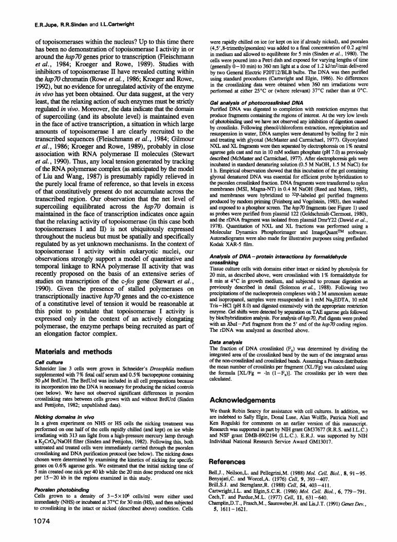
Hsp 70 Locus 87A7 rDNAFig. 1. Maps of the regions examined in this study. At the 87A7 hsp70 locus on the left the divergently transcribed coding regions (CR) andfragments from the proximal (PDS) and distal (DDS) downstream regions (indicating direction from the centromere) examined are delineated by therestriction sites shown (Goldschmidt-Clermont, 1980; Mason et al., 1982). The open box indicates the transcribed portion of the coding region andthe arrows show the direction of transcription. The XbaI fragment examined contains the coding region and a small portion of a duplicated 5' non-coding region (solid box). The locus is flanked by specialized chromatin structures (scs and scs') depicted by shaded boxes (Udvardy et al., 1985). Aportion of the rDNA locus is shown on the right. The fragment delineated by the restriction sites contains most of the 18S coding region (open box)and a small portion of the intergenic spacer (IGS). This partial map does not show the 5.8S and 28S rDNA which are located immediatelydownstream of the 18S gene (Long et al., 1981). About 200 copies of the tandemly repeated unit are present in the genome. Restriction enzymes areabbreviated as follows: B, Bgll; E, EcoRI; H, HaeIH; Hd, HindIII; P, PstI; S, Sall; X, XbaI.
1068
I m
Supercoiled domains in Drosophila chromatin
relative rates of crosslinking with good precision (Sindenet al., 1980; Sinden and Pettijohn, 1982). The results inFigure 2B also suggest that the crosslinking rates measuredwere not unduly influenced by the frequency of 5 '-TAdinucleotides in the sequences being examined. The hsp7Ocoding region fragment has the lowest frequency of 5'-TAof any sequence that we have assayed, yet it exhibits thehighest rate of crosslinking when transcriptionally induced.The constitutively active rDNA sequence contains- 10-foldmore 5'-TAs per kb, yet has a lower overall rate ofcrosslinking. Thus, under the conditions of low levels ofphoto-crosslinking at which the assay is being performed,the 5 '-TA frequency is not a major determinant of the rateof crosslinking to specific sequences.
A1 2 345
XL
NXL-~
s
4.
B
n0
-- --=
f6-
-1--i
li
Light Dose (kJ rn.'.'i
Fig. 2. Characterization of the in vivo psoralen crosslinking assay inDrosophila chromosomal DNA. (A) Agarose gel analysis of psoralenphotobinding to specific chromosomal fragments. Drosophila Schneider3 tissue culture cells were equilibrated with psoralen and exposed to360 rnm light for increasing lengths of time to give the following dosesin U/rn2; 0 (lane 1), 0.6 (lane 2), 1.2 (lane 3), 3.6 (lane 4), 6.0(lane 5), 12 (lane 6) and 24 (lane 7). DNA was isolated, digested withHaeIHI-Hindu, glyoxal denatured and analyzed on neutral agarosegels (see Materials and methods). In this experiment, crosslinking ratewas determlined for the 18S rDNA fragment (Figure 1) under NHSconditions. The NXL and XL fragment bands are designated on theleft of the autoradiogram. (B) Crosslinking rates of hsp7O and rDNAcoding regions under NHS and HS conditions. Crosslinking rates weredetermined for the hsp7O CR (Figure 1) and 18S rDNA fragments inexperiments identical to that illustrated in (A). The XL/kb weredetermlined (see Materials and methods) and plotted against the lightdose. The points on the graph are indicated as follows: El, NHS-hsp70 CR; U, HS-hsp7O CR; 0, NHS-18S rDNA; 0, HS-18SrDNA.
Detection of torsional tension in vivoThe observation that the hsp7O gene is crosslinked muchmore rapidly when transcribed than when silent does notnecessarily reflect an increase in torsional tension engenderedby transcription. It is well known from the work of severallaboratories that psoralen intercalates highly preferentiallyin the linker DNA region between nucleosomes (Cech andPardue, 1977; Sogo et at., 1984). In order to accommodatean intercalator such as psoralen a small amount of base-pairunwinding has to occur, and this is clearly relativelyunfavorable on the nucleosome core itself owing to the tightassociation between DNA and the histone octamer (Morseand Cantor, 1985). However, if such core histone-DNAinteractions are perturbed in some way, as has been clearlydemonstrated to occur for the nucleosomal array located onthe transcribed portion of the hsp7O genes upon activation(Wu et at., 1979; Nacheva et at., 1989), then access ofpsoralen to this DNA is likely to be substantially enhancedbecause the available target size for intercalation hasincreased. Such alterations in psoralen accessibility causedsolely by redistribution of DNA -protein associations havebeen clearly documented previously (Sinden et at., 1982).To control for transcription-related alterations in
DNA -protein interaction that would affect stericaccessibility alone, and to allow an estimate of thecontribution that torsional tension makes to the observed ratesof crosslinking in cells, we compared crosslinking rates inintact cells with those in cells in which potential torsionaltension had been released by DNA nicking immediately priorto crosslinking (Sinden et at., 1980; Sinden and Pettijohn,1982). This was achieved by culturing cells in mediumcontaining bromodeoxyuridine (BrdUrd) to produce BrdUrd-substituted cellular DNA, which is susceptible to strandbreakage upon irradiation with 313 nm light (see Materialsand methods). Figure 3 shows representative results fromsuch experiments derived from analysis of restrictionfragments encompassing both transcribed (panel A) andproximal downstream flanking (panel B) regions of the hsp7Ogenes. Table I summarizes the crossllnking rates per kbcalculated from this experiment. The rate of crosslinking inthe coding region under NHS conditions was very lowcompared with the rate upon HS (Figure 3A, compare lanes2 and 7; Table I; also see Figure 2B). In NHS cells, anicking light dose of 5 min led to a slight reduction in thehsp7O coding region psoralen crosslinking rate (Figure 3A,lane 3). A 2-fold reduction relative to intact cells wasdetectable following a 20 min nicking treatment (Figure 3A,lane 4; also see Table I). In HS cells, a 5 min nickingtreatment caused an immediate 2-fold reduction incrosslinking rate relative to intact cells (Figure 3A, comparelanes 7 and 8), and the rate was not reduced upon furthernicking (Figure 3A, lane 9; Table I). When psoralen wasnot added, we did not observe any DNA crosslinking dueto exposure of the cells to either the 313 nm wavelength usedfor nicking or the 360 nm wavelength used for crosslinking(Figure 3A, lanes 5 and 10). The reduction in crosslinkingrates in the nicked compared with the intact samnples observedin this experiment clearly suggests that torsional tension ispresent across the coding region in advance of, as well asfollowing, induction of transcription and that its relative levelis similar in both cases (discussed in detail below). In notablecontrast to the coding region results, similar rates ofcrosslinking were observed for hsp7O downstream flanking
1069
11- Ammm6.M
E.R.Jupe, R.R.Sinden and I.L.Cartwright
NHS HS0 0 52020 0 0 52020,0 6 6 6 6C 0 6 6 6 6C
-- H
-._ow.. Im... ,,,,I
Lane 1 2 3 4 '; 6 8 9
B
N (mir)XL (kJ Mr2)
NHS
0 0 E 2020
0 6 6 6 6C
XL
I HS0 5 2020
0 6 6 6 6C
Lane 1 2 3 4 5 6 7 8 9 10
Fig. 3. Psoralen crosslinking in vivo in intact and nicked domains ofthe hsp7O gene under NHS and HS conditions. Cells with intact ornicked domains were psoralen crosslinked to examine torsional tension.Nicking (N) indicates the time in min that the cells were exposed to313 nm light. Crosslinking (XL) indicates the dose of 360 nm light inkJ/m2. The lanes with the 6C label indicate control samples that wereboth nicked and exposed to crosslinking conditions in the absence ofpsoralen. The NXL and XL bands produced are indicated on the lefthand side. (A) Samples were digested with XbaI and analyzed asshown above (Figure 2). The hsp7O CR fragment (Figure 1) is probedin this experiment. The fragment marked H is an additional uniquehsp7O gene fragment from the 87C locus (Mason et al., 1982) thatcross-hybridized to the probe. Extended exposures detect thecrosslinked fraction for this fragment well above the position of themajor crosslinked fraction shown. (B) Samples were digested withBgl-EcoRI and probed with the PDS fragment (Figure 1) in thisexperiment.
regions under both NHS and HS conditions (Figure 3B,compare lanes 2 and 7; Table I). Moreover, under eithercondition the crosslinking rate was only slightly reducedfollowing the 20 min nicking treatment (Figure 3B, comparelane 2 with lane 4 and lane 7 with lane 9) indicating thatsubstantially less tension was present at this location,particularly under resting conditions prior to transcriptionalinduction (also see Table I).
Figure 4 summarizes the results of numerous experimentslike those shown in Figure 3 performed on the hsp70 coding
Table I. Crosslinking ratesa of intact and nicked regions of hsp7O
aThese crosslinking rates were deternined from the experimentspresented in Figure 3 at a light dose of 6 kJ/m2 as discussed in thetext, and are expressed as XL/kb.bFragment locations and abbreviations are as defined in Figure 1.
region, on hsp70 distal and proximal downstream flankingregions, and on the 18S rDNA coding region. These datareflect a complex aggregate of both differential accessibilityand torsional tension present in chromosomal DNA in vivo.To aid in the resolution and discussion of these parametersFigure 4 displays the crosslinking rate per kb for intactdomains (solid bar) adjacent to the rate for the same domainwhen relaxed by nicking for 20 min (hatched bar). The heightof the solid bars for each region investigated reflects acomposite of differential psoralen crosslinking derived fromboth the presence of unrestrained torsional tension and theoverall helix accessibility in intact cells, while the height ofhatched bars necessarily reflects that component derived fromhelix accessibility alone since any potential tension has beenreleased by nicking. Thus, the ratio of the crosslinking ratein intact (solid) versus relaxed (hatched) chromosomaldomains (shown at the bottom of Figure 4 as RI/N) reflectsDNA tension levels in a given fragment, and the relativeratio comparing one region with another reflects differentialpartitioning of this tension within a given domain.The critical basis for interpreting a reduction in
crosslinking rates upon nicking as reflecting a release ofincipient unrestrained supercoiling, equilibrated as torsionaltension, relies on the expectation that the nicking processdoes not alter nuclear protein binding or association withDNA in a manner that would cause greatly reduced stericaccessibility of psoralen to the helix. Previous studiesaddressing global tension and accessibility levels in viral(Sinden and Pettijohn, 1982; Sinden et al., 1982), bacterialand eukaryotic (Sinden et al., 1980) genomes havedemonstrated that nicking procedures as used here do notappear to induce a non-specific agglomeration ('crashing')of proteins on to DNA. However, in order to address thepossibility that such 'crashing' effects could be occurringat specific loci and thus be an artifactual cause of theobserved reduction in psoralen crosslinking, in vivoDNA-protein crosslinking experiments with formaldehydewere performed according to the procedures of Solomonet al. (1988) on both intact cells and cells exposed to nickingconditions identical to those used in the psoralen assay. Thisallowed an assessment of the effect of nicking on the levelof protein associations with the particular DNA fragmentsexamined in this study. Figure 5 shows agarose gel shiftanalyses of crosslinked DNA-protein samples derived fromthese experiments. In intact cells a retardation in migrationof both hsp7O and rDNA fragments is evident whencomparing control samples with pronase-resistant,formaldehyde treated samples (compare lanes 1 and 2 for
Fig. 4. Schematic comparison of the crosslinking rates in intact andnicked samples from NHS and HS cells. The rate of crosslinking per
kilobase is plotted on the vertical axis. The solid bars show the rate ofcrosslinking in intact cells. The rate for samples nicked for 20 min isdepicted by the hatched bars. Each of the bars represents the average
of six crosslinking rates determined from three independent cellpreparations, each of which was analyzed twice. The standarddeviation for each of the values is shown by the error bars. The ratioof the mean crosslinking rates calculated for intact/nicked domains(RI/N) is presented for each of the four restriction fragments examined.All abbreviations are as defined in Figure 1.
hsp70, and lanes 4 and 5 for rDNA). In formaldehyde treatedsamples from cells exposed to DNA nicking conditions a
shift in migration compared with purified DNA is still evident(compare lanes 1 and 3 for hsp70, and lanes 4 and 6 forrDNA), but the shift is, if anything, slightly reduced relativeto that of the formaldehyde treated samples from intact cells
(compare lanes 2 and 3 for hsp70, and lanes 5 and 6 forrDNA). If a protein 'crashing' artifact were occurring uponnicking a further retardation in the migration of hsp70 or
rDNA fragments would have been expected owing toincreased opportunities for DNA -protein crosslinkformation. We conclude from these data and other controls(see Discussion) that there is no reasonable interpretationfor the dramatic reduction of psoralen crosslinking uponnicking other than the release of a pre-existing level oftorsional tension from selected regions of the Drosophilagenome.
Discussion
A stable microdomain of torsional tensionSeveral conclusions can be drawn concerning the existenceof torsional tension in vivo as well as the relative accessibilityof the various Drosophila chromosomal regions that we havestudied. Even prior to heat induced activation, the hsp70coding region is in a state of torsional tension as indicatedby the 2-fold reduction in crosslinking rate observed afternicking (CR in Figure 4). The flanking regions show a slightreduction in psoralen binding (comparing intact and nickedsamples) suggesting that only very low levels of torsionaltension are constitutively present at these downstreamlocations (DDS and PDS in Figure 4). Thus, it appears that
Fig. 5. Analysis of DNA-protein interactions in vivo by formaldehydecrosslinking in intact and nicked chromosomal domains. Purifiedpronase-resistant nucleoprotein complexes were digested extensivelywith the restriction enzyme of choice and analyzed byblot/hybridization following TAE agarose gel electrophoresis. Thesamples analyzed are purified DNA (lanes 1 and 4), formaldehydecrosslinked DNA-protein fragments from intact cells (lanes 2 and 5)and formaldehyde crosslinked DNA-protein fragments from cellssubjected to UV nicking (lanes 3 and 6). The hsp7O regions (lanes
1-3) were examined in PstI digests (see Figure 1) and probed with an
XbaI-PstI fragment from the 5' end of the coding region. Thus, the4.2 kb band is derived from locus 87A7 (see Figure 1). The 1.1 kbband detected in this experiment is derived from hsp7O genes at locus87C1 which have an additional PstI site adjacent to the XbaI site at the5' end of the coding region. The rDNA region (lanes 4-6) examinedis the same as in Figure 2A. The marker sizes shown to the left andright of the panels refer to the size of the purified DNAs in lanes 1
and 4 respectively.
we are observing the existence of a region of stablyunrestrained supercoiling, with boundaries that are locatedsomewhere between the end of the hsp70 coding region andthe downstream flanking sequences. In this sense, theexperiments have defined an independent topologicaldomain. Following heat induced activation, the hsp70 codingregion becomes almost 3-fold more accessible to psoralen,undoubtedly a reflection of the large nucleosomalperturbation associated with active transcription (Wu et al.,1979; Wu, 1980; Karpov et al., 1984; Nacheva et al.,1989), but it again shows the same 2-fold reduction incrosslinking rate upon nicking (CR in Figure 4). Such resultslead to the conclusion that this large increase in psoralenaccessibility upon heat shock is due to the known majornucleosomal perturbation that occurs upon transcription (i.e.the target size for psoralen intercalation increases from thatdue to linker DNA alone to that of linker plus formerly core-
associated DNA): there is, however, no correspondingchange in overall tension levels as a result of gene activation.Moreover, if there were a tension-related component to theinstability of the nucleosomal array upon transcriptionalinduction (e.g. a supercoil-driven unfolding of thenucleosomes) then it would be anticipated that upon nickingthe nucleosomes would refold, and the level of psoralencrosslinking in nicked heat shocked cells would be similar
1071
DDS 18S hss -- 18S rDNA
E.R.Jupe, R.R.Sinden and I.L.Cartwright
to that in nicked control cells. This is clearly not the case.Little change in steric accessibility to psoralen in the 3'flanking regions of hsp70 is caused by heat shock (DDS andPDS in Figure 4), consistent with the apparent stability ofnucleosome structure in these particular sequences upon geneactivation (Udvardy et al., 1985). However, the RI/N doesincrease slightly following heat shock, suggesting that theflanking regions may be experiencing somewhat moretension, perhaps as a consequence of tracking RNApolymerases.The sum of the data indicated that heterogeneous torsional
tension levels exist within the 87A7 structural domainregardless of transcriptional activity. This locus is flankedby sequences (Figure 1) termed specialized chromatinstructures (scs), first identified by nuclease sensitivity assays(Udvardy et al., 1985) and recently shown to act in genetictests as effective insulators of chromosomal position effects(Kellum and Schedl, 1991, 1992). The level of constitutivetension at 87A7 is, however, significantly reducedimmediately downstream of the hsp7O coding region, wellbefore the scs sequences are encountered. If the scssequences are viewed as some form of boundary for thedefinition of a structural or genetic domain at locus 87A7(only a supposition at this point), our data indicate that sucha domain does not correspond physically to the more limitedtopological domain defined here. Interestingly, the extentof propagation of high levels of torsional tension falls withinboundaries apparently delineated by a recently describedDrosophila chromatin binding protein named B52 thatassociates with the downstream borders of activelytranscribed regions of 87A7 (Champlin et al., 1991).However, this protein is not present under conditions oftranscriptional inactivity; therefore, our detection of a limitedtopological domain prior to gene activation appears topreclude a role for B52 in delineating tension boundaries.It is certainly not necessary to hypothesize the existence ofa sequence-specific boundary to explain our observations.It could easily be the case that the tension across the hsp7Ogenes is confined by the presence on flanking DNA regionsof nucleosomes that, by virtue of tight binding to DNA, donot allow further transmission of tension. Certainly this isa property expected of nucleosomal-bound DNA based onearlier in vitro observations (Morse and Cantor, 1985). Atthis time we have no knowledge of whether the tensionpresent across the inactive hsp7O genes is confined tonucleosomal linkers or is equilibrated through the core-associated DNA.Minimal differences in the accessibility of the 18S rDNA
occur in response to heat shock (18S in Figure 4), incongruence with the reported lack of effect of thermalelevation on transcription of these genes (Bell et al., 1988).However, 2-fold reductions in crosslinking rates uponnicking are again evident, irrespective of heat shock,reflecting the presence of constitutive torsional tension at thislocation also (Figure 4). Thus, both of the coding regionsexamined in our study contain localized torsional tension atsignificantly higher levels than in non-transcribed regions.A recent study of the 5' end of the coding region of theamplified dihydrofolate reductase genes and the 18S rDNAof human cells (both constitutively expressed) has reporteda similar reduction in psoralen crosslinking levels uponnicking (Ljungman and Hanawalt, 1992a). Thus, localizedtorsional tension appears to be detectable in several
eukaryotic genes. It is important to remember that previousstudies of unrestrained supercoiling averaged globally acrosseither Drosophila (Sinden et al., 1980) or human (Sindenet al., 1980; Ljungman and Hanawalt, 1992a) genomesfailed to detect any deviation from the situation expected forrelaxed, or at least fully restrained, chromosomal DNA. Thedata we have obtained are not inconsistent with thisobservation given that only a very small fraction of thegenome is (or has the potential to be) actively transcribed.We are currently assaying other non-transcribed ortranscribed regions of the Drosophila genome in vivo toconfirm the generality of the observation that non-transcribedsequences immediately flanking active genes exist under zero(or very low) levels of tension.
Previous analyses of psoralen crosslinking rates in vitroin intact topological isomers at defined levels of superhelicaldensity (a), suggested that a 2-fold reduction in psoralencrosslinking rates upon relaxation corresponds to a a equalto -0.08, while a 1.2- to 1.5-fold reduction occurs at a aof -0.01 to -0.02 (Sinden et al., 1980). Based on a1.7-fold reduction in crosslinking rates upon nicking, theeffective a of E.coli chromosomal DNA was estimated tobe -0.05, a figure that correlated well with earlier estimates(discussed in Sinden et al., 1980) but at the high end ofmorerecent determinations made from a variety of in vivo assaysfor supercoiling in bacterial plasmids (reviewed by Drlicaet al., 1992). Thus, in principle we could be detectingmagnitudes of localized effective a in the coding regions ofhsp70 and rDNA similar to those found in the E.colichromosome, although the actual a remains to be measuredby a more quantitative approach. In contrast, the flankingregions of the hsp70 genes may have a 4- to 8-fold lowereffective a than the coding region depending on gene activity.Experiments attempting to estimate precisely the in vivo levelof a present at these locations are currently underwayutilizing strategies applied previously in prokaryoticorganisms (Kochel and Sinden, 1988; Dr6ge and Nordheim,1991; Zheng et al., 1991).
Applicability of the psoralen assay for detecting invivo tensionAs discussed above in Results, alternative explanations forthe dramatic reduction in psoralen crosslinking that occursin selected genomic locations after nicking of chromosomaldomains have been considered and tested. The results provideno evidence for a non-specific effect of nicking on overallsteric accessibility of the DNA helix to psoralen. Thus, thereduction in psoralen photobinding that we observe uponnicking is detecting the presence of unrestrained supercoiling.We also performed extensive control experiments to addresswhether rates of psoralen crosslinking are influenced by thelowered temperature conditions at which the assay isperformed. We did not observe significant differences inpsoralen crosslinking levels when the 360 nm light treatmentwas performed at 250C immediately following harvestingcompared with cells that were harvested, brought to 0°C,and left on ice for up to 30 min prior to crosslinling. Similarcrosslinking rates were also observed for samples that wereheat shocked and immediately crosslinked compared withthose kept on ice for up to 30 min (unpublished data). Thesecontrols suggest that there is no immediately detectable effectof temperature changes on the levels of negative DNAsupercoiling. Based on studies that have quantitated the
1072
Supercoiled domains in Drosophila chromatin
change in helical twist ofDNA as a function of temperature(Goldstein and Drlica, 1984), we estimate that, at the upperlimit, 1 kb of completely protein-free DNA transferred from37 to 0°C in the absence of topoisomerase activity couldhave one negative supercoil introduced. In eukaryoticchromatin, this level of change should be substantially lessby virtue of association of the DNA with nucleosomes(reported to restrain effectively temperature-induced changesin twist; Morse and Cantor, 1985) or other chromosomalproteins. Thus, any negative supercoiling that might beintroduced by lowering temperature was not detectable incontrol experiments and is likely to be a negligiblecomponent of the levels we actually observe.A particularly interesting feature of the in vivo nicking
experiments (Table I) is that the sensitivity of the activelytranscribing hsp7O domain to nicking (minimum valuereached in 5 min) was substantially greater than in thetranscriptionally inactive case (where it took 20 min to reachthe same minimum). One interpretation of this result is thatthe size of the topological microdomain increasedsubstantially upon heat shock. We do not favor thispossibility, however, and prefer an explanation based on thechemistry of the nicking reaction, which involves UV-induced free radical production mediated by BrdUrd. Recentexperiments have shown that in purified nuclei the DNA intranscriptionally active domains was far more susceptibleto oxidative damage by free radicals than was that in inactivedomains, suggesting that the difference in damage sustainedwas due to differences in chromatin condensation (Ljungmanand Hanawalt, 1992b). Our observations are consistent withthis type of mechanism operating in vivo in intact cells.The relatively constant crosslinking frequencies observed
for the hsp70 downstream, as well as the 18S rDNA coding,fragments under both NHS and HS conditions (Figure 4)provide an important internal control for the validity of thisassay. The data show that the major differences incrosslinking rates across the hsp70 coding region are not dueto psoralen delivery differences mediated by differentialmembrane permeability caused either by the elevatedtemperatures used to induce heat shock gene transcriptionor by the in vivo nicking treatment.
In summary then, we believe that when performed withthe appropriate controls (as we have been particularly carefulto do), this psoralen crosslinking approach provides anappropriate and valid method for detecting torsional tensionin eukaryotic chromatin in vivo.
Biological relevance and significanceThe existence of a stable, constitutive microdomain oftorsional tension in the hsp7O genes is a rather unexpectedfinding and is therefore of particular interest. An importantquestion is obviously what is/are the process(es) leading toits establishment and maintenance? Up to this time, therehas been no demonstration of an enzymatic activity inDrosophila (or any other eukaryote for that matter) that isable, like prokaryotic DNA gyrase, to introduce negativesupercoils into DNA (discussed in Clarke and Wolffe, 1991;recently reviewed in Freeman and Garrard, 1992).Establishment by this means thus appears to be a remotepossibility. However, there are, as noted in the Introduction,other mechanisms by which negative supercoiling could begenerated. For example, release of restrained DNAsupercoils by loss of nucleosomes (see Esposito and Sinden,
1989; Freeman and Garrard, 1992), or via alteredassociations with high mobility group proteins (Ridsdaleet al., 1990) and/or other components of the chromatin fiberin the immediate vicinity of the hsp70 genes could introducenegative supercoils. In this context, it may be relevant thatboth the promoter and transcription initiation regions ofuninduced hsp70 genes exist in a DNase I hypersensitiveconfiguration (Wu, 1980), and therefore are considered tobe nucleosome free. We are currently in the process ofmapping the precise boundaries of the supercoiled domainin order to determine if there are significant, nick-responsive,hot-spots of psoralen cross-linking through the body of thegene or in its promoter. As a further possibility it has recentlybeen reported that acetylated histones as constituents ofnucleosomes restrain only 80% of the supercoils oftheir non-acetylated counterparts (Norton et al., 1989, 1990). Sinceacetylated nucleosomes are strongly correlated with activechromatin (Hebbes et al., 1988; Tazi and Bird, 1990), itis possible that the transcriptionally competent hsp70 genesare enriched in nucleosomes of this type, and thus exist ina region of partially unrestrained supercoils. Immuno-fluorescence analysis has demonstrated the presence ofvarious forms of acetylated histone on Drosophila polytenechromosomes (Turner et al., 1992). Further analysis of theirrelative distribution at the heat shock loci will be of someinterest.A particularly intriguing feature of the hsp70 genes is that
they have been found to exist in a 'transcriptionally poised'configuration. Apparently, not only is a molecule of RNApolymerase II present on each gene in advance of activation,but it is transcriptionally engaged and elongationally stalled,having synthesized a short transcript up to 25 bases long(Rougvie and Lis, 1988; O'Brien and Lis, 1991). It is anintriguing possibility that the existence of the unrestrainedtension that we detect here is related to the presence of thispaused polymerase transcription complex. Loss, orperturbation, of nucleosomes might either accompany theestablishment of this configuration, or indeed be requiredto help promote its assembly. The importance ofunderwoundDNA in promoting efficient assembly of stable eukaryoticRNA polymerase II initiation complexes has been suggestedby a number of in vitro experiments (Tabuchi and Hirose,1988; Mizutani et al., 1991). Furthermore, recent in vivoexperimental approaches have documented the presence oftorsional tension during the assembly of competent RNApolymerase II transcription complexes within chromatin(Leonard and Patient, 1991), and the enhancement of ratesof RNA polymerase I transcriptional initiation by negativesupercoiling (Schultz et al., 1992). We are certainly veryinterested in the potential correlation between negativesuperhelicity and engaged RNA polymerase activitysuggested by our observations and are currently studyingother genes known to lack such complexes in the cell typeunder study. This is not necessarily just a trivial exerciseof looking at silent genes-the elongational blocking oftranscriptionally engaged polymerases may be a much morecommon method of regulating transcription than heretoforesupposed (Rougvie and Lis, 1990; Spencer and Groudine,1990). Consequently, it will be important to document theabsence of this 'engaged but blocked' feature in futurecrosslinking assays of inactive genes.Given the existence of a limited domain of supercoiling
in advance of transcription why is it not released by the action
1073
E.R.Jupe, R.R.Sinden and I.L.Cartwright
of topoisomerases within the nucleus? Up to this time therehas been no demonstration of topoisomerase I activity in oraround the hsp70 genes prior to transcription (Fleischmannet al., 1984; Kroeger and Rowe, 1989). Studies withinhibitors of topoisomerase II have revealed cutting withinthe hsp70 chromatin (Rowe et al., 1986; Kroeger and Rowe,1992), but no evidence for unregulated activity of the enzymein vivo has yet been obtained. Our data suggest, at the veryleast, that the relaxing action of such enzymes must be strictlyregulated invivo. Moreover, the data indicate that the domainof supercoiling (and its absolute level) is maintained evenin the face of active transcription, a situation in which largeamounts of topoisomerase I are clearly recruited to thetranscribed sequences (Fleischmann et al., 1984; Gilmouret al., 1986; Kroeger and Rowe, 1989), probably in closeassociation with RNA polymerase II molecules (Stewartet al., 1990). Thus, any local tension generated by trackingof the RNA polymerase complex (as anticipated by the modelof Liu and Wang, 1987) is presumably rapidly relieved inthe purely local frame of reference, so that levels in excessof that constitutively present do not accumulate across thetranscribed region. Our observation that the net level ofsupercoiling equilibrated across the hsp7O domain ismaintained in the face of transcription indicates once againthat the relaxing activity of topoisomerase (in this case bothtopoisomerases I and II) is not ubiquitously expressedthroughout the nucleus but must be spatially and specificallyregulated by as yet unknown mechanisms. In the context oftopoisomerase I activity within eukaryotic nuclei, ourobservations strongly support a model of quantitative andtemporal linkage to RNA polymerase II activity that wasrecently proposed on the basis of an extensive series ofstudies on transcription of the c-fos gene (Stewart et al.,1990). Given the presence of stalled polymerases ontranscriptionally inactive hsp7O genes and the co-existenceof a constitutive level of tension it would be reasonable atthis point to postulate that topoisomerase I activity isexpressed only in the context of an actively elongatingpolymerase, the enzyme perhaps being recruited as part ofan elongation factor complex.
Materials and methodsCell cultureSchneider line 3 cells were grown in Schneider's Drosophila mediumsupplemented with 7% fetal calf serum and 0.5% bactopeptone containing50 /M BrdUrd. The BrdUrd was included in all cell preparations becauseits incorporation into the DNA is necessary for producing the nicked controls(see below). We have not observed significant differences in psoralencrosslinking rates between cells grown with and without BrdUrd (Sindenand Pettijohn, 1982; unpublished data).
Nicking domains in vivoIn a given experiment on NHS or HS cells the nicking treatment wasperformed on one half of the cells rapidly chilled (and kept) on ice whileirradiating with 313 nm light from a high-pressure mercury lamp througha K2CrO4/NaOH filter (Sinden and Pettijohn, 1982). Following this, bothuntreated and treated cells were immediately carried through the psoralencrosslinking and DNA purification protocol (see below). The nicking doseschosen were determined by examining the kinetics of nicking for specificgenes on 0.6% agarose gels. We estimated that the initial nicking time of5 min created one nick per 40 kb while the 20 min dose produced one nickper 15-20 kb in the regions examined in this study.
Psoralen photobindingCells grown to a density of 3-5 x 10⁶ cells/ml were either usedimmediately (NHS) or incubated at 370C for 30 min (HS), and then subjectedto crosslinking in the intact or nicked (described above) condition. Cells
1074
were rapidly chilled on ice (or kept on ice if already nicked), and psoralen(4,5',8-trimethylpsoralen) was added to a final concentration of 0.2 ,g/mlin medium and allowed to equilibrate for 5 min (Sinden et al., 1980). Thecells were poured into a Petri dish and exposed for varying lengths of time(generally 0-10 min) to 360nmlight at a dose of 1.2 kJ/m2/min deliveredby two General Electric F20T12/BLB bulbs. The DNA was then purifiedusing standard procedures (Cartwright and Elgin, 1986). No differencesin the crosslinking data were obtained when 360 nm irradiations were
performed at either 25°C or (where relevant) 37°C rather than at0°C.Gel analysis of photocrosslinked DNAPurified DNA was digested to completion with restriction enzymes thatproduce fragments containing the regions of interest. At the very low levelsof photobinding used we have not observed any inhibition of digestion causedby crosslinks. Following phenol/chloroform extraction, reprecipitation andresuspension in water, DNA samples were denatured by boiling for 2 minand treating with glyoxal (McMaster and Carmichael, 1977). GlyoxylatedNXL and XL fragments were then separated by electrophoresis on1 % neutralagarose gels cast andrun in 10mM sodium phosphate (pH 7.0) as previouslydescribed (McMaster and Carmichael, 1977). After electrophoresis gels wereincubated in standard denaturing solution (0.5 M NaOH, 1.5 M NaCl) for1 h. Empirical observation showed that this incubation of the gel containingglyoxal denatured DNA was essential for efficient probe hybridization tothe psoralen crosslinked fraction. DNA fragments weretransferred to nylonmembranes (MSI, Magna-NT) in 0.4 M NaOH (Reed and Mann, 1985),and membranes were hybridized to 32P-labeled gel purified fragmentsproduced by random priming (Feinberg and Vogelstein, 1983), then washedand exposed to a phosphor screen. The hsp70 fragments (see Figure 1) usedas probes were purified from plasmid 122 (Goldschmidt-Clermont, 1980),and the rDNA fragment was isolated from plasmid DmrY22 (Dawid et al.,1978). Quantitation of NXL and XL fractions was performed using aMolecular Dynamics Phosphorimager and ImageQuantTM software.Autoradiograms were also made for illustrative purposes using preflashedKodak XAR-5 film.
Analysis of DNA -protein interactions by formaldehydecrosslinkingTissue culture cells with domains either intact or nicked by photolysis for20 min, as described above, were crosslinked with 1 % formaldehyde for8 min at 4°C in growth medium, and subjected to pronase digestion aspreviously described in detail (Solomon et al., 1988). Following twoprecipitations of the nucleoprotein complexes with 2 M ammonium acetateand isopropanol, samples were resuspended in 1 mM Na2EDTA, 10 mMTris-HCl (pH 8.0) and digested extensively with the appropriate restrictionenzyme. Gel shifts were detected by separation on TAE agarose gels followedby blot/hybridization analysis. For analysis of hsp70, PstI digests were probedwith an XAbaI-PstI fragment from the 5' end of the hsp70 coding region.The rDNA was analyzed as described above.
Data analysisThe fraction of DNA crosslinked (Fx) was determined by dividing theintegrated area of the crosslinked band by the sum of the integrated areasof the non-crosslinked and crosslinked bands. Assuming a Poisson distributionthe mean number of crosslinks per fragment (XL/Fg) was calculated usingthe formula [XL/Fg = -In (1-Fx)]. The crosslinks per kb were thencalculated.
AcknowledgementsWe thank Robin Searcy for assistance with cell cultures. In addition, weare indebted to Sally Elgin, Donal Luse, Alan Wolffe, Patricia Noel andKen Rogulski for comments on an earlier version of this manuscript.Research was supported in part by NIH grant GM37677 (R.R.S. and I.L.C.)and NSF grant DMB-8902194 (I.L.C.). E.R.J. was supported by NIHIndividual National Research Service Award GM13017.
ReferencesBell,J., Neilson,L. and Pellegrini,M. (1988) Mol. Cell. Biol., 8, 91-95.Benyajati,C. and Worcel,A. (1976) Cell, 9, 393-407.Brill,S.J. and Sternglanz,R. (1988) Cell, 54, 403-411.Cartwright,I.L. and Elgin,S.C.R. (1986) Mol. Cell. Biol., 6, 779-791.Cech,T. and Pardue,M.L. (1977) Cell, 11, 631-640.Champlin,D.T., Frasch,M., Saumweber,H. and Lis,J.T. (1991) Genes Dev.,
5, 1611-1621.
E.R.Jupe, R.R.Sinden and I.L.Cartwright
Cimino,G.D., Gamper,H.B., Isaacs,S.T. and Hearst,J.E. (1985) Annu. Rev.Biochem., 54, 1151-1193.
Clarke,D.J. and Wolffe,A.P. (1991) EMBO J., 10, 3419-3428.Cook,D.N., Armstrong,G.A. and Hearst,J.E. (1989) J. Bacteriol., 171,4836-4843.
Cook,P.R. and Brazell,I.A. (1976) J. Cell Sci., 22, 287-302.Dawid,I.B., Wellauer,P.K. and Long,E.O. (1978) J. Mol. Biol., 126,
749-768.Davies,J.P., Thompson,R.J. and Mosig,G. (1991) Nucleic Acids Res., 19,
5219-5225.Drlica,K. (1984) Microbiol. Rev., 48, 273-289.Drlica,K., Malik,M. and Rouviere-Yaniv,J. (1992) In Eckstein,F. and
Lilley,D.M.J. (eds), Nucleic Acids and Molecular Biology. Springer-Verlag, Berlin, Vol. 6, pp. 55-66.
Droge,P. and Nordheim,A. (1991) Nucleic Acids Res., 19, 2941-2946.Dupraw,E.J. (1970) DNA and Chromosomes. Holt, Rinehart and Winston,New York.
Eissenberg,J.C., Cartwright,I.L., Thomas,G.H. and Elgin,S.C.R. (1985)Annu. Rev. Genet., 19, 485-536.
Esposito,F. and Sinden,R.R. (1989) Oxford Surv. on Euk. Gene Exp., 5,1-50.