Superior Colliculus and Active Navigation: Role of Visual and Non-Visual Cues in Controlling Cellular Representations of Space B.G. Cooper, D.Y. Miya, and S.J.Y. Mizumori* Department of Psychology, University of Utah, Salt Lake City, Utah ABSTRACT: To begin investigation of the contribution of the superior colliculus to unrestrained navigation, the nature of behavioral representa- tion by individual neurons was identified as rats performed a spatial memory task. Similar to what has been observed for hippocampus, many superior collicular cells showed elevated firing as animals traversed particular locations on the maze, and also during directional movement. However, when compared to hippocampal place fields, superior collicular location fields were found to be more broad and did not exhibit mnemonic properties. Organism-centered spatial coding was illustrated by other neurons that discharged preferentially during right or left turns made by the animal on the maze, or after lateralized sensory presentation of somatosensory, visual, or auditory stimuli. Nonspatial movement-related neurons increased or decreased firing when animals engaged in specific behaviors on the maze regardless of location or direction of movement. Manipulations of the visual environment showed that many, but not all, spatial cells were dependent on visual information. The majority of movement-related cells, however, did not require visual information to establish or maintain the correlates. Several superior collicular cells fired in response to multiple maze behaviors; in some of these cases a dissociation of visual sensitivity to one component of the behavioral correlate, but not the other, could be achieved for a single cell. This suggests that multiple modalities influence the activity of single neurons in superior colliculus of behaving rats. Similarly, several sensory-related cells showed dramatic increases in firing rate during the presentation of multisensory stimuli compared to the unimodal stimuli. These data reveal for the first time how previous findings of sensory/motor representation by the superior colliculus of restrained/anesthetized animals might be mani- fested in freely behaving rats performing a navigational task. Furthermore, the findings of both visually dependent and visually independent spatial coding suggest that superior colliculus may be involved in sending visual information for establishing spatial representations in efferent structures and for directing spatially-guided movements. Hippocampus 1998;8:340–372. r 1998 Wiley-Liss, Inc. KEY WORDS: spatial learning; tectocortical; single unit; multisensory integration INTRODUCTION It has been suggested that adaptive navigation is mediated in part via the tecto-limbic visual pathway to hippocampus (Mizumori and Williams, 1993). Individual structures within this circuit may make unique contribu- tions to spatial memory performance. In particular, results of electrophysiological investigations and lesion studies using restrained or anesthetized animals indicate that the superior colliculus importantly contributes to spatial orientation and spatial attention (for review see Stein and Meredith, 1993). Neurons in the intermediate and deep layers of superior colliculus show greater responses to the presentation of multimodal stimuli than to presentation of the unimodal components (Meredith and Stein, 1990, 1996; Stein and Meredith, 1993; Wallace and Stein, 1994). The efficiency of this multisen- sory integration process is likely facilitated by the fact that visual and somatosensory spatiotopic maps and the auditory (computational) spatial map are aligned with each other; either type of sensory input along the horizontal meridian is located along the rostral-caudal dimension of the colliculus while the vertical meridian follows a medial-lateral organization. It is thought that neurons that comprise the multisensory maps also exert control over specific motor responses via efferent hind- brain and nigral connections (Grantyn and Grantyn, 1982; Huerta and Harting, 1984). The organization of movement ‘fields’ within the collicular motor efferent system is roughly aligned with those of the sensory maps (McIlwain, 1990; Stein and Clamann, 1981; Sparks, 1986; Jay and Sparks, 1987; Sparks and Nelson, 1987). This relationship between the numerous sensory and motor maps allows not only precise spatial coding of the sensory surround, but also quick and appropriate orien- tation and/or attentional behavioral responses to stimuli. Consequently, the superior colliculus may ultimately control behaviors important for accurate navigation such as the approach or avoidance of environmental stimuli. Of interest in this regard, stimulation of superior colliculus leads to approach or avoidance responses (Dean et al., 1986, 1988; Sahibzada et al., 1986; Westby et al., 1990), and superior colliculus lesions result in navigation, orientation, and visual attention deficits in rats (Goodale and Murison, 1975; Dean and Key, 1981; Dean and Redgrave, 1984; Lines and Milner, 1985 [but see Foreman and Stevens, 1982]). In addition to superior colliculus, other structures play a role in accurate navigation. Lesions of the hippocampal formation produce reliable and significant Grant sponsor: NSF; Grant number: BNS 9120784. *Correspondence to: S.J.Y. Mizumori, Department of Psychology, 502 Social and Behavioral Science Building, University of Utah, Salt Lake City, UT 84112. E-mail: [email protected]Accepted for publication 29 April 1998 HIPPOCAMPUS 8:340–372 (1998) r 1998 WILEY-LISS, INC.
Transcript
Superior Colliculus and Active Navigation:Role of Visual and Non-Visual Cues in ControllingCellular Representations of SpaceB.G. Cooper, D.Y. Miya, and S.J.Y. Mizumori*
Department of Psychology, University of Utah,Salt Lake City, Utah
ABSTRACT: To begin investigation of the contribution of the superiorcolliculus to unrestrained navigation, the nature of behavioral representa-tion by individual neurons was identified as rats performed a spatialmemory task. Similar to what has been observed for hippocampus, manysuperior collicular cells showed elevated firing as animals traversedparticular locations on the maze, and also during directional movement.However, when compared to hippocampal place fields, superior collicularlocation fields were found to be more broad and did not exhibit mnemonicproperties. Organism-centered spatial coding was illustrated by otherneurons that discharged preferentially during right or left turns made bythe animal on the maze, or after lateralized sensory presentation ofsomatosensory, visual, or auditory stimuli. Nonspatial movement-relatedneurons increased or decreased firing when animals engaged in specificbehaviors on the maze regardless of location or direction of movement.Manipulations of the visual environment showed that many, but not all,spatial cells were dependent on visual information. The majority ofmovement-related cells, however, did not require visual information toestablish or maintain the correlates. Several superior collicular cells firedin response to multiple maze behaviors; in some of these cases adissociation of visual sensitivity to one component of the behavioralcorrelate, but not the other, could be achieved for a single cell. Thissuggests that multiple modalities influence the activity of single neurons insuperior colliculus of behaving rats. Similarly, several sensory-related cellsshowed dramatic increases in firing rate during the presentation ofmultisensory stimuli compared to the unimodal stimuli. These data revealfor the first time how previous findings of sensory/motor representation bythe superior colliculus of restrained/anesthetized animals might be mani-fested in freely behaving rats performing a navigational task. Furthermore,the findings of both visually dependent and visually independent spatialcoding suggest that superior colliculus may be involved in sending visualinformation for establishing spatial representations in efferent structures andfor directing spatially-guided movements. Hippocampus 1998;8:340–372.r 1998 Wiley-Liss, Inc.
KEY WORDS: spatial learning; tectocortical; single unit; multisensoryintegration
INTRODUCTION
It has been suggested that adaptive navigation is mediated in part via thetecto-limbic visual pathway to hippocampus (Mizumori and Williams,1993). Individual structures within this circuit may make unique contribu-
tions to spatial memory performance. In particular,results of electrophysiological investigations and lesionstudies using restrained or anesthetized animals indicatethat the superior colliculus importantly contributes tospatial orientation and spatial attention (for review seeStein and Meredith, 1993). Neurons in the intermediateand deep layers of superior colliculus show greaterresponses to the presentation of multimodal stimuli thanto presentation of the unimodal components (Meredithand Stein, 1990, 1996; Stein and Meredith, 1993;Wallace and Stein, 1994). The efficiency of this multisen-sory integration process is likely facilitated by the factthat visual and somatosensory spatiotopic maps and theauditory (computational) spatial map are aligned witheach other; either type of sensory input along thehorizontal meridian is located along the rostral-caudaldimension of the colliculus while the vertical meridianfollows a medial-lateral organization. It is thought thatneurons that comprise the multisensory maps also exertcontrol over specific motor responses via efferent hind-brain and nigral connections (Grantyn and Grantyn,1982; Huerta and Harting, 1984). The organization ofmovement ‘fields’ within the collicular motor efferentsystem is roughly aligned with those of the sensory maps(McIlwain, 1990; Stein and Clamann, 1981; Sparks,1986; Jay and Sparks, 1987; Sparks and Nelson, 1987).This relationship between the numerous sensory andmotor maps allows not only precise spatial coding of thesensory surround, but also quick and appropriate orien-tation and/or attentional behavioral responses to stimuli.Consequently, the superior colliculus may ultimatelycontrol behaviors important for accurate navigation suchas the approach or avoidance of environmental stimuli.Of interest in this regard, stimulation of superiorcolliculus leads to approach or avoidance responses(Dean et al., 1986, 1988; Sahibzada et al., 1986; Westbyet al., 1990), and superior colliculus lesions result innavigation, orientation, and visual attention deficits inrats (Goodale and Murison, 1975; Dean and Key, 1981;Dean and Redgrave, 1984; Lines and Milner, 1985 [butsee Foreman and Stevens, 1982]).
In addition to superior colliculus, other structuresplay a role in accurate navigation. Lesions of thehippocampal formation produce reliable and significant
Grant sponsor: NSF; Grant number: BNS 9120784.*Correspondence to: S.J.Y. Mizumori, Department of Psychology, 502Social and Behavioral Science Building, University of Utah, Salt Lake City,UT 84112. E-mail: [email protected] for publication 29 April 1998
HIPPOCAMPUS 8:340–372 (1998)
r 1998 WILEY-LISS, INC.
learning impairments on a variety of spatial navigation tasks(Olton et al., 1978b; Morris et al., 1982; Sutherland et al., 1983).Moreover, hippocampal pyramidal neurons fire as a function of ananimal’s location in an environment; accordingly they are referredto as ‘‘place cells’’ (Ranck, 1973; O’Keefe and Dostrovsky, 1971;Olton et al., 1978a; McNaughton et al., 1983a; Muller et al.,1987; O’Keefe and Speakman, 1987). Visual input is critical toestablish normal hippocampal place-specific firing. If rats arecarried into the testing environment in darkness, place cells do notshow location-specific firing. Place cells can maintain their placefields in darkness, however, if the animals are allowed to view theenvironment before visual input is removed (Leonard and Mc-Naughton, 1990). This indicates that hippocampal place cells arevisually dependent and may contribute to a spatial memorysystem.
‘‘Head-direction cells’’ (i.e., cells that fire when animals orienttheir heads in particular directions in space irrespective oflocation) have been recorded within hippocampal afferents such asthe lateral dorsal nucleus of the thalamus, or LDN (Mizumori andWilliams, 1993), as well as the anterior nucleus of thalamus(Taube, 1995). The LDN receives projections from superiorcolliculus and, similar to hippocamus, it appears to be part of aspatial memory system (Thompson and Robertson, 1987b;Mizumori and Williams, 1993). LDN head-direction cells havevisual mnemonic properties that appear qualitatively similar toplace cells: When lighting is removed, LDN head-direction cellsmaintain their directional correlate for short periods of time indarkness. Furthermore, temporary inactivation of LDN impairsspatial memory performance and, concomitantly, alters spatialcoding of hippocampal place cells (Mizumori et al., 1994). Thesedata suggest that spatial processing within the LDN is critical foraccurate navigation and also normal hippocampal function.Therefore, the LDN and hippocampus may be components of alarger neural system that mediates effective visuospatial navigationin rats.
In the rat, a nocturnal animal, the tectocortical visual pathwaydominates compared to the geniculostriate pathway. Ninetypercent of the retinal ganglion cells in the rat project to superiorcolliculus (Linden and Perry, 1983). The intermediate layers ofsuperior colliculus in turn project to LDN (Thompson andRobertson, 1987b); LDN efferents synapse within a variety oflimbic structures such as the presubiculum, parasubiculum,postsubiculum, as well as retrosplenial and entorhinal cortices(Thompson and Robertson, 1987a; van Groen and Wyss, 1992).While past studies have shown that the LDN and varioussubregions of the hippocampal formation code location anddirectional information that animals may use to solve navigationproblems, the possible contribution of the superior colliculus totectolimbic function in freely navigating animals remains unclear.The extensive work that exists regarding the functional propertiesof superior collicular cells has been carried out almost exclusivelyin anesthetized or restrained cats and primates. Such studies haveshown that overlapping visual, auditory, and somatosensory mapsof sensory space are dynamic in that receptive field propertieschange as a function of behavioral context or experience (Jay andSparks, 1984). A similar phenomenon has also been demonstrated
in freely behaving rats (Weldon and Best, 1992). This type ofmodulation of superior collicular neuronal discharge is at least inpart controlled by corticotectal afferents (e.g., Ogasawara et al.,1984). These complex and dynamic sensory and motor propertiesare consistent with the hypothesis that the superior colliculus ismultifunctional; that is, it may perform both multisensory andsensory-motor integration, in the context of experience-depen-dent navigation.
Foreman and Stevens (1987) have argued for a somewhatsimilar view of dynamic interactions between superior colliculusand hippocampus. Based in part on similar behavioral effectsfollowing lesions of superior colliculus and hippocampus, namelywater maze performance, they postulated that superior colliculusmay provide input to hippocampus about novel spatial featuresrelevant for spatial coding. Cortical modulation of orientingbehaviors mediated by superior colliculus, on the other hand, wasargued to be provided by indirect limbic connections fromhippocampus.
As a first step to understanding the contribution of the superiorcolliculus to unrestrained navigation, the efferent messages of thisstructure were examined by recording single unit activity in freelybehaving animals. Experiment 1 recorded such neural activity asrats performed a spatial navigation task that has been usedextensively to study limbic navigational functions. In Experiment2, animals were tested on the same spatial memory task, but themaze was enclosed in a controlled cue environment, therebyenabling manipulations of the visual surround while behaviorallycorrelated neurons were recorded from superior colliculus. Por-tions of these experiments have been presented in abstract form(Miya et al., 1993, Experiment 1; Cooper and Mizumori, 1994,Experiment 2).
GENERAL METHODS
Subjects and Apparatus
Male Fischer-344 retired breeder rats (n 5 12) were obtainedfrom Charles River Laboratories (Raleigh, NC) at age 9 months.They were housed individually and given free access to food andwater for 1–2 weeks, after which time behavioral training began.During maze training, the rats were maintained at about 80% oftheir ad libitum body weights. The lights were on in the colonyroom between 7 a.m. and 7 p.m. Behavioral testing occurredbetween 8 a.m. and 5 p.m.
The black Plexiglas radial maze was identical to the onedescribed in previous reports (Mizumori and Williams, 1993).Briefly, the maze was comprised of a round central platformwith eight alleys (or arms) extending radially. Each maze arm(58.0 cm 3 5.5 cm) was hinged such that the experimenter couldprovide access to individual arms from the central platform. Foodreward (0.2 ml chocolate milk) was located at the distal end ofeach maze arm. The entire maze was elevated (79.0 cm) and allalleys were open. For the first experiment, the maze was located in
______________________________________ NEURAL REPRESENTATION IN SUPERIOR COLLICULUS 341
a 3.0 3 3.7-m room that was illuminated by a single 40-Wincandescent light. The testing room contained several items thatcould serve as distal visual cues to rats performing the maze task(Fig. 1A). In the second experiment, a controlled cue environmentwas constructed that surrounded the maze (Fig. 1B). This testing
room consisted of black curtains forming a square (157.5 cm 3157.5 cm) around the maze, and there were several objects in themaze room that could serve as distal visual cues. A canopy styleceiling was used that started at the camera above the maze anddraped down to the top of the curtains. Because of the cameralocation, the ceiling was slightly off-center above the maze.Animals may have used this as a spatial cue, especially when distalcues were removed. The testing room was illuminated by four15-W incandescent bulbs (mean illumination 5 3 lux) placed ineach corner of the curtained environment at the point where theceiling and walls met. The curtains of the controlled-cue environ-ment cover the floor to ceiling, restricting potential outside lightsources from reaching the maze. When the room lights areextinguished the camera is unable to detect any light sources otherthan the infrared diode (camera is sensitive to 0.5 lux). Auditoryand olfactory cues likely came from the computer located in theroom just north of the behavioral testing room and a single airvent located at the north end of the maze. A separate recordingroom was used as a novel environment for testing several animals.This room was virtually identical in size and layout as the roomused in Experiment 1, but contained a different constellation ofdistal cues (Fig. 1C).
Behavioral Training
The partial forced-choice procedure (Mizumori et al., 1990)involves presenting individually and sequentially four randomlyselected arms to the rat, followed by the simultaneous presentationof all eight arms. The rat was trained to enter each arm once pertrial to obtain the reward. Reentries resulted in no reward and assuch constituted errors. After the rat entered all eight maze arms,the trial ended, and a 2-min intertrial interval began. During theintertrial interval, the maze was rebaited with food while the ratremained on the central platform. When each rat performed eighttrials within 1 h for 7 consecutive days, free access to food wasallowed for 2–3 days. After this time, the recording electrodeswere surgically implanted. Postsurgical maze training required ratsto perform eight daily spatial memory trials within 1 h forExperiment 1 and 15 trials within 1 h for Experiment 2.
Electrode Construction and Surgical Procedure
Superior collicular single unit activity was recorded accordingto the stereotrode recording technique (McNaughton et al.,1983b). Two lacquer-coated tungsten wires (25 mm; CaliforniaFineWire, Grover Beach, CA) were twisted together, dipped inEpoxylite, then baked. The stereotrode was then threaded througha 30-gauge stainless steel tube, and two cannuli were mounted ona moveable microdrive (McNaughton et al., 1989).
For chronic implantation of the recording electrodes, animalswere initially injected (ip) with 33 mg/kg Nembutal (50 mg/ml),then supplemented with .05 ml as necessary to maintain surgicalanesthesia. Small burr holes were drilled in the skull according tothe following stereotaxic coordinates (Paxinos and Watson, 1986):A-P 6.8–7.8 mm posterior to Bregma; L 1.5 mm lateral of the
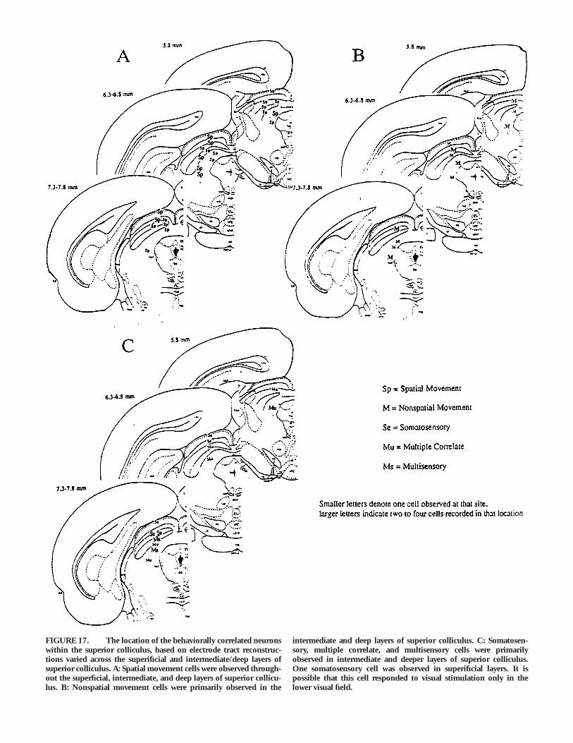
FIGURE 1. Schematic diagram of the visual environments inwhich superior colliculus neural activity was recorded. A: In Experi-ment 1, the radial maze was surrounded with various laboratoryobjects, such as tables, chairs, wall hangings, and the experimenter.The primary auditory cues available to the rat were the motor soundsof the remote controlled maze arms, and the electronic sounds of therecording equipment and computer located in an adjacent room.B: Experiment 2 was conducted with curtains surrounding the maze.Several distal cues were included inside the curtains to serve aspolarizing cues. C: The novel environment was very similar to theenvironment used in Experiment 1. Similar to Experiment 1, theexperimenter was present in the maze room during maze trials.Several other distal cues that were present in the environment arerepresented in the figure.
342 COOPER ET AL.
midsaggital suture; and D–V 1.5 mm ventral of the dural surface.In addition, a burr hole was drilled for placement of the referenceelectrode (114 mm Teflon-coated stainless steel wire) near corpuscallosum. A ground lead (125 mm Teflon-coated stainless steelwire) was attached to a stainless steel jeweler’s screw that wasfastened to the skull. All recording leads were inserted into aconnecting socket that was cemented to the skull with acrylic andnine additional jewelers screws. The animals were given 0.1 mlBicillin (300,000 units per ml) into each hindleg to guard againstinfection.
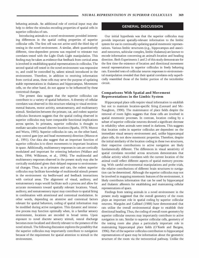
The location of the recording electrodes was verified at the endof each experiment by standard histological methods. The ratswere perfused transcardially with 0.9% NaCl followed by phos-phate buffered 10% formalin. The brains were removed, thenallowed to sink in 30% sucrose formalin. Frozen 40 mm-thicksections were mounted on microslides, stained with cresyl violet,coverslipped, and then observed under a light microscope forverification of the recording site. Electrode tract reconstructionswere made and approximate depth of recording sites in superficialand intermediate/deep layers was calculated based on the finaldepth of the electrode. These were verified by comparing histologi-cally defined depths to depth records maintained as electrodeswere lowered in superior colliculus.
Unit Recording and Behavioral Monitoring
The rat was connected to a headstage for all recording sessions.The headstage contained 5 FETs and a light-emitting diode. Therat was placed on the central platform of the maze while theelectrode was lowered into the brain. The microdrive allowed theexperimenter to advance the electrode in about 20-mm incre-ments.
Single unit activity was recorded simultaneously and indepen-dently on each wire of the stereotrode pair. Each signal wasamplified (3,000 to 10,000 times), filtered at half amplitudebetween 600 and 6 KHz, then passed through a windowdiscriminator such that a 1 msec sampling period began wheneither input surpassed a predetermined threshold. The DataWave‘‘Discovery’’ data acquisition system recorded each analog trace ata frequency of 32 KHz. The system software allowed theexperimenter to isolate individual units from the otherwisemultiunit record by comparing spike characteristics recordedsimultaneously on two closely spaced electrodes (X and Y).Scatterplots of waveform features recorded on X and Y electrodeswere displayed. For separating individual cells from each otherand noise, a variety of waveform parameters were used. Particu-larly useful features included spike amplitude, spike width (timedifferences between the peak and subsequent trough of an actionpotential signal), and relative latency to the voltage peak on X andY. In addition, a template-matching algorithm was utilized tofurther facilitate spike separation. For each cell, the experimenterstepped through a series of two-dimensional cluster plots, identify-ing the combination of spike characteristics that were most likelyassociated with a single-spike generator. Once identified, the
specific cluster boundaries that characterized each cell were savedfor use in subsequent recording sessions. This provided reasonablecertainty that the same cell was being recorded across multiple testdays. It should be noted that our intention was not to make directcomparisons to spike measures of other studies since absolutevalues of spike amplitude, spike width, etc., could vary signfi-cantly depending on filter settings, electrode impedances, etc.Rather, our goal was to use these measures for relative comparisonsacross unit behavioral correlates, to identify the same cells acrossrecording sessions, and for spike separation, when multiple singleunits were recorded simultaneously.
The animal’s location and behavior on the maze were moni-tored simultaneously with the electrophysiological data via anautomatic tracking system (Dragon Tracker, Boulder, CO, fre-quency 5 20 Hz) that recorded the X-Y coordinate position of alight emitting diode positioned above the rat’s nose.
Data Analysis
To facilitate direct comparison with data previously reportedfor hippocampal and thalamic neurons (Mizumori et al., 1989;Mizumori and Williams, 1993), the collicular units of this studywere evaluated for their spatial correlates and their sensitivity tothe movement state of the animal. Spatial correlates of single unitactivity were defined as location- and/or direction-specific firing.Location specificity was quantified by calculating the mean firingrate of the cell as the rat moved radially inward or outward on eachof the eight maze arms (McNaughton et al., 1983a; Mizumori, etal., 1989, 1992, 1994). For analysis of firing rates on the centerplatform, the maze center was subdivided into eight equalpie-shaped parts and included as part of the maze arms for analysisof firing rates on maze arms. The highest of the 16 rates on themaze arms was divided by the average of the remaining 15 toarrive at a location specificity score. Cells with location specificityscores of 2.0 or greater were classified as location-sensitive. Thelocation-specific coding was also considered directional if thefiring rate in the preferred location and direction was at least twicethe rate of the opposite direction. In addition to location-specificdirectional coding, nonlocation-specific directionality of cells wascalculated by taking the average rate in the preferred direction(towards the center of the maze or away from the center of themaze) divided by the average firing rate in the nonpreferreddirection. Cells were considered directional if they fired at twicethe rate in the preferred direction (inbound or outbound)compared to the nonpreferred direction. Graphic illustrations ofthe spatially selective discharge were accomplished in the form of‘spot-rate’ plots (McNaughton et al., 1989; Mizumori et al., 1989;see Fig. 2 for examples). Briefly, the average firing rate wascalculated as the rat remained within a 5-pixel radius of the firstposition sampled. When the diode moved outside this radius, thenew position point served as the integration center for the nextposition. The graphic output consisted of circles, whose radii areproportional to the local firing rate; and dots, indicating thelocations of the maze sampled by the rat. Vectors radiating fromthe centers of the circles illustrate the direction of diode move-
______________________________________ NEURAL REPRESENTATION IN SUPERIOR COLLICULUS 343
ment when the cell fired. A reliability index reflected theproportion of trials in which the highest mean firing rate of a celloccurred on the arm associated with the identified location/direction field of the cell.
To determine the nonspatial movement sensitivity of neurons,the mean firing rate was calculated for times when the rat movedoutward on maze arms, remained relatively still at the arm ends,
turned 1807 at the arm ends, and finally, moved inward on mazearms. A peri-event time histogram (PETH) was created by firstreplaying the data on a monitor in the same temporal and spatialsequence as that observed during the recording session, thenbehavioral event markers were entered into the datafile at pointsthat corresponded to the behaviors of interest. The firing rate ofthe cell was calculated 2.5 s before and after the occurrence of the
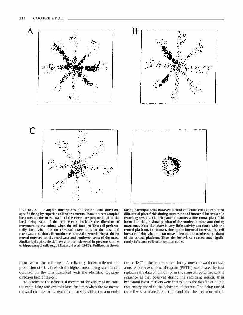
FIGURE 2. Graphic illustrations of location- and direction-specific firing by superior collicular neurons. Dots indicate sampledlocations on the maze. Radii of the circles are proportional to thelocal firing rates of the cell. Vectors indicate the direction ofmovement by the animal when the cell fired. A: This cell preferen-tially fired when the rat traversed maze arms in the west andnorthwest directions. B: Another cell showed elevated firing as the ratmoved outward on the northwest and southwest arms of the maze.Similar ‘split place fields’ have also been observed in previous studiesof hippocampal cells (e.g., Mizumori et al., 1989). Unlike that shown
for hippocampal cells, however, a third colliculus cell (C) exhibiteddifferential place fields during maze runs and intertrial intervals of arecording session. The left panel illustrates a directional place fieldlocated on the proximal portion of the southwest maze arm duringmaze runs. Note that there is very little activity associated with thecentral platform. In contrast, during the intertrial interval, this cellincreased firing when the rat moved through the northeast quadrantof the central platform. Thus, the behavioral context may signifi-cantly influence collicular location codes.
344 COOPER ET AL.
behavioral event marker. Depending on the behavioral correlate,firing rates during the behavior of interest were compared to acontrol condition.
EXPERIMENT 1: BEHAVIORALCORRELATES OF SUPERIOR
COLLICULUS NEURONS IN FREELYBEHAVING RATS
Numerous experiments have described the sensory and motorcorrelates of individual superior collicular neurons in anesthetizedor restrained animals; however, there are limited data fromanimals actively engaged in spatial learning and memory tasks. Todetermine the potential contribution of the superior colliculus toactive navigation, single units were recorded during performanceof the spatial memory task. Our working hypothesis is that thesuperior colliculus is part of a broad neural system mediatingaccurate navigation, and that single units recorded from thisstructure will reflect sensory and motor correlates useful fordirecting spatially guided behaviors.
Method
Subjects, apparatus, and procedure
Behavioral, electrophysiological, surgical, and histological tech-niques were identical to those described in General Methods.Briefly, animals (n 5 4) were trained to perform a spatial memorytask on an eight-arm radial maze. After animals performed eighttrials daily for 7 consecutive days, they were surgically implantedwith recording electrodes (two stereotrodes/hemisphere) placedjust dorsal to superior colliculus. Following recovery from surgery,animals were retrained on the spatial memory task. Upon isolationof individual collicular units, animals performed the spatialmemory task while unit activity was monitored. To maintainasymptote performance on the spatial memory task, animals ranthe maze task every third day if single units were not encountered.
Results
In Experiment 1, a total of 125 superior collicular cells wererecorded as rats performed the spatial memory task. Individualspike amplitudes were comparable to those of cells recorded inother brain areas using the stereotrode recording technique withawake animals; for example, neocortex (McNaughton et al.,1994), hippocampus (Mizumori et al., 1989), striatum (Lavoieand Mizumori, 1994), and thalamus (Mizumori and Williams,1993). The mean (6SE) spike amplitude was 144.4 mV 6 5.6mV, and the average firing rate was 5.43 Hz 6 0.61 Hz. The spikewidth on the other hand, appeared distinct from what we havetypically observed for many other brain areas, even though weused similar electrodes with identical filter settings. The averagespike width was 205.2 msec 6 6.00 msec, whereas the typical spikewidth of neocortical, hippocampal, striatal, or thalamic neurons is250–400msec. Thus, as the electrode was advanced toward the
superior colliculus, it was often possible to correctly ascertainonline whether the unit was a collicular neuron.
Cellular correlates
A variety of unit-behavioral correlates were observed as the ratsperformed the navigation task. A neuron was assigned a correlateif the peak firing rate associated with a particular behavior was atleast twice the firing rate shown during the comparison behaviors(see details below). Based on this criterion, the primary correlateof 42.4% (53/125) of the recorded cells was considered to be‘‘spatial.’’ These cells fired preferentially based on either thelocation of the animal and/or the direction of movement. Thesecond largest category of cellular correlate appeared particularlysensitive to either the general locomotion state of the animal or tosomatosensory stimulation (28.0% or 35/125). Less than one-third (29.6%; 37/125) of the cells recorded showed no obviousbehavioral correlate in our test situation.
Spatial movement correlates
Different forms of apparent spatial coding were observed. Theaverage location specificity score was 2.98 (60.61), whichcontrasts with the average score of 5.0 to 10.0 typically observedfor hippocampal CA1 and hilar/CA3 complex-spike (place)neurons. However, location specificity scores of the collicular cellswere higher than the average score of 1.0 to 2.0 typically observedfor the comparatively nonspatial hippocampal interneuron (e.g.,Barnes et al., 1990; Mizumori et al., 1989). To be consistent withpast studies (Mizumori et al., 1992), collicular cells with locationspecificity scores of 2.0 or greater were classified as location-selective neurons (n 5 35; 28% of all recorded cells). The meanlocation specificity score for the spatial location cells (3.85 60.40) was considerably higher than that of the remaining neurons(1.49 6 0.03). The reliability of the location and nonlocation cellswas also substantially different. The averages were 0.28 6 0.03and 0.21 6 0.02, respectively, for location and nonlocation cells.A regression analysis was performed to examine the relationshipbetween the location specificity of a cell and the reliability withwhich such location specificity was exhibited. An analysis ofvariance revealed a significant positive relationship betweenlocation specificity and reliability, r 5 .42; F 5 9.80 (df 5 46),P , .01. Thus, the more spatially selective correlates also tend tobe the most reliable. Nine of the place-specific collicular cells alsoexhibited a directional bias within the place field. That is, thefiring rate was at least two times greater as the rat moved either inthe inward or outward direction through the place field. Figure 2displays representative spatial correlates that show a location anddirectional firing bias during performance of the spatial memorytask. Note that the cell displayed in Figure 2A may give the initialappearance of a ‘‘head direction’’ cell. However, such a cell isqualitatively different than those that have been previouslydescribed in other areas (e.g., LDN). In this case, and for all otherdirectional cells recorded from superior colliculus in this study, thedirectional vectors were not parallel.
The directional bias to the location preference of superiorcollicular neurons appeared at least superficially similar to that
______________________________________ NEURAL REPRESENTATION IN SUPERIOR COLLICULUS 345
typically observed for hippocampal place cells (McNaughton etal., 1983a; Mizumori et al., 1989), with the exception of one cellthat exhibited different place fields depending on the phase oftraining (see Fig. 2C). This cell preferentially fired as the ratinitiated movement outward on the southwest arm of the mazeduring trial runs. During the intertrial intervals, on the otherhand, a different place field was observed for the same cell whenthe rat moved through the northeast section of the centralplatform. Importantly, during maze trials, the rat also passedthrough the same space on the central platform, but the cell didnot show elevated discharge.
Many superior collicular neurons also showed particular sensi-tivity to the direction in which the animal moved about the mazeirrespective of the absolute spatial location of the animal.Compared to firing rates as the animal remained relativelymotionless at the ends of the maze arms, four cells showedincreased firing during active locomotion either in the inward oroutward directions on the maze arms (Fig. 3). Other neurons (n 511) showed elevated discharge just prior to the cessation ofoutward movement at the arm ends (Fig. 4). Importantly, thesecells did not change their firing rates when the rat stopped on thecentral platform at the end of trials, indicating that they werecoding more than just the cessation of movement. Finally, fouradditional cells selectively increased firing by at least twofold whenthe rat made right or left turns at the arm ends. Two of theseturn-related cells additionally exhibited a clear location bias bypreferentially firing during turns on some, but not all, arms of themaze (Fig. 5). Their location specificity scores were 2.67 and 3.62.Also, only one of the four turn cells displayed a directional turnbias when the animal turned on both the ends of the arm and thecentral platform. The remaining three showed turn correlates atthe arm ends but not the maze center. It is worth noting that thenumber of cells that discriminated direction of turn may havebeen underestimated, since some of the rats in this study madeonly left or only right turns at the arm ends. Turn-related cells
recorded from these animals were not classified as directionallyselective neurons. Similar directional forward-motion and turncells have been described for posterior parietal and caudateneurons recorded from rats tested in a similar behavioral situation(McNaughton et al., 1994; Mizumori and Cooper, 1995; Mizu-mori et al., 1996).
Non-spatial movement correlates
In addition to the numerous spatial biases observed, manysuperior collicular cells were particularly sensitive to the generalmovement state of the animals. For instance, 23/125 cells (18.4%)more than doubled their firing rate when the animal traversedmaze arms relative to times when the rat remained still at the armends (See Fig. 6 for an example). This elevation in rate wasobserved irrespective of the location of the animal and thedirection of movement, with the exception of four cells whichshowed a moderate location bias (location specificity scores:2.29–2.45). Another forward movement-sensitive cell showed adramatic reduction in firing (by about two-thirds) during activelocomotion. Unlike the movement-related neurons of hippocam-pus (Mizumori et al., 1989; Ranck, 1973), the autocorrelationfunctions of these collicular units did not reveal rhythmicmodulation of discharge. Rather, these cells closely resembled themovement-related cells of the posterior parietal cortex (McNaugh-ton et al., 1994). A second type of general movement-related cellwas that which increased firing during both right and left turns(n 5 5).
Somatosensory correlates
A past study demonstrated in freely moving rats that superiorcollicular neurons are sensitive to somatosensory input (Weldonand Best, 1992). Additionally, data from anesthetized or re-strained rodents, cats, and primates have shown that the superiorcolliculus contains a somatotopic map (for reviews, Sparks and
FIGURE 3. The firing of many collicular neurons coincided withforward movement on the maze arms. In particular, cells shown in (A)and (B) increased firing as the rat moved in the outward or inward
directions, respectively. There was no apparent location-specificdischarge since movement-related firing was observed on all mazearms.
346 COOPER ET AL.
Nelson, 1987; Stein and Meredith, 1993). Therefore, we alsotested cells for their response to strokes of the right and leftvibrissae, face, and sides of the body. Six out of 125 cells (4.8%)clearly responded to some form of somatosensory input by at leastdoubling their discharge rate. While animals were performing themaze task, somatosensory cells fired at the end of maze arms orwhen the animal was on the center platform in the process ofselecting the next maze arm. (See Experiment 2 for furtherdescription of the somatosensory correlates observed in superiorcolliculus).
Discussion
The present study describes for the first time the nature ofinformation represented by superior collicular neurons as ratsperform a spatial memory task. The firing rate of a largepercentage of these cells was observed to correlate with what isgenerally considered to be spatial aspects of behavior, such as thelocation and/or the direction of movement of an animal. Otherneurons were sensitive to the general locomotion state of theanimal or to specific somatosensory stimulation.
Given the connections of the superior colliculus to motorstructures, including the spinal cord (Sahibzada et al., 1987; Yasuiet al., 1994), it is not surprising to observe cellular correlatesrelating to various motor behaviors that commonly occurredduring performance of the spatial memory task. Some cells firedduring movement on the maze arms, while others fired when theanimal was relatively still at the end of the maze arm, consumingchocolate milk. Interestingly, cells that are sensitive to changes inosmolarity have been previously reported in superior colliculus(Malmo, 1976). Although it was viewed as unlikely that superiorcolliculus has osmoreceptors for mediating thirst, it is possiblethat input from hypothalamic areas may mediate drinkingresponses of superior collicular neurons (Malmo, 1976).
Cells that correlated with turning, or angular head movements,were also observed in superior colliculus. Turn-related neuronshave also been reported in posterior parietal cortex (McNaughtonet al., 1994) and caudate nucleus (Mizumori and Cooper, 1995).Interestingly, firing rates of head direction cells in anteriorthalamic nuclei are modulated by angular head movements; thesehead direction cells fire maximally during turns 40 to 50 ms priorto reaching the preferred head direction (Blair and Sharp, 1995).At the present time, it is not known where head direction cells inanterior thalamic nuclei derive information about angular headmovement. The possiblitiy that turn-related neurons such as thesemay contribute to the neural system-mediating knowledge ofchanges in directional heading has been suggested by Skaggs,Knierim, Kudrimoti, and McNaughton (1994).
It remains to be determined what sensory information controlsthe spatial and nonspatial correlates observed in superior collicu-lus. The initial observation of this experiment suggests somesimilarities to hippocampal place cells. However, there are numer-ous possibilities for why a cell may fire when a rat is in a specificlocation or heading in a certain direction, and different sensorymodalities may have differential effects across brain structures. Forexample, grooming behavior, somatosensory cues, specific motormovements made by the animal, and visual cues may be
FIGURE 4. Illustration of a subclass of the directional forwardmovement correlate. A: The spatial plot shows that this cell tended tofire on the distal portion of maze arms. B: In particular, the PETH of5B shows a sharp increase in firing approximately 500 msec beforethe animal arrived at the end of the arms. No such increase wasobserved during inbound forward movement. Bin width is 10 msec.
______________________________________ NEURAL REPRESENTATION IN SUPERIOR COLLICULUS 347
responsible for the observed correlate in superior colliculus. Inhippocampus, however, grooming behavior is unlikely to influ-ence, and in fact suppresses complex spike-cell activity. Thus,
there is no a priori reason to suspect that similar spatial codingshould be similarly influenced by sensory input across structures.Grooming behavior is an unlikely explanation because these
FIGURE 5. Another form of spatial coding was shown byneurons that preferentially fired during turns either to the right orleft. A: For two neurons, there appeared to be a location bias to theturn selectivity. The left panel illustrates a stronger preference for leftturns on the east arm of the maze, or when the rat made left turns onthe center platform. In contrast, the right panel shows the left turnbias on only the south, southeast, and east arms of the maze. In thiscase, left turns made on the center platform did produce elevated
firing. B: Two right turn cells did not show a consistent location biasto their discharge. Notice that the cell shown in the left panel firedduring turns on the center platform, while the cell shown in the rightpanel did not. C: PETHs illustrating the turn selectivity of the cellshown in the left panel of B (above). The center of each histogramreflects the beginning of left (left panel) or right (right panel) turnsmade at the arm ends. Bin width is 10 msec.
348 COOPER ET AL.
behaviors almost exclusively occurred during the intertrial intervaland not during maze trials. Differential somatosensory input fromthe maze also appears to be an unlikely explanation for causingspatially localized discharge, because neurons sensitive to somato-sensory stimulation did not show elevated firing on particularmaze arms. Thus, it is most likely that location- and direction-specific firing of the cells in superior colliculus are due to eitherlocation-specific visual information or specific motor behaviors onthe maze. The motor interpretation may be less likely than the
visual because it seems reasonable to conclude that animals wouldmake the same motor movements across the symmetrical mazearms. Of course, without simultaneous electromyogram record-ings, a motor interpretation cannot be conclusively ruled out.
Cells sensitive to forward movement may reflect the motoractivation patterns, internally generated self-motion (i.e., idiot-hetic) cues, or visual information (e.g., optic flow). Thesepossibilties are explored in Experiment 2.
The observation of somatosensory neurons is consistent withdata from anesthetized animals showing that superior colliculuscontains a precise somatotopic map (Meredith and Stein, 1990).Previous work has shown that a discrete area of the intermediate/deep layers of colliculus serves approach behaviors. Furthermore,neurons in this ‘‘approach’’ area of the superior colliculus areactivated by somatosensory and auditory information. Thus, thesomatosensory neurons observed in the present study maymediate approach behaviors via projections to the tecto-spinaltract (Westby et al., 1990). Certainly, navigation in the naturalenvironment likely capitalizes on somatosensory information inaddition to distal visual and internally generated self-motion cues.
The diversity of spatial movement and nonspatial movementcorrelates observed in the present study is consistent with thehypothesis that the superior colliculus gives rise to spatialinformation that can be used by limbic afferent structures. Cellswith location or directional correlates were recorded in theintermediate layers of superior colliculus, and this layer projects toLDN (Thompson and Robertson, 1987b). Therefore, LDN headdirection cells may derive at least part of their directional codefrom collicular input. Experiment 2 explores critical environmen-tal stimuli that maintain and establish the different cellularcorrelates observed in superior colliculus. These data further theargument that superior colliculus is an active part of thetectocortical neural system mediating experience-dependent navi-gation.
EXPERIMENT 2: THE CONTRIBUTIONOF VISUAL INFORMATION TO
CELLULAR CORRELATES RECORDEDFROM SUPERIOR COLLICULUS OF
FREELY BEHAVING RATS
Experiment 1 demonstrated that spatial and nonspatial behav-iors are coded by individual superior collicular neurons duringactive navigation. However, the contribution of environmentalcues to establishing and maintaining the cellular correlates werenot identified. Previous work has shown that spatial correlatesrecorded from limbic structures are visually dependent, that is tosay that visual information importantly contributes to the ob-served location and/or directional coding (Leonard and McNaugh-ton, 1990; Mizumori and Williams, 1993). The present study wasdesigned to evaluate the role of global visual information anddistal visual cues in maintaining and establishing the spatialcorrelates observed in superior colliculus. It was hypothesized that
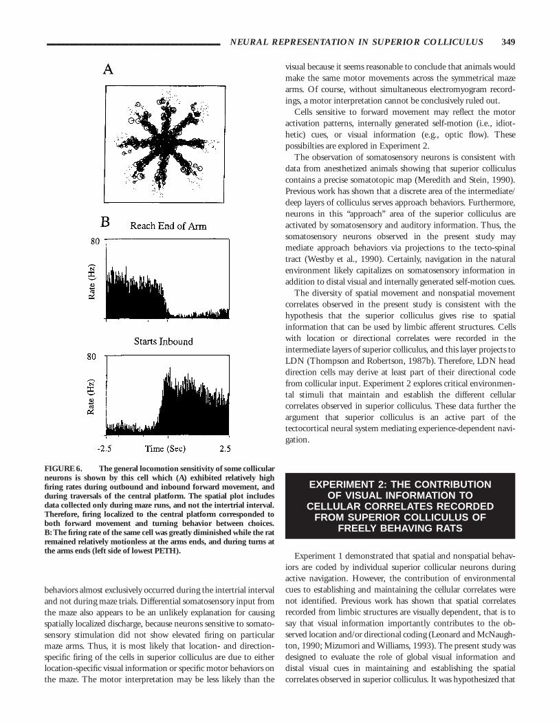
FIGURE 6. The general locomotion sensitivity of some collicularneurons is shown by this cell which (A) exhibited relatively highfiring rates during outbound and inbound forward movement, andduring traversals of the central platform. The spatial plot includesdata collected only during maze runs, and not the intertrial interval.Therefore, firing localized to the central platform corresponded toboth forward movement and turning behavior between choices.B: The firing rate of the same cell was greatly diminished while the ratremained relatively motionless at the arms ends, and during turns atthe arms ends (left side of lowest PETH).
______________________________________ NEURAL REPRESENTATION IN SUPERIOR COLLICULUS 349
visual information would be critical for maintaining spatialcorrelates and that nonspatial movement correlates would notrequire visual information to maintain the cellular correlate.
Method
Subjects, apparatus, and procedure
Nine animals were trained to perform a spatial memory task onan eight-arm radial maze (Olton and Samuelson, 1976) enclosedin a visually controlled cue environment (See Fig. 1B). Oneanimal from Experiment 1 was included in the present study.Spatial memory training was identical to Experiment 1, exceptthat animals were trained to perform 15 trials daily in less than1 h. All of the surgical, electrophysiological, and histologicaltechniques were identical to those described in General Methods,with the exception that following identification of a cell with abehavioral correlate, we attempted to perform a series of environ-mental manipulations in a pseudorandom order.
Environmental manipulations
Manipulation control trials. To determine the stability ofbehaviorally correlated neurons recorded from superior colliculus,animals performed 15 consecutive trials without explicit environ-mental manipulations.
Light–Dark–Light. To evaluate the contribution of visual inputfor maintaining a behavioral correlate, animals performed fivetrials under normal testing conditions, and during the intertrialinterval between the fifth and sixth trials, lights in the testingroom were turned off. Animals were then tested for five trials indarkness. Prior to starting the eleventh trial, all lights in the roomwere turned back on, and animals were tested for five more trialsunder standard lighting conditions.
Dark–Light. This test evaluated the requirement for visualinput to establish a behavioral correlate. To reduce vestibularinformation available about the path from the animal colonyroom to the maze room, animals were carried into the testingenvironment via a circuitous route in darkness. During transporta-tion along a circuitous route, animals were covered with alaboratory coat and the experimenter carried the animal indarkness to a variety of random locations in the outside laboratoryroom before finally entering the maze testing room. After animalswere brought into the testing room and connected to therecording equipment (in darkness), rats performed five spatialmemory trials still in darkness. Immediately prior to the sixthtrial, the lights were turned on and animals were then tested forfive trials under standard lighting conditions.
Cues–No Cues–Cues. This test was designed to evaluate thecontribution of distal visual cues to maintain the behavioralcorrelate. Animals were tested for five trials under normalconditions, and during the intertrial interval between the fifth andsixth trials, all of the distal cues were either covered with a blackcurtain or removed from the environment while the animal
remained on the center platform of the maze. Animals thenperformed five trials in the absence of distal cues. During theintertrial interval between the tenth and eleventh trial, the cueswere restored, once again in front of the animal. Animals werethen tested for five trials in the presence of the distal cues.Originally, cue rotations, and not cue removal, were going to beperformed on cells recorded from superior colliculus; however,pilot data demonstrated that simply carrying animals out of thetesting room (which was necessary to rotate the cues) disruptedthe average firing rate and the correlate of many spatial cells. Thus,all manipulations were conducted without removing the animalfrom the maze room following the onset of behavioral testing. Thecontrol procedure for cue rotation required the animal to run fivetrials, then the animal was removed from the testing room, andwaited outside of the testing room laboratory area for 5 min (SeeFig. 1B for maze room relative to outside laboratory area). Thenthe animal was carried back into the testing room, hooked up tothe recording equipment, and run for five more trials. These dataare discussed briefly in the Results section.
Visual, auditory, and somatosensory correlates. In addition torecording behavioral correlates during maze performance, allcollicular cells were tested for visual or somatosensory sensitivitywhile animals were relatively inactive on the center platform of themaze. Visual correlates of cellular activity were assessed by theexperimenter waving his hand (rostral to caudal, caudal to rostral,etc.) parallel to the rats’ heads, approximately 6 to 129 away fromone or the other eye. This allowed for reasonable lateralization ofvisual stimulus presentation. Somatosensory correlates were as-sessed by gently stroking the vibrissae (rostral to caudal) of theanimal with a pen, by touching the front legs, or by touching thehind legs of the animal. All cells that showed a visual orsomatosensory correlate were checked for auditory sensitivity.Cellular activation by auditory stimuli was typically assessed byeither jingling keys on one side of the animal or by snappingfingers under normal luminance conditions. Similar to the visualstimuli, auditory stimuli were presented parallel to the rats’ heads,about 6 to 129 away from the rat. In some cases auditorysensitivity was tested in darkness. In all cases, both sides of theanimal were tested to determine if the cell showed a lateralizedresponse. PETHs were created based on the simultaneous entry ofan event flag (via a keystroke) at the onset of stimulus presenta-tion. Because the event flag entry and sensory stimulus wereperformed by an experimenter, rather than automated, there likelywere errors in marking the precise onset of the stimulus presenta-tion (on the order of 200 to 400 ms). Data were quantified bytaking the average firing rate of the cell for 1.5 s followingstimulus presentation, compared to 1.5 s before the onset of thestimulus.
Multisensory integration was tested in several cells by jinglingkeys and waving simultaneously (visual and auditory stimulus).Visual stimuli were presented by waving a hand 6–129 away fromone side of the animal. Auditory responses were presented byjingling the keys on one side of the animal while the animal was indarkness, and presumably not able to see the visual stimulus. Themultisensory and visual alone stimulus conditions differed slightly.
350 COOPER ET AL.
In the multisensory condition a cupped hand was waved (therebyblocking the view of the keys being shaken), whereas in the visualonly condition the hand was waved open-faced. Not all cells withunimodal responses were tested for multisensory integration; datawere only quantified for those cells that the experimenter observedenhanced responses online to waving and jingling keys simulta-neously compared to waving alone.
It is important to note that only those cells which showed clearcorrelates to these sensory stimuli (i.e., experimenter couldidentify the correlate online) were recorded. Thus, the totalnumber of cells with visual, somatosensory, or auditory correlatesmay be underestimated. Additionally, these tests were conductedwhile the animal was resting on the center platform of the maze.Thus, while the movement of the animal was only slightlyrestricted (i.e., it could only circle), specific body movements werenot controlled for during these sensory tests. However, during thecourse of these sensory tests animals usually moved little, if at all,and typically were fixated on the experimenter.
Unique manipulations
In several cases, animals were tested in a novel environment(See Fig. 1C). The behavioral procedures in the novel environ-ment were identical to those previously described. Animals weretested daily in both the familiar testing room for five to eight trials,and in the novel environment for five to eight trials (a total of 10to 16 trials per day). The novel environment contained multipledistal cues (e.g., table, chair, box, large lamp) in a large rectangulartesting room (270 cm 3 435 cm) which was illuminated by asingle 40-W incandescent light (mean illumination 5 7.5 lux)placed in one corner of the room. The novel environmentprovided a much ‘‘richer’’ visual environment with a higherceiling, textured walls, more distal cues, larger size, and a morespatially extended three-dimensional structure that was notpresent in the controlled cue environment. Other unique manipu-lations, including passive movement tests and changes in thenormal testing procedure, were performed on a smaller number ofcells. Detailed descriptions of these manipulations are explained inthe Results section.
Behavioral data analysis
The mean number of errors made by each animal across eachphase of the manipulation (two to three blocks of five trials) wascalculated. A repeated measures one-way analysis of variance(ANOVA) was used to determine if the number of errors changedsignificantly (a 5 .05) across the phases of testing.
Single unit analysis
Effects of manipulations were determined by comparing theaverage premanipulation firing rate to those obtained during themanipulation and postmanipulation trials. The cellular correlatewas considered to have been altered if following baseline trials, themean firing rate increased by twofold or decreased by one-halfduring the manipulation condition, and then returned to baselineduring the postmanipulation condition. The cells recorded from
the superior colliculus in this study showed about an equalnumber of enhanced and depressed responses to the variousmanipulations. Thus, to facilitate comparisons among groups ofcells, it was necessary to calculate a change in firing rate thatwould be sensitive to both excitation and inhibition of the cell. Achange in firing rate index (CFI) was calculated for the manipula-tion conditions by subtracting the lowest firing rate from thehighest rate, then dividing by the highest firing rate, regardless ofwhether the lowest firing rate occurred during the baseline ormanipulation condition. If the average firing rate of a cell duringmaze trials increased by twofold or decreased by one-half duringthe manipulation, the CFI equaled .50.
Results
Spatial memory performance
During the Light–Dark–Light manipulation, the average num-ber of errors made by the animal increased significantly (F 5[2,14] 5 7.25; P 5 .007). For the Light–Dark–Light manipula-tion, mean number of errors across each block of five trials was0.07 (6 0.03), 0.74 (6 0.23), 0.27 (6 0.11), respectively. ANewman–Keuls post hoc analysis (a 5 .05) confirmed thatcompared to the intial light testing, the mean number of errorswas significantly greater in the dark and final light phases (P ,.05). During all other manipulations, the mean numbers of errorsmade across each phase of testing was not significantly differentfrom each other.
Cellular correlates
A total of 127 cells were recorded from nine animals. Oneanimal included from Experiment 1 contributed seven cells to thepresent experiment. The percentages of the different cellularcorrelates observed in Experiment 2 were virtually identical tothose of Experiment 1 (see Table 1 for percentages of cells in eachcategory in Experiments 1 and 2).
Similar to Experiment 1, the spatial movement categoryconsisted of the three subcategories: direction-specific (n 5 8),location-specific (n 5 16), and location- and direction-specific (n 5
TABLE 1. _____________________________________________Percentages of Cellular Correlates Recorded From SuperiorColliculus in Experiments 1 and 2
aThis category was only included in Experiment 2 because of the moreextensive sensory tests performed as part of that experiment.
______________________________________ NEURAL REPRESENTATION IN SUPERIOR COLLICULUS 351
17). The nonspatial movement category was comprised ofreach-end-of-arm (n 5 3), movement-sensitive (n 5 12), forward-movement (n 5 4), and turn (n 5 4) cells. Because almost all of theanimals made left or right turns exclusively, it was difficult to assesswhether turn cells showed directional specificity. Somatosensorycells (n 5 9) fired at twice their baseline rate during thepresentation of vibrissal sensory stimuli presented by the experi-menter. Although visual and auditory stimuli were also presentedby the experimenter, these cells only showed unimodal somatosen-sory sensitivity.
Thirteen cells displayed multiple correlates. This category isincluded in the present experiment and not in Experiment 1because of more extensive sensory testing performed as part of thisexperiment. There were three subcategories of multiple correlatecells. The first consisted of cells that responded at twice theirbaseline firing rate during the presentation of visual, auditory,and/or somatosensory stimuli (n 5 3). Multisensory integrationwas demonstrated in four multiple correlate cells (see Fig. 15).These cells showed at least twofold increases in firing rate duringthe simultaneous presentation of visual and auditory stimuli,compared to the presentation of either stimulus alone (visual orauditory). In addition to these sensory cells, neurons that fired inresponse to combinations of maze-related behaviors or stimuliwere included in the multiple correlate category. There were threetypes of cells that correlated with multiple maze-related behaviors.The first type fired when the animal was engaged in turningbehaviors only when the animal was on particular arms of themaze (turn and location, n 5 2). The second type was active onlywhen the animal was turning and moving toward the centerplatform (turn and inbound, n 5 1). The last type of multiplecorrelate cell was active when the animal remained relativelymotionless at the end of the maze arms and was turning (still andturns, n 5 3).
Only 41 out of the 127 cells recorded in this study did not showan obvious relationship to maze-related behaviors. These cellswere classified as having no obvious correlate.
The mean firing rates of the single units in superior colliculusvaried across the behavioral correlates. The average firing ratesdiffered significantly between groups, F (3,78) 5 24.48, P ,.0001. The overall mean rate (6SE) for spatial movement cells was1.29 (60.26) Hz; the nonspatial movement cells showed a higherfiring rate than all other categories of correlates. The average firingrate for this category was 11.38 (61.61) Hz. Sensory cells fired atan average rate of 4.96 (61.55) Hz during maze trials, and
multiple correlate cells fired with an average rate of 1.40 (60.55)Hz.
Environmental Manipulations
Manipulation control trials:Are cellular correlates stable?
To determine the validity of the a priori criterion chosen todetermine the effect of the manipulations, the fifteen trials werebroken down into three sets of five trials. The first five trials servedas a baseline for determining changes in firing rates across trialsduring the remaining two blocks of five trials.
Spatial movement cells. One-way repeated measures ANOVAswere calculated on the mean firing rate, location specificity, andreliability and for the 12 location and location and direction cells.These analyses demonstrated that the measures of spatial codingdid not change significantly across trials: F (2,22) 5 2.15, 0.61,and 0.41, respectively; P . .05. For the location and location anddirection cells, the average location specificity across 15 trials was3.76 (60.38) and reliability was .28 (60.02). This closelyapproximates the average location specificity and reliability scoresobserved in Experiment 1: 3.85 (6.40) and .28 (6.03), respec-tively. This indicates that spatial coding is stable across repeatedmaze trials. Table 2 displays the location specificity, reliability, andaverage rate across 15 trials for these 12 cells. Table 3 summarizes
TABLE 2. _____________________________________________Location Specificity, Reliability, and Average Firing Rate,Across 15 Trials*
TABLE 3. _____________________________________________CFI Across 15 Trials of Testing WithoutExplicit Environmental Manipulations(at the Left Side of Each Column)*
1–5 vs. 6–10 1–5 vs. 11–15
61 81
Spatial .27 62 .30 712
(n 5 15) 312
51 42
Movement .16 42 .25 41
(n 5 10) 112 212
Somato .27 22 .26 22
(n 5 2)
Multiple .25 12 0 112
(n 5 1)
NOC .19 22 .32 22
(n 5 2)
*The ‘‘1’’ sign indicates excited, ‘‘2’’ indicates inhibition, and ‘‘12’’indicates no change relative to baseline for each of the cells contributingto the mean on the right side of each column. Numbers indicate thenumber of cells which showed excitation, inhibition, or no change frombaseline. Abbreviations: Spatial, spatial movement; Movement, nonspatialmovement; Somato, Somatosensory; Multiple, Multiple correlate; NOC, Noobvious correlate.
352 COOPER ET AL.
the average CFI changes during trials 6–10, and 11–15 comparedto the initial five trials for all categories of cellular correlates. Twoof the 15 spatial movement cells tested for 15 continuous trialswithout intervening manipulations showed above criterion changesin firing rate (e.g., CFI . .50). Both of these cells showed aprogressive decline in firing rate across trials. One of the two cellswas tested on the next day, and the firing rate had returned tobaseline levels. The remaining 13 spatial cells did not show abovecriterion changes in CFI.
Although location specificity scores did not change across thefive trial blocks, the exact location of the preferred firing fieldchanged across blocks of trials. This change was restricted in thatthe field generally maintained a bias to one of three arms over thecourse of 15 trials. Figure 7 displays an example of a location and
direction correlate that maintained its spatial bias to the northern,southern, and northeastern arms across 15 trials of testing. Theprevious test day revealed the same pattern of activity. Thisindicates that the broad field of the cell remained stable acrossdays. In a different case, a location and direction cell maintainedthe same location bias across trials, but the preferred directionchanged across trials. In four other cases, location and location anddirection cells were recorded for the subsequent test day; for threeout of four cells the preferred firing field was maintained on twoout of the three arms across days. Only one cell showed a spatialbias unique from the previous day. Thus, the majority of spatialcells appear to maintain the same preferred firing field across days.When the location correlates are viewed within short periods oftime (i.e., five trials or 20 min), the preferred field is localized to a
FIGURE 7. To determine the reliability of spatial coding, 15trials were broken down in three blocks of five trials: 1–5, 6–10, and11–15, respectively. An example of spatial cell that showed adistributed firing field inbound on the northern maze arm, andoutbound on the northeastern and southern maze arms, is shown in
this figure. For reference, the spatial firing from the previous day isdisplayed in the top left corner. Given the broad distribution, it isunlikely that a single cue is controlling the activity of this cell. As isdemonstrated with this cell, the firing field was consistent acrossdays; however, the location of the preferred field varied across trials.
______________________________________ NEURAL REPRESENTATION IN SUPERIOR COLLICULUS 353
small area of the maze. But when viewed across longer periods oftime (i.e., 15 trials or 60 min), the fields are distributed across alarger area of the maze. Most of the cells recorded for multipledays show that the broad fields are maintained to the samelocation across days, and this suggests that these cells contribute tospatial mapping of environmental features. The slight variations inpreferred fields within the testing session may allow for superiorcollicular spatial cells to encode both stable features of theenvironment, and also dynamic information important for naviga-tion. During the 15 continuous trials, the three directional cells(two outbound and one inbound) maintained their behavioralcorrelates throughout the trials (i.e., fired at twice the rate in thepreferred direction across all three blocks of five trials).
Nonspatial movement cells. All 10 of the nonspatial movementcells maintained near baseline firing rates during 15 continuoustrials. An example of the activity of a forward movement selectivecell across three blocks of five trials each is shown in Figure 8. Inthis case there was a slight increase in firing rate during the last 10trials compared to the first five; however, this subtle variation infiring rate is well within the normal range for cells recorded in thisstudy. A one-way ANOVA confirmed that average firing rates didnot change: F (2,18) 5 .92, P . .05, across the three blocks oftrials. (See Table 3 for CFIs for all nonspatial movement cells.) Fournonspatial movement cells were recorded across days withoutintervening manipulations. In all of the cases, the observedbehavioral correlates were the same across test days (2–3 days).
Somatosensory and multiple correlate cells. Both of thesomatosensory cells were stable across 15 trials of testing (CFIs aredisplayed in Table 3). One somatosensory cell was tested for 2 dayswithout intervening manipulations, and in this case the correlateremained the same across days. One multisensory cell was tested for15 continuous trials, and the firing rate did not exceed criterionlevel changes (see Table 3). This cell also showed a spatial bias. Thepreferred field for this particular cell remained stable to a singlearm over the course of the maze trials. The spatial codingcombined with multisensory responses in this cell illustrates thatalthough the categories of cellular correlates are not necessarilymutually exclusive, they do however provide a useful heuristic forevaluating the types of cellular coding that occur within astructure. Three multiple correlate cells were tested across dayswithout manipulations and in all cases the correlate remained thesame for 2–3 days of repeated testing.
No obvious correlate cells. The average firing rate of two noobvious correlate cells tested for 15 trials was stable (see Table 3 for
CFIs). Two no obvious correlate cells were tested across days, and anobvious correlation with behavior on the maze was not observedacross days.
FIGURE 8. The activity of a movement cell across three blocks offive trials is shown in this figure. The top panel corresponds to thefirst five trials, middle panel to the second block of five trials, and thebottom panel to the final five trials. The PETHs display the activity ofthe cell as the animal moved toward the end of the arm, with T0
(indicated by dashed line in PETH) marking the cessation of forwardmovement. The correlate and firing rate remained stable across mazetrials. The rasters below the PETHs display a representative set of 10(out of 40) events. Bin width is 10 msec.
354 COOPER ET AL.
Summary. In this experiment, almost all spatial movement cellsmaintained a consistent firing rate across 15 trials, but thepreferred fields were not exclusively restricted to individual mazearms across trials. Instead, it appeared as if spatial movement cellsmaintain broad, distributed firing fields when animals are testedfor up to 1 h in a restricted, controlled cue environment, and thatwhen tested across days the preferred fields remain stable.Directional, movement, somatosensory, and multisensory cells main-tain consistent firing rates and behavioral correlates across trials.In the absence of experimenter-controlled environmental manipu-lations, there are minor nonsignificant fluctuations in the firingrates across trials and behavioral correlates, but these variationsnever reached a CFI of .50.
Light–Dark–Light: Is visual input requiredto maintain the behavioral correlate?
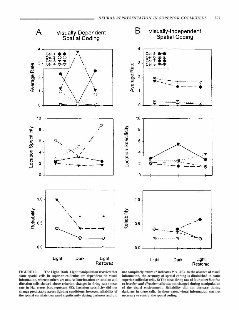
Spatial movement cells. Six of the 12 spatial cells tested in theLight–Dark–Light condition showed dramatic changes in firingrate during the dark condition. One location-sensitive, threelocation- and direction-sensitive, and two directional cells had a CFIscore of greater than .50 during dark trials. Of these six cells, twoof the cells were inhibited and four were excited during darkness.Interestingly, one location and direction cell increased its firing ratedramatically in darkness (CFI . .50) and lost its original locationand direction correlate. In this case, both the reliability andlocation specificity decreased in darkness, and did not return inthe following light trials until the fifth light recovery trial (see Fig.9A). The remaining three location and location and direction cellsshowed a similar pattern of effects. In darkness, reliability of thecorrelate was significantly decreased and did not completelyreturn to baseline levels in final light trials (F [2,6] 5 9.92, P 5.01). Newman–Keuls post-hoc analysis showed that reliabilityduring both the dark (P , .05) and final light trials (P , .05)were significantly different than the baseline condition. Theaverage reliability (mean 6 SE) for the light, dark, and lightconditions were .55 (6.09), .25 (6.01), and .20 (6.03). Meanfiring rate and location specificity did not change significantlyacross the three phases of testing (F [2,6] 5 0.01 and 0.05,respectively; P . .05). Individual cell data (presented in Fig. 10A)revealed CFIs . .50. The lack of statistical significance in averagefiring rate across phases of testing is not a discrepancy because onelocation and direction cell showed pronounced excitation, whilethe remaining three cells were inhibited.
The two directional cells (one outbound and one inbound) hada CFI greater than .50. The directional preference was maintainedin the inbound cell and lost in the outbound cell during darkness.The outbound correlate did not return in the final light trials,despite the fact that the cell was active during these trials.Unfortunately it was not possible to record the cell the next day todetermine if the correlate returned at a later time.
Two directional, three location-, and one location- and direction-sensitive cells did not reach the CFI criterion of .50 or greaterduring dark testing. Figure 10B shows that the location andlocation and direction cells that did not reach the criterion changein firing rate during darkness also did not show significant changesin mean firing rate, location specificity and reliability across thephases of the manipulation, F [2,6] 5 4.05, 0.61, and 0.22,
respectively; P . .05. The directional (outbound) cells that did notshow dark-induced changes in firing rate also maintained theirdirectional firing preference across the three phases of testing.
Nonspatial movement cells. Two out of 15 movement cells hada CFI of .50 or greater during the dark phase of the manipulation;one was excited and one was inhibited in darkness. The reach-end-of-arm cell showed a general increase in firing rate in darknessduring all maze-related behaviors, and thus the behavioral corre-late was lost. During the final light trials, the firing rate returnedto near baseline levels, as did the behavioral correlate.
The turn cell decreased its firing rate during dark trials (CFI 5.82) and returned to baseline levels in the final light trials. Thecellular correlate also changed during dark trials; the cell showed alocation and direction correlate (location specificity 5 10.23,reliability 5 .60) in darkness. When lights were restored, thelocation and direction correlate was not maintained and, althoughthe cell fired on some turns, the turn correlate did not completelyreturn (see Fig. 9B). Because of these unusual changes, theLight–Dark–Light manipulation was repeated the subsequent day,and the firing rate changes were replicated (CFI 5 .80). Thebehavioral correlate, however, was absent in this cell for the initiallight and dark trials, but in the final light trials the cell showed aclear location and direction bias (location specificity 5 5.99,reliability 5 .60). It is important to note that the clusterboundaries of this cell did not change across days and the firingrate showed a similar pattern in response to the manipulationacross days. Thus, it is likely that the same cell was being recordedacross days and that this seemingly motor-related cell wasmodulated by visual information and maze testing conditions.
The majority of movement-sensitive cells (13/15) did not reachcriterion level of change in firing rate during dark trials. Addition-ally, the behavioral correlates remained stable regardless of changesin room luminance. A nonspatial movement turn-related neuronthat was not influenced by changes in room luminance is shown inFigure 9C.
Somatosensory cells. A CFI . .50 was not observed in the sixsomatosensory cells tested with the Light–Dark–Light manipula-tion. During the course of dark testing the firing rate wasmaintained during maze trials.
Multiple correlate cells. Five out of seven multiple correlatecells had a CFI of .50 or greater during dark trials. These includeda turn and location cell, still and turn cell, a cell sensitive to visual,auditory, and somatosensory stimuli, and two multisensory cells.All of these cells were inhibited in dark conditions. In the case ofthe turn and location cell, the cell was inhibited in darkness, andthen the firing rate returned to baseline levels when room lightswere restored. Different components of the turn and location cellwere differentially affected by changes in room lighting; duringdark testing the turn correlate remained but the location bias waslost. When room lights were restored, the cell continued to fireduring turns and the location bias returned to the preferred area ofthe maze on the final light trial (see Fig. 11). A similar time-courseof restoration was observed for a location and direction celldescribed above.
______________________________________ NEURAL REPRESENTATION IN SUPERIOR COLLICULUS 355
The still and turn cell also showed a reduction by over one-halfin firing rate during dark trials (CFI 5 .77). This cell was activewhen the animal was relatively motionless at the end of the maze
arms (still component) and during turns either at the end of themaze arms or on the center platform (turn component). Thedecrease in mean firing rate was due to the fact that turn
FIGURE 9. The Light–Dark–Light manipulation disrupted halfof the spatial cells and the minority of movement cells. A: The toppanel displays a location and direction cell that increased its overallfiring rate during darkness and required five trials for the locationand direction firing field to be reestablished. B: In the middle panel aturn-related neuron that was inhibited in darkness is displayed in this
figure. Interestingly, the behavioral correlate was altered with illumi-nation changes; the cell showed a location and direction firing field indarkness. When room lights were turned on, the turn correlate onlypartially returned. C: Not all turn cells were influenced by changes inthe visual environment, and this turn-related neuron was notinfluenced by changes in room illumination (bottom panel).
356 COOPER ET AL.
FIGURE 10. The Light–Dark–Light manipulation revealed thatsome spatial cells in superior colliculus are dependent on visualinformation, whereas others are not. A: Four location or location anddirection cells showed above criterion changes in firing rate (meanrate in Hz, errors bars represent SE). Location specificity did notchange predictably across lighting conditions; however, reliability ofthe spatial correlate decreased significantly during darkness and did
not completely return (* indicates P F .05). In the absence of visualinformation, the accuracy of spatial coding is diminished in somesuperior collicular cells. B: The mean firing rate of four other locationor location and direction cells was not changed during manipulationof the visual environment. Reliability did not decrease duringdarkness in these cells. In these cases, visual information was notnecessary to control the spatial coding.
______________________________________ NEURAL REPRESENTATION IN SUPERIOR COLLICULUS 357
component was lost in darkness. Importantly, the still componentof the correlate was maintained in darkness. The original firingrate and the turn component were restored on the first trial whenroom lighting was restored.
One multiple sensory correlate and two multisensory cells had aCFI of .50 when animals performed the maze task in darkness.
These cells were inhibited in darkness and returned to baselinewhen the animal was tested in the final light trials.
The multiple correlate cells that did not show twofold changes infiring rate (e.g., CFI , .50) due to visual changes were a turn andinbound cell and a multisensory cell. The firing rate and behavioralcorrelates did not change across testing conditions. These data,combined with the data from the still and turn and turn andlocation cell, demonstrate that turn correlates recorded fromsuperior colliculus can be either visually dependent or indepen-dent of visual information.
No obvious correlate cells. Three out of nine cells tested thatdid not have an obvious correlate had a CFI of .50 or greaterduring the dark phase of the Light–Dark–Light manipulation; allthree were inhibited in darkness. In one case, a cell thatmaintained a broad-preferred firing field across several days(location specificity 5 1.76) showed a slight increase in thelocation-specific firing in the light trials following dark testing(location specificity . 2.0). This cell may have been coding ageneral area of the maze but not with the same location specificityrequired by our a priori criterion.
Summary. Taken together, these data suggest that not all spatialcells are dependent on visual information; about half of thoserecorded show visual sensitivity, the remaining did not. The utilityof examining firing rate changes is exemplified by the spatial cellswith a location bias that showed changes in firing rate correspond-ing with a decreased reliability of spatial coding. This suggests thatlocation cells whose firing rates were influenced by visual informa-tion were coding visuospatial features of the environment and indarkness the accuracy of the correlate was disrupted. In contrast tothe common argument that turn-related cells reflect angular headmovement, at least a subpopulation of superior collicular turncells were shown to be visually dependent. Most of the multiplecorrelate and multisensory cells were inhibited when visual inputwas eliminated. Movement sensitive and somatosensory cellsgenerally were not affected by changes in the visual environment.The different correlates of multiple correlate cells were differentiallysensitive to visual information: A dissociation of dark effects(within cell) suggests that individual neurons integrate both visualand nonvisual information.
Dark–Light: Is visual input required to establishthe correlate?
Spatial movement cells. In the one case in which a directionallyselective cell was tested with this manipulation, visual input wasnot required to establish the normal firing rate or the preferreddirectional firing. When tested in light, this cell fired preferentially
FIGURE 11. A cell that fired during turns also showed a spatialbias as indicated by elevated activity on a small subset of the mazearms. When the animal was tested in darkness, the spatial bias of theturn correlate was lost and did not return until the final recovery lighttrial. Importantly, the cell continued to fire during turns in darkness,illustrating that the multiple correlate can be dissociated by manipu-lation of the visual surroundings. The location bias required visualinformation, but the turning correlate did not.
358 COOPER ET AL.
when the animal was headed away from the center platform(outbound). Figure 12 shows that this correlate was observedirrespective of maze room lighting conditions. Importantly, theLight–Dark–Light manipulation performed the previous daydemonstrated that visual input was not required to maintain thecorrelate of this cell. Thus, coding of directional heading can beindependent of visual information in superior collicular neurons.
Nonspatial movement cells. One of the seven movementsensitive cells tested (forward movement) with the Dark–Lightmanipulation had a CFI of .90 during light trials compared to thedark trials. The firing rate was increased during dark trialscompared to light trials. The other movement-sensitive cells didnot show changes in firing rate when animals began maze trials indarkness.
FIGURE 12. Directional coding of space was observed to beindependent of visual input in this superior collicular neuron. Whenthe animal was brought into the testing environment in darkness, thecell showed preferential activity when the animal was heading towardthe end of the maze arm (outbound). T0 (indicated by the dashed linein the PETH) marks the cessation of forward movement in the panels
on the left half of the figure. In the right half of the figure, T0 marksthe onset of forward movement toward the center platform. When theanimal was moving toward the center of the maze (inbound), the cellwas not active. The behavioral correlate was the same regardless oflighting conditions. The rasters below the PETHs show a representa-tive 10 out of 40 events. Bin width is 10 msec.
______________________________________ NEURAL REPRESENTATION IN SUPERIOR COLLICULUS 359
Multiple correlate cells. The one multiple correlate cell testedwith this manipulation was a still and turn cell. Testing in darknesscaused a dramatic reduction in firing rate, CFI 5 .77, anddifferentially affected the multiple correlates of the cell. The stillcomponent was unaffected by starting maze trials in darkness, butthe turn correlate was abolished when visual information was notavailable. When lighting was restored, the turn correlate returned(see Fig. 13). Further dark testing revealed the same pattern ofresults: when the animal was subsequently tested in darkness, thestill correlate remained, but the turn correlate was abolished untillighting was restored to the maze room (data not shown).
No obvious correlate cells. The one no obvious correlate celltested did not show firing rate changes during this manipulation.
Summary. In the one directional cell tested, visual informationwas not required to establish or maintain the directionallyselective firing. Starting the animals in darkness affects a minorityof the movement-sensitive cells, but the majority of movementcells are not affected by this manipulation. As with the Light–Dark–Light manipulation, a dissociation between components of mul-tiple correlates can be demonstrated with these changes in thelighting conditions. This dissocation of visual sensitivity within-cell furthers the hypothesis that multiple sensory modalitiescontrol the activity of the multiple correlate cells.
Cues–No Cues–Cues: Are distal cues criticalto maintain cellular correlates?
Spatial movement cells. None of the four spatial cells testedwith the cue removal manipulation showed the above criterionchanges in firing rate. Interestingly, the location specificitycontinually increased across the phases of testing in one of the fourspatial cells tested (2.74 in the initial trials to 5.62 when the cueswere returned). The reliability of the cellular correlate alsoincreased across the three phases of testing (0 to 40). Whenanimals were removed from the testing room and then returned,all five of the spatial cells tested showed pronounced inhibition ofactivity (CFI . .50) despite the fact that the rats were returned tothe same visual environment. A spatial movement cell (inbound)was influenced by this control procedure and also the Light–Dark–Light manipulation.
Nonspatial movement and somatosensory cells. None of theseven nonspatial movement cells were affected by cue removal.Seven other nonspatial movement cells were also tested with theremoval from the testing room, and none of these were influencedby this control procedure. The two somatosensory cells tested withcue removal were not influenced by this manipulation.
Multiple correlate cells. Cue removal did not influence the onemultiple correlate cell tested with this manipulation. Two multiplecorrelate cells underwent the procedure of removing the animalfrom the testing room and returning to the maze room. The firingrate of one multiple correlate cell (the cell fired when the animalwas relatively still at the end of the arm and during turns on arm
ends) was inhibited when the animal was returned to the mazeroom and testing continued. Furthermore, as with the inboundcell described above, this cell was similarly affected by theLight–Dark–Light and Dark–Light manipulations (see Fig 13 foreffects of Dark–Light manipulation). Illumination changes anddark adaptation processes represent a possible interpretation forthese data. Under normal testing conditions the animals arebrought into the dark maze room from the brighter outer room,attached to the recording equipment, and generally remained inthe darker environment for several minutes before maze testingbegan, thereby inadvertently allowing the animal time to adapt tothe dimly lit environment. The procedure of removing the animalfrom the testing room, then returning the animal to the mazeroom and beginning maze trials may not have allowed the animalsvisual system to completely adapt to the darker maze room. Tocontrol for this possibility, the multiple correlate cell was recordedthe subsequent day, with maze trials commencing as quickly aspossible following introduction into the darker maze room. Thisprocedure did not influence the normal correlate of the cell, andreduces the likelihood that visual adaptation processes cancompletely explain these data.
No obvious correlate cells. The two no obvious correlate cellstested did not show above criterion changes in firing rate duringthe course of the Cues–No Cues–Cues manipulation. Two otherno obvious correlate cells were also tested following removal fromthe testing room. One cell was inhibited and the firing rate of theother was not affected by this procedure.
Summary. The distal cues used in this study do not appear tobe critical for cellular activity in superior colliculus. Although itmay initially appear somewhat surprising that only one spatial cellchanged during cue removal, this pattern of data suggests thatmore global environmental features (e.g., geometry, illuminationconditions of the room, etc.) are critical determinants forestablishing and maintaining spatial correlates in the superiorcolliculus. Removing the animal from the testing room inhibitedthe firing rate of a variety of cellular correlates: One potentialfactor common across two of the cells was dependency on visualinformation. The control procedure for the illumination changesindicates that dark adaptation processes alone are insufficient tocompletely explain the observed effects; therefore, contextual orattentional factors may be responsible for the inhibition of cellularactivity.
Sensory Responses and Multisensory Integration
Somatosensory cells. All of the somatosensory cells identifiedshowed clear responsivity to contralateral brushes of the vibrissaewith a pencil. Figure 14 shows an example of a somatosensoryneuron that shows a lateralized response to contralateral vibrissaestimulation. Some neurons that were activated by vibrissalstimulation also showed responses to light touches on the body bythe experimenter’s finger. The responses to body touches, however,did not always appear to clearly discriminate between sides of thebody.
360 COOPER ET AL.
Multisensory integration. Four cells showed enhanced (CFI ..50) responses to the simultaneous presentation of an auditoryand visual stimulus compared to the presentation of auditory orvisual stimuli presented alone (see Fig. 15 for an example). In the
case of the cell presented in Figure 15, the mean rate for 1.5 sfollowing visual or auditory stimulus presentations was 4.0 Hzand 6.0 Hz, respectively. The multisensory response was notsimply an additive response of the individual sensory modalities;
FIGURE 13. The Dark–Light manipulation confirmed thatmultiple correlates could be dissociated. When the animal wascarried into the testing room in darkness, this particular cell wasactive only when the animal was relatively motionless at the end ofthe maze arms, and not during turning behaviors (top of figure). T0
for the ‘‘Reach End of Arm’’ PETH marks the cessation of forwardmovement on the maze arms. T0 for the turn correlate designates theonset of turning behaviors. When room lighting was reinstated, the
cell then showed activity during both still behaviors and turning andthe end of the maze arms (bottom of figure). Turning on the centerplatform, in addition to the end of the maze arms, caused this cell tofire only when room lighting was available. This activity, occurringprior to the animal heading down toward the end of the maze arm, isapparent during the 22.5 s to 21.5 s range on the ‘‘Reach End ofArm’’ PETH in light. Bin width is 10 msec.
______________________________________ NEURAL REPRESENTATION IN SUPERIOR COLLICULUS 361
rather, when visual and auditory stimuli were presented together,this cell fired at an average rate of 15.9 Hz for the 1.5 s followingthe stimulus presentation. The multisensory cell presented inFigure 15 also responded to vibrissal stimulation (data notshown); regrettably it was not possible to reliably stroke theanimal’s vibrissae in darkness, and therefore we were not able toassess the potential for trimodal sensory integration. A similarpattern of data was observed for the other three multisensory cells.
The response to the multisensory stimulus was well above twicethe rate following unimodal input.
Summary. Similar to previous data, superior collicular cellswere activated by sensory stimuli. Furthermore, it has been welldocumented that multisensory stimulation caused enhancedresponses in individual cells recorded from anesthetized andrestrained cats. To the best of our knowledge, this represents thefirst demonstration of mulitsensory integration occurring insuperior colliculus of freely behaving rats.
Unique manipulations
Novel environment: Spatial movement cells. Two spatial move-ment cells were differentially affected by testing the animals in anovel environment (see Fig. 1C for description of novel environ-ment). One location cell did not fire for the first two days oftesting in the novel environment, while continuing to show aspatial bias in the familiar testing environment. On the third dayof testing, this cell began to fire in the novel environment but thepreferred firing field and location specificity were not isolated to asingle location. In the familiar environment, the preferred loca-tion of the firing field could be controlled by placing novel cues(e.g., visual, auditory, somatosensory, gustatory, or olfactory) onthe maze or at the end of maze arms. When an experimenterjingled keys outside of the curtains (auditory), quietly stood at theend of one arm (visual), placed novel food (gustatory andolfactory), or textured paper on a maze arm (somatosensory), thepreferred field was always located on the arm with the currentlynovel stimulus. All of these tests were conducted in a single day inthe familiar environment with three trials given for each stimulus.
One other location cell, recorded from a different animal thanthe previously described cell, was tested in both the novel andfamiliar environment. This cell showed both different and similarresults compared to the previously described cell. Unlike theprevious cell, this cell showed a relatively high location specificityscore and reliability (location specificity 5 3.97, reliability 5.50)on the first day in novel environment. The preferred firing fieldremained in the same location for the first 4 test days in the novelenvironment (outbound on arm 7), but the specificity andreliability of the field changed across days (range; spatial speci-ficity 5 1.72 to 5.75 and reliability 5 .50 to 0). After the fourthday of testing in the novel environment, the location of the firingfield began to shift unsystematically. After the first 4 days oftesting in the novel environment, the changes in the location ofthe firing field were similar to the observation that the locationbias also varied across test days in the familiar environment (seeFig. 16 for familiar environment changes across days).
In the initial novel environment testing, the location anddirection bias was outbound on arm seven. This happened to bethe arm facing directly towards the location of the experimenterduring behavioral testing (see Fig. 1C). To assess whether thepresence of the experimenter was influencing the cellular corre-late, the animal ran five trials with the experimenter in the normalposition, five trials with the experimenter rotated 180o, and fivetrials with the experimenter returned to the original position. The
FIGURE 14. Lateralized somatosensory responses were observedby several superior collicular neurons. This cell responded preferen-tially to left vibrissae stimulation. The activity prior to onset ofstimulus presentation may reflect motor activation of the vibrissaeprior to the sensory input, or may be due to slight errors in markingthe onset of stimulus presentation. The rasters display the 16stimulus presentations on the left vibrissae and the 12 presentationson the right. Bin width is 10 msec.
362 COOPER ET AL.
FIGURE 15. Multisensory integration was demonstrated in fourcells. In all cases the response to the multimodal stimuli was not asimple additive response of the unimodal components. T0 (indicatedby the dashed line) marks the approximate onset of the stimulus for arepresentative cell that showed multisensory integration. The rate
displayed in the top right corner of the figure is the average rate of thecell for 1.5 s following stimulus onset. The rasters display theindividual responses of the cell to the six auditory and visual stimuluspresentations and the seven multisensory presentations. Bin width is10 msec.
______________________________________ NEURAL REPRESENTATION IN SUPERIOR COLLICULUS 363
FIGURE 16. A spatial cell maintained a preferred firing fieldlocalized toward the location of the experimenter in the novelenvironment for the first 4 days of testing. The experimenter waspositioned in the southeast corner of the room (see Fig. 1C) duringmaze trials, and the preferred firing field fired on the center platformand maze arm directed toward the experimenter. In the familiarenvironment, this cell did not show a predictable pattern of change in
preferred firing location across days. However, it generally fired morewhen approaching the center platform in the southern direction.Changing the location of the experimenter during maze trials in thenovel environment demonstrated that the location of the preferredfiring field was largely influenced by the spatial location of theexperimenter (data not displayed).
364 COOPER ET AL.
preferred direction of the field, but not the location, rotated withthe experimenter but did not return until the subsequent test day(i.e., the cell fired when the rat moved toward the center platform(inbound) on arm seven).
Two other tests were performed on this cell to evaluate thecontribution of room geometry and salient distal cues to theobserved spatial correlate. The effects of room geometry weretested by enlarging the original controlled cue environment andadding unique distal cues to the ‘‘new’’ room. Additionally, thecurtains were removed, leaving a room similar to the originalnovel environment. When the animal was tested in the enlargedroom with novel distal cues the location specificity decreasedslightly but reliability increased. Removal of the curtains, whichleft a room virtually identical to the original novel environment(see Fig. 1C), caused location specificity to increase and did notaffect reliability. To evaluate the contribution of a salient distal cuein the environment, a novel experimenter (different from the onein the original novel environment) sat in a chair by arm 7 duringthe first four trials of testing. The cell showed high locationspecificity (4.55) and reliability (.75) as the animal moved outtoward the experimenter. Rotation of the experimenter by 907caused an equal rotation of the field (location specificity 5 9.62,reliability 5 .5), but when the experimenter was returned to theoriginal location the field did not return. These results are similiarto what was observed in the first novel environment: when theexperimenter was moved, the field shifted accordingly, but did notreturn with the reinstatement of the original test situation.Regrettably, the cell could not be isolated on the subsequent dayto determine if the correlate would return.
Novel environment: Nonspatial movement cells. One move-ment and one turn cell were tested in the novel environment. Bothof these cells were not affected by testing in the novel environ-ment.
Novel environment summary. These results make several impor-tant points. First of all, increasing the size of the testing room andproviding more geometric features within an environment en-hanced both reliability and location specificity of spatial coding insuperior colliculus. Secondly, regardless of which environment thecells were tested in, location specificity could be controlled for bythe introducing a salient stimulus (i.e., novel cues or an experi-menter) into the room. Lastly, even though the cells did notmaintain a consistent spatial bias restricted to a single arm in thefamiliar environment, the location bias remained stable in thenovel environment for the first 4 test days. Thus, spatial coding insuperior colliculus can represent both stable features of environ-ments and novel stimuli that occur within them. In contrast to thespatial cells, the movement related cells are completely unaffectedby testing in the novel environment.
Passive movement. In order to assess the contribution ofvoluntary movement to the activity of nonspatial movement (n 53) and spatial movement (n 5 1) cells, passive movement tests wereconducted by carrying the animal by hand about 15 cm aboveeach maze arm, with a 2-min ITI, for five trials. During passive
movement, three movement-sensitive cells fired at a very high rate,and one spatial movement cell fired at a low rate relative to baselinelight trials. Furthermore, the firing of all cells tested did notdiscriminate between forward movement, stopping or turningbehaviors. In three of the four cells (two nonspatial movement, onespatial movement), the passive movement tests were followed byfive maze trials, and the normal firing rate and behavioral correlatereturned during these maze trials. This suggests that if stressduring passive movement was responsible for the observedchanges in firing rate, it did not have a long-term effect on theobserved correlate. These data indicate that active movement isrequired for normal spatial and movement coding by superiorcollicular neurons.
To further investigate the role of vestibular input, a turn cell wasrecorded while the animal was subjected to passive body bends ofeither the front- or hind-half of the body while the other half ofthe body was kept still (see McNaughton et al., 1994). This celldid not discriminate between either front- or back-half bodybends in either the left or right direction. Additionally, the cell didnot fire during left or right turns on the center platform. Thus, itappears that specific motor activity occurring only at the end ofthe arm is required for this turn correlate to be observed.
Histological results
Based upon electrode reconstructions, the locations of cellsfrom each of the different categories of behavioral correlates weredetermined. Most electrode passes were through the medialportions of superior colliculus, A–P coordinates were generallyplaced about 6.3 mm to 7.3 mm posterior to bregma. Two passeswere farther anterior or posterior to these general locations (5.8 –7.8 mm). Figure 17 displays the relative distribution of therecording sites for the observed behavioral correlates. Spatialmovement cells were found in the superficial, intermediate, anddeep layers of superior colliculus and a discrete topographicalorganization did not appear to be present. The other correlatesshowed more of a topographical organization within superiorcolliculus. Nonspatial movement cells were predominantly foundin the intermediate and deep layers. Only one movement-relatedneuron was found in the superficial layers; and, not surprisingly,this cell was sensitive to changes in the visual environment. Sensorycells were also distributed across lamina. One somatosensory cellwas found in the superficial layers, the remaining in the intermedi-ate/deep layers. The observation of a somatosensory cell in thesuperficial layer may be explained by the cell responding to visualinput from the lower visual field. However, visual sensory tests didnot elicit obvious neural responses. Multiple correlate cells werefound in the intermediate and deep layers of colliculus. Consistentwith previous studies in cats, multisensory cells were also limited tothe intermediate and deep layers.
Discussion
The present experiment evaluated the contribution of visualand nonvisual cues that may influence cellular correlates recordedfrom neurons in the superior colliculus during spatial memoryperformance. Manipulations of the visual environment (e.g.,
______________________________________ NEURAL REPRESENTATION IN SUPERIOR COLLICULUS 365
FIGURE 17. The location of the behaviorally correlated neuronswithin the superior colliculus, based on electrode tract reconstruc-tions varied across the superificial and intermediate/deep layers ofsuperior colliculus. A: Spatial movement cells were observed through-out the superficial, intermediate, and deep layers of superior collicu-lus. B: Nonspatial movement cells were primarily observed in the
intermediate and deep layers of superior colliculus. C: Somatosen-sory, multiple correlate, and multisensory cells were primarilyobserved in intermediate and deeper layers of superior colliculus.One somatosensory cell was observed in superificial layers. It ispossible that this cell responded to visual stimulation only in thelower visual field.
Light–Dark–Light, novel environment) demonstrated that many,but not all, spatial cells are strongly dependent on general featuresof the visual environment. Individual distal cues do not appear toimportantly contribute to the spatially selective discharge ofsuperior collicular neurons; rather, overall geometry and relativeluminance conditions may play a more important role inmodulating spatial coding in this structure. The movement- andsomatosensory-related correlates remained reliable regardless ofchanges in the visual environment. Passive movement tests suggestthat movement-related correlates likely require motor activity,rather than vestibular input, to control the cellular correlate. Thisdoes not mean that vestibular information is not a contributingfactor, but that it is not sufficient to drive the representationsduring maze behavior. Multimodal and multisensory integrationwas demonstrated for the first time in freely behaving rats. Onemultisensory cell also had a spatial correlate, which suggests thatspatial correlates may utilize multisensory information in superiorcollicular neurons. These results strongly support the hypothesisthat the superior colliculus is multifunctional and can play anactive role in experience dependent navigation.
The results from the environmental manipulations suggest thatthere may be two kinds of spatial coding in superior colliculus:visually dependent and visually independent. Although individualdistal cues alone are unlikely determinants of spatial coding in thefamiliar environment, the Light–Dark–Light and novel environ-ment data suggest that the size of the visual environment andcomplexity of the visual surroundings may have a strong impacton spatial coding in many superior collicular cells. However,coding of both general directional heading (relative to the mazeconfiguration; i.e., inbound or outbound) and location was alsofound to be independent of visual information in other spatialcells. Hence, visual information may control spatial coding insome neurons and movement may mediate spatial coding byothers. This is in keeping with the multiple output pathways ofsuperior colliculus (Redgrave et al., 1993), in particular, thedifferential sensory responsiveness of cells in the well-identifiedapproach and avoidance pathways (Westby et al., 1990). Discreteregions of superior colliculus project to areas mediating approachbehaviors, and other regions project to areas mediating avoidanceresponses. Neurons within the ‘‘approach’’ area of superiorcolliculus were responsive to auditory and somatosensory stimuli,and neurons in the ‘‘avoidance’’ area were responsive to visual andauditory stimuli (Westby et al., 1990). The differential visualsensitivity of spatial coding suggests an input/output schemesimilar to what has been identified for orientation behaviors.Thalamic projections from superior colliculus may be stronglyinfluenced by visual information (i.e., visually dependent), whereasprojections to motor output structures from superior colliculusmay be independent of visual information. Thus, visuospatialinformation may be sent to thalamus from superior collicularneurons, and spatially guided movements may be mediated viaspatial neurons in areas of superior colliculus with motor projec-tions to approach and avoidance pathways (cf. Westby et al.,1990).
Extensive work in primates has revealed a motor map ofsaccades elicited by stimulation of the intermediate layers of
superior colliculus: Large amplitude saccades are evoked bystimulation in the caudal superior colliculus, and small amplitudesaccades in the rostral superior collicular stimulation (Robinson,1972). Furthermore, buildup and burst cells have been identifiedperhaps relating to preparation for and execution of saccades,respectively (Sparks, 1978; Munoz and Wurtz, 1995). In the rat,stimulation of the superior colliculus also evokes contraversiveconjugate eye movements with a similar topography as has beenreported in primates (McHaffie and Stein, 1982). Activation ofsuperior colliculus in the rat also evoked movement of the pinnaeand vibrissae. Therefore, it is possible that head movements wouldhave been elicited if the animal had not been restrained. Chemicaland electrical stimulation in freely moving rodents shows thatsuperior colliculus activation causes discrete behavioral responsesresembling approach and avoidance movements, depending onthe area of superior colliculus that is stimulated (Dean et al., 1986,1988; Sahibzada et al., 1986; Westby et al., 1990). The combina-tion of the primate and rat data are consistent with the hypothesisthat directing movements toward spatially relevant locations is animportant function of superior colliculus. In rats, because theyhave a very large visual field and do not have a discrete fovealrepresentation, general body movements may be preferentiallymediated by superior colliculus; whereas in primates, saccadestoward discrete locations in space are critically controlled bycollicular activity. In both species, the behavioral function may beconserved despite the difference in specific movements controlledby superior colliculus.
The relationship between turning behaviors and orientingmovements may suggest similarities between the current work andthat of approach or avoidance studies in freely moving rats. It ispossible that the turn-related neurons are part of the orientationsystem mediated by superior colliculus. In some cases, it may bethat turn correlates were dependent on visual information (e.g.,still and turn cell) and in others they were not (e.g., turn andlocation cell). This suggests that updating movement changesduring orienting responses might be coded by neurons in superiorcolliculus, and these neural codes reflect the current visual input,motor output, or visual-motor integration. Interestingly, vestibu-lar information alone does not appear to be able to driveturn-related neural activity. In the one cell subjected to passivebody bends, vestibular turn activation did not occur. Further-more, this cell only fired when the animal was turning at the endof the arm and not on the center platform of the maze. Thus,specific motor sequences are required to activate some turn-relatedcollicular neurons. In addition to turn-related neurons, individualunits were correlated with the general movement state of theanimal. These data support the argument that in freely behavingrats the superior colliculus mediates approach and avoidancebehaviors, potentially toward spatially important locations.
In a previous study involving freely moving rats, somatosensory,visual, and auditory responses were observed. Forty-eight percent(27/56) of the collicular neurons showed pronounced sensoryresponses (Weldon and Best, 1992). In the present study, only12.5% (16/127) of the cells responded to sensory stimulation.This difference in percentage of sensory correlates between thestudies may be explained by the finding that a reduction in
______________________________________ NEURAL REPRESENTATION IN SUPERIOR COLLICULUS 367
receptive field size was observed by placing the animal on acircular platform (Weldon and Best, 1992). Because all sensorytests were conducted while the animal was on the center platformof the maze, the receptive field sizes of the cells may have beenreduced, making them harder to detect. Additionally, because ofthe primary interest in spatially selective discharge, only data fromthose cells that showed clear sensory responses online werequantified. This may have reduced the total number of cellsobserved in the present study. Weldon and Best (1992) alsoreported the presence of bimodal and trimodal sensory neurons(19.6%; 11/56). In the present study we observed multimodal andmultisensory responses in 10.2% (13/127) of the neurons re-corded. Again, the difference in total number of cells may reflectdifferences in testing conditions.
Multisensory integration has been extensively studied in anes-thetized and restrained animals (see Stein and Meredith, 1993, forreview). In the cat, the location of the recording site is a criticalpredictor for the number of multisensory neurons that are likelyto be observed. For example, 84% of the neurons that wereactivated antidromically by tectoreticulospinal stimulation havebeen shown to be multisensory (Meredith et al., 1992). Conse-quently, the neurons projecting to motor structures important fororienting behaviors are predominantly multisensory neurons, andthese in turn likely guide orienting behaviors (cf. Wilkinson et al.,1996). In the present study, both multisensory and multimodalintegration were demonstrated in freely behaving rats, although ata much lower percentage (10.2%) than reported in cats. Thesmaller percentage in the current study may reflect speciesdifferences, different testing procedures, or a combination of bothfactors. Despite the difference in the absolute number of multimo-dal cells observed, the multisensory integrative responses in thisstudy are similar to those that have been reported for cats. Inaddition to multisensory integration, cells that correlated withmultiple maze behaviors showed multimodal integration. That is,one component of the multiple behavioral correlate could bevisually dependent while the other was visually independent. Forexample, when a turn and location cell was tested in darkness, theturn correlate remained, but the location bias was lost. In sum,these data suggest that multisensory integration is conservedacross species.
An important caveat to note is that precise mapping ofreceptive field properties of the sensory cells was not a goal of thepresent study. Therefore, we cannot provide more details aboutthe psychophysical properties of the multisensory integration.Clearly such a study would be of critical importance in the future.Because sensory tests were not the primary goal of the presentstudy, two potential confounds are present in the multisensorytests. The multisensory integration tests were not performed withsimultaneous recordings of muscle activity, so the influence ofmovement cannot be completely accounted for. Furthermore,subtle methodological issues may represent a potential confoundin the multisensory tests. Despite these issues, motor activationand differences in testing procedures do not seem likely to haveproduced the multisensory integration effect. First of all, if wavingand jingling the keys caused the animal to move, thereby resultingin increased cellular activity, then jingling the keys in darkness
should have caused the animal to make similar movements. Theresponse to the auditory cue in darkness was virtually equal to thevisual response, and was clearly less than the multisensoryresponse; thus movement differences are unlikely to explain thecurrent data. Lastly, the shape of the visual stimulus in themultisensory condition was not identical to that of the visualalone condition. It is possible that the enhanced response in themultisensory condition is due to this methodological difference.This explanation does not seem likely because the difference wassimply in the shape of the hand (cupped in the multisensorycondition and open in the visual alone). Given that the cuppedhand covered a smaller visual area than the open hand, it is likelythat this condition would elicit less of a visual response. Instead, adramatic enhancement was observed when waving was combinedwith the jingling of keys. Thus, although future studies will berequired to fully understand the nature of multisensory integra-tion in freely behaving rats, the present study strongly suggeststhat multisensory integration is an integral part of cellularprocessing in superior colliculus.
Weldon and Best (1992) first reported that alterations in thetesting environment could change the size of receptive fields incollicular neurons. The present study provides further support forthe effects of changing testing conditions on the normal activity ofthe cellular correlates in superior colliculus. Simply removinganimals from the testing arena and allowing them to wait in anouter laboratory room before continuing maze trials disrupted thenormal activity of many collicular cells. This procedure, whichdoes not affect place cells in hippocampus (Cooper and Mizu-mori, unpublished observations), profoundly inhibited severalsuperior collicular neurons. Those cells that showed inhibitionduring the dark phase of the Light–Dark–Light manipulationwere also inhibited by this procedure. The control for visualadaptation demonstrated that bringing the animal into the testingroom from the brighter outside laboratory room and beginningmaze trials immediately failed to change the spatial correlate.Therefore, adaptation alone cannot completely explain the ob-served effect.
One possibility is that attention and context are playing animportant role in controlling cellular representations of space insuperior colliculus. On virtually every test day, when the animalwas removed from the maze room, the animal was returned to theanimal colony room and not tested until the subsequent day. Thecontrol procedure, however, presented a novel testing situation inthat it required the animal to perform more maze trials when itwas not expecting to. It is possible that the animal ‘‘expected’’ toreturn home, hence superior colliculus activity was inhibitedwhen the animal was removed from the testing room. Thisargument would suggest that when the animal was not expectingto encounter potentially novel stimuli (i.e., behaviors that thesuperior colliculus is involved in), then superior collicular activitymay be suppressed, perhaps by top-down cortical influences. Theduration of suppression, about 15–20 min, was identical to theamount of time required for the cellular correlates to returnfollowing the dark phase of the manipulation. This delayedresponsiveness following visual and contextual changes maysuggest cortical modulation of superior colliculus activity in freely
368 COOPER ET AL.
behaving animals. An additional role of cortical input may alsohelp to define the stimulus encoding properties of spatial cells insuperior colliculus of rats.
Introducing animals to a novel environment provided interest-ing differences in the spatial coding properties of superiorcollicular cells. The first cell was not active until the third day oftesting in the novel environment. A similar, albeit quantitativelydifferent, time-dependent process was required to reinstate twocorrelates tested with the Light–Dark–Light manipulation. Thisfinding may be taken as evidence that feedback from cortical areasis involved in establishing spatial representations in colliculus. Thesecond spatial cell tested in the novel environment was responsiveto, and could be controlled by, salient stimuli introduced in theenvironment. Therefore, in addition to receiving informationfrom cortical areas, these cells may serve the purpose of updatingstable representations in thalamus and hippocampus. Movementcells, on the other hand, do not appear to be influenced by thesecontextual changes.
The present data suggest that the superior colliculus cancontribute to a variety of spatial behaviors. A diversity of cellularcorrelates was observed in this structure relating to visual environ-mental features, motor activity, somatosensory, and multisensorystimuli. Similarities between the primate, cat, and rodent superiorcolliculus literatures suggests that the spatial coding observed insuperior colliculus may have comparable functional implicationsacross species. In primates, superior colliculus is thought tomediate saccades toward discrete locations (Sparks, 1978; Munozand Wurtz, 1995). Superior colliculus in cats, on the other hand,may control gaze (eye and head movements) direction (Munoz etal., 1991). Our data also suggest that an important function ofsuperior colliculus is to direct movements to important locationsin space. Additionally, multisensory responses in cats are corticallymodulated and important for orienting behaviors (Wallace andStein, 1994; Wilkinson, et al., 1996). The multimodal andmultisensory responses observed in the present study may also becortically modulated given their delayed response to environmen-tal changes. Thus, as in primates and cats, the rodent superiorcolliculus may facilitate knowledge of multimodal stimuli presentin the environment via feedforward and feedback interactionswith cortical areas. The alignment of visual, auditory, andsomatosensory maps would facilitate such a process and allow foraccurate movements toward spatially relevant locations. Visual,auditory, and somatosensory input may contribute to spatial firingin combination with attentional and contextual modulation. Inother words, depending on attentive and contextual factorsrelevant for spatial behaviors, coding of spatial information maybe modified during active navigation. This dynamic and interac-tive process may function optimally when, in a familiar sensoryenvironment, locations are encoded in broad terms. Uponexposure to novel discrete sensory stimuli, neural dischargebecomes more localized and directly relevant to the presence of thenovel stimuli. The following discussion explores the possiblity thatthe superior colliculus may importantly contribute to navigationbecause of the requirement for spatial attention to stimuli in theenvironment.
GENERAL DISCUSSION
Our initial hypothesis was that the superior colliculus mayprovide important spatially-relevant information to the limbicsystem for use in continually updating spatial mnemonic represen-tations. Various limbic structures (e.g., hippocampus and associ-ated neocortex, subicular complex, limbic thalamus) are known toencode information concerning an animal’s location and headingdirection. Both Experiments 1 and 2 of this study demonstrate forthe first time the existence of location and directional movementneural representations in superior colliculus in freely behavingrats. Extended tests of collicular neuron responses to environmen-tal manipulation revealed that their spatial correlates only superfi-cially resembled those of the limbic portion of the tectolimbiccircuit.
Comparisons With Spatial and MovementRepresentations in the Limbic System
Hippocampal place cells require visual information to establishbut not to maintain location-specific firing (Leonard and Mc-Naughton, 1990). The maintenance of place fields despite theremoval of room lights suggests that these cells are involved inspatial mnemonic processes. In contrast, location coding by asubset of superior collicular neurons showed a significant decreasein reliability when animals were tested in darkness. This suggeststhat location codes in superior colliculus are dependent on theimmediate visual sensory environment and, unlike hippocampalplace cells, do not show mnemonic properties. Therefore, despitethe initial similarity of the location coding in these two structures,their respective contributions to active navigation are likelyfundamentally different. The differences in visual sensitivity ofspatial correlates recorded across structures demonstrates thatcellular activity which correlates with the current location of theanimal could reflect different aspects of spatial memory process-ing. With careful environmental manipulations and probe trials,the relative contributions of different brain structures to naviga-tion can be determined. Although the superior colliculus may notbe involved in mapping mnemonic features of the environment, itlikely contributes information that can be used by hippocampaland thalamic afferents for establishing and maintaining cellularrepresentations of space.
Findings from testing animals in a novel environment in thepresent study suggested that the overall geometry of the roomplays an important role in spatial coding by superior collicularneurons. Margules and Gallistel (1988) have demonstrated thatrats utilize the overall environmental shape to establish theirdirectional heading. Thus, the coding of overall room geometry bysuperior collicular neurons may importantly contribute to activenavigation in rats. Similar to superior collicular cells, geometry ofthe testing room also plays a particularly important role inmaintaining hippocampal place fields (O’Keefe and Burgess,1996). Part of the superior colliculus contribution to hippocampalrepresentations of space may be information about the geometricstructure of the room via the tectocortical pathway. Unlike the
______________________________________ NEURAL REPRESENTATION IN SUPERIOR COLLICULUS 369
superior collicular location coding in familiar environments,many place cells in hippocampus are controlled by the location ofthe visual cues (O’Keefe and Speakman, 1987). Informationabout the constellation of distal cues may arrive in hippocampusvia geniculostriate pathway rather than the tectocortical system.Integrating information about the relationship of the distal visualcues and room geometry likely provides hippocampus with stablerepresentations of the environment. The absence of well localizedspatially selective discharge in superior colliculus, compared tohippocampus, may be a result of the type of spatial informationbeing integrated to form the spatial firing pattern. Because thehippocampus integrates information about both room geometryand distal visual cues, more precise fields may be formed. Incontrast, only general features of the environment may beencoded by superior colliculus resulting in cellular coding oflocations that is less specific.
LDN head direction cells show qualitatively similar mnemonicproperties as place cells in hippocampus; they are maintained forshort periods of time in darkness, and require visual informationto establish the preferred directional firing (Mizumori andWilliams, 1993). In contrast to heading direction codes in LDN,the superior collicular directional cells fired in response tomovement either toward or away from the center platform. Thus,instead of coding a particular direction in absolute space, superiorcollicular directional cells appear to be guided by the localgeometry of the behavioral testing apparatus. The findings ofdirectional coding in superior colliculus irrespective of lightingconditions, and the fact that superior colliculus projects to LDN,suggests that maintenance of LDN directional activity in the darkmay be derived in part by superior colliculus input. Consistentwith this view, lesion data and the current experiments indicatethat superior colliculus can importantly contribute to knowledgeof directional heading within an environment and that thisinformation can be used by efferent structures to mediate activenavigation (Lines and Milner, 1985; Foreman and Stevens, 1987).
A large percentage of behaviorally correlated neurons insuperior colliculus were related to the movement state of theanimal. Cells that fired in response to forward movement,stopping, and turning were identified, particularly in the interme-diate and deep layers. Similar movement-related activity inneurons has been observed in posterior cortical areas (McNaugh-ton et al., 1994) and caudate nucleus (Mizumori and Cooper,1995; Mizumori, et al., 1996). Although the correlates are similaracross these structures, it is not clear that the sensory control ofthese cells is the same. For example, a posterior cortical neuronthat fired in response to turning behavior was modulated by acombination of visual and vestibular input (McNaughton et al.,1994). For the one superior collicular turn cell tested with passivebody bends, vestibular input did not play a particularly importantrole in controlling the turn-related activity, and many turn-relatedcells recorded in superior colliculus did not require visualinformation to maintain the behavioral correlate. As with thelocation and direction codes of superior colliculus, the movementcorrelates recorded in this structure may share similarities andexhibit differences in processing sensory information when com-pared to other structures. In the case of the turn correlates,
superior colliculus activity co-occurs with movement, whereassome posterior cortical neurons appear to be modulated byvestibular–visual interactions.
A Possible Role for Superior Colliculusin Active Navigation
The present data combined with previous reports on thesensory control of spatial coding in limbic structures arguestrongly that a broad neural systems analysis is required inunderstanding how navigation is accomplished. In addition to thedifferences in sensory-dependent processing across brain struc-tures of spatial information, there are also differences in the role ofexperience on spatial processing. On several occassions it wasdemonstrated that a manipulation would change a correlate, butthe correlate would either return after 20 min of testing; or, inother cases, not until the next day. Thus, following restoration oforiginal environmental conditions, the time course of representa-tional reorganization varies substantially from midbrain to corticalstructures.
Consistent with the finding that collicular location codesrequire prolonged periods of time to become reliable after majordisruption in an expected environment is the result that onespatial cell tested in the novel environment did not become activeuntil the third day of testing. Although the preferred firing fieldwas first observed at this time, the cell did not show a consistentpattern of change in location specificity across subsequent testdays. It appears that experience-dependent changes in superiorcolliculus neural activity are fundamentally different than thoseoccurring in thalamus and hippocampus where spatial representa-tions become stable in at most tens of minutes (Wilson andMcNaughton, 1993).
The superior colliculus is classically viewed as part of anorientation system mediating approach and avoidance behaviorsto stimuli in the environment. In cats the multisensory integrationthat occurs within the intermediate and deep layers of superiorcolliculus is dependent on cortical input (Wallace and Stein,1994). Importantly, cortical input is also critical for multisensoryapproach behaviors (Wilkinson, et al., 1996). In the present studymultisensory integration was observed in several cells, as wasmultimodal integration. It seems likely, therefore, that in the ratmultisensory integration serves a similar purpose and is controlledby similar mechanisms as it is in cats.
Based on these data we postulate that upon exposure to a newenvironment, the superior colliculus is initially sensitive to specificfeatures of the environment. Our novel environment data supportthis view since, in contrast to location fields recorded in a familiarenvironment, the location fields in the novel environment wereselective to single maze arms. With continued exposure, animalssoon learn to attend to only a subset of available cues. Perhaps thesuperior collicular neurons function in a similar way; that is,collicular neurons become tuned to more general, rather thanspecific environmental features. This form of experience-dependent modulation may occur because of (top-down) corticalinfluences. Thus, cue removal in a familiar environment had noeffect on cell firing, and the location cells exhibited comparatively
370 COOPER ET AL.
broad fields during asymptote performance in a familiar environ-ment. Despite this rather broad spatial code, the colliculusremains adapted to detecting potentially significant changes in itssensory surround. In support of this argument, the location anddirection coding of one cell could be controlled by the presence ofnew stimuli introduced into the familiar environment. Such dataimply that the superior colliculus may provide current visualinformation that can be used by limbic structures to update spatialrepresentations. If the environmental change is sufficiently great(e.g., sudden darkness), cells may reorganize their coding proper-ties in an attempt to adapt. Stabilization of the location fieldsfollowing dark testing may take time because it depends oncortical input to guide the experience-dependent collicular repre-sentations of space.
In conclusion, a main functional contribution of the superiorcolliculus to navigation may be to continually update limbiccortical structures with significant information concerning thecurrent sensory surround. Its neural codes do not, however,passively reflect the available sensory cues. Rather, there is likelyimportant cortical modulation of collicular responsiveness tofamiliar cues. In the absense of this modulatory influence (i.e.,when not in a familiar environment), the superior colliculusdefaults to a function of ‘‘detecting’’ novel stimuli.
Acknowledgments
We thank Troy Chatwin for assistance with behavioral tests,and James Canfield for helpful comments and suggestions on themanuscript.
REFERENCES
Barnes CA, McNaughton BL, Mizumori SJY, Leonard BW, Lin LH.Comparison of spatial and temporal characteristics of neuronalactivity in sequential stages of hippocampal processing. Prog BrainRes 1990;83:287–300.
Blair HT, Sharp PE. Anticipatory head direction signals in anteriorthalamus: Evidence for a thalamocortical circuit that integratesangular head motion to compute head direction. J Neurosci 1995;15:6260–6270.
Cooper BG, Mizumori SJY. Sensory dependence of spatial cells recordedfrom superior colliculus of freely-behaving rats. Soc Neurosci Abstr1994;20:805.
Dean P, Key P. Spatial deficits on radial maze after large tectal lesions inrats: Possible role of impaired scanning. Behav Neur Bio 1981;32:170–190.
Dean P, Redgrave P. The superior colliculus and visual neglect in rat andhamster. I. Behavioral evidence. Brain Res Rev 1984;8:129–141.
Dean P, Mitchell IJ, Redgrave P. Contralateral head movements producedby microinjection of glutamate into superior colliculus of rats:Evidence for mediation by multiple output pathways. Neuroscience1988;24:491–500.
Dean P, Redgrave P, Sahibzada N, Tsuji K. Head and body movementsproduced by electrical stimulation of the superior colliculus in rats:Effects of interruption of crossed tectoreticulospinal pathway. Neuro-science 1986;19:367–380.
Foreman N, Stevens R. Visual lesions and radial maze performance inrats. Behav Neur Bio 1982;36:126–136.
Foreman N, Stevens R. Relationships between the superior colliculus andhippocampus: Neural and behavioral considerations. Behav Brain Sci1987;10:101–152.
Goodale MA, Murison RCC. The effects of lesions of the superiorcolliculus on locomotor orientation and the orienting reflex in the rat.Brain Res 1975;88:243–261.
Grantyn A, Grantyn R. Axonal patterns and sites of termination of catsuperior colliculus neurons projecting in the tecto-bulbo-spinal tract.Exp Brain Res 1982;46:243–256.
Huerta MF, Harting JK. The mammalian superior colliculus: Studies ofits morphology and connections. In: Vanegas H, ed. ComparativeNeurobiology of the Optic Tectum. New York: Plenum, 1984:687–773.
Jay MF, Sparks DL. Auditory receptive fields in primate superiorcolliculus shift with changes in eye position. Nature 1984;309:345–347.
Jay MF, Sparks DL. Sensorimotor integration in the primate superiorcolliculus. I. Motor convergence. J Neurophysiol 1987;57:22–34.
Lavoie AM, Mizumori SJY. Spatial-, movement- and reward-sensitivedischarge by medial ventral striatum neurons of rats. Brain Res1994;638:157–168.
Leonard B, McNaughton BL. Spatial representation in the rat: Concep-tual, behavioral, and neurophysiological perspectives. In: Kesner RP,Olton DS, eds. Neurobiology of Comparative Cognition. New Jersey:Erlbaum, 1990:363–422.
Linden R, Perry V. Massive retinotectal projection in rats. Brain Res1983;272:145–149.
Lines CR, Milner AD. A deficit in ambient visual guidance followingsuperior colliculus lesions in rats. Behav Neurosci 1985;99:707–716.
Malmo RB. Osmosensitive neurons in the rat’s dorsal midbrain. BrainRes 1976;105:105–120.
Margules J, Gallistel CR. Heading in the rat: Determination byenvironmental shape. Anim Learn Behav 1988;16:404–416.
McHaffie JG, Stein BE. Eye movements evoked by electrical stimulationin the superior colliculus of rats and hamsters. Brain Res 1982;247:243–253.
McIlwain JT. Topography of eye-position sensitivity of saccades evokedelectrically from the cat’s superior colliculus. Visual Neurosci 1990;4:289–298.
McNaughton BL, Barnes CA, O’Keefe JA. The contributions of position,direction, and velocity to single cell unit activity in the hippocampusof freely moving rats. Exp Brain Res 1983a;52:41–49.
McNaughton BL, O’Keefe J, Barnes CA. The stereotrode: A newtechnique for simultaneous isolation of several single units in thecentral nervous system from multiple unit records. J Neurosci Meth1983b;8:391–397.
McNaughton BL, Barnes CA, Meltzer J, Sutherland RJ. Hippocampalgranule cells are necessary for normal spatial learning but not forspatially-selective pyramidal cell discharge. Exp Brain Res 1989;76:485–496.
McNaughton BL, Mizumori SJY, Barnes CA, Leonard BJ, Marquis M,Green EJ. Cortical representation of motion during unrestrainedspatial navigation in the rat. Cerebral Cortex 1994;4:27–39.
Meredith MA, Wallace MT, Stein BE. Visual, auditory and somatosensory convergence in output neurons of the cat superior colliculus:Multisensory properties of the tecto-reticulo-spinal projection. ExpBrain Res 1992;88:181–186.
Meredith MA, Stein BE. The visuotopic component of the multisensorymap in the deep laminae of the cat superior colliculus. J Neurosci1990;10:3727–3742.
Meredith MA, Stein BE. Spatial determinants of multisensory integrationin cat superior colliculus neurons. J Neurophysiol 1996;75:1843–1857.
Miya DY, Mizumori SJY, Chatwin T. Spatial movement properties ofsuperior colliculus cells in behaving rats. Soc Neurosci Abst 1993;19:793.
______________________________________ NEURAL REPRESENTATION IN SUPERIOR COLLICULUS 371
Mizumori SJY, Cooper BG. Spatial representations of dorsal caudateneurons of freely-behaving rats. Soc Neurosci Abst 1995;21:1929.
Mizumori SJY, Williams JD. Directionally selective mnemonic propertiesof neurons in the lateral dorsal nucleus of the thalamus of rats. JNeurosci 1993;13:4015–4028.
Mizumori SJY, McNaughton BL, Barnes CA, Fox KB. Preserved spatialcoding in hippocampal CA1 pyramidal cells during reversible suppres-sion of CA3 output: Evidence for pattern completion in hippocam-pus. J Neurosci 1989;9:3915–3928.
Mizumori SJY, Perez GM, Alvarado MC, Barnes CA, McNaughton BL.Reversible inactivation of the medial septum differentially affects twoforms of learning in rats. Brain Res 1990;528:12–20.
Mizumori SJY, Ward KE, Lavoie AM. Medial septal modulation ofentorhinal single unit activity in anesthetized and freely moving rats.Brain Res 1992;570:188–197.
Mizumori SJY, Miya DY, Ward KE. Reversible inactivation of the lateraldorsal thalamus disrupts hippocampal place representation andimpairs spatial learning. Brain Res 1994;644:168–174.
Mizumori SJY, Unick KE, Cooper BG. Dynamic reward and spatialcodes of caudate nucleus neurons. Soc Neurosci Abst 1996;22:680.
Morris RGM, Garrud P, Rawlins JNP, O’Keefe J. Place navigationimpaired in rats with hippocampal lesions. Nature 1982;297:681–683.
Muller RU, Kubie JL, Ranck JB Jr. Spatial firing pattern of hippocampalcomplex-spike cells in a fixed environment. J Neurosci 1987;7:1935–1950.
Munoz DP, Wurtz RH. Saccade-related activity in monkey superiorcolliculus I. Characteristics of burst an buildup cells. J Neurophys1995;73:2313–2333.
Munoz DP, Guitton D, Pellsion D. Control of orienting gaze shifts by thetectoreticulospinal system in the head-free cat. III. Spatio-temporalcharacteristics of phasic motor discharges. J Neurophys 1991;66:1642–1666.
Ogasawara K, McHaffie JG, Stein BE. Two visual systems in cat. JNeurophysiol 1984;52:1226–1245.
O’Keefe J, Burgess N. Geomtetric determinants of the place fields ofhippocampal neurons. Nature 1996;381:425–428.
O’Keefe J, Dostrovsky J. The hippocampus as a spatial map, preliminaryevidence from unit activity in the freely moving rat. Brain Res1971;34:171–175.
O’Keefe J, Speakman A. Single unit activity in the rat hippocampusduring a spatial memory task. Exp Brain Res 1987;68:1–27.
Olton D, Branch M, Best PJ. Spatial correlates of hippocampal unitactivity. Exp Neurol 1978a;58:387–409.
Olton D, Walker JA, Gage FH. Hippocampal connections and spatialdiscrimination. Brain Res 1978b;139:295–308.
Paxinos G, Watson C. The Rat Brain in Stereotaxic Coordinates.Australia: Academic Press, 1986.
Ranck JB Jr. Studies on single neurons in dorsal hippocampus formationand septum in unrestrained rats. Part I. Behavioral correlates andfiring repertoires. Exp Neurol 1973;41:461–535.
Redgrave P, Westby GWM, Dean P. Functional archtecture of rodentsuperior colliculus: Relevance of multiple output channels. Prog BrainRes 1993;85:69–77.
Robinson DA. Eye movements evoked by collicular stimulation in thealert monkey. Vision Res 1972;12:1795–1808.
Sahibzada N, Dean P, Redgrave P. Movements resembling orientation oravoidance elicited by electrical stimulation of the superior colliculus inrats. J Neurosci 1986;6:723–733.
Sahibzada N, Yamasaki D, Rhoades RW. The spinal commissuralprojections from the superior colliculus in rat and hamster arise fromdistinct neuronal populations. Brain Res 1987;415:242–256.
Skaggs WE, Knierim JJ, Kudrimoti HS, McNaughton BL. A model ofthe neural basis of the rat’s sense of direction. Adv Neural Proc Sys1994;7:173–180.
Sparks DL. Functional properties of neurons in the monkey superiorcolliculus: Coupling of neuronal activity and saccade onset. Brain Res1978;156:1–16.
Sparks DL. Translation of sensory signals into commands for control ofsaccadic eye movements: role of primate superior colliculus. PhysiolRev 1986;66:118–170.
Sparks DL, Nelson JS. Sensory and motor maps in the mammaliansuperior colliculus. TINS 1987;10:312–317.
Stein BE, Clamann HP. Control of pinna movements and sensorimotorregister in cat superior colliculus. Brain Behav Evol 1981;19:180–192.
Stein BE, Meredith MA. The Merging of the Senses. Boston: MIT Press,1993.
Sutherland RJ, Whishaw IQ, Kolb B. A behavioural analysis of spatiallocalization following electrolytic, kainate-, or colchicine-induceddamage to the hippocampal formation in the rat. Behav Brain Res1983;7:133–153.
Taube JS. Head direction cells recorded in the anterior thalamic nuclei offreely moving rats. J Neurosci 1995;15:70–86.
Thompson SM, Robertson RT. Organization of subcortical pathways forsensory projections to the limbic cortex I. Subcortical projections tothe medial limbic cortex in the rat. J Comp Neurol 1987a ;265:175–188.
Thompson SM, Robertson RT. Organization of subcortical pathways forsensory projections to the limbic cortex II. Afferent projections to thethalamic lateral dorsal nucleus in the rat. J Comp Neurol 1987b;265:189–202.
van Groen T, Wyss JM. Projections from the laterodorsal nucleus of thethalamus to the limbic and visual cortices in the rat. J Comp Neurol1992;324:427–448.
Wallace MT, Stein BE. Cross-modal synthesis in the midbrain dependson input from cortex. J Neurophys 1994;71:429–432.
Weldon DA, Best PJ. Changes in sensory responsivity in deep layerneurons of the superior colliculus of behaving rats. Behav Brain Res1992;47:97–101.
Westby GWM, Keay KA, Redgrave P, Dean P, Bannister M. Outputpathways from the rat superior colliculus mediating approach andavoidance have different sensory properties. Exp Brain Res 1990;81:626–638.
Wilkinson LK, Meredith MA, Stein BE. The role of anterior ectosylviancortex in cross-modality orientation and approach behavior. ExpBrain Res 1996;112:1–10.
Wilson MA, McNaughton BL. Dynamics of the hippocampal ensemblecode for space. Science 1993;261:993–994.
Yasui Y, Tsumori T, Ando A, Domoto T, Kayahara T, Nakano K.Descending projections from the superior colliculus to the reticularformation around the motor trigeminal nucleus and the parvicellularreticular formation of the medulla oblongata in the rat. Brain Res1994;656:420–426.