Page 1
1
Supplementary Information
Hijacking a biosynthetic pathway yields a glycosyltransferase inhibitor
within cells
Tracey M. Gloster1, Wesley F. Zandberg1, Julia E. Heinonen1, David L. Shen2, Lehua Deng1,
and David J. Vocadlo1,2
1 Department of Chemistry, Simon Fraser University, 8888 University Drive, Burnaby, V5A
1S6, Canada.
2 Department of Molecular Biology and Biochemistry, Simon Fraser University, 8888
University Drive, Burnaby, V5A 1S6, Canada.
Corresponding author: David J. Vocadlo, Tel: 001 778 782 3530, Fax: 001 778 782 3765.
Nature Chemical Biology: doi:10.1038/nchembio.520
Page 2
2
Contents of the Supplementary Information.
Page 3: Supplementary Methods
Page 8: Synthesis of Ac-5SGlcNAc (5) and 5SGlcNAc (3)
Page 15: Synthesis of pMP-5SGlcNAc (8)
Page 19: Synthesis of Me-5SGlcNAc (10)
Page 21: Synthesis of Ac-5SGlcNAz (14)
Page 23: Characterization of UDP-5SGlcNAc (4)
Page 24: Supplementary Results
Page 24: Supplementary Figure 1
Page 25: Supplementary Figure 2
Page 26: Supplementary Figure 3
Page 27: Supplementary Figure 4
Page 28: Supplementary Figure 5
Page 29: Supplementary Figure 6
Page 30: Supplementary Figure 7
Page 31: Supplementary Figure 8
Page 32: Supplementary Figure 9
Page 33: Supplementary Figure 10
Page 34: Supplementary Figure 11
Page 35: Supplementary Figure 12
Page 36: Supplementary Figure 13
Page 37: Supplementary Figure 14
Page 38: Supplementary Figure 15
Page 40: Supplementary Figure Legends
Page 46: References
Nature Chemical Biology: doi:10.1038/nchembio.520
Page 3
3
Supplementary Methods
Chemoenzymatic synthesis of UDP-5SGlcNAc (4): Inorganic pyrophosphatase (PPA), ATP
and UTP were purchased from Sigma. GNK, AGM, and AGX1 were buffer exchanged into
50 mM Tris, pH 7.5, 2 mM MgCl2 using PD10 columns (GE Healthcare). GDP-Glc (68 μM)
was included as an internal standard for CE analysis. A one-pot reaction consisting of 2.5
mM ATP, 1 mM UTP, 0.6 mM 5SGlcNAc (3), 0.4 μM GNK, 2.9 μM AGM, 0.3 μM AGX
and 50 mU/mL PPA was incubated for 14 h at 37 °C. The reaction mixture was treated for an
additional 4 h at 37 °C with calf alkaline phosphatase (Roche). The reaction was monitored
by CE (see below). Epimerization of UDP-5SGlcNAc (4) to UDP-5SGalNAc was carried out
using UDP-GlcNAc 4-epimerase (14 h at 37 °C).
Purification of UDP-5SGlcNAc (4): Enzymes were removed by passing the mixture through
a centrifugal filtration device (10 kDa molecular weight cut-off; Centricon). The filtrate
containing the desired product was desalted using a column containing Dowex AG 1-X4 ion
exchange resin (BioRad; 7 mL bed volume) converted into the formate form and pre-
equilibrated in water. After loading, the column was washed with 10 bed volumes of H2O and
10 bed volumes of 4 M formic acid, and eluted with 10 bed volumes of 550 mM ammonium
formate, pH 4.01. Fractions containing UDP-5SGlcNAc (4) were concentrated in vacuo and
further purified by HPLC on a Hewlett Packard series 1100 instrument, equipped with an
Eclipse XDB-C18 (5 µm, 9.4 x 250 mm) column (Agilent Technologies), using ion-paired
conditions (adapted from Ref. 2). Compounds were detected by monitoring the UV
absorbance at 254 nm and UV-active peaks were assessed for purity by HPLC and CE.
Fractions containing the desired product were pooled and lyophilized.
Capillary electrophoresis (CE): CE was performed on a ProteomeLab PA800 (Beckman-
Coulter) using fused silica capillaries of 50 µM internal diameter x 44 cm (to detector). The
running buffer was 40 mM Na2B4O7 (Sigma), pH 9.5, containing 1.0% (w/v) polyethylene
Nature Chemical Biology: doi:10.1038/nchembio.520
Page 4
4
glycol (MW 20,000; Fluka) and was filtered prior to use. The capillary was conditioned by
washing with 1 N NaOH (2 min, 20 psi), 18 MΩ H2O (3 min, 20 psi) and running buffer (5
min, 40 psi). After injecting a short (15 nL) H2O plug, samples were electrostatically
introduced according to an established field-amplified sample injection technique3.
Electrophoresis was carried out at a constant voltage of 30 kV and capillary temperature of
22 °C. Electropherograms were derived by measuring the absorbance at 254 (+/- 10) nm at a
rate of 4 Hz. Peaks were integrated using 32 Karat 5.0 software (Beckman-Coulter) and all
peaks were normalized to the GDP-Glc internal standard.
OGT transfer: The ability of OGT (over-expressed and purified as described in Ref. 4) to
transfer UDP-5SGlcNAc (4) was tested using recombinant nup62 as the acceptor. Assays
contained 30 μM nup62, 20 μM UDP-5SGlcNAc (4) or UDP-GlcNAc (2) and 0.4 μM OGT
in phosphate buffered saline (PBS), in a total volume of 100 μL, and were allowed to proceed
for an appropriate time between 10 min and 2 h at 37 °C in order to maintain a constant rate
without substrate depletion. Reactions were quenched by the addition of an equal volume of
ethanol, frozen at -20 °C for 1 h to precipitate proteins and centrifuged at 13,000 rpm for 20
min. 5 μM GDP-Glc, an internal standard, was added prior to freezing. The supernatant was
removed and lyophilized. Nucleotides and nucleotide sugars were extracted as described
below for the cell lysates, resuspended in 200 μL H2O, and analyzed by CE. Production of
UDP was monitored at 254 nm; the concentration was determined from a standard curve of
UDP standards, which were prepared in triplicate using the same procedure as for the
reactions, at a concentration of between 1 and 20 μM. Controls in the absence of nup62 and
OGT were subtracted to account for hydrolysis of the UDP-sugars.
OGT inhibition: The ability of 5SGlcNAc (3) and UDP-5SGlcNAc (4) to inhibit OGT
activity was assessed using radiolabelled UDP-[3H]-GlcNAc (American Radiolabel) as the
donor and recombinant nup62 as the acceptor. Assays contained 18 µM of nup62, 1 µM of
Nature Chemical Biology: doi:10.1038/nchembio.520
Page 5
5
UDP-GlcNAc (2, constant specific activity of 0.5 Ci/mmol of UDP-[3H]-GlcNAc), 100 nM
OGT, and either various concentrations (0-25 µM) of UDP-5SGlcNAc (4) or 1 mM
5SGlcNAc (3) in PBS. The Vmax for OGT was determined using high (30, 40 and 50 µM)
concentrations of UDP-GlcNAc (2, containing 0.033, 0.025, 0.02 Ci/mmol specific activity,
respectively). Assays were carried out essentially as described previously4 and the Ki
determined by Dixon analysis.
Western/lectin blots: Samples were electrophoresed through 10% SDS polyacrylamide gels
and transferred onto nitrocellulose membranes (Biorad). Membranes were blocked for 1 h in
PBS with 0.1% Tween-20 (PBS-T) containing 1% (unless otherwise stated) bovine serum
albumin (BSA), and probed with the appropriate primary antibody or lectin in 1% BSA in
PBS-T overnight at 4 °C. Blots were washed for 1 h with PBS-T, and blocked for a further 30
min with 1% BSA in PBS-T at room temperature. They were then probed with the
appropriate horseradish peroxidise (HRP)-conjugated secondary antibody/probe in 1% BSA
in PBS-T at room temperature for 1 h, followed by washing in PBS-T for 1 h. Blots were
developed with SuperSignal West Pico Chemiluminescent Substrate (Pierce) and exposed to
CL-XPosure film (Pierce). Densitometry was performed using ImageQuant 5.2 (Molecular
Dynamics), and fits to the data were made using GRAFIT5.
Antibodies/lectins: O-GlcNAc levels were assessed primarily using CTD110.6 (Covance),
but RL2 (Abcam) and HGAC85 (Abcam) were also tested. Actin was probed using JLA20
(Developmental Studies Hybridoma Bank). OGA levels were assessed using a polyclonal
anti-OGA antibody and OGT levels using H-300 (Santa Cruz Biotechnology). nup62 was
probed using mAb 414 (Covance) and Sp1 using anti-Sp1 (PEP2) (Santa Cruz
Biotechnology). Secondary antibodies employed (HRP-conjugated goat anti-mouse IgM, goat
anti-mouse IgG, goat anti-chicken IgY and goat anti-rabbit IgG) were obtained from Santa
Cruz Biotechnology. Biotinylated-ConA, PHA-L, SNA, and MAA were purchased from EY
Nature Chemical Biology: doi:10.1038/nchembio.520
Page 6
6
Laboratories, and biotinylated-GNA from Vector Laboratories. HRP-conjugated streptavidin
(Pierce) was used as a probe for the biotinylated lectins and for ligating biotin phosphine.
Immunocytochemistry: Following removal of media, cells were fixed in 4%
paraformaldehyde at 37 °C for 12 min and washed 3 times with PBS for 5 min each. Cells
were permeabilized using 0.3% Triton X-100 in PBS-T at rt for 30 min and blocked with
10% normal goat serum (NGS) and 5% BSA in PBS-T at room temperature for a further 30
min. Cells were probed with primary antibody (CTD110.6) overnight at 4 °C, washed 3 times
with PBS for 5 min each, and then incubated with secondary antibody (donkey anti-mouse
IgM, conjugated to FITC) in the dark, at room temperature, for 1.5 h. Cells were washed
again in PBS (3 times, 5 min each), coverslipped with Vectashield Mounting Medium with
DAPI (Vector Laboratories) and mounted onto slides. Cells were imaged using a Leica
DM4000B fluorescent microscope with appropriate filter sets for FITC and DAPI.
Cell growth curves: CHO and EMEG+/- cells were seeded at an approximate initial density of
15,000 cells/mL, in a volume of 1 mL, and allowed to settle for 8 hours. Following this they
were treated with 50 μM Ac-5SGlcNAc (5), 50 μM Ac-GlcNAc (6), or vehicle. Cells were
then counted and approximately every 24 hours afterwards for 5 days. Each time, cells were
washed with PBS then 100 μL trypsin was added and incubated at 37 °C for 3 minutes.
Trypsinization was stopped upon the addition of 100 μL media and the cells were
resuspended. 10 μL of the cells were added to an appropriate dilution of trypan blue, and
counted using a haemocytometer. All measurements were performed in triplicate.
Click-iT® kit for labelling O-GlcNAc residues: The ‘Click-iT® O-GlcNAc Enzymatic
Labeling System’ was purchased from Invitrogen, and essentially the manufacturer’s protocol
was followed with some alterations. nup62 was immunoprecipitated and eluted from the
beads in 1% SDS in 20 mM Hepes, pH 7.9, and boiled for 10 min; this was used as the
substrate for the chemoenzymatic labeling using UDP-GalNAz and GalT1. Following the
Nature Chemical Biology: doi:10.1038/nchembio.520
Page 7
7
overnight reaction, biotinylated phosphine (in 20% DMF) was added at a final concentration
of 50 μM and incubated at 37 °C for 90 min. The reaction was stopped by the addition of
SDS-PAGE loading buffer, and the samples were boiled for 10 min. The same procedure as
described for the western blots was used to analyze the samples, starting from the second
blocking step. Blots were then probed with HRP-conjugated streptavidin (Pierce).
Extraction of nucleotide sugars from cell lysates: COS-7 cells were treated with Ac-
5SGlcNAc (5, 0 to 1000 μM) for 24 h in triplicate. Cells were washed gently with PBS prior
to the addition of 1 mL PBS containing 10 mM EDTA. Cells were incubated at 37 °C for 4
min, the cells scraped from the plate, and pelleted by centrifugation (1000 rpm, 10 min, 4
°C). Cell pellets were immediately lysed by the addition of 750 µL 75% (v/v) ethanol (-20
°C) and sonicated using a W-375 ultrasonic processor (Heat Systems Ultrasonics, Inc.).
Insoluble material was removed by centrifugation (14,000 rpm, 15 min, 4 °C) and the
supernatant was snap frozen and lyophilized6,7. The crude extract was spiked with GDP-Glc
(200 pmol) as an internal standard, dissolved in 0.5 mL 18 MΩ H2O and nucleotide sugars
extracted using Envi-Carb graphite solid-phase extraction cartridges (200 mg) (Supelco) as
described by Räbinä et al2. Briefly, Envi-Carb cartridges were conditioned with 80% (v/v)
CH3CN and 20% 0.1% (v/v) TFA (3 mL), followed by 18 MΩ H2O (6 mL), before samples
were applied. After passing the samples through the SPE cartridge, the cartridge was
sequentially washed with 2 mL each of 18 MΩ H2O, 25% (v/v) CH3CN, and 50 mM
triethylammonium acetate (TEAA), pH 7.0. Sugar nucleotides were eluted with 3 x 1 mL
25% CH3CN in 50 mM TEAA, pH 7.0, passed through a 0.22 µm filter (Millipore),
lyophilized, and stored at -20 °C until analysis. Extracted sugar nucleotides were dissolved in
200 µL 18 MΩ H2O and an aliquot was diluted 1:4 prior to characterization by CE using a
method adapted from Feng et al8.
Nature Chemical Biology: doi:10.1038/nchembio.520
Page 8
8
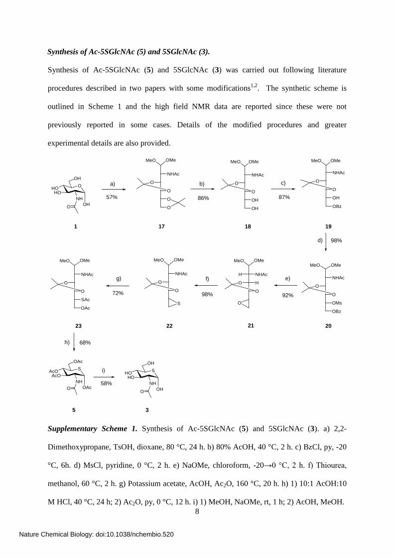
Synthesis of Ac-5SGlcNAc (5) and 5SGlcNAc (3).
Synthesis of Ac-5SGlcNAc (5) and 5SGlcNAc (3) was carried out following literature
procedures described in two papers with some modifications1,2. The synthetic scheme is
outlined in Scheme 1 and the high field NMR data are reported since these were not
previously reported in some cases. Details of the modified procedures and greater
experimental details are also provided.
OMeMeO
NHAc
O
O
O
O
OMeMeO
NHAc
O
O
OH
OH
OMeMeO
NHAc
O
O
OBz
OH
NHAc
O
O
OBz
OMs
H NHAc
HO
O
O
NHAc
O
O
S
57% 86% 87%
98%
92%98%72%
68%
1 17 18 19
20212223
a) b) c)
d)
e)f)g)
h)
5 3
i)
58%
OMeMeO
SAcOAcO
NHO
OAc
OAc
OMeMeO
OHOHO
NH
O
OH
OH
SHOHO
NH
O
OH
OH
OMeMeO
NHAc
O
O
OMeMeO
SAc
OAc
Supplementary Scheme 1. Synthesis of Ac-5SGlcNAc (5) and 5SGlcNAc (3). a) 2,2-
Dimethoxypropane, TsOH, dioxane, 80 °C, 24 h. b) 80% AcOH, 40 °C, 2 h. c) BzCl, py, -20
°C, 6h. d) MsCl, pyridine, 0 °C, 2 h. e) NaOMe, chloroform, -20→0 °C, 2 h. f) Thiourea,
methanol, 60 °C, 2 h. g) Potassium acetate, AcOH, Ac2O, 160 °C, 20 h. h) 1) 10:1 AcOH:10
M HCl, 40 °C, 24 h; 2) Ac2O, py, 0 °C, 12 h. i) 1) MeOH, NaOMe, rt, 1 h; 2) AcOH, MeOH.
Nature Chemical Biology: doi:10.1038/nchembio.520
Page 9
9
2-Acetamido-2-deoxy-3,4,5,6-di-O-isopropylidine-aldehydo-D-glucose dimethyl acetal (17)9
– 15.0 g (67.8 mmol) of 1 was suspended in 1,4 dioxane (150 mL) at 80 °C. 2,2-
dimethoxypropane (60 mL) was added to the suspension, together with 2.25 g (11.8 mmol) of
p-toluene sulphonic acid. The reaction was stirred for 24 hours at 80 °C. The mixture was
cooled and subsequently treated with enough potassium carbonate to obtain a neutral
solution. The reaction was filtered and directly concentrated under high vacuum. The
concentrated crude reaction was subjected to flash column silica chromatography using a
solvent system of 1-2 % methanol in chloroform, affording the title compound as a caramel
coloured viscous syrup (13.4 g, 57%). 1H-NMR (400 MHz, CDCl3) δ (ppm) 5.86 (d, J = 10.0
Hz, 1H), 4.45 (ddd, J = 9.60, 6.00, 1.20 Hz, 1H), 4.35 (d, J = 6.00 Hz, 1H), 4.23 (dd, J =
8.00, 1.20 Hz, 1H), 4.14-4.04 (m, 2H), 3.95 (dd, J = 8.40, 4.40 Hz, 1H), 3.61 ( t, J = 8.00
Hz, 1H), 3.40 (s, 3H), 3.36 (s, 3H), 2.01 (s, 3H), 1.46 (s, 3H), 1.38 (s, 3H), 1.36 (s, 3H), 1.33
(s, 3H); 13C-NMR (100 MHz, CDCl3) δ (ppm) 169.58, 109.93, 109.46, 103.46, 77.90, 77.60,
67.41, 54.68, 53.55, 49.05, 26.99, 26.93, 26.38, 25.24, 23.36.
2-Acetamido-2-deoxy-3,4-O-isopropylidine-aldehydo-D-glucose dimethyl acetal (18)9 – 10.35
g (29.80 mmol) of 17 was dissolved in a solution of 80% acetic acid in water, and stirred at
40 °C for 2 hours. Upon completion of the reaction, the mixture was concentrated under high
vacuum in small aliquots until the whole reaction mixture was concentrated to afford a thick
oil. The reaction was dried directly under high vacuum until the acetic acid was removed.
The crude reaction mixture was purified via flash column silica chromatography using a
solvent system of increasing polarity, beginning at 7% methanol and increasing to 10%
methanol in chloroform. The pure fractions were concentrated and dried on the high vacuum
pump to afford the title compound as a yellow syrup. (8.55 g, 86 %). 1H-NMR (500 MHz,
Nature Chemical Biology: doi:10.1038/nchembio.520
Page 10
10
CDCl3) δ (ppm) 6.32 (d, J = 9.67 Hz, 1H), 4.42 (d, J = 6.98 Hz, 1H), 4.34 (t, J = 9.68 Hz,
1H), 4.23 (d, J = 9.48 Hz, 1H), 3.73 (dd, J =11.18, 3.08 Hz, 1H), 3.60 (m, 3H), 3.36 (s, 3H),
3.27 (s, 3H), 2.02 (s, 3H), 1.33 (s, 3H); 13C-NMR (125 MHz, CDCl3) δ (ppm) 171.23, 109.08,
102.64, 78.02, 76.21, 73.45, 64.17, 54.61, 52.48, 50.04, 26.96, 23.19.
2-Acetamido-6-O-benzoyl-2-deoxy-3,4-O-isopropylidene-aldehydo-D-glucose dimethyl acetal
(19)10 – To a stirred solution of 18 (7.70 g, 22.2 mmol) in pyridine, dry benzoyl chloride
(3.14 g, 22.2 mmol) was added at -20 °C. The mixture was stirred for 6 hours between -10 to
-5 °C. Upon reaction completion, the mixture was extracted with chloroform. The extract was
washed two times with 2 M HCl, saturated sodium carbonate, and water. Each aqueous layer
was back extracted with chloroform. The combined organic layers were dried with
magnesium sulphate, filtered, and concentrated. The concentrated crude reaction mixture
was purified via flash column silica chromatography using a solvent system of 1% methanol
in chloroform with increasing polarity to 2% methanol in chloroform. The pure fractions
were concentrated, dried, and then crystallized from ethyl acetate and hexanes to afford the
title compound as white needles (7.68 g, 87 %). 1H-NMR (500 MHz, CDCl3) δ (ppm) 8.11
(dd, J = 8.45, 1.33 Hz, 2H), 7.53 (t, J = 7.49 Hz, 1H), 7.41 (t, J = 8.18 Hz, 2H), 4.45 (m, 4H),
4.24 (d, J = 10.1 Hz, 1H), 3.90 (m, 1H), 3.71 (t, J = 8.71 Hz, 1H), 3.36 (s, 3H), 3.26 (s, 3H),
2.03 (s, 3H), 1.35 (s, 3H), 1.35 (s, 3H), 1.32 (s, 3H); 13C-NMR (125 MHz, CDCl3) δ (ppm)
171.20, 166.62, 132.99, 129.82, 128.29, 109.07, 102.16, 79.03, 75.38, 71.62, 66.83, 54.67,
51.95, 50.36, 26.95, 26.93, 23.12.
2-Acetamido-6-O-benzoyl-2-deoxy-3,4-O-isopropylidine-5-O-mesyl-aldehydo-D-glucose
dimethyl acetal (20)10 – 6.52 g (16.4 mmol) of 19 was cooled to 0 °C in pyridine (14 mL). To
this solution mesyl chloride (2.50 g, 20.8 mmol) was added. The reaction was kept at 0 °C
Nature Chemical Biology: doi:10.1038/nchembio.520
Page 11
11
until judged complete (2 hours). The mixture was concentrated under high vacuum. The
remaining residue was extracted with chloroform, and successively washed two times with 2
M HCl, saturated sodium carbonate solution and water. Each aqueous layer was back
extracted with chloroform. All organic layers were combined, dried with magnesium
sulphate, concentrated in vacuo, and pumped under high vacuum until residual pyridine was
removed. The crude reaction mixture was purified via flash column silica chromatography in
a solvent system of increasing polarity of 1-2% methanol in chloroform. Pure fractions were
concentrated and dried on the high vacuum pump, giving rise to pure product as an
amorphous white solid (7.64 g, 98 %). 1H-NMR (500 MHz, CDCl3) δ (ppm) 8.12 (d, J = 7.11
Hz, 1H), 7.60 (t, J = 7.44 Hz, 1H), 7.48 (t, J =8.12 Hz, 2H), 5.84 (d, J =9.66 Hz, 1H), 5.14
(m, 1H), 4.80 (dd, J =12.54, 2.64 Hz, 1H), 4.49 (dd, J = 12.54, 7.10 Hz, 1H), 4.45 (m, 2H),
4.39 (m, 1H), 4.00 (dd, J = 7.94, 6.23 Hz, 1H), 3.45 (s, 3H), 3.34 (s, 3H), 3.16 (s, 3H), 2.07
(s, 3H), 1.45 (s, 3H), 1.43 (s, 3H); 13C-NMR (125 MHz, CDCl3) δ (ppm) 170.07, 166.10,
133.29, 129.91, 129.50, 128.48, 110.39, 103.18, 78.40, 76.78, 75.65, 63.55, 55.78, 52.79,
49.69, 38.89, 26.99, 26.90, 23.36.
2-Acetamido-5,6-anhydro-2-deoxy-3,4-O-isopropylidene-aldehydo-L-idose dimethyl acetal
(21)10 – To a solution of 20 (6.23 g, 13.1 mmol) in dry chloroform (62 mL) was added drop-
wise at -20 °C a fresh solution of sodium methoxide (0.50 g Na in 20 mL of methanol). The
reaction mixture was kept between -10 °C and 0 °C until the reaction was complete (5 hours).
Upon completion, the reaction was treated with 102 mL of additional methanol. The reaction
was neutralized with H+ amberlite ion exchange resin (IR-120) until the reaction mixture
reached a pH of 7. The resin was filtered and washed with additional methanol. The filtered
reaction mixture was concentrated in vacuo and the remaining material triturated with ether
and hexanes to afford a pure white amorphous solid (3.51 g, 92 %). 1H-NMR (500 MHz,
Nature Chemical Biology: doi:10.1038/nchembio.520
Page 12
12
CDCl3) δ (ppm) 5.91 (d, J = 9.83 Hz, 1H), 4.37 (d, J = 7.06 Hz, 1H), 4.18 (m, 2H), 3.36 (s,
3H), 3.35 (m, 1H), 3.25 (s, 3H), 2.98 (m, 1H), 2.80 (m, 1H), 2.75 (dd, J = 5.13, 4.17 Hz, 1H),
1.97 (s, 3H), 1.35 (s, 3H), 1.32 (s, 3H); 13C-NMR (125 MHz, CDCl3) δ (ppm) 170.03, 109.79,
102.72, 78.36, 76.00, 55.43, 52.20, 50.92, 48.67, 43.75, 26.88, 26.76, 23.17.
2-Acetamido-2,5,6-trideoxy-5,6-epithio-3,4-O-isopropylidine-aldehydo-D-glucose dimethyl
acetal (22)10 – To a solution of 21 (3.65 g, 12.64 mmol) in dry methanol (78 mL), was added
thiourea (2.88 g, 37.87 mmol), and the mixture was stirred at 60 °C. Upon completion, the
reaction mixture was cooled to room temperature and then concentrated in vacuo. The title
compound was purified using a solvent system of 2% methanol and chloroform (3.77 g, 98
%). 1H-NMR (500 MHz, CDCl3) δ (ppm) 5.85 (d, J = 9.43 Hz, 1H), 4.33 (m, 3H), 3.40 (s,
3H), 3.36 (s, 3H), 3.26 (t, J = 7.94 Hz, 1H), 2.92 (m, 1H), 2.51 (d, J = 6.03 Hz, 1H), 2.26 (d,
J = 5.19 Hz, 1H), 1.99 (s, 3H), 1.44 (s, 3H), 1.39 (s, 3H); 13C-NMR (125 MHz, CDCl3) δ
(ppm) 169.67, 109.95, 103.38, 81.91, 78.22, 55.38, 53.56, 49.39, 33.92, 27.04, 27.03, 22.96.
2-Acetamido-6-O-acetyl-5-S-acetyl-2-deoxy-3,4-O-isopropylidine-5-thio-aldehydo-D-glucose
dimethyl acetal (23)10. A solution of 22 (1.02 g, 3.34 mmol), acetic anhydride (31 mL),
potassium acetate (1.53 g, 15.6 mmol), and acetic acid (5 mL) was brought to reflux at 160
°C with stirring for 20 hours. After completion of the reaction, the mixture was cooled to
room temperature and evaporated under high vaccum. The remaining residue was extracted
with chloroform and successively washed with 2 M HCl, saturated sodium bicarbonate, and
brine. Each aqueous layer was back extracted with chloroform. The pooled organic layers
were dried with magnesium sulphate, and evaporated to a dark syrup. Flash column
chromatography using a solvent system of 70% ethyl acetate/30% hexanes furnished the title
compound as a white crystalline solid (0.980 g, 72 %).1H-NMR (500 MHz, CDCl3) δ (ppm)
Nature Chemical Biology: doi:10.1038/nchembio.520
Page 13
13
5.83 (d, J = 9.69 Hz, 1H), 4.37 (m, 2H), 4.32 (d, J = 6.77 Hz, 1H), 4.26 (dd, J = 11.48, 5.11
Hz, 1H), 4.16 (d, J = 7.52 Hz, 1H), 3.96 (m, 1H), 3.82 (t, J = 7.90 Hz, 1H), 3.37 (s, 3H), 3.30
(s, 3H), 2.36 (s, 3H), 2.04 (s, 3H), 2.02 (s, 3H), 1.42 (s, 3H), 1.39 (s, 3H); 13C-NMR (100
MHz, CDCl3) δ (ppm) 193.76, 170.51, 169.74, 109.77, 103.08,, 76.62, 63.78, 55.38, 52.57,
50.27, 44.94, 30.46, 27.03, 26.80, 23.27, 20.66.
2-Acetamido-1,3,4,6-tetra-O-acetyl-2-deoxy-5-thio-α-D-glucopyranose (5)10 – In a solution of
10:1 acetic acid: 2 M HCl (77 mL), 23 (2.57 g, 6.31 mmol) was dissolved and the mixture
was warmed to 40 °C for 48 hours, under an atmosphere of nitrogen. The reaction mixture
turned a bright pink colour. Upon completion of the reaction, the mixture was concentrated
without heating under high vacuum. The resulting dark brown residue was dried thoroughly
under high vacuum until residual acid was removed and a syrup was obtained. The syrup was
dissolved in acetic anhydride (13 mL) and pyridine (51 mL) and stirred overnight at 0 °C.
Upon completion of the reaction, the mixture was concentrated under high vacuum without
heating. The resulting syrup was subjected to flash column silica chromatography using a
solvent system of 2-3% methanol in chloroform to give the title compound as an off-white
solid that upon crystallization from ethyl acetate and hexanes gave rise to pure white needles
(1.74 g, 68 % over two steps). 1H-NMR (500 MHz, CDCl3) δ (ppm) 5.92 (d, J = 3.04 Hz,
1H), 5.70 (d, J = 8.86 Hz, 1H), 5.37 (dd, J =10.74, 9.64 Hz, 1H), 5.16 (dd, J = 10.89, 9.70
Hz, 1H), 4.63 (m, 1H), 4.34 (dd, J = 12.11, 4.95 Hz, 1H), 4.03 (dd, J =12.11, 3.15 Hz, 1H),
3.50 (ddd, J = 10.8, 4.80, 3.20, 1H), 2.17 (s, 3H), 2.05 (s, 3H), 2.03 (s, 3H), 2.02 (s, 3H), 1.90
(s, 3H); 13C-NMR (125 MHz, CDCl3) δ (ppm) 171.73, 170.60, 169.52, 169.16, 168.77, 72.79,
71.78, 71.47, 61.10, 55.20, 39.75, 23.08, 21.14, 20.65, 20.53.
Nature Chemical Biology: doi:10.1038/nchembio.520
Page 14
14
2-Acetamido-2-deoxy-5-thio-α-D-glucopyranose (3)10 – In a solution of methanol (1.9 mL),
0.050 g (0.123 mmol) of 5 was dissolved and a catalytic amount of sodium methoxide
(enough to raise the pH of the reaction solution to between 9 and 10) was added. The reaction
was followed closely by TLC and was found to be complete after 1 hour at room temperature.
The reaction was subsequently neutralized by the drop-wise addition of a dilute solution of
acetic acid in methanol (pH 4) until the reaction solution was neutralized to pH 7. The
reaction mixture was concentrated without heating under high vacuum. The final product was
isolated by flash silica column chromatography using a solvent system of 12:2:1 ethyl
acetate:methanol:water as a white solid (0.017 g, 58 %).1H-NMR (600 MHz, MeOH-d4) δ
(ppm) 4.89 (d, J = 3.17 Hz, 1H), 4.07 (dd, J =10.50, 2.82 Hz, 1H), 3.87-3.79 (m, 2H), 3.65
(dd, J = 10.46, 8.82 Hz, 1H), 3.55 (dd, J = 10.37, 8.93 Hz, 1H), 3.25-3.22 (m, 1H), 1.96 (s,
3H); 13C-NMR (150 MHz, MeOH-d4) δ (ppm) 173.35, 76.88, 73.52, 73.50, 62.65, 60.01,
44.89, 22.72.
Nature Chemical Biology: doi:10.1038/nchembio.520
Page 15
15
Synthesis of pMP-5SGlcNAc (8).
SAcOAcO
NH
O
OAc
OAc
SAcOAcO
NH
OAc
O
O
SHOHO
NH
OH
O
O
O
80% 76% 12%
66%
a) b) c)
d)
5 24 25 26
8
SAcOAcO
NHO
OAc
OH
SAcOAcO
NH
O
OAc
O CCl3
NH
O
Supplementary Scheme 2. Synthesis of pMP-5SGlcNAc (8). a) Hydrazine acetate, DMF, rt,
6 h. b) Trichloroacetonitrile, DBU (cat.), DCM, -20 °C→room temperature, 1h. c) p-
Methoxyphenol, BF3OEt2 (cat.), DCM, -20 °C→room temperature, 1h. d) 1) NaOMe (cat.)
MeOH, room temperature, 0.5 h; 2) AcOH, MeOH.
2-Acetamido-3,4,6-tri-O-acetyl-2-deoxy-5-thio-α-D-glucopyranose (24) – Anomeric
deacetylation was effected in a manner similar to that reported previously for different
saccharides11. To a solution of 5 (0.200 g, 0.49 mmol) in DMF (1 mL), stirring at room
temperature, hydrazine acetate (0.050 g, 0.54 mmol) was added. The mixture was left to stir
until all starting material was consumed (6 hours). The mixture was concentrated under high
vacuum, and co-evaporated twice with toluene, without heating. The resulting residue was
taken up in ethyl acetate and washed two times with saturated sodium bicarbonate, water
(until a neutral pH was obtained), and brine. Each aqueous layer was back extracted with
ethyl acetate. The pooled organic layers were dried using magnesium sulphate, filtered, and
concentrated in vacuo. The residue was subjected to flash column silica chromatography
using a solvent system of 70% ethyl acetate, 30% hexanes, and subsequently concentrated
Nature Chemical Biology: doi:10.1038/nchembio.520
Page 16
16
under high vacuum to afford the title compound as a white solid. Crystallization from ethyl
acetate and hexanes afforded a white powder (0.144 g, 80 %).1H-NMR (600 MHz, CDCl3) δ
(ppm) 6.14 (d, J = 9.31 Hz, 1H), 5.36-5.27 (m, 2H), 5.03 (d, J = 2.74 Hz, 1H), 4.55-4.51 (m,
1H), 4.36 (dd, J = 11.99, 5.04 Hz, 1H), 4.10 (dd, J = 12.00, 3.39 Hz, 1H), 3.67-3.64 (m, 1H),
2.08 (s, 3H), 2.04 (s, 3H), 2.03 (s, 3H), 1.97 (s, 3H); 13C-NMR (150 MHz, CDCl3) δ (ppm)
171.31, 170.78, 169.97, 169.39, 72.78, 72.14, 71.69, 61.58, 55.42, 38.67, 23.19, 20.68, 20.64,
20.57. HRMS (m/z): [M+H]+ calcd for C14H21NO8S: 364.1066; found 364.1070.
2-Acetamido-3,4,6-tri-O-acetyl-2-deoxy-5-thio-α-D-glucopyranosyl trichloroacetimidate (25)
– Preparation of the trichloroacetmidiate donor was carried out by modification of previously
described procedures used for different saccharides12-15. 24 was dissolved in dichloromethane
at 0 °C, and to this solution was added trichloroacetonitrile (0.181 g, 1.27 mmol) and a
catalytic amount of DBU (~1 drop). The reaction was allowed to stir at 0 °C until completion
(0.5 hours). The mixture was diluted with benzene and concentrated under high vacuum to
yield a residue. The crude mixture was subjected to flash column silica chromatography using
a solvent system of 60% ethyl acetate in hexanes. The desired material was isolated as a clear
oil, which upon standing, solidified to give a white amorphous solid (0.086 g, 76 %).1H-
NMR (600 MHz, CDCl3) δ (ppm) 8.81 (s, 1H), 6.13 (d, J = 3.00 Hz, 1H), 5.82 (d, J = 8.80
Hz, 1H), 5.43 (dd, J = 10.68, 9.79 Hz, 1H), 5.27 (dd, J = 10.68, 9.78 Hz, 1H), 4.73-4.69 (m,
1H), 4.35 (dd, J =12.14, 4.98 Hz, 1H), 4.11 (dd, J = 12.11, 3.13 Hz, 1H), 4.07-4.05 (m, 1H),
2.05 (s, 3H), 2.05 (s, 3H), 2.05 (s, 3H), 1.91 (s, 3H); 13C-NMR (150 MHz, CDCl3) δ (ppm)
170.42, 170.40, 169.51, 169.12, 159.83, 90.70, 77.73, 71.60, 71.22, 60.90, 55.71, 40.05,
22.98, 20.54, 20.52, 20.43. HRMS (m/z): [M+H]+ calcd for C16H121N2O8SCl3: 507.0162;
found 507.0173.
Nature Chemical Biology: doi:10.1038/nchembio.520
Page 17
17
para-Methoxyphenyl 2-acetamido-3,4,6-tri-O-acetyl-2-deoxy-5-thio-β-D-glucopyranoside
(26) – Glycosylation was effected by adaptation of reported methods used for glycosylation
of trichloroacetimidate donors of other saccharides13,15. A solution of 25 (0.050 g, 0.100
mmol) in dichloromethane (0.77 mL) and p-methoxyphenol (0.025 g, 0.20 mmol) was stirred
at -20 °C. To this solution a catalytic amount of boron trifluoroetherate (~ 1 drop) was added,
and the reaction was immediately allowed to warm to room temperature. The reaction was
stirred for 1 h, after which time the reaction was judged complete. Two equivalents of
triethylamine (0.026 mL) were then added to the reaction mixture. The mixture was
concentrated under high vacuum without heating. Flash column silica chromatography was
carried out to isolate the products and the 1H NMR spectrum revealed a mixture of two
products; the α- and β-anomers in a ratio of 70% α and 30% β. The two anomers were
separated via flash column chromatography, using a solvent system of 60% ethyl acetate in
hexanes, to afford the pure β-anomer (0.056 g, 12 %), and α-anomer (0.096 g, 21 %). (β-
anomer) 1H-NMR (600 MHz, CDCl3) δ (ppm) 6.95 (d, J = 9.07 Hz, 2H), 6.83 (d, J = 9.09
Hz, 2H), 5.95 (d, J = 8.82 Hz, 1H), 5.38 (t, J = 7.44 Hz, 1H), 5.20 (d, J = 6.33 Hz, 1H), 5.11
(t, J = 7.36 Hz, 1H), 4.61-4.58 (m, 1H), 4.40-4.33 (m, 2H), 3.70 (s, 3H), 3.20 (dd, J = 13.23,
6.23 Hz, 1H), 2.08 (s, 3H), 2.07 (s, 3H), 2.05 (s, 3H), 1.97 (s, 3H); 13C-NMR (150 MHz,
CDCl3) δ (ppm) 170.52, 170.12, 169.67, 168.92, 155.41, 150.43, 118.17, 114.68, 80.53,
71.01, 69.13, 63.40, 55.63, 40.57, 29.68, 23.38, 20.77, 20.69, 20.57. HRMS (m/z): [M+H]+
calcd for C21H27N2O9S: 470.1485; found 470.1478.
para-Methoxyphenyl 2-acetamido-2-deoxy-5-thio-β-D-glucopyranoside (8) – To a solution of
26 (0.0062 g, 0.013 mmol) in methanol (0.25 mL) was added a catalytic amount of sodium
methoxide (enough to bring the reaction solution to pH 10). The reaction was allowed to stir
at room temperature. After twenty minutes a white solid began to precipitate out of solution.
Nature Chemical Biology: doi:10.1038/nchembio.520
Page 18
18
The reaction had reached completion after 30 minutes. The reaction was first diluted with
additional methanol (5 mL), and subsequently brought to a neutral pH by the drop-wise
addition of a dilute mixture of acetic acid in methanol (pH 4). The reaction was concentrated
under high vacuum without heating. The title compound was isolated by precipitation from
ethanol and ether as a fine white powder (0.0033 g, 66 %). 1H-NMR (600 MHz, MeOH-d4) δ
(ppm) 6.96 (d, J =9.16 Hz, 2H), 6.81 (d, J = 9.16 Hz, 2H), 5.07 (d, J = 9.29 Hz, 1H), 4.16 (t,
J = 9.60Hz, 1H), 3.93 (dd, J = 11.44, 3.73 Hz,1H), 3.75 (dd, J = 11.44, 6.47 Hz, 1H), 3.72 (s,
3H), 3.57 (dd, J = 10.07, 8.95 Hz, 1H), 3.38 (dd, J = 9.89, 8.91 Hz, 1H), 2.89-2.85 (m, 1H),
1.93 (s, 3H); 13C-NMR (150 MHz, D2O) δ (ppm) 173.69, 155.72, 153.24, 118.96, 115.59,
82.24, 76.53, 75.78, 62.69, 61.32, 55.10, 47.53, 23.03; HRMS (m/z): [M+H]+ calcd for
C15H21NO6S: 344.1168; found 344.1157.
Nature Chemical Biology: doi:10.1038/nchembio.520
Page 19
19
Synthesis of Me-5SGlcNAc (10).
SAcOAcO
NH
OAc
O O
NH
CCl3
25
SAcOAcO
NH
OAc
O
27
OSHO
HONH
OH
O
10
Oa) b)
42% 29%
Supplementary Scheme 3. Synthesis of Me-5SGlcNAc (10). a) MeOH (2 eq), BF3OEt2 (cat.),
DCM, -20 °C→room temperature. b) 1) NaOMe (cat.), MeOH, rt; 2) AcOH, MeOH.
Methyl 2-acetamido-3,4,6-tri-O-acetyl-2-deoxy-5-thio-β-D-glucopyranoside – (27). 25 (0.050
g, 0.11 mmol) was dissolved in dry dichloromethane (0.76 mL). Two equivalents of dry
methanol (0.009 mL, 0.211 mmol) were added to the solution at -20°C and then a catalytic
amount of boron trifluoroethyletherate was added, after which the reaction mixture was
allowed to warm to room temperature. Once the reaction was judged complete, two
equivalents of triethylamine (0.03 mL) were added. The reaction was concentrated under high
vacuum and the residue was immediately subjected to silica gel column chromatography
using a solvent system of ethyl acetate. The ratio of α- to β-anomers was found to be 1:1 by
1H NMR. The desired β-anomer was isolated through a second purification using flash
column chromatography in ethyl acetate in order to separate the two anomers. The title
compound was isolated as a white powder (0.0154 g , 42%). (β-anomer) 1H-NMR (600 MHz,
MeOH-d4) δ (ppm) 5.17 (t, J = 9.74 Hz, 1H), 5.02 (t, J = 9.53 Hz, 1H), 4.60 (d, J = 8.76 Hz,
1H), 4.34 (dd, J = 11.86, 5.23 Hz, 1H), 4.22 (t, J = 9.21 Hz, 1H), 4.10 (dd, J = 11.82, 3.87
Hz, 1H), 3.45 (s, 3H), 3.28-3.26 (m, 1H), 2.02 (s, 3H), 1.97 (s, 3H), 1.96 (s, 3H), 1.89 (s,
3H); 13C-NMR (150 MHz, MeOH-d4) δ (ppm) 173.37, 172.26, 171.57, 171.32, 84.41,74.83,
Nature Chemical Biology: doi:10.1038/nchembio.520
Page 20
20
73.41,63.64, 59.21, 58.65, 41.91, 22.72, 20.64, 20.61, 20.54. HRMS (m/z): [M+H]+ calcd for
C15H23NO8S: 378.1223; found 378.1214.
Methyl 2-acetamido-2-deoxy-5-thio-β-D-glucopyranoside (10) – 27 (0.0088 g, 0.023 mmol)
was dissolved in methanol (0.35 mL) at room temperature. To this solution a catalytic amount
of sodium methoxide was added (the pH of the reaction solution was 8-9). The reaction was
followed closely by TLC and upon completion was neutralized (to pH 7) using a dilute
solution of acetic acid in methanol (pH 4). The neutralized mixture was concentrated under
high vacuum without heating. The desired product was purified by silica gel column
chromatography using a solvent system of 15% methanol in chloroform. The pure fractions
were concentrated in vacuo and the residue precipitated from ethanol by addition of ether to
afford the title compound as a white residue (1.68 mg, 29%). 1H-NMR (600 MHz, MeOH-d4)
δ (ppm) 4.55 (s, 1H), 4.42 (d, J = 8.89 Hz, 1H), 3.97 (t, J = 9.26 Hz, 1H), 3.93 (dd, J = 11.35,
3.93 Hz, 1H), 3.72 (dd, J = 11.35, 6.59 Hz, 1H), 3.51 (t, J = 8.95 Hz, 1H), 3.41 (s, 3H), 3.26
(dd, J = 9.53, 8.90 Hz, 1H), 2.76-2.73 (m, 1H), 1.95 (s, 3H). 13C-NMR (150 MHz, MeOH-d4)
δ (ppm) 173.79, 84.76, 76.89, 75.63, 63.13, 60.96, 58.36, 47.48, 22.96. HRMS (m/z): [M+H]+
calcd for C9H17NO5S: 252.0906; found 252.0913.
Nature Chemical Biology: doi:10.1038/nchembio.520
Page 21
21
Synthesis of Ac-5SGlcNAz (14)
S
OAc
AcOAcO
N3OAc
S
OAc
AcOAcO
H2NOAc
S
OAc
AcOAcO
NHOAc
a)
28
b)
O
N3
2914
HCl
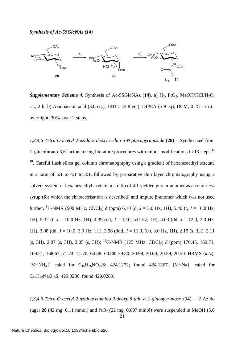
Supplementary Scheme 4. Synthesis of Ac-5SGlcNAz (14). a) H2, PtO2, MeOH/HCl/H2O,
r.t., 2 h; b) Azidoacetic acid (3.0 eq.), HBTU (3.8 eq.), DIPEA (5.9 eq), DCM, 0 °C → r.t.,
overnight, 30% over 2 steps.
1,3,4,6-Tetra-O-acetyl-2-azido-2-deoxy-5-thio-α-D-glucopyranoside (28) – Synthesized from
D-glucofurano-3,6-lactone using literature procedures with minor modifications in 13 steps16-
18. Careful flash silica gel column chromatography using a gradient of hexanes:ethyl actetate
in a ratio of 5:1 to 4:1 to 3:1, followed by preparative thin layer chromatography using a
solvent system of hexanes:ethyl acetate in a ratio of 4:1 yielded pure α-anomer as a colourless
syrup (for which the characterisation is described) and impure β-anomer which was not used
further. 1H-NMR (500 MHz, CDCl3) δ (ppm) 6.10 (d, J = 3.0 Hz, 1H), 5.40 (t, J = 10.0 Hz,
1H), 5.32 (t, J = 10.0 Hz, 1H), 4.39 (dd, J = 12.0, 5.0 Hz, 1H), 4.03 (dd, J = 12.0, 3.0 Hz,
1H), 3.88 (dd, J = 10.0, 3.0 Hz, 1H), 3.56 (ddd, J = 11.0, 5.0, 3.0 Hz, 1H), 2.19 (s, 3H), 2.11
(s, 3H), 2.07 (s, 3H), 2.05 (s, 3H); 13C-NMR (125 MHz, CDCl3) δ (ppm) 170.45, 169.71,
169.51, 168.67, 71.74, 71.70, 64.88, 60.88, 39.80, 20.98, 20.60, 20.59, 20.50. HRMS (m/z):
[M+NH4]+ calcd for C16H26NO10S: 424.1272; found 424.1267. [M+Na]+ calcd for
C16H22NaO10S: 429.0286; found 429.0280.
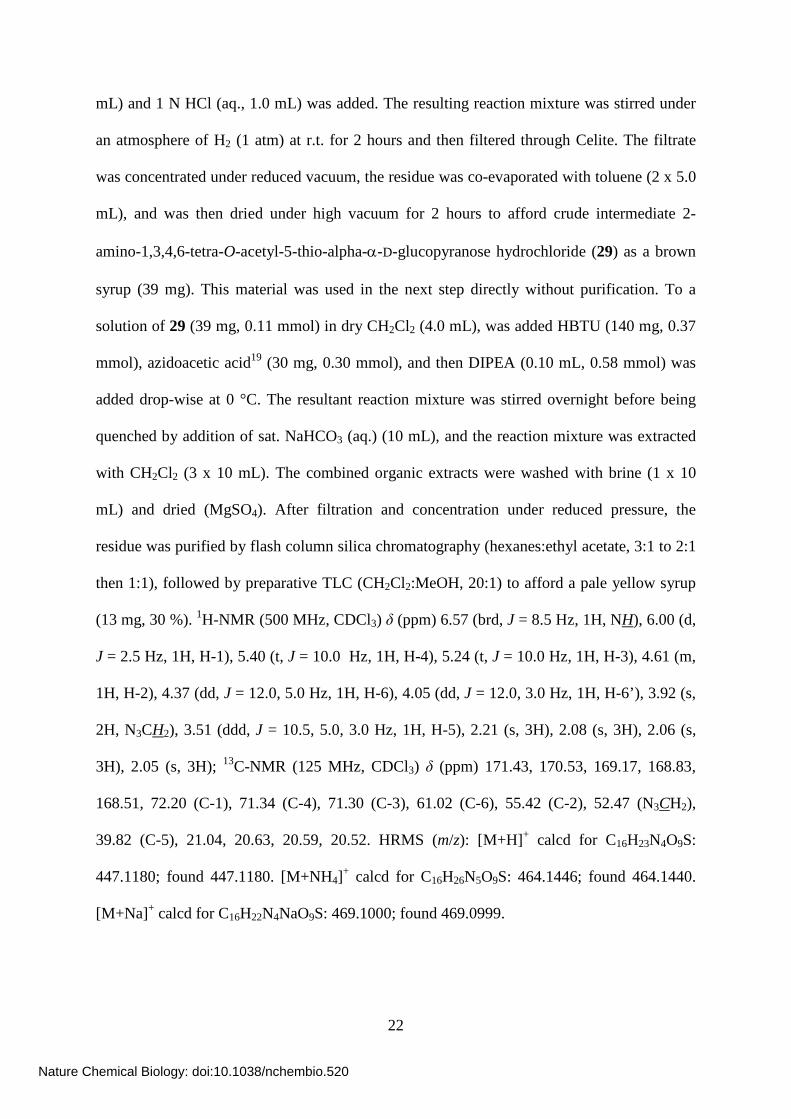
1,3,4,6-Tetra-O-acetyl-2-azidoacetamido-2-deoxy-5-thio-α-D-glucopyranose (14) – 2-Azido
sugar 28 (42 mg, 0.11 mmol) and PtO2 (22 mg, 0.097 mmol) were suspended in MeOH (5.0
Nature Chemical Biology: doi:10.1038/nchembio.520
Page 22
22
mL) and 1 N HCl (aq., 1.0 mL) was added. The resulting reaction mixture was stirred under
an atmosphere of H2 (1 atm) at r.t. for 2 hours and then filtered through Celite. The filtrate
was concentrated under reduced vacuum, the residue was co-evaporated with toluene (2 x 5.0
mL), and was then dried under high vacuum for 2 hours to afford crude intermediate 2-
amino-1,3,4,6-tetra-O-acetyl-5-thio-alpha-α-D-glucopyranose hydrochloride (29) as a brown
syrup (39 mg). This material was used in the next step directly without purification. To a
solution of 29 (39 mg, 0.11 mmol) in dry CH2Cl2 (4.0 mL), was added HBTU (140 mg, 0.37
mmol), azidoacetic acid19 (30 mg, 0.30 mmol), and then DIPEA (0.10 mL, 0.58 mmol) was
added drop-wise at 0 °C. The resultant reaction mixture was stirred overnight before being
quenched by addition of sat. NaHCO3 (aq.) (10 mL), and the reaction mixture was extracted
with CH2Cl2 (3 x 10 mL). The combined organic extracts were washed with brine (1 x 10
mL) and dried (MgSO4). After filtration and concentration under reduced pressure, the
residue was purified by flash column silica chromatography (hexanes:ethyl acetate, 3:1 to 2:1
then 1:1), followed by preparative TLC (CH2Cl2:MeOH, 20:1) to afford a pale yellow syrup
(13 mg, 30 %). 1H-NMR (500 MHz, CDCl3) δ (ppm) 6.57 (brd, J = 8.5 Hz, 1H, NH), 6.00 (d,
J = 2.5 Hz, 1H, H-1), 5.40 (t, J = 10.0 Hz, 1H, H-4), 5.24 (t, J = 10.0 Hz, 1H, H-3), 4.61 (m,
1H, H-2), 4.37 (dd, J = 12.0, 5.0 Hz, 1H, H-6), 4.05 (dd, J = 12.0, 3.0 Hz, 1H, H-6’), 3.92 (s,
2H, N3CH2), 3.51 (ddd, J = 10.5, 5.0, 3.0 Hz, 1H, H-5), 2.21 (s, 3H), 2.08 (s, 3H), 2.06 (s,
3H), 2.05 (s, 3H); 13C-NMR (125 MHz, CDCl3) δ (ppm) 171.43, 170.53, 169.17, 168.83,
168.51, 72.20 (C-1), 71.34 (C-4), 71.30 (C-3), 61.02 (C-6), 55.42 (C-2), 52.47 (N3CH2),
39.82 (C-5), 21.04, 20.63, 20.59, 20.52. HRMS (m/z): [M+H]+ calcd for C16H23N4O9S:
447.1180; found 447.1180. [M+NH4]+ calcd for C16H26N5O9S: 464.1446; found 464.1440.
[M+Na]+ calcd for C16H22N4NaO9S: 469.1000; found 469.0999.
Nature Chemical Biology: doi:10.1038/nchembio.520
Page 23
23
Characterization of UDP-5SGlcNAc (4).
5'-(2-acetamido-2-deoxy-5-thio-α-D-glucopyranosyl)diphosphate triethylammonium salt (4) -
The chemoenzymatic synthesis was carried out as described in the methods section. 1H NMR
(600 MHz, D2O): δ (ppm) 2.08 (s, 3H, NH(CO)CH3), 3.38 (ddd, 1H, J5'', 6a'' = 5.4 Hz, J5'',6b'' =
3.0 Hz, H-5''), 3.71 (dd, 1H, J4'',5'' = 10.2 Hz, H-4''), 3.75 (dd, 1H, J3'',2'' = 9.6 Hz, J3'',4'' = 9.0
Hz, H-3''), 3.89 (dd, 1H, J6b'',6a'' = 12.0 Hz, H-6b''), 3.97 (dd, 1H, H-6a''), 4.18-4.21 (m, 1H, H-
2''), H-5b' = 4.21-4.24 (m, 1H, H-5b'), H-5a' = 4.24-4.27 (m, 1H, H-5a'), 4.28-4.30 (m, 1H,
H-4'), 4.36-4.38 (m, 2H, H-2',3'), 5.32 (dd, 1H, J1'',2'' = 2.4 Hz, J1'',P = 7.8 Hz, H-1''), 5.97 (d,
1H, H-5), 6.00 (d, 1H, J1',2' = 4.2 Hz, H-1'), 7.96 (d, 1H, J6,5 = 7.8 Hz, H-6); 13C NMR (150
MHz, D2O): δ (ppm) 22.06 (NH(CO)CH3), 43.59 (C-5''), 57.74 (C-2''), 59.91 (C-6''), 64.89
(C-5'), 69.61 (C-3'), 72.03 (C-3''), 73.62 (C-4''), 73.75 (C-2'), 76.62 (C-1''), 83.17 (C-4'),
88.39 (C-1'), 102.63 (C-5), 141.63 (C-6), 151.78 (C-2), 166.20 (C-4), 169.13 (NH(CO)CH3).
31P NMR (242 MHz, D2O): δ (ppm) -10.66 (d, Jp,p = 21.8 Hz), -12.09 (d). HRMS (m/z): [M-
H]- calcd for C17H26N3O16P2S: 622.0519, found 622.0514.
Nature Chemical Biology: doi:10.1038/nchembio.520
Page 24
24
Supplementary Results
Supplementary Figure 1
Nature Chemical Biology: doi:10.1038/nchembio.520
Page 25
25
Supplementary Figure 2
a
b
Nature Chemical Biology: doi:10.1038/nchembio.520
Page 26
26
Supplementary Figure 3
b
a
Nature Chemical Biology: doi:10.1038/nchembio.520
Page 27
27
Supplementary Figure 4
ppm (t2)3.504.004.505.005.506.00
40
50
60
70
80
90
100
ppm (t1)
ppm (t2)3.003.504.00
2.50
3.00
3.50
4.00
ppm (t1)
a
b
Nature Chemical Biology: doi:10.1038/nchembio.520
Page 28
28
Supplementary Figure 5
Nature Chemical Biology: doi:10.1038/nchembio.520
Page 29
29
Supplementary Figure 6
Nature Chemical Biology: doi:10.1038/nchembio.520
Page 30
30
Supplementary Figure 7
Nature Chemical Biology: doi:10.1038/nchembio.520
Page 31
31
Supplementary Figure 8
Nature Chemical Biology: doi:10.1038/nchembio.520
Page 32
32
Supplementary Figure 9
Nature Chemical Biology: doi:10.1038/nchembio.520
Page 33
33
Supplementary Figure 10
Nature Chemical Biology: doi:10.1038/nchembio.520
Page 34
34
Supplementary Figure 11
Nature Chemical Biology: doi:10.1038/nchembio.520
Page 35
35
Supplementary Figure 12
a
b
Nature Chemical Biology: doi:10.1038/nchembio.520
Page 36
36
Supplementary Figure 13
Nature Chemical Biology: doi:10.1038/nchembio.520
Page 37
37
Supplementary Figure 14
Nature Chemical Biology: doi:10.1038/nchembio.520
Page 38
38
Supplementary Figure 15
Nature Chemical Biology: doi:10.1038/nchembio.520
Page 39
39
Supplementary Figure Legends
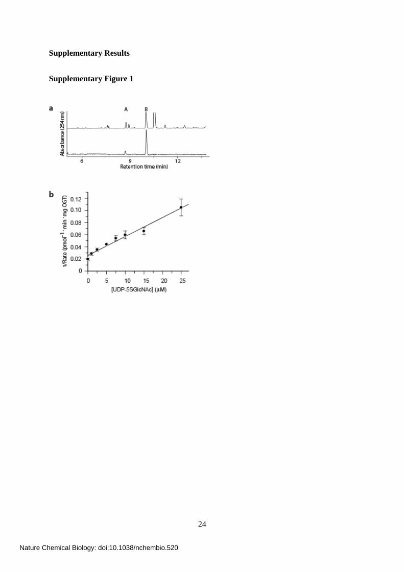
Supplementary Figure 1. In vitro characterization of the ability of the human enzymes
of the HBP to convert 5SGlcNAc (3) to UDP-5SGlcNAc (4) and in vitro evaluation of
UDP-5SGlcNAc (4) as an inhibitor of OGT. (a) In vitro enzymatic synthesis of UDP-
5SGlcNAc (4) catalyzed by the human enzymes of the HBP monitored by capillary
electrophoresis at 254 nm. Upper trace, crude reaction mixture; lower trace, UDP-5SGlcNAc
(4) following ion exchange and HPLC purification. Peak A, GDP-Glc (internal standard
spiked into samples prior to analysis); peak B, UDP-5SGlcNAc (4). (b) Inhibition by UDP-
5SGlcNAc (4) of OGT-catalyzed transfer of O-GlcNAc onto nup62.
Supplementary Figure 2. 1H NMR spectrum of UDP-5SGlcNAc (4). (a) 1H-NMR
spectrum of UDP-5SGlcNAc (4). Peaks at 8.35, 3.10 and 1.18 ppm arise from the
triethylammonium counterion. (b) Expansion of 1H NMR spectrum of UDP-5SGlcNAc (4)
covering the region from 4.40 to 3.20 ppm.
Supplementary Figure 3. NMR spectra of UDP-5SGlcNAc (4). (a) 13C1H NMR
spectrum of UDP-5SGlcNAc (4). Peaks at 8.1 (CH3) and 46.6 (CH2) are derived from the
triethylammonium cation. The peak at 174.2 ppm is from the carbonyl of the acetyl group. (b)
31P1H NMR spectrum of UDP-5SGlcNAc (4) referenced to 85% H3PO4 at 0 ppm.
Supplementary Figure 4. Two dimensional NMR spectra of UDP-5SGlcNAc (4). (a)
HMQC spectrum and (b) COSY spectrum.
Nature Chemical Biology: doi:10.1038/nchembio.520
Page 40
40
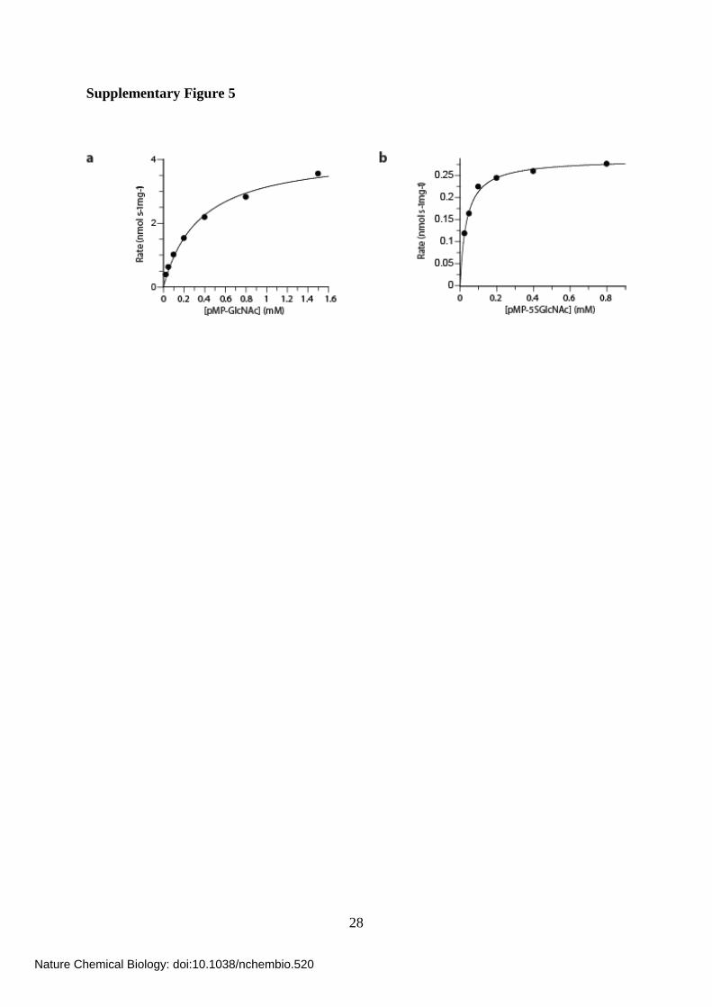
Supplementary Figure 5. Processing of oxygen- and thio-containing glycosides by OGA.
(a) Michaelis-Menten kinetics for OGA hydrolysis of pMP-GlcNAc (7) performed at 37 °C.
The fit gives a kcat of 14.3 nmol s-1 mg-1 and a KM of 350 μM. (b) Michaelis-Menten kinetics
for OGA hydrolysis of pMP-5SGlcNAc (8) performed at 37 °C. The fit gives a kcat of 0.28
nmol s-1 mg-1 and a KM of 35 μM.
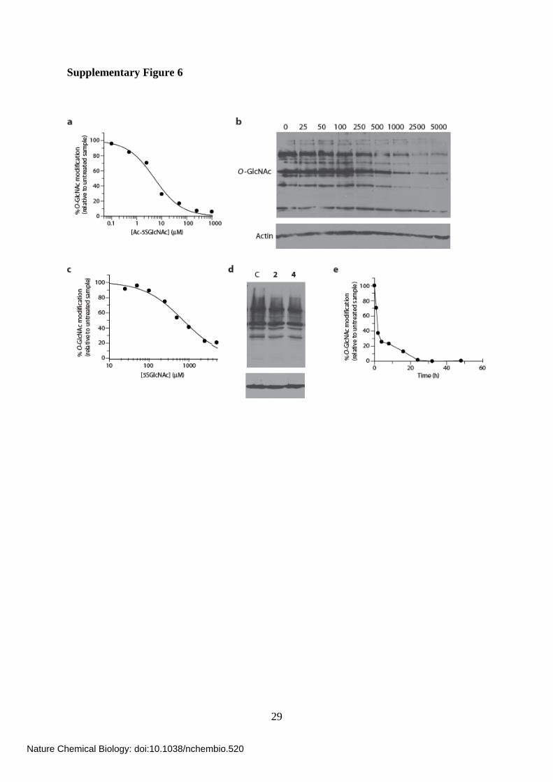
Supplementary Figure 6. Effects of treating cells with 5SGlcNAc (3), UDP-5SGlcNAc
(4) and Ac-5SGlcNAc (5). (a) Densitometry analysis from western blots (converted to a %
O-GlcNAc modification (corrected for actin levels) relative to the untreated control sample)
of COS-7 cell lysates following Ac-5SGlcNAc (5) administration at different doses (0-1000
μM) for 24 h (Figure 2a), which yields an EC50 value of 5 μM. (b) Western blots of COS-7
cell lysates following 5SGlcNAc (3) administration at different doses (0-5000 μM) for 24 h.
Upper panel, probed with anti-O-GlcNAc antibody (CTD110.6); lower panel, probed with
anti-actin antibody. (c) Densitometry analysis from western blots of COS-7 cell lysates
following 5SGlcNAc (3) administration at different doses (0-5000 μM) for 24 h (panel b),
which yields an EC50 value of 700 μM. (d) Western blots of COS-7 cell lysates following
either vehicle, 50 μM UDP-GlcNAc (2), or 50 μM UDP-5SGlcNAc (4) treatment for 24 h.
Upper panel, probed with anti-O-GlcNAc antibody (CTD110.6); lower panel, probed with
anti-actin antibody. (e) Densitometry analysis from western blots of COS-7 cell lysates
following Ac-5SGlcNAc (5) administration at 50 μM for various times (Figure 2b).
Supplementary Figure 7. Probing the effectiveness of the anti-O-GlcNAc antibodies.
(a) Western blots of recombinantly modified p62 protein, probed with the anti-O-GlcNAc
antibody CTD110.6 in the presence of increasing concentrations (in mM) of either Me-
GlcNAc (9) or Me-5SGlcNAc (10). (b) Western blots of COS-7 cell lysates following no (C),
Nature Chemical Biology: doi:10.1038/nchembio.520
Page 41
41
Ac-GlcNAc (6) or Ac-5SGlcNAc (5) administration at 50 μM for 24 h. Probed with different
anti-O-GlcNAc antibodies: CTD110.6, RL2 and HGAC85, from left to right.
Supplementary Figure 8. Effect of Ac-5SGlcNAc (5) on cell growth and Sp1 levels and
testing the reversibility of Ac-5SGlcNAc (5) treatment. (a) Cell proliferation curves over
the course of 5 days for CHO cells following no (circles), 50 μM Ac-GlcNAc (6) (squares) or
50 μM Ac-5SGlcNAc (5) (triangles) treatment. The error bars indicate the deviation from the
mean for triplicate measurements. The western blots for the last time point indicate O-
GlcNAc levels are still significantly decreased in cells treated with Ac-5SGlcNAc (5). Upper
panel, probed with anti-O-GlcNAc antibody (CTD110.6); lower panel, probed with anti-actin
antibody. (b) Cell proliferation curves over the course of 5 days for EMEG heterozygous
mouse embryonic fibroblast cells following no (circles), 50 μM Ac-GlcNAc (6) (squares) or
50 μM Ac-5SGlcNAc (5) (triangles) treatment. The error bars indicate the deviation from the
mean for triplicate measurements. The western blots for the last time point indicate O-
GlcNAc levels are still significantly decreased in cells treated with Ac-5SGlcNAc (5). Upper
panel, probed with anti-O-GlcNAc antibody (CTD110.6); lower panel, probed with anti-actin
antibody. (c) Western blots of CHO cell lysates following no (C) or Ac-5SGlcNAc (5)
administration at 50 μM for 4 days. Probed with (from top to bottom) anti-Sp1 antibody, anti
O-GlcNAc (CTD110.6) antibody and anti-actin antibody. Full blot of the anti-Sp1 is shown
in panel (d). (d) Western blots of CHO cell lysates following no (C) or Ac-5SGlcNAc (5)
administration at 50 μM for 4 days. The full blot probed with anti-Sp1 antibody is shown. (e)
Western blots of COS-7 cell lysates following no (C) or 50 μM Ac-5SGlcNAc (5) treatment
for 24 h. Following treatment, the media was replaced with no inhibitor, and cells were
harvested at the time points indicated (between 0 and 24 h). Upper panel, probed with anti-O-
GlcNAc antibody (CTD110.6); lower panel, probed with anti-actin antibody.
Nature Chemical Biology: doi:10.1038/nchembio.520
Page 42
42
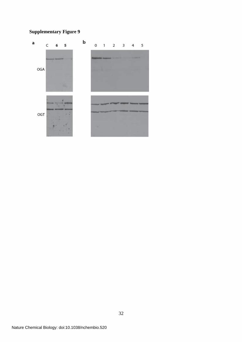
Supplementary Figure 9. Full versions of blots shown in Figure 2; Ac-5SGlcNAc (5) acts
in cells to decrease OGA levels and increase OGT levels in a compensatory manner. (a)
Full western blots of COS-7 cell lysates administered specified agents at 50 μM for 24 h;
vehicle only (C), Ac-GlcNAc (6) or Ac-5SGlcNAc (5). Upper panel, probed with anti-OGA
antibody; lower panel, probed with anti-OGT antibody. (b) Full western blots of COS-7 cell
lysates following Ac-5SGlcNAc (5) administration at 50 μM for different amounts of time.
Upper panel, probed with anti-OGA antibody; lower panel, probed with anti-OGT antibody.
Supplementary Figure 10. Effect of treating CHO cells and other cells lines with Ac-
5SGlcNAc (5). (a) Western blots of CHO cell lysates following Ac-5SGlcNAc (5)
administration at different doses (0-250 μM) for 24 h. Upper panel, probed with anti-O-
GlcNAc antibody (CTD110.6); lower panel, probed with anti-actin antibody. (b)
Densitometry analysis of the western blots shown in panel a, converted to a % O-GlcNAc
modification (corrected for actin levels) relative to the untreated control sample) of as a
function of dose; this gives an EC50 of 0.8 μM. (c) Western blots of CHO cell lysates
following Ac-5SGlcNAc (5) administration at 50 μM for different amounts of time (shown in
hours). Upper panel, probed with anti-O-GlcNAc antibody (CTD110.6); lower panel, probed
with anti-actin antibody. (d) Densitometry analysis of the western blots shown in panel c,
converted to a % O-GlcNAc modification (corrected for actin levels) relative to the untreated
control sample as a function of time. (e) Western blots of cell lysates from different cell lines
following no (C), Ac-GlcNAc (6) or Ac-5SGlcNAc (5) administration at 50 μM (or 100 μM
for PC12 cells) for 24 h. Upper panel, probed with anti-O-GlcNAc antibody (CTD110.6);
lower panel, anti-actin antibody. Cell lines used: COS-7 (African green monkey kidney cell
line), CHO (Chinese hamster ovary cell line), SK-N-SH (human neuroblastoma cell line),
HepG2 (human liver carcinoma cell line), PC12 (rat adrenal medulla pheochromocytoma cell
Nature Chemical Biology: doi:10.1038/nchembio.520
Page 43
43
line, which terminally differentiate upon nerve growth factor treatment), mouse hybridoma
cell line and EMEG32-/- (mouse embryonic fibroblasts deficient in glucosamine-6-phosphate
acetyltransferase (GAT, shown in Figure 1b)).
Supplementary Figure 11. Full versions of blots shown in Figure 3; evaluation of the
effects of Ac-5SGlcNAc treatment of cells on the O-GlcNAc modification state of nup62.
(a) Full western blots of immunoprecipitated nup62 from cell lysates following vehicle (C) or
250 μM Ac-5SGlcNAc (5) treatment for 24 h. Following immunoprecipitation, nup62 was
incubated with UDP-GalNAz in the absence (-) or presence (+) of GalT1 and
chemoselectively labelled. Upper panel, probed with anti-nup62 antibody; lower panel,
probed with streptavidin. (b) Full western blots of immunoprecipitated nup62 from cell
lysates following vehicle (C) or 250 μM Ac-5SGlcNAc (5) treatment for 24 h. Following
immunoprecipitation, nup62 was incubated with buffer (-) or with BtGH84 (+) for 2 h to
remove O-GlcNAc. Upper panel, probed with anti-nup62 antibody; lower panel, probed with
anti-O-GlcNAc antibody (CTD110.6) (long exposure at top and short exposure below). (c)
Full western blots of COS-7 cell lysates following Ac-5SGlcNAc (5) administration at
different doses (0-1000 μM) for 24 h. Upper panel, probed with anti-nup62 antibody; lower
panel, probed with anti-O-GlcNAc antibody (CTD110.6).
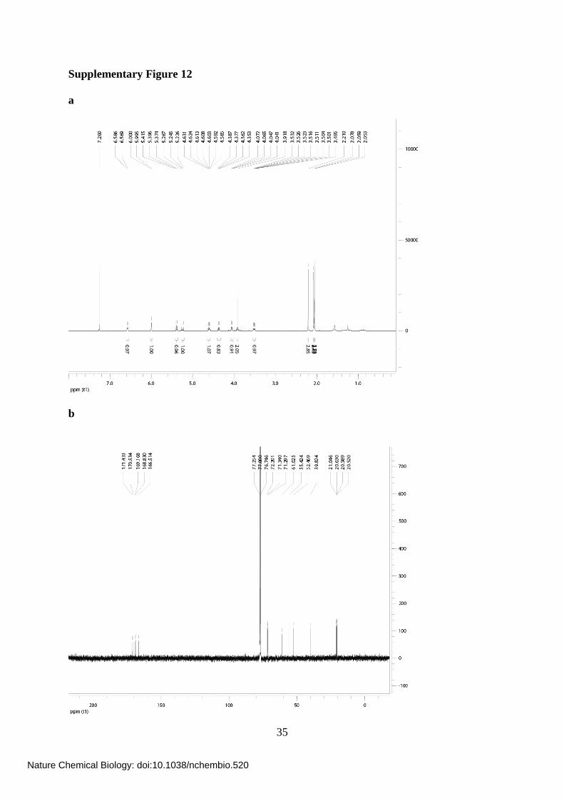
Supplementary Figure 12. NMR spectra of Ac-5SGlcNAz (14). (a) 1H-NMR spectrum of
Ac-5SGlcNAz (14). (b) 13C-NMR spectrum of Ac-5SGlcNAz (14). Both were taken in
CDCl3.
Supplementary Figure 13. Full version of blot shown in Figure 4c; metabolic feeding of
Ac-5SGlcNAz (14) to cells and subsequent chemoselecctive ligation demonstrates there
Nature Chemical Biology: doi:10.1038/nchembio.520
Page 44
44
is no accumulation of 5SGlcNAz (13) on nup62. Full western blot of immunoprecipitated
nup62 from cell lysates following vehicle (C), 50 μM Ac-GlcNAz (15) or Ac-5SGlcNAz (14)
treatment for 24 h. Following immunoprecipitation, nup62 was incubated with buffer (-) or
with BtGH84 (+) for 2 h to remove O-GlcNAc, and then underwent the Staudinger ligation
with biotin phosphine. Blots are probed with streptavidin-HRP.
Supplementary Figure 14. Analysis of nucleotide sugars by capillary electrophoresis
and the effects of treating cells with Ac-5SGlcNAc (5) on nucleotide sugar pools. (a)
UDP-sugar analysis by CE to validate identity of peaks from cells; trace shows absorbance at
254 nm as a function of retention time. Run 1, UDP-5SGlcNAc and UDP-5SGalNAc
standards; run 2, UDP-GlcNAc and UDP-GalNAc standards; run 3, UDP-sugars extracted
from COS-7 cells following treatment with 250 μM Ac-5SGlcNAc for 24 h; run 4, sample
from run 3 spiked with standards from run 1; run 5, sample from run 3 spiked with standards
from run 2. (A) GDP-Glc (internal standard); (B) UDP-GlcNAc (2); (C) UDP-5SGalNAc;
(D) UDP-5SGlcNAc (4); (E) UDP-GalNAc. (b) UDP-5SGlcNAc (4) and UDP-Gal co-eluted
during CE analysis. The contribution from each molecule to the peak could be calculated
based on the concentration of UDP-GlcNAc (2) and UDP-5SGal, and the epimeric ratio
determined from the standards of 2.1:1 GlcNAc:GalNAc. This graph shows a comparison of
the total peak area compared to the sum of the estimated peak areas from the epimeric ratios
for each concentration of Ac-5SGlcNAc (5) administered to cells. (c) Monitoring UDP-
5SGlcNAc (4) and UDP-5SGalNAc in vitro by capillary electrophoresis; trace shows
absorbance at 254 nm as a function of retention time. Run 1, UDP-5SGlcNAc (4); run 2,
UDP-5SGlcNAc (4) treatment with UDP-GlcNAc 4-epimerase; run 3, UDP-GlcNAc (2) and
UDP-GalNAc standards. (A) GDP-Glc (internal standard); (B) UDP-GlcNAc (2); (C) UDP-
5SGalNAc; (D) UDP-5SGlcNAc (4); (E) UDP-GalNAc.
Nature Chemical Biology: doi:10.1038/nchembio.520
Page 45
45
Supplementary Figure 15. Effect of Ac-GlcNAc (5) treatment on cell surface
glycosylation and N-glycosylation of a secreted IgG. (a) Full western blots of COS-7 cell
lysates following Ac-5SGlcNAc (5) administration at different doses (0-1000 μM) for 24 h.
C+ denotes untreated cell lysate incubated with PNGase F and C- denotes untreated cell
lysate incubated with vehicle. Blots are probed with (from top to bottom) anti-O-GlcNAc
antibody (CTD110.6), anti-actin antibody, ConA lectin (recognizes α-D-mannose, α-D-
glucose and branched mannose), GNA lectin (recognizes mannose), PHA-L (recognizes
complex branched chain oligosaccharide structure), SNA lectin (recognizes sialic acid
α(2,6)Gal/GalNAc) and MAA lectin (recognizes sialic acid α(2,3)Gal). (b) Full western blots
of mouse hybridoma cell lysates (O-GlcNAc) and immunoprecipitated mouse hybridoma
antibody (IgG, ConA and GNA) following administration of Ac-5SGlcNAc (5) at different
doses (0-1000 μM) for 24 h. C+, untreated cell lysate incubated with PNGase F; C-, untreated
cell lysate incubated with vehicle. Blots are probed with (from top to bottom) anti-O-GlcNAc
antibody (CTD110.6), anti-IgG antibody, ConA lectin and GNA lectin.
Nature Chemical Biology: doi:10.1038/nchembio.520
Page 46
46
References:
1. Leiting, B., Pryor, K.D., Eveland, S.S. & Anderson, M.S. One-day enzymatic
synthesis and purification of UDP-N-[1-14C]acetyl-glucosamine. Anal Biochem 256,
185-191 (1998).
2. Rabina, J. et al. Analysis of nucleotide sugars from cell lysates by ion-pair solid-phase
extraction and reversed-phase high-performance liquid chromatography. Glycoconj J
18, 799-805 (2001).
3. Chien, R.L. & Burgi, D.S. Field-amplified polarity-switching sample injection in
high-performance capillary electrophoresis. J Chromatogr 559, 153-161 (1991).
4. Martinez-Fleites, C. et al. Structure of an O-GlcNAc transferase homolog provides
insight into intracellular glycosylation. Nat Struct Mol Biol 15, 764-765 (2008).
5. Leatherbarrow, R.J. GraFit Version 5, (Erithacus Software Ltd., Horley, UK, 2001).
6. Tomiya, N., Ailor, E., Lawrence, S.M., Betenbaugh, M.J. & Lee, Y.C. Determination
of nucleotides and sugar nucleotides involved in protein glycosylation by high-
performance anion-exchange chromatography: sugar nucleotide contents in cultured
insect cells and mammalian cells. Anal Biochem 293, 129-137 (2001).
7. Turnock, D.C. & Ferguson, M.A. Sugar nucleotide pools of Trypanosoma brucei,
Trypanosoma cruzi, and Leishmania major. Eukaryot Cell 6, 1450-1463 (2007).
8. Feng, H.T., Wong, N., Wee, S. & Lee, M.M. Simultaneous determination of 19
intracellular nucleotides and nucleotide sugars in Chinese Hamster ovary cells by
capillary electrophoresis. J Chromatogr B Analyt Technol Biomed Life Sci 870, 131-
134 (2008).
9. Hasegawa, A. & Kiso, M. A simple preparation of 2-(acylamino)-2-deoxy-3,4:5,6-di-
O-isopropyladine-aldehydo-D-glucose dimethyl and dibenzylacetal. Carbohydr Res
79, 265-270 (1980).
Nature Chemical Biology: doi:10.1038/nchembio.520
Page 47
47
10. Tanahashi, E., Kiso, T. & Hasegawa, A. A facile synthesis of 2-acetamido-2-deoxy-5-
thio-D-glucopyranose. Carbohydr Res 117, 304-308 (1983).
11. Damager, I., Numao, S., Chen, H., Brayer, G.D. & Withers, S.G. Synthesis and
characterisation of novel chromogenic substrates for human pancreatic alpha-amylase.
Carbohydr Res 339, 1727-1737 (2004).
12. Baudry, M., Barberousse, V., Descotes, G., Pires, J. & Praly, J.P. Synthetic studies in
the 5-thio-D-xylopyranose series part 2: Coupling of 5-thio-D-xylopyranosyl donors
with electron-rich aryl moieties: Access to C-aryl 5-thio-D-xylopyranosides. Tet Lett
54, 7447-7456 (1998).
13. Hashimoto, H. & Izumi, M. Efficient and stereoselective 1,2-cis glycoside formation
of 5-thioaldopyranoses: glycosylation with peracetylated 5-thio-D-arabinopyranosyl
and 5-thio-L-fucopyranosyl trichloroacetimidates. Tet Lett 34, 4949-4952 (1993).
14. Tsuruta, O., Yuasa, H., Hashimoto, H., Kurono, S. & Yazawa, S. Affinity of 5-thio-L-
fucose-containing Lewis X (LeX) trisaccharide analogs to anti-LeX monoclonal
antibody. Bioorg Med Chem Lett 9, 1019-1022 (1999).
15. Yuasa, H., Suga, Y. & Hashimoto, H. Glycosidation reactions of 5-thioxylpyranosyl
Donors. Lett Org Chem 5, 429-431 (2008).
16. Driguez, H. & Henrissat, B. A novel synthesis of 5-thio-D-glucose. Tet Lett 22, 5061-
5062 (1981).
17. Tsuda, Y. et al. Thio Sugars. I. Radical promoted thione-thiol rearrangement of cyclic
thionocarbonates: synthesis of 5-thioglucose. Chem Pharm Bull 44, 1465-1475
(1996).
18. Csuk, R. & Glanzer, B.I. A short synthesis of 2-acetimido-2-deoxy-5-thio-D-glucose
and D-mannose from 5-thio-glucal. J Chem Soc Chem Comm, 343-344 (1986).
Nature Chemical Biology: doi:10.1038/nchembio.520
Page 48
48
19. Taggi, A.E. et al. The development of the first catalyzed reaction of ketenes and
imines: catalytic, asymmetric synthesis of beta-lactams. J Am Chem Soc 124, 6626-
6635 (2002).
Nature Chemical Biology: doi:10.1038/nchembio.520