Total-group tetrapods (Tetrapodomorpha) Dipnomorpha Dipnoi Porolepi- formes Coelacanths Onycho- donts limbed tetrapods Megalichthys Outgroup Gooloogongia Rhizo- dontidae ‘Osteo- lepididae’ Youngolepis Guiyu Psarolepis Achoania Onychodus Strunius Gavinia Miguashaia Diplocercides Latimeria Powichthys Styloichthys Diabolepis Dipterus Dipnorhynchus Glyptolepis Porolepis Kenichthys Tungsenia Barameda Gyroptychius Osteolepis Gogonasus Medoevia Cladarosymblema Beelarongia Canowindra Koharalepis Marsdenichthys Spodichthys Tristichopterus Eusthenopteron Panderichthys Tikttalik Acanthostega Ichthyostega ‘Osteolepiformes’ Cano- windridae Megal- ichthyidae ‘Tristicho- pteridae’ Elpistostegalia Total-group tetrapods (Tetrapodomorpha) Dipnomorpha Dipnoi Porolepi- formes Coelacanths Onycho- donts limbed tetrapods Megalichthys Outgroup Gooloogongia Rhizo- dontidae ‘Osteo- lepididae’ Youngolepis Guiyu Psarolepis Achoania Onychodus Strunius Gavinia Miguashaia Diplocercides Latimeria Powichthys Styloichthys Diabolepis Dipterus Dipnorhynchus Glyptolepis Porolepis Kenichthys Tungsenia Barameda Gyroptychius Osteolepis Gogonasus Medoevia Cladarosymblema Beelarongia Canowindra Koharalepis Marsdenichthys Spodichthys Tristichopterus Eusthenopteron Panderichthys Tikttalik Acanthostega Ichthyostega ‘Osteolepiformes’ Cano- windridae Megal- ichthyidae ‘Tristicho- pteridae’ Elpistostegalia a b Supplementary Informaon of “The earliest known stem-tetrapod from the Lower Devonian of China” Jing Lu 1 , Min Zhu 1 , John A. Long 2 , Wenjin Zhao 1 , Tim J. Senden 3 , Liantao Jia 1 & Tuo Qiao 1 1 Key Laboratory of Evoluonary Systemacs of Vertebrates, Instute of Vertebrate Paleontology and Paleoanthropology, Chinese Academy of Sciences, P.O Box 643, Beijing 100044; 2 Natural History Museum of Los Angeles County, Los Angeles, California 90007; 3 Department of Applied Mathemacs, Research School of Physics and Engineering, The Australian Naonal University, ACT 0200, Australia. I. Supplementary Figures Supplementary Figure S1. Phylogenec placement of Tungsenia shown in the strict and 50% majority-rule consensus trees. (a) The strict consensus tree of the 5 shortest trees. (b) The 50% majority-rule consensus tree of the 5 shortest trees. 1
Supplementary Information of “The earliest known stem-tetrapod
from the Lower Devonian of China”
Jing Lu1, Min Zhu1, John A. Long2, Wenjin Zhao1, Tim J. Senden3, Liantao Jia1 & Tuo Qiao1
1Key Laboratory of Evolutionary Systematics of Vertebrates, Institute of Vertebrate Paleontology and Paleoanthropology, Chinese Academy of Sciences, P.O Box 643, Beijing 100044; 2Natural History Museum of Los Angeles County, Los Angeles, California 90007; 3Department of Applied Mathematics, Research School of Physics and Engineering, The Australian National University, ACT 0200, Australia. I. Supplementary Figures
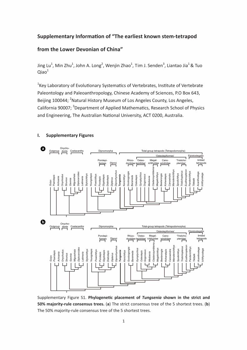
Supplementary Figure S1. Phylogenetic placement of Tungsenia shown in the strict and 50% majority-rule consensus trees. (a) The strict consensus tree of the 5 shortest trees. (b) The 50% majority-rule consensus tree of the 5 shortest trees.
1
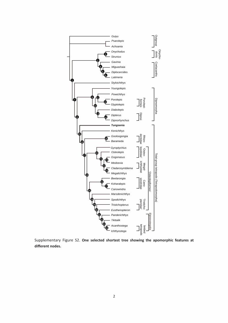
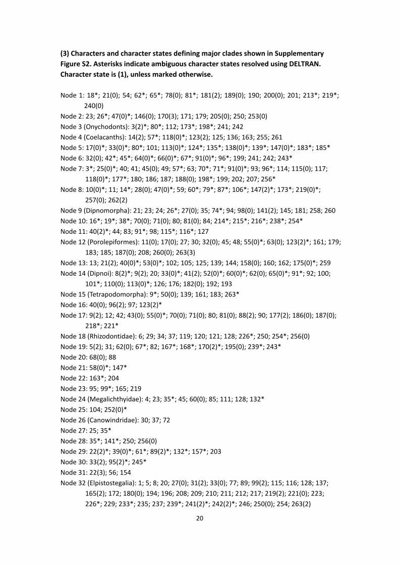
Supplementary Figure S2. One selected shortest tree showing the apomorphic features at different nodes.
2
Total-group tetrapods (Tetrapodomorpha)
Dipnom
orphaDipnoi
Porolepi-form
es
Coelacanths
Onycho-donts
limbed
tetrapods
Megalichthys
Outgroup
Gooloogongia
Rhizo-
dontidae‘O
steo-lepididae’
Youngolepis
Guiyu
Psarolepis
Achoania
Onychodus
Strunius
Gavinia
Miguashaia
Diplocercides
Latimeria
Powichthys
Styloichthys
Diabolepis
Dipterus
Dipnorhynchus
Glyptolepis
Porolepis
Kenichthys
Tungsenia
Barameda
GyroptychiusOsteolepis
Gogonasus
Medoevia
Cladarosymblema
Beelarongia
Canowindra
Koharalepis
Marsdenichthys
Spodichthys
Tristichopterus
Eusthenopteron
Panderichthys
Tikttalik
Acanthostega
Ichthyostega
‘Osteolepiform
es’
Cano-
windridae
Megal-
ichthyidae‘Tristicho-pteridae’
Elpistostegalia
1
9
2
8
15
7
16
17
19
25
22
20
21
18
23
24
2627
28
29
30
31
32
3334
3
4
56
10
1112
1314
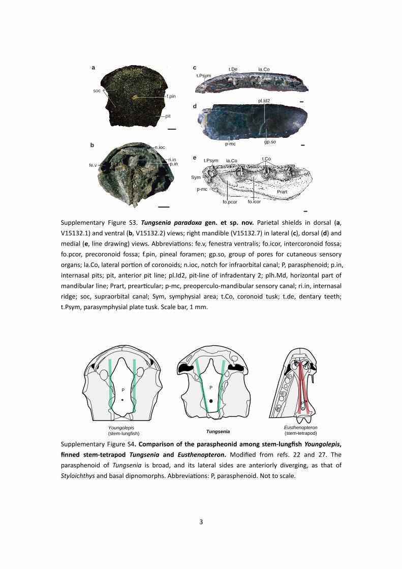
Supplementary Figure S3. Tungsenia paradoxa gen. et sp. nov. Parietal shields in dorsal (a, V15132.1) and ventral (b, V15132.2) views; right mandible (V15132.7) in lateral (c), dorsal (d) and medial (e, line drawing) views. Abbreviations: fe.v, fenestra ventralis; fo.icor, intercoronoid fossa; fo.pcor, precoronoid fossa; f.pin, pineal foramen; gp.so, group of pores for cutaneous sensory organs; la.Co, lateral portion of coronoids; n.ioc, notch for infraorbital canal; P, parasphenoid; p.in, internasal pits; pit, anterior pit line; pl.Id2, pit-line of infradentary 2; plh.Md, horizontal part of mandibular line; Prart, prearticular; p-mc, preoperculo-mandibular sensory canal; ri.in, internasal ridge; soc, supraorbital canal; Sym, symphysial area; t.Co, coronoid tusk; t.de, dentary teeth; t.Psym, parasymphysial plate tusk. Scale bar, 1 mm.
Supplementary Figure S4. Comparison of the paraspheonid among stem-lungfish Youngolepis, finned stem-tetrapod Tungsenia and Eusthenopteron. Modified from refs. 22 and 27. The parasphenoid of Tungsenia is broad, and its lateral sides are anteriorly diverging, as that of Styloichthys and basal dipnomorphs. Abbreviations: P, parasphenoid. Not to scale.
3
n.ioc
f.pin
pit
soc
ri.in
P
p.infe.vt.Cola.Co
la.Cot.Psym
t.Psym
t.De
gp.so
pl.Id2
p-mc
Prartp-mc
Sym
ca
b
d
e
fo.icorfo.pcor
Youngolepis(stem-lungfish) Tungsenia
Eusthenopteron(stem-tetrapod)
P P
P
cav.nc cav.nc
socolf
soc
cv
re.papre.pin
cc.II
c.sup.oph
gr.r.pal
cv
fo.hy c.a.ci+r.pal
c.v.pitioc
soc
c.a.ci+r.pal
a b
d
fe
c
a b c
d e
jucMx
Lap.SQP
p.SQP
SqSq
Sq+Qj+Pop
QjQj
JuJu Ju
JuJu
Po Po
PoPo
Po
Pop
pl.Sqpl.Qj
Sq+Qj+Pop
Pop
p.SQP
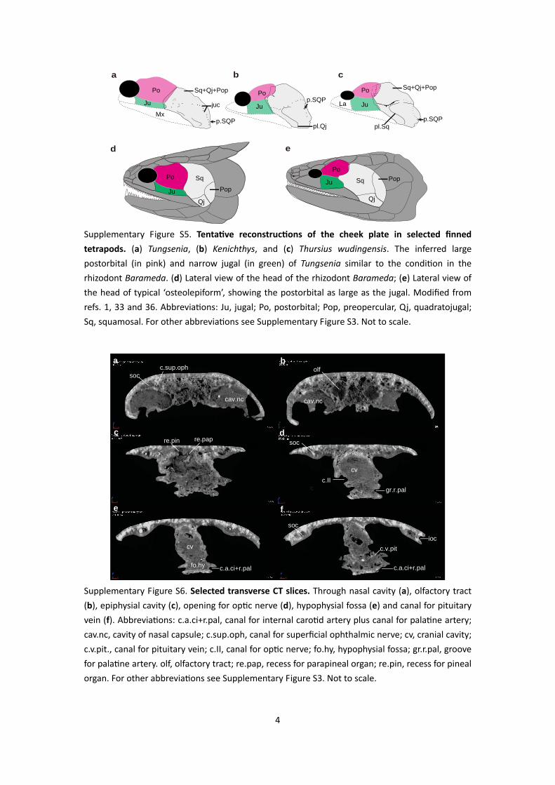
Supplementary Figure S5. Tentative reconstructions of the cheek plate in selected finned tetrapods. (a) Tungsenia, (b) Kenichthys, and (c) Thursius wudingensis. The inferred large postorbital (in pink) and narrow jugal (in green) of Tungsenia similar to the condition in the rhizodont Barameda. (d) Lateral view of the head of the rhizodont Barameda; (e) Lateral view of the head of typical ‘osteolepiform’, showing the postorbital as large as the jugal. Modified from refs. 1, 33 and 36. Abbreviations: Ju, jugal; Po, postorbital; Pop, preopercular, Qj, quadratojugal; Sq, squamosal. For other abbreviations see Supplementary Figure S3. Not to scale.
Supplementary Figure S6. Selected transverse CT slices. Through nasal cavity (a), olfactory tract (b), epiphysial cavity (c), opening for optic nerve (d), hypophysial fossa (e) and canal for pituitary vein (f). Abbreviations: c.a.ci+r.pal, canal for internal carotid artery plus canal for palatine artery; cav.nc, cavity of nasal capsule; c.sup.oph, canal for superficial ophthalmic nerve; cv, cranial cavity; c.v.pit., canal for pituitary vein; c.II, canal for optic nerve; fo.hy, hypophysial fossa; gr.r.pal, groove for palatine artery. olf, olfactory tract; re.pap, recess for parapineal organ; re.pin, recess for pineal organ. For other abbreviations see Supplementary Figure S3. Not to scale.
4
c.II
c.hyp
?PT
cv
fo.hy
c.II
?PT c.hyp
cv cv
fo.hyc.hyp fo.hy
fo.hy
c.hyp
ca
f
PTad.hy
PT
ad.hy
‘PT’
‘PT’
c.II
b d f
e
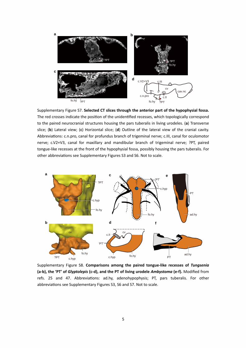
Supplementary Figure S7. Selected CT slices through the anterior part of the hypophysial fossa. The red crosses indicate the position of the unidentified recesses, which topologically correspond to the paired neurocranial structures housing the pars tuberalis in living urodeles. (a) Transverse slice; (b) Lateral view; (c) Horizontal slice; (d) Outline of the lateral view of the cranial cavity. Abbreviations: c.n.pro, canal for profundus branch of trigeminal nerve; c.III, canal for oculomotor nerve; c.V2+V3, canal for maxillary and mandibular branch of trigeminal nerve; ?PT, paired tongue-like recesses at the front of the hypophysial fossa, possibly housing the pars tuberalis. For other abbreviations see Supplementary Figures S3 and S6. Not to scale.
Supplementary Figure S8. Comparisons among the paired tongue-like recesses of Tungsenia (a-b), the ‘PT’ of Glyptolepis (c-d), and the PT of living urodele Ambystoma (e-f). Modified from refs. 25 and 47. Abbreviations: ad.hy, adenohypophysis; PT, pars tuberalis. For other abbreviations see Supplementary Figures S3, S6 and S7. Not to scale.
5
?PTfo.hy?PT
fo.hy ?PT
c.V2+V3
c.II
c.III
c.n.procav.nc
cv
ba
c
d
?PTfo.hy
cv
olf
mes
olf olfolfolf
mes
mesmes
Tungsenia Youngolepis(stem-lungfish)
Eusthenopteron(stem-tetrapod)
Dicksonosteus(placoderm)
Xenacanthus(elasmobranch)
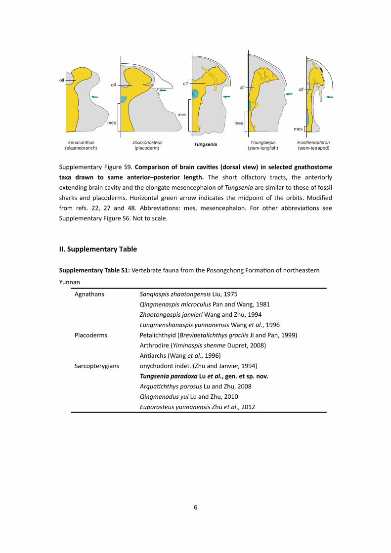
Supplementary Figure S9. Comparison of brain cavities (dorsal view) in selected gnathostome taxa drawn to same anterior–posterior length. The short olfactory tracts, the anteriorly extending brain cavity and the elongate mesencephalon of Tungsenia are similar to those of fossil sharks and placoderms. Horizontal green arrow indicates the midpoint of the orbits. Modified from refs. 22, 27 and 48. Abbreviations: mes, mesencephalon. For other abbreviations see Supplementary Figure S6. Not to scale. II. Supplementary Table Supplementary Table S1: Vertebrate fauna from the Posongchong Formation of northeastern
Yunnan
Agnathans Sanqiaspis zhaotongensis Liu, 1975 Qingmenaspis microculus Pan and Wang, 1981
Zhaotongaspis janvieri Wang and Zhu, 1994 Lungmenshanaspis yunnanensis Wang et al., 1996
Placoderms Petalichthyid (Brevipetalichthys gracilis Ji and Pan, 1999) Arthrodire (Yiminaspis shenme Dupret, 2008)
Antiarchs (Wang et al., 1996) Sarcopterygians onychodont indet. (Zhu and Janvier, 1994)
Tungsenia paradoxa Lu et al., gen. et sp. nov. Arquatichthys porosus Lu and Zhu, 2008 Qingmenodus yui Lu and Zhu, 2010 Euporosteus yunnanensis Zhu et al., 2012
6
7
III. Supplementary Methods (1) Character list Characters are based on refs. 1, 4, 8, 39, 49‐58. 1. Position of orbits: lateral and widely separated (0); dorsal and close together (1). 2. Proportion of skull roof (measured as length from tip of snout to posterior margin of
postparietals) lying anterior to middle of orbits: <50% (0); ≥50% (1). 3. Parietal portion of skull roof relative to postparietal portion in length: parietal portion
roughly as long as postparietal portion (0), parietal portion much longer postparietal portion (1), parietal portion much shorter than postparietal portion (2).
4. Orientation of anterior tectal and lateral rostral relative to (anterior) external nostril: dorsal/ventral (0); posterior/anterior (1).
5. Postrostral: absent (0); postrostral mosaic of small variable bones (1); large median postrostral, with or without accessory bones (2); paired E bones (3).
6. Number of nasals: many (0); one or two (1). 7. Paired nasals meet in midline of skull: absent (0); present (1). 8. Position of anterior external nostril: facial (0); edge of mouth (or marginal) (1); palatal
(2). 9. Position of posterior nostril: external, far from jaw margin (0); external, close to jaw
margin (1); palatal (2). 10. Posterior nostril enclosed posteriorly by preorbital or preorbital process of premaxilla:
sensu stricto): absent (0); present (1). 13. B‐bone: absent (0); present (1). 14. Number of supraorbitals: one (0); two (1); more than two (2). 15. Relative size of anterior (or anteriormost) supraorbital (prefrontal, posterior tectal of
Jarvik) and posterior (or posteriormost) supraorbital (postfrontal): similar (0); prefrontal much bigger (1).
16. Contact between parietal and supraorbital: present (0); absent (1). 17. Dermosphenotic: absent (0); present (1). 18. Intertemporal: present (0); absent (1). 19. Number of tectals: one (0); two or more (1). 20. Paired bones anterior to parietals: absent (0); present (1). 21. Anterior margin of parietal: between or in front of orbits (0); slightly posterior to orbits
(1); much posterior to orbits (2). 22. Width of ethmoid relative to length from snout tip to posterior margin of parietals: more
than 80% (0); 70 – 79% (1); 50 – 69% (2); less than 50% (3). 23. Pineal foramen: present (0); absent (1). 24. Pineal eminence (in those taxa with no pineal foramen): absent (0); present (1). 25. Location of pineal foramen/eminence: level with, or anterior to posterior margin of
orbits (0); well posterior to orbits (1). 26. Parietals surround pineal foramen/eminence: yes (0); no (1).
8
27. Dermal intracranial joint: absent (0); present (1). 28. Margins of postparietals and parietals participating in dermal intracranial joint: smooth
(0); jagged (1) 29. Postparietals narrow to a point posteriorly: no (0); yes (1). 30. Proportions of postparietal shield: not very wide posteriorly (0); very wide posteriorly
(1). 31. Spiracular notch: absent‐small (0); narrow groove (1); wide notch (2). 32. Number of marginal bones alongside postparietal: single (0); two or more (1). 33. Extratemporal: absent (0); present lateral to tabular (1); present, separating lateral
extrascapular from squamosal (‘postspiracular’ condition) (2). 34. Contact between extratemporal and supratemporal: absent (0); present (1). 35. Posterior margin of tabulars: anterior to posterior margin of postparietals (0); level with
posterior margin of postparietals (1). 36. Extrascapular bones: present (0); absent (1). 37. Anterior margin of median extrascapular: long (0); very short (1). 38. Median extrascapular overlap: median extrascapular overlapped by lateral extrascapulars
(0); median extrascapular overlapping lateral extrascapulars (1); median extrascapular abutting lateral extrascapulars (2).
39. Internasal septum: narrow (0); broad (1). 40. Internasal pits: undifferentiated or anterior palatal fossa (0); shallow paired pits with
strong midline ridge (1); deep pear‐shaped pits (2); absent (3). 41. Fenestra ventralis: absent (0); present (1); common ventral fenestra for anterior and
posterior nostrils (2). 42. Postorbital process on braincase: present (0); absent (1). 43. Tectum orbitale: narrow (0); extensive (1). 44. Large median opening and several small dorsolateral openings in postnasal wall: absent
(0); present (1). 45. Ethmoid articulation for palatoquadrate: placed on postnasal wall (0); majority of facet
located anterior to postnasal wall (1); extends posteriorly to the level of N.II (2). 46. Eyestalk or unfinished area for similar structure: absent (0); present (1). 47. Postorbital pillar: absent (0); present (1). 48. Autopalatine fossa bearing unfinished articular surfaces: absent (0); present (1). 49. Basipterygoid process: small knob‐like process (0); developed as a broad platform (1). 50. Position of exit of pituitary vein: in front of basipterygoid process (0); dorsal to vertical
portion of basipterygoid process (1). 51. Descending process of sphenoid (with its posterior extremity lacking periostegeal lining):
anterior to dorsal (1); dorsal anterior to ventral (2). 59. Hyomandibular facets in those taxa where they straddle the level of the jugular canal:
narrowly separated or confluent (0), broadly separated (1). 60. Vestibular fontanelles: absent (0); present (1). 61. Anterior margin of vestibular fontanelle anterior to posterior margin of basicranial
fenestra: no (0); yes (1). 62. Accessory fenestration in otic capsule: absent (0); present (1). 63. Parachordal plates: separated from the otic capsule by cartilage or a persistent fissure (0);
sutured or co‐mineralised with the otic capsule (1). 64. Otoccipital fissure: absent (0); present (1). 65. Basicranial fenestra: absent (0); present (1). 66. Supraotic cavity proper: absent (0); present (1). 67. Posttemporal fossae: absent (0); present (1). 68. Extent of crista parotica: does not reach posterior margin of tabular (0); reaches
posterior margin of tabular (1). 69. Otical process (an outgrowth from the lateral wall of the braincase penetrated by the
branches of the r. oticus lateralis): absent (0); present (1). 70. Foramina (similar to infradentary foramina) on cheek bones: absent (0); present (1). 71. Quadratojugal, squamosal and preopercular fused: no (0); yes (1). 72. Postorbital bone: contributes to orbital margin (0); excluded from orbital margin (1). 73. Jugal extends anterior to middle of orbit: no (0); yes (1). 74. Lacrimal posteriorly enclosing posterior nostril: absent (0); present (1). 75. Lacrimal excluded from orbit: no (0); yes (1). 76. Contact between postorbital and lacrimal: absent (0); present (1). 77. Jugal‐quadratojugal contact: absent (0); present (1). 78. Most posterior major bone of cheek bearing preopercular canal ('preopercular')
extending forward, close to orbit: absent (0); present (1). 79. Contact between most posterior major bone of cheek bearing preopercular canal
('preopercular') and maxilla: present (0); absent (1). 80. Number of cheek bones bearing preopercular canal posterior to jugal: one (0); two (1). 81. Bone bearing both quadratojugal pit‐line and preopercular canal: absent (0); present (1). 82. Verticle bar‐like preopercular: absent (0); present (1). 83. Subsquamosals: absent (0); present (1). 84. Preoperculosubmandibular: absent (0); present (1). 85. Vomer proportions: not much broader than long (0); much broader than long (1). 86. Vomeral area with grooves and raised areas: absent (0); present (1). 87. Vomerine fangs: absent (0); present (1). 88. Anteromedial process of vomer: absent, vomers separated (0); present (1); absent,
vomers in close contact (2). 89. Posterior process of vomers: absent (0); short (1); long (2).
10
90. Articulation of vomers: vomers not articulating with each other (0); vomers articulating with each other (1).
91. Parasphenoid: protruding forward into ethmoid region of endocranium (0); behind ethmoid region (1).
92. Position of parasphenoid: beneath sphenethmoid part of endocranium (0); beneath sphenethmoid and otico‐occipital part (1).
93. Parasphenoid denticle field: terminates at or anterior to level of foramina for internal carotid arteries (0); extends posterior to foramina for internal carotid arteries (1).
94. Denticulated field of parasphenoid: without spiracular groove (0); with spiracular groove (1).
95. Relationship of vomer to parasphenoid: no contact, or simple abutment (0); dorsal/ventral overlap anteriorly; side to side overlap between posterior processes of vomers and lateral sides of parasphenoid (1).
97. Lateral sides of parasphenoid: parallel or slightly diverging anteriorly (0); converging anteriorly (1). (New character, illustrated in Supplementary Figure S4)
98. Posterior carotid opening in parasphenoid: large (0); small (1); absent (2). 99. Shape of spiracular chamber: very large, steeply inclined anteriorly (0); acutely inclined
anteriorly (1); horizontal (2). 100. Articulation of pterygoids: pterygoids not articulating with each other (0); pterygoids
articulating with each other (1). 101. Posterior margin of palatoquadrate: sloping forward (0); erect or sloping backward (1). 102. Dentition of palatoquadrate complex: marginal teeth (0); tooth plates (1). 103. Number of fang pairs on ectopterygoid: one (0); two (1); none (2). 104. Proportions of entopterygoid: anterior end level with processus ascendens (0); anterior
end considerably anterior to processus ascendens (1). 105. Dental plate: denticles on entopterygoid or naked bone (0); tooth plate on
entopterygoid (1); dentine plate on entopterygoid (2). 106. Number of dermopalatines: multiple (0); single (1). 107. Palatal fangs mesial to marginal tooth row: yes (0); no (1). 108. Tooth‐bearing median rostral: absent (0), present (1). 109. Subterminal mouth: absent (0); present (1). 110. Premaxilla: absent (0); present (1). 111. Enlarged anterior tooth on premaxilla: absent (0); present (1). 112. Premaxillae with inturned symphysial processes: absent (0); present (1). 113. Maxilla: absent (0); present (1). 114. Posterior expansion of maxilla (maxilla cleaver‐shaped): present (0); absent (1). 115. Posterodorsal process of maxilla: present (0); very weak or absent (1). 116. Posteriorly deep maxilla: present (0); absent (1). 117. Contribution by maxilla to posterior margin of cheek: present (0); absent (1). 118. Strong ascending flexion of symphysial region of mandible: absent (0); present (1). 119. Anterior projection of the dentary forming a dorsal border for the anteromesial rugosity
and symphysial plate: absent (0); present (1).
11
120. Protrusion of lower jaw in front of skull: absent (0); present (1). 121. Anteroventral lower jaw profile: rounded (0); squared off (1). 122. Large ventromesially directed flange of symphysial region of mandible: absent (0);
present (1). 123. Teeth of outer dental arcade: several rows of disorganized teeth (0); two rows, with large
teeth lingually and small teeth labially (1); single row of teeth (2). 124. Flange‐like extension composed of Meckelian ossification and prearticular that extends
below ventral margin of infradentaries: absent (0), present (1). 125. Length of dentary: long (0); short with lip fold (1). 126. Marginal teeth on dentary: present (0); absent (1). 127. Teeth of dentary: reaching anterior end of dentary (0); not reaching anterior end (1). 128. Dentary fang pair: absent (0); present (1). 129. Accessory tooth rows on dentary: present (0); absent (1). 130. Dermal ornament on each infradentary: even (0); separate “starburst” on each bone (1). 131. Infradentary foramina: always present (0); variable (1); always absent (2). 132. Splenial: not sutured to prearticular (0); sutured to prearticular (1). 133. Mesial lamina of splenial: absent (0); present (1). 134. Suture between splenial and anterior coronoid: absent (0); present (1). 135. Postsplenial: present (0); absent (1). 136. Raised crest on angular: absent (0); present (1). 137. Angular and prearticular: separated by ventral exposure of Meckelian bone (0); in
contact (1). 138. Surangular: absent (0); present (1). 139. Parasymphysial tooth whorl: present (0), absent (1). 140. Fang pair on parasymphysial dental plate: absent (0); present (1). 141. Parasymphysial dental plate (= foremost coronoid of stem sarcopterygians): long with
posterior corner, sutured to anterior coronoid, denticulated or with tooth row (0); short, rounded, not sutured to anterior coronoid, denticulated (1); absent (2).
142. Tooth row on parasymphysial dental plate: absent (0); present (1). 143. Mesial parasymphysial foramen: absent (0); present (1). 144. Coronoids (sensu stricto, excluding parasymphysial dental plate or foremost bone of the
coronoid series): present (0); absent (1). 145. Number of coronoids (sensu lato, including parasymphysial dental plate but
parasymphysial tooth whorl): more than three (0); three (1). 146. Fangs of coronoids (sensu stricto): absent (0); present (1). 147. Marginal denticle band on coronoids: broad band, at least posteriorly (0); narrow band
with 2‐4 denticle rows (1); single tooth row or absent (2). 148. Coronoid fangs larger than marginal teeth: yes (0); no (1). 149. Fangs on anterior coronoid recognisable because noticeably mesial to vertical lamina
and marginal tooth row: (0) yes; (1) no. 150. Fangs on anterior coronoid recognisable because much bigger than marginal teeth: yes
(0); no (1). 151. Fangs on middle coronoid recognisable because noticeably mesial to vertical lamina and
marginal tooth row: yes (0); no (1).
12
152. Fangs on middle coronoid recognisable because much bigger than marginal teeth: yes (0); no (1).
153. Fangs on posterior coronoid recognisable because much bigger than marginal: yes (0); no (1).
154. Coronoid proportions: posterior coronoid similar in length to, or shorter than middle coronoid (0); posterior coronoid significantly longer than middle coronoid (1).
155. Number of fang pairs on posterior coronoid: one (0); two (1); none (2). 156. Anterior end of prearticular: not forked (0); forked (1). 157. Prearticular: rear part with conspicuous horizontal ledge (0); rear part flat (1). 158. Prearticular‐dentary contact: present (0); absent (1). 159. Longitudinal dorsal ridge on prearticular: absent (0); present (1). 160. Teeth radial rows on prearticular: absent (0); present (1). 161. Anterior mandibular (precoronoid) fossa: absent (0); present (1). 162. Labial pit: absent (0); present (1). 163. Retroarticular process: absent (0); present (1). 164. Meckelian bone exposed immediately anterior to first coronoid: yes (0); no (1). 165. Hyomandibula orientation: posteroventral, distally terminating near jaw joint (0); almost
horizontal orientation, opercular process high up dorsally (1); very short, laterally directed (2).
166. Foramen in hyomandibular: absent (0); present (1). 167. ‘Urohyal’ morphology: dorsoventrally compressed and rod‐like, may bifurcate posteriorly
(0); vertical plate (1). 168. Sublingual rod: absent (0); present (1). 169. Opercular: present (0); absent (1). 170. Number of branchiostegal rays per side: ten or more (0); two to seven (1); one (2); none
(3). 171. Median gular: present (0); absent (1). 172. Size of median gular: small (0); large (1). 173. Submandibulars: absent (0); present (1). 174. Width of submandibulars: narrow (0); broad (1). 175. Pore clusters: absent (0); present (1). 176. Ethmoid commissure: present (0); absent (1). 177. Course of ethmoid commissure: middle portion through median rostral (0); sutural
course (1); through bone center of premaxilla (2). 178. Direction of anterior pit‐line on parietal shield: antero‐posterior, reaching or almost
reaching parietal‐postparietal border (0); essentially transverse, anterior or almost anterior to pineal foramen/eminence (1).
179. Middle and posterior pit‐lines on postparietal: posteriorly situated (0), mesially situated (1).
180. Course of supraorbital canal: straight (0); lyre‐shaped (1). 181. Posterior end of supraorbital canal: in postparietal (0); in parietal (1); in intertemporal
(2). 182. Contact of supraorbital and infraorbital canals: in contact rostrally (0); not in contact
rostrally (1).
13
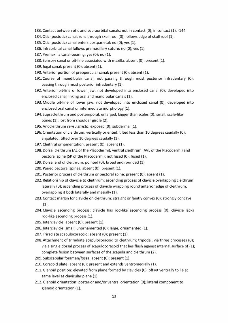
183. Contact between otic and supraorbital canals: not in contact (0); in contact (1). ‐144 184. Otic (postotic) canal: runs through skull roof (0); follows edge of skull roof (1). 185. Otic (postotic) canal enters postparietal: no (0); yes (1). 186. Infraorbital canal follows premaxillary suture: no (0); yes (1). 187. Premaxilla canal‐bearing: yes (0); no (1). 188. Sensory canal or pit‐line associated with maxilla: absent (0); present (1). 189. Jugal canal: present (0); absent (1). 190. Anterior portion of preopercular canal: present (0); absent (1). 191. Course of mandibular canal: not passing through most posterior infradentary (0);
passing through most posterior infradentary (1). 192. Anterior pit‐line of lower jaw: not developed into enclosed canal (0); developed into
enclosed canal linking oral and mandibular canals (1). 193. Middle pit‐line of lower jaw: not developed into enclosed canal (0); developed into
enclosed oral canal or intermediate morphology (1). 194. Supracleithrum and postemporal: enlarged, bigger than scales (0); small, scale‐like
bones (1); lost from shoulder girdle (2). 195. Anocleithrum sensu stricto: exposed (0); subdermal (1). 196. Orientation of cleithrum: vertically oriented: tilted less than 10 degrees caudally (0);
angulated: tilted over 10 degrees caudally (1). 197. Cleithral ornamentation: present (0); absent (1). 198. Dorsal cleithrum (AL of the Placodermi), ventral cleithrum (AVL of the Placodermi) and
pectoral spine (SP of the Placodermi): not fused (0); fused (1). 199. Dorsal end of cleithrum: pointed (0); broad and rounded (1). 200. Paired pectoral spines: absent (0); present (1). 201. Posterior process of cleithrum or pectoral spine: present (0); absent (1). 202. Relationship of clavicle to cleithrum: ascending process of clavicle overlapping cleithrum
laterally (0); ascending process of clavicle wrapping round anterior edge of cleithrum, overlapping it both laterally and mesially (1).
203. Contact margin for clavicle on cleithrum: straight or faintly convex (0); strongly concave (1).
204. Clavicle ascending process: clavicle has rod‐like ascending process (0); clavicle lacks rod‐like ascending process (1).
205. Interclavicle: absent (0); present (1). 206. Interclavicle: small, unornamented (0); large, ornamented (1). 207. Triradiate scapulocoracoid: absent (0); present (1). 208. Attachment of triradiate scapulocoracoid to cleithrum: tripodal, via three processes (0);
via a single dorsal process of scapulocoracoid that lies flush against internal surface of (1); complete fusion between surfaces of the scapula and cleithrum (2).
209. Subscapular foramen/fossa: absent (0); present (1). 210. Coracoid plate: absent (0); present and extends ventromedially (1). 211. Glenoid position: elevated from plane formed by clavicles (0); offset ventrally to lie at
same level as clavicular plane (1). 212. Glenoid orientation: posterior and/or ventral orientation (0); lateral component to
glenoid orientation (1).
14
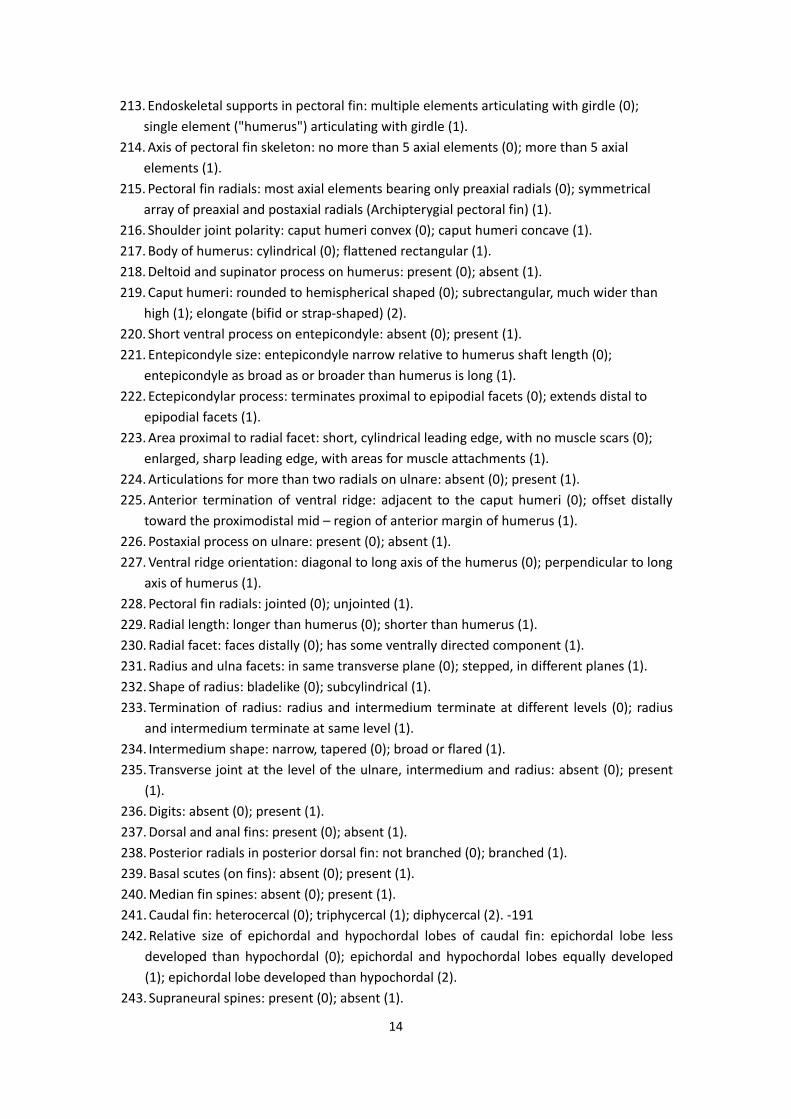
213. Endoskeletal supports in pectoral fin: multiple elements articulating with girdle (0); single element ("humerus") articulating with girdle (1).
214. Axis of pectoral fin skeleton: no more than 5 axial elements (0); more than 5 axial elements (1).
215. Pectoral fin radials: most axial elements bearing only preaxial radials (0); symmetrical array of preaxial and postaxial radials (Archipterygial pectoral fin) (1).
216. Shoulder joint polarity: caput humeri convex (0); caput humeri concave (1). 217. Body of humerus: cylindrical (0); flattened rectangular (1). 218. Deltoid and supinator process on humerus: present (0); absent (1). 219. Caput humeri: rounded to hemispherical shaped (0); subrectangular, much wider than
high (1); elongate (bifid or strap‐shaped) (2). 220. Short ventral process on entepicondyle: absent (0); present (1). 221. Entepicondyle size: entepicondyle narrow relative to humerus shaft length (0);
entepicondyle as broad as or broader than humerus is long (1). 222. Ectepicondylar process: terminates proximal to epipodial facets (0); extends distal to
epipodial facets (1). 223. Area proximal to radial facet: short, cylindrical leading edge, with no muscle scars (0);
enlarged, sharp leading edge, with areas for muscle attachments (1). 224. Articulations for more than two radials on ulnare: absent (0); present (1). 225. Anterior termination of ventral ridge: adjacent to the caput humeri (0); offset distally
toward the proximodistal mid – region of anterior margin of humerus (1). 226. Postaxial process on ulnare: present (0); absent (1). 227. Ventral ridge orientation: diagonal to long axis of the humerus (0); perpendicular to long
axis of humerus (1). 228. Pectoral fin radials: jointed (0); unjointed (1). 229. Radial length: longer than humerus (0); shorter than humerus (1). 230. Radial facet: faces distally (0); has some ventrally directed component (1). 231. Radius and ulna facets: in same transverse plane (0); stepped, in different planes (1). 232. Shape of radius: bladelike (0); subcylindrical (1). 233. Termination of radius: radius and intermedium terminate at different levels (0); radius
and intermedium terminate at same level (1). 234. Intermedium shape: narrow, tapered (0); broad or flared (1). 235. Transverse joint at the level of the ulnare, intermedium and radius: absent (0); present
(1). 236. Digits: absent (0); present (1). 237. Dorsal and anal fins: present (0); absent (1). 238. Posterior radials in posterior dorsal fin: not branched (0); branched (1). 239. Basal scutes (on fins): absent (0); present (1). 240. Median fin spines: absent (0); present (1). 241. Caudal fin: heterocercal (0); triphycercal (1); diphycercal (2). ‐191 242. Relative size of epichordal and hypochordal lobes of caudal fin: epichordal lobe less
developed than hypochordal (0); epichordal and hypochordal lobes equally developed (1); epichordal lobe developed than hypochordal (2).
243. Supraneural spines: present (0); absent (1).
15
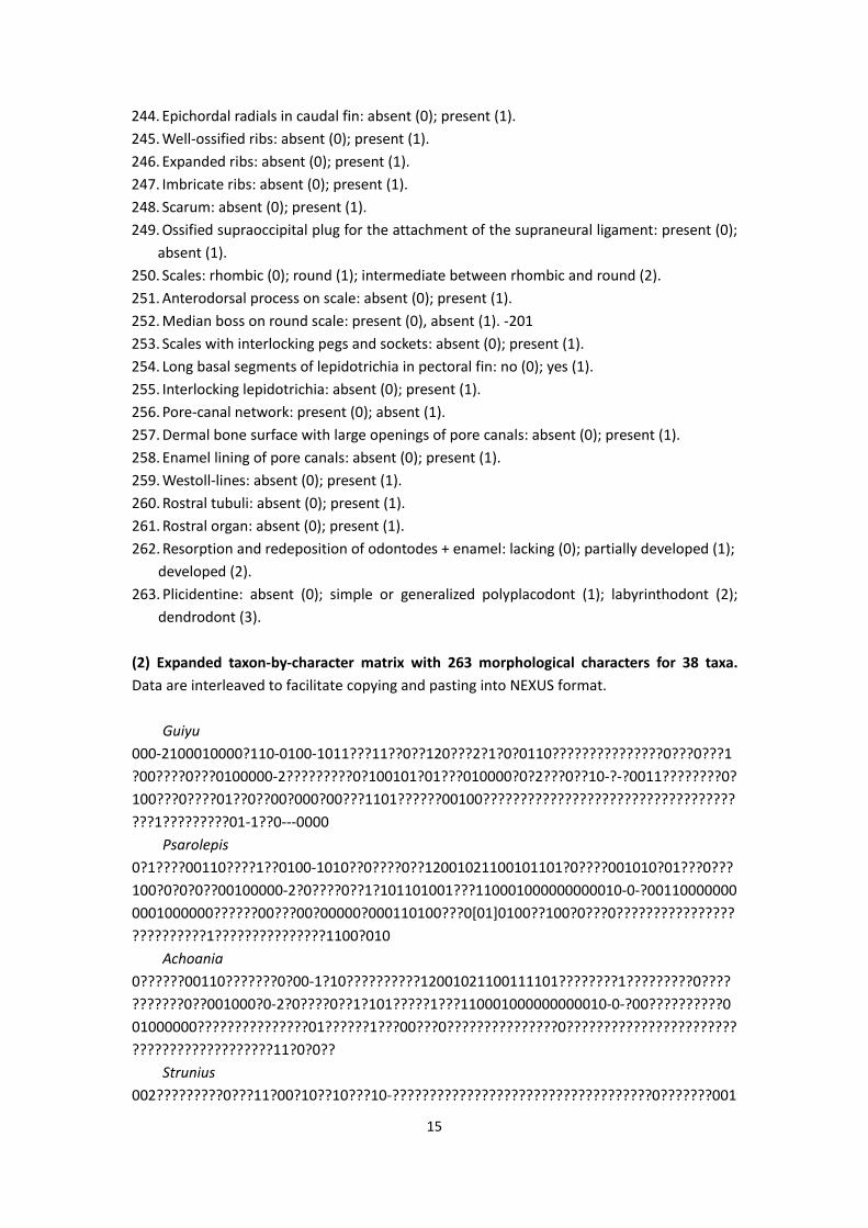
244. Epichordal radials in caudal fin: absent (0); present (1). 245. Well‐ossified ribs: absent (0); present (1). 246. Expanded ribs: absent (0); present (1). 247. Imbricate ribs: absent (0); present (1). 248. Scarum: absent (0); present (1). 249. Ossified supraoccipital plug for the attachment of the supraneural ligament: present (0);
absent (1). 250. Scales: rhombic (0); round (1); intermediate between rhombic and round (2). 251. Anterodorsal process on scale: absent (0); present (1). 252. Median boss on round scale: present (0), absent (1). ‐201 253. Scales with interlocking pegs and sockets: absent (0); present (1). 254. Long basal segments of lepidotrichia in pectoral fin: no (0); yes (1). 255. Interlocking lepidotrichia: absent (0); present (1). 256. Pore‐canal network: present (0); absent (1). 257. Dermal bone surface with large openings of pore canals: absent (0); present (1). 258. Enamel lining of pore canals: absent (0); present (1). 259. Westoll‐lines: absent (0); present (1). 260. Rostral tubuli: absent (0); present (1). 261. Rostral organ: absent (0); present (1). 262. Resorption and redeposition of odontodes + enamel: lacking (0); partially developed (1);
developed (2). 263. Plicidentine: absent (0); simple or generalized polyplacodont (1); labyrinthodont (2);
dendrodont (3). (2) Expanded taxon‐by‐character matrix with 263 morphological characters for 38 taxa. Data are interleaved to facilitate copying and pasting into NEXUS format. Guiyu 000‐2100010000?110‐0100‐1011???11??0??120???2?1?0?0110???????????????0???0???1?00????0???0100000‐2?????????0?100101?01???010000?0?2???0??10‐?‐?0011????????0?100???0????01??0??00?000?00???1101??????00100?????????????????????????????????????1?????????01‐1??0‐‐‐0000 Psarolepis 0?1????00110????1??0100‐1010??0????0??12001021100101101?0????001010?01???0???100?0?0?0??00100000‐2?0????0??1?101101001???110001000000000010‐0‐?001100000000001000000??????00???00?00000?000110100???0[01]0100??100?0???0??????????????????????????1???????????????1100?010 Achoania 0??????00110???????0?00‐1?10??????????12001021100111101????????1?????????0???????????0??001000?0‐2?0????0??1?101?????1???110001000000000010‐0‐?00??????????001000000???????????????01??????1???00???0???????????????0??????????????????????????????????????????11?0?0?? Strunius 002?????????0???11?00?10??10???10‐???????????????????????????????????0???????001
(3) Characters and character states defining major clades shown in Supplementary Figure S2. Asterisks indicate ambiguous character states resolved using DELTRAN. Character state is (1), unless marked otherwise. Node 1: 18*; 21(0); 54; 62*; 65*; 78(0); 81*; 181(2); 189(0); 190; 200(0); 201; 213*; 219*;
47 Atwell, W. J. The morphogenesis of the hypophysis in the tailed amphibia. The Anatomical Record 22, 373–390 (1921).
48 Schultze, H.‐P. in The Skull, Vol. 2: Patterns of Structural and Systematic Diversity. (eds. Hanke J. & Hall B. K.)189–254 (Chicago Univ, 1993).
49 Cloutier, R. & Ahlberg, P. E. in Interrelationships of Fishes. (eds. Stiasnny M. L. J., Parenti, L. R. & Johnson G. D.) 445–479 (Academic Press, 1996).
50 Zhu, M. & Schultze, H.‐P. in Major Events in Early Vertebrate Evolution: Palaeontology, Phylogeny, Genetics and Development. (ed. Ahlberg P. E.) 289–314 (Taylor & Francis, 2001).
51 Zhu, M., Yu, X.‐B. & Ahlberg, P. E. A primitive sarcopterygian fish with an eyestalk. Nature 410, 81–84 (2001).
52 Daeschler, E. B., Shubin, N. H. & Jenkins, F. A. A Devonian tetrapod‐like fish and the evolution of the tetrapod body plan. Nature 440, 757–763 (2006).
53 Long, J. A., Young, G. C., Holland, T., Senden, T. J. & Fitzgerald, E. M. G. An exceptional Devonian fish from Australia sheds light on tetrapod origins. Nature 444, 199‐202 (2006).
54 Friedman, M. Styloichthys as the oldest coelacanth: implications for early osteichthyan interrelationships. J. Syst. Paleontol. 5, 289–343 (2007).
55 Ahlberg, P. E., Lukševičs, E. & Mark‐Kurik, E. A near‐tetrapod from the Baltic Middle Devonian. Palaeontology 43, 533–548 (2000).
56 Zhu, M., Yu, X.‐B., Wang, W., Zhao, W.‐J. & Jia, L.‐T. A primitive fish provides key characters bearing on deep osteichthyan phylogeny. Nature 441, 77–80 (2006).
57 Zhu, M. et al. The oldest articulated osteichthyan reveals mosaic gnathostome characters. Nature 458, 469–474 (2009).
58 Holland, T. et al. A new species of Barameda (Rhizodontida) and heterochrony in the rhizodontid pectoral fin. J. Vertebr. Paleontol. 27, 295–315 (2007).