+ All Categories
Home > Documents > Supplementary Materials for - Sciencescience.sciencemag.org/.../1251915.Karakas.SM.pdf ·...

Supplementary Materials for - Sciencescience.sciencemag.org/.../1251915.Karakas.SM.pdf ·...

Date post: 22-Jun-2018
Category:
Upload: dotruc
View: 221 times
Download: 0 times
Share this document with a friend
28
www.sciencemag.org/content/344/6187/992/suppl/DC1 Supplementary Materials for Crystal structure of a heterotetrameric NMDA receptor ion channel Erkan Karakas and Hiro Furukawa* *Corresponding author. E-mail: [email protected] Published 30 May 2014, Science 344, 992 (2014) DOI: 10.1126/science.1251915 This PDF file includes Materials and Methods Figs. S1 to S16 Table S1 References
Transcript

www.sciencemag.org/content/344/6187/992/suppl/DC1

Supplementary Materials for

Crystal structure of a heterotetrameric NMDA receptor ion channel

Erkan Karakas and Hiro Furukawa*

*Corresponding author. E-mail: [email protected]

Published 30 May 2014, Science 344, 992 (2014) DOI: 10.1126/science.1251915

This PDF file includes

Materials and Methods Figs. S1 to S16 Table S1 References

2

Materials and Methods

Construct Design

The genes encoding the C-terminally truncated rat GluN1a (residues 1-847,

accession code: P35439) and GluN2B (residues 27-852, accession code: Q00960), which

is C-terminally truncated and N-terminally fused to GluN1 signal peptide, OneStrep tag

and a thrombin recognition site, were cloned into pUCDM and pFL vectors, respectively

(40). Polyhedrin promoter in the original vectors was replaced by Hsp70 promoter. Those

two plasmids were recombined using Cre recombinase as described previously (40). To

improve crystal quality of GluN1a/GluN2B NMDA receptors, the following mutations

were initially incorporated: 1) 6 out of 11 putative glycosylation sites on GluN1a and 1

out of 6 putative glycosylation sites on GluN2B were removed by mutations on GluN1a

(Asn61Gln, Asn239Asp, Asn350Gln, Asn471Gln, Asn491Gln and Asn771Gln) and on

GluN2B (Asn348Asp); 2) cysteine residues, Cys22 on GluN1a and Cys588, Cys838 and

Cys849 on GluN2B were mutated to serine to avoid non-specific disulfide bond

formation; 3) six residues, Cys395, Pro396, Glu397, Glu399, Glu400 and Glu402, were

removed from the ATD-LBD linker on GluN2B; and 4) patches of charged residues on

the cytoplasmic loop of GluN1a was neutralized by mutations, Glu594Gln, Glu595Ser,

Glu597Ser, Glu598Thr, Arg844Asn, Arg845Gly and Lys846Ala. This construct,

GluN1a/GluN2Bcryst, crystallized and resulted in 5.7 Å resolution structure. Based on the

structure, GluN1a/GluN2Bcrystx construct was designed and made by introducing the

GluN2B Ser214Cys mutation to cross-link the two GluN2B ATDs and GluN1a-

Thr561Cys/GluN2B-Ile815Cys and GluN1a-Phe810Cys/GluN2B-Asp557Cys mutation

pairs at the TMD to further stabilize the heterotetramer.

Expression and purification

Large scale expression of GluN1a/GluN2Bcrystx NMDA receptors were performed by

infecting Sf9 insect cells at a cell density of 4 x 10⁶ cells/ml using baculovirus harboring

both GluN1acrystx and GluN2Bcrystx genes under the control of HSP70 promoter from

Drosophila melanogaster. At 48 hours post infection, cells were harvested by

centrifugation at 1,000 g for 20 min. and lysed in a buffer composed of 150 mM NaCl, 20

mM HEPES-NaOH pH 7.3 and 1 mM phenylmethylsulphonyl fluoride (PMSF) using

Avestin EmulsiFlex-C5. The cell lysate was centrifuged at 6,000 g for 25 minutes and the

membrane was pelleted by centrifugation at 185,000 g for 1 hour. The membranes were

homogenized and solubilized in 180 mM NaCl, 40 mM HEPES-NaOH pH 7.3 and 0.5%

lauryl maltose neopentyl glycol (MNG-3) for 3 hours, prior to centrifugation at 185,000 g

for 40 minutes to remove insoluble materials. The GluN1a/GluN2Bcrystx proteins in the

supernatant were loaded onto Strep-tactin Superflow, washed with 15 column volumes of

the wash buffer composed of 500 mM NaCl, 20 mM HEPES-NaOH pH 7.3, 0.01%

MNG-3 and 0.01 mg/ml 1-palmitoyl-2-oleoyl-sn-glycero-3-phosphocholine (POPC), and

eluted with the same buffer containing 3 mM d-desthiobiotin. Following thrombin

digestion to remove the strep tag at 18°C overnight, protein was further purified by size

exclusion chromatography using Superose 6 (10/300 GL, GE Healthcare) equilibrated

with 200 mM NaCl, 20 mM HEPES-NaOH pH 7.3, 0.01% MNG-3 and 0.01 mg/ml

POPC at room temperature. The fractions that correspond to GluN1a/GluN2Bcrystx were

concentrated to 4.5-5 mg/ml, supplemented with 10 mM glycine, 10 mM L-glutamate

3

and 0.1 mM ifenprodil and used for crystallization immediately. To produce SeMet-

incorporated GluN1a/GluN2Bcrystx proteins, the culture medium was substituted with

methionine free ESF921 media prior to viral infection. After one hour, L-SeMet at 75

mg/L was added to the culture and the media was harvested 48 h post infection. The

proteins were purified as described above.

Crystallization

Crystals of GluN1a/GluN2Bcryst NMDA receptors were initially obtained by vapor

diffusion using a reservoir solution of 15% PEG 4000, 0.1 M Tris-HCl (pH 8.5) and 0.2

M MgCl2 (MembFac, Hampton Research). However, the diffraction was limited to 8.0 Å

after extensive optimization of the crystallization conditions. Use of Mg(OAc)2 instead of

MgCl2 dramatically increased crystal size by 3 fold and increased the diffraction power to

~7.0 Å. Diffraction power was further improved by including synthetic lipids, POPC, in

purification buffers and incubating crystals in 1 M Mg(OAc)2 containing buffer, which

also acted as cryo-protectant, for 24 hours prior to flash freezing. The most dramatic

improvement was observed on crystals soaked with the heavy atom cluster, hexa-sodium

metatungstate, where diffraction reached up to 5.7 Å. At this point, we were able to

obtain a structure of GluN1a/GluN2Bcryst NMDA receptor by molecular replacement as

described below. This low resolution structure was used to design cross-linking mutations

to stabilize the tetramer. After extensive screening of cross-link constructs for

crystallization and x-ray diffraction, the GluN1a/GluN2Bcrystx (described above) yielded

crystals that diffracted beyond 4.0 Å. The best diffracting crystals of

GluN1a/GluN2Bcrystx NMDA receptor were obtained by vapor diffusion at 18 °C in

hanging drops containing 1.4 µl of protein solution, 0.5 µl of reservoir solution (10-12%

PEG 4000, 0.2 M NaCl, 0.1 M Tris-HCl pH 8.8 and 0.4 M Mg(OAc)2), and 0.4 µl of 0.1

mM hexa-sodium metatungstate. Crystals were first incubated in a buffer containing 12%

PEG 4000, 0.08 M Tris-HCl pH 8.8, 0.02 M HEPES-NaOH pH 7.3, 0.2 M NaCl, 1 M

Mg(OAc)2, 0.01% MNG-3, 10 mM glycine, 10 mM L-glutamate, 0.1 mM ifenprodil,

0.01 mg/ml POPC, and 0.05 mM hexa-sodium metatungstate for 24 hours and flash

frozen by liquid nitrogen after soaking in the same buffer but with higher hexa-sodium

metatungstate concentration (1 mM) for 2 hours. Holmium and gadolinium derivatives

were obtained by including 100 mM HoCl3 or GdCl3 in the final soaking buffer.

Data collection and structure determination

Data collection was performed using synchrotron radiation at the beamlines ID23-B

and ID23-D at the Advanced Photon Source (APS) at Argonne National Laboratory and

at the beamline BL41XU at SPring-8. Datasets were indexed, integrated and scaled using

HKL2000 (41). Diffraction power of the datasets collected from single crystals was

limited to 4.2 Å due to radiation damage. Therefore, datasets collected from 15 different

crystals of GluN1a/GluN2Bcrystx construct were indexed and integrated separately and

scaled together using HKL2000 (41). The structure was solved by molecular replacement

using the coordinates of GluN1b/GluN2B ATD (PDB code: 3QEL) (7) and

GluN1/GluN2B LBD model built based on the GluN1/GluN2A LBD structure (PDB

code: 2A5T) (12) with the program PHASER (42). The model was built using COOT

(43) and initial structural refinement was performed using jellybody refinement

implemented in the program REFMAC (17) along with PROSMART (44) to create

4

restraints based on the high resolution structures of the isolated ATD and LBD structures

and using deformable elastic network (DEN) refinement (18). Final refinement cycles

were performed using Phenix (45) with restraints based on two-fold non-crystallographic

symmetry, secondary structure and high resolution structures of ATD and LBDs.

Cysteine cross-linking and western blot

Proteins with incorporated cysteine mutations on GluN1a/GluN2Bcryst constructs

were expressed as described above. Harvested cell pellets were solubilized in a buffer

composed of 150 mM NaCl, 20 mM HEPES-NaOH pH 7.3, 0.5% MNG-3, 10 mM

glycine, 10 mM L-glutamate, 0.01 mM ifenprodil and 1mM PMSF and centrifuged at

185,000g. The GluN1a/GluN2B proteins in the supernatant were purified using Strep-

tactin Superflow resin and subjected to SDS-polyacrylamide gel electrophoresis (7%) in

the presence and absence of 150 mM β-mercaptoethanol. The proteins were transferred to

Hybond-ECL nitrocellulose membranes (GE Healthcare). The membrane was blocked

with 5% milk in TBST (20 mM Tris-HCl pH 8.0, 150 mM NaCl and 0.05% Tween-20),

then incubated with mouse monoclonal antibodies against GluN1 (MAB1586, Millipore)

or GluN2B (ab93610, Abcam), followed by HRP-conjugated anti-mouse antibodies (GE

Healthcare). Protein bands were detected by ECL detection kit (GE Healthcare).

Electrophysiology

Recombinant GluN1a/GluN2B NMDA receptors were expressed by co-injecting

0.1-0.5 ng of the wild-type or mutant rat GluN1a and GluN2B cRNAs at a 1:2 ratio (w/w)

into defolliculated Xenopus laevis oocytes. The two-electrode voltage-clamp recordings

were performed using agarose-tipped microelectrodes (0.4-1.0 MΩ) filled with 3 M KCl

at a holding potential of -60 mV. The bath solution contained 5 mM HEPES, 100 mM

NaCl, 0.3 mM BaCl2 and 10 mM Tricine at pH 7.4 (adjusted with KOH). Currents were

evoked by applications of 100 μM of glycine and L-glutamate and potentiated or

allosterically inhibited by spermine and ifenprodil, respectively. Whole-cell patch-clamp

recordings were performed on HEK 293 cells transfected with GluN1acryst in pUCDM

and GluN2Bcryst in pFL under CMV promoter. All of the recordings were done at −60 mV

(23 °C) with micropipettes containing (in mM) 110 D-gluconate, 110 CsOH, 30 CsCl, 5

HEPES, 4 NaCl, 0.5 CaCl2, 2 MgCl2, 5 BAPTA, 2 NaATP and 0.3 NaGTP (pH 7.35).

The external solution contained (in mM) 150 NaCl, 10 HEPES, 30 D-mannitol, 3 KCl,

1.0 CaCl2 and 0.01 EDTA at 23°C and pH 7.4. Rapid solution exchange was

accomplished with a piezoelectric driven two barreled theta glass pipette (Burleigh

Instruments, Newton NJ); typical 10-90% exchange times for solutions around the cells

were < 3ms. Deactivation and desensitization time course were fitted with the double

exponential equation: Response = AmplitudeFAST (exp(-time/tauFAST)) +

AmplitudeSLOW(exp(-time/tauSLOW). Data acquisition was performed with pClamp version

9-10 and analysis performed using Channelab.

5

Fig. S1. Construct design of NMDA receptor subunits. Schematic representation

of rat GluN1acrystx (yellow) and GluN2Bcrystx (cyan) subunits highlighting the

modification performed on both constructs. Amino terminal domain (ATD), ligand

binding domain (LBD), transmembrane domain (TMD) and carboxy terminal domain

(CTD) for both subunits are shown as separate domains. The GluN1a subunit is truncated

at the C-terminus (Gln 847) and expressed with the native signal peptide. C-terminally

truncated GluN2B subunit is fused to the signal peptide from GluN1a, OneStrep tag, and

a thrombin recognition site at the N-terminus (Arg 27). Mutations to remove predicted

glycosylation sites are highlighted in green. Cysteine to serine mutations to avoid non-

specific disulfide bond formation are shown in orange and residues that are removed

from the ATD and LBD linker on GluN2B subunits are shown in blue. Point mutations to

neutralize the charged groups of residues at the cytoplasmic side of GluN1a subunit are

indicated in red. To stabilize the tetrameric arrangement of the NMDA receptor, cysteine

mutations (purple) are introduced to cross-link M1 helices to the M4 helices of the

neighboring subunits at the TMD and the two GluN2B ATDs.

6

Fig. S2: Expression and purification of GluN1a/GluN2B NMDA receptors. (A)

GluN1acryst that is fused to EGFP at the C-terminal end is co-expressed with GluN2Bcryst

in Sf9 insect cells under heat-shock protein promoter from Drosophila melanogaster (red,

dHSP) or polyhedrin promoter (blue, polH) using baculovirus. The GluN1acryst-

EGFP/GluN2Bcryst NMDA receptor proteins are extracted by MNG-3 and detected by

fluorescence coupled size exclusion chromatography (FSEC) (475 nm excitation/507 nm

emission) (46). The major peak at the retention time of ~2,000 second for the sample

expressed under dHSP represents heterotetramers. Such peak is almost absent when the

proteins are expressed under polH. (B) Purification of GluN1a/GluN2Bcrystx NMDA

receptor proteins from insect cells using Strep-tactin Superflow. SDS-PAGE showing

MNG-3 solubilized material (lane 1), flow through fraction from the column (lane 2), the

elution fraction (lane 3), and the elution fraction treated with thrombin to remove

OneStrep tag fused to GluN2Bcrystx (lane 4). (C) Size exclusion chromatogram of purified

GluN1a/GluN2Bcrystx NMDA receptor proteins detected by intrinsic tryptophan

fluorescence (280 nm excitation/330 nm emission).

7

8

Fig. S3: Multiple sequence alignment of GluN1acrystx with other ionotropic

glutamate receptor subunits. Shown here are the primary sequences of GluN1acrystx, rat

GluN1a (P35439), rat GluN2B (Q00960), rat GluA2i (NP_058957) and GluK1

(P22756_2). Sequences for the C-terminal domain are excluded from the alignment. The

sequence alignment is annotated with arrows for β-strands, cylinders for α- and η (310)-

helices and lines for loops based on the crystal structure of GluN1acrystx. The structural

annotation is colored according to domains as magenta (ATD), orange (LBD) and cyan

(TMD). Disordered regions with no clear electron density are shown as dashed lines. The

names of the helices and strands are kept the same as in high resolution structures of

isolated GluN1b/GluN2B ATD (α, η and β prefixed) and GluN1/GluN2A LBD (capital

letters and numbers). Mutated residues are highlighted with the same color code as in Fig.

S1.

9

10

Fig. S4: Multiple sequence alignment of GluN2Bcrystx with ionotropic glutamate

receptors. Shown here are the primary sequences of GluN2Bcrystx, rat GluN2B (Q00960),

rat GluN1a (P35439), rat GluA2i (NP_058957) and GluK1 (P22756_2). Sequences for

the C-terminal domain are excluded from the alignment. The sequence alignment is

annotated with arrows for β-strands, cylinders for α- and η (310)-helices and lines for

loops based on the crystal structure of GluN2Bcrystx. The structural annotation is colored

according to domains as magenta (ATD), orange (LBD) and cyan (TMD). Disordered

regions with no clear electron density are shown as dashed lines. The names of the

helices and strands are kept the same as in high resolution structures of isolated

GluN1b/GluN2B ATD (α, η and β prefixed) and GluN1/GluN2A LBD (capital letters and

numbers). Mutated residues are highlighted with the same color code as in Fig. S1.

11

Fig. S5: Electrophysiological measurement of the response time course of

mutant GluN1a/GluN2B NMDA receptors. (A-D) Representative whole cell patch-

clamp recordings of wild-type GluN1a/GluN2B (A and C) and GluN1a/GluN2Bcryst (B

and D) expressed in HEK293 cells and activated with long (A and B; 1.5 sec) and brief

(C and D; 5 msec) application of 1 mM glutamate in the constant presence of 100 µM

glycine. The cells were voltage-clamped at −60 mV. (E-G) Comparison of time courses

for activation (E), desensitization (F), and deactivation (G) of wild-type GluN1a/GluN2B

and GluN1a/GluN2Bcryst.

12

Fig. S6: Electrophysiological properties of GluN1a/GluN2B NMDA receptor

mutants. (A and B) Allosteric inhibition of the wild-type (A) and GluN1a/GluN2Bcryst

(B) NMDA receptors by two different concentrations of ifenprodil (IF). Glut represents

100 µM of L-glutamate. (C and D) Potentiation of Glut-induced currents in the wild-type

(C) and GluN1a/GluN2Bcryst (D) NMDA receptors by spermine (100 µM). The patterns

of both ifenprodil mediated allosteric inhibition and spermine mediated potentiation are

similar between the wild-type and GluN1a/GluN2Bcryst NMDA receptors indicating the

physiological relevance of the mutant construct. (E to H) Redox experiments using two-

electrode voltage clamp on Xenopus oocytes injected with cRNAs for

GluN1a/GluN2Bcryst NMDA receptors (E), GluN1a/GluN2Bcryst (Ser214Cys) NMDA

receptors (F), GluN1acryst-Thr561Cys-Phe810Cys/GluN2Bcryst-Asp557Cys-Ile815Cys

(G), and GluN1a/GluN2Bcrystx NMDA receptors (equivalent to GluN1acryst Thr561Cys-

Phe810Cys/GluN2Bcryst Ser214Cys-Ile815Cys-Asp557Cys) (H). There is little or no

change upon addition of 2 mM of DTT in GluN1a/GluN2Bcryst NMDA receptors whereas

the major potentiation in GluN1a/GluN2Bcryst (Ser214Cys) and GluN1a/GluN2Bcrystx

NMDA receptor is observed. The current for GluN1a/GluN2Bcrystx NMDA receptor is

typically small compared to that for GluN1a/GluN2Bcryst NMDA receptors likely caused

by inefficient surface expression in Xenopus oocytes. (I-K) The redox experiment done

by whole-cell patch clamp on HEK293 cells expressing GluN1a/GluN2Bcryst (Ser214Cys)

NMDA receptors (I), GluN1a-Thr561Cys-Phe810Cys/GluN2Bcryst-Ile815Cys-

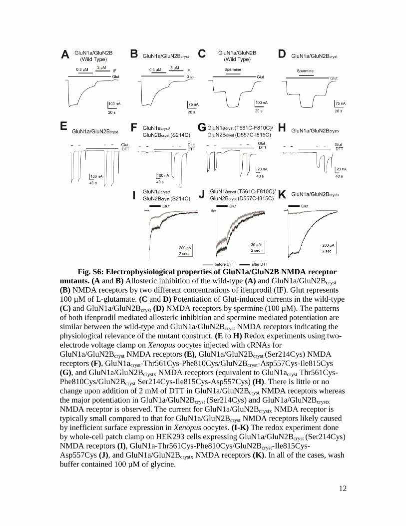
Asp557Cys (J), and GluN1a/GluN2Bcrystx NMDA receptors (K). In all of the cases, wash

buffer contained 100 µM of glycine.

13

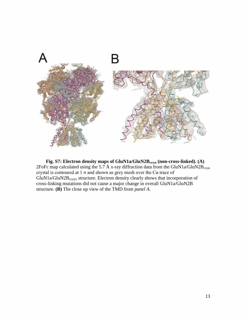
Fig. S7: Electron density maps of GluN1a/GluN2Bcryst (non-cross-linked). (A) 2FoFc map calculated using the 5.7 Å x-ray diffraction data from the GluN1a/GluN2Bcryst

crystal is contoured at 1 σ and shown as grey mesh over the Cα trace of

GluN1a/GluN2Bcrystx structure. Electron density clearly shows that incorporation of

cross-linking mutations did not cause a major change in overall GluN1a/GluN2B

structure. (B) The close up view of the TMD from panel A.

14

Fig. S8: Electron densities in the transmembrane domains in

GluN1a/GluN2Bcrystx. 2FoFc maps for the M1, M2, M3 and M4 helices of both GluN1a

(α) and GluN2B (β) TMDs are prepared with a B-factor sharpening factor of -90 Å2,

countered at 1.0 σ and shown as grey mesh. The models of GluN1a (α) and GluN2B (β)

are shown as yellow and cyan Cα traces, respectively. Cαs of methionine residues are

shown as spheres. Anomalous difference Fourier maps calculated from data collected on

two different selenomethionine-derivative crystals are averaged around the 2-fold non-

crystallographic axis using COOT (43) and shown as red mesh. The maps are contoured

at the following σ levels; 2.8 σ for GluN1a Met 555, 3.6 σ for GluN1a Met 576, 2.5 σ for

GluN1a Met 607, 2.5 σ for GluN1a Met 634, 4.2 σ for GluN1a Met 641, 3.3 σ for

GluN1a Met 813, 2.7 σ for GluN1a Met 818, 2.5 σ for GluN2B Met 561, 2.8 σ for

GluN2B Met 631, 2.5 σ for GluN2B Met 654, 3.0 σ for GluN2B Met 824 and 2.5 σ for

GluN2B Met 829. No significant signal was observed for residues GluN2B Met 562 and

Met 565.

15

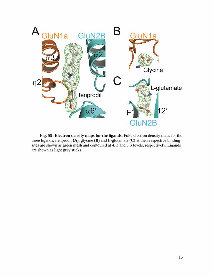
Fig. S9: Electron density maps for the ligands. FoFc electron density maps for the

three ligands, ifenprodil (A), glycine (B) and L-glutamate (C) at their respective binding

sites are shown as green mesh and contoured at 4, 3 and 3 σ levels, respectively. Ligands

are shown as light grey sticks.

16

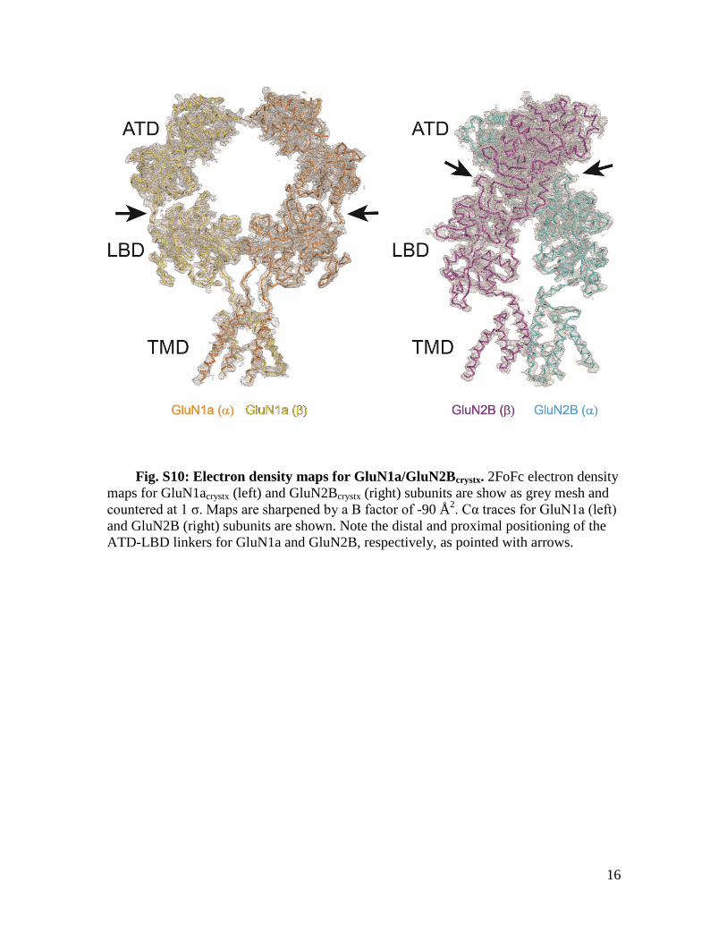
Fig. S10: Electron density maps for GluN1a/GluN2Bcrystx. 2FoFc electron density

maps for GluN1acrystx (left) and GluN2Bcrystx (right) subunits are show as grey mesh and

countered at 1 σ. Maps are sharpened by a B factor of -90 Å2. Cα traces for GluN1a (left)

and GluN2B (right) subunits are shown. Note the distal and proximal positioning of the

ATD-LBD linkers for GluN1a and GluN2B, respectively, as pointed with arrows.

17

Fig. S11: Dimer-of-dimers assembly of GluN1a/GluN2B NMDA receptors.

Tetrameric arrangement of ATD (A), LBD (B) and TMD (C) of GluN1a/GluN2Bcrystx

NMDA receptors viewed from top (left panels) and side (right panels). Regions at the

dimer-of-dimers interfaces are highlighted and residues in close proximity at ATD and

LBD are shown as sticks on the side view. Side view of LBD (panel B, right) is one

(between GluN1a (α) and GluN2B (α)) of the two equivalent sites of dimer-of-dimers

interaction at LBD.

18

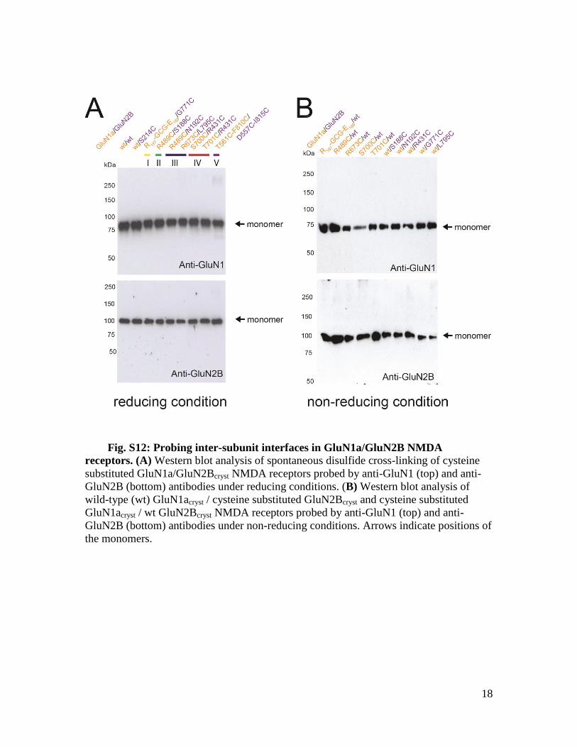
Fig. S12: Probing inter-subunit interfaces in GluN1a/GluN2B NMDA

receptors. (A) Western blot analysis of spontaneous disulfide cross-linking of cysteine

substituted GluN1a/GluN2Bcryst NMDA receptors probed by anti-GluN1 (top) and anti-

GluN2B (bottom) antibodies under reducing conditions. (B) Western blot analysis of

wild-type (wt) GluN1acryst / cysteine substituted GluN2Bcryst and cysteine substituted

GluN1acryst / wt GluN2Bcryst NMDA receptors probed by anti-GluN1 (top) and anti-

GluN2B (bottom) antibodies under non-reducing conditions. Arrows indicate positions of

the monomers.

19

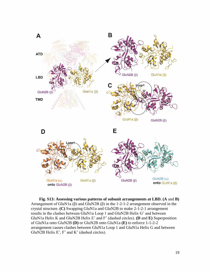
Fig. S13: Assessing various patterns of subunit arrangements at LBD. (A and B)

Arrangement of GluN1a (β) and GluN2B (β) in the 1-2-1-2 arrangement observed in the

crystal structure. (C) Swapping GluN1a and GluN2B to make 2-1-2-1 arrangement

results in the clashes between GluN1a Loop 1 and GluN2B Helix G’ and between

GluN1a Helix K and GluN2B Helix E’ and F’ (dashed circles). (D and E) Superposition

of GluN1a onto GluN2B (D) or GluN2B onto GluN1a (E) to enforce 1-1-2-2

arrangement causes clashes between GluN1a Loop 1 and GluN1a Helix G and between

GluN2B Helix E’, F’ and K’ (dashed circles).

20

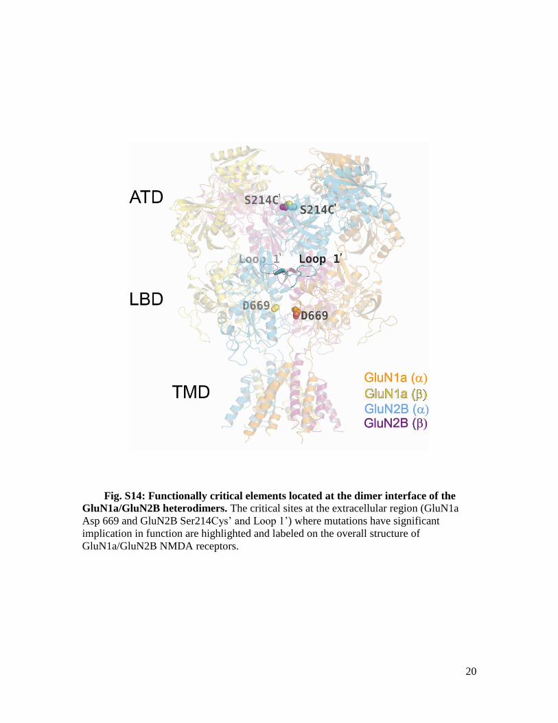
Fig. S14: Functionally critical elements located at the dimer interface of the

GluN1a/GluN2B heterodimers. The critical sites at the extracellular region (GluN1a

Asp 669 and GluN2B Ser214Cys’ and Loop 1’) where mutations have significant

implication in function are highlighted and labeled on the overall structure of

GluN1a/GluN2B NMDA receptors.

21

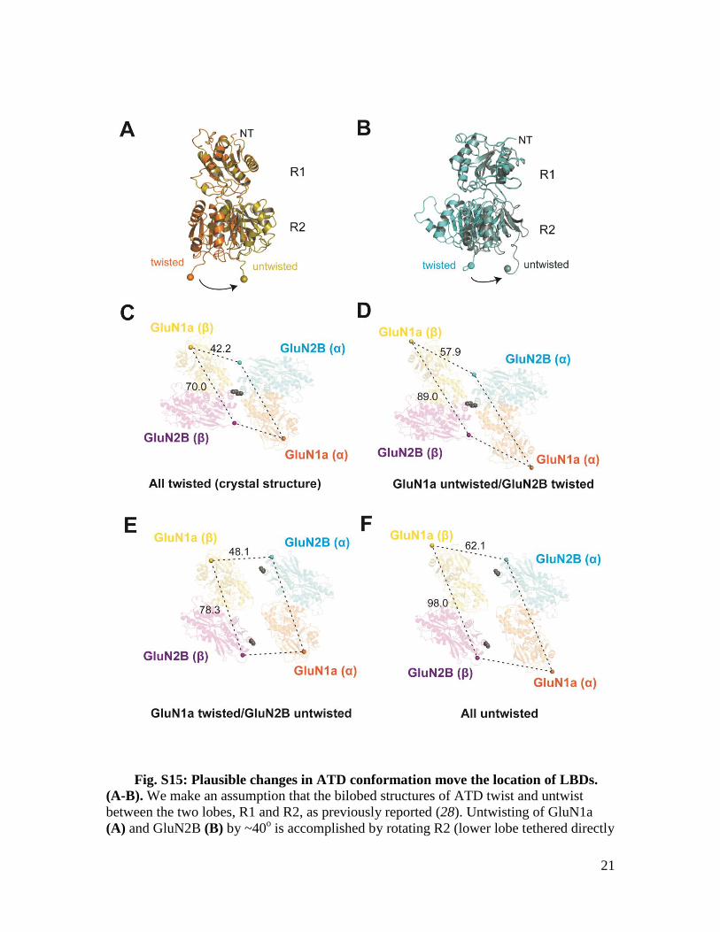
Fig. S15: Plausible changes in ATD conformation move the location of LBDs.

(A-B). We make an assumption that the bilobed structures of ATD twist and untwist

between the two lobes, R1 and R2, as previously reported (28). Untwisting of GluN1a

(A) and GluN2B (B) by ~40o is accomplished by rotating R2 (lower lobe tethered directly

22

to LBD) using GluK1 ATD structure (30) as a guide. (C to F) Shown here are structures

of ATD tetramers viewed “up” from the LBDs. The ATDs in the current crystal structure

and the previously published GluN1b/GluN2B ATD structures have “twisted”

conformations in both GluN1 and GluN2B (C). Untwisting of GluN1a (D), GluN2B (E),

or both (F) dramatically changes the location of LBDs. Numbers next to the dashed lines

represent distance (in Å) between the beginning points of LBD defined as the Cαs of

GluN1a Thr396 and GluN2B His405 (spheres). Note that untwisting of both GluN1a and

GluN2B ATDs result in separation of the LBDs in both length and width to significantly

more extent in GluN1a than GluN2B. This separation likely results in rearrangement of

subunits within and between the GluN1a/GluN2B LBD thereby affecting patterns of ion

channel gating. Shown in gray spheres are GluN2B Ser214 residues from both GluN2B

(α) and (β). Formation of a disulfide bond by the GluN2B Ser214Cys mutation at the

lower lobe (R2) not only traps the inter-subunit arrangement between the two GluN2B

subunits, but also movement in the GluN2B ATD lobes.

23

Fig. S16: Structural comparison of NMDA receptor ion channel with potassium

channels. TMDs of GluN1a subunits (yellow, left panel) and GluN2B subunits (cyan,

middle panel) are superposed onto the ion channel domains (red) of the closed

conformation of KcsA (PDB ID: 1K4C) (A), open conformation of MthK (PDB ID:

3LDC) (B). The superposed structures are viewed from the side (left and middle panels)

or from the extracellular side (right panel). Superposition is performed using Secondary-

structure matching (SSM) tool. Loops are excluded from the figure for clarity.

24

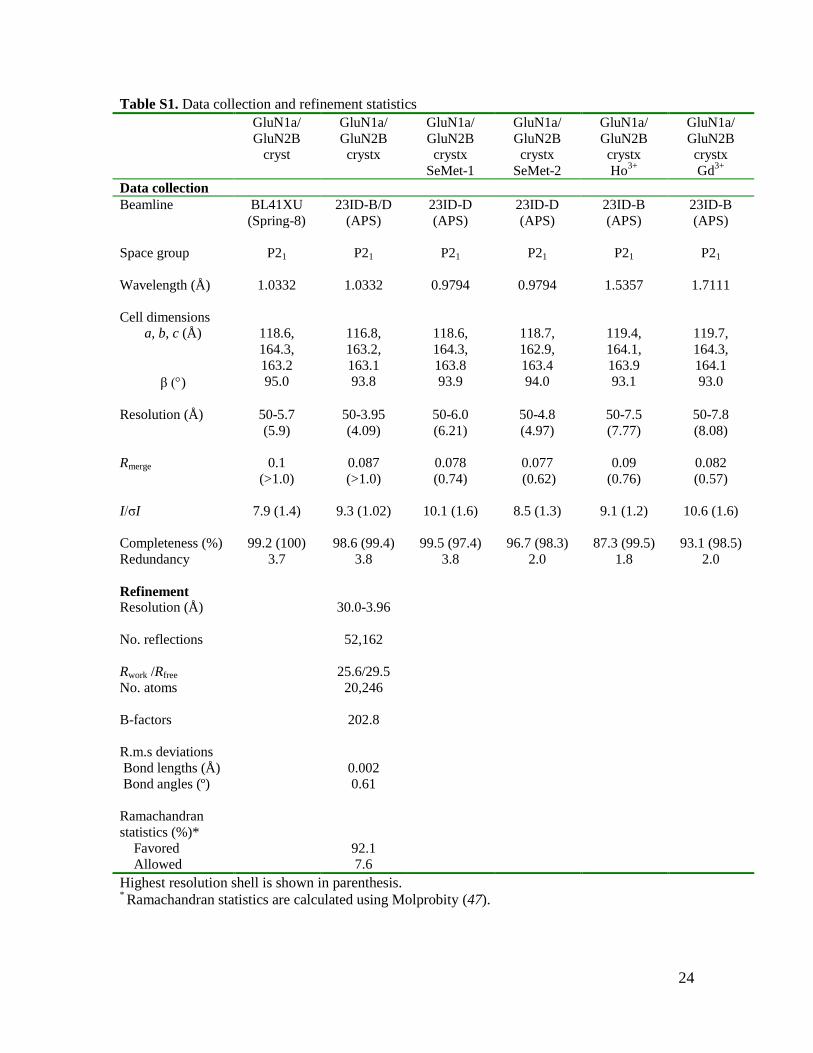
Table S1. Data collection and refinement statistics

GluN1a/

GluN2B

cryst

GluN1a/

GluN2B

crystx

GluN1a/

GluN2B

crystx

SeMet-1

GluN1a/

GluN2B

crystx

SeMet-2

GluN1a/

GluN2B

crystx

Ho3+

GluN1a/

GluN2B

crystx

Gd3+

Data collection

Beamline BL41XU 23ID-B/D 23ID-D 23ID-D 23ID-B 23ID-B

(Spring-8) (APS) (APS) (APS) (APS) (APS)

Space group P21 P21 P21 P21 P21 P21

Wavelength (Å) 1.0332 1.0332 0.9794 0.9794 1.5357 1.7111

Cell dimensions

a, b, c (Å) 118.6,

164.3,

163.2

116.8,

163.2,

163.1

118.6,

164.3,

163.8

118.7,

162.9,

163.4

119.4,

164.1,

163.9

119.7,

164.3,

164.1

β () 95.0 93.8 93.9 94.0 93.1 93.0

Resolution (Å) 50-5.7

(5.9)

50-3.95

(4.09)

50-6.0

(6.21)

50-4.8

(4.97)

50-7.5

(7.77)

50-7.8

(8.08)

Rmerge 0.1

(>1.0)

0.087

(>1.0)

0.078

(0.74)

0.077

(0.62)

0.09

(0.76)

0.082

(0.57)

I/σI 7.9 (1.4) 9.3 (1.02) 10.1 (1.6) 8.5 (1.3) 9.1 (1.2) 10.6 (1.6)

Completeness (%) 99.2 (100) 98.6 (99.4) 99.5 (97.4) 96.7 (98.3) 87.3 (99.5) 93.1 (98.5)

Redundancy 3.7 3.8 3.8 2.0 1.8 2.0

Refinement

Resolution (Å)

30.0-3.96

No. reflections

52,162

Rwork /Rfree 25.6/29.5

No. atoms

20,246

B-factors

202.8

R.m.s deviations

Bond lengths (Å) 0.002

Bond angles (º)

0.61

Ramachandran

statistics (%)*

Favored 92.1

Allowed 7.6

Highest resolution shell is shown in parenthesis. * Ramachandran statistics are calculated using Molprobity (47).

25

References and Notes 1. T. Hayashi, Effects of sodium glutamate on the nervous system. Keio J. Med. 3, 183–192

(1954). doi:10.2302/kjm.3.183

2. M. L. Mayer, G. L. Westbrook, P. B. Guthrie, Voltage-dependent block by Mg2+ of NMDA responses in spinal cord neurones. Nature 309, 261–263 (1984). Medline doi:10.1038/309261a0

3. S. F. Traynelis, L. P. Wollmuth, C. J. McBain, F. S. Menniti, K. M. Vance, K. K. Ogden, K. B. Hansen, H. Yuan, S. J. Myers, R. Dingledine, Glutamate receptor ion channels: structure, regulation, and function. Pharmacol. Rev. 62, 405–496 (2010). Medline doi:10.1124/pr.109.002451

4. M. Gielen, B. Siegler Retchless, L. Mony, J. W. Johnson, P. Paoletti, Mechanism of differential control of NMDA receptor activity by NR2 subunits. Nature 459, 703–707 (2009). Medline doi:10.1038/nature07993

5. H. Yuan, K. B. Hansen, K. M. Vance, K. K. Ogden, S. F. Traynelis, Control of NMDA receptor function by the NR2 subunit amino-terminal domain. J. Neurosci. 29, 12045–12058 (2009). Medline doi:10.1523/JNEUROSCI.1365-09.2009

6. K. B. Hansen, H. Furukawa, S. F. Traynelis, Control of assembly and function of glutamate receptors by the amino-terminal domain. Mol. Pharmacol. 78, 535–549 (2010). Medline doi:10.1124/mol.110.067157

7. E. Karakas, N. Simorowski, H. Furukawa, Structure of the zinc-bound amino-terminal domain of the NMDA receptor NR2B subunit. EMBO J. 28, 3910–3920 (2009). Medline doi:10.1038/emboj.2009.338

8. E. Karakas, N. Simorowski, H. Furukawa, Subunit arrangement and phenylethanolamine binding in GluN1/GluN2B NMDA receptors. Nature 475, 249–253 (2011). Medline doi:10.1038/nature10180

9. L. Mony, S. Zhu, S. Carvalho, P. Paoletti, Molecular basis of positive allosteric modulation of GluN2B NMDA receptors by polyamines. EMBO J. 30, 3134–3146 (2011). Medline doi:10.1038/emboj.2011.203

10. A. I. Sobolevsky, M. P. Rosconi, E. Gouaux, X-ray structure, symmetry and mechanism of an AMPA-subtype glutamate receptor. Nature 462, 745–756 (2009). Medline doi:10.1038/nature08624

11. A. N. Farina, K. Y. Blain, T. Maruo, W. Kwiatkowski, S. Choe, T. Nakagawa, Separation of domain contacts is required for heterotetrameric assembly of functional NMDA receptors. J. Neurosci. 31, 3565–3579 (2011). Medline doi:10.1523/JNEUROSCI.6041-10.2011

12. H. Furukawa, S. K. Singh, R. Mancusso, E. Gouaux, Subunit arrangement and function in NMDA receptors. Nature 438, 185–192 (2005). Medline doi:10.1038/nature04089

13. K. M. Vance, N. Simorowski, S. F. Traynelis, H. Furukawa, Ligand-specific deactivation time course of GluN1/GluN2D NMDA receptors. Nat. Commun. 2, 294 (2011). Medline doi:10.1038/ncomms1295

26

14. Y. Yao, C. B. Harrison, P. L. Freddolino, K. Schulten, M. L. Mayer, Molecular mechanism of ligand recognition by NR3 subtype glutamate receptors. EMBO J. 27, 2158–2170 (2008). Medline doi:10.1038/emboj.2008.140

15. A. Jespersen, N. Tajima, G. Fernandez-Cuervo, E. C. Garnier-Amblard, H. Furukawa, Structural insights into competitive antagonism in NMDA receptors. Neuron 81, 366–378 (2014). Medline doi:10.1016/j.neuron.2013.11.033

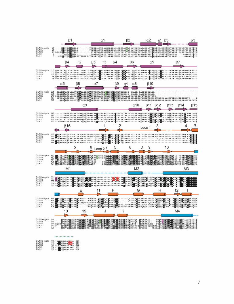
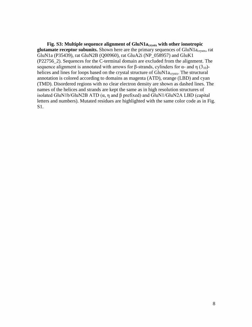
16. Single-letter abbreviations for the amino acid residues are as follows: A, Ala; C, Cys; D, Asp; E, Glu; F, Phe; G, Gly; H, His; I, Ile; K, Lys; L, Leu; M, Met; N, Asn; P, Pro; Q, Gln; R, Arg; S, Ser; T, Thr; V, Val; W, Trp; and Y, Tyr[BPO1].

17. G. N. Murshudov, P. Skubák, A. A. Lebedev, N. S. Pannu, R. A. Steiner, R. A. Nicholls, M. D. Winn, F. Long, A. A. Vagin, REFMAC5 for the refinement of macromolecular crystal structures. Acta Crystallogr. D Biol. Crystallogr. 67, 355–367 (2011). Medline doi:10.1107/S0907444911001314

18. G. F. Schröder, M. Levitt, A. T. Brunger, Super-resolution biomolecular crystallography with low-resolution data. Nature 464, 1218–1222 (2010). Medline doi:10.1038/nature08892

19. C. L. Salussolia, M. L. Prodromou, P. Borker, L. P. Wollmuth, Arrangement of subunits in functional NMDA receptors. J. Neurosci. 31, 11295–11304 (2011). Medline doi:10.1523/JNEUROSCI.5612-10.2011

20. M. Riou, D. Stroebel, J. M. Edwardson, P. Paoletti, An alternating GluN1-2-1-2 subunit arrangement in mature NMDA receptors. PLoS ONE 7, e35134 (2012). Medline doi:10.1371/journal.pone.0035134

21. U. Das, J. Kumar, M. L. Mayer, A. J. Plested, Domain organization and function in GluK2 subtype kainate receptors. Proc. Natl. Acad. Sci. U.S.A. 107, 8463–8468 (2010). Medline doi:10.1073/pnas.1000838107

22. J. Rachline, F. Perin-Dureau, A. Le Goff, J. Neyton, P. Paoletti, The micromolar zinc-binding domain on the NMDA receptor subunit NR2B. J. Neurosci. 25, 308–317 (2005). Medline doi:10.1523/JNEUROSCI.3967-04.2005

23. M. P. Regalado, A. Villarroel, J. Lerma, Intersubunit cooperativity in the NMDA receptor. Neuron 32, 1085–1096 (2001). Medline doi:10.1016/S0896-6273(01)00539-6

24. K. Kashiwagi, J. Fukuchi, J. Chao, K. Igarashi, K. Williams, An aspartate residue in the extracellular loop of the N-methyl-D-aspartate receptor controls sensitivity to spermine and protons. Mol. Pharmacol. 49, 1131–1141 (1996). Medline

25. A. Y. Lau, H. Salazar, L. Blachowicz, V. Ghisi, A. J. Plested, B. Roux, A conformational intermediate in glutamate receptor activation. Neuron 79, 492–503 (2013). Medline doi:10.1016/j.neuron.2013.06.003

26. F. Zheng, K. Erreger, C. M. Low, T. Banke, C. J. Lee, P. J. Conn, S. F. Traynelis, Allosteric interaction between the amino terminal domain and the ligand binding domain of NR2A. Nat. Neurosci. 4, 894–901 (2001). Medline doi:10.1038/nn0901-894

27. K. M. Vance, K. B. Hansen, S. F. Traynelis, GluN1 splice variant control of GluN1/GluN2D NMDA receptors. J. Physiol. 590, 3857–3875 (2012). Medline doi:10.1113/jphysiol.2012.234062

27

28. S. F. Traynelis, M. Hartley, S. F. Heinemann, Control of proton sensitivity of the NMDA receptor by RNA splicing and polyamines. Science 268, 873–876 (1995). Medline doi:10.1126/science.7754371

29. A. Inanobe, H. Furukawa, E. Gouaux, Mechanism of partial agonist action at the NR1 subunit of NMDA receptors. Neuron 47, 71–84 (2005). Medline doi:10.1016/j.neuron.2005.05.022

30. J. Zuo, P. L. De Jager, K. A. Takahashi, W. Jiang, D. J. Linden, N. Heintz, Neurodegeneration in Lurcher mice caused by mutation in delta2 glutamate receptor gene. Nature 388, 769–773 (1997). Medline doi:10.1038/42009

31. Y. Zhou, J. H. Morais-Cabral, A. Kaufman, R. MacKinnon, Chemistry of ion coordination and hydration revealed by a K+ channel-Fab complex at 2.0 A resolution. Nature 414, 43–48 (2001). Medline doi:10.1038/35102009

32. S. B. Long, X. Tao, E. B. Campbell, R. MacKinnon, Atomic structure of a voltage-dependent K+ channel in a lipid membrane-like environment. Nature 450, 376–382 (2007). Medline doi:10.1038/nature06265

33. S. Ye, Y. Li, Y. Jiang, Novel insights into K+ selectivity from high-resolution structures of an open K+ channel pore. Nat. Struct. Mol. Biol. 17, 1019–1023 (2010). Medline doi:10.1038/nsmb.1865

34. W. Li, R. W. Aldrich, Activation of the SK potassium channel-calmodulin complex by nanomolar concentrations of terbium. Proc. Natl. Acad. Sci. U.S.A. 106, 1075–1080 (2009). Medline doi:10.1073/pnas.0812008106

35. R. C. Liddington, Y. Yan, J. Moulai, R. Sahli, T. L. Benjamin, S. C. Harrison, Structure of simian virus 40 at 3.8-A resolution. Nature 354, 278–284 (1991). Medline doi:10.1038/354278a0

36. J. Watanabe, C. Beck, T. Kuner, L. S. Premkumar, L. P. Wollmuth, DRPEER: A motif in the extracellular vestibule conferring high Ca2+ flux rates in NMDA receptor channels. J. Neurosci. 22, 10209–10216 (2002). Medline

37. K. Imoto, C. Busch, B. Sakmann, M. Mishina, T. Konno, J. Nakai, H. Bujo, Y. Mori, K. Fukuda, S. Numa, Rings of negatively charged amino acids determine the acetylcholine receptor channel conductance. Nature 335, 645–648 (1988). Medline doi:10.1038/335645a0

38. T. Kawate, J. C. Michel, W. T. Birdsong, E. Gouaux, Crystal structure of the ATP-gated P2X(4) ion channel in the closed state. Nature 460, 592–598 (2009). Medline doi:10.1038/nature08198

39. B. Siegler Retchless, W. Gao, J. W. Johnson, A single GluN2 subunit residue controls NMDA receptor channel properties via intersubunit interaction. Nat. Neurosci. 15, 406–413, S1–S2 (2012). Medline doi:10.1038/nn.3025

40. D. J. Fitzgerald, P. Berger, C. Schaffitzel, K. Yamada, T. J. Richmond, I. Berger, Protein complex expression by using multigene baculoviral vectors. Nat. Methods 3, 1021–1032 (2006). Medline doi:10.1038/nmeth983

28

41. Z. Otwinowski, W. Minor, Processing of X-ray diffraction data collected in oscillation mode. Methods Enzymol. 276, 307–326 (1997). doi:10.1016/S0076-6879(97)76066-X

42. A. J. McCoy, R. W. Grosse-Kunstleve, P. D. Adams, M. D. Winn, L. C. Storoni, R. J. Read, Phaser crystallographic software. J. Appl. Crystallogr. 40, 658–674 (2007). Medline doi:10.1107/S0021889807021206

43. P. Emsley, K. Cowtan, Coot: Model-building tools for molecular graphics. Acta Crystallogr. D Biol. Crystallogr. 60, 2126–2132 (2004). Medline doi:10.1107/S0907444904019158

44. R. A. Nicholls, F. Long, G. N. Murshudov, Low-resolution refinement tools in REFMAC5. Acta Crystallogr. D Biol. Crystallogr. 68, 404–417 (2012). Medline doi:10.1107/S090744491105606X

45. P. D. Adams, P. V. Afonine, G. Bunkóczi, V. B. Chen, I. W. Davis, N. Echols, J. J. Headd, L. W. Hung, G. J. Kapral, R. W. Grosse-Kunstleve, A. J. McCoy, N. W. Moriarty, R. Oeffner, R. J. Read, D. C. Richardson, J. S. Richardson, T. C. Terwilliger, P. H. Zwart, PHENIX: A comprehensive Python-based system for macromolecular structure solution. Acta Crystallogr. D Biol. Crystallogr. 66, 213–221 (2010). Medline doi:10.1107/S0907444909052925

46. I. W. Davis, L. W. Murray, J. S. Richardson, D. C. Richardson, MOLPROBITY: Structure validation and all-atom contact analysis for nucleic acids and their complexes. Nucleic Acids Res. 32 (Web Server), W615–W619 (2004). Medline doi:10.1093/nar/gkh398


Recommended