Supporting Information Lane et al. 10.1073/pnas.0812020106 SI Methods General. Semipreparative high performance liquid chromatogra- phy (HPLC) was performed using a Waters 1525 or 515 pump with a Waters 2996 diode-array UV detector or a Waters 2487 dual-wavelength absorbance detector. Compound purification was achieved with Agilent Zorbax SB-C18 and RX-SIL columns (5 m, 9.4 250 mm). 1 H NMR spectra were collected in CDCl 3 on a Bruker DRX-500 instrument with a 5-mm broadband probe and referenced to residual CHCl 3 (7.24 ppm). LC-MS analyses were conducted with a Waters 2695 HPLC interfaced to a 2996 diode-array UV detector and Micromass ZQ 2000 electrospray ionization mass spectrometer, using MassLynx 4.0 software and an Alltech Alltima C 18 reversed-phase column (3 m, 2.1 150 mm). HPLC grade solvents were used in semipreparative HPLC and DESI-MS, and optima grade solvents applied in LC-MS experiments (Fisher Scientific). NMR solvents were obtained from Cambridge Isotope Laboratories. High resolution mass spectra were acquired using electrospray ionization with an Applied Biosystems QSTAR-XL hybrid quadrupole-time-of- flight tandem mass spectrometer and Analyst QS software. Epifluorescence and light microscopy experiments were con- ducted with an Olympus IX50 inverted microscope, and images collected with MagnaFire software (Optronics). Additional light micrographs were obtained with an Olympus dissecting scope (i.e., Fig. S5). All statistical analyses were completed with either SYSTAT version 9 or GraphPad version 4. The Marine Alga Callophycus serratus. The red macroalga C. ser- ratus (Harvey ex Kutzing 1957) (family Solieriaceae, order Gigartinales, class Rhodophyceae, phylum Rhodophyta) was collected at depths from 1 to 30 m at several sites in Fiji. Ten collections were made in 2006 at Yanuca, Waitabu in Taveuni, Lavena in Taveuni, and Dravuni in Kadavu; GPS coordinates for each collection are provided in Table S1. Immediately following collection, portions for quantitative whole tissue LC-MS exper- iments were extracted as described in the main text. Remaining material was frozen at 20 °C until further processing. Samples for DESI-MS experiments and microscopic analyses were col- lected in 2008 at Yanuca (18°2235S, 177°5972E) and immediately preserved with 1% or 10% formalin in natural seawater until analysis. Samples for DESI-MS analyses were from separate plants collected on the reef at distances from 3 to 1,000 m. Algal samples were identified based on comparison with previously described morphological traits (S. Fredericq, personal communication) and by 18S rRNA sequencing (see below). Vouchers are housed at the Georgia Institute of Technology and the University of the South Pacific in Suva, Fiji. 18S rRNA Sequencing. Genomic DNA from ethanol-preserved Callophycus serratus samples (G0004, G0021, G0039, G0049, G0052, G0100, G0113, G0118 and G0171) was extracted using the DNeasy Tissue Extraction Kit (Qiagen) and purified using polyethylene glycol. The nuclear small subunit ribosomal RNA (18S rRNA) gene was amplified via PCR, in 3 separate reactions, using primers (G01/G09, G02/G08, and G04/G07) and reaction conditions from Saunders and Kraft (1). Each of the 3 18S rRNA fragments was sequenced in both directions. Sequences were manually edited in BioEdit vers 7.0.5.3 (2) and aligned using ClustalW (3). Sequences were deposited in GenBank (accessions FJ660605-FJ660613). Phylogenetic relationships among se- quences were determined in PAUP* (4), using parsimony cri- teria and 1,000 bootstrap replicates, including Callophycus op- positifolius as an outgroup (GenBank accession no. AY437654) Antimicrobial Assays. Assays with Lindra thalassiae (ATCC 56663) were completed as described in ref. 5. HP20ss fractions were solubilized in a minimal volume of acetone and incorporated into molten YPM agar (16 g/L granulated agar, 2 g/L yeast extract, 2 g/L peptone, 4 g/L D-mannitol, 250 mg/L of both streptomycin sulfate and penicillin G in 1 L of natural seawater) at concentrations approximating natural whole algal tissue con- centrations. For each fraction, three 400-L subsamples of this mixture were dispensed into sterile 24-well microtiter plates, allowed to solidify, and an aliquot of L. thalassiae suspension in sterile seawater added to each well. Control wells were prepared with YPM agar and acetone but no algal material. Plates were incubated at 28 °C for 3 days and digital photographs collected for each well. The area calculator feature of ImageJ software (National Institutes of Health) was applied to determine the percentage of each well covered in fungal hyphae, and for each chromatographic fraction, the average fungal coverage for the 3 subsamples was treated as a single replicate. Assays were con- ducted for the bromophycolide- or callophycoic acid/ callophycol-containing fractions from n 4 independent bro- mophycolide chemotype C. serratus collections and n 6 callophycoic acid/callophycol chemotype collections. Fungal coverage for treatments vs. controls and among different treat- ments was compared using 1-way ANOVA with Dunnett’s post test (6). Antifungal assays with pure bromophycolides, callophycoic acids, and callophycols were completed as with extract fractions. Individual compounds were incorporated into molten YPM agar at 1:1 serially diluted concentrations ranging from 300 M to 0.15 M and n 3 assays completed at each concentration. Significant growth inhibition at the maximum tested concentra- tion was established by comparison of individual treatments with solvent-only controls applying 1-way ANOVA with Dunnett’s post test (6). For compounds significantly inhibitory (P 0.05) at the maximum tested concentration, inhibition data were fit to a sigmoidal dose-response curve; mean log IC 50 and standard error values were calculated. Reported IC 50 values were deter- mined by computing the antilog of mean log IC 50 values; standard errors for IC 50 values were not determined, because such values are not directly correlated with log IC 50 standard errors (7). Significant antifungal activity differences among active compounds were analyzed with an F test of the log IC 50 value for each compound (7). Antibacterial assays using Pseudoalteromonas bacteriolytica (ATCC 700679) were adapted from reported methods (5). A 24-h shake culture of P. bacteriolytica was diluted 1:160 in Difco Marine Broth 2216 (BD Biosciences) and 195 L of this mixture added to duplicate treatment and control wells of a 96-well plate. An equal amount of sterile marine broth was added to blank wells. Five microliters of 40concentrated HP20ss fractions in DMSO were then dispensed into all treatment and blank wells, giving a final concentration approximating natural whole tissue concentrations in the alga; 5 L of DMSO were added to corresponding control wells. Plates were incubated at 30 °C for 24 h and turbidity measured at a wavelength of 600 nm. We corrected for the natural absorbance of extract fractions by subtracting extract-only sterile blank turbidities from values obtained for treatments. For chromatographic fractions from each C. serratus collection, the average turbidity value from 2 Lane et al. www.pnas.org/cgi/content/short/0812020106 1 of 10
Transcript
Supporting InformationLane et al. 10.1073/pnas.0812020106SI MethodsGeneral. Semipreparative high performance liquid chromatogra-phy (HPLC) was performed using a Waters 1525 or 515 pumpwith a Waters 2996 diode-array UV detector or a Waters 2487dual-wavelength absorbance detector. Compound purificationwas achieved with Agilent Zorbax SB-C18 and RX-SIL columns(5 �m, 9.4 � 250 mm). 1H NMR spectra were collected in CDCl3on a Bruker DRX-500 instrument with a 5-mm broadband probeand referenced to residual CHCl3 (7.24 ppm). LC-MS analyseswere conducted with a Waters 2695 HPLC interfaced to a 2996diode-array UV detector and Micromass ZQ 2000 electrosprayionization mass spectrometer, using MassLynx 4.0 software andan Alltech Alltima C18 reversed-phase column (3 �m, 2.1 � 150mm). HPLC grade solvents were used in semipreparative HPLCand DESI-MS, and optima grade solvents applied in LC-MSexperiments (Fisher Scientific). NMR solvents were obtainedfrom Cambridge Isotope Laboratories. High resolution massspectra were acquired using electrospray ionization with anApplied Biosystems QSTAR-XL hybrid quadrupole-time-of-f light tandem mass spectrometer and Analyst QS software.Epifluorescence and light microscopy experiments were con-ducted with an Olympus IX50 inverted microscope, and imagescollected with MagnaFire software (Optronics). Additional lightmicrographs were obtained with an Olympus dissecting scope(i.e., Fig. S5). All statistical analyses were completed with eitherSYSTAT version 9 or GraphPad version 4.
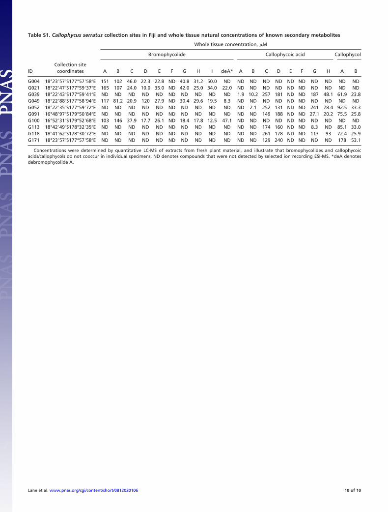
The Marine Alga Callophycus serratus. The red macroalga C. ser-ratus (Harvey ex Kutzing 1957) (family Solieriaceae, orderGigartinales, class Rhodophyceae, phylum Rhodophyta) wascollected at depths from 1 to 30 m at several sites in Fiji. Tencollections were made in 2006 at Yanuca, Waitabu in Taveuni,Lavena in Taveuni, and Dravuni in Kadavu; GPS coordinates foreach collection are provided in Table S1. Immediately followingcollection, portions for quantitative whole tissue LC-MS exper-iments were extracted as described in the main text. Remainingmaterial was frozen at �20 °C until further processing. Samplesfor DESI-MS experiments and microscopic analyses were col-lected in 2008 at Yanuca (18°22�35� S, 177°59�72� E) andimmediately preserved with 1% or 10% formalin in naturalseawater until analysis. Samples for DESI-MS analyses werefrom separate plants collected on the reef at distances from 3 to1,000 m. Algal samples were identified based on comparison withpreviously described morphological traits (S. Fredericq, personalcommunication) and by 18S rRNA sequencing (see below).Vouchers are housed at the Georgia Institute of Technology andthe University of the South Pacific in Suva, Fiji.
18S rRNA Sequencing. Genomic DNA from ethanol-preservedCallophycus serratus samples (G0004, G0021, G0039, G0049,G0052, G0100, G0113, G0118 and G0171) was extracted usingthe DNeasy Tissue Extraction Kit (Qiagen) and purified usingpolyethylene glycol. The nuclear small subunit ribosomal RNA(18S rRNA) gene was amplified via PCR, in 3 separate reactions,using primers (G01/G09, G02/G08, and G04/G07) and reactionconditions from Saunders and Kraft (1). Each of the 3 18S rRNAfragments was sequenced in both directions. Sequences weremanually edited in BioEdit vers 7.0.5.3 (2) and aligned usingClustalW (3). Sequences were deposited in GenBank (accessionsFJ660605-FJ660613). Phylogenetic relationships among se-quences were determined in PAUP* (4), using parsimony cri-
teria and 1,000 bootstrap replicates, including Callophycus op-positifolius as an outgroup (GenBank accession no. AY437654)
Antimicrobial Assays. Assays with Lindra thalassiae (ATCC 56663)were completed as described in ref. 5. HP20ss fractions weresolubilized in a minimal volume of acetone and incorporatedinto molten YPM agar (16 g/L granulated agar, 2 g/L yeastextract, 2 g/L peptone, 4 g/L D-mannitol, 250 mg/L of bothstreptomycin sulfate and penicillin G in 1 L of natural seawater)at concentrations approximating natural whole algal tissue con-centrations. For each fraction, three 400-�L subsamples of thismixture were dispensed into sterile 24-well microtiter plates,allowed to solidify, and an aliquot of L. thalassiae suspension insterile seawater added to each well. Control wells were preparedwith YPM agar and acetone but no algal material. Plates wereincubated at 28 °C for 3 days and digital photographs collectedfor each well. The area calculator feature of ImageJ software(National Institutes of Health) was applied to determine thepercentage of each well covered in fungal hyphae, and for eachchromatographic fraction, the average fungal coverage for the 3subsamples was treated as a single replicate. Assays were con-ducted for the bromophycolide- or callophycoic acid/callophycol-containing fractions from n � 4 independent bro-mophycolide chemotype C. serratus collections and n � 6callophycoic acid/callophycol chemotype collections. Fungalcoverage for treatments vs. controls and among different treat-ments was compared using 1-way ANOVA with Dunnett’s posttest (6).
Antifungal assays with pure bromophycolides, callophycoicacids, and callophycols were completed as with extract fractions.Individual compounds were incorporated into molten YPM agarat 1:1 serially diluted concentrations ranging from 300 �M to0.15 �M and n � 3 assays completed at each concentration.Significant growth inhibition at the maximum tested concentra-tion was established by comparison of individual treatments withsolvent-only controls applying 1-way ANOVA with Dunnett’spost test (6). For compounds significantly inhibitory (P � 0.05)at the maximum tested concentration, inhibition data were fit toa sigmoidal dose-response curve; mean log IC50 and standarderror values were calculated. Reported IC50 values were deter-mined by computing the antilog of mean log IC50 values;standard errors for IC50 values were not determined, becausesuch values are not directly correlated with log IC50 standarderrors (7). Significant antifungal activity differences amongactive compounds were analyzed with an F test of the log IC50value for each compound (7).
Antibacterial assays using Pseudoalteromonas bacteriolytica(ATCC 700679) were adapted from reported methods (5). A24-h shake culture of P. bacteriolytica was diluted 1:160 in DifcoMarine Broth 2216 (BD Biosciences) and 195 �L of this mixtureadded to duplicate treatment and control wells of a 96-well plate.An equal amount of sterile marine broth was added to blankwells. Five microliters of 40� concentrated HP20ss fractions inDMSO were then dispensed into all treatment and blank wells,giving a final concentration approximating natural whole tissueconcentrations in the alga; 5 �L of DMSO were added tocorresponding control wells. Plates were incubated at 30 °C for24 h and turbidity measured at a wavelength of 600 nm. Wecorrected for the natural absorbance of extract fractions bysubtracting extract-only sterile blank turbidities from valuesobtained for treatments. For chromatographic fractions fromeach C. serratus collection, the average turbidity value from 2
Lane et al. www.pnas.org/cgi/content/short/0812020106 1 of 10
subsample assays was treated as a single replicate. Assays wereconducted for the bromophycolide- or callophycoic acid/callophycol-containing fractions from n � 4 independent bro-mophycolide chemotype C. serratus collections and n � 6callophycoic acid/callophycol chemotype collections. Turbiditieswere statically compared with 1 those obtained for no-extractcontrols, using 1-tailed paired t test (6).
To establish a role of bromophycolides in antifungal defenseof algal surfaces, 2:1 bromophycolide A and B solutions wereserially diluted (1:1) in ethyl ether and 30-�L aliquots dispensedas evenly as possible over the surface of 400 �L of solidified YPMagar blocks (200-mm2 area) in 24-well microtiter plates, andallowed to air dry. Assuming even distribution of bromophycol-ides A and B across agar surfaces and negligible absorption ofcompounds into agar blocks, this corresponded to combined
surface concentrations ranging from 340 pmol/mm2 to 1.3 pmol/mm2 (n � 2 assays at each concentration). Solvent-only controlwells were prepared equivalently. Surfaces of treatment andcontrol wells were inoculated with a suspension of L. thalassiaein sterile seawater and incubated at 28 °C for 3 days. L. thalassiaegrowth was assessed and the log IC50 value computed as before.
ACKNOWLEDGMENTS. We thank A. Chequer, P.R. Jensen, C. Kauffman, C.Kicklighter, A. Prusak, and G. Toth for collection assistance; W. Aalbersberg forlaboratory facilities and collection support in Fiji; K. Feussner and M. Sharmafor extraction assistance; S. Fredericq for taxonomic analyses; P.R. Jensen andT. Hazen for microbiological advice; S. Engel and P.R. Jensen for stock culturesof test microbes; P.R. Jensen, C. Kauffman, and M. Woolery for cultivation andchemical analyses of microbes; S. Asberry for microtome preparation of algalsamples; D. Bostwick and C. Sullards for mass spectral analyses; and L. Gelbaumfor NMR spectroscopy assistance. We thank the Government of Fiji for per-mission to perform research in their territorial waters and the people of Serua,Kadavu, and Cakaudrove provinces for facilitating this work.
1. Saunders GW, Kraft GT (1994) Small-subunit rRNA gene sequences from representa-tives of selected families of the Gigartinales and Rhodymeniales (Rhodophyta). 1.Evidence for the Plocamiales ord. nov. Can J Bot 72:1252–1263.
2. Hall TA (1999) BioEdit: A user-friendly biological sequence alignment editor andanalysis program for Windows 95/98/NT. Nucleic Acids Symp Ser 41:95–98.
3. Larkin MA, et al. (2007) ClustalW and ClustalX version 2. Bioinformatics 23:2947–2948.
4. Swofford DL (2002) PAUP*: Phylogenetic analysis using parsimony (and other meth-ods) 4.0 Beta (Sinauer, Sutherland, MA).
5. Kubanek J, et al. (2003) Seaweed resistance to microbial attack: A targeted chemicaldefense against marine fungi. Proc Natl Acad Sci USA 100:6916–6921.
Fig. S2. Experimental log IC50 values for growth inhibition of the fungus L. thalassiae, as determined by analysis of dose-response curves. Error bars represent1 SD. IC50 values are indicated in white text within each data bar. NSA denotes compounds that were not significantly active at the maximum evaluatedconcentration of 300 �M (P � 0.05, n � 3, 1-way ANOVA with Dunnett’s post test).
Lane et al. www.pnas.org/cgi/content/short/0812020106 4 of 10
Fig. S3. Negative-ion DESI mass spectrum of pure bromophycolide E (10 �L, 1 mg/mL). The ion cluster centered at m/z 583 represents the [M�H]� molecularion and m/z 619 represents the [M�Cl]� chloride adduct of bromophycolide E.
Lane et al. www.pnas.org/cgi/content/short/0812020106 5 of 10
Fig. S4. Light micrographs (100� magnification) of representative C. serratus fragment before (Left) and after (Right) DESI-MS analysis, indicating no obviouscell lysis resulting from exposure to the DESI source.
Lane et al. www.pnas.org/cgi/content/short/0812020106 6 of 10
Fig. S5. Light micrographs of bromophycolide-containing patches observed on intact C. serratus surfaces. (a) Micrographs of specimens preserved with 1–10%aqueous formalin. (b) Images of algal samples stored at �20 °C before analysis. Patches were observed on all morphological features of the algal thallus.
Lane et al. www.pnas.org/cgi/content/short/0812020106 7 of 10
Fig. S6. Light micrographs of 5-�m sections from C. serratus fragments associated with bromophycolide-containing patches (preserved with 1% formalin). Inall cases, sample processing dislodged patches from algal surfaces. (A) (Left) 100� magnification of C. serratus section. (Right) 400� magnification of regionhighlighted by black box in A. (B–E) Typical 400� micrographs of C. serratus, illustrating that patched regions were not associated with large-scale tissue damage.External algal surfaces are pointed downward.
Lane et al. www.pnas.org/cgi/content/short/0812020106 8 of 10
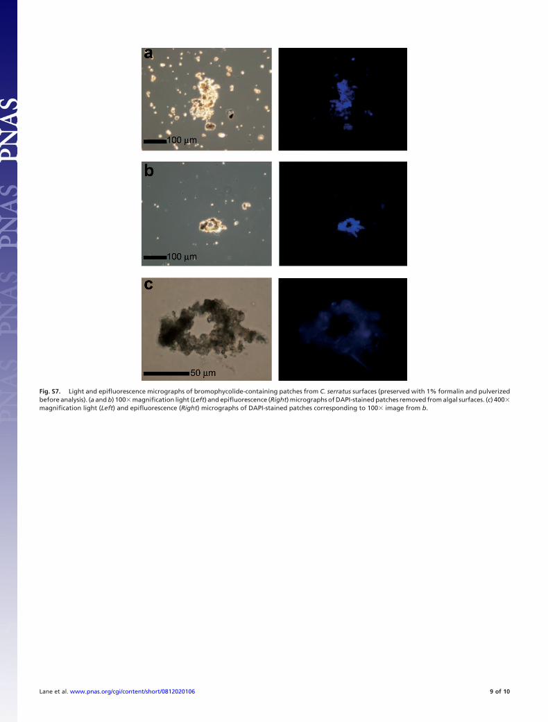
Fig. S7. Light and epifluorescence micrographs of bromophycolide-containing patches from C. serratus surfaces (preserved with 1% formalin and pulverizedbefore analysis). (a and b) 100� magnification light (Left) and epifluorescence (Right) micrographs of DAPI-stained patches removed from algal surfaces. (c) 400�magnification light (Left) and epifluorescence (Right) micrographs of DAPI-stained patches corresponding to 100� image from b.
Lane et al. www.pnas.org/cgi/content/short/0812020106 9 of 10
Concentrations were determined by quantitative LC-MS of extracts from fresh plant material, and illustrate that bromophycolides and callophycoicacids/callophycols do not cooccur in individual specimens. ND denotes compounds that were not detected by selected ion recording ESI-MS. *deA denotesdebromophycolide A.
Lane et al. www.pnas.org/cgi/content/short/0812020106 10 of 10