Page 1
S1
Supporting Information
New H-bonding patterns in biphenyl-based
synthetic lectins; pyrrolediamine bridges enhance
glucose-selectivity
Gururaj Joshi and Anthony P. Davis*
School of Chemistry, University of Bristol,
Cantock’s Close, Bristol BS8 1TS, UK
E-mail: [email protected]
Fax: +44-117 9298611; Tel: +44-117 9546334.
Electronic Supplementary Material (ESI) for Organic & Biomolecular ChemistryThis journal is © The Royal Society of Chemistry 2012
Page 2
S2
Synthesis and Characterisation
General Methods
All commercially available compounds were purchased from Aldrich, Alfa-Aesar, or Sigma
and were used without further purification except where stated. Solvents for synthesis were
dried by passing through a modified Grubbs system, of alumina column, manufactured by
Anhydrous Engineering. Routine monitoring of reactions was performed using precoated
silica gel TLC plates (Merck silica gel 60 F254). Spots were visualized by either UV light,
ethanolic solution of phosphomolybdic acid, potassium permanganate, or ninhydrin. Rf
values are given under these conditions. Flash column chromatography was performed using
silica gel (Fisher brand silica 60Å particle size 35-70 micron) as the absorbent. Both
analytical and preparative High Performance Liquid Chromatography (HPLC) were
performed on reversed phase-HPLC (RPHPLC) instruments, using C18 columns and a binary
solvent system (CH3OH and H2O). 1H and 13C NMR spectra were recorded on a Varian 400-
MR spectrometer, or on respectively VNMRSYS 500 (proton sensitive) or VNMRSYS 500
(carbon sensitive) spectrometers. Chemical shifts were reported in ppm downfield from
tetramethylsilane for proton and carbon or relative to the signals corresponding to the residual
non-deuterated solvents (CDCl3: δ 7.26 ppm, D2O: δ 4.79 ppm / 298 K). Mass spectra were
recorded on a VG Analytical Autospec for Electron Impact (EI), a VG Analytical Quattro for
ESI and Nanospray, or an Applied Biosystems 4700 spectrometer for MALDI.
.3,5-Bis(azidomethyl)-3’,5’-bisaminomethyl biphenyl 9
N3
N3
NH2
NH2
9
Boronic ester 81 (0.80 g, 1.73 mmol) and 3,5-bis(azidomethyl)-iodobenzene 72 (0.55 g, 1.73
mmol) were suspended in DMSO (10 mL). To this was added [1,1’-
bis(diphenylphosphino)ferrocene]-dichloropalladium(II) (18 mg, 0.02 mmol) and sodium
1 T. J. Ryan, G. Lecollinet, T. Velasco and A. P. Davis, Proc. Natl. Acad. Sci. USA, 2002, 99, 4863. 2 B. Sookcharoenpinyo, E. Klein, Y. Ferrand, D. B. Walker, P. R. Brotherhood, C. Ke, M. P. Crump and A. P. Davis, Angew. Chem., Int. Ed., 2012, 51, 4586.
Electronic Supplementary Material (ESI) for Organic & Biomolecular ChemistryThis journal is © The Royal Society of Chemistry 2012
Page 3
S3
carbonate (0.448 g, 4.23 mmol) in water (2.0 mL). The resulting mixture was heated to 80°C
for 6 hours before being cooled to room temperature. The reaction mixture was dissolved in
water (30 mL) and extracted with ethyl acetate (3 × 25 mL). The combined organic fractions
were washed with water (2 × 25 mL), brine (25 mL), dried over sodium sulfate, filtered and
concentrated in vacuo to yield a black oil. The oil was purified by flash column
chromatography (hexane/ethyl acetate 3:2) producing the expected bis-Boc-protected
biphenyl as a white solid (0.7 g, 77%). Trifluoroacetic acid (0.5 mL) was added to a solution
of this material (0.7 g, 1.3 mmol) in DCM (5 mL) at 5°C under nitrogen atmosphere and the
mixture was stirred for 5 hours at room temperature. Evaporation under reduced pressure
gave an oil which was purified by flash column chromatography (DCM/methanol saturated
with NH3 95:5) to yield di-amine 9 (0.42 g, 97%). Rf = 0.4 (DCM/methanol saturated NH3
9:1). 1H NMR (500 MHz, CD2Cl2) δ = 2.56 (brs., 4H, NH2), 3.91 (s, 4H, CH2NH2), 4.44 (s,
4H, CH2N3), 7.28 (s, 1H, ArH) 7.32 (s, 1H, ArH), 7.45 (d, J = 1.2 Hz, 2H, ArH) 7.53 (d, J =
1.4 Hz, 2H, ArH). 13C NMR (126 MHz, CD2Cl2) δ = 45.8 (CH2NH2), 54.5 (CH2N3), 124.7
(ArCH), 125.7 (ArCH), 126.7 (ArCH), 136.9 (ArC), 140.4 (ArC), 142.1 (ArC), 143.4 (ArC).
HRMS (ESI): m/z calculated for C16H19N8+ [M + H] +: 323.1733, found 323.1245.
Electronic Supplementary Material (ESI) for Organic & Biomolecular ChemistryThis journal is © The Royal Society of Chemistry 2012
Page 4
S4
Figure S1. 1H NMR (top) and 13C NMR (bottom) spectra of biphenyl 9 (CDCl3, 400 MHz).
10 9 8 7 6 5 4 3 2 1 0Chemical Shift (ppm)
7.987.832.002.003.993.96
DCM
7.4
45
47
.44
36
7.3
57
57
.23
29
7.1
89
0
4.3
50
3
3.8
19
1
2.4
66
9
180 170 160 150 140 130 120 110 100 90 80 70 60 50 40 30 20 10 0Chemical Shift (ppm)
14
3.4
07
41
42
.11
58
14
0.3
88
51
36
.87
94
12
6.6
47
71
25
.69
06
12
4.6
94
7
54
.51
23
45
.82
11
24
.61
86
Electronic Supplementary Material (ESI) for Organic & Biomolecular ChemistryThis journal is © The Royal Society of Chemistry 2012
Page 5
S5
Macrocyclic tetra-amine 11
11
H2N
H2NHN
NH
NH2
NH2
O
O
HN
O
NH
O
HN
NH
O
O
O
Ot-Bu
O
O
Ot-Bu
O
O
t-BuO
O
O
t-BuO
O
Ot-BuO
O
O Ot-Bu
O
A solution of bis-pentafluorophenyl ester 103 (1.5 g, 1.46 mmol) in dry THF (50 mL) was
added drop-wise over 30 hours to a solution of di-amine 9 (0.42 g, 1.36 mmol) and DIPEA
(1.5 mL, 8.3 mmol) in dry THF (ca. 1200 mL) under nitrogen atmosphere at room
temperature. The reaction mixture was further stirred vigorously for 24 hours at room
temperature. The solvent was removed under reduced pressure to give a solid that was taken
up in chloroform (200 mL) and washed with saturated aqueous solution of NH4Cl (100 mL),
water (100 mL), saturated aqueous solution of NaHCO3 (100 mL) and brine (100 mL). The
solution was dried over sodium sulfate, filtered and concentrated in vacuo to yield an off-
white solid which was purified by flash column chromatography (eluent toluene/DCM/ethyl
acetate/EtOH 35:30:30:0 to 35:30:30:6) to afford a crude mixture of macrocycles. The
mixture was further purified by preparative HPLC (Hichrom Kromasil column, 150 x 21.2
mm, 5 µm, eluent: methanol/water 80:20 to 95:5 in 10 min, then methanol/water 99:1 after 40
min, and 100:0 after 48 min, flow rate 21 mL.min-1, retention time = 17 minutes) to give pure
[2+2] macrocycle (0.7 g, 55%). Activated Pd on C (0.2 g) was added to the [2+2] macrocycle
(0.2 g, 1.05 mmol) in dry THF/methanol saturated with NH3 1:1 (15 mL) and the flask was
evacuated and filled with H2 (1 atm) gas. The reaction mixture was stirred at room
temperature for an hour and the Pd catalyst was removed by filtration and washed with ethyl
acetate. The filtrate and washings were concentrated and purified by flash column
3 E. Klein, M. P. Crump and A. P. Davis, Angew. Chem., Int. Ed., 2005, 44, 298.
Electronic Supplementary Material (ESI) for Organic & Biomolecular ChemistryThis journal is © The Royal Society of Chemistry 2012
Page 6
S6
chromatography (DCM/methanol saturated with NH3 95:5 to 4:1) to obtain tetra-amine 11 as
a white solid (0.18 g, 94%). Rf = 0.15 (DCM/methanol saturated with NH3 4:1). 1H NMR
(500 MHz, CD3OD) δ = 1.38 (s, 54H, C(CH3)3), 2.45 (t, J = 6.11 Hz, 12H, CH2CH2O), 3.71
(t, J = 6.11 Hz, 12H, CH2CH2O), 3.89 (s, 12H, C(CH2O)3), 3.93 (s, 8H, CH2NH2), 4.64 (s,
8H, CH2NH), 7.34 (s, 2H, ArH), 7.39 (s, 2H, ArH), 7.56 (s, 4H, ArH), 7.59 (s, 4H, ArH),
8.23 (s, 6H, Linker- ArH). 13C NMR (126 MHz, CD3OD) δ = 27.8 (C(CH3)3), 36.1
(CH2CH2CO), 44.0 (CH2NH), 45.6 (CH2NH2), 60.5 (C(CH2O)3), 67.0 (CH2CH2O), 68.8
(C(CH2O)3), 80.6 (C(CH3)3), 124.6 (ArCH), 125.3 (ArCH), 126.1 (ArCH), 128.2 (ArCH),
129.0 (ArCH), 134.4 (ArC), 136.0 (ArC), 139.2 (ArC), 140.9 (ArC), 141.2 (ArC), 142.8
(ArC), 166.1 (CONHCH2), 166.7 (CONHC), 171.1 (COOC(CH3)3). HRMS (ESI): m/z
calculated for C100H139N10O24+ [M + H]+ is 1863.9964, found 1863.9998.
Figure S2. 1H NMR spectrum of tetra-amine 11 (CD3OD, 500 MHz).
0.00.51.01.52.02.53.03.54.04.55.05.56.06.57.07.58.08.5f1(ppm)
CDH2ODCD3OH
Electronic Supplementary Material (ESI) for Organic & Biomolecular ChemistryThis journal is © The Royal Society of Chemistry 2012
Page 7
S7
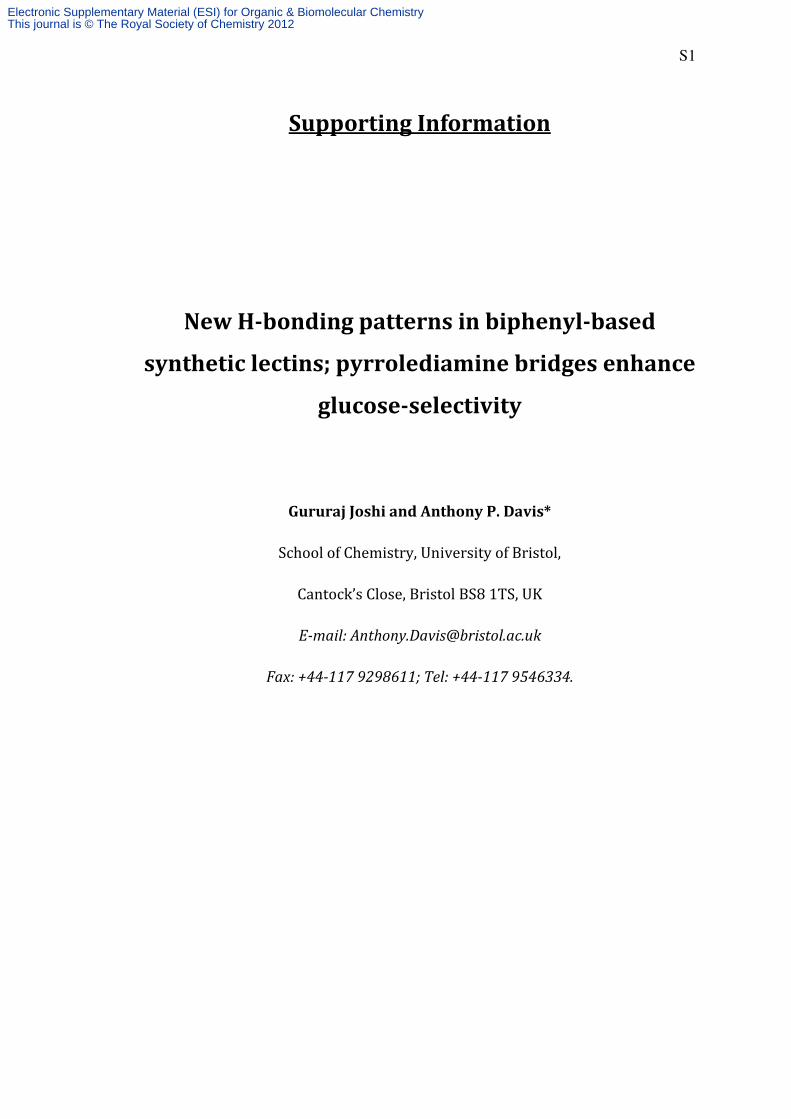
Protected macrotricycle 13
13
OBut
N
NN
NN
N
N
N
O
O
N
O
O
H
H
O
O
H
HN
Y
H
H
O
O
OBut
OBut
O
OBut
O
Boc
Boc
Boc
Boc
Y
NHC(CH2OCH2CH2CO2But)3Y =
Pyrrolediadehyde 124 (13.4 mg, 40 µmol) was added to a solution of tetra-amine 11 (38.5 mg,
20 µmol) in dry degassed CHCl3/CH3OH (9:1, 20 mL) and stirred at room temperature
overnight. Analysis by 1H NMR suggested quantitative imine formation. The solvent was
removed under reduced pressure to yield a yellow solid, presumed to be the corresponding
macrotricyclic tetra-imine (50 mg, quantitative). This material was dissolved in
CHCl3/CH3OH (1:1, 10 mL), NaBH4 (38 mg, 1 mmol) was added and the resultant mixture
was stirred for an hour. The reaction was monitored by 1H NMR (for the disappearance of
imine-H). The solvent was removed under reduced pressure to yield a white solid. DCM (25
mL) was added to form a turbid solution which was washed with water (20 mL), HCl (0.1 N,
20 mL), brine (20 mL), dried over sodium sulfate, filtered and evaporated. The resulting
solid was partially purified by flash column chromatography (DCM/methanol saturated with
NH3 100:0 to 98:2), collecting the fraction with Rf = 0.2 (DCM/methanol saturated with NH3
99:1). The obtained amine was dissolved in dry THF (10 mL) and di-tert-butyl dicarbonate
(50 mg, 23 mmol) was added. The mixture was heated under reflux overnight under a
nitrogen atmosphere. The solvent was removed and the residue redissolved in DCM (25 mL)
and washed with saturated aqueous NaHCO3 (20 mL), water (20 mL), brine (20 mL), dried
over Na2SO4, filtered and evaporated. The resulting solid was subjected to preparative HPLC
(Hichrom Kromasil column, 250 x 21.2 mm, 5 µm, eluent: methanol/water 95:5 to methanol
100 in 10 min, flow rate 21 mL.min-1, retention time = 24 minutes), to yield protected
macrotricycle 13 as a white solid (20 mg, 34% over two steps). Rf = 0.6 (hexane/ethyl acetate
4 M. Galezowski and D. T. Gryko, J. Org. Chem., 2006, 71, 5942.
Electronic Supplementary Material (ESI) for Organic & Biomolecular ChemistryThis journal is © The Royal Society of Chemistry 2012
Page 8
S8
3:2). 1H NMR (500 MHz, CD2Cl2/CD3OD) δ = 1.24 -1.38 (s, 90H, C(CH3)3), 1.58 (brs.,
36H, C(CH3)3), 2.45 (t, J = 6.26 Hz, 12H, CH2CH2O), 3.67 (t, J = 6.26 Hz, 12H, CH2CH2O),
3.81 (s, 12H, C(CH2O)3), 4.43 (brs., 24H, ArCH2NHCO, ArCH2NH, Pyr-CH2NH), 6.57 (brs.,
2H, ArH), 7.00 (brs., 4H, ArH), 7.39 (brs., 6H, ArH), 7.89 (brs., 2H, Linker-ArH), 8.37 (brs.,
4H, ArH). HRMS (ESI): m/z calculated for C152H212N12O40Na+ [M + Na]+ is 2868.4821,
found 2868.3006.
Electronic Supplementary Material (ESI) for Organic & Biomolecular ChemistryThis journal is © The Royal Society of Chemistry 2012
Page 9
S9
Figure S3. 1H NMR (top) and 13C NMR (bottom) spectra of protected macrotricycle 13
(CDCl3, 400 MHz).
10 9 8 7 6 5 4 3 2 1 0Chemical Shift (ppm)
8.3
08
6
7.8
50
7
7.3
28
2
4.3
66
7
3.7
51
43
.62
20
3.6
09
13
.59
69
3.2
88
0
2.3
98
02
.38
51
2.3
72
9
1.5
22
51
.32
23
1.1
82
5
0.0
00
0
180 160 140 120 100 80 60 40 20 0Chemical Shift (ppm)
Electronic Supplementary Material (ESI) for Organic & Biomolecular ChemistryThis journal is © The Royal Society of Chemistry 2012
Page 10
S10
Receptor 6
6
O-
N
NN
NN
N
N
N
O
O
N
O
O
H
H
O
O
H
HN
HN
H
H
O
O
O-
O-
O
O-
O
H
H
HH
HN
O
OO
OO
OO
O
O
O
OO
O
O
O
O
OO
Protected macrotricycle 13 (10 mg, 3.5 µmol) was dissolved in DCM (2 mL) and cooled to
5°C under a nitrogen atmosphere. Trifluoroacetic acid (200 µL) was added and the solution
was stirred at room temperature for 6 hours. Evaporation under reduced pressure gave a
residue which was dissolved in methanol/water (3:2, 5 mL). Sodium hydroxide (0.5 M) was
added till pH 7.0. The solvent was removed by freeze drying to obtain pure macrotricycle 6
as a white solid (7 mg, 93%). The solid, however, was not soluble in D2O at pD 7.0. NaOD
was added to the turbid solution slowly while stirring until the solution was clear at pD 13. 1H NMR (500 MHz, D2O at pD 13.0) δ = 2.37 (t, J = 6.72 Hz, 12H, CH2CH2O), 3.30 (AB
quartet, d, J = 13.69 Hz, 4H, Pyr-CH2NH), 3.40 (AB quartet, d, J =13.69 Hz, 4H, Pyr-
CH2NH), 3.59 (AB quartet, d, J = 14.92 Hz, 4H, ArCH2NHCH2), 3.67 (t, J = 6.72 Hz, 12H,
CH2CH2O), 3.70 - 3.85 (m, 16H, C(CH2O)3 and ArCH2NHCH2), 4.39 (AB quartet, d, J =
14.43 Hz, 4H, ArCH2NHCO) 4.52 (AB quartet, d, J=14.18 Hz, 4H, ArCH2NHCO), 6.87 (s,
2H, ArH), 7.12 (s, 4H, ArH), 7.27 (s, 2H, ArH), 7.43 (s, 4H, ArH), 7.75 (s, 2H, Linker-ArH),
8.17 (d, J = 1.47 Hz, 4H, Linker-ArH). 13C NMR (126 MHz, D2O) δ = 37.7 (CH2CH2O),
42.8 (Pyr-CH2NH), 44.0 (ArCH2NH), 48.8 (ArCH2NH), 61.1 (C(CH2O)3), 68.6 (CH2CH2O),
68.8 (C(CH2O)3), 115.1 (Pyr-CCO2Na), 117.4 (ArCH), 118.2 (ArCH), 120.0 (ArCH), 125.3
(ArCH), 126.1 (ArCH), 129.2 (Pyr-CCH2), 134.8 (ArC), 136.0 (ArC), 139.1 (ArC-Ar), 139.9
(ArC-Ar), 168.8 (Linker-CONHC), 169.4 (ArCONHCH2), 174.5 (Pyr-CO2Na), 180.3
(CO2Na). HRMS (ESI): m/z calculated for C46H50N6O16Na22+ [M + 2Na]2+ (corresponding
deca-carboxylic acid macrotricycle) is 965.3175, found 966.3170.
Electronic Supplementary Material (ESI) for Organic & Biomolecular ChemistryThis journal is © The Royal Society of Chemistry 2012
Page 11
S11
Figure S4. 1H NMR (top) and 13C NMR (bottom) spectra of receptor 6 (D2O, 400 MHz).
The HOD peak is suppressed in the 1H NMR spectrum.
10 9 8 7 6 5 4 3 2 1 0Chemical Shift (ppm)
8.1
72
4
7.7
46
17
.43
44
7.2
70
97
.12
27
6.8
70
1
4.5
30
44
.50
20
4.4
06
64
.37
77
3.7
72
83
.75
67
3.6
79
93
.66
71
3.6
54
43
.60
79
3.5
78
13
.38
28
3.3
13
8
2.3
78
12
.36
49
2.3
52
7
220 200 180 160 140 120 100 80 60 40 20 0 -20Chemical Shift (ppm)
18
0.3
09
11
74
.48
12
16
9.3
53
51
68
.82
44
13
9.8
94
71
39
.12
44
13
5.9
88
61
34
.84
48
12
9.8
72
81
29
.18
80
12
6.1
14
51
25
.27
42
12
0.0
37
61
18
.20
13
11
7.4
38
71
15
.12
78
68
.80
75
68
.55
85
61
.04
99
50
.60
00
48
.81
04
44
.02
51
42
.74
90
37
.70
69
29
.82
48
-9.0
80
2
Electronic Supplementary Material (ESI) for Organic & Biomolecular ChemistryThis journal is © The Royal Society of Chemistry 2012
Page 12
S12
NMR Binding Studies
Standard procedure for NMR titrations.
Solutions of guest in were prepared in phosphate buffer (pD = 13, 50 mM, prepared using
D2O*) and allowed to equilibrate overnight. Aliquots were added to 500 µl of a solution
receptor 6 (1.15 mmol, [6]initial = 2.3 mM) in phosphate buffer (pD = 13, 50 mM, prepared
using D2O*) in an NMR sample tube. The tube was shaken carefully after each addition and 1H NMR spectra were recorded at 296 K. Variations in chemical shifts for receptor protons
were entered into a specifically written non-linear least squares curve-fitting program
implemented within Excel. Assuming 1:1 stoichiometry, the programme calculates Ka and
the limiting change in chemical shift ∆δ. The assumption of 1:1 stoichiometry is supported
by the generally good fits between observed and calculated data.
*The phophate buffer solution (PBS) was prepared as follows: A 1 M PBS stock solution
was prepared by dissolving disodium hydrogen phosphate (2.68 g) in D2O (10 mL). The pD
was adjusted to 13.0 by addition of 4% NaOD solution. A portion of this solution (500 µL)
was diluted to 10 mL using D2O and the pD readjusted to 13.0 to give the 50 mM PBS used
for the titrations.
Electronic Supplementary Material (ESI) for Organic & Biomolecular ChemistryThis journal is © The Royal Society of Chemistry 2012
Page 13
Titration of receptor 6 with GlcNAc
Figure S5. Partial 1H NMR spectra from the addition of
receptor aromatic protons. [3] = 0.50 M
Figure S6. Experimental and calculated values
[6]initial = 2.3 mmol, [3]titrant = 0.5 M.
Limiting ∆δ = 0.07 ppm.
0.00
0.01
0.02
0.03
0.04
0.05
0.06
0.07
0.08
0.00 0.05 0.10
∆δ
[Guest] added / mol.dm
Titration of receptor 6 with GlcNAc 3
H NMR spectra from the addition of 3 to 6. The signals shown are due to
] = 0.50 M.
Experimental and calculated values for the 1H NMR binding study of
= 0.5 M. Ka = 6.8 M-1. Peak at 7.28 ppm used for
0.10 0.15 0.20 0.25
[Guest] added / mol.dm-3
∆δ calculated
∆δ observed
S13
. The signals shown are due to
H NMR binding study of 6 + 3.
. Peak at 7.28 ppm used for analysis.
Electronic Supplementary Material (ESI) for Organic & Biomolecular ChemistryThis journal is © The Royal Society of Chemistry 2012
Page 14
Titration of receptor 6 with methyl
Figure S7. Partial 1H NMR spectra from the addition of
to receptor aromatic protons. [
Figure S8. Experimental and calculated values
[6]initial = 2.3 mmol, [14]titrant = 0.96 M
Limiting ∆δ = 0.08 ppm.
0.00
0.01
0.02
0.03
0.04
0.05
0.06
0.07
0.08
0.09
0.00 0.10
∆δ
∆δ
∆δ
∆δ
[Guest] added / mol.dm
receptor 6 with methyl β-D-glucoside 14
H NMR spectra from the addition of 14 to 6. The signals shown are due
to receptor aromatic protons. [14] = 0.96 M.
Experimental and calculated values for the 1H NMR binding study of
= 0.96 M. Ka = 4.2 M-1. Peak at 7.89 ppm used for
0.20 0.30 0.40
[Guest] added / mol.dm-3
∆δ calculated
∆δ observed
S14
. The signals shown are due
binding study of 6 + 14.
ppm used for analysis.
Electronic Supplementary Material (ESI) for Organic & Biomolecular ChemistryThis journal is © The Royal Society of Chemistry 2012
Page 15
Titration of receptor 6 with D
Figure S9. Partial 1H NMR spectra from the addition of
to receptor aromatic protons. [
Figure S10. Experimental and calculated values
[6]initial = 0.9 mmol, [16]titrant = 0.61 M.
Limiting ∆δ = 0.053 ppm.
0.00
0.01
0.02
0.03
0.04
0.05
0.06
0.00 0.05 0.10
∆δ
[Guest] added / mol.dm
Titration of receptor 6 with D-galactose 16
H NMR spectra from the addition of 16 to 6. The signals shown are due
to receptor aromatic protons. [16] = 0.61 M.
Experimental and calculated values for the 1H NMR binding study of
= 0.61 M. Ka = 2.9 M-1. Peak at 7.53 ppm used for
0.15 0.20 0.25 0.30
[Guest] added / mol.dm-3
∆δ calculated
∆δ observed
S15
. The signals shown are due
H NMR binding study of 6 + 16.
. Peak at 7.53 ppm used for analysis.
Electronic Supplementary Material (ESI) for Organic & Biomolecular ChemistryThis journal is © The Royal Society of Chemistry 2012
Page 16
Titration of receptor 6 with D
Figure S11. Partial 1H NMR spectra from the addition of
to receptor aromatic protons. [
Figure S12. Experimental and calculated values
[6]initial = 2.3 mmol, [17]titrant = 0.96 M
Limiting ∆δ = 0.029 ppm.
0.00
0.01
0.01
0.02
0.02
0.03
0.03
0.04
0.00 0.10 0.20
∆δ
[Guest] added / mol.dm
Titration of receptor 6 with D-xylose 17
H NMR spectra from the addition of 17 to 6. The signals shown are due
to receptor aromatic protons. [17] = 0.96 M.
Experimental and calculated values for the 1H NMR binding study of
= 0.96 M. Ka = 4.2 M-1. Peak at 7.89 ppm used for
0.20 0.30 0.40 0.50
[Guest] added / mol.dm-3
∆δ calculated
∆δ observed
S16
. The signals shown are due
binding study of 6 + 17.
ppm used for analysis.
0.60
Electronic Supplementary Material (ESI) for Organic & Biomolecular ChemistryThis journal is © The Royal Society of Chemistry 2012


![Biphenyl 4-Hydroxylases Involved in Aucuparin Biosynthesis ...Biphenyl 4-Hydroxylases Involved in Aucuparin Biosynthesis in Rowan and Apple Are Cytochrome P450 736A Proteins1[OPEN]](https://static.documents.pub/doc/80x56/604367eb187f3f61c47fbd55/biphenyl-4-hydroxylases-involved-in-aucuparin-biosynthesis-biphenyl-4-hydroxylases.jpg)


