Surface mechanical properties, corrosion resistance, and cytocompatibility of nitrogen plasma-implanted nickel–titanium alloys: A comparative study with commonly used medical grade materials K.W.K. Yeung, 1 R.W.Y. Poon, 2 P.K. Chu, 1 C.Y. Chung, 2 X.Y. Liu, 2 W.W. Lu, 1 D. Chan, 3 S.C.W. Chan, 1 K.D.K. Luk, 1 K.M.C. Cheung 1 1 Division of Spine Surgery, Department of Orthopaedics and Traumatology, Faculty of Medicine, University of Hong Kong, Pokfulam, Hong Kong 2 Department of Physics and Materials Science, City University of Hong Kong, Kowloon, Hong Kong 3 Department of Biochemistry, Faculty of Medicine, University of Hong Kong, Pokfulam, Hong Kong Received 10 January 2006; revised 8 September 2006; accepted 17 October 2006 Published online 12 February 2007 in Wiley InterScience (www.interscience.wiley.com). DOI: 10.1002/jbm.a.31154 Abstract: Stainless steel and titanium alloys are the most common metallic orthopedic materials. Recently, nickel–ti- tanium (NiTi) shape memory alloys have attracted much attention due to their shape memory effect and super-elas- ticity. However, this alloy consists of equal amounts of nickel and titanium, and nickel is a well known sensitizer to cause allergy or other deleterious effects in living tis- sues. Nickel ion leaching is correspondingly worse if the surface corrosion resistance deteriorates. We have therefore modified the NiTi surface by nitrogen plasma immersion ion implantation (PIII). The surface chemistry and corro- sion resistance of the implanted samples were studied and compared with those of the untreated NiTi alloys, stainless steel, and Ti-6Al-4V alloy serving as controls. Immersion tests were carried out to investigate the extent of nickel leaching under simulated human body conditions and cytocompatibility tests were conducted using enhanced green fluorescent protein mice osteoblasts. The X-ray pho- toelectron spectroscopy results reveal that a thin titanium nitride (TiN) layer with higher hardness is formed on the surface after nitrogen PIII. The corrosion resistance of the implanted sample is also superior to that of the untreated NiTi and stainless steel and comparable to that of titanium alloy. The release of nickel ions is significantly reduced compared with the untreated NiTi. The sample with sur- face TiN exhibits the highest amount of cell proliferation whereas stainless steel fares the worst. Compared with coatings, the plasma-implanted structure does not delami- nate as easily and nitrogen PIII is a viable way to improve the properties of NiTi orthopedic implants. Ó 2007 Wiley Periodicals, Inc. J Biomed Mater Res 82A: 403–414, 2007 Key words: cell viability; osteoblast; surface treatment; stainless steel; titanium; nickel–titanium alloy INTRODUCTION Stainless Steels, titanium, and titanium alloys are the most widely used metallic orthopedic materials. Stainless steel is the oldest and most preferred mate- rial for internal fixation devices because of its me- chanical properties, cost effectiveness, and acceptable biocompatibility. 1,2 Commercial titanium and tita- nium alloys are the best choice for dental and cementless orthopedic implants since they possess superior biocompatibility and corrosion resistance as well as low modulus. 3 A new class of materials, nickel–titanium (NiTi) shape memory alloy, has re- cently attracted much attention due to its distinctive shape memory effect and super-elasticity, which may not be found in stainless steels and titanium alloys. Other favorable properties of this material as medical implants have also been reported. 4–16 A number of studies suggest that this material is com- patible with living tissues, 7,17–27 but adverse effects have also been reported. A study found that the Correspondence to: Dr. K.M.C. Cheung; e-mail: ken-cheung@ hku.hk or Prof. P.K. Chu; e-mail: [email protected]Contract grant sponsor: Hong Kong Research Grants Council (RGC); contract grant numbers: HKU 7283/00M, CityU 1/04C Contract grant sponsor: City University of Hong Kong; contract grant number: 9667002 Contract grant sponsor: Scoliosis Research Society; con- tract grant number: 9667002 Contract grant sponsor: Hong Kong Innovation Technol- ogy; contract grant number: GHP 019/05 ' 2007 Wiley Periodicals, Inc.
Transcript
Surface mechanical properties, corrosion resistance, andcytocompatibility of nitrogen plasma-implantednickel–titanium alloys: A comparative studywith commonly used medical grade materials
S.C.W. Chan,1 K.D.K. Luk,1 K.M.C. Cheung11Division of Spine Surgery, Department of Orthopaedics and Traumatology, Faculty of Medicine,University of Hong Kong, Pokfulam, Hong Kong2Department of Physics and Materials Science, City University of Hong Kong, Kowloon, Hong Kong3Department of Biochemistry, Faculty of Medicine, University of Hong Kong, Pokfulam, Hong Kong
Received 10 January 2006; revised 8 September 2006; accepted 17 October 2006Published online 12 February 2007 in Wiley InterScience (www.interscience.wiley.com). DOI: 10.1002/jbm.a.31154
Abstract: Stainless steel and titanium alloys are the mostcommon metallic orthopedic materials. Recently, nickel–ti-tanium (NiTi) shape memory alloys have attracted muchattention due to their shape memory effect and super-elas-ticity. However, this alloy consists of equal amounts ofnickel and titanium, and nickel is a well known sensitizerto cause allergy or other deleterious effects in living tis-sues. Nickel ion leaching is correspondingly worse if thesurface corrosion resistance deteriorates. We have thereforemodified the NiTi surface by nitrogen plasma immersionion implantation (PIII). The surface chemistry and corro-sion resistance of the implanted samples were studied andcompared with those of the untreated NiTi alloys, stainlesssteel, and Ti-6Al-4V alloy serving as controls. Immersiontests were carried out to investigate the extent of nickelleaching under simulated human body conditions andcytocompatibility tests were conducted using enhanced
green fluorescent protein mice osteoblasts. The X-ray pho-toelectron spectroscopy results reveal that a thin titaniumnitride (TiN) layer with higher hardness is formed on thesurface after nitrogen PIII. The corrosion resistance of theimplanted sample is also superior to that of the untreatedNiTi and stainless steel and comparable to that of titaniumalloy. The release of nickel ions is significantly reducedcompared with the untreated NiTi. The sample with sur-face TiN exhibits the highest amount of cell proliferationwhereas stainless steel fares the worst. Compared withcoatings, the plasma-implanted structure does not delami-nate as easily and nitrogen PIII is a viable way to improvethe properties of NiTi orthopedic implants. � 2007 WileyPeriodicals, Inc. J Biomed Mater Res 82A: 403–414, 2007
Stainless Steels, titanium, and titanium alloys arethe most widely used metallic orthopedic materials.
Stainless steel is the oldest and most preferred mate-rial for internal fixation devices because of its me-chanical properties, cost effectiveness, and acceptablebiocompatibility.1,2 Commercial titanium and tita-nium alloys are the best choice for dental andcementless orthopedic implants since they possesssuperior biocompatibility and corrosion resistance aswell as low modulus.3 A new class of materials,nickel–titanium (NiTi) shape memory alloy, has re-cently attracted much attention due to its distinctiveshape memory effect and super-elasticity, whichmay not be found in stainless steels and titaniumalloys. Other favorable properties of this material asmedical implants have also been reported.4–16 Anumber of studies suggest that this material is com-patible with living tissues,7,17–27 but adverse effectshave also been reported. A study found that the
Correspondence to: Dr. K.M.C. Cheung; e-mail: [email protected] or Prof. P.K. Chu; e-mail: [email protected] grant sponsor: Hong Kong Research Grants
Council (RGC); contract grant numbers: HKU 7283/00M,CityU 1/04CContract grant sponsor: City University of Hong Kong;
contract grant number: 9667002Contract grant sponsor: Scoliosis Research Society; con-
tract grant number: 9667002Contract grant sponsor: Hong Kong Innovation Technol-
ogy; contract grant number: GHP 019/05
' 2007 Wiley Periodicals, Inc.
osteogenesis process and osteonectin synthesis activ-ity in NiTi alloys were unfavorable compared withstainless steels and titanium alloys.28 Other studiesreported that the cell death rate was severe on NiTialloy.29,30 This problem is suspected to stem fromthe poor corrosion resistance that may lead to anincrease in cytotoxicity. The toxic materials releasedfrom the substrate result in cell death rather thancell apoptosis.31 The supernatant and corrosive prod-ucts from NiTi substrate may result in the death ofsmooth muscle cells, especially when leached nickelexceeds 9 ppm.31 Other studies also reported thatthe nickel ions20,21 from the alloys caused detrimen-tal effects to humans, especially for nickel hypersen-sitive patients resulting in strong allergic reac-tions.32–35 It is known that the corrosion resistance ofNiTi alloys can be varied by the material microstruc-tures and surface morphology. Undoubtedly, thecorrosion and wear resistance of the materials mustbe enhanced before this material can be widely usedclinically, since fretting at the interface of couplingsof orthopedic implants is expected. Some researchershave implanted tantalum and oxygen using plasma-based techniques to improve the surface mechanicalproperties of NiTi alloy.36–38 Our group has alsoinvestigated the enhancement of the corrosion andwear resistance of NiTi using plasma surface treat-ment. Our previous studies39–44 showed that the cor-rosion and wear resistance could be significantlyimproved by using acetylene, nitrogen, and oxygenplasma immersion ion implantation (PIII). However,a comparative study on PIII-treated NiTi with othercommon medical grade metals has not been per-formed based on our knowledge. The objectives ofthis study are to compare: (1) the surface mechanicalproperties; (2) the surface chemistry; and (3) osteo-blast viability on nitrogen PIII NiTi, untreated NiTi,medical grade stainless steels, as well as Ti-6Al-4Valloys.
METHODOLOGY
Materials preparation
Circular NiTi bars with 50.8% Ni (SE508; NitinolDevice Company, Fremont, CA) were cut into discsof 5 mm in diameter and 1 mm in thickness. Theywere ground and polished to a shiny surface, andthen ultrasonically cleaned with acetone and ethanolbefore implantation was conducted in our plasmaimmersion ion implanter.45–47 The implantation pa-rameters are displayed in Table I. All the treatedsamples were ultrasonically cleaned again after PIII.
Medical grade stainless steel spinal rods (ISOLA Sys-tem; DePuy Spine) and Ti-6Al-4V alloy spinal rods(Universal Spine System; Synthes), both 6 mm in diam-eter, were trimmed down to 5 mm diameter and thensliced into 1-mm-thick discs. The samples were groundand polished under the same conditions as those in thepreparation of the NiTi discs. All the samples wereultrasonically cleaned with acetone and ethanol beforesurface composition analysis and cell culturing.
Surface chemical composition analysis
The surface chemical compositions were investi-gated by using the survey scanning mode of X-rayphotoelectron spectroscopy (XPS) (PHI 5802 System;Physical Electronics, MN). The survey scans wereacquired after Ar ion sputtering to remove interfer-ences from surface contamination. A monochromaticaluminum X-ray source was employed and thesampled area was 0.8 mm in diameter. The scanningstep size was 0.8 eV. The energy scale was calibratedusing the Cu2p3 (932.67 eV) and Cu3p (75.14 eV)peaks from a pure copper standard.
Electrochemical tests
The electrochemical tests48 based on ASTM G5-94(1999) and G61-86 (1998) were performed by apotentiostat (VersaStat II EG&G) using a standardsimulated body fluid (SBF) at a pH of 7.4249 andtemperature of (37 6 0.5)8C. The ion concentrationsin the SBF are shown in Table II.49 A cyclic potential
TABLE INitrogen Plasma Immersion Ion Implantation Parameters
ParameterNiTi WithoutImplantation
NiTi WithNitrogen
Implantation
Gas type Control N2
RF – 1000 WHigh voltage – �40 kVPulse width – 30 lsFrequency – 50 HzDuration of
TABLE IIIon Concentration of Saturated Body Fluidin Comparison With Human Blood Plasma
Concentration (mM)
Naþ Kþ Ca2þ Mg2þ HCO3� Cl� HPO4
2� SO42�
SBF 142.0 5.0 2.5 1.5 4.2 148.5 1.0 0.5Blood
plasma 142.0 5.0 2.5 1.5 27.0 103.0 1.0 0.5
404 YEUNG ET AL.
Journal of Biomedical Materials Research Part A DOI 10.1002/jbm.a
spanning between �500 mV and þ1500 mV wasapplied at a scanning rate of 600 mV/h. Before theelectrochemical tests, the medium was purged withnitrogen for 1 h to remove dissolved oxygen. A con-dition potential of �800 mV was applied to the sam-
ples for 300 s to remove the outermost surfaceoxides due to atmospheric oxidation at room condi-tions. The cyclic potential was scanned after 10 s ofdelay time during which no potential was applied.The surface morphology of each sample after the
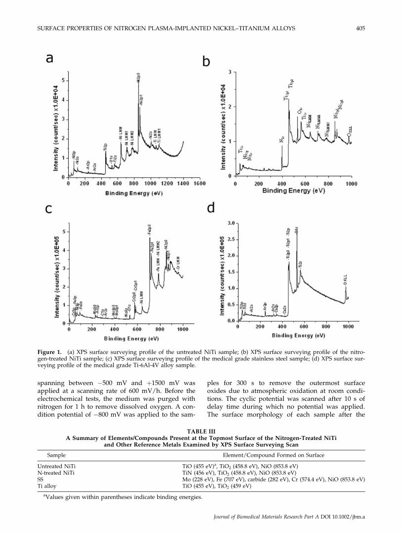
Figure 1. (a) XPS surface surveying profile of the untreated NiTi sample; (b) XPS surface surveying profile of the nitro-gen-treated NiTi sample; (c) XPS surface surveying profile of the medical grade stainless steel sample; (d) XPS surface sur-veying profile of the medical grade Ti-6Al-4V alloy sample.
TABLE IIIA Summary of Elements/Compounds Present at the Topmost Surface of the Nitrogen-Treated NiTi
and Other Reference Metals Examined by XPS Surface Surveying Scan
Sample Element/Compound Formed on Surface
Untreated NiTi TiO (455 eV)a, TiO2 (458.8 eV), NiO (853.8 eV)N-treated NiTi TiN (456 eV), TiO2 (458.8 eV), NiO (853.8 eV)SS Mo (228 eV), Fe (707 eV), carbide (282 eV), Cr (574.4 eV), NiO (853.8 eV)Ti alloy TiO (455 eV), TiO2 (459 eV)
aValues given within parentheses indicate binding energies.
SURFACE PROPERTIES OF NITROGEN PLASMA-IMPLANTED NICKEL–TITANIUM ALLOYS 405
Journal of Biomedical Materials Research Part A DOI 10.1002/jbm.a
test was studied using scanning electron microscopy(JSM-820; JEOL, Japan).
Immersion tests
Two samples of each type were immersed in 25 mLof SBF in polypropylene (pp) bottles. The pp bottleswere closed tightly and incubated in a thermostaticchamber at (37 6 0.1)8C for 5 weeks. All the bottleswere shaken gently for a few seconds every 3 days.After 5 weeks, the SBF in the bottles were analyzedby inductively coupled plasma mass spectrometry(ICPMS) (PE SCIEX ELAN6100; Perkin Elmer) todetermine the amount of ions leached from eachspecimen.41
Nanoindentation tests
Nanoindentation tests41 (MTS Nano Indenter XP)were conducted on five areas to determine the averagehardness of the samples. Readings were recordedthrough a depth of 200 nm. A three-sided pyramidalBerkovich diamond indenter was employed.
Cell culture experiments
To investigate the cytocompatibility of the plasma-treated and untreated samples, osteoblasts isolatedfrom calvarial bones of 2-day-old mice that ubiqui-
tously expressed an enhanced green fluorescent pro-tein (EGFP) were used in our culture in a Dulbecco’sModified Eagle Medium (DMEM) (Invitrogen) sup-plemented with 10% (v/v) fetal bovine serum (Bio-west, France), antibiotics (100 U/mL of penicillinand 100 lg/mL of streptomycin), and 2 mM L-gluta-mine at 378C in an atmosphere of 5% CO2 and 95%air. The specimens (1 mm thick and 5 mm in diame-ter) were fixed onto the bottom of a 24-well tissueculture plate (Falcon) using 1% (w/v) agarose. A cellsuspension consisting of 5000 cells was seeded ontothe surface of the untreated NiTi samples, the nitro-gen-implanted NiTi, stainless steel, Ti alloy, andwells without any metal discs serving as a controlfor normal culturing conditions. Cells were grown in1 mL of medium and changed every 3 days. Cellattachment was examined after the second day ofculture, and cell proliferation examined after 4, 6,and 8 days of culture. Four samples were taken ateach time point to obtain better statistics. In ourstudy, cells were allowed to reach confluence duringthe examination period. To determine the cell num-ber, the attached cells were released by digestionwith trypsin-EDTA (Invitrogen) and counted using ahaematocytometer (Tiefe Depth Profondeur, Marien-feld, Germany). Cell viability was assessed by stain-ing with 0.2% Trypan blue (Sigma). The number ofcells was expressed as a mean value 6 standarddeviation (SD). The data were analyzed by usingunpaired two-sample t test and the statistical analy-sis was performed using the SPSS program (SPSS forWindows, Release 11.0.0).
Cell proliferation was observed by using a fluores-cent microscope (Axioplan 2; Carl Zeiss, Germany).The attached living EGFP-expressing osteoblastswere visualized using a 450–490-nm incident filterand the fluorescence images emitted at 510 nm werecaptured using a Sony DKS-ST5 digital camera.
Static surface contact angle measurement
Static contact angle analysis was conducted usingthe Rame–Hart Goniometer. A 5-lL droplet of 13DMEM solution (Invitrogen) at room temperaturewas injected on the sample surfaces. The 13 DMEMsolution was used as the contact liquid instead ofwater or Hank’s solution to better simulate theactual environment for cell adhesion. To get goodstatistical averages, five measurements were per-formed on each sample including the untreatedNiTi, nitrogen-treated NiTi, stainless steel, and Tialloy. The data presented here represent the meanvalues 6 SDs of the measurements of the left andright angles of the droplets. The unpaired two-sam-ple t test was performed on the data and the SPSS
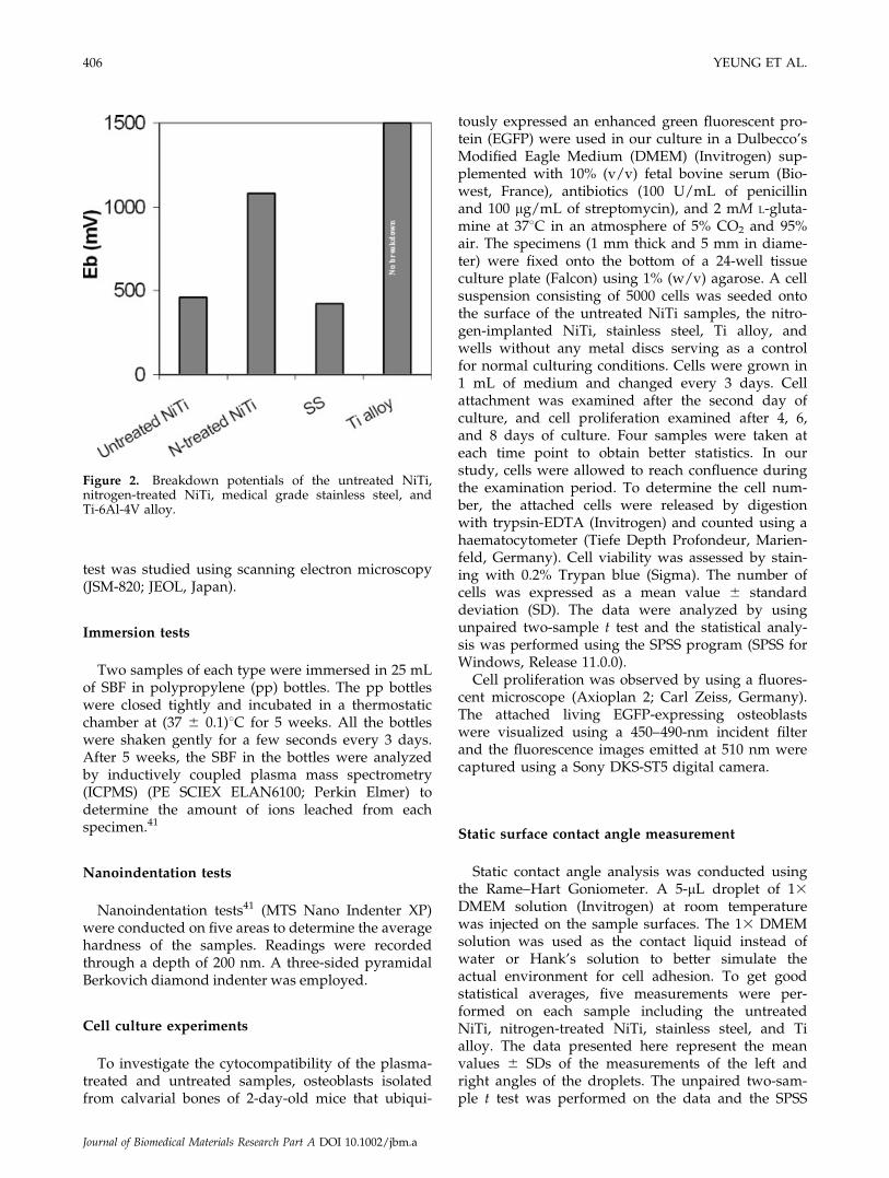
Figure 2. Breakdown potentials of the untreated NiTi,nitrogen-treated NiTi, medical grade stainless steel, andTi-6Al-4V alloy.
406 YEUNG ET AL.
Journal of Biomedical Materials Research Part A DOI 10.1002/jbm.a
program (SPSS for Windows, Release 11.0.0) wasused for statistical analysis
Surface roughness and topography analysis
The surface roughness and topography of un-treated and plasma-treated samples were character-ized by atomic force microscopy (AFM) (Auto ProbeCP; Park Scientific Instruments). The measurementwas operated in the contact mode. The scanningarea was 1 3 1 lm2 under room ambient condition.
EXPERIMENTAL RESULTS
Surface chemical composition analysis
Figure 1(a–d) reveal the surface chemical composi-tions of the untreated NiTi, N-PIII NiTi, medicalgrade stainless steel, and Ti-6Al-4V alloy, respec-tively. Table III lists the detected elements and com-pounds derived from their binding energies. Themajor compounds found in the untreated NiTi sam-
ples surface are TiO, TiO2, and NiO. On the nitro-gen-implanted surfaces, TiN, TiO2, and smallamounts of NiO are detected. However, the depthprofiles (data not shown here) of the N-PIII samplesuggest that the NiO concentration is quite low com-pared with that on the untreated NiTi sample. Ironis abundant on the medical grade stainless steel sur-face (depth profile data not shown here). In additionto small amounts of Cr and Mo, some carbides andNiO are present. On the medical grade Ti alloy sam-ples, TiO and TiO2 are found.
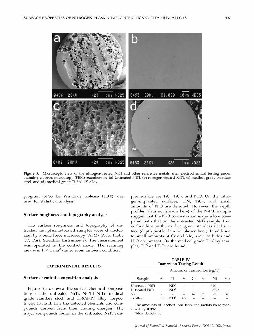
Figure 3. Microscopic view of the nitrogen-treated NiTi and other reference metals after electrochemical testing underscanning electron microscopy (SEM) examination. (a) Untreated NiTi, (b) nitrogen-treated NiTi, (c) medical grade stainlesssteel, and (d) medical grade Ti-6Al-4V alloy.
The amounts of leached ions from the metals were mea-sured by ICPMS.
aNon detectable.
SURFACE PROPERTIES OF NITROGEN PLASMA-IMPLANTED NICKEL–TITANIUM ALLOYS 407
Journal of Biomedical Materials Research Part A DOI 10.1002/jbm.a
Corrosion resistance analysis
The essential readings from our electrochemicaltests in lieu of the complete potentiodynamic curvesare shown on Figure 2. The breakdown potential isdesignated by Eb. Larger Eb values represent bettercorrosion resistance. The Eb values measured fromthe untreated NiTi, N-PIII NiTi, and SS sample are461, 1080, and 422 mV, respectively. No breakdownis observed in the Ti alloy sample under the mea-surement conditions. Therefore, the corrosion resist-ance of the four samples in descending order is Tialloy > N-PIII NiTi > untreated NiTi > SS. The N-PIII samples exhibit higher Eb values than theuntreated NiTi and SS samples. These results sug-gest that the corrosion resistance of the N-PIII sam-ples is better than both the untreated NiTi and SS.
The surface morphologies of the samples afterelectrochemical tests are shown in Figure 3. Theholes on the nitrogen-treated surfaces are very tiny,whereas much bigger holes with irregular shapes arefound on the untreated NiTi and SS sample surfaces.No trace of corrosion is observed on the Ti sample.The corrosion resistance of NiTi has been signifi-cantly improved after nitrogen plasma treatment.However, its ability to resist corrosion is still not asgood as Ti alloy.
Ions leached from the materials
The amounts of ions leached from the untreated,N-PIII, SS, and Ti alloy samples after the immersiontests are listed on Table IV. The ion concentrationsare determined by ICPMS. The Ni concentration
leached from the untreated sample is 320 lg/L,whereas that from the N-PIII sample is 57.9 lg/L. NoTi ion is found to be leached from the untreated andN-PIII NiTi samples. With regard to the SS sample,the leached Cr concentration is 47 lg/L, Fe 35 lg/L,Ni 22 lg/L, and Mo 3.8 lg/L. In addition to 18 lg/Lof Al, there is about 4.2 lg/L of V leached from theTi alloy. Again, no Ti ion release is observed.
Surface hardness analysis
Nanoindentation is applied to evaluate the hard-ness (H) of the untreated control and implantedsample surfaces. The hardness profiles of theuntreated, N-PIII, SS, and Ti alloy samples areshown in Figure 4 and the results are summarizedin Table V.
In the N-PIII NiTi sample, the maximum hardnessis 7.7 GPa on the surface. It gradually decreases to4.5G Pa at a depth of 165 nm. In the untreated NiTisample, the maximum hardness is 5.2 GPa at around30 nm from the surface and gradually diminishes to4.7 GPa at 150 nm. In the SS sample, the hardnessexhibits the maximum value of 6.7 GPa at 50 nmfrom the surface and then decreases gradually to arather constant value of 5.2 GPa at 200 nm from the
Figure 4. Hardness profiles of the untreated NiTi, nitro-gen-treated NiTi, medical grade stainless steel, and Ti-6Al-4V alloy.
TABLE VSurface Hardness Testing Result of the Untreated NiTi,Nitrogen-Treated NiTi, Medical Grade Stainless Steel,
and Ti-6Al-4V Alloy
Sample Hardness (GPa)
Untreated NiTi 5.2–4.7N-treated NiTi 7.7–4.5SS 6.7–5.2Ti alloy 9.2–6.0
Figure 5. Plot of cell proliferation on the untreated NiTi,nitrogen-treated NiTi, medical grade stainless steel, andTi-6Al-4V alloy versus number of days. [Color figure canbe viewed in the online issue, which is available at www.interscience.wiley.com.]
408 YEUNG ET AL.
Journal of Biomedical Materials Research Part A DOI 10.1002/jbm.a
surface. In the Ti alloy sample, the maximum hardnessis found to be 9.2 GPa at 40 nm from the surface andprogressively decreases to 6.0 GPa at 200 nm.
Our results suggest that the hardness of the N-PIIIsurface is generally higher than that of the untreatedNiTi substrate at 0–75 nm. Compared with the SSsample, the hardness of the N-PIII layer in the first25 nm from the surface is higher. However, thehardness of the N-PIII NiTi sample is lower thanthat of the Ti sample except in the topmost region.In general, the surface hardness of the N-PIII NiTi ismore superior than that of the untreated NiTi andSS samples, but inferior to that of Ti.
Cell proliferation analysis
Figure 5 plots the cell proliferation versus numberof days and shows that the N-PIII sample is well tol-
erated by the EGFP-expressing osteoblasts. After cul-turing for 2 days, the cells start to attach to and pro-liferate on all the samples except for stainless steel.After 4 days, cell proliferation on the untreated NiTialloy samples is slightly higher than that of the N-PIII NiTi, SS, and Ti alloy samples. However, theN-PIII samples exhibit the highest degree of cell pro-liferation among the samples after 6 and 8 days ofculturing. Cell proliferation on the SS and Ti alloysamples are significantly lower than that on thenitrogen-treated NiTi and the untreated NiTi controlsample after 8 days (p < 0.05). The cell proliferationobserved on the untreated NiTi, N-PIII NiTi, stain-less steel, and titanium alloy samples after 2 and 8days of culturing are shown in Figures 6 and 7,respectively. It can be clearly observed that cells areattached to and proliferate on all the samples. Theresults of cell culturing unequivocally demonstrate
Figure 6. Microscopic view of the nitrogen-treated NiTi and other reference metals after 2 days of cell culture, using theEGFP-expressing mouse osteoblasts. Proliferation clusters are obviously seen on all the surfaces (3100). (a) UntreatedNiTi, (b) nitrogen-treated NiTi, (c) medical grade stainless steel, and (d) medical grade Ti-6Al-4V alloy. [Color figure canbe viewed in the online issue, which is available at www.interscience.wiley.com.]
SURFACE PROPERTIES OF NITROGEN PLASMA-IMPLANTED NICKEL–TITANIUM ALLOYS 409
Journal of Biomedical Materials Research Part A DOI 10.1002/jbm.a
that there is no immediate short-term cytotoxiceffects on the N-PIII NiTi samples. The stainless steelsamples show the least degree of cell proliferation af-ter 8 days of cell culturing. An insignificant amountof dead cells emerges after 8 days of culturing, per-haps due to cell apoptosis.
Surface contact angle analysis
Figure 8 shows the surface contact angles meas-ured from the different surfaces in descending order:stainless steel > titanium alloy > nitrogen treatedNiTi > untreated NiTi. The stainless steel samplepossesses the largest contact angle of 91.138, whereasthe untreated NiTi has the smallest contact angle of77.998. The untreated NiTi and nitrogen-treated NiTiare shown to be potentially more favorable for cell
adhesion than stainless steel and Ti alloy (p < 0.05).It seems that the contact angle of the untreated NiTiis slightly superior than that of the N-PIII NiTi.However, the difference is not statistically signifi-cant.
Surface roughness and topography measurement
Figure 9(a–d) reveal the surface topographies ofthe untreated NiTi, nitrogen-treated NiTi, stainlesssteel, and titanium alloy. The RMS surface roughnessvalues of the untreated NiTi, nitrogen-treated NiTi,stainless steel, and titanium are 1.61, 2.04, 1.61, and2.42 nm, respectively (Table VI). It seems that theroughness is slightly increased after plasma treat-ment. However, compared with the surface topogra-phies of the untreated NiTi, stainless steel, and tita-
Figure 7. Microscopic view of the nitrogen-treated NiTi and other reference metals after 8 days of cell culture, using theEGFP-expressing mouse osteoblasts (3100). (a) Untreated NiTi, (b) nitrogen-treated NiTi, (c) medical grade stainlesssteel, and (d) medical grade Ti-6Al-4V alloy. [Color figure can be viewed in the online issue, which is available at www.interscience.wiley.com.]
410 YEUNG ET AL.
Journal of Biomedical Materials Research Part A DOI 10.1002/jbm.a
nium alloy, the nitrogen-treated NiTi demonstrates avery specific pattern on the nanoscale.
DISCUSSION
Nitrogen PIII produces a thin layer of TiN on thesurface together with a graded interface with thebulk NiTi substrate. In our previous studies,41 a TiNlayer about 120 nm thick was produced by using200-Hz PIII. In addition to establishing a protectivelayer against corrosion and wear, this treatment can
suppress the surface Ni concentration and reducethe possibility of Ni ion leaching. Poon et al.41
reported that a higher implantation frequency withpostimplantation annealing could enhance the corro-sion resistance and reduce the leaching of Ni.Annealing can further suppress the surface Ni con-tent possibly due to the consolidation of titaniumnitride and titanium oxide, which have higher heatsof formation compared with nickel nitride and nickeloxide. As a result, Ni is segregated away from thesurface. The temperature on the specimen surfaceduring 200-Hz PIII can reach as high as 3508C,which may change the super-elastic transition tem-perature of the shape memory alloy. Thus, in thisstudy, we used a lower pulsing frequency of 50 Hzto achieve a sample temperature of only about 1508Cduring PIII. Under the lower frequency conditions,the TiN layer is about 60 nm. In spite of a lowernitrogen implant dose, improved corrosion resistanceand mitigated Ni out-diffusion can still be observed.Moreover, the surface TiN which can be classified ashard ceramic50 possesses higher surface hardnesscompared with the untreated NiTi and SS at the top25 nm surface region.
Medical grade stainless steels and titanium alloysare the most common implantable materials in medi-cine. These metals, especially commercial pure Tiand Ti-based alloys, are believed to possess goodcorrosion resistance and deformability. A number of
Figure 8. Plot of surface contact angle measurements ofthe untreated NiTi, nitrogen-treated NiTi, medical gradestainless steel, and Ti-6Al-4V alloy.
Figure 9. 3-D AFM topograph of the nitrogen-treated NiTi and other reference metals. Scanned area is 1 3 1 lm2, andAFM used in tapping mode at 258C. (a) Untreated NiTi, (b) nitrogen-treated NiTi, (c) medical grade stainless steel, and (d)medical grade Ti-6Al-4V alloy. [Color figure can be viewed in the online issue, which is available at www.interscience.wiley.com.]
SURFACE PROPERTIES OF NITROGEN PLASMA-IMPLANTED NICKEL–TITANIUM ALLOYS 411
Journal of Biomedical Materials Research Part A DOI 10.1002/jbm.a
studies show that they are compatible with livingtissues.51–57 However, it is known that stainlesssteels contain small amount of nickel (Ni) and chro-mium (Cr) to enhance the corrosion resistance. Ni istoxic to living tissues and reported to be carcino-genic as well. Furthermore, Cr may cause impair-ment of osteoblast proliferation and differentiationas well as cytokine release.20,58,59 Our XPS surveyreveals that Cr and Ni are detected on the surface ofthe medical grade stainless steel samples. Immersiontest results also confirm out-leaching of these ions.Schmidt et al.1 and Okazaki et al.60 also reportedthat stainless steel had poor corrosion resistanceunder physiological conditions that resulted into Niand Cr ions release. These findings may explain theinsignificant growth of osteoblast on the stainlesssteel samples in our study. In order to improve thecorrosion resistance of stainless steel, many methodshave been investigated to suppress the release ofsuch toxic ions.61–64
The use of NiTi alloy in human implants is stillcontroversial due to its extremely high nickel con-centration compared with other medical grade met-als such as stainless steel and titanium alloy. Ad-verse effects, such as nickel ion leaching fromimplants, have been reported in humans.65 In vivoand in vitro studies indicate that the rate of cell pro-liferation on NiTi samples is lower compared withstainless steels and Ti alloys.66 However, our cellculturing experiments show higher proliferation onuntreated NiTi samples than medical grade stainlesssteel and Ti alloy after 8 days of culturing (p < 0.05).Furthermore, the N-PIII samples show more superiorcell results than stainless steel and Ti alloy samplesafter 8 days of culturing (p < 0.05). In addition tomore superior surface mechanical properties,67,68 theN-PIII NiTi favors osteoblast proliferation. Basedon reports in the literature, the TiN coating is welltolerated by different cells, particularly bonecells.55,65,69,70 This phenomenon can be attributed tothe growth of the calcium phosphate phase on thesurface of TiN-coated titanium implant, whereassuch activities do not take place on the untreated ti-tanium implants.71 Our surface composition analysisreveals that the surface-treated layer consists of themixed precipitates of TiOxNy oxynitride. This coatingis favorable to bone-like material formation under
in vivo conditions. Our cell culturing results alsosuggest that the N-PIII samples are as good as, if notbetter than, the untreated NiTi alloy.17,20,21,24,27,55,72
Czarnowska et al. confirmed our results that thenitriding layer possesses better cell proliferation overthe untreated layer with oxide.55 Regarding the Tialloy samples, our study suggests that the cellgrowth on this metal is significantly less than thaton the untreated NiTi alloy and N-PIII samples (p <0.05). The surface composition analysis indicates thata protective and biocompatible layer (TixOy) isformed on the surface. However, this layer is not asgood as the nitriding layer in terms of cell prolifera-tion. Additionally, our static surface contact anglemeasurement results indicate that the nitrogen-treated and untreated NiTi surfaces are more hydro-philic than the SS and Ti alloy. Our results agree withother previous reports that the hydrophilic surfacefavors cell adhesion and proliferation.73,74 Therefore,this may be the explanation of why there no cells arefound on SS after 2 days of culturing.
In addition to the surface free energy and surfacechemistry, it should be noted that other parameterssuch as surface roughness may also affect the rate ofcell attachment and proliferation.18,73,75 The nitrogen-treated NiTi demonstrates a specific pattern afterplasma treatment. The surface with the nanomor-phology very likely favor to cell proliferation. Thesurface roughness also likely alters the corrosion re-sistance of the materials. If corrosion resistance iscompromised, leaching of metal ions is very possi-ble, thereby lowering the cytocompatibility of thesubstrate. PIII is a better surface modification tech-nique to improve the corrosion resistance and cellproliferation of medical implants, especially implantswith sophisticated shapes.55 However, it should bementioned that this study only reveals the short-termcytocompatibility effects on those surface-treatedsamples. A long-term cytotoxicity test up to a year isnecessary prior to subjecting these surface-treatedmaterials to clinical use.
CONCLUSION
This study reveals that a graded TiN layer is formedon the surface of NiTi alloy after nitrogen PIII. Theenhanced surface possesses better corrosion and wearproperties than the untreated NiTi and medical gradestainless steel, and the surface properties are compara-ble to those of Ti alloy. In terms of cytocompatibility,the cell viability on stainless steel and titanium is infe-rior to that on the untreated NiTi and N-PIII NiTi.Medical grade stainless steels show the least amount ofcells. Our data suggest that nitrogen PIII is favorable toosteoblast proliferation. All in all, this new surface-
TABLE VISurface Roughness (RMS) of the Untreated NiTi,
Nitrogen-Treated NiTi, Medical Grade Stainless Steel,and Ti-6Al-4V Alloy
Sample RMS (nm)
Untreated NiTi 1.61N-treated NiTi 2.08SS 1.61Ti alloy 2.42
412 YEUNG ET AL.
Journal of Biomedical Materials Research Part A DOI 10.1002/jbm.a
modification treatment will advance the implant tech-nology in the biomedical area.
We thank Mr. Wilson W.C. Chan for his expert technicalwork.
References
1. Schmidt C, Ignatius AA, Claes LE. Proliferation and differen-tiation parameters of human osteoblasts on titanium and steelsurfaces. J Biomed Mater Res 2000;54:209–215.
2. Disegi J, Eschbach L. Stainless steel in bone surgery. Injury2000;31(Suppl 4):4–6.
3. Woodman JL, Jacobs JJ, Galante JO, Urban RM. Metal ionrelease from titanium-based prosthetic segmental replace-ments of long bones in baboons: A long-term study. J OrthopRes 1984;1:421–430.
4. Chen MF, Yang XJ, Hu RX, Cui ZD, Man HC. Bioactive NiTishape memory alloy used as bone bonding implants. MaterSci Eng C 2004;24:497–502.
5. Cragg AH, De-Jong SC, Barnhart WH, Landas SK, Smith TP.Nitinol intravascular stent: Results of preclinical evaluation.Radiology 1993;189:775–778.
6. Dai K, Chu Y. Studies and applications of NiTi shape mem-ory alloys in the medical field in China. Biomed Mater Eng1996;6:233–240.
7. Es-Souni M, Es-Souni M, Brandies HF. On the transformationbehaviour, mechanical properties and biocompatibility of twoNiTi-based shape memory alloys: NiTi42 and NiTi42Cu7.Biomaterials 2001;22:2153–2161.
8. Gil FX, Planell JA, Manero JM. Relevant aspects in the clincalapplications of NiTi shape memory alloys. J Mater Sci MaterMed 1996;7:403–406.
9. Kong X, Grabitz RG, van Oeveren W, Klee D, van KootenTG, Freudenthal F, Qing M, von Bernuth G, Seghaye M-C.Effect of biologically active coating on biocompatibility ofNitinol devices designed for the closure of intra-atrial com-munications. Biomaterials 2002;23:1775–1783.
10. Kujala S, Ryhanen J, Danilov A, Tuukkanen J. Effect of porosityon the osteointegration and bone ingrowth of a weight-bearingnickel–titanium bone graft substitute. Biomaterials 2003;24:4691–4697.
11. Kujala S, Ryhanen J, Jamsa T, Danilov A, Saaranen J, PramilaA, Tuukkanen J. Bone modeling controlled by a nickel–tita-nium shape memory alloy intramedullary nail. Biomaterials2002;23:2535–2543.
12. Yang PJ, Zhang YF, Ge MZ. Internal fixation with NiTi shapememory alloy compressive staples in orthopedic surgery.Chin Med J 1987;100:712–714.
13. Ryhanen J, Kallioinen M, Serlo W, Peramaki P, Junila J, Sand-vik P, Niemela E, Tuukkanen J. Bone healing and mineraliza-tion, implant corrosion, and trace metals after nickel–titaniumshape memory metal intramedullary fixation. J Biomed MaterRes 1999;47:472–480.
14. Miyazaki S. Medical and dental application of shape memoryalloys. In: Otsuka K, Wayman CM, editors. Shape MemoryMaterials. Cambridge: Cambridge University Press; 1998. p 267–281.
15. Sanders JO, Sanders AE, More R, Ashman RB. A preliminaryinvestigation of shape memory alloys in the surgical correc-tion of scoliosis. Spine 1993;18:1640–1646.
16. Schmerling MA, Wilkov MA, Sanders AE, Woosley JE. Usingthe shape recovery of nitinol in the Harrington rod treatmentof scoliosis. J Biomed Mater Res 1976;10:879–892.
17. Bogdanski D, Koller M, Muller D, Muhr G, Bram M, Buchk-remer HP, Stover D, Choi J, Epple M. Easy assessment of the
biocompatibility of Ni-Ti alloys by in vitro cell culture experi-ments on a functionally graded Ni-NiTi-Ti material. Biomate-rials 2002;23:4549–4555.
18. El Medawar L, Rocher P, Hornez J-C., Traisnel M, Breme J,Hildebrand HF. Electrochemical and cytocompatibility assess-ment of NiTiNOL memory shape alloy for orthodontic use.Biomol Eng 2002;19:153–160.
19. Putters J, Kaulesar S, De Z, Bijma A, Besselink P. Compara-tive cell culture effects of shape memory metal (Nitinol),nickel and titanium: A biocompatibility estimation. Eur SurgRes 1992;24:378–382.
20. Kapanen A, Ilvesaro J, Danilov A, Ryhanen J, Lehenkari P,Tuukkanen J. Behaviour of Nitinol in osteoblast-like ROS-17cell cultures. Biomaterials 2002;23:645–650.
21. Kapanen A, Kinnunen A, Ryhanen J, Tuukkanen J. TGF-[b]1secretion of ROS-17/2.8 cultures on NiTi implant material.Biomaterials 2002;23:3341–3346.
22. Kapanen A, Ryhanen J, Danilov A, Tuukkanen J. Effect ofnickel–titanium shape memory metal alloy on bone forma-tion. Biomaterials 2001;22:2475–2480.
23. Kujala S, Pajala A, Kallioinen M, Pramila A, Tuukkanen J, Ryha-nen J. Biocompatibility and strength properties of nitinol shapememory alloy suture in rabbit tendon. Biomaterials 2004;25:353–358.
24. Rocher P, El Medawar L, Hornez J-C., Traisnel M, Breme J,Hildebrand HF. Biocorrosion and cytocompatibility assessmentof NiTi shapememory alloys. ScrMater 2004;50:255–260.
25. Ryhanen J, Kallioinen M, Tuukkanen J, Junila J, Niemela E,Sandvik P, Serlo W. In vivo biocompatibility evaluation ofnickel–titanium shape memory metal alloy: Muscle and peri-neural tissue responses and encapsule membrane thickness.J Biomed Mater Res 1998;41:481–488.
26. Ryhanen J, Niemi E, Serlo W, Niemela E, Sandvik P, PernuH, Salo T. Biocompatibility of nickel–titanium shape memorymetal and its corrosion behavior in human cell cultures.J Biomed Mater Res 1997;35:451–457.
27. Wever DJ, Veldhuizen AG, Sanders MM, Schakenraad JM,van Horn JR. Cytotoxic, allergic and genotoxic activity of anickel–titanium alloy. Biomaterials 1997;18:1115–1120.
28. Berger-Gorbet M, Broxup B, Rivard C, Yahia LH. Biocompatibil-ity testing of NiTi screws using immunohistochemistry on sec-tions containing metallic implants. J Biomed Mater Res 1996;32:243–248.
29. Jia W, Beatty MW, Reinhardt RA, Petro TM, Cohen DM,Maze CR, Strom EA, Hoffman M. Nickel release from ortho-dontic arch wires and cellular immune response to variousnickel concentrations. J Biomed Mater Res 1999;48:488–495.
30. Es-Souni M, Es-Souni M, Fischer-Brandies H. On the proper-ties of two binary NiTi shape memory alloys. Effects of sur-face finish on the corrosion behaviour and in vitro biocom-patibility. Biomaterials 2002;23:2887–2894.
31. Shih CC, Lin SJ, Chen YL, Su YY, Lai ST, Wu GJ, Kwok CF,Chung KH. The cytotoxicity of corrosion products of nitinolstent wire on cultured smooth muscle cells. J Biomed MaterRes 2000;52:395–403.
32. Dalmau L, Alberty H, Parra J. A study of nickel allergy.J Prosthet Dent 1984;52:116–119.
33. Lamster I, Kalfus D, Steigerwald P, Chasens A. Rapid loss ofalveolar bone associated with nonprecious alloy crowns intwo patients with nickel hypersensitivity. J Periodontol 1987;58:486–492.
34. Espana A, Alonso ML, Soria C, Guimaraens D, Ledo A.Chronic urticaria after implantation of 2 nickel-containingdental prostheses in a nickel-allergic patient. Contact Derma-titis 1989;21:204–206.
35. Sanford WE, Niboer E.Renal toxicity of nickel in humans. In:Niboer E, Nriagu JO, editors. Nickel and Human HealthCurrent Perspectives. Canada:Wiley; 1992. p 123–134.
SURFACE PROPERTIES OF NITROGEN PLASMA-IMPLANTED NICKEL–TITANIUM ALLOYS 413
Journal of Biomedical Materials Research Part A DOI 10.1002/jbm.a
36. Tan L, Dodd RA, Crone WC. Corrosion and wear-corrosionbehavior of NiTi modified by plasma source ion implanta-tion. Biomaterials 2003;24:3931–3939.
37. Tan L, Crone WC. Surface characterization of NiTi modified byplasma source ion implantation. ActaMater 2002;50:4449–4460.
38. Cheng Y, Wei C, Gan KY, Zhao LC. Surface modification ofTiNi alloy through tantalum immersion ion implantation.Surf Coat Technol 2004;176:261–265.
39. Poon RWY, Ho JPY, Liu X, Chung CY, Chu PK, Yeung KWK,Lu WW, Cheung KMC. Anti-corrosion performance of oxi-dized and oxygen plasma-implanted NiTi alloys. Mater SciEng A 2004;390:444–451.
40. Chu PK, Chen JY, Wang LP, Huang N. Plasma-surface modi-fication of biomaterials. Mater Sci Eng R Rep 2002;36:143–206.
41. Poon RWY, Ho JPY, Liu X, Chung CY, Chu PK, Yeung KWK,Lu WW, Cheung KMC. Formation of titanium nitride barrierlayer in nickel–titanium shape memory alloys by nitrogenplasma immersion ion implantation for better corrosion re-sistance. Thin Solid Films 2005;488:20–25.
42. Poon RWY, Liu XY, Chung CY, Chu PK, Yeung KWK, LuWW, Cheung KMC. Surface and corrosion characteristics ofcarbon plasma implanted and deposited nickel–titaniumalloy. J Vac Sci Technol A Vac Surf Films 2005;23:525–530.
43. Poon RWY, Yeung KWK, Liu XY, Chu PK, Chung CY, LuWW, Cheung KMC, Chan D. Carbon plasma immersion ionimplantation of nickel–titanium shape memory alloys. Bioma-terials 2005;26:2265–2272.
44. Yeung KWK, Poon RWY, Liu XY, Ho JPY, Chung CY, ChuPK, Lu WW, Chan D, Cheung KMC. Investigation of nickelsuppression and cytocompatibility of surface-treated nickel–titanium shape memory alloys by using plasma immersionion implantation. J Biomed Mater Res A 2005;72:238–245.
45. Chu PK. Recent developments and applications of plasmaimmersion ion implantation. J Vac Sci Technol B Microelec-tron Nanometer Struct 2004;22:289–296.
46. Chu P, Tang B, Cheng Y, Ko P. Principles and characteristicsof a new generation plasma immersion ion implanter. RevSci Instrum 1997;68:1866–1874.
47. Chu P, Tang B, Wang L, Wang X, Wang S, Huang N. Third-generation plasma immersion ion implanter for biomedicalmaterials and research. Rev Sci Instrum 2001;72:1660–1665.
48. Starosvetsky D, Gotman I. Corrosion behavior of titaniumnitride coated Ni–Ti shape memory surgical alloy. Biomateri-als 2001;22:1853–1859.
49. Cho S, Nakanishi K, Kokubo T, Soga N. Dependence of apa-tite formation on silica gel on its structure: Effect of heattreatment. J Am Ceram Soc 1995;78:1769–1774.
50. Vaz F, Cerqueira P, Rebouta L, Nascimento S, Alves E, Gou-deau P, Riviere J, Pischow K, Rijk J. Structural, optical andmechanical properties of coloured TiNxOy thin films. ThinSolid Films 2004;447/448:449–454.
51. Shih CC, Shih CM, Su YY, Su LHJ, Chang MS, Lin SJ. Effect ofsurface oxide properties on corrosion resistance of 316L stainlesssteel for biomedical applications. Corros Sci 2004;46:427–441.
52. Niinomi M. Recent research and development in titaniumalloys for biomedical applications and healthcare goods. SciTechnol Adv Mater 2003;4:445–454.
53. Sgambato A, Cittadini A, Ardito R, Dardeli A, Facchini A,Dalla Pria P, Colombo A. Osteoblast behavior on nanostruc-tured titanium alloys. Mater Sci Eng C 2003;23:419–423.
54. Tsyganov I, Wieser E, Matz W, Reuther H, Richter E. Modifi-cation of the Ti-6Al-4V alloy by ion implantation of calciumand/or phosphorus. Surf Coat Technol 2002;158/159:318–323.
55. Czarnowska E, Wierzchon T, Maranda-Niedbala A. Propertiesof the surface layers on titanium alloy and their biocompatibil-ity in in vitro tests. J Mater Process Technol 1999;92/93:190–194.
56. Lagneau C, Farges J-C., Exbrayat P, Lissac M. Cytokeratinexpression in human oral gingival epithelial cells: In vitro
regulation by titanium-based implant materials. Biomaterials1998;19:1109–1115.
57. Guleryuz H, Cimenoglu H. Effect of thermal oxidation oncorrosion and corrosion-wear behaviour of a Ti-6Al-4V alloy.Biomaterials 2004;25:3325–3333.
58. Swiontkowski M, Agel J, Schwappach J, McNair P, Welch M.Cutaneous metal sensitivity in patients with orthopaedic inju-ries. J Orthop Trauma 2001;2:86–89.
59. Wang J, Wicklund B, Gustilo R, Tsukayama D. Prostheticmetals interfere with the functions of human osteoblast cellsin vitro. Clin Orthop 1997;339:216–226.
60. Okazaki Y, Gotoh E. Comparison of metal release from vari-ous metallic biomaterials in vitro. Biomaterials 2005;26:11–21.
61. Tian X, Fu RKY, Wang L, Chu PK. Oxygen-induced nickelsegregation in nitrogen plasma implanted AISI 304 stainlesssteel. Mater Sci Eng A 2001;316:200–204.
62. Fini M, Nicoli Aldini N, Torricelli P, Giavaresi G, Borsari V,Lenger H, Bernauer J, Giardino R, Chiesa R, Cigada A. Anew austenitic stainless steel with negligible nickel content:An in vitro and in vivo comparative investigation. Biomateri-als 2003;24:4929–4939.
63. Olivares R, Rodil SE, Arzate H. In vitro studies of the biomi-neralization in amorphous carbon films. Surf Coat Technol2004;177/178:758–764.
64. Bordjih K, Jouzeau J-Y., Mainard D, Payan E, Delagoutte J-P.,Netter P. Evaluation of the effect of three surface treatmentson the biocompatibility of 316L stainless steel using humandifferentiated cells. Biomaterials 1996;17:491–500.
65. Boyan BD, Hummert TW, Dean DD, Schwartz Z. Role of ma-terial surface in regulating bone and cartilage response. Bio-materials 1996;17:137–146.
66. Ponsonnet L, Comte V, Othmane A, Lagneau C, CharbonnierM, Lissac M, Jaffrezic N. Effect of surface topography andchemistry on adhesion, orientation and growth of fibroblastson nickel–titanium substrates. Mater Sci Eng C 2002;21:157–165.
67. Girardeau T, Bouslykhane K, Mimault J, Villain JP, Chartier P.Wear improvement and local structure in nickel–titanium coat-ings produced by reactive ion sputtering. Thin Solid Films1996;283:67–74.
68. Grant DM, Green SM, Wood JV. The surface performance ofshot peened and ion implanted NiTi shape memory alloy.Acta Metall Mater 1995;43:1045–1051.
69. Rie KT, Stucky T, Silva RA, Leitao E, Bordji K, Jouzeau J-Y.,Mainard D. Plasma surface treatment and PACVD on Ti alloysfor surgical implants. Surf Coat Technol 1995;74/75(Part 2):973–980.
70. Aronsson BO, Lausmaa J, Kasemo B. Glow discharge plasmatreatment for surface cleaning and modification of metallicbiomaterials. J Biomed Mater Res 1997;35:49–73.
71. Piscanec S, Colombi Ciacchi L, Vesselli E, Comelli G, Sbai-zero O, Meriani S, De Vita A. Bioactivity of TiN-coated tita-nium implants. Acta Mater 2004;52:1237–1245.
72. Narayan J, Fan WD, Narayan RJ, Tiwari P, Stadelmaier HH.Diamond, diamond-like and titanium nitride biocompatiblecoatings for human body parts. Mater Sci Eng B 1994;25:5–10.
73. Hallab NJ, Bundy KJ, O’connor K, Moses RL, Jacobs JJ. Eval-uation of metallic and polymeric biomaterial surface energyand surface roughness characteristics for directed cell adhe-sion. Tissue Eng 2001;7:55–71.
74. Spriano S, Bosetti M, Bronzoni M, Verne E, Maina G, BergoV, Cannas M. Surface properties and cell response of lowmetal ion release Ti-6Al-7Nb alloy after multi-step chemicaland thermal treatments. Biomaterials 2005;26:1219–1229.
75. Ponsonnet L, Reybier K, Jaffrezic N, Comte V, Lagneau C,Lissac M, Martelet C. Relationship between surface properties(roughness, wettability) of titanium and titanium alloys andcell behaviour. Mater Sci Eng C 2003;23:551–560.
414 YEUNG ET AL.
Journal of Biomedical Materials Research Part A DOI 10.1002/jbm.a