Page 1
Surface microstructuring of Ti plates by
femtosecond lasers in liquid ambiences: a new
approach to improving biocompatibility
Yang Yang,1 Jianjun Yang,
1,* Chunyong Liang,
2 Hongshui Wang,
2 Xiaonong Zhu,
1 and
Nan Zhang1
1Institute of Modern Optics, Nankai University, Key Laboratory of Opto-electronic Information Science and
Technology, Education Ministry of China, Tianjin, 300071, China 2School of Materials Science and Engineering, Hebei University of Technology, Tianjin 300130, China
*[email protected]
Abstract: Microstructuring of Ti plates with femtosecond laser pulses is
investigated in three different liquids. In these ambiences, complex
microstructures with voids and islands are produced on the sample surfaces,
whose feature sizes are controlled by the laser parameters. Through adopting
supersaturated Hydroxyapatite suspension with higher incident laser
fluences, it is for the first time to observe the firm deposition of
biocompatible elements Ca-P on the microstructures. At lower laser fluence,
only porous structure is present but without additional elements deposition.
Both plasma-related ablation under the confinement of liquids and
micro-bubbles striking are employed to discuss such structures formation.
Tight combining elements Ca-P onto the structured surfaces provide a new
way to improve the biocompatibility of body-embedded devices.
©2009 Optical Society of America
OCIS codes: (140.3390) Laser materials processing; (140.7090) Ultrafast lasers; (220.4000)
Microstructure fabrication.
References and links
1. Y. Yang, J. Yang, C. Liang, and H. Wang, “Ultra-broadband enhanced absorption of metal surfaces structured by
femtosecond laser pulses,” Opt. Express 16(15), 11259–11265 (2008).
2. G. H. Welsh, N. T. Hunt, and K. Wynne, “Terahertz-pulse emission through laser excitation of surface plasmons in
a metal grating,” Phys. Rev. Lett. 98(2), 026803 (2007).
3. W. Q. Han, L. Wu, R. F. Klie, and Y. Zhu, “Enhanced optical absorption induced by dense nanocavities inside
titania nanorods,” Adv. Mater. 19(18), 2525–2529 (2007).
4. Y. B. Gerbig, S. I. U. Ahmed, D. G. Chetwynd, and H. Haefke, “Topography-related effects on the lubrication of
nanostructured hard surfaces,” Tribol. Int. 39(9), 945–952 (2006).
5. T. J. Webster, and J. U. Ejiofor, “Increased osteoblast adhesion on nanophase metals: Ti, Ti6Al4V, and CoCrMo,”
Biomaterials 25(19), 4731–4739 (2004).
6. M. Birnbaum, “Semiconductor surface damage produced by ruby lasers,” J. Appl. Phys. 36(11), 3688–3689
(1965).
7. A. Y. Vorobyev, and C. Guo, “Femtosecond laser nanostructuring of metals,” Opt. Express 14(6), 2164–2169
(2006).
8. Q. Z. Zhao, S. Malzer, and L. J. Wang, “Formation of subwavelength periodic structures on tungsten induced by
ultrashort laser pulses,” Opt. Lett. 32(13), 1932–1934 (2007).
9. Q. Wu, Y. Ma, R. Fang, Y. Liao, Q. Yu, X. Chen, and K. Wang, “Femtosecond laser-induced periodic surface
structure on diamond film,” Appl. Phys. Lett. 82(11), 1703–1705 (2003).
10. Y. Shimotsuma, P. G. Kazansky, J. Qiu, and K. Hirao, “Self-organized nanogratings in glass irradiated by
ultrashort light pulses,” Phys. Rev. Lett. 91(24), 247405 (2003).
11. F. Keilmann, and Y. H. Bai, “Periodic surface structures frozen into CO2 laser- melted quartz,” Appl. Phys., A
Mater. Sci. Process. 29(1), 9–18 (1982).
12. P. P. Rajeev, M. Gertsvolf, C. Hnatovsky, E. Simova, R. S. Taylor, P. B. Corkum, D. M. Rayner, and V. R.
Bhardwaj, “Transient nanoplasmonics inside dielectrics,” J. Phys. At. Mol. Opt. Phys. 40(11), S273–S282 (2007).
13. M. Shen, C. Crouch, J. E. Carey, and E. Mazur, “Femtosecond laser-induced formation of submicrometer spikes on
silicon in water,” Appl. Phys. Lett. 85(23), 5694–5696 (2004).
#117130 - $15.00 USD Received 16 Sep 2009; revised 25 Oct 2009; accepted 27 Oct 2009; published 5 Nov 2009
(C) 2009 OSA 9 November 2009 / Vol. 17, No. 23 / OPTICS EXPRESS 21124
Page 2
14. Y. Yang, J. Yang, C. Liang, H. Wang, X. Zhu, D. Kuang, and Y. Yang, “Sub-wavelength surface structuring of
NiTi alloy by femtosecond laser pulses,” Appl. Phys., A Mater. Sci. Process. 92(3), 635–642 (2008).
15. A. Y. Vorobyev, V. S. Makin, and C. Guo, “Periodic ordering of random surface nanostructures induced by
femtosecond laser pulses on metals,” J. Appl. Phys. 101(3), 034903 (2007).
16. T. H. Her, R. J. Finlay, C. Wu, S. Deliwala, and E. Mazur, “Microstructuring of silicon with femtosecond laser
pulses,” Appl. Phys. Lett. 73(12), 1673–1675 (1998).
17. M. Shen, C. Crouch, J. E. Carey, and E. Mazur, “Femtosecond laser-induced formation of submicrometer spikes on
silicon in water,” Appl. Phys. Lett. 85(23), 5694–5696 (2004).
18. M. Shen, J. E. Carey, C. H. Crouch, M. Kandyla, H. A. Stone, and E. Mazur, “High-density regular arrays of
nanometer-scale rods formed on silicon surfaces via femtosecond laser irradiation in water,” Nano Lett. 8(7),
2087–2091 (2008).
19. K. Katayama, H. Yonekubo, and T. Sawada, “Formation of ring patterns surrounded by ripples by single- shot laser
irradiation with ultrashort pulse width at the solid/liquid interface,” Appl. Phys. Lett. 82(24), 4244–4246 (2003).
20. G. Daminelli, J. Kruger, and W. Kautek, “Femtosecond laser interaction with silicon under water confinement,”
Thin Solid Films 467(1-2), 334–341 (2004).
21. T. Sakka, S. Iwanaga, Y. H. Ogata, A. Matsunawa, and T. Takemoto, “Laser ablation at solid–liquid interfaces: An
approach from optical emission spectra,” J. Chem. Phys. 112(19), 8645–8653 (2000).
22. S. Bharati, M. K. Sinha, and D. Basu, “Hydroxyapatite coating by biomimetic method on titanium alloy using
concentrated SBF,” Bull. Mater. Sci. 28(6), 617–621 (2005).
23. X. L. Zhu, J. Chen, L. Scheideler, R. Reichl, and J. Geis-Gerstorfer, “Effects of topography and composition of
titanium surface oxides on osteoblast responses,” Biomaterials 25(18), 4087–4103 (2004).
24. C. Aparicio, J. M. Manero, F. Conde, M. Pegueroles, J. A. Planell, M. Vallet-Regí, and F. J. Gil, “Acceleration of
apatite nucleation on microrough bioactive titanium for bone-replacing implants,” J. Biomed. Mater. Res. A
82A(3), 521–529 (2007).
25. J. P. Sylvestre, A. V. Kabashin, E. Sacher, and M. Meunier, “Femtosecond laser ablation of gold in water: influence
of the laser-produced plasma on the nanoparticle size distribution,” Appl. Phys., A Mater. Sci. Process. 80(4),
753–758 (2005).
26. R. M. Tilaki, A. Irajizad, and S. M. Mahdava, “The effect of liquid environment on size and aggregation of gold
nanoparticles prepared by pulsed laser ablation,” J. Nanopart. Res. 9(5), 853–860 (2007).
27. D. Grojo, J. Hermann, and A. Perrone, “Plasma analyses during femtosecond laser ablation of Ti, Zr, and Hf,” J.
Appl. Phys. 97(6), 063306 (2005).
28. A. Vogel, N. Linz, S. Freidank, and G. Paltauf, “Femtosecond-laser-induced nanocavitation in water: implications
for optical breakdown threshold and cell surgery,” Phys. Rev. Lett. 100(3), 038102 (2008).
29. A. Vogel, J. Noack, G. Huttman, and G. Paltauf, “Mechanisms of femtosecond laser nanosurgery of cells and
tissues,” Appl. Phys. B 81(8), 1015–1047 (2005).
30. J. Noack, D. Hammer, G. Noojin, B. Rockwell, and A. Vogel, “Influence of pulse duration on mechanical effects
after laser-induced breakdown in water,” J. Appl. Phys. 83(12), 7488–7495 (1998).
31. P. V. Kazakevich, A. V. Simakin, and G. A. Shafeev, “Formation of periodic structures by laser ablation of metals
in liquids,” Appl. Surf. Sci. 252(13), 4457–4461 (2006).
1. Introduction
Laser-induced surface microstructures on different materials have been attractive in recent
years, not only because they benefit to exploring laser-matter interaction, but also provide great
applications in the field of physics, chemistry and material science [1–5]. Ever since the first
observation by Birnbaum in 1960s [6], surface microstructures formation under the radiation of
intense laser beam has been reported on a variety of solid materials [7–10]. Although some
mechanisms, including scattered light interference [11], self-organized plasma modification
[12], secondary bubbles ablation in liquids [13] and so on, have been proposed, no single theory
can be commonly accepted for different structuring cases.
For example, laser-induced periodic surface structure has been now mostly attributed to
interference between the incident and scattered lights, wherein the ripple spacing was verified to
depend closely on the incident angles [8,9,11]. However, such theory could not be employed
simply to explain our previous report that the ripple’s periodicity could keep about 630 nm with
varying incident angles [14]. Recently, Guo et al. pointed out that the periodicity of ripples
induced by femtosecond lasers on the metals could be shortened with increasing the number of
laser shots [15]. Anyway, previous studies suggest that the formation of surface microstructures
should be related to the incident laser parameters and material properties.
In fact, the ambient medium parameters have also an important influence on the quality of
laser treatments. For instance, Mazur reported that in the presence of SF6 gases quasi-ordered
arrays of conical spikes could be formed spontaneously on silicon under the irradiation of
#117130 - $15.00 USD Received 16 Sep 2009; revised 25 Oct 2009; accepted 27 Oct 2009; published 5 Nov 2009
(C) 2009 OSA 9 November 2009 / Vol. 17, No. 23 / OPTICS EXPRESS 21125
Page 3
femtosecond lasers [16]. In water, high-density regular arrays of nanometer-scale rods were
induced on silicon surface via femtosecond laser irradiation [17,18]. These phenomena indicate
that laser-induced surface structures formation also depends on the surrounding environments.
Generally, the current femtosecond laser-induced surface morphology experiment in liquids
has mostly employed de-ionized water as the surrounding medium [17,19,20]. During these
experiments, nonlinear phenomena such as supercontinuum and plasma generation could be
observed due to the interaction of water phase with the incoming high-fluence laser beam.
Moreover, for the laser ablation at solid-liquid interface, spectroscopic study revealed that
various chemical reactions between the species ablated from the solid target and the ambient
liquid molecules could take place [21]. Namely, not only the species did come from the solid
target, but also the species originated from the liquid are involved in the interface plasma. So
some new phenomena could be expected if the laser ablation of targets in the presence of
biocompatible solutions.
In this letter, we investigate the ambient medium-related surface microstructures formation
by femtosecond laser pulses when Ti alloy plate samples are submerged into liquid
environments. In distilled water ambience, a new kind of micro-texture consisting of
micro-islands and nano-voids is generated on the laser-exposed surfaces. The feature size of
such structures can be controlled by both the incident laser fluences and the number of laser
shots. In the supersaturated Hydroxyapatite (HA, Ca10(PO4)6(OH)2) suspension and the mixture
solution of CaCl2 and Na3PO4, similar surface microstructures are also produced. The influence
of solution concentration on the surface microstructures formation process is also studied. Both
energy dispersive X-ray (EDX) spectra and X-ray diffraction (XRD) analyses reveal that
bio-compatible elements of Ca and P (Ca-P) are deposited firmly onto the structured surfaces
when the sample are ablated within supersaturated HA suspension. Finally underlying
mechanisms for the formation of surface structures and the deposition of the elements Ca-P in
liquid environments are discussed.
2. Experimental setup
In our experiments, a commercial chirped pulse amplification of Ti:sapphire laser system
(HP-Spitfire, New Port Inc.) was employed as a light source, which operated at a repetition rate
of 1 kHz and provided 50 fs laser pulse trains with the central wavelength of around 800 nm.
The maximum energy of each amplified laser pulse was 2 mJ. The output laser beam was
polarized horizontally and had a diameter of about 8 mm. A single shot auto-correlator (SSA,
Positive Light Inc.) was used to monitor the time duration of the laser pulses in real time.
Selection of pure titanium (Ti) plates as a sample in the experiments is due to their wide
applications in space manufacturing, medical embedment and so on, wherein the modification
of metal surface properties through generating microstructures is very important and much
desirable.
After surfaces of the sample with a dimension of 10 × 10 × 1 mm3 were polished with SiC
emery paper to remove the oxide layers, it was placed on the bottom of an open glass vessel
filled with suspension. The height of the liquid layer above the targets was 2.5 mm. The vessel
was mounted on a computer-controlled x-y-z translation stage (UTM100PPE1, New Port Inc.)
to allow its displacement with pre-set scanning velocity. Alternatively, exposure of the Ti plate
under the layer of liquid was performed by the stationary focal laser beam at the normal
incidence angle through a 10×microscope objective with a numerical aperture of 0.25. The
upper surface of the target was set 100 µm below the focal plane in air but about 760 µm above
the focal plane in liquid, which was determined from a microscopic analysis of craters on the Ti
plate. In this case, the diameter of laser spot on the target was estimated about 62 µm. A
schematic diagram of our experimental setup is shown in Fig. 1.
#117130 - $15.00 USD Received 16 Sep 2009; revised 25 Oct 2009; accepted 27 Oct 2009; published 5 Nov 2009
(C) 2009 OSA 9 November 2009 / Vol. 17, No. 23 / OPTICS EXPRESS 21126
Page 4
Fig. 1. Schematic diagram for microstructuring of Ti plates with femtosecond lasers in liquids.
In order to investigate the influences of liquid media on the surface microstructuring of
metals by femtosecond lasers, we adopted the distilled water and two different kinds of
solutions, including supersaturated HA aqueous suspension and the admixture of CaCl2 and
Na3PO4. The reason why we choose these two kinds of solutions is that their solutes have
essential elements for human bones, so that the elements deposition on Ti surfaces might
improve the biocompatibility of metals for the body embedment. In the supersaturated HA
aqueous solution, the ions of Ca2+
, PO43-
, HPO42-
, H2PO4- as well as HA nanoparticles will
dissolve in the solution, while in the admixture of CaCl2 and Na3PO4, Ca2+
, Na+, PO4
3-, HPO4
2-,
H2PO4- and Cl
- will dissolve in the solution.
All the experiments were carried out in a Class 1000 clean room. Before and after the
experiments, the samples were cleaned by an ultrasonic cleaner with de-ionized water. The
morphology of the laser-exposed metal surfaces was examined with the help of a scanning
electron microscope (SEM, Hitachi S-4800). The deposition chemical elements and their
structures were analyzed through the energy dispersive X-ray spectra and X-ray diffraction.
3. Experimental results
3.1 Formation of surface microstructures in distilled water
Firstly, we put some distilled water into the glass vessel to study the surface morphological
evolution of the targets with varying femtosecond laser fluences and the sample scan speeds, the
measured results are shown by SEM images in Fig. 2. As it can be seen, for a given laser energy
of 300 µJ, corresponding to the laser fluence of 9.9 J/cm2, numerous irregular-shaped small
islands and tiny elliptical voids were generated on the laser-exposed Ti surface at the sample
scan speed of 1.8 mm/s. In this case, the number of laser pulses partially overlapping on each
spot reaches N = 35. The characteristic size of these islands, more or less connected spatially,
ranges from 2.27 µm to 3.4 µm. And the average length of the void is about 880 nm. The
estimated density of micro-islands on the Ti plate amounts to 3.4×107 cm
−2. Particularly, the
top surfaces of the micro-islands appear relatively flat and smooth.
#117130 - $15.00 USD Received 16 Sep 2009; revised 25 Oct 2009; accepted 27 Oct 2009; published 5 Nov 2009
(C) 2009 OSA 9 November 2009 / Vol. 17, No. 23 / OPTICS EXPRESS 21127
Page 5
Fig. 2. Surface morphologies induced by femtosecond laser with different fluences at variation of
the scan speeds in distilled water. The scale bar that applied to all six pictures is 5 µm.
When the sample scan speed was decreased to 0.8 mm/s with partially overlapped laser
pulses of N = 80 at this given laser fluence, very similar microstructures could be observed on
the laser-exposed surface. However, in this case the feature size of the micro-islands began to
increase and the length of the elliptical voids is also enlarged to about 1.43 µm, which is bigger
than those at 1.8 mm/s speed. Moreover, the estimated densities of micro-islands and
micro-voids on the Ti plate also decreased at this time.
If the scan speed continued to reduce as small as 0.2 mm/s at the given laser fluence of 9.9
J/cm2, which corresponds to the partially overlapped laser pulses of N = 320, the laser-scribed
region became much different from the above two cases. As shown by the corresponding image
in Fig. 2, the micro-islands look like somewhat spikes. Voids between the micro-islands were
pronounced resulting in the estimated density of islands and the void diameter on the Ti plate to
be 1.2× 107 cm
−2 and 1.55 µm, respectively. Another interesting feature of such surface
structures is that stratified patterns on the micro-islands and inside the micro-voids can be seen
evidently. These observations could be understood as follows: when the scan speed decreased to
be 0.2 mm/s, surface defects as a result of the ablation by previous pulses will increase the
radiation absorption of the subsequent laser pulses. Moreover, the radiation absorption can also
increase due to the varied incident angle of the laser beam at the pre-machined spot area.
Therefore, the nonlinear interaction between the targets and the incident laser pulses will
become much stronger under this situation.
When the laser fluence was reduced to 3.3 J/cm2 at the lower energy of 100 µJ, as shown by
SEM images in the lower row of Fig. 2, laser-induced surface microstructures on the targets
could still be seen at different scan speeds. Apparently, the evolution of surface morphologies
with decreasing scan speeds or increasing pulse overlap is very similar to that at the higher laser
fluence. However, at the same scan speed, both micro-islands and micro-voids dimensions are
be closely related to the incident laser fluence, that is, the lower the laser fluence is, the smaller
the microstructures become. The dependence of the micro-void size on the sample scan speed or
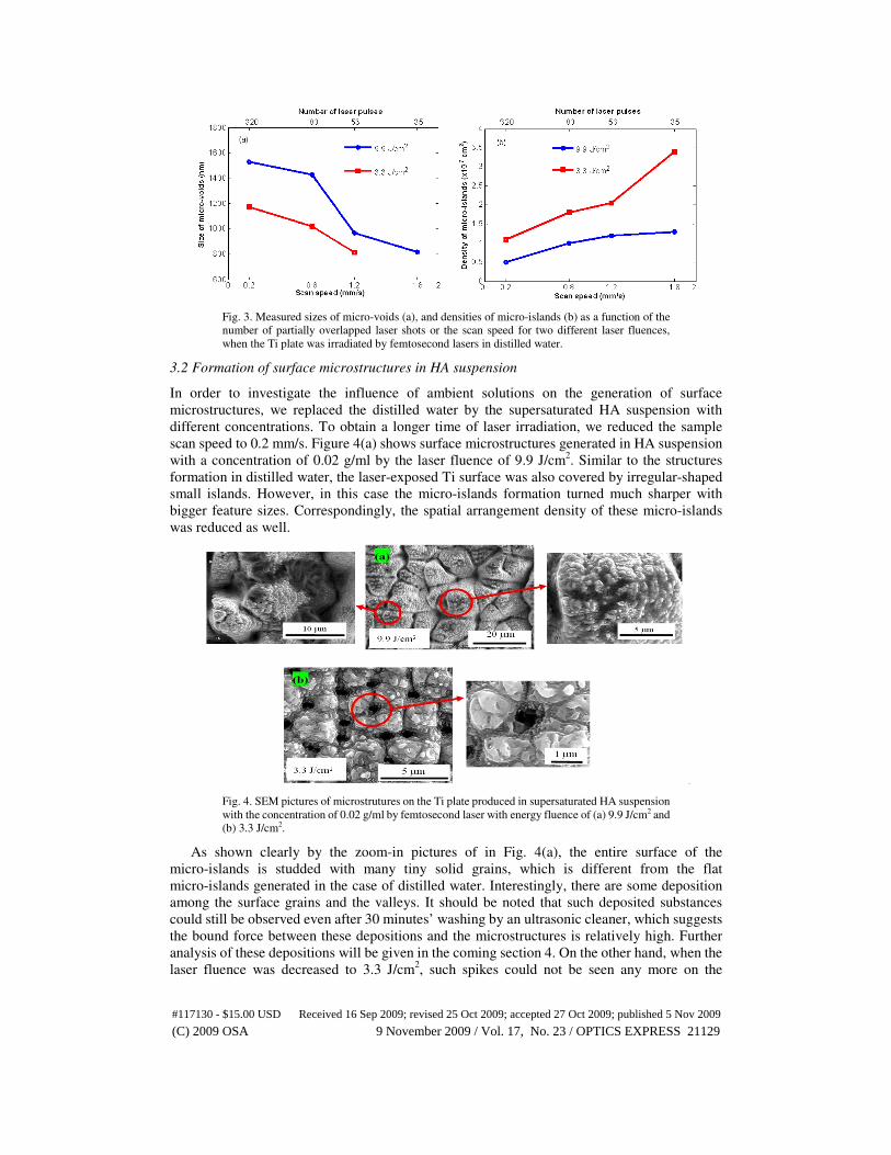
the partially overlapped laser pulses for two different laser fluences is shown by Fig. 3(a). For
the same laser fluence, the length of voids decreases gradually with the increase of scan speed
from 0.2 mm/s to 1.8 mm/s, or the decrease of the number of laser pulses from N = 320 to N =
35. While for a given scan speed, the higher laser fluence will induce bigger voids. Figure 3(b)
describes the variation of the micro-islands density as the function of the laser fluence and the
scan speed. Clearly, the density of the micro-islands gets larger as the scan speed increases or
the number of overlapped laser pulses decreases.
#117130 - $15.00 USD Received 16 Sep 2009; revised 25 Oct 2009; accepted 27 Oct 2009; published 5 Nov 2009
(C) 2009 OSA 9 November 2009 / Vol. 17, No. 23 / OPTICS EXPRESS 21128
Page 6
Fig. 3. Measured sizes of micro-voids (a), and densities of micro-islands (b) as a function of the
number of partially overlapped laser shots or the scan speed for two different laser fluences,
when the Ti plate was irradiated by femtosecond lasers in distilled water.
3.2 Formation of surface microstructures in HA suspension
In order to investigate the influence of ambient solutions on the generation of surface
microstructures, we replaced the distilled water by the supersaturated HA suspension with
different concentrations. To obtain a longer time of laser irradiation, we reduced the sample
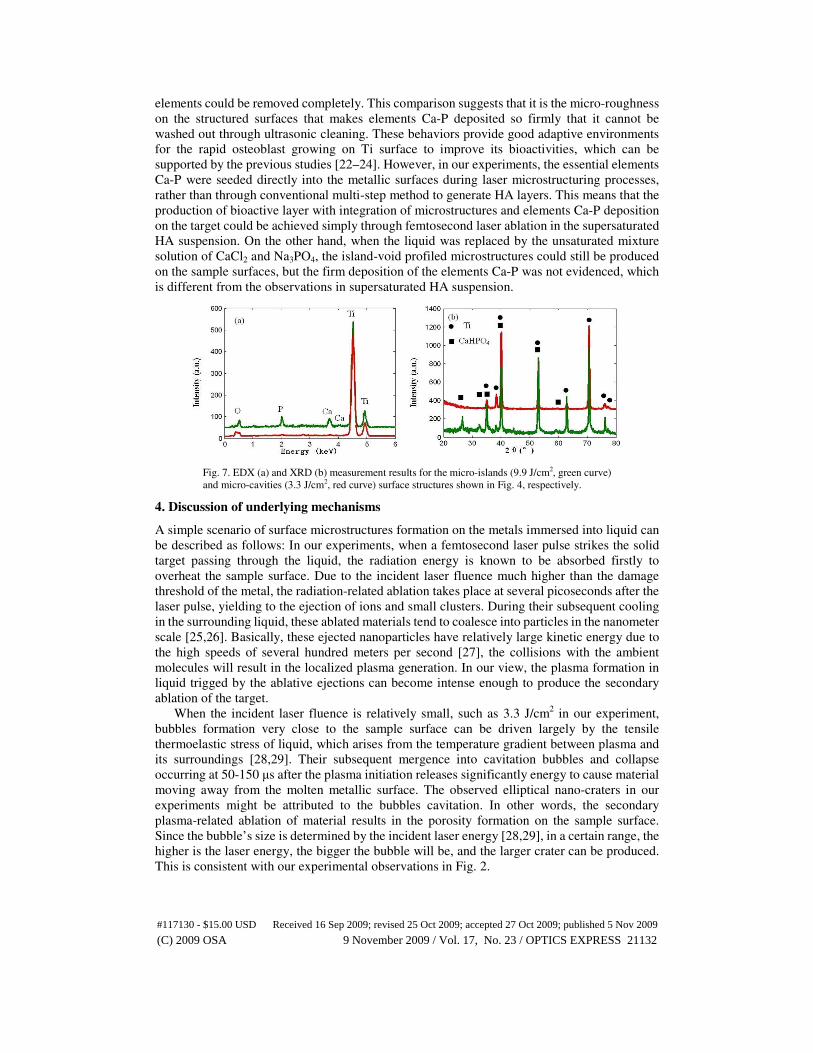
scan speed to 0.2 mm/s. Figure 4(a) shows surface microstructures generated in HA suspension
with a concentration of 0.02 g/ml by the laser fluence of 9.9 J/cm2. Similar to the structures
formation in distilled water, the laser-exposed Ti surface was also covered by irregular-shaped
small islands. However, in this case the micro-islands formation turned much sharper with
bigger feature sizes. Correspondingly, the spatial arrangement density of these micro-islands
was reduced as well.
Fig. 4. SEM pictures of microstrutures on the Ti plate produced in supersaturated HA suspension
with the concentration of 0.02 g/ml by femtosecond laser with energy fluence of (a) 9.9 J/cm2 and
(b) 3.3 J/cm2.
As shown clearly by the zoom-in pictures of in Fig. 4(a), the entire surface of the
micro-islands is studded with many tiny solid grains, which is different from the flat
micro-islands generated in the case of distilled water. Interestingly, there are some deposition
among the surface grains and the valleys. It should be noted that such deposited substances
could still be observed even after 30 minutes’ washing by an ultrasonic cleaner, which suggests
the bound force between these depositions and the microstructures is relatively high. Further
analysis of these depositions will be given in the coming section 4. On the other hand, when the
laser fluence was decreased to 3.3 J/cm2, such spikes could not be seen any more on the
#117130 - $15.00 USD Received 16 Sep 2009; revised 25 Oct 2009; accepted 27 Oct 2009; published 5 Nov 2009
(C) 2009 OSA 9 November 2009 / Vol. 17, No. 23 / OPTICS EXPRESS 21129
Page 7
laser-exposed Ti surfaces within ambience of HA suspension. Instead, porous surface structures
with the diameter of around 910 nm could be generated under this condition, which is similar to
the observations in distilled water. However, as shown by Fig. 4(b), the generated pores seem to
be smaller than those in distilled water. Particularly, the deposition of substances on the
structured area was not found in this case, which indicates the higher laser energies should be
very necessary for the substances deposition among the surface microstructures.
Figure 5 shows surface microstructures formation in HA suspension with a larger
concentration of 0.04 g/ml, where the incident fluence of femtosecond laser was 3.3 J/cm2 and
the sample was scanned at moving speed of 0.2 mm/s. Expectably, the generated porous surface
is similar to the structure in Fig. 4(b), which suggests that the concentration of the solution has
little influence on the microstructures formation. Moreover, the size of micro-cavity was found
to remain about 910 nm even with different solute concentrations. Comparing with the results in
Fig. 2, we can understand that the size of the micro-cavity on the targets is rather influenced by
the incident laser fluence than by the solution concentration. Remarkably, although the higher
concentration of the solution was employed, deposition of the particular substances could not be
found in the regime of low laser fluence.
Fig. 5. Porous surface microstructure formation on the Ti plate sample in HA solution with the
larger concentration of 0.04 g/ml by femtosecond laser with fluence of 3.3 J/cm2.
3.3 Formation of surface microstructures in the mixed solution of CaCl2 and Na3PO4
Figure 6 shows the typical surface structures on the Ti plate in the mixed solution of CaCl2 and
Na3PO4 with a concentration of 0.018 g/ml, when the sample was scanned at the speed of 0.2
mm/s under the irradiation of femtosecond lasers with two different fluences. When the laser
fluence was 9.9 J/cm2, the micro-islands structures were evidenced again, but with
better-organized spatial distribution, and the shape of the micro-islands turns to be more
regular. At the laser fluence of 3.3 J/cm2, micro-cavity structures could also be produced on the
sample surfaces. Compared with the structures in Fig. 5, micro-cavities in this case had almost
the same size of 910 nm. However, the deposition substances could not be found at this
concentration, even with higher laser fluence of 9.9 J/cm2.
If the solution concentration was reduced to 0.008 g/ml and the sample was scribed by
femtosecond laser with less fluence of 3.3 J/cm2, the porous surface microstructures on the
laser-exposed region can be observed with the void size of 910 nm, which confirms that the size
of micro-voids is insensitive to the variation of the solution concentration.
#117130 - $15.00 USD Received 16 Sep 2009; revised 25 Oct 2009; accepted 27 Oct 2009; published 5 Nov 2009
(C) 2009 OSA 9 November 2009 / Vol. 17, No. 23 / OPTICS EXPRESS 21130
Page 8
Fig. 6. Surface microstructuring of the Ti plate in the mixture solution of CaCl2 and Na3PO4 with
concentration of 0.018 g/ml by femtosecond laser fluence of (a) 9.9 J/cm2 and (b) 3.3 J/cm2.
3.4 Chemical element deposition on the microstructured metal surfaces
In order to investigate chemical evolution of the metal surfaces structured by femtosecond
lasers in the suspension of HA, we produced several large areas of these structures on different
Ti plates, each of which has an area of 10 × 10 mm2. After the laser-structured samples were
washed by the ultrasonic cleaner, their surface compositions were analyzed through EDX and
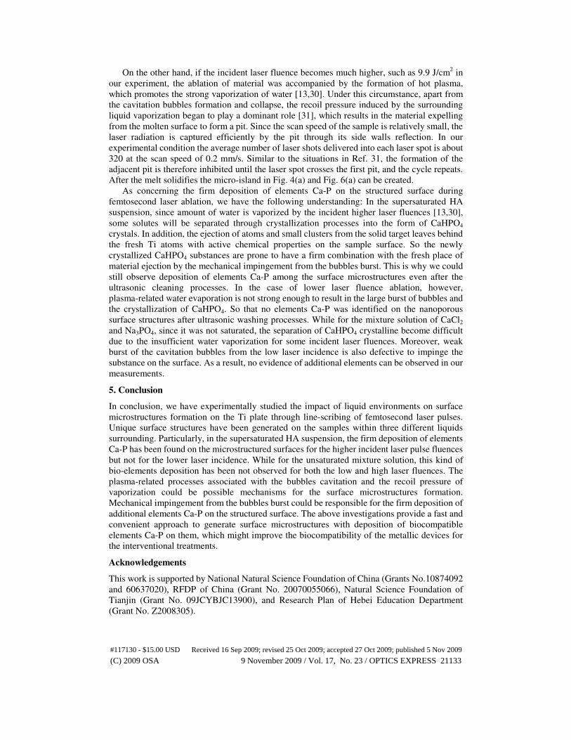
XRD measurements. The obtained results are depicted by Fig. 7, where two different laser
fluences were employed. In the case of EDX measurements with peak signals corresponding to
chemical components, multiple peaks standing in the case of laser treatment with the higher
fluence of 9.9 J/cm2 suggests that two important chemical elements Ca and P have been
deposited during the laser treatment. While for the laser fluence of 3.3 J/cm2, as shown in Fig.
7(a), only peaks of Ti and Oxygen elements are displayed in the curve, and signals relevant to
both Ca and P elements seem to disappear, which indicates that little additional elements Ca-P
are deposited on the micro-structured sample surfaces under this situation .
In the case of XRD measurement, distinct narrow peak signals represent diffraction
intensities at different angles from the structured sample. Among them, information about Ti
and CaHPO4 materials are marked by the solid cycles and squares, respectively. As shown in
Fig. 7(b), the measured peaks at θ = 35°, 40° and 53° coincide in the position with Ti peaks,
which cannot be employed to identify the production of CaHPO4 crystals. However, for the
laser fluence of 9.9 J/cm2, there are three other distinct peaks appearing at the diffraction angles
of θ = 27°, 33° and 59°. Through comparing them with the JCPDS (Joint Committee on Powder
Diffraction Standards) data sheet, we can identify such signals from CaHPO4 rather than HA
and other crystals. While for the laser fluence of 3.3 J/cm2, all diffraction peaks coincide in the
position with Ti peaks, so that we could not recognize the existence of CaHPO4 crystal. These
measurements provide another proof that the deposition of elements Ca-P in the form of
CaHPO4 crystal could take place at the higher laser fluence of 9.9 J/cm2, while no elements
Ca-P could be found to deposit into the structured surfaces at the lower laser fluence of 3.3
J/cm2. This result is consistent with the observations in Fig. 7(a).
It should be stressed that the deposited elements Ca-P could not be removed even after 30
minutes of ultrasonic cleaning, indicating a firm combination between the elements Ca-P and
the structured surface. For a direct comparison, we submerged a flat and smooth Ti plate into
the HA suspension with the supersaturated concentration for several days. With the water
vaporizing naturally, the deposition of elements Ca-P on the sample surface could be seen by
naked eyes. However, after washing by the ultrasonic cleaner, all these deposited chemical
#117130 - $15.00 USD Received 16 Sep 2009; revised 25 Oct 2009; accepted 27 Oct 2009; published 5 Nov 2009
(C) 2009 OSA 9 November 2009 / Vol. 17, No. 23 / OPTICS EXPRESS 21131
Page 9
elements could be removed completely. This comparison suggests that it is the micro-roughness
on the structured surfaces that makes elements Ca-P deposited so firmly that it cannot be
washed out through ultrasonic cleaning. These behaviors provide good adaptive environments
for the rapid osteoblast growing on Ti surface to improve its bioactivities, which can be
supported by the previous studies [22–24]. However, in our experiments, the essential elements
Ca-P were seeded directly into the metallic surfaces during laser microstructuring processes,
rather than through conventional multi-step method to generate HA layers. This means that the
production of bioactive layer with integration of microstructures and elements Ca-P deposition
on the target could be achieved simply through femtosecond laser ablation in the supersaturated
HA suspension. On the other hand, when the liquid was replaced by the unsaturated mixture
solution of CaCl2 and Na3PO4, the island-void profiled microstructures could still be produced
on the sample surfaces, but the firm deposition of the elements Ca-P was not evidenced, which
is different from the observations in supersaturated HA suspension.
Fig. 7. EDX (a) and XRD (b) measurement results for the micro-islands (9.9 J/cm2, green curve)
and micro-cavities (3.3 J/cm2, red curve) surface structures shown in Fig. 4, respectively.
4. Discussion of underlying mechanisms
A simple scenario of surface microstructures formation on the metals immersed into liquid can
be described as follows: In our experiments, when a femtosecond laser pulse strikes the solid
target passing through the liquid, the radiation energy is known to be absorbed firstly to
overheat the sample surface. Due to the incident laser fluence much higher than the damage
threshold of the metal, the radiation-related ablation takes place at several picoseconds after the
laser pulse, yielding to the ejection of ions and small clusters. During their subsequent cooling
in the surrounding liquid, these ablated materials tend to coalesce into particles in the nanometer
scale [25,26]. Basically, these ejected nanoparticles have relatively large kinetic energy due to
the high speeds of several hundred meters per second [27], the collisions with the ambient
molecules will result in the localized plasma generation. In our view, the plasma formation in
liquid trigged by the ablative ejections can become intense enough to produce the secondary
ablation of the target.
When the incident laser fluence is relatively small, such as 3.3 J/cm2 in our experiment,
bubbles formation very close to the sample surface can be driven largely by the tensile
thermoelastic stress of liquid, which arises from the temperature gradient between plasma and
its surroundings [28,29]. Their subsequent mergence into cavitation bubbles and collapse
occurring at 50-150 µs after the plasma initiation releases significantly energy to cause material
moving away from the molten metallic surface. The observed elliptical nano-craters in our
experiments might be attributed to the bubbles cavitation. In other words, the secondary
plasma-related ablation of material results in the porosity formation on the sample surface.
Since the bubble’s size is determined by the incident laser energy [28,29], in a certain range, the
higher is the laser energy, the bigger the bubble will be, and the larger crater can be produced.
This is consistent with our experimental observations in Fig. 2.
#117130 - $15.00 USD Received 16 Sep 2009; revised 25 Oct 2009; accepted 27 Oct 2009; published 5 Nov 2009
(C) 2009 OSA 9 November 2009 / Vol. 17, No. 23 / OPTICS EXPRESS 21132
Page 10
On the other hand, if the incident laser fluence becomes much higher, such as 9.9 J/cm2 in
our experiment, the ablation of material was accompanied by the formation of hot plasma,
which promotes the strong vaporization of water [13,30]. Under this circumstance, apart from
the cavitation bubbles formation and collapse, the recoil pressure induced by the surrounding
liquid vaporization began to play a dominant role [31], which results in the material expelling
from the molten surface to form a pit. Since the scan speed of the sample is relatively small, the
laser radiation is captured efficiently by the pit through its side walls reflection. In our
experimental condition the average number of laser shots delivered into each laser spot is about
320 at the scan speed of 0.2 mm/s. Similar to the situations in Ref. 31, the formation of the
adjacent pit is therefore inhibited until the laser spot crosses the first pit, and the cycle repeats.
After the melt solidifies the micro-island in Fig. 4(a) and Fig. 6(a) can be created.
As concerning the firm deposition of elements Ca-P on the structured surface during
femtosecond laser ablation, we have the following understanding: In the supersaturated HA
suspension, since amount of water is vaporized by the incident higher laser fluences [13,30],
some solutes will be separated through crystallization processes into the form of CaHPO4
crystals. In addition, the ejection of atoms and small clusters from the solid target leaves behind
the fresh Ti atoms with active chemical properties on the sample surface. So the newly
crystallized CaHPO4 substances are prone to have a firm combination with the fresh place of
material ejection by the mechanical impingement from the bubbles burst. This is why we could
still observe deposition of elements Ca-P among the surface microstructures even after the
ultrasonic cleaning processes. In the case of lower laser fluence ablation, however,
plasma-related water evaporation is not strong enough to result in the large burst of bubbles and
the crystallization of CaHPO4. So that no elements Ca-P was identified on the nanoporous
surface structures after ultrasonic washing processes. While for the mixture solution of CaCl2
and Na3PO4, since it was not saturated, the separation of CaHPO4 crystalline become difficult
due to the insufficient water vaporization for some incident laser fluences. Moreover, weak
burst of the cavitation bubbles from the low laser incidence is also defective to impinge the
substance on the surface. As a result, no evidence of additional elements can be observed in our
measurements.
5. Conclusion
In conclusion, we have experimentally studied the impact of liquid environments on surface
microstructures formation on the Ti plate through line-scribing of femtosecond laser pulses.
Unique surface structures have been generated on the samples within three different liquids
surrounding. Particularly, in the supersaturated HA suspension, the firm deposition of elements
Ca-P has been found on the microstructured surfaces for the higher incident laser pulse fluences
but not for the lower laser incidence. While for the unsaturated mixture solution, this kind of
bio-elements deposition has been not observed for both the low and high laser fluences. The
plasma-related processes associated with the bubbles cavitation and the recoil pressure of
vaporization could be possible mechanisms for the surface microstructures formation.
Mechanical impingement from the bubbles burst could be responsible for the firm deposition of
additional elements Ca-P on the structured surface. The above investigations provide a fast and
convenient approach to generate surface microstructures with deposition of biocompatible
elements Ca-P on them, which might improve the biocompatibility of the metallic devices for
the interventional treatments.
Acknowledgements
This work is supported by National Natural Science Foundation of China (Grants No.10874092
and 60637020), RFDP of China (Grant No. 20070055066), Natural Science Foundation of
Tianjin (Grant No. 09JCYBJC13900), and Research Plan of Hebei Education Department
(Grant No. Z2008305).
#117130 - $15.00 USD Received 16 Sep 2009; revised 25 Oct 2009; accepted 27 Oct 2009; published 5 Nov 2009
(C) 2009 OSA 9 November 2009 / Vol. 17, No. 23 / OPTICS EXPRESS 21133