i Surface-selective and Controllable Photo-grafting for Synthesis of Tailored Macroporous Membrane Adsorbers (Oberflächenselektive und kontrollierbare Photopfropfung für die Synthese von maßgeschneiderten makroporösen Membranadsorbern) by Dongming He Jiangxi, China Thesis submitted to the Department of Chemistry of Universität Duisburg-Essen, in partial fulfillment of the requirements of the degree of Dr. rer. nat. Approved by the examining committee on July 3, 2008: Chair : Prof. Dr. Heinz Wilhelm Siesler Advisor : Prof. Dr. Mathias Ulbricht Reviewer : Prof. Dr. Thomas Schrader Essen, 2008
Transcript
i
Surface-selective and Controllable Photo-grafting for Synthesis
of Tailored Macroporous Membrane Adsorbers
(Oberflächenselektive und kontrollierbare Photopfropfung für die Synthese von maßgeschneiderten makroporösen Membranadsorbern)
by
Dongming He Jiangxi, China
Thesis submitted to the Department of Chemistry of
Universität Duisburg-Essen, in partial fulfillment of
the requirements of the degree of
Dr. rer. nat.
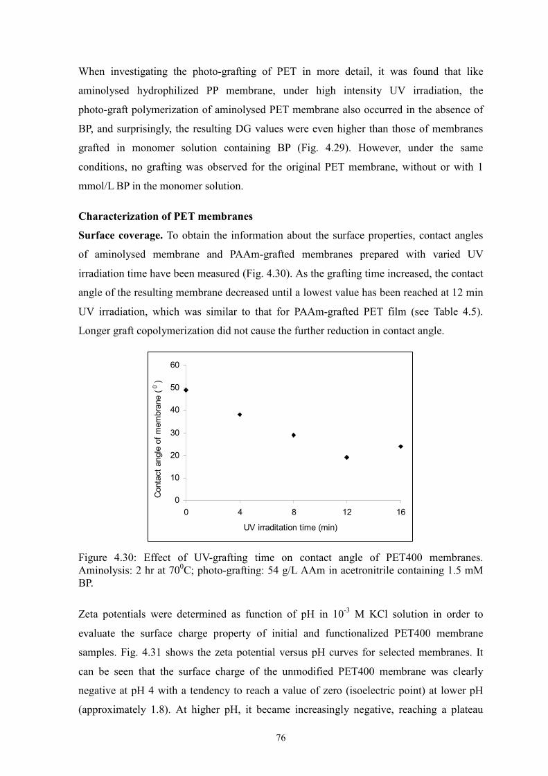
Approved by the examining committee on July 3, 2008:
Chair : Prof. Dr. Heinz Wilhelm Siesler
Advisor : Prof. Dr. Mathias Ulbricht
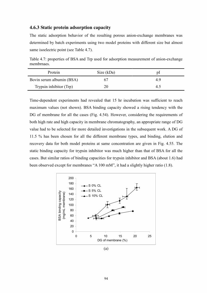
Reviewer : Prof. Dr. Thomas Schrader
Essen, 2008
ii
Abstract
Photo-grafting is a straightforward and promising technique for surface modification of
polymeric membranes. This work emphasized on the development and investigation of
surface-selective photo-grafting method from polar organic solution; on the other hand, on
the preparation of membrane adsorbers via the proposed grafting methods and evaluation
of the resulting membrane adsorbers.
Two novel surface-selective photo-grafting methods have been developed: synergist
immobilization and iniferter immobilization methods. Hydrophilized polypropylene (PP)
microfiltration (MF) membrane, whose surface polymer layer contains polyacrylate, was
used as base membrane for both methods; track-etched polyethylene terephthalate (PET)
MF membranes (PET200 and PET400) were used for extension of synergist
immobilization method and further investigation; methanol or acetonitrile solution of
acrylamide (AAm) with/without cross-linker (EDMA) was applied for investigation of
grafting mechanism.
For synergist immobilization method, the synergist (tertiary amino groups) for
photo-initiator benzophenone (BP) was introduced onto the membrane surface via an
aminolysis reaction with diethyl ethylenediamine (DEEDA). The reaction conditions have
been optimized. The proposed grafting mechanism was verified by the significant
difference in degree of grafting (DG) between original and aminolysed membranes. In
order to better understand and improve this novel method, detailed investigation of
functionalization parameters and affecting factors has been carried out. The grafted
membranes were characterized by ATR-IR, contact angle, SEM, permporometry, liquid
permeability and zeta potential. The obtained results demonstrated that the highest
surface-selectivity of photo-grafting could be achieved only under the optimum grafting
conditions, i.e., inert solvent to excited BP should be used to reduce/avoid
homopolymerization in bulk solution; appropriately low UV intensity should be applied to
exclude the uncontrolled side grafting reaction (another functionalization mechanism was
discovered at high UV intensity based on direct generation of starter radical); appropriately
low BP concentration was used to reduce the non-selective photo-grafting. Thus, the
grafted layer could be well controlled by immobilized synergist concentration, UV
irradiation time, monomer concentration and initiator concentration. In addition, this
method has been successfully applied to track-etched PET membrane, and it is also
expected to functionalize other polymeric membranes with similar chemical structure.
iii
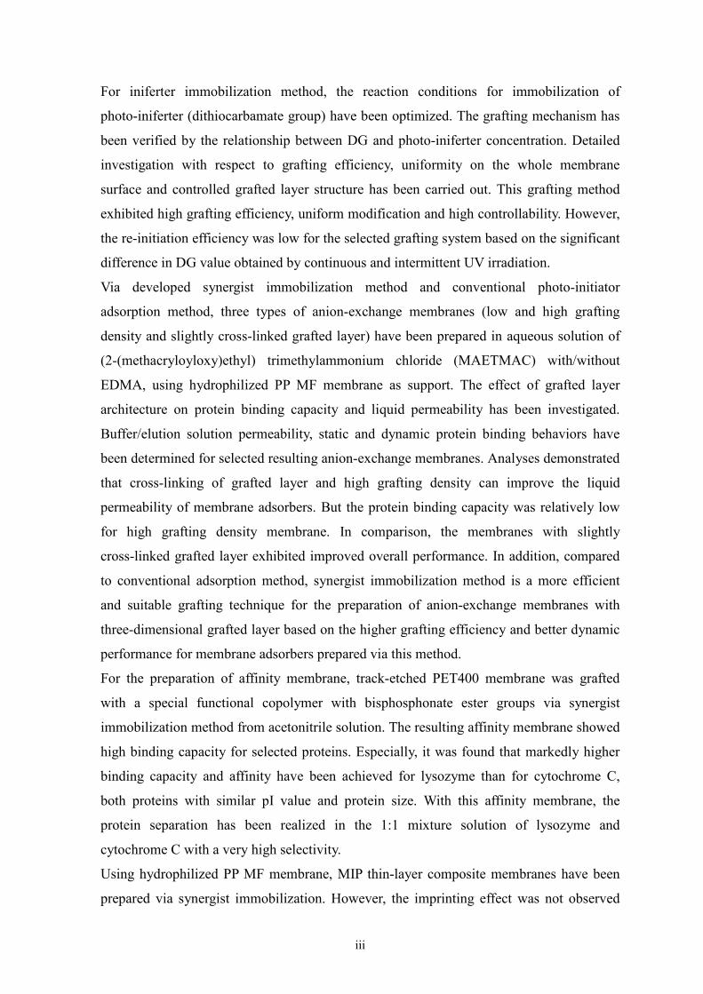
For iniferter immobilization method, the reaction conditions for immobilization of
photo-iniferter (dithiocarbamate group) have been optimized. The grafting mechanism has
been verified by the relationship between DG and photo-iniferter concentration. Detailed
investigation with respect to grafting efficiency, uniformity on the whole membrane
surface and controlled grafted layer structure has been carried out. This grafting method
exhibited high grafting efficiency, uniform modification and high controllability. However,
the re-initiation efficiency was low for the selected grafting system based on the significant
difference in DG value obtained by continuous and intermittent UV irradiation.
Via developed synergist immobilization method and conventional photo-initiator
adsorption method, three types of anion-exchange membranes (low and high grafting
density and slightly cross-linked grafted layer) have been prepared in aqueous solution of
Figure 4.66 Syntheses of MIP/NIP composite membranes with different DG
adjusted by varied UV irradiation time using iniferter immobilization
method.
107
Figure 4.67 Effect of DG of MIP/NIP membranes grafted via synergist
immobilization method on amount of adsorbed and eluted template
(Boc-L-PhA).
108
Figure 4.68 Effect of cross-linker concentration on amount of eluted template. 109
Figure 4.69 Chemical structure of DMPAP. 110
Figure 4.70 Separation factor (a) and total binding capacity of Boc-D/L-PhA (b)
for MIPs and NIPs.
110
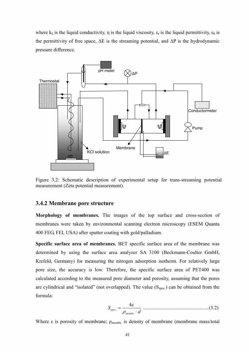
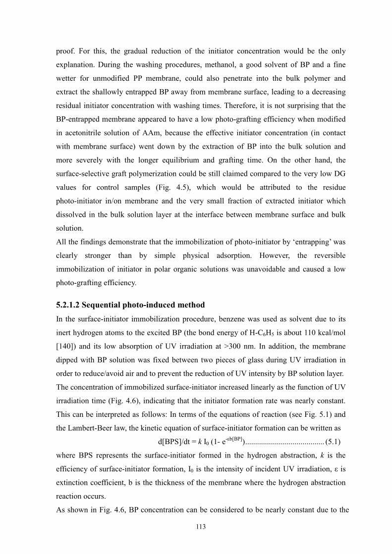
Figure 5.1: Schematic depiction of surface-initiator formation via sequential
photo-induced method.
114
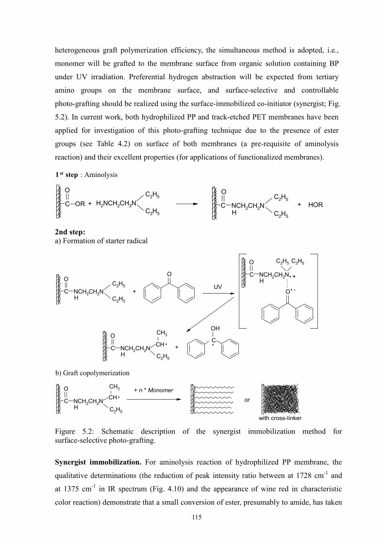
Figure 5.2: Schematic description of the synergist immobilization method for
surface-selective photo-grafting.
115
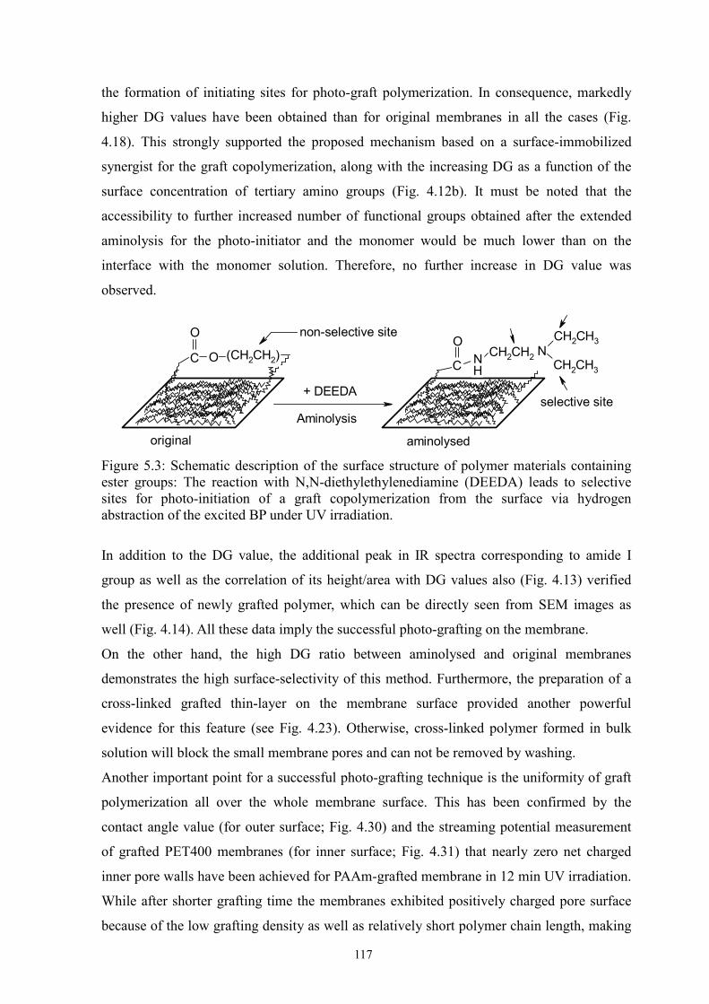
Figure 5.3: Schematic description of the surface structure of polymer materials
containing ester groups…
117
Figure 5.4: Schematic description of mechanism for direct generation of starter
radicals on the aminolysed PET surface under high intensity UV
irradiation.
122
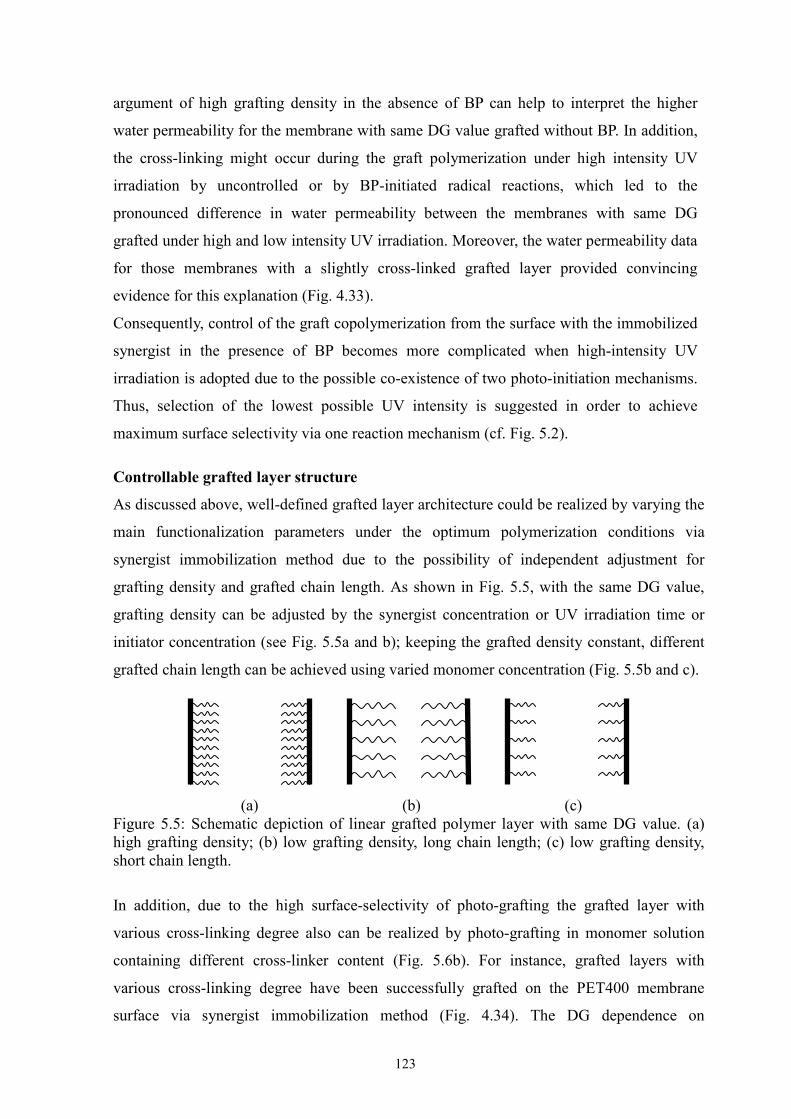
Figure 5.5: Schematic depiction of linear grafted polymer layer with same DG
value.
123
Figure 5.6: Schematic description of grafted layer structure in PET track-etched
pores in ethanol and water.
124
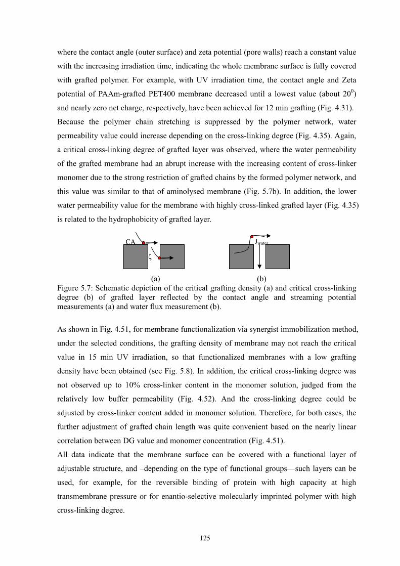
Figure 5.7: Schematic depiction of the critical grafting density (a) and critical
cross-linking degree (b) of grafted layer…
125
Figure 5.8: Schematic description of mechanisms and controlled photo-grafting of
synergist immobilization and adsorption.
126
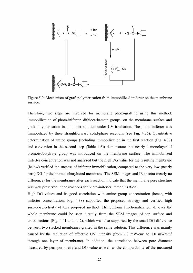
Figure 5.9: Mechanism of graft polymerization from immobilized iniferter on the
membrane surface.
127
Figure 5.10: Schematic overall depiction of four photo-grafting methods. 130
xvii
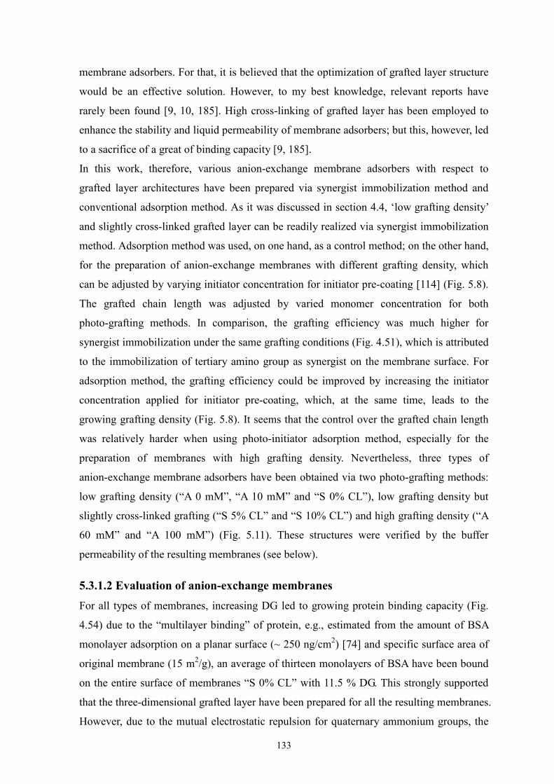
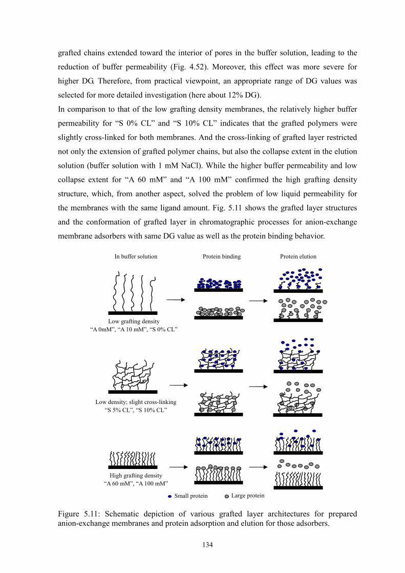
Figure 5.11: Schematic depiction of various grafted layer architectures for
prepared anion-exchange membranes and protein adsorption and
elution for those adsorbers.
134
Figure 5.12 Schematic depiction of protein binding of functional copolymer in
various architectures.
137
Figure 5.13 Chemical structures of grafted layer for synthesized affinity
membranes.
137
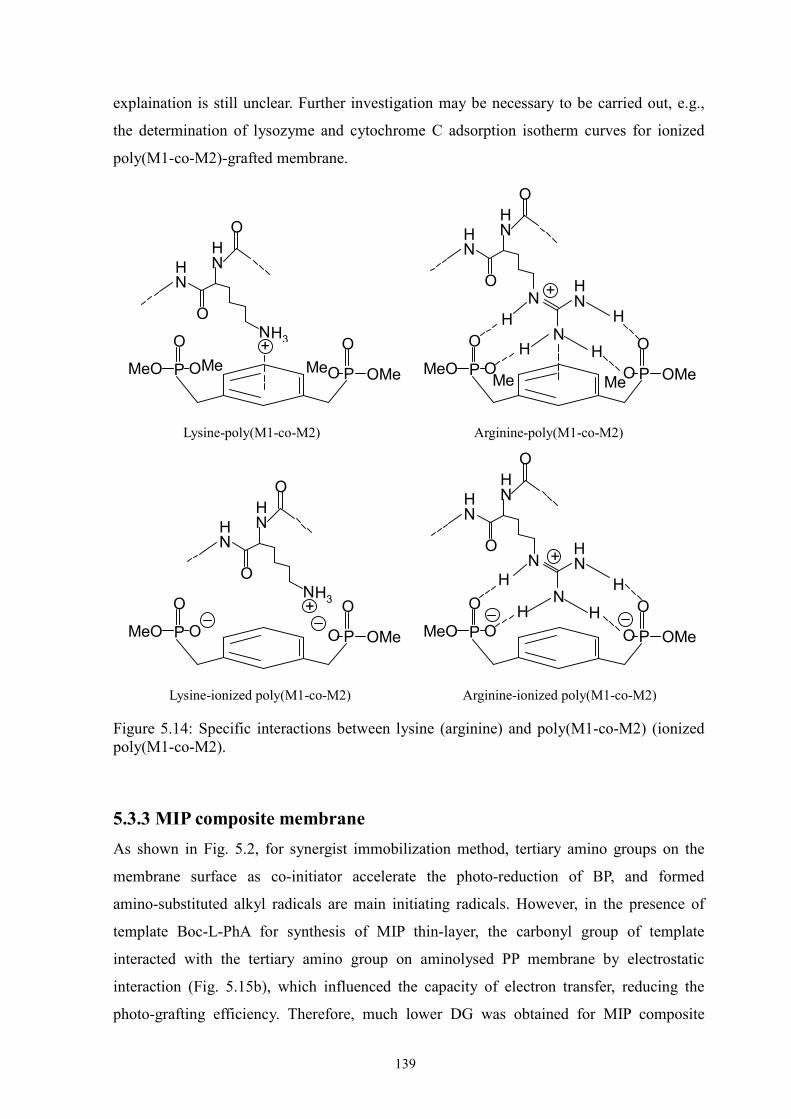
Figure 5.14 Specific interactions between lysine (arginine) an poly(M1-co-M2)
(ionized poly(M1-co-M2)).
139
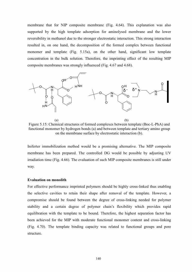
Figure 5.15 Chemical structures of formed complexes… 140
1
Chapter 1 Introduction
1.1 Background and existing problems
Chromatography is widely used for the separation, isolation, purification and analytical
characterization of biomolecules. Due to some limitations associated with the conventional
packed bed column chromatography, such as high pressure drop, internal diffusion
limitation or compaction of the soft particles, many efforts have been made to improve the
performance of the processes by alternative stationary phase materials. Afeyan et al. [1]
prepared a functionalized bead having pores through which a protein can be transported by
convection and showed that diffusive resistance in the bead-packed bed could be neglected.
Brandt et al. [2] designed a porous affinity chromatography membrane which allows the
mass transfer of protein molecules to the ligands mainly by convection, and the
aforementioned limitations of traditional particles could also be eliminated. Ease of scale-up
makes the membrane format very attractive. Furthermore, Muller [3] first proposed the idea
of “multilayer binding” of protein on the extending polymer chains attached on the surface
of adsorber particles. This idea has also been successfully applied for obtaining higher
binding capacity of porous membrane adsorbers [4, 5].
So far, membrane chromatography has been intensively investigated and industrially applied
as a promising alternative to the conventional technology [6-8]. However, it has been
realized that membrane chromatography has its limitations such as unclear mass transport
phenomena and relatively lower binding capacity compared to adsorber particles. Therefore,
recent research work mainly focuses on the clarification of transport phenomena in
membrane chromatographic processes and the enhancement of binding capacity. For the
latter, the effective solution would be emphasized on membrane chemistry (i.e., synthesis of
membrane adsorbers with high binding capacity), although the more effective ligand
utilization by the optimized chromatographic processes could also improve the binding
capacity.
Due to the limited surface area of porous membrane, enhancing the binding capacity by
“multilayer binding” becomes a main research direction. This certainly will lead to the
reduction of liquid permeability due to the extension of longer grafted chains into the pore
interior. However, from the viewpoint of practicability, the tradeoff between high binding
capacity and low liquid permeability needs to be overcome. This requires a new design for
grafted functional active layers. For example, grafted chains containing ion-exchange
groups expand or shrink depending on the ionic strength, pH and kind of solvent used. The
2
swelling of grafted polymer is helpful for protein binding but reduces the liquid
permeability, affecting the mass transfer of proteins. Cross-linking of the grafted chains has
been shown to be effective in reducing the swelling and improving the stability [9], but in
this case, the cross-linked grafted layer led to a low protein binding capacity and nearly no
protein could be eluted. In addition, with the same ligand amount, the increase of grafting
density can also increase the liquid permeability of membrane adsorbers. Therefore, this
may be another approach to overcome the trade-off on condition that the ligand utilization
yield can be guaranteed. However, rare reports regarding improving membrane adsorbers
performance by adjustment of grafted layer structure have been found [10].
For the preparation of membrane adsorbers with high binding capacity, surface graft
modification on commercial membranes has shown itself a good solution. Commercially
available Sartobind membrane adsorbers have been prepared by grafting cross-linked
functional polymer chains onto the base cellulose membrane and used for the same
purpose later as well (cf. ref. [8]). However, those adsorbers showed a limited dynamic
binding capacity. Investigators have developed several alternative techniques to attach the
functional ligands in an appropriate form onto the surface of commercial macroporous
membranes. Saito et al. [4] have employed radiation-induced graft polymerization to
prepare various ion-exchange membranes. Husson et al. have developed a method for
surface-initiated atom transfer radial polymerization (ATRP) for the preparation of
cation-exchange membrane adsorbers [11]. However, to achieve a fully controlled grafting
via ATRP requires significant efforts which may be hard to implement in large technical
scale. Photo-initiated graft copolymerization had been proposed and successfully applied
as an easy and robust approach for the preparation of bio-affinity [12] or of high-capacity
cation-exchange membranes [10, 13].
More photo-grafting techniques have also been proposed for other membrane
functionalities [14, 15]. To some extent, the developed photo-grafting methods exhibit high
surface-selectivity and controllability when they are applied to modify membranes from
aqueous solution. However, to my best knowledge, there is still a technique gap that no
efficient photo-grafting method, via which polymeric membranes can be
surface-selectively grafted from polar organic solution of monomer, had been developed.
This greatly limits the use of a variety of functional monomers. In addition, precise design
of grafted layer is still a challenging issue.
3
1.2 Objectives of the research
The overall objectives of this research are
(i) to find efficient surface-selective photo-grafting methods for polymeric membranes
according to the main criteria: graft polymerization is initiated from the whole membrane
surface; effective grafting can be manipulated in polar organic solution of monomer; grafted
polymer layer can be well controlled.
(ii) to prepare membrane adsorbers with well-defined grafted layer architectures via the
proposed grafting techniques and evaluate the performance of such membrane adsorbers.
To be more specific, the research tasks involve
i. Study on the already developed photo-grafting methods according to the criteria proposed
above.
ii. Proposal of novel photo-grafting strategies and study on the grafting mechanisms.
iii. Investigation of various factors affecting grafting efficiency and surface-selectivity.
iv. Proof / extension of the proposed technique by applying to another membrane made of
similar polymer structure.
v. Preparation of anion-exchange membrane adsorbers with designed grafted layer from
aqueous solution via the proposed grafting method.
vi. Preparation of affinity membrane adsorbers from polar organic solution.
vii. Preparation of molecularly imprinted polymer (MIP) composite membranes from polar
organic solution.
viii. Evaluation of the performance of the novel membrane adsorbers.
1.3 Scope of this dissertation
Already known photo-initiator entrapping method and sequential photo-induced method
were used for graft functionalization of unmodified polypropylene (PP) microfiltration (MF)
membrane. Membrane functionalizations were performed from acrylamide (AAm) aqueous
solution and in AAm acetonitrile solution via respective grafting approaches. The difference
in grafting efficiency was analysed by more detailed experiments and determinations.
A novel photo-grafting strategy—synergist immobilization—was proposed toward the
functionalization of hydrophilized PP MF membrane from polar organic solutions. Tertiary
amino groups as synergist (co-initiator) of benzophenone (BP) were immobilized on the
whole membrane surface via aminolysis reaction. The proposed grafting mechanism was
evaluated by comparing grafting efficiency of aminolysed and original membranes using
simultaneous method (BP dissolved in monomer solution). In order to better control the
4
grafting behavior, the factors that affect the surface-selectivity of photo-grafting such as
solvent, UV irradiation time and intensity, monomer and photo-initiator concentration have
been investigated in detail. To extend the application of this grafting technique, the
functionalization of track-etched polyethylene terephthalate (PET) membranes (PET200 and
PET400) have been performed and the controllability of grafted polymer layer was further
verified, and characterization of prepared layer was completed.
Another photo-grafting method—iniferter immobilization method—has also been
developed based on the introduction of dithiocarbamate groups as photo-iniferter onto the
hydrophilized PP membrane surface. The grafting mechanism was studied and the
investigation of grafting controllability was carried out by modifying membrane at varied
functionalization parameters and by various characterizations of grafted membranes. The
“livingness” of graft polymerization via this method was also investigated.
Anion exchange membrane adsorbers with various grafted layer architectures (low and high
grafting density, with and without slight cross-linking) have been prepared from
(2-(methacryloyloxy)ethyl) trimethylammonium chloride aqueous solution via synergist
immobilization method and conventional photo-initiator adsorption method. The
performances of various membrane adsorbers have been evaluated by buffer (with and
without extra salt) permeability, static and dynamic binding capacity as well as
breakthrough and elution curves. The grafting efficiency and controllability during surface
functionalization as well as the performances of the resulting membrane adsorbers (with
similar degree of grafting (DG) and grafted layer structure) were compared between two
photo-grafting methods.
Affinity membranes have been prepared from acetonitrile solution of functional monomers
via synergist immobilization method. Track-etched PET400 membrane was used as support.
A series of proteins with different size and isoelectric point have been used to detect the
binding capacity on the resulting affinity membranes. Based on the significant difference in
binding capacity between lysozyme and cytochrome C, the separation of protein mixture
(lysozyme:cytochrome C = 1:1) was performed using poly(M1-co-M2)-grafted membrane
adsorber (M1 and M2 stand for methacryloylamino-2-hydroxy-propane and
5-(methacryloylamino-m-xylylene bisphosphonic acid tetramethylester, respectively), and
the separation effect has been qualitatively evaluated using UV-Vis spectrophotometer.
In order to realize the separation of anantiomers Boc-D/L-phenylalanine (Boc-D/L-PhA),
Boc-L-PhA imprinted composite membranes have been synthesized via synergist
immobilization and also via iniferter immobilization method. Non-imprinted polymer (NIP)
5
composite membranes as blank samples have been prepared in the absence of template
(Boc-L-PhA) under the identical other conditions. MIP/NIP polymers with various
composites were prepared by UV-induced radical polymerization to optimize the recipe
(ratio among templat, functional monomer and cross-linker) for better imprinting effect. UV
spectrophotometer and high performance liquid chromatography (HPLC) were used to
determine the binding capacity of amino acid and the separation factor of enantiomers for
both MIP and NIP composites membranes and polymers.
6
Chapter 2 Theory
2.1 Surface grafting techniques for polymeric membranes
With the extending applications of membrane technology in many fields such as water
purification, food and pharmaceutical industry and life sciences, the increasing demands on
the quantity and the function of polymeric membranes need to be met. Since demanded
interfacial characteristics can rarely be achieved by bulk modifications of the membrane
forming polymer without complications in membrane fabrication or with respect to
membrane stability, membrane surface modification becomes a unique alternative
approach to confer new and improved properties and hence fulfill the higher requirements.
Of the modification techniques, surface grafting turned out to be a promising technique to
design appropriate membranes for target applications. Overall, the ultimate aim of
membrane surface modification is either to minimize undesired interactions (adsorption or
adhesion) which reduce the performance (membrane fouling), or to introduce additional
interactions (charge, affinity, responsiveness or catalytic properties) for improving the
selectivity or creating an entirely novel separation function. Basically, the grafting
techniques, which have been developed for polymer surface modification [16], are
applicable to polymeric membranes. Therefore, a fast development with respect to
membrane modification has been seen in recent years. However, it must be noted that the
controllability of membrane modifications is still a challenging issue due to the
significantly different reactions in membrane pores from that on the nonporous polymer
surface and the limited characterization techniques, especially for the small and irregular
pores, as well as the less stability of polymer pore structure under modification conditions.
So far, the exploited grafting techniques for polymeric membranes mainly include
chemical grafting, radiation-induced, plasma-induced and photo-chemical grafting
techniques.
2.1.1 Chemical grafting
Chemical grafting can proceed along two major routes: direct chemical reactions and graft
polymerization from membrane surface. Surface modification by direct chemical reactions
is usually applied for the membranes with reactive surface groups. Otherwise, a strong
chemical environment (e.g., strongly basic or acidic or oxidizable) has to be provided to
change the surface properties of membranes.
7
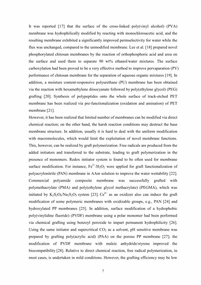
It was reported [17] that the surface of the cross-linked poly(vinyl alcohol) (PVA)
membrane was hydrophilically modified by reacting with monochloroacetic acid, and the
resulting membrane exhibited a significantly improved permselectivity for water while the
flux was unchanged, compared to the unmodified membrane. Lee et al. [18] prepared novel
phosphorylated chitosan membranes by the reaction of orthophosphoric acid and urea on
the surface and used them to separate 90 wt% ethanol/water mixtures. The surface
carboxylation had been proved to be a very effective method to improve pervaparation (PV)
performance of chitosan membrane for the separation of aqueous organic mixtures [19]. In
addition, a moisture content-responsive polyurethane (PU) membrane has been obtained
via the reaction with hexamethylene diisocyanate followed by poly(ethylene glycol) (PEG)
grafting [20]. Synthesis of polypeptides onto the whole surface of track-etched PET
membrane has been realized via pre-functionalization (oxidation and amination) of PET
membrane [21].
However, it has been realized that limited number of membranes can be modified via direct
chemical reaction; on the other hand, the harsh reaction conditions may destruct the base
membrane structure. In addition, usually it is hard to deal with the uniform modification
with macromolecules, which would limit the exploitation of novel membrane functions.
This, however, can be realized by graft polymerization. Free radicals are produced from the
added initiators and transferred to the substrate, leading to graft polymerization in the
presence of monomers. Redox initiator system is found to be often used for membrane
surface modification. For instance, Fe2+/H2O2 were applied for graft functionalization of
polyacrylonitrile (PAN) membrane in AAm solution to improve the water wettability [22].
Commercial polyamide composite membrane was successfully grafted with
polymethacrylate (PMA) and poly(ethylene glycol methacrylate) (PEGMA), which was
initiated by K2S2O8/Na2S2O5 system [23]. Ce4+ as an oxidizer also can induce the graft
modification of some polymeric membranes with oxidizable groups, e.g., PAN [24] and
hydroxylated PP membranes [25]. In addition, surface modification of a hydrophobic
poly(vinylidine fluoride) (PVDF) membrane using a polar monomer had been performed
via chemical grafting using benzoyl peroxide to impart permanent hydrophilicity [26].
Using the same initiator and supercritical CO2 as a solvent, pH sensitive membrane was
prepared by grafting poly(acrylic acid) (PAA) on the porous PP membrane [27]; the
modification of PVDF membrane with maleic anhydride/styrene improved the
biocompatibility [28]. Relative to direct chemical reaction, free radical polymerization, in
most cases, is undertaken in mild conditions. However, the grafting efficiency may be low
8
because of the occurrence of homopolymerization in bulk solutions. Apart from the general
free-radical mechanism, atom transfer radical polymerization (ATRP) is also an interesting
technique to carry out grafting. Prior to graft polymerization, halogen should be introduced
on the membrane surface generally by chemical reactions. Husson et al. modified the
microprous PVDF membrane using ATRP [29]. It was claimed that it is possible to tune the
ion-exchange capacity and the average pore size of the resulting grafted membrane in
rational ways. Later, a cation-exchange membrane with high binding capacity has been
prepared via the same method on the basis of cellulose membrane [11]. This technique had
been also used for the graft modification of PP microporous membrane [30] and
track-etched PET membrane [31, 32].
2.1.2 Radiation-induced grafting
In a review paper by Nasef and Hegazy [33], it was reported in detail about the
radiation-induced graft copolymerization for the preparation of ion-exchange membranes.
Because the excitation with high energy irradiation has a low selectivity, bond scissions in
the volume of a membrane material cannot be avoided. This grafting method is relatively
rarely used for polymeric membrane modification except for some membranes with inert
surfaces, e.g., polyethylene (PE) and PVDF. For the formation of radicals, electron beam
and Co-60 (γ-radiation) are usually applied. Saito and his co-workers have developed a
grafting method based on the radical initiation by electron beam [34]. Using this method a
variety of functional groups had been introduced on the porous PE hollow fiber as well as
HDPE membranes for different applications, e.g., various membrane adsorbers [35-37].
Graft modification of PVDF porous membrane with AA and sodium 4-styrenesulfonate
(SSS) was also initiated by electron beam [38]. In addition, the graft functionalization of
PE sheet membrane was induced by γ-irradiation for the preparation of ion-exchange
membrane [39]; Temperature-responsive [40] PVDF membrane as well as a dialysis
chloroprene rubber membrane [41] had also been realized by grafting PolyNIPAAm and
PolyHEMA, respectively, on the membrane surface under γ -irradiation.
2.1.3 Plasma-induced grafting
Plasma-induced grafting is a useful technique in the modification of surface properties.
The excitation with plasma is very surface selective [42]. Currently, more attention is
being given to its applications in membrane separation science. Low temperature plasma
technique has extensively been used to modify membrane surface. Overall, three effective
9
routes could be involved: plasma treatment, plasma treatment followed by graft
polymerization and plasma polymerization.
Via direct plasma treatment membrane surface property can be readily modified (e.g.,
usually the hydrophilicity) by the introduction of various polar functional groups due to the
optional process gases. For example, He [43], CO2 [44], N2 [45], O2 [46], H2O [47], NH3
[48, 49] and He/H2O [50] have been used to improve the hydrophilicity of membranes.
One of the major drawbacks of plasma treatment is, however, that the physicochemical
characteristics of the modified surfaces, including surface composition, can be time
dependent. Polar and chain group reorientation in the surface region can result in gradual
deterioration of the surface functionality. This process, called aging or “hydrophobic
recovery”, partially restores the original hydrophobic surface to the extent that it adapts
composition to the interfacial force. This hydrophobic recovery should be reduced or
eliminated to obtain long-term hydrophilicity for practical applications of the membrane.
This can be avoided by further graft functionalization, i.e., two steps are involved:
Polymeric membrane is first plasma-treated in the presence of inert gas (e.g., Ar or He);
subsequently, monomer in solution or in vapor phase is initiated to polymerize from
membrane surface. In many cases, a layer of peroxide groups are formed upon the
plasma-treated membrane was taken out of the plasma reactor. Subsequent graft
polymerization proceeds in monomer solution via thermal decomposition of peroxide
groups. Graft polymerization of PAN and polysulfone (PSf) UF membrane surfaces, which
were treated by pure helium plasma, was performed in hydrophilic monomer solutions [51].
By this means, stimuli-responsive composite membranes have been prepared by grafting
environmental sensitive monomer (AA or NIPAAm) onto membrane surface [52, 53]. In
another route, membrane modification is achieved by Ar- or He-plasma treatment followed
by graft copolymerization with monomer in the vapor phase. PAAm and PSt have been
grafted on the microporous PES and PAN UF membrane surfaces, reapectively, via this
modification route [54, 55].
Plasma polymerization is a procedure, in which gaseous monomers (saturated or
unsaturated), stimulated through a plasma, condense on freely selectable substrates. In this
case, cross-linking structure is generally formed; grafted layer with functional groups has
high stability but very low controllability due to the complicated reactions. Generally, this
route is applied for simple modifications (e.g., improvement of hydrophilicity) [56, 57]. In
some cases, the “hydrophobic recovery” was also observed [58].
However, the ablation tendency of the base polymer may be significant [50], although the
10
plasma treatment conditions could be adjusted and optimized. In addition, modifications in
small pores (diameter <100 nm) are complicated because this dimension is smaller than the
average free path length of the active species in the plasma [59].
2.1.4 Photo-chemical grafting
The excitation with UV irradiation has the great advantage that the wavelength can be
adjusted selectively to the reaction to be initiated, and, hence, undesired side reactions can
be avoided or at least reduced very much. Photo-initiation can be used without problems
also in small pores. The UV technology can be integrated into continuous manufacturing
processes simply and cost-efficiently. Photo-initiated processes have their largest potential
when complex polymer morphologies need to be surface-selectively functionalized with
minimal degradation of the base membrane, and when they are used to create
macromolecular layers via ‘grafting-to’ or ‘grafting-from’ [59]. So far, many
photo-grafting routes have been developed and used for various modifications of
polymeric membranes; and various membrane functionalies has been realized via
photo-grafting methods (detailed see section 2.2).
A key feature of a successful surface modification is to realize a synergy between the
useful properties of the base membrane and the changed chemical structure within the
barrier or the novel functional polymer (layer) added to the barrier. This synergy can only
be achieved by a mild and controllable modification technique, and photo-chemical
processes have a large potential in that regard.
2.2 Photo-graft functionalization on polymeric membranes
For surface selective photo-chemical processes, two alternative approaches are
distinguished: (i) coupling small molecular entities or larger macromolecules to the surface
(“grafting-to”) or (ii) heterogeneous graft copolymerization where monomers are
polymerized using starter groups on the surface (“grafting-from”).
2.2.1 “Photo-grafting-to” functionalization
“Photo-grafting-to” functionalization of a polymeric membrane is based on the
photo-reaction between membrane polymers and functionalization agents. Therefore, at
least one type of photo-reactive group which has a distinct, selective and efficient
reactivity is required. Aromatic azides or diazo compounds including their carbonyl
“cousins” (azidocarbonyl or diazocarbonyl) are important examples. Under the photo-
11
irradiation the decomposition of these compounds takes place along with the release of
nitrogen, which can subsequently lead to addition (via nitren / isocyanate or carben /
ketene) or to cross-linking reactions (via nitren dimerization) (Fig. 2.1). Such reactions are
also being used for “photo-affinity labeling” of bio-molecules, e.g., proteins [45]. Another
important principle is the photo-induced dimerization via [2+2] cycloaddition, e.g., of
cinnamate, coumarin or styrylpyridine groups (this reaction is only possible via the excited
state). Depending on the chemistry of the photo-reactive groups, controlled elimination or
addition reactions are possible, also including dimerization reactions leading to
cross-linking of macromolecule chains.
Therefore, according to the location of these photo-reactive moieties, two routes can be
classified: via photo-reactive membrane polymer and via photo-reactive functionalization
agents (Fig. 2.2). Since this approach is relatively independent on the chemical
composition of materials (cf. above), various functionalization agents can be attached onto
a membrane surface with photo-reactive moieties, or a variety of membranes (non-porous
and porous, different membrane polymers) can be modified without any pre-treatment by
using photo-reactive functional molecules.
N N N+ hv
N2
_Ar-
Ar N:
Ar C N N N+ -
O
hv
N2_ Ar C N
O
: Ar N C O
Aromatic azides
R
R'C N N
+ - hv
N2
_ :
C N N+ -
O
R C
O
Rhv
N2
_ : R C C O
Diazo compounds
R
Ar
Ar
R
+hv
R Ar
Ar R
Aromatic vinyl compounds ([2+2] cycloaddition)
R
R'C
Figure 2.1: Three groups of photo-reactive moieties and their main photo-initiated reaction pathways.
12
Figure 2.2: Depiction of principal mechanisms for photo-functionalization of polymeric membranes: via photo-reactive membrane polymer (from left) and via photo-reactive functionalization agents (from right).
Photo-reactive membrane polymer. Taking advantage of the photo-reactivity of such
typical moieties, membrane surface modification had been extensively investigated. One of
the approaches is by direct synthesis of membrane forming polymer with photo-reactive
groups. For example, Darkow et al. [61] synthesized a new type of membrane polymer,
The stability and controllability of grafted polymer layers on the surface of a base polymer
membrane are two crucial evaluation parameters for functionalization techniques.
Especially the controllability has received increasing attention in terms of architecture and
property of grafted polymers. Due to disadvantages of “photo-grafting-to”, especially with
respect to grafting density, “photo-grafting-from” technique has been increasingly used and
developed.
In order to really tailor and optimize the membrane performance, various routes have been
developed depending on initial membrane materials and structures, architectures of grafted
polymer, and used modification system including properties of monomer and solvent. It
should be mentioned that radical polymerization has almost exclusively been used until
now. Photo-grafting can proceed in two ways: without or with an added photo-initiator.
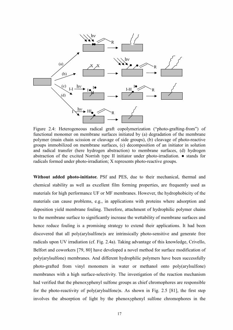
The approach “without added photo-initiator” involves the direct generation of free
radicals from the base membrane polymers under UV irradiation. Therefore, such methods
require either a photo-sensitive base polymer (photo-reactive side group or part of polymer
backbone) (cf. Fig. 2.4a) or the introduction of photo-sensitive groups onto the membrane
surfaces prior to graft copolymerization (Fig. 2.4b). For approach “with added
photo-initiator”, initiating radical sites should be generated on the membrane surface by
the reaction of photo-initiator with the base membrane polymer under UV irradiation. One
possibility is that the formed radicals by homolysis of initiator transfer to the base polymer,
which initiates graft copolymerization (cf. Fig. 2.4c). But the homopolymerization may
simultaneously take place in bulk solution, affecting surface grafting. Another more
surface-selective grafting approach is to use Norrish type II initiator (e.g., benzophenone
(BP) or xanthone, and their derivatives). The excited initiator by UV irradiation abstracts
hydrogen from the membrane polymer. The resulting radicals on membrane surface are
reactive to initiate polymerization, whereas the radicals formed in initiator side are
relatively inert (Fig. 2.4d). Therefore, the surface-selective photo-grafting can be reached.
In addition, the addition of the additives or co-initiators may enhance the
surface-selectivity of photo-grafting.
17
Figure 2.4: Heterogeneous radical graft copolymerization (“photo-grafting-from”) of functional monomer on membrane surfaces initiated by (a) degradation of the membrane polymer (main chain scission or cleavage of side groups), (b) cleavage of photo-reactive groups immobilized on membrane surfaces, (c) decomposition of an initiator in solution and radical transfer (here hydrogen abstraction) to membrane surfaces, (d) hydrogen abstraction of the excited Norrish type II initiator under photo-irradiation. ● stands for radicals formed under photo-irradiation; X represents photo-reactive groups.
Without added photo-initiator. PSf and PES, due to their mechanical, thermal and
chemical stability as well as excellent film forming properties, are frequently used as
materials for high performance UF or MF membranes. However, the hydrophobicity of the
materials can cause problems, e.g., in applications with proteins where adsorption and
deposition yield membrane fouling. Therefore, attachment of hydrophilic polymer chains
to the membrane surface to significantly increase the wettability of membrane surfaces and
hence reduce fouling is a promising strategy to extend their applications. It had been
discovered that all poly(arylsulfone)s are intrinsically photo-sensitive and generate free
radicals upon UV irradiation (cf. Fig. 2.4a). Taking advantage of this knowledge, Crivello,
Belfort and coworkers [79, 80] have developed a novel method for surface modification of
poly(arylsulfone) membranes. And different hydrophilic polymers have been successfully
photo-grafted from vinyl monomers in water or methanol onto poly(arylsulfone)
membranes with a high surface-selectivity. The investigation of the reaction mechanism
had verified that the phenoxyphenyl sulfone groups as chief chromophores are responsible
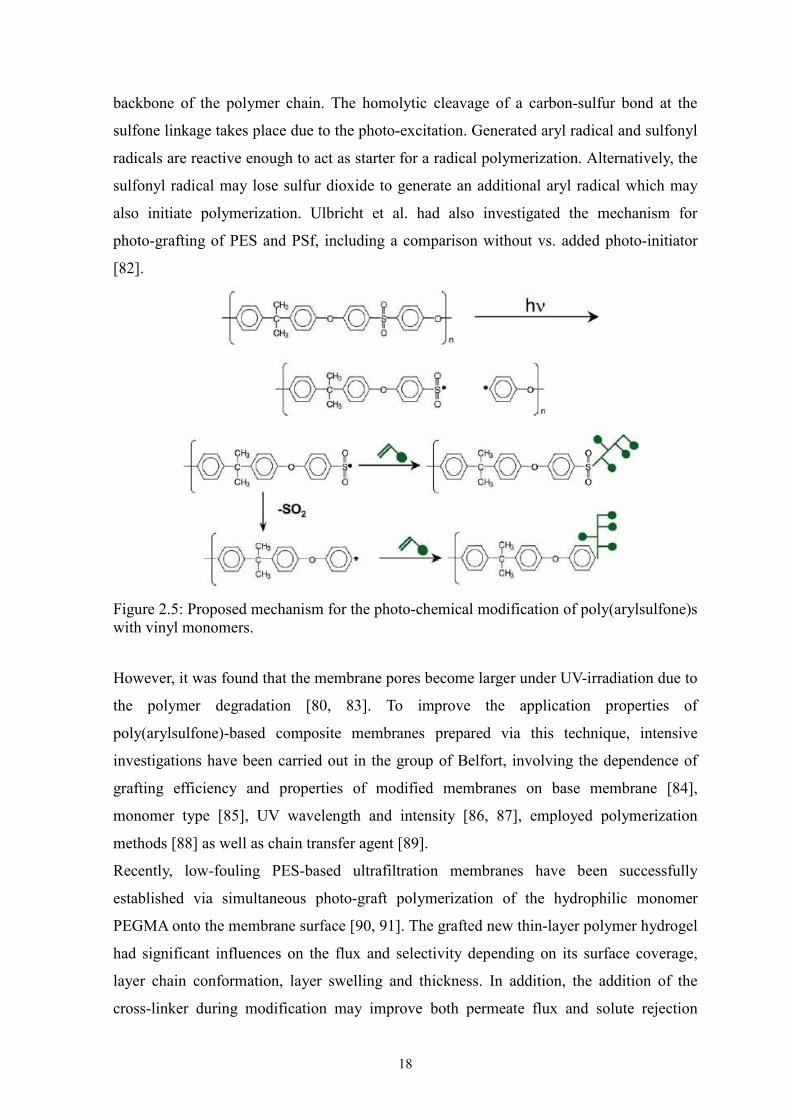
for the photo-reactivity of poly(arylsulfone)s. As shown in Fig. 2.5 [81], the first step
involves the absorption of light by the phenoxyphenyl sulfone chromophores in the
. . R
hν
X X R
hν . .
H
. I R . I-H
. I
. I . HI hν R
I-I . hν I
(a)
(b)
(c)
(d)
18
backbone of the polymer chain. The homolytic cleavage of a carbon-sulfur bond at the
sulfone linkage takes place due to the photo-excitation. Generated aryl radical and sulfonyl
radicals are reactive enough to act as starter for a radical polymerization. Alternatively, the
sulfonyl radical may lose sulfur dioxide to generate an additional aryl radical which may
also initiate polymerization. Ulbricht et al. had also investigated the mechanism for
photo-grafting of PES and PSf, including a comparison without vs. added photo-initiator
[82].
Figure 2.5: Proposed mechanism for the photo-chemical modification of poly(arylsulfone)s with vinyl monomers.
However, it was found that the membrane pores become larger under UV-irradiation due to
the polymer degradation [80, 83]. To improve the application properties of
poly(arylsulfone)-based composite membranes prepared via this technique, intensive
investigations have been carried out in the group of Belfort, involving the dependence of
grafting efficiency and properties of modified membranes on base membrane [84],
monomer type [85], UV wavelength and intensity [86, 87], employed polymerization
methods [88] as well as chain transfer agent [89].
Recently, low-fouling PES-based ultrafiltration membranes have been successfully
established via simultaneous photo-graft polymerization of the hydrophilic monomer
PEGMA onto the membrane surface [90, 91]. The grafted new thin-layer polymer hydrogel
had significant influences on the flux and selectivity depending on its surface coverage,
layer chain conformation, layer swelling and thickness. In addition, the addition of the
cross-linker during modification may improve both permeate flux and solute rejection
19
during ultrafiltration [90].
The first example of a combination of two different photo-irradiation techniques is an
optically reversible switching PES membrane surface, obtained by photo-grating a
monomer with photo-chromic side groups [92].
Due to its potential, this technique is also being seriously evaluated by membrane
manufacturers. An interesting example with respect to both, photo-irradiation and
membrane technologies, had been made via the continuous photo-functionalization of the
outer skin of PSf hollow-fiber membranes with an anionic grafted polymer layer to obtain
nanofiltration (NF) membranes [93].
With other photo-sensitive polymers, where UV irradiation leads to formation of polymer-
bound radical, addition of photo-initiator is also not required for “photo-grafting-from”. A
typical example is that the BP structure is present along the membrane polymer chain. The
photo-reduction of BP structure under UV irradiation can generate radicals by abstracting
hydrogen from monomer and/or bulk polymer itself (similar to Fig. 2.4d, but without
addition of initiator). In this case, cross-linking may occur in the absence of monomer upon
UV irradiation. Yanagishita et al. [94] functionalized an UF membrane made of a
polyimide (PI) with benzophenone structure (BTDA-p-PDA) via UV irradiation in
presence of monomer in gas phase or liquid phase. The obtained pore-filled composite
membrane showed benzene permselectivity for benzene/cyclohexane mixture in PV.
Successful graft modifications have also been observed for other membranes with BP
structures [95, 96].
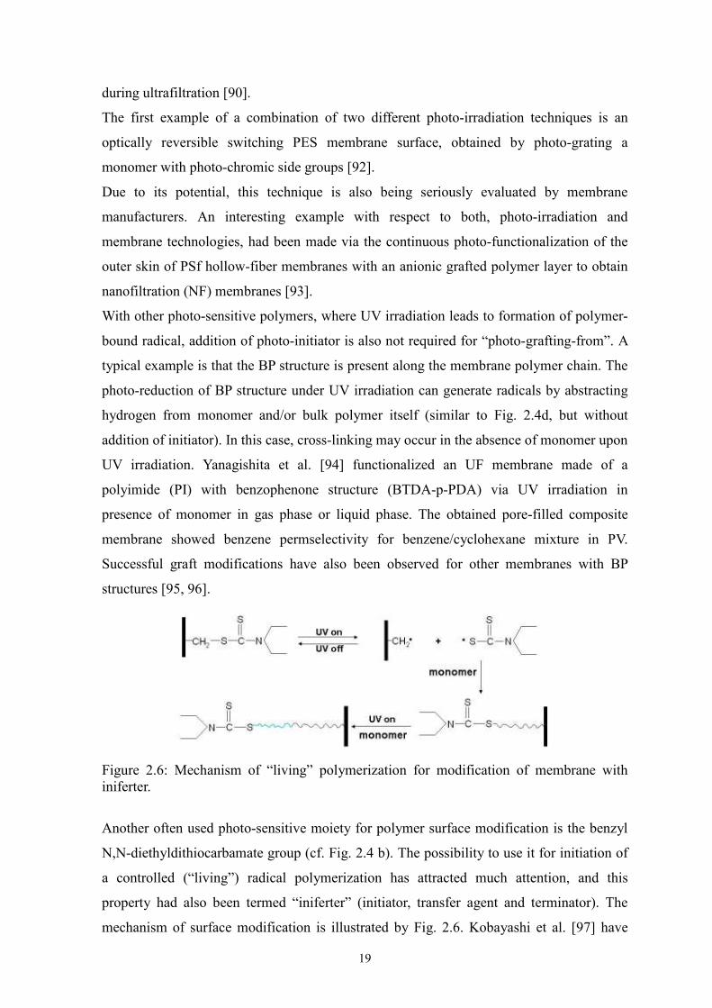
Figure 2.6: Mechanism of “living” polymerization for modification of membrane with iniferter.
Another often used photo-sensitive moiety for polymer surface modification is the benzyl
N,N-diethyldithiocarbamate group (cf. Fig. 2.4 b). The possibility to use it for initiation of
a controlled (“living”) radical polymerization has attracted much attention, and this
property had also been termed “iniferter” (initiator, transfer agent and terminator). The
mechanism of surface modification is illustrated by Fig. 2.6. Kobayashi et al. [97] have
20
successfully grafted a thin-layer of a theophylline (THO) imprinted polymer onto the UF
membrane, which had been prepared from synthesized poly(acrylonitrile-co
-diethylaminodithiocarbamoylmethylstyrene). The resulting MIP composite membrane has
been evaluated as a membrane adsorber, and it had been found that it could recognize the
THO vs. the similar substance caffeine (CAF) with a high selectivity.
However, the synthesis of these special polymers containing photo-sensitive groups and
the following formation of membrane might be a problem in many cases. Therefore,
alternatively, a benzyl N,N-diethyldithiocarbamate group had been chemically
immobilized on the surface of an established membrane via simple coupling reactions.
Using immobilized photo-active iniferter, a molecule-responsive “gate” membrane was
prepared via surface functionalization of the skin layer in the pores of a commercial
cellulosic dialysis membrane with a hydrophilic MIP [98]. The increase in DG value for
re-initiation via multiple sequential UV irradiation periods indicated that graft
copolymerization may proceed via a controlled (“living”) mechanism. Such an approach
should be helpful for the preparation of more sophisticated architectures, e.g., block
structures, of the grafted layer.
Guan et al. have developed another photo-grafting route, based on the combined use of
photo-oxidization and UV irradiation grafting [99]. Hydroperoxide groups were created on
the membrane surface by photo-oxidation in hydrogen peroxide in the first step. Grafted
copolymer layer on the membrane has then been obtained in the presence of monomer
under UV irradiation in the second step. To minimize the homopolymerization, an
appropriate amount of iron (II) was added in the monomer solution as a reductant. The
grafted membranes have been applied for promotion of human endothelial cell adhesion
and growth [100-102].
With added photo-initiator. Many surface modifications of porous membranes in
laboratory and technical scale are performed using cross-linking polymerization to form
thin layers covering the entire pore surface. For such modification, conventional (“type I”)
photo-initiators (for example, benzoin derivatives, or organic peroxides or azo compounds,
i.e., starter radials are directly formed by cleavage of a weak bond in the initiator; cf. Fig.
2.4c) can be very efficient because very fast processes can be realized under ambient
conditions. During the reaction, radical transfer to base polymer is possible (except for
chemically very stable materials such as Teflon). Therefore, chemical grafting of the newly
formed polymer layer to the support membrane may take place to some extent. The most
important example for a commercial application is the hydrophilization of MF membranes
21
made, for instance, from PVDF [103] or PP [104], using hydrophilic polyacrylates; this
modification improves the wetting of the porous membranes by water and reduces the
non-specific binding of (bio)macromolecules (i.e., membrane fouling). Another very
special example is the synthesis of thin-layer MIP composite membranes based on MF
membranes via a cross-linking polymerization initiated with help of the photo-initiator
benzoin ethyl ether [105, 106].
In order to achieve better control over the heterogeneous “photo-grafting-from” of polymer
surfaces, the “type II” photo-initiators, mostly BP or its derivatives, were frequently used.
This type of photo-initiator undergoes photo-reduction by hydrogen atom abstraction from
surrounding chemical species, which leads to the generation of initiating radicals (cf. Fig.
2.4d). Therefore, a preferential hydrogen abstraction from the substrate polymer is an
essential prerequisite for high surface selectivity of graft copolymerization; the
homopolymerization in solution should be minimized. For this purpose, several strategies
have been proposed (Fig. 2.7).
Figure 2.7: Schematic depiction of methods for the immobilization of a “type II” photo-initiator (here benzophenone) for photo-grafting functionalization of membranes.
As known for rather long time for the modification of polymer surfaces (not in the context
of membranes), BP and its derivatives can be used to graft various functional polymers by
just dissolving the initiator in the monomer solution [107-109]. Recently, Yang et al. [77,
110, 111] modified PET Nucleopore membranes using the same approach, i.e., the
simultaneous method. They observed that the photo-grafting occurred only on the top
surface rather than in the membrane pores. Membranes with thermo- or pH-sensitive
22
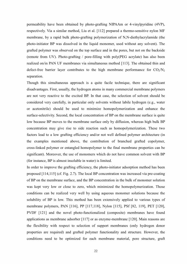
permeability have been obtained by photo-grafting NIPAAm or 4-vinylpyridine (4VP),
respectively. Via a similar method, Liu et al. [112] prepared a thermo-sensitive nylon MF
membrane, by a rapid bulk photo-grafting polymerization of N,N-diethylacrylamide (the
photo-initiator BP was dissolved in the liquid monomer, used without any solvent). The
grafted polymer was observed on the top surface and in the pores, but not on the backside
(remote from UV). Photo-grafting / pore-filling with poly(PEG acrylate) has also been
realized on/in PAN UF membranes via simultaneous method [113]. The obtained thin and
defect-free barrier layer contributes to the high membrane performance for CO2/N2
separation.
Though this simultaneous approach is a quite facile technique, there are significant
disadvantages. First, usually, the hydrogen atoms in many commercial membrane polymers
are not very reactive to the excited BP. In that case, the selection of solvent should be
considered very carefully, in particular only solvents without labile hydrogen (e.g., water
or acetonitrile) should be used to minimize homopolymerization and enhance the
surface-selectivity. Second, the local concentration of BP on the membrane surface is quite
low because BP moves to the membrane surface only by diffusion, whereas high bulk BP
concentration may give rise to side reaction such as homopolymerization. These two
factors lead to a low grafting efficiency and/or not well defined polymer architecture (in
the examples mentioned above, the contribution of branched grafted copolymer,
cross-linked polymer or entangled homopolymer to the final membrane properties can be
significant). Moreover, the use of monomers which do not have common solvent with BP
(for instance, BP is almost insoluble in water) is limited.
In order to improve the grafting efficiency, the photo-initiator adsorption method has been
proposed [114,115] (cf. Fig. 2.7). The local BP concentration was increased via pre-coating
of BP on the membrane surface, and the BP concentration in the bulk of monomer solution
was kept very low or close to zero, which minimized the homopolymerization. Those
conditions can be realized very well by using aqueous monomer solutions because the
solubility of BP is low. This method has been extensively applied to various types of
membrane polymers, PAN [116], PP [117,118], Nylon [115], PSf [82, 119], PET [120],
PVDF [121] and the novel photo-functionalized (composite) membranes have found
applications as membrane adsorber [117] or as enzyme-membrane [120]. Main reasons are
the flexibility with respect to selection of support membranes (only hydrogen donor
properties are required) and grafted polymer functionality and structure. However, the
conditions need to be optimized for each membrane material, pore structure, graft
23
functionality and layer design to meet the needs of different applications; this will be
discussed with the help of examples below.
Pore-filled PAN UF membrane has been successfully established by grafted functional
polymer chains attached to surface when two necessary conditions have been met: DG ≥
DGcritical and small pores in the skin layer of the support membrane (≤ 12 nm in diameter)
[122]. An extension of that work for the separation of aromatic/aliphatic mixture has also
been reported [123-126].
PAN UF membranes was also grafted with monomethoxy poly(ethylene glycol)
methacrylates (MePEG200MA) [127]. A hydrophilic and low-protein-adsorbing UF
membrane with relatively high permeability has been established by adjusting the degree
of grafting via UV irradiation time and monomer concentration.
By applying longer UV wavelength and the photo-initiator BP, PSf UF membranes also
have been modified in a controlled way with PAA layer for covalent immobilization of
biomolecules [82]. This technique avoids the negative effect of using direct UV irradiation
above that desired functionalization was usually accompanied by strong pore etching in the
UF membrane active layer (see above). Borcherding et al. [117] reported that a PP MF
membranes functionalized with the reactive poly(glycidyl methacrylate) (polyGMA)
preserved its high permeability and exhibited high receptor coupling capacity for
recombinant protein A, due to the thin grafted layer and a great deal of epoxy groups
introduced on membrane surface. Based on the very promising results, it is possible to
tailor both membrane structure and application protocols towards other attractive affinity
separations of biomolecules.
Due to the high surface-selectivity of this method, a thin and compact cross-linked layer
can also be synthesized onto the whole surface of a porous membrane, which maintains a
high permeability and relatively large specific surface area, e.g., for affinity binding.
Thin-layer MIP composite membranes, covered with an imprinted polymer layer selective
to small molecules (shown for herbicides) in a mixture with similar compounds, have been
prepared by Piletsky et al. [118] and Sergeyeva et al. [121]. The high affinity of these
synthetic affinity membranes to templates together with their straightforward and
inexpensive preparation provides a good basis for the development of applications of
imprinted polymers in fast separation processes such as solid-phase extraction [128, 129].
This photo-grafting method was successfully adopted also by other groups [130, 131].
The even, tight and defect-free filling of large membrane pores (from PP or PET) with
porous polyacrylate-based monoliths was based on a pre-functionalization of the pore wall
24
with a compatible photo-grafted copolymer [132]. MIP-pore-filled membranes of such
type are currently evaluated for continuous enantio-selective separations [128].
Enzyme membrane reactors also have been prepared based on modified PP MF membranes
with polyGMA [133]. An enzymatic chain elongation of maltooligosaccharide molecules
was achieved during passage through a membrane activated with immobilized
amylosucrase. Commercially available capillary pore membranes (PET) have also been
modified for covalent enzyme immobilization within the pores using this approach [120].
It should be kept in mind that the aforementioned extensive applications of photo-initiator
adsorption method were based on the graft modification in aqueous solutions, where
solvent water does not strongly influence the local BP concentration on the surface.
However, to preserve the tenet of this photo-initiator adsorption technique and to achieve
the high surface-selectivity, the parameters which could reduce the local BP concentration
should be taken into account. For example, a good solvent for BP should not be chosen for
high grafting efficiency, though in one special example, a MIP composite PVDF membrane
has been successfully prepared from the methanol solution, in which a small amount of BP
was added to prevent the rapid reduction of high BP concentration on the surface [118].
To further improve the grafting efficiency and controllability and to extend the application
of “photo-grafting from” technique, more efforts have been made in our group, mainly
focusing on the immobilization of BP. One improvement is that the weak physical
adsorption of BP onto the membrane surface was replaced by ionic interaction between
respective functional groups on the surface and the photo-initiator [14], which can be
realized by the introduction of charged groups onto the base membrane and the application
of counter charged BP derivatives. This stronger immobilization of photo-initiator enabled
an efficient and surface selective functionalization because a better control of grafting
density and a reduction of photo-initiated side reactions along with a more efficient use of
the photo-initiator were possible. Thermo- and pH-responsive PET membranes have been
prepared via this method [14, 134]. Various measurements (for example, the
trans-membrane zeta potential) indicated the even surface coverage of the pore walls with
the grafted polymer. This was the basis for a quantitative analysis of effective layer
thickness as function of synthesis conditions and solution conditions.
Another improved strategy −“photo-initiator entrapping”− also has been proposed to
strengthen the immobilization of BP in the membrane surface [13]. In this process, BP
entrapping was realized by the procedures as follows: membrane was soaked in the BP
solution whose solvent can swell base membrane polymer. After drying, slight washing
25
with non-swelling solvent of membrane polymer followed for removing the adsorbed BP
on the surface. The entrapped BP density in the membrane surface could be tuned by the
initial BP concentration. Controllable three-dimensional grafted layer structure has been
achieved via BP entrapping method to lead to an improved protein binding capacity. Xu et
al. modified PP MF membranes via this technique to improve the biocompatibility
[135-137]. For example, a novel sugar-containing monomer (D-gluconamidoethyl
methacrylate) was grafted on PP via UV-induced graft copolymerization [135]. Results
with respect to BSA adsorption and platelet adhesion imply strongly that a considerable
enhancement of biocompatibility had been achieved. Using the same conditions, PP hollow
fiber MF membranes have been grafted with a hydrophilic layer to improve the
anti-fouling characteristics in a submerged membrane-bioreactor [138]. This method had
also been used to prepare cross-linked grafted layers, and it had been demonstrated that the
dynamic performance of porous membrane adsorbers could be improved [10]; a very high
surface selectivity is mandatory for such reaction conditions, because otherwise
cross-linked insoluble polymer would be immobilized in an uncontrolled way in the pore
space.
In addition, the membrane grafting efficiency has been improved by addition of ferric
chloride with optimum concentration, when simultaneous method or adsorption method
was adopted [139]. This is ascribed to the “synergistic effect” between Fe3+ and BP.
Ma et al. [15, 140] proposed and investigated another photo-induced variant of this graft
copolymerization method ―“covalent photo-initiator immobilization”— consisting of two
steps. This method is based on the discovery of Yang and Ranby that also with BP, a more
controlled (“living”) grafting mechanism can be realized [108]. In the first step, in the
absence of monomer, BP abstracts hydrogen from the substrate to generate surface radicals
and semipinacol radicals, which combine to form surface-bound photo-initiators. In the
second step, the monomer solutions are added onto the active substrate and a living graft
copolymerization is initiated under UV irradiation. In this method, graft density and graft
polymer chain length can be controlled by choosing the reaction conditions in the first step
and in the subsequent step independently. Moreover, the formation of undesired
homopolymer and cross-linked or branched polymer can be substantially eliminated. This
approach has been employed to modify membranes for fouling reduction [141, 142].
However, in this process, the cleavage of covalent bond requires high energy, which leads
to a low grafting efficiency and might also cause photo-degradation of many graft
copolymers.
26
Combining adsorption and covalent photo-initiator immobilization methods,
poly(N,N-dimethylaminoethyl methacrylate) had been photo-grafted onto the PP MF
membrane [143]. Based on this reactive polymer layer, phospholipids-analogous polymers
have been tethered on the membrane to improve surface biocompatibility in the next
reaction.
In brief, many routes for “photo-grafting-from” technique have been developed for
polymeric membrane modification via heterogeneous graft copolymerization. In contrast,
the synthesis of surface anchored polymers via “photo-grafting-from” is often less
controlled with respect to polymer structure, but a very wide variation of grafting densities
and chain lengths can be obtained under relatively convenient reaction conditions. Various
applications of obtained composite membrane have been found in many fields depending
on its membrane material and structure, properties and architecture of grafted layer.
2.3 Membrane functionalities achieved by “photo-grafting-from”
Generally, there are three main composite membrane types, which could meet various
demands in different fields: thin-film-, pore surface-functionalized and pore-filled
membranes (Fig. 2.8 [59]).
Figure 2.8: Depiction of three main composite membrane types: (a) thin-film, (b) pore surface-functionalized, (c) pore-filled.
In principle, all three types of composite membranes introduced could be prepared via
“photo-grafting-from” techniques. However, thin-film composite membranes with
non-porous barriers for gas separation or reverse osmosis have been only rarely prepared
by grafting (and no report on photo-grafting), because the very high grafting density
required for using the intrinsic properties of the new polymer in a non-swollen state as
selective barrier are hard to achieve (see recent work with controlled surface-initiated
polymerization, e.g., [144]). Many examples can be found for more “loose” grafted layers
which should still be permeable, typical examples are anti-fouling modifications, e.g., for
UF membranes. Consequently, most composite membranes prepared via
“photo-grafting-from” are pore surface-functionalized. Depending on initial pore size
(micro/meso pores vs. macropores), grafted layer thickness (ultrathin or extending over 10s
27
or 100s of nanometers) and its distribution over the membrane cross-section (outer surface
vs. entire internal surface) largely different membrane functionalities can be achieved.
Representative membrane functionalities achieved via “photo-grafting from”
functionalization are illustrated (cf. Fig. 2.9).
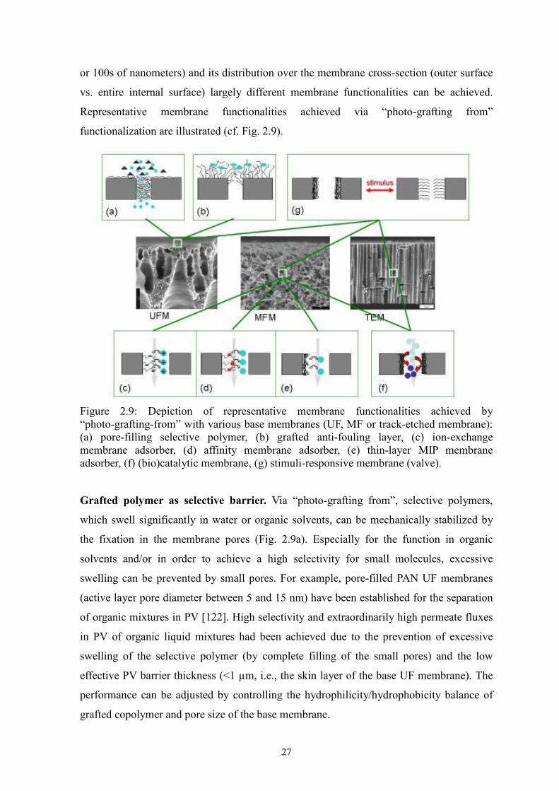
Figure 2.9: Depiction of representative membrane functionalities achieved by “photo-grafting-from” with various base membranes (UF, MF or track-etched membrane): (a) pore-filling selective polymer, (b) grafted anti-fouling layer, (c) ion-exchange membrane adsorber, (d) affinity membrane adsorber, (e) thin-layer MIP membrane adsorber, (f) (bio)catalytic membrane, (g) stimuli-responsive membrane (valve).
Grafted polymer as selective barrier. Via “photo-grafting from”, selective polymers,
which swell significantly in water or organic solvents, can be mechanically stabilized by
the fixation in the membrane pores (Fig. 2.9a). Especially for the function in organic
solvents and/or in order to achieve a high selectivity for small molecules, excessive
swelling can be prevented by small pores. For example, pore-filled PAN UF membranes
(active layer pore diameter between 5 and 15 nm) have been established for the separation
of organic mixtures in PV [122]. High selectivity and extraordinarily high permeate fluxes
in PV of organic liquid mixtures had been achieved due to the prevention of excessive
swelling of the selective polymer (by complete filling of the small pores) and the low
effective PV barrier thickness (<1 µm, i.e., the skin layer of the base UF membrane). The
performance can be adjusted by controlling the hydrophilicity/hydrophobicity balance of
grafted copolymer and pore size of the base membrane.
28
Grafted polymer as anti-fouling layer. Low-fouling UF membranes (Fig. 2.9b) can be
prepared under photo-grafting conditions where the degradation of the base membrane
pore structure is minimized. The composition, surface coverage and thickness of the
grafted layer are crucial for final membrane performance, i.e., low fouling at preserved size
exclusion property and competitive flux [90, 114].
Grafted polymer layer comprising functional groups for reversible binding. Surface
functionalized MF membrane adsorbers for fast protein purification (Fig. 2.9c,d) have been
prepared via photo-grafting of two- or three-dimensional layers with suited functional
groups [13, 117]. A novel type of MIP composite membranes (Fig. 2.9e), with high binding
specificity at high throughput, has been obtained by surface initiated photo-grafting of a
very thin cross-linked functional layer [118, 121]. All these cases require a sufficient
permeability, in order to use the main advantage of porous membrane adsorbers, i.e., the
reduction of mass transfer limitations by convective flow though the membrane pores.
Grafted polymer layer for immobilization of (bio)catalyst. Iso-porous track-etched
membranes with a larger pore diameter (between 100 nm and 3 µm) as well as PP MF
membrane had been functionalized via “grafting-from” reactions in order to prepare
enzyme-membranes as convective flow microreactors (Fig. 2.9f) [120, 133]. Enzyme-
membranes with high permeability and essentially unchanged FTF activities have been
yielded with grafted poly-(2-aminoethyl methacrylate) layer followed by glutardialdehyde
activation and coupling of fructosyltransferase (FTF, inulinsucrase from Streptococcus
mutans). A continuous enzymatic reaction driven by trans-membrane substrate flow has
been realized by using membranes with rather spacious cylindrical pores (3 µm) to avoid
their blocking by the product polyfructan (inulin) with a molecular weight between 30-50
Mio g/mol. The covalent immobilization of epoxy-reactive nanoparticles on the pore walls
of PET membrane, subsequently photo-grafted with poly(amino-ethyl acrylate), also
increased the productivity and lowered pore-blocking tendency [145].
Grafted stimuli-responsive polymer layer. Using tailored grafted functional polymer
layers on the pore walls of membranes, it is possible to reversibly change the permeability
and/or selectivity. The most straightforward mechanism is the alteration of the effective
pore diameter by changing the conformation of a grafted polymer via solution conditions
as ‘stimulus’ (Fig. 2.9g). For example, reversible switching of permeability had been
achieved using photo-grafted pH-responsive (PAA or PMA) [13, 14, 115] or temperature-
responsive chains (polyNIPAAm) [134].
29
2.4 Membrane adsorbers
As mentioned in section 1.1, membrane adsorber can overcome the limitations associated
with packed bed. In membrane chromatography processes, the transport of solutes to their
binding sites takes place predominantly by convection, reducing both process time and
recovery liquid volume. The pressure drop is significantly lower compared to packed beds
as the flow path, even for a stack of multiple membranes, is much shorter. Another major
advantage of membrane adsorbers is the relative ease of scale-up. In addition, for large
proteins, the surface area available for binding is significantly greater with membranes
[146]. However, membrane chromatography has its limitations towards pore size
distribution and uneven thickness of membrane, inlet flow distribution and lower binding
capacity, which indirectly or directly influences the performance of membrane
chromatography such as the efficiency of protein separation and ligands utilization.
Therefore, intensive investigations on membrane adsorbers have been carried out (detailed
see below).
2.4.1 Classification of membrane adsorbers
Membrane adsorbers are chromatographic membranes carrying functional groups (ligands)
for the binding of biomolecules. They are not filters, although the structure looks similar.
Separation is achieved by reversibly binding the interests to the ligands. Generally,
macroporous membranes are used.
According to the module geometry, membrane adsorbers can be classified into flat sheet
(including single and stacked-membrane), hollow fiber and spiral wound membranes (Fig.
2.10) [6, 8]. Previously, membrane adsorbers were sold in the form of single thin sheet
having pore size of the order of 1 µm. However, the capacity of these membranes was low,
and small thickness and porosity variations severely degraded membrane performance. The
next generation is stacks of several flat sheets, which are housed within membrane
modules. In addition to providing more adsorbent volume, the use of membrane stacks can
average out the side effect of variation of pore size distribution and membrane thickness
for single flat sheet, leading to sharper breakthrough curves and higher binding capacities
(cf. below). Based on the statistics of the literatures [8, 147], flat sheet membranes are by
far most widely used.
A single thin sheet wound around a permeable cylindrical core can be similarly packaged
(spiral wound membrane). It is claimed to be suitable for large-scale applications and for
use in the bind and elute mode. However, the biggest challenge would be the poor flow
30
distribution because the membrane area increases in a radially outward direction, causing a
drop in superficial velocity of the liquid stream during its flow through the membrane. In
addition, the binding and elution processes might be difficult to be modeled and predicted.
Nevertheless, this type of membrane adsorbers has been used by companies such as
Sartorius and Pall (provided in ref. [8]).
A hollow fiber membrane adsorber usually has a tubular geometry, consisting of a bundle
of several hundred fibers potted together within a module in a shell and tube
heat-exchanger-type configuration. It provides a high membrane surface area to volume
ratio. The reduction in accumulation of particles near the pore entrance is another
advantage using hollow fibers due to the cross-flow. However, this type of adsorber can not
be used for pulse chromatography because the duration is insignificant when compared
with the overall processing time. Even in the bind and elute mode, the breakthrough is
expected to be broadened, leading to poor adsorber utilization.
Figure 2.10: Schematic description for membrane adsorber geometries. Arrows illustrate the direction of bulk flow; pattern indicates the membrane cross-sectional area [6].
According to the interaction mode (the active ligands), membrane adsorbers involve
affinity membrane, ion-exchange membrane, hydrophobic interaction based membrane and
reversed-phase based membrane. Based on the collected literatures [8, 147], affinity
separation seems to be the largest segment. The ligands used for affinity membrane
chromatography mainly include immunoaffinity ligands e.g., [148, 149], protein A or G
e.g., [150, 151], dyes e.g., [152, 153] and immobilized metal ions e.g., [154, 155] as well
as peptides e.g., [156]. In terms of chromatographic membranes commercially available,
ion-exchange membranes constitute the largest segment. To achieve a corresponding
interest binding, membranes have been modified with different charged groups such as
sulfonic acid [157], acrylic acid [10] and di/ethylamino group [37]. Along with affinity and
ion-exchange chromatographies, chromatography based on hydrophobic interaction has
(a) Single flat sheet (c) Hollow fiber
(b) Membrane stack
(d) Spiral wound
31
wide applications for protein purifications. Alkyl, e.g., butyl and octyl, and aryl, e.g.,
phenyl, groups are representative ligands for this type of adsorber. For the reversed-phase
based membrane, few reports had been found. The different types of membrane adsorbers
used for chromatographic separation have been reviewed in the review papers (e.g., refs.
[8,147]).
In addition, a novel type of membrane adsorber has been received a great interest, which is
based on a novel molecular imprinting technology. The MIP composite membrane (see Fig.
2.9e) can be obtained by in situ preparation [158] or grafting MIP thin-layer on the
membrane surface [118]. The interaction mode can be covalent bonding, hydrogen
bonding, ionic interaction or hydrophobic interaction; more importantly, the shapes of the
synthesized fixed recognition cavities take more responsible for the selective affinity of
interest. The resulting membrane adsorbers can be used for separation of the compounds
with similar chemical and physical properties such as enantio-separation.
2.4.2 Investigation on membrane adsorbers
In order to clearly understand adsorption behavior of membrane adsorbers and improve the
overall performance of membrane chromatography, both chromatographic processes and
membrane chemistries have been being simultaneously investigated.
Chromatographic processes. To properly design, operate and apply the membrane-based
separation processes, hence maximize the throughput and the degree of separation, many
efforts have been directed towards the process engineering aspects, especially to the mass
transport phenomena of membrane chromatography, usually using flat sheet membranes as
the model system. The earliest work regarding transport phenomena was reported by Briefs
and Kula [159]. A mathematical formulation for an idealized membrane adsorber based on
100 stacks of flat sheets was presented and solved to predict breakthrough and elution
profiles. The mathematical analysis was experimentally verified by dynamic adsorption
and elution studies using the enzyme formate dehydrogenase and pyruvate decarboxylase.
Suen and Etzel [160] formulated a mathematical model including convection, diffusion and
Langmuir-type adsorption to analyze the design and operation of affinity membrane
bioseparations. From the analysis it was predicted that for thin membranes, the flow-rate is
limited by the ligand-protein association kinetics and even small variations of thickness
and porosity severely degrade membrane performance, which would be overcome by the
use of stacks of several thin membranes. The mass transport implications of this
mathematical model were also explored using experimental studies [161]. Later, using
32
ion-exchange membranes with larger pore size (150 µm), Etzel and his co-workers found
based on new mathematical models that the breakthrough curve and elution peak strongly
depend on flow-rate, and the maximum allowable pore size which eliminates this
limitation was calculated for different molecular weight solutes [162]. Also, the
performance of ion-exchange membrane was analyzed using different models, which
incorporated nonlinear sorption isotherm and mass transfer coefficient. The models allow
the determination of the rate-controlling mass-transfer phenomena and solid-phase
concentration, and prediction of the operating and membrane-design parameters needed to
obtain sharp breakthrough curves [163]. Tennikova and Svec [164] investigated the mass
transport phenomena of membrane chromatography primarily based on operating
parameters. They claimed that the process efficiency of the systems was not limited by
flow-rate. It was explained that the increase in flow rate led to the enhancement of
diffusive protein transport (i.e., increase in mass transfer coefficient). From the calculations,
the diffusivity of the proteins within the pores is about four orders of magnitude higher
than their respective free solution diffusivities. Tejeda et al. [165] have also discussed the
implications of mass transfer in the design of membrane adsorbers.
Recently, the mathematical models have been developed using different adsorption
mechanisms rather than Langmuir adsorption, such as bi-Langmuir [166] or Freundlich
adsorption [167, 168]. In addition, Ghosh et al. [169] designed a new membrane module
with enhanced both feed flow distribution and effluent collection. The membrane modules
showed significant higher lysozyme binding capacities than the corresponding
conventional modules, measured both in the breakthrough mode and in the pulse
chromatography mode.
In addition to the analyses of breakthrough and elute curves, the visual confocal laser
scanning microscopy (CLSM) images for protein adsorption may provide a helpful
guidance for grafted layer design [170, 171].
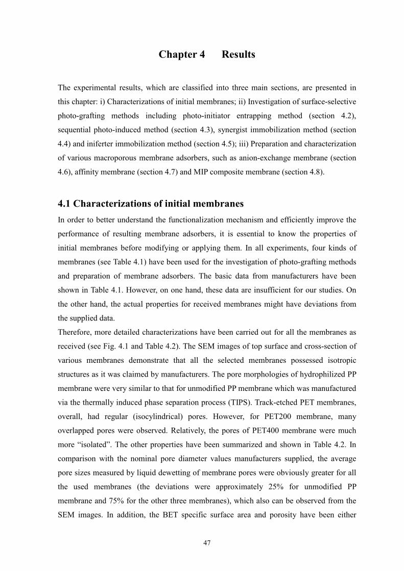
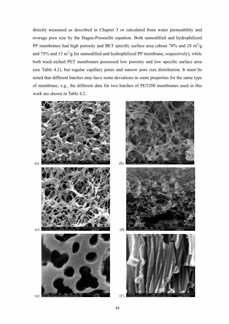
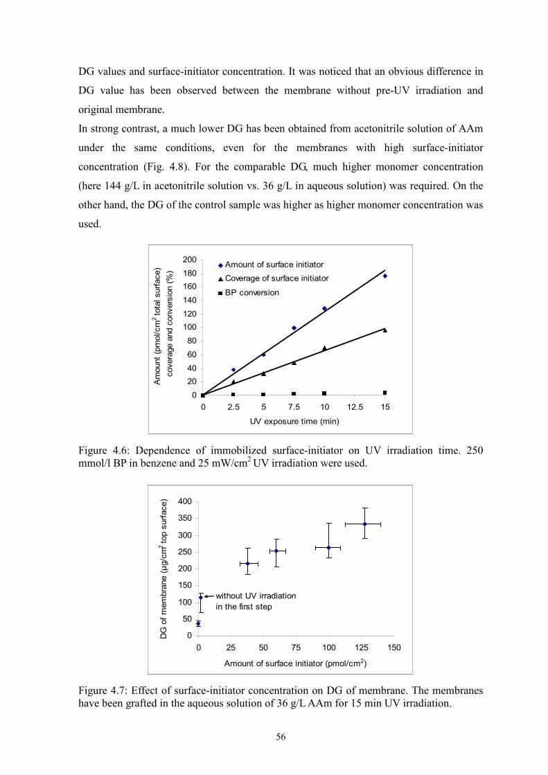
Membrane chemistry (Immobilization of ligands). The immobilization of ligands, on
one hand, reduces the non-specific binding; on the other hand, provides specific binding
sites for interests. The motivations for membrane chemistry investigation are 2-fold: one is
to immobilize ligands as many as possible on the membrane surface for high binding
capacity (or the prerequisite of high binding capacity); another is to ensure the availability
of ligands (i.e., the structure of modified layer should be considered) and the operable
chromatographic processes (e.g., membrane permeability related to flow rate). In the
review papers, Klein [7] and Zeng and Ruckenstein [172] summarized the modifications of
33
existing membranes for membrane adsorbers, concentrating on the chemical modifications
as well as radiation-induced grafting method. Chemical modifications mainly are
applicable to the membranes with reactive groups on the surfaces (e.g., polysulfone and
nylon-6 hollow fibers). Two important issues should be considered: one is the effect of
reaction conditions on the stability of base membranes. Another is that either small
functional molecules are immobilized (it is hard to improve the binding capacity) or
macromolecules are bound to the membrane surface via “grafting-to”. For the latter, the
structure of grafted polymer and uniformity of functionalization would be complicated to
control. As for radiation-induced grafting method, the major body of work has been
conducted by Saito and his co-workers. Graft of commercially available PE MF with
PolyGMA had been perfomed through initiating by electron beam and then exposed to
air-free vapors of glycidylmethacrylate. The introduced epoxide groups offer many
possibilities for the immobilization of various ligands. The resulting membranes can be
applied as affinity membranes [35], cation/anion-exchange membranes [157, 36] and
hydrophobic interaction-based membranes [173]. To improve the performance of
membrane adsorbers, the grafting layer property was adjusted by varied ligand density [4]
and cross-linking [9]. In addition, photo-induced grafting method has been used for
synthesis of membrane adsorbers. As discussed above, this method including several
grafting routes is more flexible to synthesize desired grafted layer architectures. Main work
came from Ulbricht and his co-workers. For example, affinity membranes [82, 117, 119],
Table 4.2 Measured data for various initial membranes.
Membrane Average pore size (a) nm
Thickness µm
BET specific surface area m2g-1
Porosity %
Pore type Chemical structure on the surface
Unmodified PP ~250 186 28 78 irregular (CH2CH)
CH3
n
Hydrophilized PP ~350 90 15 75 irregular
-COOR(c)
CH2CH2O )n MeR =
or
CH2CH2O( )n C
O
(
PET200 ~3501, 3702 23 2.8
(3.91, 2.72)(b) 341, 242(b) cylindrical
PET400 ~700 23 1.7 (b) 28(b) cylindrical C
O
OC2H4O)C
O
( n HHO
Note: 1 and 2 (for PET200 membrane) represent different batches. (a) Measured via liquid dewetting technique using Capillary Flow Porometer; (b)
calculated from water permeability and average pore size; (c) deduced from IR spectrum and contact angle, referring to the patent [104].
51
4.2 Photo-initiator entrapping method
This photo-grafting method had been developed previously in our group [13] and
cation-exchange functional layers had been prepared on the unmodified PP membrane
from aqueous solution of acrylic acid [10, 13]. Based on the more stable immobilization of
photo-initiator in the membrane surface (by BP entrapping; compared with the simple
physical adsorption for conventional adsorption method [114]), in this study, an attempt
was made to extend the application to the photo-graft functionalization of membranes from
polar organic solution system.
4.2.1 Photo-initiator immobilization
According to the proposed mechanism (see Fig. 5.10), moderate swelling of base
membrane is the pre-requisite for the effective immobilization of photo-initiator. In this
work, therefore, five kinds of good solvents of BP have been chosen to investigate the
swelling property of unmodified PP membrane. As shown in Table 4.3, two groups can be
classified according to the obtained results. The group including chloroform, benzene and
heptane occupied significantly larger volume in the membrane and also caused a much
greater mass loss of bulk polymer after 24-hr immersion than another group consisting of
ethanol and acetonitrile, indicating that a better swelling of PP membrane has been
achieved with the former. Here, therefore, heptane was chosen as the solvent of initiator.
Nevertheless, the porosity of PP membrane has been estimated from the occupied volume
of methanol and acetonitrile (see Table 4.3), which was similar to the porosity value
determined with water (after pre-wetting).
Table 4.3 Swelling property of unmodified PP membrane in various solvents.
Solvent Occupied volume by
solvent* (10-3mL/mg) Mass loss
(‰) Estimated porosity**
(%)
Ethanol 5.37 1.39 77.8
Acetonitrile 5.39 1.62 78.4
Chloroform 5.91 7.64 -
Benzene 5.96 6.19 -
Heptane 6.01 6.55 -
Unmodified PP membrane was immersed in solvent for 24 hr. *: calculated from the equation 3.5. **: obtained through the equation 3.6. The amount of immobilized photo-initiator is a crucial factor which determines the
subsequent photo-grafting efficiency and grafting density. Therefore, the BP uptake has
52
been determined gravimetrically (accuracy of the balance was 10 µg) after twice quick
washing (2 min and 1 min, respectively) with non-swelling solvent, methanol, by which
the adsorbed BP on the membrane surface had been verified to be completely removed
[13]. Note that control samples have been analyzed with pure solvent (heptane) under the
same conditions for correction for mass loss by extraction. Fig. 4.3 shows BP uptake of
membrane as function of soaking time or BP concentration. It seems that no more BP was
effectively entrapped in the membrane surface for the further immersion after about 2.5
µg/cm2 (relative to top surface) was achieved in 15 min. In addition, the amount of
immobilized BP only had a slow increase with the BP concentration used, and did not
show a pronounced difference when the BP concentration was varied from 10 mmol/L to
100 mmol/L.
0
1
2
3
4
5
0 10 20 30 40 50 60
Soaking time (min)
BP
upta
ke (
µg/c
m2)
(a)
0
1
2
3
4
5
0 20 40 60 80 100
BP concentration (mM)
BP
upta
ke (
µg/c
m2)
(b)
Figure 4.3: Effect of soaking time and initiator concentration on BP uptake. a) Effect of soaking time (10 mmol/L BP in heptane); b) Effect of BP concentration (15 min soaking).
53
4.2.2 Effect of washing on initiator immobilization
In the original literature for this method [13], water, as the medium of polymerization
system, had little influence on the entrapped BP during photo-graft polymerization because
of the negligible solubility of BP and no swelling of PP in water in limited time.
However, this work aimed at the membrane functionalization from polar organic solution.
Therefore, it is important and necessary to know the immobilization stability of entrapped
initiator during graft polymerization because the initiator BP is soluble in most of organic
solvents. For this purpose, pure polar solvent, methanol, has been used to simulate the
environment of graft polymerization system instead of the monomer solution. Moreover,
this part of study was merged into the washing procedure prior to photo-grafting. Therefore,
the washing procedure was different from that previously adopted, but similar to the
procedures for membrane functionalization in monomer solution, i.e., including 10-15 min
equilibrium and 10-15 min photo-irradiation time. In this case, multi-time-washing was set
up (Table 4.4). The washed solutions were detected spectrophotometrically at 256 nm. As
it is shown in Table 4.4, washing of the dried BP-adsorbed membrane was carried out with
methanol for three or four times depending on the BP concentration used. After soaking in
BP solution of 10 mM and 100 mM, respectively, BP still could be detected in the solutions
after the second and third time-washing, respectively. After that, the extraction of BP has
been performed with heptane. However, very low absorbance (in the error range) was
observed for the extraction solution, i.e., residual amount of BP in membrane was
negligible (not shown).
Table 4.4 UV absorbance of washed solutions at 256 nm.
BP concentration (mmol/L) Washing conditions
100 10
1st (10 mL, 10 min) 3.855 1.123
2nd (5 mL, 5 min) 1.110 0.334
3rd (5 mL, 5 min) 0.197 -0.002
4th (5 mL, 5 min) 0.001 -
Note: The membrane with a diameter of 44 mm was used for this experiment.
4.2.3 Effect of washing on membrane photo-grafting
Photo-graft polymerization of the membranes with different washing times has been
performed in AAm aqueous solution. As comparisons, same membranes have also been
grafted via conventional photo-initiator adsorption method (i.e., heptane was replaced with
54
methanol). Fig. 4.4 shows that for adsorption method, the DG decreased rapidly with the
washing time and a low constant value was achieved at only 1.5 time-washing, whereas, a
gradual reduction in DG value can be observed in the case of entrapping method. However,
a tendency towards the constant low value seems to be unavoidable.
0
50
100
150
200
250
300
350
400
1 1.5 2 3
Washing times
DG
of
mem
bra
nes (
µg/c
m2 to
p s
urf
ace)
BP in heptane
BP in methanol
Figure 4.4: Effect of washing times on unmodified PP membrane photo-grafting via entrapping and conventional adsorption methods. Each time: one sample was washed in 5 mL methanol for 5 min. half time: 3 samples were washed in 5 mL methanol for 2 min. 10 mM BP was used for the pre-treatment of PP membrane; Photo-grafting conditions: 36 g/L AAm aqueous solution, 15 min UV irradiation.
0
50
100
150
200
250
300
350
400
acrylamide in
water (36g/L)
acrylamide in AN
(36g/L)
acrylamide in AN
(144g/L)
DG
of
mem
bra
ne (
µg/c
m2 t
op s
urf
ace)
entrapping method
without initiator
Figure 4.5: Effect of solvent on DG of membrane. Membranes were washed once prior to graft polymerization. 10 mmol/L BP was used for the pre-treatment of PP membrane; Polymerization conditions: 15 min UV irradiation.
55
4.2.4 Effect of solvent on membrane photo-grafting
As shown in Fig. 4.5, photo-graft polymerization of BP-entrapped and without
initiator-treated (as blank sample; treated by same procedures but no initiator in heptane)
PP membranes have also been conducted from acetonitrile solution of AAm. Compared to
that from aqueous solution, the obtained DG value was significantly lower under the same
grafting conditions. A DG value of approximately 135 µg/cm2 has been obtained even
though the high monomer concentration of 144 g/L was applied. Moreover, in this case, the
DG value of the blank membrane also clearly increased.
4.3 Sequential photo-induced method
Earlier, photo-grafting of polymer films had been investigated using BP as initiator and it
was discovered that the termination reaction of this graft system was mainly by
combination of growing chain radicals and semipinacol radicals [104, 177]. Taking
advantage of this characteristic, Bowman and co-workers had developed a novel two-step
method for unmodified PP membrane photo-grafting (see Fig. 5.10) [15]. The current work
focuses on the investigation of photo-graft functionalization in polar organic solution using
the same method and base membrane.
The PP membrane saturated with benzene solution of 250 mmol/L BP was fixed between
two pieces of glass and exposed under UV irradiation. At a given time, initiator has been
covalently bond to the PP membrane surface (see reaction mechanism in Fig. 5.1). The
amount and surface coverage of surface-initiator as well as BP conversion are shown in Fig.
4.6. The concentration of immobilized surface-initiator increased nearly linearly as a
function of UV irradiation time in the studied range where BP conversion was very low
(the maximum conversion was less than 4%). This is in agreement with the results reported
by Bowman [15]. Assuming that the occupied area of one BP molecule is 90 Å2 [13], a
surface coverage of approximately 95% has been achieved at 15 min UV irradiation, while
further irradiation led to the degradation of membrane due to the rising temperature.
Surface-initiator-immobilized PP membranes have been UV-functionalized in aqueous and
acetonitrile solution of AAm for 15 min, respectively, along with the control samples (one
was original membrane; another was treated with all the procedures except for UV
irradiation). Fig. 4.7 shows the relationship between DG value and surface-initiator amount,
as membranes were grafted from aqueous solution. The DG values were significantly
higher for initiator-immobilized membranes than for original membrane and the membrane
without pre-UV-irradiation. Moreover, a good correlation can be seen between obtained
56
DG values and surface-initiator concentration. It was noticed that an obvious difference in
DG value has been observed between the membrane without pre-UV irradiation and
original membrane.
In strong contrast, a much lower DG has been obtained from acetonitrile solution of AAm
under the same conditions, even for the membranes with high surface-initiator
concentration (Fig. 4.8). For the comparable DG, much higher monomer concentration
(here 144 g/L in acetonitrile solution vs. 36 g/L in aqueous solution) was required. On the
other hand, the DG of the control sample was higher as higher monomer concentration was
used.
0
20
40
60
80
100
120
140
160
180
200
0 2.5 5 7.5 10 12.5 15
UV exposure time (min)
Am
ount
(pm
ol/cm
2 to
tal surf
ace)
covera
ge a
nd c
onvers
ion (
%)
Amount of surface initiator
Coverage of surface initiator
BP conversion
Figure 4.6: Dependence of immobilized surface-initiator on UV irradiation time. 250 mmol/l BP in benzene and 25 mW/cm2 UV irradiation were used.
0
50
100
150
200
250
300
350
400
0 25 50 75 100 125 150
Amount of surface initiator (pmol/cm2)
DG
of
mem
bra
ne (
µg/c
m2 to
p s
urf
ace)
without UV irradiation
in the first step
Figure 4.7: Effect of surface-initiator concentration on DG of membrane. The membranes have been grafted in the aqueous solution of 36 g/L AAm for 15 min UV irradiation.
57
0
50
100
150
200
250
300
350
AAm in water
(36g/L)
AAm in AN
(36g/L)
AAm in AN
(144g/L)
DG
of
mem
bra
ne (
µg/c
m2)
sequential photoinduced method
without initiator
Figure 4.8: Comparison of photo-graft functionalization from water and acetonitrile solutions. Surface-initiator concentration on membrane surface was about 160 pmol/cm2. Grafting conditions: 15 UV irradiation time.
4.4 Synergist immobilization method
4.4.1 Immobilization of tertiary amino groups
Thermally induced aminolysis, which is based on the nucleophilic reaction of an organic
amine agent with the ester group along a polymer chain, had already been used to treat
polymer fibers or membrane surfaces [178, 179]. According to the chemical structure of
hydrophilic polymer layer (Table 4.2), hydrophilized PP membrane would be possible to be
modified using the same reaction. Therefore, in this work, DEEDA (Fig. 4.9) was used to
pre-functionalize the hydrophilized PP membrane surface with tertiary amino groups.
H2N NCH2 CH2
C2H5
C2H5
Figure 4.9: Chemical structure of diethyl ethylenediamine (DEEDA).
The immobilized tertiary amino groups have been determined by ATR-IR (Fig. 4.10a).
From the spectra of aminolysed membranes, unfortunately, no amide absorption peaks
could be detected after the reaction. However, the intensity ratio between the bands at 1728
and at 1375 cm-1, which represents ester and methyl groups (the latter as reference peak for
PP), respectively, has been observed as function of reaction conditions, i.e., the intensity
ratio decreased with the rising aminolysis temperature and reaction time (Fig. 4.10b and
4.10c). In addition, a characteristic color reaction has been used to qualitatively detect
58
tertiary amino group as well (using “D-solution”; see Section 3.4.3). A wine-red color was
observed on the aminolysed membrane samples, while no color was found for the control
samples.
0
0.05
0.1
0.15
0.2
0.25
500100015002000
Wavenumber (cm -1)
Ab
so
rba
nce
1
2
- 1 - original hydrophilic membrane
- 2 - aminolysed membrane
(a)
0.2
0.25
0.3
0.35
0.4
0 20 40 60 80
Temperature (℃)
Absorp
tion p
eak inte
nsity r
atio
(1728cm
-1/1
375 c
m-1
)
original membrane
0
0.1
0.2
0.3
0.4
0 1 2 3 4
Reaction time (hr)
Absorp
tion p
eak inte
nsity r
atio
(1728cm
-1/1
375cm
-1)
(b) (c)
Figure 4.10: (a) ATR-FTIR spectra for original hydrophilized and aminolysed membranes (2 hr at 600C); and the dependence of absorption peak intensity ratio between the bands at 1728 cm-1 and at 1375 cm-1 on aminolysis temperature (reaction time: 1 hr) (b) and reaction time (aminolysis temperature: 600C) (c).
Reversible staining with an anionic dye is a simple, effective and frequently applied
approach for qualitative and quantitative determination of amino groups [14. 21, 179].
Here AO (Fig. 4.11) was used to detect the extent of aminolysis quantitatively.
NaO3S N N
OH
Figure 4.11: Chemical structure of acid orange II (AO).
59
At a given time (1 hr), the influence of aminolysis temperature on the surface amount of
tertiary amino groups was measured (Fig. 4.12a). It can be seen that tertiary amino group
concentration increased with the reaction temperature. For convenience, 600C was adopted
for more detailed studies. As shown in Fig. 4.10b, at 600C, the maximum value of surface
amount of tertiary amino groups has been achieved at the longest aminolysis time.
However, the surface amount of tertiary amino groups as a function of the reaction time
seemed to grow in two stages, i.e., in the ranges of 1.5-2.0 hr and 3.0-4.0 hr, respectively.
0
100
200
300
400
500
20 40 60 80
Temperature (°C)
Te
rtia
ry a
min
o g
rou
p
co
nce
ntr
atio
n,c
(p
mo
l/cm
2)
0
1
2
3
4
5
6
DG
of m
em
bra
ne
(%
)
(a)
0
100
200
300
400
500
600
0 1 2 3 4
Aminolysis time (hr)
Te
rtia
ry a
min
o g
rou
p
co
nce
ntr
atio
n, c (
pm
ol/cm
2)
0
0.5
1
1.5
2
2.5
3
3.5
4
4.5
DG
of m
em
bra
ne
(%
)
(b)
Figure 4.12: Effect of aminolysis process parameters on the amount of tertiary amino groups formed on the membrane surface (relative to the specific surface area of the membrane) and DG value of the membrane obtained in the subsequent photo-grating step (36 g/L AAm and 5 mM BP in methanol, 10 min UV irradiation). (a) Effect of aminolysis temperature, reaction time 1 hr. (b) Effect of aminolysis time, reaction temperature 600C. (Note that a DG = 1% corresponds to 24 µg cm-2 relative to the top surface area and to 0.067 µg cm-2 relative to the specific surface area.)
60
4.4.2 Photo-graft polymerization on membranes
To evaluate the proposed method, photo-grafting functionalization of original and
aminolysed membranes has been carried out in methanol solution of AAm via
simultaneous method (i.e., photo-initiator BP dissolved in monomer solution). The results
demonstrate that for the original hydrophilized PP membrane, DG was very low and hardly
affected by varying the parameters in the studied range. Even for the highest monomer
concentration, the DG value of the membrane reached only about 0.17 µg/cm2 (Fig. 4.18).
In contrast, for the aminolysed membranes, much higher DG values have been achieved.
Furthermore, for the membranes after varied aminolysis conditions, there was a good
correlation between the surface content of tertiary amino groups and the DG values (Fig.
4.12).
In addition, ATR-IR spectra of functionalized membranes with different DG values have
been recorded to characterize the chemical structure of grafted layer (Fig. 4.13). An amide
I band at 1665 cm-1 appeared which was not present in the case of the original
hydrophilized and not detectable for the aminolysed membrane (cf. Fig. 4.10a). Moreover,
the intensity of the absorption peak at 1665 cm-1 was correlated with the DG values.
0
0.01
0.02
0.03
0.04
0.05
0.06
0.07
0.08
0.09
0.1
130014001500160017001800
Wavenumber (cm-1)
Absorb
ance
1
2
3
- 1 - original membrane
- 2 - DG 3.8% membrane
- 3 - DG 8.5% membrane
Figure 4.13: ATR-FTIR spectra for original hydrophilized and PAAm-grafted PP membranes with different DG. For the modification of porous membranes, a key issue is that the original pore structure
should not be degraded during the intended functionalization. The morphologies of the top
surface of the membranes were observed using SEM (Fig. 4.14). No differences in
morphology could be detected between original and aminolysed membranes. However, it
should be noted that in the original and aminolysed membranes a large amount of fiber-like
61
polymer was seen. These features were less pronounced for the membrane with the lower
DG, and they had disappeared nearly completely after photo-grafting at a DG of 8.5%. That
additional polymer layer has been attached evenly and tightly on the pore surface of the
membrane, and no aggregates or polymer globules were observed on the surface and in the
pores.
(a) (b)
(c) (d)
Figure 4.14: SEM top surface images of porous PP membranes: (a) original hydrophilized; (b) aminolysed (2 hr at 700C); (c) photo-grafted with PAAm (DG = 3.8%); (d) photo-grafted with PAAm (DG = 8.5%).
Figure 4.16: Effect of BP concentration on grafting on original hydrophilized PP membrane. (■) 8 min UV irradiation, 54 g/l AAm in methanol, ♦) 12 min UV irradiation, 54 g/l AAm in acetonitrile.
0
10
20
30
40
50
60
0 1 2 3 4 5 6
BP conc. (mmol/L)
DG
of
ori
gin
al
me
mbra
ne (
%)
0
1
2
0 1 2 3 4 5
BP conc. (mmol/L)
DG
of
me
mb
ran
e
(%)
64
-1
4
9
14
19
24
29
34
0 0.5 1 1.5 2
BP concentration (mmol/L)
DG
of
mem
bra
ne (
%)
Original PP membrane
Aminolysed membrane
(a)
0
10
20
30
40
50
60
70
0 1 2 3 4 5 6
BP conc. (mmol/L)
DG
of
mem
bra
ne (
%)
aminolysed
original
(b)
Figure 4.17: Effect of BP concentration on the surface selectivity of grafting on hydrophilized PP membranes from acetonitrile solution containing 54 g/l AAm. a) 10 min UV irradiation; b) 12 min UV irradiation.
Effect of UV irradiation time
The DG values could also be adjusted by the UV irradiation time (Fig.18b). Low graft
copolymerization efficiency below 2 min exposure was observed. Afterwards, a
continuously increasing DG was achieved with UV irradiation time. In addition, a much
higher DG ratio (aminolysed vs. original), i.e., surface-selectivity of photo-grafting was
found for shorter grafting time.
Effect of monomer concentration
As expected, the DG values increased with monomer concentration (Fig.18c). Similar to
the maximum for short UV irradiation time, the functionalization process exhibited the
65
0
1
2
3
4
5
6
7
8
9
0 0.5 1 1.5 2 2.5 3 3.5 4 4.5 5
BP concentration (mM)
DG
of
mem
bra
ne (
%)
0
1
2
3
4
5
6
7
8
9
DG
ratio b
etw
een a
min
oly
sed
and o
rigin
al m
em
bra
nes
original membrane
aminolysed membrane
(a)
0
1
2
3
4
5
6
7
8
9
10
0 2 4 6 8 10
UV exposure time (min)
DG
of
mem
bra
ne (
%)
0
5
10
15
20
25
30
DG
ratio b
etw
een a
min
oly
sed
and o
rigin
al m
em
bra
nes
original
aminolysed
(b)
0
2
4
6
8
10
12
14
0 20 40 60 80
Monomer concentration (g/l)
DG
of
mem
bra
ne (
%)
0
1
2
3
4
5
6
7
8
9
DG
ratio b
etw
een a
min
oly
sed
and o
rigin
al m
em
bra
nesoriginal
aminolysed
(c)
Figure 4.18: Effect of main functionalization parameters on DG and surface-selectivity of photo-grafting from methanol monomer solution. (a) Effect of BP concentration. 54 g/l AAm in methanol, 8 min UV irradiation time. (b) Effect of UV irradiation time. 1.25 mM BP, 54 g/L AAm, (c) Effect of monomer concentration. 1.25 mM BP, 8 min UV irradiation.
66
highest grafting surface-selectivity at the lowest monomer concentration; no DG was
obtained for the original membrane. However, the large DG differences have been
achieved at the high monomer concentration, although the surface-selectivity was
relatively lower.
Effect of UV intensity
It is well known that increasing the intensity of UV irradiation can accelerate the
photo-reduction of BP. Therefore, UV irradiation at 7.5 mW/cm2 and 38 mW/cm2 have
been applied to investigate the influence of UV intensity on the DG and the surface-
selectivity of photo-grafting, using the two different polymerization systems (Fig. 4.19).
In the case of original membrane, at high UV intensity significant DG values (6.2% and
5.2%; cf. Fig. 4.17b) were obtained from both solutions containing BP, which were higher
than those DG data at low UV intensity (1.6% and ~0%; cf. Fig. 4.19a) at same UV
irradiation time. As expected, in the absence of BP, no grafting has been observed, even
when exposed to high-intensity UV irradiation (~38 mW/cm2). However, surprisingly,
under the identical conditions, relatively high DG values of 4.2% (from methanol) and
15.4% (from acetonitrile) have been achieved for the aminolysed membranes. But the
results were still low compared with the data obtained in the presence of BP (10.8% and
46.3% from methanol and acetonitrile solutions, respectively).
In order to clarify the grafting phenomenon under high-intensity UV irradiation in the
absence of photo-initiator, aminolysed membranes in 54 g/L AAm solutions without BP
were exposed to UV irradiation with systematically varied intensity (Fig. 4.20). It is clear
that under the selected conditions, grafting of the aminolysed membranes could be
gravimetrically detected only for UV irradiation intensities beyond ~20 mW/cm2.
without BP
with 1mM BP
1.6
9.4
0
23.1
0 00
00
5
10
15
20
25
DG
of
mem
bra
ne (
%)
methanolacetonitrile
original
aminolysed
(a)
67
without BP
with 1mM BP
6.210.8
5.2
46.3
0 4.2
0
15.4
0
10
20
30
40
50
DG
of
mem
bra
ne (
%)
aminolysed
methanolacetonitrile
original
(b)
Figure 4.19: Effect of UV intensity on hydrophilized PP membrane photo-grafting from methanol or acetonitrile solutions containing 54 g/l AAm and using 10 min UV irradiation. UV intensity: a) ≈7.5 mW/cm2, b) ≈38 mW/cm2.
-2
0
2
4
6
8
10
12
5 10 15 20 25 30
UV intensity (mW/cm2)
DG
of
mem
bra
ne (
%)
Figure 4.20: Effect of UV intensity on grafting of aminolysed PP membrane. Grafting conditions: 12 min UV irradiation, 54 g/l AAm in acetonitrile without BP.
4.4.4 Characterizations of grafted membranes
Pore size distribution
For MF membranes with a relatively isotropic pore size distribution, the layer with the
smallest trans-membrane pores (i.e., the selective “barrier”) is typically located inside the
membrane. Further information about this barrier pore size can be retrieved from
permporometry which is based on a liquid dewetting of the pores as a function of
trans-membrane pressure (Fig. 4.21). Obviously, the aminolysis reaction had a small but
significant effect on the internal pore structure; the pore size distribution became wider and
68
the characteristic pore diameter was somewhat larger (about 20 nm) than for the original
membrane. As expected, the pore size decreased gradually with the increase of the DG (cf.
Fig. 4.21). The reductions in pore radius due to photo-grafting, i.e., the differences
compared to the data for the aminolysed membrane, were 8 nm for DG = 3.8% and 20 nm
for DG = 8.5%. Assuming that the entire surface area of porous membrane was evenly
covered with grafted polymer, the thickness of grafted layers can be calculated from DG
and specific surface area of hydrophilized PP membrane (15 m2/g): 2.5 nm for DG = 3.8%
and 6 nm for DG = 8.5%. These data are comparable with the differences in pore radius
measured by liquid dewetting method (above). While the relative increase with DG was in
good correlation, although a significant discrepancy was observed for the absolute values.
0.1 0.2 0.3 0.4 0.5 0.6 0.7 0.8
0
5
10
15
80
Pore
siz
e d
istr
ibution
(%)
Pore size (µm)
original
aminolysed
3.8% grafted
8.5% grafted
Figure 4.21: Pore size distribution from liquid dewetting permporometry for original hydrophilized, aminolysed and two photo-grafted membranes (the peak values of pore diameter were 351 nm for the original membrane, 374 nm for the aminolysed membrane, as well as 358 nm and 334 nm for the functionalized membranes with DG of 3.8% and 8.5%, respectively).
Water permeability
The water permeability of a MF membrane is an important criterion for its performance,
but these data can also be analysed with respect to porosity and average pore diameter (Fig.
4.22). In general, the water permeability reduced with the DG values. For the photo-grafted
membranes with a DG of 8.5%, a value of 8000 L/m2hbar was obtained, while the water
permeability of the aminolysed membrane was almost identical to the data for the original
hydrophilized membrane (15000 L/m2hbar). Very similar results had been found for
functionalization of the unmodified PP membrane with similar nominal pore size using the
69
BP entrapping method, i.e., a permeability reduction from 16000 L/m2hbar for the original
membrane to 8000 L/m2hbar for photo-grafted membranes with an average dry polymer
layer thickness of 5-7 nm [13]. Using Hagen-Poiseuille equation and the known membrane
thickness and porosity, this flux reduction corresponded to an average pore radius
reduction of about 45 nm for the grafted membrane with DG=8.5%. In addition, the curves
of water flux as a function of DG from different polymerization parameters were not
identical (cf. Fig. 4.22). At the same DG value, the water permeability of the membranes
modified at high BP concentrations (2.5 mM and 5 mM) was much higher than for the
membranes modified at lower BP concentrations (< 1.25 mM).
4000
6000
8000
10000
12000
14000
16000
0 2 4 6 8 10 12 14
DG of membrane (%)
Wate
r perm
eabili
ty (
L/m
2 h
bar)
2.5 and 5 mM BP conc.
Figure 4.22: Effect of DG for photo-grafted membranes onto water permeability. ▲: varied BP concentration, 54 g/l AAm in methanol, 8 min UV irradiation (cf. Fig. 4.18a); ■: varied UV irradiation time at 54 g/l AAm in methanol, 1.25 mM BP (cf. Fig. 4.18b); ♦: varied AAm concentration, 1.25 mM BP, 8 min UV irradiation (cf. Fig. 4.18c).
Cross-linked grafted layer
In many cases, the grafted layer on membrane surface is required to be cross-linked in
order to improve its stability or achieve certain special performance (e.g., MIP structure
[118,121]). Therefore, high surface-selectivity of graft functionalization is necessary to
avoid the uncontrollability of grafted layer architecture. According to the results obtained
above, it seems to be feasible to meet this requirement. Therefore, two monomer solutions
were employed for this study. Fig. 4.23 shows the morphologies of grafted polymer layers
on original and aminolysed PP membranes functionalized from methanol and acetonitrile
solutions containing cross-linker monomer (MBAA and EDMA, respectively) and AAm in
molar ratio of 5:1. Totally different structures have been observed for the membranes
grafted from methanol and acetonitrile solutions. A very even and compact thin layer could
70
be seen on the membrane, which has been grafted from acetonitrile solution, while a
number of loose aggregates or globules were attached on the surface when methanol was
employed as the medium, in particular, the deposition of polymer aggregates/globules was
more pronounced for the grafted original membrane (Fig. 4.23a and 4.23b).
Cross-linked structure improves not only the chemical and mechanical properties of
grafted layer, but also suppresses the swelling of grafted polymer chains in good solvent,
which may be beneficial to some desired performance [9, 10]. As expected, all the
membranes grafted from acetonitrile solution maintained high water flux, which was close
to that of original membrane. The reduction could be observed only for the membranes
with high DG (Fig. 4.24). However, the significantly lower water permeability has been
obtained for all the membranes grafted from methanol solution even with low DG.
Furthermore, this reduction was more obvious for the grafted original membrane with the
same DG.
(a) (b)
(c) (d)
Figure 4.23: SEM images of original and grafted membranes from different solutions. (a) original membrane, (b) grafted original membrane from methanol solution of MBAA and AAm (5:1) (DG: ~9%), (c) grafted aminolysed membrane from methanol solution (DG: ~8%), (d) grafted aminolysed membrane from acetonitrile solution of EDMA and AAm (5:1) (DG: ~ 20%).
71
0
20
40
60
80
100
120
0 5 10 15 20 25
DG of membrane (%)
Re
lative
wa
ter
flu
x, J/J
orig
inal (%
)
grafted aminolysed membrane in system A
grafted aminolysed membrane in system B
grafted original membrane in system A
grafted original membrane in system B
Figure 4.24: Effect of DG and grafting conditions of membranes on relative water flux.
4.4.5 Application of synergist immobilization method to PET membrane
Poly(ethylene terephthalate) (PET), like the hydrophilic polyacrylate layer of the
hydrophilized PP membrane, has an ester group in its repeating unit, what is a pre-requisite
for synergist immobilization via aminolysis. In addition, track-etched PET membranes
have regular capillary pores with a very narrow size distribution (Fig. 4.1). Consequently,
they are good candidates as model surface for fundamental research. As an example for
extending the application of the novel method, the surface functionalization of track-etched
PET via “grafting-from” using the synergist immobilization method has been investigated
and characterized.
First of all, some explorative experiments had been done on PET film under similar
conditions used above for hydrophilized PP membrane photo-grafting. IR spectra and
contact angle of aminolysed and grafted film have been determined (Fig. 4.25 and Table
4.5). An additional absorption peak at 1665 cm-1 appeared in the grafted PET film, which
corresponds to the amide I band, and the contact angle reduced dramatically from about
730C for aminolysed PET film to approximately 200C for the grafted membrane with a DG
of 6%. All the data indicate that synergist immobilization method could be successfully
applied to PET substrate.
Table 4.5 Contact angle of various PET films.
Membrane Original PET Aminolysed PET Grafted PET
Contact angle / 0 86 ± 4 73 ± 4 20 ± 8
72
0
0.01
0.02
0.03
0.04
0.05
0.06
0.07
0.08
10001200140016001800
Wavenumber (cm-1)
Absorb
ance
Aminolysed
Grafted
Figure 4.25: ATR-FTIR spectra of aminolysed and grafted PET film. (aminolysis reaction: 700C for 2 hr; graft polymerization: 54 g/L AAm in acetonitrile, 10 min UV irradiation, 1.25 mmol/L BP).
Optimization of aminolysis reaction for PET membrane
In order to examine the conditions which are suited for the heterogeneous reaction, the
mass loss of membranes was measured after varied time of aminolysis by DEEDA at
varied temperatures (Fig. 4.26a). Higher temperature and longer reaction time led to larger
mass losses under the selected conditions. For better controllability of the reaction, 700C
had been used for further studies. The amino group concentration on the membrane surface,
which has been determined using reversible staining with an anionic dye (AO), increased
as a function of the reaction time at 700C. 374 pmol/cm2 amino groups (relative to the
specific surface area) were determined at 2 hr. This was almost identical to the data
obtained after 2 hr aminolysis of hydrophilized PP membrane at 600C.
0
0.5
1
1.5
2
2.5
3
3.5
4
0 0.5 1 1.5 2 2.5 3
Aminolysis time (h)
Mass loss a
fter
am
inoly
sis
(%
)
60 ºC 70 ºC 80 ºC
(a)
73
0
50
100
150
200
250
300
350
400
0 0.5 1 1.5 2
Aminolysis time (h)
Tert
iary
am
ino g
roup
concentr
ation,
c (
pm
ol/cm
2)
-0.6
-0.4
-0.2
0
0.2
0.4
0.6
0.8
1
1.2
DG
of
mem
bra
ne (
µg/c
m2)
(b)
Figure 4.26: Optimization of aminolysis process parameters for PET200 membranes (batch 1). a) Effect of aminolysis temperature and time on mass of membrane (open symbols represent broken membranes). b) Effect of aminolysis time at 700C on tertiary amino group concentration and subsequent DG (grafting conditions: 10 min UV irradiation, 54 g/l AAm in acetonitrile with 1 mmol/l BP). In addition, the reaction caused also an increase of the specific surface area: from 2.79
m2/g for the original membrane, to 3.54 m2/g for the membrane aminolysed for 2 h at 70°C.
Moreover, a shift of the peak of the pore size distribution by 10 nm had been detected (Fig.
4.27).
0
10
20
30
40
50
60
70
80
0.25 0.3 0.35 0.4 0.45
Pore size (µm)
Pore
dis
trib
ution (
%)
original
aminolysed
grafted
Figure 4.27: Pore size distribution of various PET200 membranes (batch 1). Original (maximum: 334 nm), aminolysed for 2 h at 70 ºC (maximum: 344 nm), and grafted (DG = 1.34 µg/cm2; maximum: 342 nm). Photo-grafting on aminolysed PET membrane
The graft copolymerization of aminolysed PET200 membrane has been performed in
acetonitrile solution of AAm. As expected, the DG of membrane increased with the
74
0
0.2
0.4
0.6
0.8
1
1.2
0 0.5 1 1.5 2 2.5
BP conc. (mmol/L)
DG
of
mem
bra
ne (
µg/c
m2)
aminolysed
original
(a)
0
0.2
0.4
0.6
0.8
1
1.2
1.4
0 20 40 60 80 100
Monomer conc. (g/L)
DG
of
mem
bra
ne (
µg/c
m2)
(b)
0
0.2
0.4
0.6
0.8
1
1.2
1.4
0 5 10 15 20
UV irradiation time (min)
DG
of
mem
bra
ne (
µg/c
m2)
(c)
Figure 4.28: Effect of functionalization parameters on grafting onto PET200 membrane (batch 2) from monomer solution in acetonitrile at low UV intensity (7.0 mW/cm2). a) 54 g/l AAm, 10 min UV irradiation, b) 10 min UV irradiation, 1.5 mmol/l BP, c) 54 g/l AAm and 1.5 mmol/l BP.
75
increasing concentration of tertiary amino groups on the membrane surface (Fig. 4.26b).
As a consequence, the pore size of grafted PET was observed to shift towards smaller
values compared to the aminolysed membrane (cf. Fig. 4.27).
Under low intensity UV irradiation, for the original PET membrane no grafting was
detected, but a pronounced dependence of DG of the aminolysed PET membrane on the BP
concentration was observed (Fig. 4.28a). Hence, complete surface selectivity of the
synergist immobilization method has been achieved. The DG grew up to a saturation value
of ~0.9 µg/cm2 when the BP concentration reached about 1.5 mM under the selected
conditions. Like for the hydrophilic PP membrane, the graft polymerization of aminolysed
PET membrane can also be well adjusted by the varied UV irradiation time and monomer
concentration (see Fig. 4.28).
0
0.2
0.4
0.6
0.8
1
1.2
1.4
1.6
0 5 10 15
UV irradiation time (min)
DG
of
mem
bra
ne (
µg/c
m2)
with 1 mM BP
without BP
(a)
0
0.5
1
1.5
2
2.5
0 20 40 60 80 100
Monomer conc. (g/L)
DG
of
mem
bra
ne (
µg/c
m2)
with 1 mM BP
without BP
(b)
Figure 4.29: Effect of monomer concentration and UV irradiation time on grafting onto PET200 membrane (batch 1) from monomer solutions in acetonitrile at high UV intensity (32 mW/cm2). a) 54 g/l AAm, b) 15 min UV irradiation.
76
When investigating the photo-grafting of PET in more detail, it was found that like
aminolysed hydrophilized PP membrane, under high intensity UV irradiation, the
photo-graft polymerization of aminolysed PET membrane also occurred in the absence of
BP, and surprisingly, the resulting DG values were even higher than those of membranes
grafted in monomer solution containing BP (Fig. 4.29). However, under the same
conditions, no grafting was observed for the original PET membrane, without or with 1
mmol/L BP in the monomer solution.
Characterization of PET membranes
Surface coverage. To obtain the information about the surface properties, contact angles
of aminolysed membrane and PAAm-grafted membranes prepared with varied UV
irradiation time have been measured (Fig. 4.30). As the grafting time increased, the contact
angle of the resulting membrane decreased until a lowest value has been reached at 12 min
UV irradiation, which was similar to that for PAAm-grafted PET film (see Table 4.5).
Longer graft copolymerization did not cause the further reduction in contact angle.
0
10
20
30
40
50
60
0 4 8 12 16
UV irraditation time (min)
Conta
ct
angle
of
mem
bra
ne (
0 )
Figure 4.30: Effect of UV-grafting time on contact angle of PET400 membranes. Aminolysis: 2 hr at 700C; photo-grafting: 54 g/L AAm in acetronitrile containing 1.5 mM BP.
Zeta potentials were determined as function of pH in 10-3 M KCl solution in order to
evaluate the surface charge property of initial and functionalized PET400 membrane
samples. Fig. 4.31 shows the zeta potential versus pH curves for selected membranes. It
can be seen that the surface charge of the unmodified PET400 membrane was clearly
negative at pH 4 with a tendency to reach a value of zero (isoelectric point) at lower pH
(approximately 1.8). At higher pH, it became increasingly negative, reaching a plateau
77
value of -25 mV at pH >6. The aminolysed PET membrane had a positive charge with a
zeta potential approaching 5 mV at the lowest pH value. An increase in pH caused a
decrease in the zeta potential, and a value of -5 mV was reached at pH 7. The isoelectric
point of this membrane was determined at pH 6. When the aminolysed membranes have
been modified with PAAm, the concentration of positive charge decreased with the UV
irradiation time up to no detectable net charge at 12 min UV irradiation.
-30
-25
-20
-15
-10
-5
0
5
10
2 3 4 5 6 7 8
PH value
Zeta
pote
ntial (m
V)
Original
Aminolysed
4 min grafted
8 min grafted
12 min grafted
Figure 4.31: Zeta potential vs. pH for original, aminolysed (2 hr at 700C) and selected grafted PET400 membranes. Grafting conditions: 54 g/L AAm in acetonitrile containing 1.5 BP.
Liquid permeability. Water and ethanol fluxes were measured for the selected aminolysed
and grafted membranes and converted into the pore radius change using the
Hagen-Poiseuille equation. Fig. 4.32 shows the relationships between DG of the
functionalized membranes and the change of pore size in water and ethanol. For all
membranes, the measured pore sizes with ethanol were close to those of the aminolysed
membranes. The grafted layer thickness calculated from the ethanol flux was similar to the
one for the dry layer, estimated from DG value and assuming an even coverage on the
specific surface area of 3.54 m2/g, determined by nitrogen adsorption/BET analysis (for a
DG of 1.35 µg/cm2, 30 nm was estimated from ethanol flux, and 27 nm from gravimetrical
data). However, a strong reduction of permeability (and hence pore radius) was observed
for grafted membranes with water as test fluid.
With a certain DG value, the grafted membrane adjusted by UV irradiation time exhibited
the similar water permeability. However, a pronounced reduction was found with the
increase of DG values adjusted by monomer concentration (cf. Fig. 4.32 and 4.33).
Interestingly, under high-intensity UV irradiation, pore radii of the membranes grafted
78
from the monomer solution containing BP appeared smaller than those of the membranes
grafted in the absence of BP (cf. Fig. 4.32).
0
0.2
0.4
0.6
0.8
1
0 0.4 0.8 1.2 1.6 2
DG of membrane (µg/cm2)
Pore
radiu
s r
atio (
r gr./r
am
.)
in ethanolin waterin ethanolin ethanolin waterin waterin waterin ethanol
Figure 4.32: Effect of DG for PET200 membranes (batch 1), grafted with PAAm from acetonitrile at high intensity UV (32 mW/cm2) on reduction of apparent pore radius in ethanol or water. DG had been adjusted by UV irradiation time (circle and square) and monomer concentration (triangle and diamond). Filled and open symbols refer to the grafted membranes from solution with and without BP, respectively.
0
0.2
0.4
0.6
0.8
1
0 0.4 0.8 1.2 1.6 2
DG of membrane (µg/cm2)
Pore
radiu
s r
atio (
r gr./r
am
.)
Figure 4.33: Effect of DG for PET200 membranes (batch 2), grafted with PAAm from acetonitrile at low intensity UV (7.0 mW/cm2) on reduction of apparent pore radius in water or ethanol (colorful symbols). DG had been adjusted by UV irradiation time (filled square, open square – for comparison: membrane grafted at 32 mW/cm2 UV in the absence of BP), BP concentration (diamond), and monomer concentration (triangle). Chemical cross-linking in the grafted layer had been achieved by addition of 10 wt% EDMA in the monomer solution (circle).
79
In addition, at the same DG value, the pore radius ratio between grafted and aminolysed
membranes was much higher after high intensity than that after low intensity UV
irradiation (at ~ 0.8 µg/cm2: 0.5 (0.7 from batch 1) and 0.25, for high and low intensity UV,
respectively; cf. Fig. 4.32 and 4.33). For this phenomenon, a hypothesis has been brought
forward that cross-linking has occurred during the graft polymerization under high
intensity UV irradiation. Thus, to provide evidence for this hypothesis, aminolysed
membranes have been photo-grafted from a monomer solution containing a certain fraction
of a cross-linker (EDMA) under low intensity UV irradiation. These membranes showed a
significantly lower reduction of pore radius with the same DG, especially when compared
with the membranes grafted under controlled conditions, i.e., the combination of BP,
synergist and low intensity UV irradiation (cf. Fig. 4.33).
Cross-linked grafted layer
Cross-linked polymer layers have been prepared on the aminolysed PET400 membrane
from AAm solution in acetonitrile containing varied EDMA contents (Fig. 4.34). The DG
increased with the rising EDMA content until a highest value has been achieved at 10 wt%
EDMA in total monomer, and then the DG went down with the higher cross-linker
concentration. It seems that DG value started to be constant at the EDMA concentration of
25 wt%.
0
0.5
1
1.5
2
2.5
3
3.5
4
4.5
5
0 20 40 60 80 100
Crosslinker content (wt%)
DG
of
mem
bra
ne (
%)
Figure 4.34: Effect of crosslinker content on DG of PET400 membrane (12 min UV irradiation, 54 g/L total monomer concentration, 1.5 mM BP).
Membranes with similar DG value but differing in the composition of grafted layer have
been prepared from the acetonitrile solution containing varied molar ratio of AAm and
EDMA. Water and ethanol permeability have been measured for those grafted membranes
80
(Fig. 4.35). As expected, ethanol permeability values for all membranes are similar and
close to that of aminolysed membrane. However, depending on the cross-linker content
used, the water permeability of grafted membranes differed. When the cross-linker content
was in the range of 0-5 wt%, the water permeability was low and almost constant for the
grafted membranes, then going up with the further increasing cross-linker concentration
until a plateau, which is close to that of aminolysed membrane, was achieved at 15 wt%
cross-linker. Only at high cross-linker content, the slightly lower values were observed.
0
10000
20000
30000
40000
50000
60000
0 20 40 60 80 100
Crosslinker content (%)
Perm
eabili
ty (
L/h
*bar*
m2)
In ethanol In water
Figure 4.35: Effect of crosslinker content on water or ethanol permeability of grafted PET400 membrane (DG = (2.6 ± 0.3)% for all grafted membranes).
4.5 Iniferter immobilization method
Dithiocarbamate has been investigated as a photo-reactive moiety to initiate graft
polymerization on various surfaces, especially for the preparation of well-defined grafted
layer due to its characteristic as an iniferter [180-182]. However, the application of this
technique to polymeric membranes has been rarely reported [183]. Therefore, we
attempted to further exploit the photo-grafting technique for polymeric membranes using
the concept of iniferter, mainly aiming at the preparation of controllable grafted functional
layer on the membranes from polar organic solution.
4.5.1 Photo-iniferter immobilization
As shown in Fig. 4.36, in this work, the iniferter, dithiocarbamate was immobilized on the
hydrophilized PP membrane by 3 steps.
81
O
ORC
(TEPA)
NH2
Br CC Br
O
C
ON
NaS C
S
Nmonomer
hv
N
H
( )4
HCC Br
O
C C
S
NS C C
S
NSMn
NH2
NH
( )4
H
aminolysis
Figure 4.36: description for immobilization of iniferter and subsequent photo-grafting of membrane.
Preliminary experiments have been undertaken to optimize the reaction conditions. In the
first step, the amino groups were introduced by aminolysis reaction with 25 wt% TEPA in
DMF. The amount of amino groups on the resulting aminated membranes was determined
using dye staining method with AO as before. Fig. 4.37 shows the dependence of amino
group concentration on reaction temperature and time. At both 600C and 700C, the amino
group concentration increased with the reaction time until a saturated value of around 500
pmol/cm2 was achieved at 4 hr, which is comparable with the data reported in previous
literature (400 and 700 pmol/cm2 on track-etched PET membrane [14, 21]). Therefore, the
aminated membranes with ~500 pmol/cm2 amino group have been prepared for the
following studies.
0
100
200
300
400
500
600
0 1 2 3 4 5 6
Reaction time (hr)
Am
ino g
roups c
oncentr
ation (
pm
ol/cm
2)
▲ 600C
■ 700C
Figure 4.37: Effect of reaction time and temperature on amino group concentration. Reaction proceeded in DMF solution of 25% TEPA. Different concentrations of 2-bromoisobutyryl bromide in the range of 10 to 150 mM have
been adopted in the 2nd step, which proceeded at 300C for 4 hr (Table 4.6). The conversion
of amino groups increased with the bromide concentration until approximately 70% was
82
achieved at 50 mM. Therefore, this concentration was chosen for all the following
investigations unless otherwise mentioned. Similar to the reaction for the introduction of
dithiocarbamate on the planar surface [182], the subsequent immobilization of iniferter (the
3rd step) was carried out in ethanol solution of 0.3 M sodium N,N-diethyldithiocarbamate
at room temperature overnight.
Table 4.6: Optimization of 2-bromoisobutyryl bromide concentration in the 2nd step (cf. Fig. 4.36).
1# 2# 3# 4#
2-bromoisobutyryl bromide (mM) 10 50 100 150
Triethylamine (mM) 13 55 105 155
DMAP (mM) 5 5 5 5
Acetonitrile (ml) 9.96 9.86 9.72 9.6
Amino group (pmol/ cm2) 230 149 137 141
Conversion (%) 55 71 73 73
Reaction was performed at 300C for 4 hr; the used membranes had been treated by the 1st step at 700C for 4 hr.
0
5
10
15
20
25
30
35
40
45
50
0 1 2 3 4 5
Aminolysis reaction time /hr
DG
of
mem
bra
ne /
%
Figure 4.38: Effect of aminolysis reaction time on photo-grafting of iniferter-immobilized membrane. Grafting conditions: 54 g/L AAm in acetonitrile, 10 min UV irradiation. ♦: 7.5 mW/cm2, without controlling temperature (with UV irradiation, the temperature in UV chamber was rising up to about 600C after 10 min); ■: 11.0 mW/cm2, at ambient temperature (cooling water was used).
4.5.2 Photo-graft polymerization on iniferter-immobilized membrane
Graft polymerization of the iniferter-immobilized membrane and bromoisobutylated
membrane (treated by the first two steps) has been performed in acetonitrile solution of
AAm under 7.5 mW/cm2 UV irradiation. No grafted polymer was detectable
83
gravimetrically for the bromoisobutylated membrane, whereas a quite higher DG value
was achieved on the iniferter-immobilized membrane under the same grafting conditions
(e.g., the DG values of ≈ 0% and ≈ 38% were obtained from acetonitrile solution of 54 g/L
AAm under 8 min UV irradiation, respectively). In addition, the DG rose with the iniferter
concentration under two different graft polymerization conditions until a plateau value was
reached at 3 hr aminolysis reaction (Fig. 4.38).
0
10
20
30
40
50
60
0 2 4 6 8 10 12
UV irradiation time (min)
DG
of
mem
bra
ne (
%)
(a)
0
10
20
30
40
50
60
0 20 40 60 80 100
Monomer concentration (g/L)
DG
of
mem
bra
ne (
%)
(b)
Figure 4.39: Effect of graft functionalization parameters on DG of membrane. (a) Effect of UV irradiation. 54 g/L AAm in acetonitrile. (b) Effect of monomer concentration. 6 min UV irradiation.
To better understand the proposed mechanism, the factors affecting graft functionalization
of iniferter-immobilized membrane have been investigated. Fig. 4.39 shows the DG values
of grafted membranes as function of UV irradiation time and monomer concentration.
Under the studied conditions, the iniferter-immobilized membranes exhibited a higher
84
photo-grafting rate at the short irradiation time, and then the propagation rate declined with
the longer irradiation time. As expected, the DG of membrane increased with the rising
monomer concentration. However, like for the long UV irradiation time, the grafting rate
slightly decreased at high monomer concentration. Nevertheless, the DG of grafted
membranes can be well adjusted by varied UV irradiation time or monomer concentration.
Fig. 4.40 shows the IR spectra of PAAm-grafted membranes with various DG values
adjusted by varied AAm concentration. The peak intensity at about 1675 cm-1,
corresponding to amide I group, increased and had a good correlation with the DG value.
0
0.02
0.04
0.06
0.08
0.1
0.12
0.14
0.16
0.18
0.2
1000 1500 2000 2500 3000 3500
Wavenumber (cm-1)
Ab
so
rba
nce
no grafting
3.30%
12.30%
14.80%
17.60%
19.90%
32.10%
Figure 4.40: IR spectra of iniferter-immobilized and PAAm-grafted membranes with various DG (beside the curves). The morphologies of the selected membranes have been observed using SEM (Fig. 4.41).
Negligible change can be seen between original and iniferter-immobilized membranes. In
strong contrast, from the images of functionalized membrane, a compact grafted polymer
layer was observed uniformly covering on the top surface and inner wall of membrane
pores rather than in the pores (Fig. 4.42c). In order to further verify the uniformity of
grafted layer in the axial direction of membrane (through the membrane pores), the
structure of cross-section of grafted membrane was also examined. Grafted polymer can be
seen and no clear difference has been found among the upper, middle and bottom layers of
the same membrane (Fig. 4.42a-c). In addition, another experiment has been done, i.e., two
sheets of iniferter-immobilized membranes were used as stack and photo-grafted in
acetonitrile solution of 54 g/L AAm for 8 min. The obtained DG values were 40% and 32%
for upper membrane and bottom membrane, respectively.
85
(a) (b) (c) Figure 4.41: SEM top surface images for various membranes. (a) Original, (b) iniferter-immobilized, (c) PAAm-grafted (DG = 40%).
(a) (b) (c) Figure 4.42: SEM images of cross-section of grafted membrane (DG = 40%). (a) Upper layer, (b) middle layer, (c) bottom layer.
0
0.1
0.2
0.3
0.4
0.5
0.2 0.3 0.4 0.5 0.6
Pore size (µm)
Pore
siz
e d
istr
ibution DG=10.3%
DG=16.3%
DG=34.8%
DG=49.8%
Figure 4.43: Pore size distribution of grafted membranes with different DG values. 10.3% (348.4 nm), 16.3% (340.8 nm), 34.8% (334.5 nm), 49.8% (310.4 nm).
Further information about the barrier pore size can be obtained from permporometry (Fig.
4.43). As expected, the pore size decreased gradually with the increase of the DG value
from 348 nm for about 10% to 310 nm for approximately 50%, which is comparable with
calculated data (dry grafted polymer thickness is 6.5 nm for 10% and 33 nm for 50%) from
86
DG and specific surface area (15 m2/g), assuming uniform grafting on the whole surface.
In addition, the water permeability of a MF membrane can also characterize the change of
average pore diameter. Fig. 4.44 shows the pore radius ratio between grafted and
iniferter-immobilized membranes as a function of DG value achieved by varied UV
irradiation time and monomer concentration, respectively. The water permeability reduced
almost linearly with the increasing DG value, and the straight lines nearly overlapped
independent of the varied functionalization parameters (cf. Fig. 4.44).
0
0.2
0.4
0.6
0.8
1
1.2
0 10 20 30 40 50 60
DG of membrane (%)
Po
re r
ad
ius r
atio
(rg
r./r
ini )
by irradiation time
by monomer conc.
Figure 4.44: Dependence of pore radius ratio obtained from water permeability on DG values of membranes.
Grafted layers with various cross-linking degree have been prepared in acetonitrile solution
containing AAm and EDMA with various ratios. Under the same graft polymerization
conditions, the DG of membrane decreased with the rising EDMA content at the same total
monomer concentration (Fig. 4.45). Then, similar DG value (here 14.8%) was obtained by
modulating the total monomer concentration. Water permeability of these membranes has
been measured (Fig. 4.46). The water permeability had a slight increase with the EDMA
content at the low values (below 10%), and then a steep increase has been observed for
15% EDMA content, and the value for this membrane was close to that of
iniferter-immobilized membrane. In addition, Fig. 4.47 shows the IR spectra of the selected
membrane with similar DG grafted in the solution with different EDMA contents. For
100% EDMA, no amide I adsorption peak at 1675 cm-1 was observed, but the peak at 1728
cm-1, corresponding to ester group, has been intensified compared with that for unmodified
membrane. With the increase of AAm content, the signal at 1675 cm-1 gradually
strengthened. Using these IR spectra, it is hard to determine the precise quantitative
87
cross-linking degree of actual grafted layers. The main reason is that the quantification of
double bond in cross-linker is hardly achieved due to its weak absorption peak (covered by
other absorption peak). However, compared with the IR spectrum of PAAm-grafted
membrane with same DG (Fig. 4.40) and the water permeability data (cf. Fig. 4.46), the
information could be acquired that the cross-linking degree in the grafted layer increased
with the higher cross-linker content.
0
5
10
15
20
25
30
35
40
45
0 20 40 60 80 100
Cross-linker content (%)
DG
of
mem
bra
ne (
%)
Figure 4.45: Dependence of DG value on cross-linker (EDMA) content in total monomer. Grafting conditions: 54 g/L total monomer in acetonitrile, 10 min UV irradiation.
5000
10000
15000
20000
25000
30000
0 20 40 60 80 100 120
Cross-linker content (%)
DG
of
mem
bra
ne (
%)
Figure 4.46: Dependence of water permeability on cross-linker (EDMA) content in total monomer concentration. All the membranes have similar DG (14.8%).
88
0
0.05
0.1
0.15
0.2
1000 1500 2000 2500 3000
Wavenumber (cm-1
)
Absorb
ance
100%
50%
25%
15%
10%
5%
1728 cm-1
1675 cm-1
no grafting
Figure 4.47: IR spectra of iniferter-immobilized and grafted membranes with various cross-linking degree (DG:14.8%); the percentages: EDMA contents in total monomer.
4.5.3 Livingness of graft polymerization
Theoretically, the grafted chains will remain “living” due to the photo-iniferter caps at the
end of chains. The livingness of graft polymerization can be characterized generally by the
linear relationship between molar mass of grafted chains and UV irradiation time. However,
due to the inconvenience for molar mass measurement on the surface, growing polymer
thickness and grafted polymer mass are very often used [184]; the synthesis of block
copolymer gives indirectly information for living polymerization as well [181]. The
obtained data above motivated the interest in studying the livingness of graft polymer. For
this study, acetonitrile solution of 54 g/L AAm was applied to polymerize from
iniferter-immobilized membranes by two methods: continuous (without interruption of UV
irradiation) and intermittent UV irradiation (grafting of membrane was performed twice,
each time with identical irradiation time). DG of membrane was used for characterization of
livingness. Fig. 4.48 shows the effect of twice intermittent UV irradiation on DG of
membrane. It is worth noting that the polymerization system was untouched during 1 min
interval. It was found that only a little lower DG was observed via twice intermittent
irradiation than via continuous method at short irradiation time. However, the reduction in
DG became somewhat greater with the longer UV irradiation time. In addition, multi-time
UV irradiation (each 1 min) has been carried out in the same grafting system (Fig. 4.49).
The results demonstrate that with the repeating times of UV irradiation the amount of
grafted PAAm increased based on the gravimetrical measurement and the increasing peak
height or area at 1665 cm-1 in the IR spectra of grafted membranes, which are attributed to
89
amide I band (not shown). However, the grafting efficiency decreased rapidly with the
repeating times compared with the data for continuous UV irradiation (Fig. 4.49).
To clarify the undesired phenomenon, more experiments have been done. One is to study
the effect of interval between twice UV irradiation on membrane photo-grafting. It was
found that the obtained DG value reduced gradually by about 10% when the interval was
prolonged from 1 min to 20 min. On the other hand, with the constant interval 1 min, the
location of membrane was changed in the same monomer solution during the interval or
the membrane was shifted to a fresh monomer solution. Surprisingly, the transfer of
membrane caused a clear decrease in DG vale, even the second grafting was undetectable
gravimetrically in most of cases.
0
5
10
15
20
25
30
35
0 2 4 6 8 10
UV irradiation time / min
DG
of
mem
bra
ne /
%
once irradiation
identical twice irradiation
Figure 4.48: Effect of continuous and intermittent methods on DG of membrane. For intermittent irradiation, during 1 min interval, the polymerization system was untouched. 54 g/L AAm in acetonitrile was used.
0
5
10
15
20
25
0 1 2 3 4 5
repeating time (each time 1 min UV irradiation)
DG
of
mem
bra
ne (
%)
Figure 4.49: Effect of repeating times of UV irradiation on photo-grafting of membrane. Dotted line was obtained from the data for continuous grafting method. 54 g/L AAm in acetonitrile was used. During 1 min interval, the polymerization system was untouched.
90
4.6 Preparation and characterization of anion-exchange membrane
adsorber
We have developed a novel highly surface-selective photo-grafting technique—synergist
immobilization method—for functionalization of polymeric membranes (see section 4.4).
Its high surface-selectivity and controllability of photo-grafting would make it feasible to
prepare a well-defined grafted layer on the membrane surface. Therefore, this section
focuses onto the direct preparation of anion-exchange membranes via this method using
the functional monomer (2-(methacryloyloxy)ethyl)-trimethylammonium chloride (Fig.
4.50) and a hydrophilized PP MF membrane as porous support. The aim is 2-fold: one is to
identify an efficient grafting technique for the preparation of membrane adsorbers with
three-dimensional grafted layer; another aim is to investigate the dependence of protein
binding capacity on architecture of grafted layer.
O
O
NCl
-+
Figure 4.50: Chemical structure of (2-(methacryloyloxy)ethyl)-trimethylammonium chloride
4.6.1 Preparation of anion-exchange membranes
For the purposes above, anion-exchange membranes with various structures of grafted
layer were prepared via two photo-grafting routes (cf. Fig. 5.8). For both synergist
immobilization (see section 4.4) and photo-initiator adsorption methods [114], the length
of grafted chains can be mainly adjusted by varied monomer concentration, while the
grafting density remained essentially unchanged under constant other conditions. It should
be noted that for synergist immobilization method, membranes with full coverage of the
entire membrane surface with tertiary amino groups have been prepared for photo-grafting
(cf. Fig. 4.12). As shown in Fig. 4.51, DG of membranes increased as a function of
monomer concentration for all studied cases. However, evidently, the photo-grafting
efficiency was significantly higher using synergist immobilization method, and a longer
time delay for graft copolymerization was observed when using adsorption method. At the
identical grafting conditions a higher monomer concentration was required to achieve the
same DG value using only dissolved (0 mM in coating step) or dissolved and coated BP
(10 to 100 mM). Furthermore, in the case of synergist immobilization method, a good
linear correlation was observed between DG and monomer concentration in the studied
91
range. In contrast, for adsorption method, the DG increased as monomer concentration
increased for all the cases at a given BP concentration. But as seen in Fig. 4.51, an irregular
dependence of DG on monomer concentration was found. In addition, the DG value rose
slightly at low monomer concentration with the increasing BP concentration employed for
BP pre-adsorption. However, at high monomer concentration, overall, the coating of BP
did not much enhance the DG value of membrane.
In the next stage, via synergist immobilization method an attempt was made to prepare
anion-exchange membranes with a cross-linked functional layer. The DG of membrane
slightly increased with the increasing fraction of cross-linker, MBAA, in monomer solution
(Fig. 4.51). Similar to the preparations without cross-linker, the amount of grafted
copolymer could be tuned readily according to the linear correlation of DG with monomer
concentration.
0
5
10
15
20
25
0 10 20 30 40 50 60
Monomer conc.(g/L)
DG
of
mem
bra
ne (
%)
S 0% CL
S 5% CL
S 10% CL
A 0 mM
A 10 mM
A 20 mM
A 60 mM
A 100 mM
Figure 4.51: Dependence of DG of membranes on monomer concentration and photo-grafting route (BP concentration in the pre-coating step has been varied for the adsorption method; content of crosslinker in the reaction mixture has been varied for the synergist immobilization method).
4.6.2 Buffer permeability of anion-exchange membranes
One of the advantages of membrane chromatography over the conventional packed-bed
column chromatography is its convective mass transfer, which can reduce both process
time and recovery liquid volume. Therefore, the buffer permeability has been measured as
one of the important parameters to evaluate the performances of anion-exchange
membranes (Fig. 4.52).
Buffer flux reduced with increasing DG values, but strong influences of preparation
conditions were seen. The influence of added salt concentration in the buffer was also
92
investigated in order to obtain information about the mobility of the grafted chains;
selected results are shown in Fig. 4.53. Flux increased with increasing salt concentration
until almost constant values were obtained for concentrations beyond 500 mM. Again,
significant influences of the preparation conditions were also observed.
0
500
1000
1500
2000
2500
3000
3500
4000
4500
5000
0 5 10 15 20 25
DG of membrane (%)
Buff
er
perm
eabili
ty (
L/h
*bar*
m2) S 0% CL
S 5% CL
S 10% CL
(a)
0
4000
8000
12000
16000
20000
0 5 10 15 20
DG of membrane (%)
Buff
er
perm
eabili
ty (
L/h
*bar*
m2) 0 mM
10 mM
20 mM
60 mM
100 mM
(b)
Figure 4.52: Effect of structure of grafted layer on buffer permeability of membranes, prepared (a) via synergist immobilization method, (b) via adsorption method. Buffer: 50 mM Tris-HCl, pH=7. 0.2-1.5 bar transmembrane pressure was used depending on the buffer flux.
For synergist immobilization method, as it was observed in Fig. 4.52a and Fig. 4.53a,
of membranes enhanced with the increasing fraction of cross-linker. Furthermore, the
collapse extent of the graft chains, which can be evaluated by the ratio of permeability
93
values of buffer with 1 M and without NaCl, was smaller for the membranes “S 5% CL”
and “S 10% CL”. For instance, at the DG of about 16% the ratios were 7.4 and 3.2 for “S
0% CL” and “S 10% CL”, respectively (Fig. 4.53a). When using adsorption method, the
configuration of grafted chains could change at varied BP concentration used in the
BP-pre-coating step (cf. Fig. 5.8). Similar to membrane “S 0% CL”, low buffer
permeability has been obtained for “A 0 mM” and “A 10 mM” membranes. In contrast, the
buffer permeability of membranes “A 60 mM” and “A 100 mM” was significantly higher,
as well as the collapse extent of their grafted chains in salt solutions was much less
pronounced than those of membranes “A 0 mM”, “A 10 mM” and “A 20 mM” (Fig. 4.52b
and 4.53b), all for similar DG values.
0
1000
2000
3000
4000
5000
6000
7000
0 0.2 0.4 0.6 0.8 1
Salt conc. (mol/L)
Buff
er
perm
eabili
ty (
L/h
*bar*
m2)
S 10% CL S 5% CL S 0% CL
7.8%
15.9%
15.8%15.6%
7.8%
7.1%
(a)
0
4000
8000
12000
16000
20000
0 0.2 0.4 0.6 0.8 1
Salt conc.(mol/L)
Buff
er
perm
eabili
ty (
L/h
*bar*
m2)
A 0 mM 8.1% A 0 mM 16.9%
A 100 mM 7.7% A 100 mM 14.9%
(b)
Figure 4.53: Effect of extra salt concentration in buffer solution (50 mM Tris-HCl, pH=7) on permeability of porous anion-exchange membranes. 0.2-1.5 bar transmembrane pressure was used depending on the buffer flux.
94
4.6.3 Static protein adsorption capacity
The static adsorption behavior of the resulting porous anion-exchange membranes was
determined by batch experiments using two model proteins with different size but almost
same isoelectric point (see Table 4.7).
Table 4.7: properties of BSA and Trp used for adsorption measurement of anion-exchange membrnaes.
Protein Size (kDa) pI
Bovin serum albumin (BSA) 67 4.9
Trypsin inhibitor (Trp) 20 4.5
Time-dependent experiments had revealed that 15 hr incubation was sufficient to reach
maximum values (not shown). BSA binding capacity showed a rising tendency with the
DG of membrane for all the cases (Fig. 4.54). However, considering the requirements of
both high rate and high capacity in membrane chromatography, an appropriate range of DG
value had to be selected for more detailed investigations in the subsequent work. A DG of
11.5 % has been chosen for all the different membrane types, and binding, elution and
recovery data for both model proteins at same concentration are given in Fig. 4.55. The
static binding capacity for trypsin inhibitor was much higher than that of BSA for all the
cases. But similar ratios of binding capacities for trypsin inhibitor and BSA (about 1.6) had
been observed except for membranes “A 100 mM”, it had a slightly higher ratio (1.8).
0
20
40
60
80
100
120
140
160
180
200
0 5 10 15 20 25
DG of membrane (%)
BS
A b
indin
g c
apacity
(mg/m
L m
em
bra
ne)
S 0% CL
S 5% CL
S 10% CL
(a)
95
0
20
40
60
80
100
120
140
0 5 10 15 20 25
DG of membrane (%)
BS
A b
indin
g c
apacity
(mg/m
L m
em
bra
ne)
A 0 mM
A 10 mM
A 20 mM
A 60 mM
A 100 mM
(b)
Figure 4.54: Dependence of BSA binding capacity on DG of membranes and structure of grafted layer. 3 mg/ml BSA solution was used. Membrane preparation: (a) via synergist immobilization method, (b) via adsorption method.
0
20
40
60
80
100
120
140
160
180
S 0% CL S 5% CL S 10% CL A 0 mM BP A 100mM BP
Am
ount
of
adsorb
ed a
nd e
lute
d p
rote
in
(mg/m
L m
em
bra
ne)
Adsorbed (Trp)
Eluted (Trp)
Adsorbed (BSA)
Eluted (BSA)
98%
90%
91%
80%
100%
98%
96%
97%
87%
98%
Figure 4.55: Effect of molecular size of proteins (BSA and trypsin inhibitor) on binding and recovery. DG was 11.5% for all the membranes. 3 mg/ml protein concentration was used. The percentage values in the figure are recovery estimated from data for adsorbed and eluted protein (cf. Experimental).
It can be observed from the Fig. 4.54 that at the same DG value, the BSA binding capacity
was decreasing with the higher cross-linking. Nevertheless, rather high binding capacities
have been achieved for “S 5% CL” and “S 10% CL” (~80 and 70 mg/ml membrane,
respectively at 12% DG) compared with the previously reported values for BSA (20-50
mg/ml) [13, 174, 185]. In addition, with the increasing BP concentration for BP
pre-coating the obtained membrane showed a reducing BSA binding capacity as well.
96
Similar to that of membrane “S 0% CL”, the binding capacities of membranes “A 0 mM”
and “A 10 mM” were about 100 mg/mL membrane at 12 % DG. In contrast, for membranes
“A 60 mM” and “A 100 mM”, quite low BSA binding capacities were obtained.
In the batch experiments, a lower recovery was observed for the membranes with
cross-linked grafted layer than that of “S 0% CL”. However, the effect of cross-linking on
protein recovery was weaker for small size protein. Exceptionally, for membrane “A 100
mM”, the recovery of trypsin inhibitor was lower than that of BSA (Fig. 4.55).
Fig. 4.56 shows the BSA adsorption isotherm curves for various anion-exchange
membranes with similar DG value. These curves fit well the Langmuir adsorption isotherm
model; from the fitted Langmuir adsorption equations, the binding constants and maximum
normalized BSA adsorption capacities were obtained (Table 4.8). The difference in binding
constant was not obvious (in the range of 1.9-6.7*106 M-1) for all the membrane adsorbers,
although the significant difference in binding capacity has been observed (cf. Table 4.8).
0
20
40
60
80
100
120
140
0 0.5 1 1.5 2 2.5 3
Equilibrium conc. (mg/mL)
BS
A a
dsorp
tion c
apacity
(mg/m
L m
em
bra
ne)
S 0% CL S 5% CL S 10% CL
A 0 mM A 20 mM A 100 mM
Figure 4.56: BSA adsorption isotherm curves (pH=7.0) of various membranes (all DG = 11.5%).
Table 4.8: Binding constant and BSA binding capacity for selected anion-exchange membrane adsorbers (obtained from the fitted Langmuir adsorption equations)
The dynamic adsorption behavior of membranes was evaluated by loading 3 g/L BSA
solutions at a flow rate of 1 ml/min or 5 ml/min and monitoring the breakthrough curve.
Irrespective of flow rate, identical breakthrough curves were obtained for all different
anion-exchange membranes. The overview on all data from these experiments for the
selected membranes is shown in Table 4.9. Except for two cases (“S 0% CL”, “A 0 mM”),
the normalized dynamic BSA binding capacity was identical or even slightly higher (by up
to 10%) than the static binding capacity (cf. Table 4.8). For the two membranes without
cross-linking (“S 0% CL”) and the lowest grafting density (“A 0 mM”), each within the
respective series, significantly lower values (in both cases by about 12%) have been
observed.
Table 4.9: Overall data from BSA dynamic experiments for the selected porous anion-exchange membranes.
Membrane Flow rate
(ml/min)
Eluted
BSA*
(mg/ml)
Average
eluted BSA
(mg/mL)
Normalized
capacity with DG*
(mgBSA/mgpolym)
Recovery
(%)
BTC
slope**
(ml-1)
1 72 “S 0% CL”
5 72 72 3.13 96 1.36
1 59 “S 5% CL”
5 59 59 2.98 96 1.92
1 55 “S 10% CL”
5 55 55 2.63 100 2.05
1 87 “A 0 mM”
5 87 87 3.15 91 0.97
1 60 “A 20 mM”
5 61 61 2.55 97 1.58
1 31 “A 100 mM”
5 31 31 1.33 100 3.85
* The sample diameter of 13 mm has been used for calculation of the membrane volume. ** Calculated from the breakthrough curves (BTC) in the range of 0-0.5 for the ratio “effluent
conc./feed conc.” (cf. Fig. 4.57).
Fig. 4.57 shows the breakthrough curves at a flow rate of 5 ml/min. In the case of
membrane “A 100 mM”, the breakthrough curve was very steep but the BSA binding
capacity was relatively low, while “A 0 mM” and “A 20 mM” showed similar BSA
adsorption behavior. However, for these membranes, the time delay for reaching the
maximum capacity was very long. The dynamic capacities, i.e., the protein uptakes of the
98
membranes before the effluent concentration reached 10% of the loading concentration,
were quite high and similar for all “S x% CL” cases, though the maximum capacity was
slightly higher for “S 0% CL” than for “S 5% CL” and “S 10% CL”. However, like for the
membranes “A 0 mM” and “A 20 mM”, the further BSA adsorption on the “S 0% CL”
membrane to reach the maximum capacity occurred with a long time delay. This undesired
effect was strongly reduced by cross-linking; both membranes prepared with cross-linker
showed lower dispersion in the dynamic experiments as well as higher protein recovery. In
addition, “S 0% CL” membrane exhibited the faster dynamic BSA binding than “A 0 mM”
membrane from the breakthrough curves (Fig. 4.57a), although both membranes had
similar maximum binding capacity.
(a)
0
0.1
0.2
0.3
0.4
0.5
0.6
0.7
0.8
0.9
1
0 1 2 3 4 5 6 7 8
Effluent volume (mL)
Eff
luent
conc./
Feed c
onc.(
-) S 0%CL
S 5%CL
S 10%CL
Original
(b)
Figure 4.57: Breakthrough curves for various membranes, prepared via (a) synergist immobilization method, (b) adsorption method (BSA concentration: 3 g/l; Flow rate: 5 ml/min).
0
0.1
0.2
0.3
0.4
0.5
0.6
0.7
0.8
0.9
1
0 1 2 3 4 5 6 7 8
Effluent volume (mL)
Eff
luent
conc./
Feed c
onc.
(-)
A 0 mM
A 20 mM
A 100 mM
S 0%CL
99
In addition, it can be observed from Fig. 4.58 that upon adsorbing protein, the backpressure
went down quickly (in the first minute, i.e., the time it takes to reach saturation of binding
sites). Especially for membrane “S 0% CL”, the pressure declined strongly, from 0.95 MPa
to 0.5 MPa. In comparison, the collapse of the polymer chains during the protein
adsorption and elution phase was not that strong for “S 5% CL” and “S 10% CL”.
Moreover, these membranes showed a much lower backpressure before and during protein
binding, and this is in line with buffer permeability data (cf. Fig. 4.52a).
The slope values calculated from the breakthrough curves in the range of 0-50% ratio of
effluent conc./feed conc. (cf. Table 4.9) have been used as simple quantitative indicator
because a detailed analysis within the frame of state-of-the-art models (cf., e.g., ref. [186])
is more complicated, especially because some membranes (“A 0 mM”, “A 20 mM”, “S 0%
CL”) revealed significant dispersion at higher degrees of saturation. This data could also be
used to evaluate the potential selectivity of anion-exchange membranes in separations of
proteins. The selected anion-exchange membranes could be classified into three groups: “S
Figure 4.58: Chromatography system pressure during protein adsorption and elution for membranes prepared via synergist immobilization (BSA concentration: 3 g/l; Flow rate: 5 ml/min).
100
4.7 Preparation and Characterization of affinity membrane
This work was based on the previous work of the Schrader’s group [187]. A functional
copolymer of bisphosphonato-xylylene methacrylamide was synthesized in water (with
bisphosphonate salt groups) and the high association constants were observed for several
proteins due to the host-guest interactions between proteins and the functional copolymer.
Interestingly, in aqueous solution of this copolymer, a significantly higher value was
achieved for lysozyme than for cytochrome C. However, the difference in association
constant between both proteins disappeared, when the same copolymer was coated on a
planar surface. This was explained by the less flexibility of the copolymer chains on the
planar surface.
In this study, affinity membranes were prepared using track-etched PET400 membrane as
base membrane, on whose surface a similar bisphosphonato copolymer to that mentioned
above was grafted via synergist immobilization method. Protein binding capacity has been
measured and protein selectivity has been evaluated for the resulting affinity membranes.
4.7.1 Preparation of affinity membrane
In this study, the aminolysed PET400 membranes with constant tertiary amino group
concentration and fixed UV irradiation time were applied, implying that a constant grafting
density would be obtained for all the grafted membranes via synergist immobilization
method. Therefore, the adjustment of DG value was attributed to the controllable grafted
chain length, which has been realized by photo-graft polymerization of membranes in
acetonitrile solutions with varied functional monomer concentration. As shown in Table
4.10, the DG value of the resulting membrane increased with M1 concentration until a
plateau value (here 2.3 µg/cm2) was reached at 0.8 mol/L. Therefore, the total
concentration of 0.8 mol/L was also adopted for preparation of copolymer-grafted
membranes (Table 4.10). Very similar DG value was obtained for poly(M1-co-M2)-grafted
membrane. Moreover, the DG value was hardly varied by the subsequent ionization of
poly(M1-co-M2)-grafted membrane with LiBr in dry acetonitrile.
Table 4.10: Preparation of various homo- and co-polymer grafted membranes
Figure 4.59: Chemical structures of monomers (M1 and M2) for preparation of affinity membranes.
In order to confirm the composition of the grafted layer, the polyM1- and
copolymer-grafted membranes (including Li-salt of grafted membrane) have been analysed
by 31P solid state NMR. As shown in Fig. 4.60, compared with that of polyM1-grafted
membrane, two additional resonance peaks (a strong and a weak peak at about 30 ppm and
about 20 ppm, respectively) were observed in the spectrum of poly(M1-co-M2)-grafted
membrane, most likely corresponding to phosphonate methyl ester and its partially
phosphonate salt, respectively (typical 31P NMR chemical shift in solution). After
Li-ionization reaction, however, the peak became much weaker at 30 ppm but significantly
stronger at 20 ppm.
-50
0
50
100
150
200
250
300
350
-100-50050100150
ppm
Inte
nsity
PolyM1-grafted
Poly(M1-co-M2)-grafted
Ionized poly(M1-co-M2)-grafted
Figure 4.60: 31P solid state NMR spectra of various grafted membranes. In addition, contact angle and water permeability have been measured for various resulting
membranes to determine the hydrophilic property of grafted layer (Table 4.11). Compared
to the contact angle (approximately 480) of aminolysed PET membrane, three types of
102
functionalized membranes exhibited high hydrophilicity based on the significantly lower
contact angle values. In comparison, a strong rise in hydrophilicity has been achieved for
grafted copolymer, so that only a small difference in contact angle was observed after the
subsequent Li-ionization reaction. As intended, the grafted polymer chains grew from the
inner wall of membrane pores. Therefore, water permeability of membranes has measured
to evaluate the stretching (swelling) degree of grafted polymer in water and to estimate
membrane pore diameter (cf. section 5.2.3). As shown in Table 4.11, the water
permeability values were significantly lower for all the functionalized membranes than for
aminolysed membrane. Poly (M1-co-M2) exhibited a much greater swelling in water than
polyM1, and the Li-ionization did not significantly improve its swelling much more, as
judged from the obtained water permeability values. These results are in agreement with
the contact angle measurements of various membranes.
Table 4.11: Contact angle and water permeability of various membranes
Membrane Estimated DG
(µg/cm2) Contact angle (0)
Water permeability (L/h bar m2)
47500 Aminolysed - 48 ± 4
61319*
2.3 36700 PolyM1-grafted
3.5 36 ± 5
52500*
2.3 10570 Poly(M1-co-M2)-grafted (partially ionized as mentioned above) 3.5 25450*
Before ionization
2.2
29 ± 5
6579
Poly(M1-co-M2)-grafted After
ionization 2.2 23 ± 7 4586
*: 2nd batch of PET membranes with similar pore size to the 1st batch but higher porosity.
4.7.2 Static binding capacity of proteins
In order to evaluate the protein affinities of the above-described new membrane adsorbers,
a series of proteins with different pI and size have been used to determine the binding
capacity under the same conditions. As shown in Fig. 4.61, polyM1-grafted membranes, as
blank samples, exhibited low binding capacity for all six selected proteins. Moreover, the
non-specific binding reduced with the increase of protein size. However, a relatively higher
binding capacity of poly(M1-co-M2)-grafted membrane has been observed to all the
proteins. On the other hand, the specific binding correlated with pI values of proteins.
103
Interestingly, significantly higher binding capacity of lysozyme on this membrane has been
observed than for the other proteins and for the blank membrane. Especially, cytochrome C,
with the similar pI value and size to lysozyme, exhibited a comparatively lower binding as
well.
In the case of Li-ionized poly(M1-co-M2)-grafted membrane, relatively high binding
capacities have also been achieved for both lysozyme and cytochrome C, even a higher
cytochrome C binding capacity was also observed than that of poly(M1-co-M2)-grafted
membrane. However, no selective binding was observed between both proteins based on
the similar binding capacities.
0
200
400
600
800
1000
1200
1400
1600
1800
2000
Cytochrome C Lysozyme Myoglobin Trypsin Proteinase BSA
Pro
tein
bin
din
g c
ap
acity (
µg
/mL
me
mb
r.)
PolyM1-grafted
poly(M1-co-M2)-Grafted
Lithiated poly(M1-co-M2)-grafted
size / kDa 12 14 17 22 29 66
PI 9.2 9.1 5.4 8.3 7.7 5.8
Figure 4.61: Static binding capacity of proteins for various grafted membranes.
Since poly(M1-co-M2)-grafted membrane had pronounced difference in binding capacity
between lysozyme and cytochrome C, they were chosen to determine the isotherm
adsorption curves for poly(M1-co-M2)-grafted membranes by batch experiments. Both
adsorption curves fitted well the Langmuir adsorption isotherm model (see Fig. 4.62).
Under the used conditions, the binding sites on the resulting affinity membranes have been
saturated at the concentration of approximately 70 µg/mL for both proteins. According to
the equations obtained from respective fitted Langmuir isotherm curve, the maximum
binding capacities have been achieved to be about 2000 µg/mL per membrane volume and
800 µg/mL per membrane volue for lysozyme and cytochrome C, respectively (Fig. 4.62);
104
the corresponding binding constant was also much greater for lysozyme (1.3*107 M-1) than
for cytochrome C (8.6*105 M-1).
0
500
1000
1500
2000
2500
0 100 200 300 400 500
Equilibrium conc. (µg/mL)
Pro
tein
adsorp
tion o
n m
em
bra
ne
(µg/m
L m
em
br.
)
Lysozyme Cytochrome C
Figure 4.62: Lysozyme and cytochrome C isotherm adsorption curves of poly(M1-co-M2)-grafted membrane. The equations obtained from fitted Langmuir adsorption isotherm curves are A=1966*0.95*c/(1+0.95*c) (binding constant K=0.95 mL/µg (=1.3*107 M-1) for lysozyme and A=805*0.072*c/(1+0.072*c) (K=0.072 mL/µg (=8.6*105 M-1) for cytochrome C).
4.7.3 Separation of proteins
Based on the obtained static binding capacities and binding constant of various proteins on
poly(M1-co-M2)-grafted membranes, the resulting affinity membrane showed strongly
higher binding performance toward lysozyme. Taking advantage of this property, lysozyme
was expected to be separated from protein mixtures. Therefore, in this work a 1:1 mixture
solution of lysozyme and cytochrome C was used (Fig. 4.63), which are difficult to be
separated by the conventional technique due to the high degree of similarity in physical
and chemical properties. Same procedures were performed as for static binding capacity
measurement. A total protein binding capacity of about 1500 µg/mL per membrane volume
was obtained, which is comparable with pure lysozyme binding capacity but much higher
than for pure cytochrome C (cf. Fig. 4.62). To identify the component of protein, the
elution solution was analysed using UV-Vis spectrophotometer. For comparison, the UV
spectra have been recorded for lysozyme and cytochrome C solutions with the same
concentration like the eluted protein (18 µg/mL). As shown in Fig. 4.63, the only peak was
found at 280 nm for lysozyme, where a very weak peak was also observed for cytochrome
C. However, a strong representative peak appeared at around 410 nm for cytochrome C
solution. In the spectrum of elution solution, an only apparent absorption peak was found
105
at around 280 nm, while no adsorption peak was observed at around 410 nm, indicating
that no detectable cytochrome C was found in the elution solution.
0
0.1
0.2
0.3
0.4
0.5
0.6
0.7
0.8
0.9
1
200 300 400 500
Wavelength (nm)
Absorb
ance
Cytochrome C
Lysozyme
Eluted
Figure 4.63: UV spectra of solutions of pure cytochrome C and pure lysozyme as well as solution eluted after binding from a 1:1 mixture of the two proteins to membrane grafted with poly(M1-co-M2), all at a concentration of 18 µg/mL in buffer.
4.8 Preparation and characterization of MIP composite membrane Molecular imprinting, first reported by Wulff and Sarhan [188], is an appealing strategy to
create specific enantioselective binding sites with well-defined shape and functionalities in
prearranged fashion. And the application of molecularly imprinted polymers to membrane
separation was first reported by Piletsky et al. [189]. However, in membrane separation,
not only permselectivity, but also flux, is an important factor for the evaluation of
membrane performance. For this purpose, surface functionalization of porous membranes
as one of the effective solutions has attracted great attention to prepare thin-layer
molecularly imprinted polymer (MIP) composite membranes with controlled specificity for
individual compounds. Several routes have been developed for obtaining stable thin-layer
MIPs [97, 105, 118]. However, to my best knowledge, the controllable surface-selective
grafting from porous polymer membrane from organic solvents, which would enable to use
more different templates and functional monomers for molecular imprinting, is not yet
found to be reported. Therefore, in this study, it was attempted to prepare thin-layer MIP
composite membrane for separation of Boc-D/L-PhA enantiomers (Fig. 4.64) using the
proposed synergist immobilization and iniferter immobilization methods. Hydrophilized
PP membrane was used as base membrane, Boc-L-PhA as template, AAm/MAA, EDMA
and acetronitrile as functional monomer, cross-linker and solvent, respectively.
106
O NH
O
OH
O
O NH
O
OH
O
Boc-D-phenylalanine Boc-L-phenylalanine
Figure 4.64: Chemical structures of enantiomers Boc-D/L-PhA.
0
5
10
15
20
25
0 4 8 12 16
UV irradiation time (min)
DG
of
mem
bra
ne (
%)
MIP
NIP
(a)
0
5
10
15
20
25
0 0.5 1 1.5 2
BP concentration (mM)
DG
of
mem
bra
ne (
%)
MIP
NIP
(b)
Figure 4.65: Effect of functionalization parameters on DG of MIP and NIP composite PP membranes grafted from acetonitrile solution of Boc-L-PhA / AAm / EDMA (10 / 40 / 200 mM) for MIP; 0 / 40 / 200 mM for NIP) via synergist immobilization method. (a) Effect of UV irradiation time; 1.0 mM BP concentration. (b) Effect of BP concentration. 8 min UV irradiation for NIP; 15 min for MIP.
107
4.8.1 Preparation of MIP composite membrane
First, synergist immobilization method was used to synthesize the Boc-L-PhA imprinted
composite membrane. The DG value was adjusted by varied UV irradiation time and BP
concentration (Fig. 4.65). The DG increased with UV irradiation time; with the increasing
BP concentration, a plateau of DG value was reached at 1.0 mM BP concentration. Under
the same grafting conditions, the significantly higher DG was obtained for NIP membranes
than for MIP composite membrane (Fig. 4.65a). With similar DG value much longer
grafting time was required for the synthesis of MIP layer (Fig. 4.65b).
In addition, using iniferter immobilization method, similar MIP/NIP composite PP
membranes have been also prepared (Fig. 4.66). The DG was adjusted by UV irradiation
time, and very similar DG was obtained for both MIP and NIP composite membranes
under the same grafting conditions.
0
5
10
15
20
25
30
6 8 10 12
UV irradiation time (min)
DG
of
mem
bra
ne (
%)
NIP
MIP
Figure 4.66: Syntheses of MIP/NIP composite membranes with different DG adjusted by varied UV irradiation time using iniferter immobilization method. Grafting proceeded in an acetronitrile solution of Boc-L-PhA / MAA / EDMA (10 / 50 / 250 mM) for MIP; 0 / 50 / 250 mM for NIP).
4.8.2 Characterization of MIP/NIP composite membranes
The amount of adsorbed and eluted Boc-L-PhA was determined spectrophotometrically at
217 nm for MIP membranes, together with NIP membranes as control samples. Fig. 4.67
shows the effect of DG on template (Boc-L-PhA) binding for the membranes, which was
functionalized via synergist immobilization method. Compared with original hydrophilized
PP membrane, all the MIP and NIP membranes exhibited significantly higher reversible
Boc-L-PhA binding. In comparison, a pronounced higher Boc-L-PhA adsorption has been
108
observed for aminolysed membrane, but the eluted amount was even lower than that for
MIP and NIP membranes. Viewing the data in Fig. 4.67 for all the MIP and NIP
membranes, the very little influence of DG value was observed on the template binding
capacity. Furthermore, imprinting effect was not achieved for MIP membranes since
similar binding capacity was obtained for NIP membranes with the same DG value.
0
0.5
1
1.5
2
2.5
3
3.5
4
0 8.5 11 14 17.5
DG of membrane (%)
Am
ou
nt o
f a
dso
rbe
d B
oc-L
-Ph
A
(1
0-7
mo
l/m
2)
NIP MIP
Original
Aminolysed
(a)
0
0.2
0.4
0.6
0.8
1
0 8.5 11 14 17.5
DG of membrane (%)
Am
ou
nt o
f e
lute
d B
oc-L
-Ph
A
(10
-7 m
ol/m
2)
NIP MIP
Original
Aminolysed
(b)
Figure 4.67: Effect of DG of MIP/NIP membranes grafted via synergist immobilization method on amount of adsorbed and eluted template (Boc-L-PhA). (DG=0% stands for aminolysed PP membrane). Membrane sample with a diameter of 25 mm was immersed in 2 mL 0.15 mM Boc-L-PhA solution in acetonitrile. After twice washing with acetonitrile, elution was carried out with 2 mL pure methanol.
Therefore, to optimize the composition of grafted MIP layer, various cross-linker
concentration were used for the synthesis of MIP layer on membrnaes, and template
binding capacities of the resulting MIP and NIP membranes have been measured (Fig.
109
4.68). However, the imprinting effect was not seen for any case.
0
0.2
0.4
0.6
0.8
1
1.2
230 mM 260 mM 290 mM 320 mM
Concentration of crosslinker
Am
ou
nt o
f e
lute
d B
oc-L
-Ph
A
(1
0-7
mo
l/m
2)
NIP MIP
Figure 4.68: Effect of cross-linker concentration on amount of eluted template. All the membranes had similar DG value (about 16%). 0.15 mM Boc-L-PhA solution in acetonitrile was used as initial solution.
In the case of MIP/NIP composite membranes prepared via iniferter immobilization, the
template binding measurements have been performed, but the obtained results were not
highly reproducible (not shown). Therefore, further experiments and evaluation are
required for more reliable data.
4.8.3 Preparation and characterization of MIP/NIP monoliths
In order to optimize the composition for preparation of grafted MIP thin-layer, MIP and
NIP monoliths have been synthesized using different ratios among template, functional
monomer and cross-linker via photo-induced radical polymerization (Table 4.12). The
conversions of monomers were close to 100% for all the cases.
Table 4.12: Preparation of MIPs and NIPs with different compositions.
Boc-L-PhA MAA EDMA
Molar ratio Acetonitrile DMPAP
MIP1 1 3 20
MIP2 1 5 20
MIP3 1 8 20
MIP4 1 10 20
MIP5 1 12 20
50 wt% 1 wt% of monomers
NIPs All the conditions were same but without template
Note: polymerization was performed under UV irradiation for 15 min. UV intensity: 3.5 mW/cm2.
110
C
O
OCH3
C
OCH3
Figure 4.69: Chemical structure of DMPAP.
The enantioseparation of the synthesized MIPs and NIPs have been performed by
incubating the polymers in racemic mixture solution of Boc-D/L-PhA, and the respective
concentrations of initial and equilibrium solutions were measured by HPLC for
determination of the separation factor (calculated though equation 3.10) and enatiomers
binding capacity (Fig. 4.70). The samples M2 and M3 exhibited the highest Boc-D/L-PhA
binding capacity and enantioselectivity, respectively.
0.96
0.98
1
1.02
1.04
1.06
1.08
1.1
1.12
1.14
1.16
N1 M1 N2 M2 N3 M3 N4 M4 N5 M5
Separa
tion f
acto
r
1.03
1.05
1.09
1.061.05
(a)
0
0.1
0.2
0.3
0.4
0.5
0.6
0.7
0.8
0.9
1
N1 M1 N2 M2 N3 M3 N4 M4 N5 M5
Tota
l bin
din
g c
apacity (
µm
ol/g)
(b)
Figure 4.70: Separation factor (a) and total binding capacity of Boc-D/L-PhA (b) for MIPs and NIPs. The data above the bars in figure (a) are normalized separation factor (αMIP / αNIP). All the samples (N1-M5) were prepared according to Table 4.12. The racemic solution of Boc-D/L-PhA with a concentration of 0.1 mM, respectively, was used.
111
Chapter 5 Discussion
5.1 Characterizations of initial membranes
It is worth noting that the data from both liquid dewetting permporometry and liquid
permeability measurements have been jointly used to determine some properties such as
porosity. For liquid dewetting method, the pore diameter is determined by the effective
barrier, which to large extent depends on the pore structure within the membrane. For
instance, crossed pores, pore cavities and connected pores are often observed within the
track-etched PET membrane and have much influence on the determination of pore
diameter [190]. While water permeability values of membranes are more representative for
the entire pore volume than the “barrier” permporometry data. Therefore, the combined
evaluation by both methods would be more accurate for the pore-volume-related
properties.
The pronounced discrepancy in pore diameter between data measured by liquid dewetting
permporometry and supplied by manufacturers (Table 4.1 and 4.2) is probably due to the
imprecise controllability in the preparation process, e.g., for PET membrane the “over
etching” in the basic bath may lead to, on one hand, the over-increase of pore diameter; on
the other hand, the part overlap of pores (but still isoporous structure), which is even more
pronounced for the membranes with dense tracks (e.g., PET200 membrane). As a result,
apparent pore diameter would be greater than that expected. At the same time, this also
causes a significantly higher porosity. This explanation can be supported by the significant
deviation between two batches of PET200 membranes as well (Table 4.2). As for
conventional MF membrane, the nominal pore size diameter is determined by the
efficiency of removal of size-known micron-particles (usually 95% or 98% retention). In
addition, the high liquid permeability of MF membrane as another important property is
taken into consideration. Therefore, for most of MF membranes, the characteristic pore
sizes measured by permporometry are higher than the nominal one. The particle removal
by MF membrane usually occurs by depth filtration and the effect of particle removal is
similar to the specification [191].
From the comparisons of IR spectra and contact angle between both PP membranes (note
that the high contact angle value of unmodified PP membrane can be explained by the
porous PP surface, with air-filled pores contacting the test liquid water.), it can be deduced
that the hydrophilic polymer coating on the hydrophilized PP membrane seems to contain
ester and ether groups. This result is in agreement with the assumption that the
112
pre-modification of the PP membranes is performed via synthesis of a thin cross-linked
hydrophilic polyacrylate, probably based on mono and bismethacrylates of ethyleneglycol
derivatives (e.g., hydroxyethyl methacrylate and diethylene glycol bismethacrylate),
covering the entire surface of the porous membrane [104]. According to the proposed
polymer structure, the ester may be either part of the polymer chain (e.g., in an ethylene
glycol-derived cross-linker) or the linker to a hydrophilic side group. The possible
structures are shown in Table 4.2.
5.2 Photo-grafting technique for polymeric membranes
5.2.1 Photo-grafting methods
5.2.1.1 Photo-initiator entrapping method
Based on the occupied volume and mass loss values, a moderate swelling of bulk PP
membrane has been achieved for chloroform, benzene and heptane due to their closer
solubility parameters to that of PP membrane. Earlier, it had also been reported that the
heptane sorption in PP films at room temperature had been estimated experimentally
between 12 wt% [192] and 7.6 wt% [193], while the value for ethanol had been only 0.015
wt % [193]. But due to the short immersion time, the degree of swelling did not reach such
a high value in this study. As expected, the BP uptake as function of soaking time and
initial BP concentration demonstrates the successful initiator-entrapping in the membrane.
Because there is a balance between swelling and dissolution of base polymer in heptane, a
saturated swollen surface layer may be reached in a certain immersion time, leading to no
further BP uptake with longer time (Fig. 4.3). However, more BP molecules could remain
in the membrane surface volume at a high BP concentration. The slight increase of
entrapped BP amount with BP concentration may be related to the drying and washing
processes. In addition, the effective initiator-entrapping also has been supported by the
subsequent photo-grafting of various membranes in aqueous solution (Fig. 4.4).
Surprisingly, more detailed investigations indicate that the entrapped photo-initiator in
membrane can be extracted by the further washing with methanol, based on the detected
data from multi-time-washed solutions (Table 4.4) and subsequent extraction solution with
heptane (nearly no BP was detectable). This phenomenon is inconsistent with the
previously reported results [13]. Earlier, it had been claimed that less than 10% could be
removed with the polar methanol as compared with the non-polar heptane. Furthermore, as
shown in Fig. 4.4, the reduction in DG value with the washing times provided another
113
proof. For this, the gradual reduction of the initiator concentration would be the only
explanation. During the washing procedures, methanol, a good solvent of BP and a fine
wetter for unmodified PP membrane, could also penetrate into the bulk polymer and
extract the shallowly entrapped BP away from membrane surface, leading to a decreasing
residual initiator concentration with washing times. Therefore, it is not surprising that the
BP-entrapped membrane appeared to have a low photo-grafting efficiency when modified
in acetonitrile solution of AAm, because the effective initiator concentration (in contact
with membrane surface) went down by the extraction of BP into the bulk solution and
more severely with the longer equilibrium and grafting time. On the other hand, the
surface-selective graft polymerization could be still claimed compared to the very low DG
values for control samples (Fig. 4.5), which would be attributed to the residue
photo-initiator in/on membrane and the very small fraction of extracted initiator which
dissolved in the bulk solution layer at the interface between membrane surface and bulk
solution.
All the findings demonstrate that the immobilization of photo-initiator by ‘entrapping’ was
clearly stronger than by simple physical adsorption. However, the reversible
immobilization of initiator in polar organic solutions was unavoidable and caused a low
photo-grafting efficiency.
5.2.1.2 Sequential photo-induced method
In the surface-initiator immobilization procedure, benzene was used as solvent due to its
inert hydrogen atoms to the excited BP (the bond energy of H-C6H5 is about 110 kcal/mol
[140]) and its low absorption of UV irradiation at >300 nm. In addition, the membrane
dipped with BP solution was fixed between two pieces of glass during UV irradiation in
order to reduce/avoid air and to prevent the reduction of UV intensity by BP solution layer.
The concentration of immobilized surface-initiator increased linearly as the function of UV
irradiation time (Fig. 4.6), indicating that the initiator formation rate was nearly constant.
This can be interpreted as follows: In terms of the equations of reaction (see Fig. 5.1) and
the Lambert-Beer law, the kinetic equation of surface-initiator formation can be written as
d[BPS]/dt = k I0 (1- e-εb[BP])........................................ (5.1)
where BPS represents the surface-initiator formed in the hydrogen abstraction, k is the
efficiency of surface-initiator formation, I0 is the intensity of incident UV irradiation, ε is
extinction coefficient, b is the thickness of the membrane where the hydrogen abstraction
reaction occurs.
As shown in Fig. 4.6, BP concentration can be considered to be nearly constant due to the
114
very low BP conversion. As a result, a constant surface-initiator formation rate should be
achieved under the ideal conditions, e.g., constant UV intensity, avoidance of undesired
radical consumption.
HC C
O
+hv
C. C
OH
.+ C C OH
Figure 5.1: Schematic depiction of surface-initiator formation via sequential photo-induced method.
Based on the DG values in Fig. 4.7, the proposed mechanism turned out to be effective for
photo-grafting of unmodified PP membrane. And the increase in DG was mainly attributed
to the increasing grafting density. In addition, benzene can swell unmodified PP membrane
(Table 4.3). Hence, initiator could be entrapped in the membrane volume during the
immersion of membrane in the initiator solution in benzene, which was similar to that for
photo-initiator entrapping method. Therefore, even though the system was not exposed
under UV irradiation in the first step (the control sample without UV irradiation in the first
step), a higher DG value was achieved than that of original membrane without any
pre-treatment (Fig. 4.7).
However, when it comes to photo-grafting efficiency (DG value vs. initiator concentration),
sequential photo-induced method would not be an efficient photo-grafting technique due to
the requirement of high energy for the formation of initiating radicals (covalent bond
dissociation). Moreover, the low photo-grafting efficiency appeared much more pronounced
for membrane functionalization in polar organic solutions (Fig. 4.8).
5.2.1.3 Synergist immobilization method
Study on proposed mechanism
The synergist immobilization method was proposed, taking advantage of the characteristics
of type II photo-initiator BP that a tertiary amine as co-initiator (“synergist”) can accelerate
its photo-reduction, and the amino-substituted alkyl radical formed is believed to be the
main initiating radical. The detailed process for surface-selective functionalization of
membrane is shown in Fig. 5.2. In the proposed process, the tertiary amino groups, acting
as synergist of BP, are firstly introduced to the surface by aminolysis reaction between
ester groups on the membrane surface and DEEDA. Subsequently, to evaluate
115
heterogeneous graft polymerization efficiency, the simultaneous method is adopted, i.e.,
monomer will be grafted to the membrane surface from organic solution containing BP
under UV irradiation. Preferential hydrogen abstraction will be expected from tertiary
amino groups on the membrane surface, and surface-selective and controllable
photo-grafting should be realized using the surface-immobilized co-initiator (synergist; Fig.
5.2). In current work, both hydrophilized PP and track-etched PET membranes have been
applied for investigation of this photo-grafting technique due to the presence of ester
groups (see Table 4.2) on surface of both membranes (a pre-requisite of aminolysis
reaction) and their excellent properties (for applications of functionalized membranes).
1 st step : Aminolysis
H2NCH2CH2N
C2H5
C2H5
+
O
C NCH2CH2NH
+ HOR
C2H5
C2H5
O
ORC
2nd step:
a) Formation of starter radical
O+
O
C NCH2CH2N
C2H5 C2H5
H+
-
O
O
C NCH2CH2NH C2H5
CH
CH3
OH
C+
UV
O
C NCH2CH2NH
C2H5
C2H5
b) Graft copolymerization
O
C NCH2CH2NH C2H5
CH
CH3 + n * Monomer
with cross-linker
or
Figure 5.2: Schematic description of the synergist immobilization method for surface-selective photo-grafting.
Synergist immobilization. For aminolysis reaction of hydrophilized PP membrane, the
qualitative determinations (the reduction of peak intensity ratio between at 1728 cm-1 and
at 1375 cm-1 in IR spectrum (Fig. 4.10) and the appearance of wine red in characteristic
color reaction) demonstrate that a small conversion of ester, presumably to amide, has taken
116
place, and the tertiary amino groups have been immobilized successfully onto the
membrane surface under the selected conditions. Further evidence has been provided by the
quantitative determination of cationic (tertiary amino) group amount. Its two-stage growth
with the reaction time (Fig. 4.12b) could be explained by the following analysis. Assuming
a two-dimensional interface structure and considering the specific surface area of the
membrane (about 15 m2/g), the tertiary amino group concentration of 350 pmol/cm2 (after 2
hr) corresponds to an area of about 47.5 Å2 per tertiary amino group. Estimated using
Gaussian 98 software, DEEDA has an area of ~45 Å2. Therefore, the first stage appeared
probably because the whole specific surface of the membrane has been covered
approximately with a monolayer of functional groups when the aminolysis proceeded up to
2 hr. A longer reaction time resulted in an even larger amount of amino groups due to further
aminolysis deeper in the hydrophilic polymer layer, until reaching the significantly larger
values of the second stage. This explanation was also supported by the DG data for
heterogeneous graft copolymerization on the other membranes in Fig. 4.26b. By this means,
700C for 2 hr turned out to be the optimum conditions for the immobilization of a
monolayer of tertiary amino groups on PET200 membrane, along with the analysis of
membrane mass loss. During aminolysis reaction, PET chain scission led to the desired
introduction of tertiary amino groups, on the other hand, to a fractional dissolution of the
membrane polymer. Thus, the polymer layer on the membrane surface could be rearranged
and surface roughness may significantly increase (larger specific surface area and average
pore diameter (Fig. 4.27)). However, the membrane pore structure was still very well
preserved, based on the similar SEM images and water permeability values for both
membranes.
Photo-graft polymerization on membranes. According to the molecular structure of
surface polymer layer, original hydrophilized PP and PET membranes have a weak
hydrogen donor capacity. Therefore, only a small amount of starter radicals can be formed
on the membrane surface by hydrogen abstraction of the excited BP, which leads to very
low DG values. For instance, for hydrophilized PP membrane, a relatively high DG would
be achieved only when the BP concentration in monomer solution was more than a certain
value and graft polymerization proceeded under the optimum conditions (Fig. 4.16). While
in most of cases, original PET membranes showed a very low surface selectivity of
photo-grafting (nearly no grafting has been observed (Fig. 4.28a).
As shown in Fig. 5.3, however, the immobilized tertiary amino groups on the aminolysed
membranes contain much more reactive hydrogen atoms to the exited BP, which accelerates
117
the formation of initiating sites for photo-graft polymerization. In consequence, markedly
higher DG values have been obtained than for original membranes in all the cases (Fig.
4.18). This strongly supported the proposed mechanism based on a surface-immobilized
synergist for the graft copolymerization, along with the increasing DG as a function of the
surface concentration of tertiary amino groups (Fig. 4.12b). It must be noted that the
accessibility to further increased number of functional groups obtained after the extended
aminolysis for the photo-initiator and the monomer would be much lower than on the
interface with the monomer solution. Therefore, no further increase in DG value was
observed.
C O
O
(CH2CH2)
non-selective site
selective site+ DEEDA
Aminolysis
CN
CH2CH3ON
CH2CH3
CH2CH2
H
original aminolysed
Figure 5.3: Schematic description of the surface structure of polymer materials containing ester groups: The reaction with N,N-diethylethylenediamine (DEEDA) leads to selective sites for photo-initiation of a graft copolymerization from the surface via hydrogen abstraction of the excited BP under UV irradiation.
In addition to the DG value, the additional peak in IR spectra corresponding to amide I
group as well as the correlation of its height/area with DG values also (Fig. 4.13) verified
the presence of newly grafted polymer, which can be directly seen from SEM images as
well (Fig. 4.14). All these data imply the successful photo-grafting on the membrane.
On the other hand, the high DG ratio between aminolysed and original membranes
demonstrates the high surface-selectivity of this method. Furthermore, the preparation of a
cross-linked grafted thin-layer on the membrane surface provided another powerful
evidence for this feature (see Fig. 4.23). Otherwise, cross-linked polymer formed in bulk
solution will block the small membrane pores and can not be removed by washing.
Another important point for a successful photo-grafting technique is the uniformity of graft
polymerization all over the whole membrane surface. This has been confirmed by the
contact angle value (for outer surface; Fig. 4.30) and the streaming potential measurement
of grafted PET400 membranes (for inner surface; Fig. 4.31) that nearly zero net charged
inner pore walls have been achieved for PAAm-grafted membrane in 12 min UV irradiation.
While after shorter grafting time the membranes exhibited positively charged pore surface
because of the low grafting density as well as relatively short polymer chain length, making
118
the non-grafted (i.e., positive amino groups-covered) surface exposed in the measuring
solution. In addition, the uniformity of grafting was also supported by the pore diameter
measured by liquid dewetting method and calculated from ethanol permeability of grafted
membranes. The pore diameters analyzed from both measurements were comparable with
the results estimated from DG and specific surface area, on condition that membrane
surfaces are uniformly covered by the grafted copolymer (cf. section 4.4.4 and 4.4.5).
Finally, from SEM images and liquid permeability values, it can be known that the
membrane pore structure was well preserved after all the processes, which provided
convincing evidences for the feasibility of the process conditions.
All the data and findings demonstrate that the proposed synergist immobilization method
has succeeded in membrane photo-grafting from polar organic solution in terms of high
photo-grafting efficiency, high surface-selectivity and uniform functionalization on the
whole membrane surface as well as negligible impairment of base membranes.
However, to deeply understand the proposed strategy and to better control the membrane
graft functionalization, more detailed investigations had been carried out and will be
discussed below.
Factors affecting photo-graft polymerization
Immobilized synergist concentration. In the proposed mechanism (Fig. 5.2), tertiary
amino groups immobilized on the membrane surface play key roles as co-initiator (synergist)
of BP and subsequently initiating sites for graft polymerization, determining the
photo-grafting efficiency and surface-selectivity. Therefore, a high synergist concentration
promotes a more effective formation of initiating radicals because the reaction rate would be
enhanced between excited BP and synergist. As a result, a high grafting density and DG
value will be achieved (Fig. 4.12b). As already discussed before, the synergist concentration
can be controlled by the varied aminolysis reaction conditions. Therefore, it is possible to
adjust the grafting density and efficiency by controlling the synergist surface concentration.
However, grafting density can also be modulated by other parameters such as grafting time
and initiator concentration (below). In this work, therefore, considering photo-grafting
efficiency and surface-selectivity, the aminolysed membranes with a monolayer of synergist
had been prepared for more detailed investigations, i.e., aminolysis reaction proceeded at
600C for 2 hr and at 700C for 2 hr for hydrophilized PP membrane and PET membrane,
respectively.
Solvent. Generally, there is an inhibition period during radical polymerization due to the
119
presence of oxygen in monomer solution. In current work, due to the relatively high
hydrogen donor capacity of methanol, BP in its excited triplet state probably can react with
methanol. Thus, a more effective consumption of oxygen in this photochemical reaction
causes a more rapid depletion of oxygen in the solution, which shortens the inhibition
period of graft polymerization in methanol solution (Fig. 4.15). However, for longer UV
irradiation time, the photo-grafting rate of AAm appeared much higher from acetonitrile
solution than from methanol solution. Although the polymerization reactivity of monomer in
different solvents should be taken into account, it is believed that the reactivity of the
solvents was mainly responsible for the pronounced difference. The hydrogen abstraction of
the excited BP from methanol as a competing reation leads to a decrease in starter radical
concentration on the membrane surface, and on the other hand, to the formation of initiating
radicals in the bulk solution. Consequently, the graft copolymerization may slow down due
to the fewer surface-initiating sites and the slower diffusion of monomer caused by the
hindrance of homopolymer in the bulk solution. Similar results had been reported earlier by
Tazuke and Kimura [194]. However, the difference is that in the early study, the addition of
methanol inhibited the graft copolymerization from plain PP film surface, while in our work
− due to the immobilization of synergist tertiary amino groups − the hydrogen donor
capability of the membrane surface should be significantly larger than that of methanol and
much higher than that of acetonitrile. This also explains the pronounced difference for
original membrane grafted in methanol and acetonitrile solutions. Owing to the less reactive
original surface, polymerization took place mainly in bulk methanol solution, leading to
very low DG values in any case. While in acetonitrile solution, original membrane may
exhibit its activity (but still weak) and relatively high DG could be obtained under the
optimum conditions.
On the other hand, the low DG values obtained from methanol solutions suggest that the
recombination of growing homopolymer chains with macroradicals at the surface should not
play a significant role. Geuskens et al. [109] discussed this recombination as main
mechanism for surface grafting initiated by “type II” photo-initiators, based on the fact that
homopolymerization was always associated with surface grafting. In contrast, Tazuke et al.
[195] claimed that such a termination reaction had been negligible under their experimental
conditions. Consequently, the surface-selectivity of photo-grafting was much higher from
acetonitrile solutions than from methanol solutions. This is even clearer when comparing
the images and the water permeability of the membranes grafted in acetontile and methanol
solutions containing a high amount of cross-linker, respectively (Fig. 4.23 and 4.24). Due to
120
the occurrence of polymerization in bulk methanol solution, a great number of polymer
aggregates or globules have been formed on the grafted membrane surface probably by
covalent or physical but loose combination of polymer formed in bulk solution with the
grafted copolymer layer on the surface, leading to significantly reduced water permeability.
While the high surface-selectivity of graft polymerization in acetonitrile solution induced an
even and compact cross-linked grafted layer, preserving the high water flux, due to the
suppression of swelling by polymer network.
Photo-initiator concentration. Since the surface layer functionalized with tertiary amino
groups largely improved reactivity and selectivity for the graft copolymerization, only low
photo-initiator concentration is required for the desired high DG value. Certainly, the
formation rate for starter radicals would increase rapidly with the increase of BP
concentration due to the accelerated reaction between BP and synergist. However, the
formation of starter radicals on the surface is limited by the limited amount of synergist.
Thus the further increase of BP concentration may not bring more benefit for the grafting of
aminolysed membrane. On the other hand, when there exists a component with relatively
high hydrogen donor capacity in bulk solution (e.g., in methanol solution), the increasing
BP concentration will enhance the competing homopolymerization in the solution, which
may inversely affect the photo-grafting from the membrane surface, e.g., by a reduction of
monomer concentration or slowing down the monomer transport in the pores. Therefore,
under the studied polymerization conditions, the highest DG value has been achieved at
0.625 mmol/L BP concentration in methanol solution. In contrast, a plateau of DG value
was observed with the increasing BP concentration due to the inert character of acetonitrile
to the excited BP, avoiding/largely suppressing the occurrence of homopolymerization in
bulk solution; on the other hand, due to the limited synergist amount. In addition, it should
be noted that with the increasing BP concentration, non-selective grafting of original
membrane increased as well due to the greater possibility for generation of starter radicals,
which would lead to a negative influence on the surface-selective of photo-grafting. For
instance, the surface-selectivity is close to 1 at BP concentration of 5 mmol/L, when
grafting proceeds in acetonitrile solution (Fig. 4.17).
In addition, at a given photo-grafting time, higher initiator concentration induces a greater
amount of initiating radicals on the membrane surface, hence increases the grafting density.
Therefore, with the same DG value, the water permeability would be higher than the
membrane with low grafting density due to the shorter grafted chain length of the membrane
with high grafting density (Fig. 4.22 and 5.5).
121
In conclusion, an appropriately low initiator concentration is required to reach a balance
between high efficiency and high surface-selectivity of photo-grafting. Moreover, under a
given UV irradiation time the adjustment of grafting density would be possible by the varied
BP concentration.
UV irradiation time. The conversion of functional groups to starter radicals was enhanced
with UV irradiation time, by which the grafting density could be adjusted. At the first few
minutes of irradiation, formed starter radicals have been quenched by the dissolved oxygen
(estimated concentration 1 to 10 mM) in the monomer solutions, so that the graft
copolymerization efficiency was very low. However, the dependency of DG on UV
irradiation time indicates that this inhibition effect could be overcome via consumption of
oxygen by photo-chemically generated radicals. Afterwards, the continuous increase in DG
may be due to the production of new initiation sites with time and longer period for polymer
chain growth, which was supported by the reduction of water permeability with DG value
adjusted by varied UV irradiation time (Fig. 4.22). In addition, the highest
surface-selectivity has been found for shorter UV irradiation time (Fig. 4.18) due to the high
photo-grafting efficiency for aminolysed membrane and the occurrence of non-selective
grafting for original membrane at longer polymerization time.
Monomer concentration. Higher monomer concentration causes the greater chain
propagation rate, leading to a longer polymer chain and keeping a constant grafting density
under the identical conditions. Like for DG adjusted by UV irradiation time, the dependence
of water permeability on DG value also reflected the increase in grafted chain length (Fig.
4.22). Combining the interplay of initiation efficiency (large differences between original
and aminolysed membranes) and the chain propagation rate, the highest surface-selectivity
would be achieved at an appropriately high monomer concentration (e.g., at 54 g/L AAm for
methanol solution under current conditions (Fig. 4.18)). On the other hand, the large
absolute DG differences obtained at the highest monomer concentrations offer a great
potential for the application of the method with respect to an efficient change of surface
properties. However, it should be noted that the DG may reach a plateau value at a certain
high monomer concentration due to the slow diffusion of monomer to the initiating or
propagating sites at a high DG.
UV irradiation intensity. It was confirmed that the higher UV intensity accelerated the
reaction of BP with the substrate (including non-selective and selective surfaces) based on
the significantly higher DG values obtained under high intensity UV irradiation (see Fig.
122
4.19). However, it has been discovered that a side graft polymerization would occur in the
absence of BP, and it becomes more pronounced at the higher intensity UV irradiation.
This implies that starter radicals could be formed by UV irradiation but a certain threshold
concentration of these starter radicals is necessary in order to effectively compete with the
inhibition by dissolved oxygen. Therefore, only detectable DG could be achieved under the
with zero DG for original membranes under the same conditions, the changed chemical
structure of the polymer surface layer after aminolysis reaction would take responsibility
for this side reaction. The explanation could be based on a mechanism according to the
work of Coyle [196]: Aromatic amides with a 2-diethylaminoethyl substituent undergo a
photo-induced Norrish type II cleavage reaction, and the quantum yields, although quite
low, were 15 to 40 times higher than for amides without the amino substituent. Yang et al.
[197] also believed that an effective grafting on a polymer film in the absence of a
photo-sensitizer was initiated by the solvent DMF, which may generate starter radicals
under UV irradiation mainly by means of hydrogen abstraction from the substrate.
Consequently, it is believed that high-intensity UV irradiation will lead to the direct
generation of starter radicals on the aminolysed membrane surface (Fig. 5.4). This new
mechanism proposed above can help to explain the surprising effect for both aminolysed
PP and PET membranes. While the difference in efficiency of direct cleavage (much higher
for aminolysed PET membrane) can be interpreted by the aliphatic and aromatic character
of the amide formed on the surface of hydrophilized PP and PET membranes, respectively.
C NH
O
NCH2CH3
CH2CH3C
NH
N
OH
H3CHC
H3CH2C
hv
Figure 5.4: Schematic description of mechanism for direct generation of starter radicals on the aminolysed PET surface under high intensity UV irradiation.
Under high intensity UV irradiation two types of radicals are formed on the membrane
surface, which can lead to a high grafting density. The addition of BP will reduce the
generation of starter radicals via the intramolecular pathway (cf. above) because it absorbs
UV light, at the same time the efficient bimolecular hydrogen abstraction by excited BP
from the surface will also create starter radicals (cf. Fig. 5.2). Therefore, for example, in
the case of PET200 membrane, under high intensity UV irradiation even higher DG was
obtained for the cases without BP than for those with BP (Fig. 4.49). Furthermore, the
123
argument of high grafting density in the absence of BP can help to interpret the higher
water permeability for the membrane with same DG value grafted without BP. In addition,
the cross-linking might occur during the graft polymerization under high intensity UV
irradiation by uncontrolled or by BP-initiated radical reactions, which led to the
pronounced difference in water permeability between the membranes with same DG
grafted under high and low intensity UV irradiation. Moreover, the water permeability data
for those membranes with a slightly cross-linked grafted layer provided convincing
evidence for this explanation (Fig. 4.33).
Consequently, control of the graft copolymerization from the surface with the immobilized
synergist in the presence of BP becomes more complicated when high-intensity UV
irradiation is adopted due to the possible co-existence of two photo-initiation mechanisms.
Thus, selection of the lowest possible UV intensity is suggested in order to achieve
maximum surface selectivity via one reaction mechanism (cf. Fig. 5.2).
Controllable grafted layer structure
As discussed above, well-defined grafted layer architecture could be realized by varying the
main functionalization parameters under the optimum polymerization conditions via
synergist immobilization method due to the possibility of independent adjustment for
grafting density and grafted chain length. As shown in Fig. 5.5, with the same DG value,
grafting density can be adjusted by the synergist concentration or UV irradiation time or
initiator concentration (see Fig. 5.5a and b); keeping the grafted density constant, different
grafted chain length can be achieved using varied monomer concentration (Fig. 5.5b and c).
(a) (b) (c) Figure 5.5: Schematic depiction of linear grafted polymer layer with same DG value. (a) high grafting density; (b) low grafting density, long chain length; (c) low grafting density, short chain length.
In addition, due to the high surface-selectivity of photo-grafting the grafted layer with
various cross-linking degree also can be realized by photo-grafting in monomer solution
containing different cross-linker content (Fig. 5.6b). For instance, grafted layers with
various cross-linking degree have been successfully grafted on the PET400 membrane
surface via synergist immobilization method (Fig. 4.34). The DG dependence on
124
cross-linker content in monomer solution can be interpreted as follows: The reaction of the
second double bond of EDMA in grafted chains with other growing polymer chains in the
boundary layer leads to a grafted polymer network. By this means, macroradicals from
chain transfer reactions (which would otherwise lead to polymer in solution), will also be
incorporated in the grafted layer, leading to a higher DG than without cross-linker. But at
high EDMA concentration, the highly cross-linked network would hinder the accessibility to
the initiating sites for monomer, which affected the propagation rate, leading to lower DG
values. In addition, the low polymerization reactivity of EDMA would be another reason,
because EDMA was used as received, i.e., with a content of a small amount of stabilizer.
Figure 5.6: Schematic description of grafted layer structure in PET track-etched pores in ethanol and water. (a) linear polymer, (b) cross-linked polymer.
These structures can be characterized by ethanol and water permeability measurement due
to the low solubility/swelling of PAAm in ethanol and its high swelling in water. Therefore,
all grafted membranes exhibited high ethanol permeability irrespective of the grafted layer
structure due to the very small grafted layer thickness relative to the large pore size, whereas,
the water permeability would be much lower for the membrane with linear grafted polymer
due to the stretching of polymer chains, leading to the reduced apparent pore diameter (Fig.
5.6a). Therefore, with the same DG value, the functionalized membranes with dense grafted
chains show higher water permeability due to the shorter chain length. While with the same
grafted chain length, the grafted membranes with varied grafting density could have similar
water permeability. In this case, the surface coverage with grafted polymer (related to the
grafting density) can be indirectly evaluated by contact angle and surface charge (streaming
potential) measurements. As it is described in Fig. 5.7a, there is a critical grafting density,
ethanol
ethanol
water
water
r
r
r
r
(a)
(b)
125
where the contact angle (outer surface) and zeta potential (pore walls) reach a constant value
with the increasing irradiation time, indicating the whole membrane surface is fully covered
with grafted polymer. For example, with UV irradiation time, the contact angle and Zeta
potential of PAAm-grafted PET400 membrane decreased until a lowest value (about 200)
and nearly zero net charge, respectively, have been achieved for 12 min grafting (Fig. 4.31).
Because the polymer chain stretching is suppressed by the polymer network, water
permeability value could increase depending on the cross-linking degree (Fig. 4.35). Again,
a critical cross-linking degree of grafted layer was observed, where the water permeability
of the grafted membrane had an abrupt increase with the increasing content of cross-linker
monomer due to the strong restriction of grafted chains by the formed polymer network, and
this value was similar to that of aminolysed membrane (Fig. 5.7b). In addition, the lower
water permeability value for the membrane with highly cross-linked grafted layer (Fig. 4.35)
is related to the hydrophobicity of grafted layer.
(a) (b) Figure 5.7: Schematic depiction of the critical grafting density (a) and critical cross-linking degree (b) of grafted layer reflected by the contact angle and streaming potential measurements (a) and water flux measurement (b).
As shown in Fig. 4.51, for membrane functionalization via synergist immobilization method,
under the selected conditions, the grafting density of membrane may not reach the critical
value in 15 min UV irradiation, so that functionalized membranes with a low grafting
density have been obtained (see Fig. 5.8). In addition, the critical cross-linking degree was
not observed up to 10% cross-linker content in the monomer solution, judged from the
relatively low buffer permeability (Fig. 4.52). And the cross-linking degree could be
adjusted by cross-linker content added in monomer solution. Therefore, for both cases, the
further adjustment of grafted chain length was quite convenient based on the nearly linear
correlation between DG value and monomer concentration (Fig. 4.51).
All data indicate that the membrane surface can be covered with a functional layer of
adjustable structure, and –depending on the type of functional groups—such layers can be
used, for example, for the reversible binding of protein with high capacity at high
transmembrane pressure or for enantio-selective molecularly imprinted polymer with high
cross-linking degree.
Jwater CA
ζ
126
Figure 5.8: Schematic description of mechanisms and controlled photo-grafting of synergist immobilization and adsorption.
5.2.1.4 Iniferter immobilization method
Study on photo-grafting mechanism
As shown in Fig. 5.9, the covalent bond (C-S) between dithiocarbamate group and
membrane surface is cleaved under UV irradiation and a pair of radicals are yielded. The
radical on the membrane surface will initiate graft polymerization in the presence of
monomer, while the dithiocarbamyl radical is less- or non-reactive but exclusively leads to
termination reaction through recombination with a growing polymer chain on the membrane
surface. The dithiocarbamyl end capped chain thus formed can be dissociated into a radical
pair by further UV irradiation, and reinitiate polymerization.
127
+ hvC C
S
NS C. C
S
NS.+
nM+
C .(M)n-1
M
C (M)n C
S
NS
- hv
Figure 5.9: Mechanism of graft polymerization from immobilized iniferter on the membrane surface.
Therefore, two steps are involved for membrane photo-grafting using this method:
immobilization of photo-iniferter, dithiocarbamate groups, on the membrane surface and
graft polymerization in monomer solution under UV irradiation. The photo-iniferter was
immobilized by three straightforward solid-phase reactions (see Fig. 4.36). Quantitative
determination of amino groups (including immobilization in the first reaction (Fig. 4.37)
and conversion in the second step (Table 4.6)) demonstrate that nearly a monolayer of
bromoisobutylrate group was introduced on the membrane surface. The immobilized
iniferter concentration was not analyzed but the high DG value for the resulting membrane
(below) verified the success of iniferter immobilization, compared to the very low (nearly
zero) DG for the bromoisobutylrated membrane. The SEM images and IR spectra (nearly no
difference) for the membranes after each reaction indicate that the membrane pore structure
was well preserved in the reactions for photo-iniferter immobilization.
High DG values and its good correlation with amino group concentration (hence, with
iniferter concentration; Fig. 4.38) supported the proposed strategy and verified high
surface-selectivity of this proposed method. The uniform functionalization all over the
whole membrane could be seen directly from the SEM images of top surface and
cross-sections (Fig. 4.41 and 4.42), which was also supported by the small DG difference
between two stacked membranes grafted in the same solution. This difference was mainly
caused by the reduction of effective UV intensity (from 7.0 mW/cm2 to 1.0 mW/cm2
through one layer of membrane). In addition, the correlation between pore diameter
measured by permporometry and DG value as well as the comparability of the measured
128
value with the calculated dry grafted layer thickness provided another proof (see 4.5.2).
Controllability of photo-graft polymerization
As shown in Fig. 5.9, initiating sites are formed by the dissociation of photo-reactive
covalent bond. Therefore, the photo-grafting efficiency, on one hand, depends on the bond
dissociation energy. In this aspect, detailed investigations have been carried out by Ishizu et
al [198]. It was concluded that steric factors are important in determining bond dissociation
energy, hence the formation rate of initiating radical. Therefore, in this work, a relatively
high C-S bond dissociation ability would be expected due to the influence of two methyl
groups on the bond length of C-S. From this viewpoint, the structure of monomer used will
also strongly affect its propagation rate, because dithiocarbamyl radical plays a role of
terminator as well. Re-cleavage of formed monomer-S bond needs to be taken into account
for re-initiation (in this case, the polymerization is undertaken by successive reactions rather
than chain reactions). On the other hand, higher iniferter concentration would promote an
effective graft polymerization. Higher grafting density can be achieved due to the more
initiating sites created under UV irradiation. Therefore, the higher DG has been obtained for
longer aminolysis reaction (finally leading to more immobilized dithiocarbamyl groups)
(Fig. 4.38).
As presented in Fig. 4.39, DG of membrane can be adjusted by varied UV irradiation time
and monomer concentration mainly by the growing grafted chains. Due to the uniform
grafting, a good correlation has been observed between DG and characteristic IR adsorption
peak intensity for newly grafted polymer (Fig. 4.40). However, for longer grafting time and
high monomer concentration, the graft polymerization rate was affected by the quenching of
initiating radicals and/or slower monomer transport at high DG, leading to a slower growth
of DG value. Furthermore, according to the overlapped curves (i.e., water permeability vs.
DG value adjusted by UV irradiation time and monomer concentration), similar grafting
effect could be achieved, i.e., by the increase of grafting chain length. Moreover, the linear
decline of apparent pore diameter with DG value implies that all the grafted chains seem to
be growing simultaneously and the depletion of initiating radicals was negligible during the
graft polymerization in the studied range. In addition, the grafted layers with various
cross-linking degree have been prepared due to the high surface-selective photo-grafting
(Fig. 4.45). The cross-linking degree in the actual grafted polymer could be adjusted by
added cross-linker content (a precise measurement should be found for the detection of
cross-linking degree). It should be noted that the quantitative determination by IR spectra
was tried to detect the cross-linking degree, but failed because the adsorption peak of double
129
bond in the grafted polymer layer was weak and covered by other peaks. Anyway, a good
correlation has been obtained between added cross-linker content and the height/area of
characteristic IR adsorption peak (Fig. 4.47), which was also supported by the water
permeability of various membranes (Fig. 4.46). Using this feature of high controllability,
various membrane functions would be realized by grafting well-defined polymer layers,
such as MIP composite membrane, high performance ion-exchange membrane.
However, all the data for the investigation of “living” character of polymerization indicate
that the re-grafting of membrane has a low efficiency; in some cases even non-livingness
can be detected. The undesired phenomena can be interpreted as follows: At the beginning
of UV irradiation, there were a certain amount of radicals quenched by such as dissolved
oxygen in monomer solution or by coupling of macroradicals. However, these side reactions
were suppressed due to the depletion of oxygen and the higher dithiocarbamyl radical
concentration in the system (when the growing radicals are terminated by side reactions, the
excess dithiocarbamyl radicals still stay in the pores under UV irradiation), leading to
smooth graft polymerization (the excess dithiocarbamyl radicals promote termination
reaction). Even though UV irradiation was interrupted for a short time (here, 1 min), the
grafting of membrane continued due to the re-dissociation of C-S at the end of grafted
chains (Fig. 4.48). But with the prolonging interval between twice irradiation, the grafting
efficiency fell down because the dithiocarbamate formed in bulk solution may diffuse out of
the pores, which occurred more severely when the membrane was moved or shifted to a
fresh solution. As a result, the balance between initiation and termination by dithiocarbamyl
radicals has been destructed, leading to the sacrifice of more initiating sites.
5.2.2 Comparison of photo-grafting methods
As discussed above, this work covered four UV-induced approaches for polymeric
method, synergist immobilization method and iniferter immobilization method. To some
extent, all those methods are successful for membrane surface modification. However, each
method also has its own disadvantages. In order to select a suitable method according to the
functionalization system and the demands, a comprehensive understanding of each strategy
is indispensable. An overall comparison will be given as follows (see Fig. 5.10).
130
Figure 5.10: Schematic overall depiction of four photo-grafting methods.
Immobilization of photo-initiator/co-initiator: The strategy “photo-initiator entrapping”
had been brought forward to improve/solve the disadvantage of reversible immobilization of
initiator for conventional photo-initiator adsorption method. Due to the fixing action of base
polymer chains towards entrapped initiator, the local concentration and distribution of
photo-initiator would be more stable (Fig. 5.10). In this case, the swelling of base membrane
polymer in initiator solution is the key factor to ensure entrapped initiator amount, along
with the initiator concentration used. For sequential photo-induced method, the obtained
surface-initiator binds covalently on the membrane surface by photo-reaction between BP
I I I I I I I I I I I I
SH SH SH SH
I I I I
S S S S
Photo-initiator entrapping method
Sequential photo-induced method
Synergist immobilization method
H H H H H H H
BP in heptane
I─Semipinacol
SH─Synergist Tertiary amino group
Washing with methanol
Initiator/co-initiator immobilization
PR PR PR PR
PR PR PR PR
PR PR PR PR
Iniferter immobilization method
PR─Iniferter Dithiocarbamate group
Reactions
Reaction
e.g. aminolysis
BP in benzene
hν
Photo-grafting from monomer solution
In water In polar organic solvent
with BP
131
and base polymer (Fig. 5.10). Therefore, on one hand, the factors affecting the reaction rate
for surface-initiator formation must be taken into account, for example, the hydrogen donor
capacity of base polymer and solvent. On the other hand, side reactions would influence the
recombination of two radicals formed under UV irradiation, for instance, the quenching of
radicals by oxygen. Nevertheless, the initiators formed are strongly immobilized on the
membrane surface. The surface-initiator concentration would be adjusted by UV irradiation
time and initial initiator concentration. In synergist immobilization method, a new strategy
had been proposed that co-initiator or synergist (tertiary amino groups) of BP instead of
photo-initiator is introduced on the membrane surface by aminolysis reaction or other
reactions depending on the base membrane and used reagent. Synergist concentration can be
readily controlled by pre-functionalization conditions. In the case of iniferter
immobilization method, like for sequential photo-induce method, dithiocarbamate groups as
photo-iniferter was also covalently immobilized on the membrane surface by reactions. It
should be noted that the reactions for synergist and photo-iniferter immobilization are
limited with respect to base polymer.
Photo-grafting efficiency: For all four methods, the initiating sites are generated from
membrane surface, which guarantees the surface-selectivity of graft copolymerization. For
photo-initiator entrapping and synergist immobilization methods, the excited BP in the
membrane surface and in bulk solution, respectively, abstracts hydrogen atom from
membrane surface under UV irradiation. For the former, nearly all the immobilized initiator
is in contact with base polymer in monomer aqueous solution, making the reaction relatively
easy and effective. However, the hydrogen reactivity of base membrane should be
considered, because generally, commercial membranes have quite low hydrogen donor
capacity to the excited BP. Therefore, surface-grafting is prone to suffer from influence by
the competition from grafting surroundings. As for synergist immobilization method, the
photo-reduction of BP significantly is enhanced due to the introduction of highly reactive
tertiary amino groups (Fig. 5.10), leading to more efficient formation of initiating sites on
the surface. High surface-selectivity of photo-grafting would be expected. Because of the
high grafting efficiency and surface-selectivity, membrane can be functionalized at the very
low initiator concentration. For the other two methods, the initiating radicals are formed by
dissociations of covalent bonds (Fig. 5.10). In comparison, higher energy is required to split
the bond between membrane surface and semipinacol, which may cause a relatively low
grafting efficiency. Furthermore, the degradation of base membrane should also be taken
into account under the high-energy irradiation. In the case of iniferter immobilization
132
method, unlike conventional radical polymerization, the chain propagation proceeds by
successive reactions rather than chain reactions due to the special photo-reactive moiety as
initiator, transfer agent and terminator. As a result, the graft polymerization rate may be
slower but more controllable. Moreover, the efficiency of scission is lower for re-initiation.
Sensitivity of photo-grafting to grafting system: As discussed above, the photo-grafting
efficiency for entrapping method is much lower in polar organic solution than in aqueous
solution due to the extraction of entrapped BP by solvent, leading to the reduction of
effective initiator concentration and wide distribution on the surface. Therefore, this grafting
method is more suitable to modify membrane with non- or poor-solvents of initiator as
polymerization media, at the same time, the use of monomers which are insoluble in those
solvents is also limited. In addition, as mentioned before, the base polymer with inert
hydrogen atom to the excited BP would not be efficiently grafted via entrapping method. In
sequential photo-induced method, due to the relatively low grafting efficiency caused by the
requirement of high energy, the high polymerization reactivity of monomer in system may
be required to achieve desired functionalization. For instance, in this work, this grafting
method exhibited a quite low efficiency in acetonitrile solution of AAm. Via synergist
immobilization method, so far, a high grafting efficiency and surface-selectivity has been
achieved in whether polar organic or aqueous solution. However, it must be noted that the
immobilized tertiary amino groups are weakly basic, thus its reactivity with excited BP
could be influenced by the acid surroundings, i.e., the acidic monomer might show
relatively lower graft polymerization reactivity. As for iniferter immobilization method,
monomer structure has much influence on the photo-grafting efficiency due to the
re-dissociation of monomer-S covalent bonds formed. For some monomers, the grafting
time may be very long for a desired DG.
5.3 Membrane functionalities
5.3.1 Anion-exchange membrane
5.3.1.1 Preparation of anion-exchange membranes
Surface-graft functionalization has been proven to be efficient technique to enhance the
protein binding capacity of membranes by “multilayer binding” of protein along the
grafted polymer chains. On the other hand, it has been realized that a tradeoff between high
protein binding capacity and low liquid permeability is still a challenging issue and must
be overcome for full employment of the excellent mass transfer characteristics of
133
membrane adsorbers. For that, it is believed that the optimization of grafted layer structure
would be an effective solution. However, to my best knowledge, relevant reports have
rarely been found [9, 10, 185]. High cross-linking of grafted layer has been employed to
enhance the stability and liquid permeability of membrane adsorbers; but this, however, led
to a sacrifice of a great of binding capacity [9, 185].
In this work, therefore, various anion-exchange membrane adsorbers with respect to
grafted layer architectures have been prepared via synergist immobilization method and
conventional adsorption method. As it was discussed in section 4.4, ‘low grafting density’
and slightly cross-linked grafted layer can be readily realized via synergist immobilization
method. Adsorption method was used, on one hand, as a control method; on the other hand,
for the preparation of anion-exchange membranes with different grafting density, which
can be adjusted by varying initiator concentration for initiator pre-coating [114] (Fig. 5.8).
The grafted chain length was adjusted by varied monomer concentration for both
photo-grafting methods. In comparison, the grafting efficiency was much higher for
synergist immobilization under the same grafting conditions (Fig. 4.51), which is attributed
to the immobilization of tertiary amino group as synergist on the membrane surface. For
adsorption method, the grafting efficiency could be improved by increasing the initiator
concentration applied for initiator pre-coating, which, at the same time, leads to the
growing grafting density (Fig. 5.8). It seems that the control over the grafted chain length
was relatively harder when using photo-initiator adsorption method, especially for the
preparation of membranes with high grafting density. Nevertheless, three types of
anion-exchange membrane adsorbers have been obtained via two photo-grafting methods:
low grafting density (“A 0 mM”, “A 10 mM” and “S 0% CL”), low grafting density but
slightly cross-linked grafting (“S 5% CL” and “S 10% CL”) and high grafting density (“A
60 mM” and “A 100 mM”) (Fig. 5.11). These structures were verified by the buffer
permeability of the resulting membranes (see below).
5.3.1.2 Evaluation of anion-exchange membranes
For all types of membranes, increasing DG led to growing protein binding capacity (Fig.
4.54) due to the “multilayer binding” of protein, e.g., estimated from the amount of BSA
monolayer adsorption on a planar surface (~ 250 ng/cm2) [74] and specific surface area of
original membrane (15 m2/g), an average of thirteen monolayers of BSA have been bound
on the entire surface of membranes “S 0% CL” with 11.5 % DG. This strongly supported
that the three-dimensional grafted layer have been prepared for all the resulting membranes.
However, due to the mutual electrostatic repulsion for quaternary ammonium groups, the
134
grafted chains extended toward the interior of pores in the buffer solution, leading to the
reduction of buffer permeability (Fig. 4.52). Moreover, this effect was more severe for
higher DG. Therefore, from practical viewpoint, an appropriate range of DG values was
selected for more detailed investigation (here about 12% DG).
In comparison to that of the low grafting density membranes, the relatively higher buffer
permeability for “S 0% CL” and “S 10% CL” indicates that the grafted polymers were
slightly cross-linked for both membranes. And the cross-linking of grafted layer restricted
not only the extension of grafted polymer chains, but also the collapse extent in the elution
solution (buffer solution with 1 mM NaCl). While the higher buffer permeability and low
collapse extent for “A 60 mM” and “A 100 mM” confirmed the high grafting density
structure, which, from another aspect, solved the problem of low liquid permeability for
the membranes with the same ligand amount. Fig. 5.11 shows the grafted layer structures
and the conformation of grafted layer in chromatographic processes for anion-exchange
membrane adsorbers with same DG value as well as the protein binding behavior.
Figure 5.11: Schematic depiction of various grafted layer architectures for prepared anion-exchange membranes and protein adsorption and elution for those adsorbers.
Low grafting density
“A 0mM”, “A 10 mM”, “S 0% CL”
Protein binding In buffer solution Protein elution
Small protein Large protein
Low density; slight cross-linking
“S 5% CL”, “S 10% CL”
High grafting density
“A 60 mM”, “A 100 mM”
135
For the low grafting density membranes the fully extended chains favored the protein
binding. However, it was found that in the progress of protein binding, the grafted chains
collapsed due to the charge screening by the bound proteins (Fig. 4.58). The same
phenomenon had been reported by Saito et al. [199]. Therefore, with the longer incubation
time, the protein was transported to ligands mainly by diffusion, leading to a further but
slow increase in protein binding. A high static binding capacity could be achieved from
batch experiments. Moreover, the influence of chain shrinkage on protein binding capacity
and recovery was relatively weak because of the flexibility of linear polymer chains.
However, from the breakthrough curves (Fig. 4.57), a long time delay was observed for
this type of membrane to reach the maximum binding capacity, indicating a significant
dispersion. In addition, the maximum dynamic binding capacity was significantly lower
than the static one because the binding from the solution flowing through the pores may
become less efficient with increasing thickness of the grafted layer.
Cross-linking to a certain extent hindered the accessibility of proteins to ligands, especially
for large size protein (here, BSA), leading to a slightly lower protein binding capacity.
Nevertheless, the binding capacity was still high enough (~80 and 70 mg/ml membrane for
“S 5% CL” and “S 10% CL”, respectively at 12% DG) compared with the data reported in
literatures (20-50 mg/ml) [13, 174, 185]. Morover, a relatively lower protein recovery was
obtained for large size protein because the bound proteins inside the grafted layer in the
long incubation time may be not released due to probable further collapse of polymer
networks in elution solution, whereas this effect was weaker for small proteins due to the
Figure 5.14: Specific interactions between lysine (arginine) and poly(M1-co-M2) (ionized poly(M1-co-M2).
5.3.3 MIP composite membrane
As shown in Fig. 5.2, for synergist immobilization method, tertiary amino groups on the
membrane surface as co-initiator accelerate the photo-reduction of BP, and formed
amino-substituted alkyl radicals are main initiating radicals. However, in the presence of
template Boc-L-PhA for synthesis of MIP thin-layer, the carbonyl group of template
interacted with the tertiary amino group on aminolysed PP membrane by electrostatic
interaction (Fig. 5.15b), which influenced the capacity of electron transfer, reducing the
photo-grafting efficiency. Therefore, much lower DG was obtained for MIP composite
140
membrane that for NIP composite membrane (Fig. 4.64). This explanation was also
supported by the high template adsorption for aminolysed membrane and the lower
reversibility in methanol due to the stronger electrostatic interaction. This strong interaction
resulted in, on one hand, the decomposition of the formed complex between functional
monomer and template (Fig. 5.15a), on the other hand, significant low template
concentration in the bulk solution. Therefore, the imprinting effect of the resulting MIP
composite membranes was strongly influenced (Fig. 4.67 and 4.68).
(a) (b) Figure 5.15: Chemical structures of formed complexes between template (Boc-L-PhA) and functional monomer by hydrogen bonds (a) and between template and tertiary amino group
on the membrane surface by electrostatic interaction (b).
Iniferter immobilization method would be a promising alternative. The MIP composite
membrane has been prepared. The controlled DG would be possible by adjusting UV
irradiation time (Fig. 4.66). The evaluation of such MIP composite membranes is still under
way.
Evaluation on monolith
For effective performance imprinted polymers should be highly cross-linked thus enabling
the selective cavities to retain their shape after removal of the template. However, a
compromise should be found between the degree of cross-linking needed for polymer
stability and a certain degree of polymer chain’s flexibility which provides rapid
equilibration with the template to be bound. Therefore, the highest separation factor has
been achieved for the MIP with moderate functional monomer content and cross-linking
(Fig. 4.70). The template binding capacity was related to functional groups and pore
structure.
O NH O
O
O
H NH
O
HNH
O
H
O NH O
O
O
H
N NH
- +
O
δ δ
141
Chapter 6 Conclusions
Already known photo-initiator entrapping method and sequential photo-induced method
have been evaluated with respect to the photo-grafting efficiency from polar organic
solutions. Low photo-grafting efficiency has been obtained for both grafting methods in
selected grafting system based on the much lower DG value compared with that for
grafting in aqueous solution. For photo-initiator entrapping method, the entrapped initiator
BP was extracted by organic solvent and local effective initiator concentration was much
reduced during graft polymerization, which led to the low grafting efficiency. While in the
case of sequential photo-induced method, the low grafting efficiency should be attributed
to the requirement of high energy for initiating radical formation.
Two novel surface-selective photo-grafting methods have been developed: synergist
immobilization and iniferter immobilization methods. Synergist immobilization method,
based on the immobilization of tertiary amino groups as synergist for the photo-initiator BP,
has been evaluated for commercial hydrophilized PP MF membrane. The introduction of
tertiary amino groups via aminolysis reaction has been optimized. Compared with original
membrane, aminolysed membrane demonstrated high surface-selectivity for photo-grafting
of PAAm from methanol or acetonitrile solution based on the significantly higher DG
values. Various factors affecting photo-grafting efficiency, surface-selectivity and grafting
controllability such as solvent, UV irradiation time and intensity, concentration of
monomer and photo-initiator have been investigated in detail. The obtained results showed
that the competition of solvent with membrane surface towards the generation of starter
radicals can lead to a decrease of DG and surface-selectivity of photo-grafting. High UV
intensity accelerated the graft polymerization, but it significantly reduced the
surface-selectivity of grafting. Furthermore, under such conditions, radical sites were
probably generated on the propagating polymer chains or already formed polymer that led
to cross-linking in the grafted layer. In addition, at high UV intensity and in the absence of
BP, another functionalization mechanism was discovered that is based on the direct
generation of a new type of starter radical from the synergist group on the membrane
surface. An appropriately low photo-initiator concentration was also required to assure
maximum surface-selectivity. Under the optimized conditions (inert solvent, appropriately
low UV intensity and BP concentration), the grafted layer could be well controlled by
synergist concentration, UV irradiation time, monomer and initiator concentration.
Furthermore, synthesis of cross-linked grafted layer structure has also been realized due to
142
the high surface-selectivity of grafting. Based on the measurements for surface properties
(contact angle and zeta potential) and water permeability measurement, two critical values
have been observed: critical grafting density for full surface coverage with grafted polymer
and critical cross-linking degree where the water permeability value showed an abrupt
increase, i.e., swelling of prepared layer was minimized by cross-linking. In addition, this
photo-grafting method was successfully applied to track-etched PET membrane. Therefore,
selective photo-grafting on other polymer materials with a similar chemical structure will
be possible by this method, and hence more applications for the surface design of
polymeric materials are expected (such as various membrane adsorbers).
Iniferter immobilization method has been developed based on the immobilization of
dithiocarbamate group as photo-iniferter. The reaction conditions for iniferter
immobilization have been optimized. The photo-grafting efficiency, uniformity and
controllability have been confirmed base on DG, SEM images, permeability and pore size
distribution measurements. In addition, the livingness of graft polymerization was
investigated by continuous and intermittent UV irradiation methods. Results demonstrated
that the re-initiation efficiency was quite low under the selected conditions. Nevertheless, it
is an efficient photo-grafting technique for membrane surface modification; various grafted
layer structures have been obtained via this method.
Various anion-exchange membrane adsorbers have been prepared from aqueous solution
via synergist immobilization and photo-initiator adsorption methods such as low grafting
density and high grafting density membranes as well as the membrane adsorbers with
slightly cross-linked grafted layer. Based on the permeability, static and dynamic binding
capacity measurements for the resulting membrane adsorbers, it could be concluded that
low grafting density membrane exhibited high protein binding capacity, but low buffer
permeability and a significant dispersion (a long time delay to reach the maximum binding
capacity). Both cross-linking structure and high grafting density improved the liquid
permeability. Moreover, high grafting density membrane exhibited a steep breakthrough
curve, however, the protein binding capacity was relatively low and molecular size
exclusion has been observed. In comparison, the membranes with slightly cross-linked
grafted layer showed an improved overall performance (relatively high protein binding
capacity and low dispersion). In addition, compared with photo-initiator adsorption method,