2
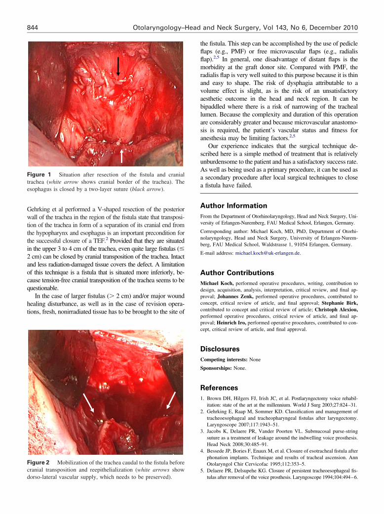
CLINICAL TECHNIQUES AND TECHNOLOGY Surgical closure of persistent tracheoesophageal fistulas by esophageal suturing and cranial transposition of the trachea Michael Koch, MD, PhD, Johannes Zenk, MD, PhD, Stephanie Birk, MD, Christoph Alexiou, MD, PhD, and Heinrich Iro, MD, PhD, Erlangen, Germany No sponsorships or competing interests have been disclosed for this article. I mplantation of a tracheoesophageal voice prosthesis is the gold standard for voice rehabilitation after a total laryngec- tomy. The incidence of complications generally appears to be greater in patients who have received radiotherapy. 1,2 Peripros- thetic leakage, which persists despite conservative treatment, necessitates surgical treatment. This generally creates a prob- lem that is difficult to solve, especially in the case of patients who underwent radiotherapy, as evidenced by the many sur- gical approaches described in the literature. Besides local techniques, more elaborate procedures, such as the combination of local suturing with an interposition of local or distant pedicle flaps between the esophagus and trachea and clo- sure by means of free microvascular flaps, have been published. 2-5 Cranial transposition of the trachea has been described in only one publication. 3 In the present report, our experiences with a two- layer inverting suture of the esophagus combined with a cranial transposition of the residual trachea to close refractory tracheo- esophageal fistulas (TEFs) are described. Materials and Methods Patients Five patients developed therapy-resistant TEFs (size maximum 1.5 1 cm) after laryngectomy with implantation of a Provox voice prosthesis. All received radiochemotherapy postoperatively (mean radiation dose 52.6 Gy). Fistulas were located in the cranial 3 to 4 cm of the trachea. In one patient, previous closure of TEF by the use of a local technique failed. Before the treatment was performed, informed consent was received from all patients and approved by the review board of the Friedrich-Alexander Univer- sity of Erlangen-Nuremberg. Surgical Technique The trachea was mobilized by means of a skin incision extending from the eight-o’clock to four-o’clock position and then separated from the esophagus so that the fistula was displayed. The fistula and cranial trachea were resected, and the esophageal defect was closed by means of a two- layer inverting suture (Fig 1). The trachea caudal to the fistula was mobilized (Fig 2) and then transposed cranially and sutured tension-free to the skin of the neck. Results Successful closure of the fistula could be achieved in four patients (80%). The mean follow-up time was 3.5 years (range, 0.5-5 years). One patient developed a filiform recurrent tra- cheoesophageal fistula 10 weeks after the operation. A revision operation was performed, during which severe necrotic changes were found in the tissues concerned. This patient received booster radiotherapy because of an R1 situation in the postcricoid region at the level of the entrance of the esophagus (total radiation dose of 64 Gy). Closure of the fistula was achieved by means of a two-layer esophageal suture with interposition of a pectoralis major myofascial flap (PMF). The patient has now been free of symptoms for 2.5 years. Discussion More than 90 percent of TEFs after implantation of a voice prosthesis can be treated by conservative means; surgical closure is required in approximately five percent. 1 The difficulties of a surgical treatment of TEF are evidenced by the multiplicity of surgical techniques described in the literature. 2-5 For small fistulas (5-10 mm) and acceptable wound conditions, the use of local techniques is suitable and associated with success rates of 60 to 100 percent. In case of poor tissue conditions, replacement tissue, from the sternocleidomastoid muscle or the infrahyoid muscula- ture, can be interposed between the esophagus and the posterior wall of the trachea. Reduced amount of muscle material that remains after an extensive prior operation may limit the applica- tion of this type of operation. 2-4 Defect coverage by cranial trans- position of the trachea has been described only by Bessede et al. 4 Received July 12, 2010; revised August 28, 2010; accepted September 20, 2010. Drs. Koch and Zenk contributed equally to this article. Otolaryngology–Head and Neck Surgery (2010) 143, 843-844 0194-5998/$36.00 © 2010 American Academy of Otolaryngology–Head and Neck Surgery Foundation. All rights reserved. doi:10.1016/j.otohns.2010.09.034