Journal of Agricultural Science and Technology B 5 (2015) 701-713 doi: 10.17265/2161-6264/2015.10.007 Surveillance of Antibiotic Resistant Staphylococcus aureus in Agricultural Production Chain of Mongolia Tumuruu Gantsetseg 1 , Jargalsaikhan Enkhtuya 1 , Tundev Odgerel 2 , Ochirpurev Ariuntuya 3 and Sodnom Lkhagvasuren 1 1. Institute of Veterinary Medicine, Ulaanbaatar 17024, P.O. Box 53/24, Mongolia 2. National Center for Communicable Disease, Ulaanbaatar 17024, P.O. Box 53/24, Mongolia 3. WHO Representative Office in Mongolia, Ulaanbaatar P.O. Box 46/78, Mongolia Abstract: Monitoring of food borne pathogens in food is the primary tool for the implementation of food safety systems. It is necessary to monitor the prevalence of food borne pathogens for effective food safety planning and targeted interventions. Staphylococcus aureus is considered as the third largest cause of food related illness in worldwide. The present study aimed at surveillance of S. aureus contamination of meat on meat supply chain stages, which is a common benchmark of meat market in Mongolia, and characterization of isolated and collected strains from other agricultural sources. The cultural and polymerase chain reaction (PCR) methods were used for isolation, identification and characterization of S. aureus. In 216 cultures of S. aureus among 634 Staphylococci isolates obtained from different sources throughout the agricultural production chain in this study, common gene for S. aureus (98.74%), and nuc (97.47%), mecA (44.12%), msrA (9.66%), gyrA (32.77%) and ermC (29.41%) genes were identified. As seen in the surveillance result, the prevalence of methicillin-resistance S. aureus (MRSA) is 44% among S. aureus isolates from agricultural production chain. Confirmed cases of food-borne infections and intoxications caused by S. aureus should be considered as one of mean criteria of food safety issues in Mongolia, and special attentions should be paid on antibiotic resistant bacteria, such as S. aureus. Key words: Meat supply chain, mecA positive Staphylococcus aureus, polymerase chain reaction. 1. Introduction Staphylococcus aureus is a bacterium, which causes food-borne and fomite-borne infections and intoxications [1]. Among healthy humans, 30% carry S. aureus, which causes various infections, intoxications, postsurgical infections, pustule and sepsis [2]. Virulence factors of S. aureus include: (1) surface proteins that promote colonization of host tissues; (2) invasins (leukocidin, kinases, hyaluronidase); (3) surface factors (capsule, protein A); (4) biochemical properties (carotenoids, catalase production); (5) immunological disguises (protein A, coagulase); (6) membrane-damaging toxins (hemolysins, leukotoxin, leukocidin); (7) exotoxins (SEA-G, TSST, ET); (8) inherent and acquired resistance to antimicrobial agents [3]. Corresponding author: Sodnom Lkhagvasuren, associate professor, research field: food safety. S. aureus is considered as the third largest cause of food related illness in worldwide [4]. Monitoring the presence of food borne pathogens in food is the primary tool for the implementation of food safety systems. It is necessary to monitor the prevalence of food borne pathogens for effective food safety planning and targeted interventions [5]. Methicillin-resistant S. aureus (MRSA) that is resistant to virtually all β-lactam antibiotics is mediated by the chromosomally located mecA gene [6]. Livestock constitutes a potential reservoir of MRSA isolates belonging to a recently derived lineage within clonal complex 398 (MRSA CC398-IIa). Since its discovery in the early 2000s, this lineage has become a major cause of human disease in Europe, posing a serious public health challenge in countries with intensive livestock production. Various studies suggest that environmental contamination of air and D DAVID PUBLISHING
Transcript
Journal of Agricultural Science and Technology B 5 (2015) 701-713 doi: 10.17265/2161-6264/2015.10.007
Surveillance of Antibiotic Resistant Staphylococcus
aureus in Agricultural Production Chain of Mongolia
Tumuruu Gantsetseg1, Jargalsaikhan Enkhtuya1, Tundev Odgerel2, Ochirpurev Ariuntuya3 and Sodnom
Lkhagvasuren1
1. Institute of Veterinary Medicine, Ulaanbaatar 17024, P.O. Box 53/24, Mongolia
2. National Center for Communicable Disease, Ulaanbaatar 17024, P.O. Box 53/24, Mongolia
3. WHO Representative Office in Mongolia, Ulaanbaatar P.O. Box 46/78, Mongolia
Abstract: Monitoring of food borne pathogens in food is the primary tool for the implementation of food safety systems. It is necessary to monitor the prevalence of food borne pathogens for effective food safety planning and targeted interventions. Staphylococcus aureus is considered as the third largest cause of food related illness in worldwide. The present study aimed at surveillance of S. aureus contamination of meat on meat supply chain stages, which is a common benchmark of meat market in Mongolia, and characterization of isolated and collected strains from other agricultural sources. The cultural and polymerase chain reaction (PCR) methods were used for isolation, identification and characterization of S. aureus. In 216 cultures of S. aureus among 634 Staphylococci isolates obtained from different sources throughout the agricultural production chain in this study, common gene for S. aureus (98.74%), and nuc (97.47%), mecA (44.12%), msrA (9.66%), gyrA (32.77%) and ermC (29.41%) genes were identified. As seen in the surveillance result, the prevalence of methicillin-resistance S. aureus (MRSA) is 44% among S. aureus isolates from agricultural production chain. Confirmed cases of food-borne infections and intoxications caused by S. aureus should be considered as one of mean criteria of food safety issues in Mongolia, and special attentions should be paid on antibiotic resistant bacteria, such as S. aureus. Key words: Meat supply chain, mecA positive Staphylococcus aureus, polymerase chain reaction.
1. Introduction
Staphylococcus aureus is a bacterium, which causes
food-borne and fomite-borne infections and
intoxications [1]. Among healthy humans, 30% carry
S. aureus, which causes various infections,
intoxications, postsurgical infections, pustule and
sepsis [2]. Virulence factors of S. aureus include: (1)
surface proteins that promote colonization of host
tissues; (2) invasins (leukocidin, kinases,
hyaluronidase); (3) surface factors (capsule, protein
at 230 rpm for 2 min, then incubated at 35 °C for 24 h
[15]. Then, smear was prepared and stained by Gram’s
method and Gram positive clustered cocci were
selected. Colonies were selected based on whether the
cocci cause beta hemolysis on blood agar and form
black colonies on Baird Parker selective agar. In order
to differentiate staphylococci from other cocci,
catalase test was used, while coagulase test was used
to identify S. aureus from other Staphylococci [15].
2.3 Biochemical Test
For identification of Staphylococci by biochemical
characteristics, API Staph test kit (BioMerieux) was
used as described in the manufacturers instruction [16].
Briefly, the following steps and procedures were used.
The first step in this procedure is to make a saline
suspension of the organism from an isolated colony. A
Table 1 Sampling and collection strains.
Collection Sampling from food chains
From slaughtering house From food market From beef production From broiler From horse
From patients
Total Sources Animals
Meat samples
Swabs CarriersMeat samples
Swabs Carriers FeedSlaughteranimals
Processing plant
Retail Broilers SlaughterProcessing plant
RetailNasa swabs
Nasa swabs
No. of samples
52 52 52 52 104 102 87 - - - - - - - - 360 - 861
No. of strains
2 3 5 2 15 31 3 7 8 6 9 23 22 18 13 13 36 216
Cultures taken from laboratories, such as NCCD Bacteriological Laboratory, SCVL, VLUC, NRLFS, FSHL-IVM and LIDI-IVM, and samples collected for last 3-4 years in these laboratories from the above mentioned sources. -: unknown numbers of samples. Table 2 Primers used for the study.
No. Name of gene Target sequences PCR primer’ sequences (5l to 3l) Product size (bp) Reference
1 Common S. aureus AAT CTT TGT CGG TAC ACG ATA TTC TTC ACG CGT AAT GAG ATT TCA GTA GAT AAT ACA ACA
108 [17]
2 ermC Erythromycin resistance of S. aureus CTT GTT GAT CAC GAT AAT TTC CC ATC TTT TAG CAA ACC CGT ATT C
190 [18]
3 msrA Macrolide resistance efflux of S. aureus TCC AAT CAT TGC ACA AAA TC AAT TCC CTC TAT TTG GTG GT
163 [19]
4 mecA Methicillin resistance of S. aureus AAC AGG TGA ATT ATT AGC ACT TGT AAG AAT TCC CTC TAT TTG GTG GT
174 [20]
5 nuc Thermostable nuclease of S. aureus GCGATTGATGGTGATACGGTT CAAGCCTTGACGAACTAAAGC
276 [21]
6 VSMec Penicillin binding protein of S. aureus TGG CTA TCG TGT CAC AAT CG CTG GAA CTT GTT GAG CAG AG
310 [22]
Surveillance of Antibiotic Resistant Staphylococcus aureus in Agricultural Production Chain of Mongolia
704
staph strip is then placed in a tray that has a small
amount of water added to it to provide humidity
during incubation. Next, a sterile pipette is used to
dispense 2-3 drops of the bacterial suspension to each
micro cupule. The inoculated tray is covered and
incubated aerobically for 18-24 h at 35-37 °C. Finally,
a seven-digit profile number is obtained and used to
identify the bacteria.
2.4 Antibiotic Resistance Test
Antibiotic resistance and susceptibility of 216 S.
aureus cultures, isolated and collected during the
study, were checked by use of disc diffusion test [23],
DNA of antibiotic resistant strains was extracted and
the gene for antibiotics resistance was amplified by
PCR using primer shown in Table 2.
2.4.1 Disk Diffusion Test (Kirby-Bauer Method)
The test is performed by applying a bacterial
inoculation of approximately 1 CFU/mL to the surface
diameter Mueller-Hinton agar plate.
Commercially-prepared by Biolab, Zrt and HiMedia
fixed concentration, antibiotic disks are placed on the
inoculated agar surface. Plates are incubated for 16-24
h at 35 °C prior to determination of results. The zones
of growth inhibition around each of the antibiotic
disks are measured to the nearest millimeter. The
diameter of the zone is related to the susceptibility of
the isolate and to the diffusion rate of the drug through
the agar medium [24].
2.4.2 DNA Extraction and Purification
To extract bacterial DNA, 400 µL isolate was
placed into microtube and centrifuged at 1,000 rpm
for 30 min. Then, 200 µL of pellet was pipetted into
new microtube and centrifuged at 8,000 rpm for 15
min. Supernatant was removed and 200 µL distilled
water was added into the precipitate, followed by
mixing in vortex. Then, the mixture was placed in
boiling water for 15 min and template was prepared
by centrifuging at 1,0000 rpm for 10 min.
DNA was extracted from blood and nasal swabs
using the QIAamp DNA mini kit from Qiagen. The
QIAamp DNA mini kit was used for the protocol of
commercial guideline. To isolate DNA from meat,
phenol-chloroform extraction method was used. Yield
and purity of isolated DNA were measured by
spectrophotometer at 260 nm and 280 nm wavelength
and the purity ranged between 1.72 to 1.94.
2.4.3 PCR Method
For the surveillance of genes of S. aureus, which is
resistant to both β-lactam and non β-lactam antibiotics,
including oxacillin, methiciliin and erythromycin, the
following primers shown in Table 2 and both of PCR
and multiplex PCR methods for surveillance of
antibiotics resistant genes were used [25].
2.4.3.1 Reaction Mixture
Total of 25 µL of mixture, containing 2.5 µL 10×
PCR buffer, 2 µL dNTP (GeneAmp, UK) (each 2.5
mM), 2 µL template, 1.5 µL MgCl2, 1 µL of each of
primers 1 and 2, 0.175 µL of 5 unit/µM taq DNA
polymerase (TaKaRa, Japan), and 14.815 µL
ddH2O/DW, was taken. In using multiplex PCR,
subtractions form water equal to amounts of primer
and MgCl2 were estimated and each primer was taken
in dependent on concentrations [25].
2.4.3.2 Amplification
In total of 35 cycles, there were such steps as
initialization at 95 °C for 7 min, denaturation at 94 °C
for 1 min, annealing at 55 °C for 1 min, elongation at
72 °C for 1 min and final elongation at 72 °C for 7
min, which was 10 min for multiplex PCR [25].
2.4.3.3 Gel Electrophoresis
Mixture of 8 µL PCR product and 2 µL loading
buffer was loaded in wells on the gel, and run in
1.5%-2% agarose gel depending on its DNA length.
The gel was stained by ethidium bromide for 15 min
and DNA fragments were visualized on
transilluminator at 320 nm wavelength.
3. Results and Discussion
3.1 Prevalence and Identification of S. aureus
Of 861 samples taken from meat production and
distribution chain, coagulase-positive Staphylococcus
Surveillance of Antibiotic Resistant Staphylococcus aureus in Agricultural Production Chain of Mongolia
705
samples represent 10% by microbiological method,
whilst 12% were coagulase positive Staphylococcus
by PCR method (Table 3). In these positive samples, S.
aureus was detected in 3.8% of animal samples, 5.8%
of meat samples, 9.6% of environmental swabs and
3.8% of patients (Table 3, Fig. 1).
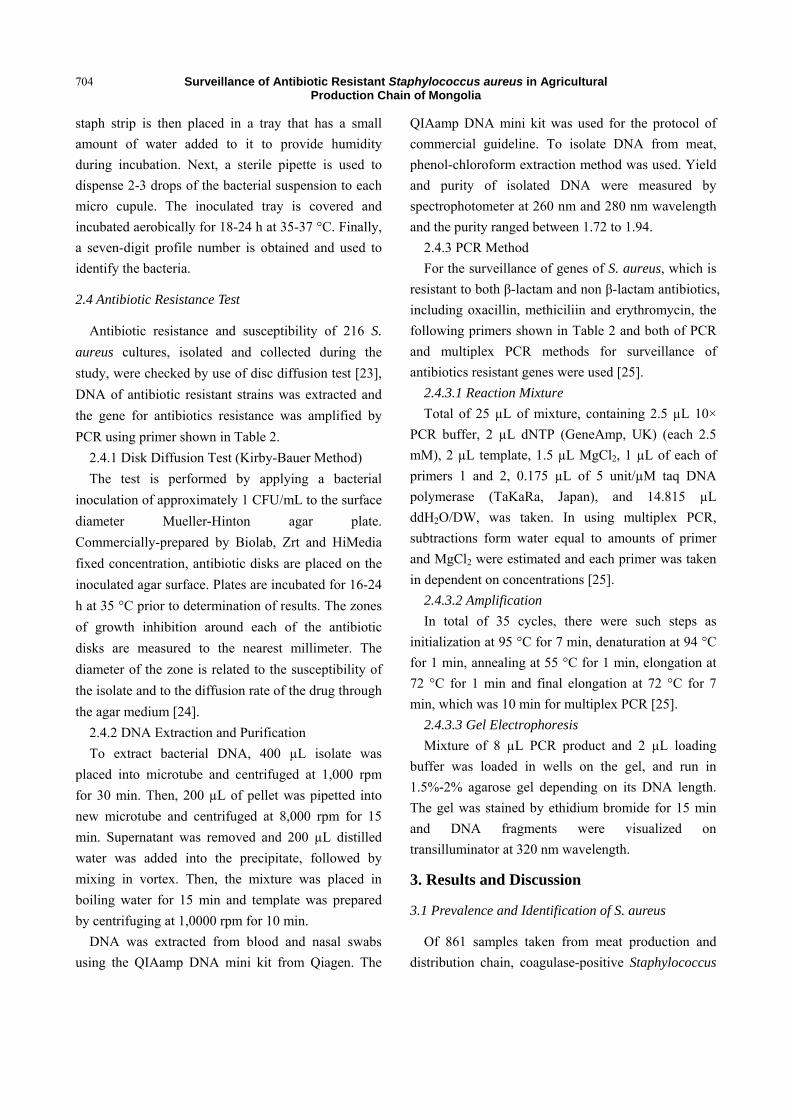
For samples taken from food markets, S. aureus
was detected in 14.42% of meat, 3.4% in carriers and
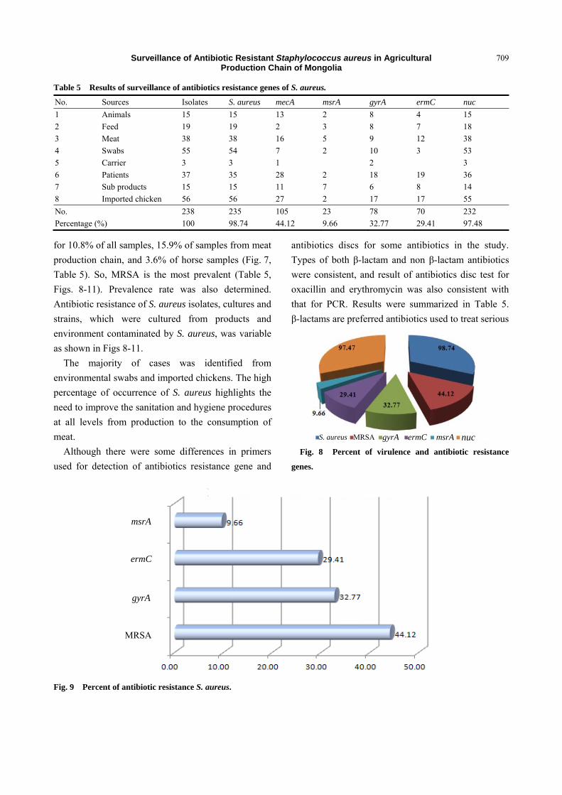
30.4% in fomite surface swabs (Fig. 2). Of 156 strains
and cultures identified by laboratory examinations in
the last three years, 83.7% were positive for mecA S.
aureus. Furthermore, 3.6% of S. aureus identified
from 360 horse samples (nasal swab and blood) of
Selenge, Darkhan, Orkhon-Uul and Bulgan provinces
were positive for mecA S. aureus (Table 4).
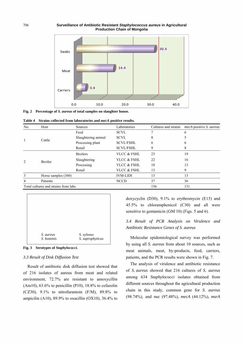
3.2 Result of API Staph Test
Isolates from fomites and animal products
accounted for 61.3% and 28.7%, respectively, in total
isolates and serotyping of staphylococci by API test.
When serotype of 634 cultures of Staphylococcus by
their biochemistry and enzyme activity identified, there
were S. aureus (35.3%), S. xylosus (29.4%), S. hominis
(17.6%) and S. saprophyticus (8.8%) (Fig. 3). Results
of the study demonstrated that portion of S. aureus,
which is the cause of infection and intoxication, was
greater than other types and S. aureus is seen to be
indicator of fomite borne infection (Fig. 4).
Table 3 Results of isolation and identification of S. aureus from meat chain.
Collection Kind No. of isolation and identification of S. aureus
[7] Casey, J. A., Curriero, F. C., Cosgrove, S. E., Nachman, K. E., and Schwartz, B. S. 2013. “High-Density Livestock Operations, Crop Field Application of Manure, and Risk of Community-Associated Methicillin-Resistant Staphylococcus aureus Infection in Pennsylvania.” JAMA Intern Med. 173 (21): 1980-90.
[8] Schulz, J., Friese, A., Klees, S., Tenhagen, B. A., Fetsch, A., Rösler, U., and Hartung, J. 2012. “Longitudinal Study of the Contamination of Air and of Soil Surfaces in the Vicinity of Pig Barns by Livestock-Associated Methicillin Resistant Staphylococcus aureus.” Appl. Environ. Microbiol. 78 (16): 5666-71.
[9] Eko, K. E., Forshey, B. M., Carrel, M., Schweizer, M. L., Perencevich, E. N., and Smith, T. C. 2015. “Molecular Characterization of Methicillin-Resistant Staphylococcus aureus (MRSA) Nasal Colonization and Infection Isolates in a Veterans Affairs Hospital.” Antimicrobial Resistance and Infection Control 4: 10.
[10] Gibbs, S. G., Green, C. F., Tarwater, P. M., Mota, L. C., Mena, K. D., and Scarpino, P. V. 2006. “Isolation of Antibiotic-Resistant Bacteria from the Air Plume Downwind of a Swine Confined or Concentrated Animal Feeding Operation.” Environ Health Perspect. 114 (7): 1032-7.
[11] Wendlandt, S., Schwarz, S., and Silley, P. 2013. “Methicillin-Resistant Staphylococcus aureus: A Food-Borne Pathogen.” Annu. Rev. Food Sci. Technol. 4: 117-39.
[12] Enkhtuya, J. 2015. Molecular Epidemiology of Some Zoonotic Diseases and Improvement of Vaccine Technology. Report of Science Technology Project.
[13] National Center for Communicable Disease of Mongolia. “Annual Report 2013.” Accessed July, 2016. http://www.nccd.gov.mn/index.php.
[14] Xu, Y. G., Cui, L. C., Tain, C. Y., Li, S. L., Cao, J. J., Liu, Z. M., and Zhang, G. C. 2012. “A Multiplex Polymerase Chain Reaction Coupled with High Performance Liquid Chromatography Assay for Simultaneous Detection of Six Foodborne Pathogens.” Food Control 25 (2): 778-83.
[15] ISO. 2003 “Enumeration of Coagulase-Positive Staphylococci (Staphylococcus aureus and Other Species).” Microbiology of Food and Animal Feeding Stuffs, ISO 6888-2:1999/Amd1:2003.
[16] Isenberg, H. D. 1992. Clinical Microbiology Procedures
Handbook. Vol. 1. Washington: American Society of Microbiology.
[17] Martineau, F., Picard, F. J., Grenier, L., Roy, P. H., Ouellette, M., and Bergeron, M. G. 2000. “Multiplex PCR Assays for the Detection of Clinically Relevant Antibiotic Resistance Genes in Staphylococci Isolated from Patients Infected after Cardiac Surgery: The ESPRIT Trial.” J. Antimicrob. Chemother. 46 (4): 527-34.
[18] Projan, S. J., Monod, M., Narayanan, C. S., and Dubnau, D. 1987. “Replication Properties of pIM13, a Naturally Occurring Plasmid Found in Bacillus subtilis, and of Its Close Relative pE5, a Plasmid Native to Staphylococcus aureus.” J. Bacteriol. 169 (11): 5131-9.
[19] Ryffel, C., Bucher, R., Kayser, F. H., and Berger-Bachi, B. 1991. “The Staphylococcus aureus mec Determinant Comprises an Unusual Cluster of Direct Repeats and Codes for a Gene Product Similar to the Escherichia coli sn-Glycerophosphoryl Diester Phosphodiesterase.” J. Bacteriol. 173 (23): 7416-22.
[20] Ross, J. I., Eady, E. A., Cove, J. H., Cunliffe, W. J., Baumberg, S., and Wootton, J. C. 1990. “Inducible Erythromycin Resistance in Staphylococci Is Encoded by a Member of the ATP-Binding Transport Super-Gene Family.” Mol. Microbiol. 4 (7): 1207-14.
[21] Brakstad, O. G., Aasbakk, K., and Maeland, J. A. 1992. “Detection of Staphylococcus aureus by Polymerase Chain Reaction Amplication of the nuc Gene.” Journal of Clinical Microbiology 30 (7): 1654-60.
[22] Killgore, G. E., Halloway, B., and Tenover, F. C. 2000. “A 5’ Nuclease PCR (Taq Man) High-Throughput Assay for Detection of the mecA Gene in Staphylococci.” Journal of Clinical Microbiology 38 (7): 2516-9.
[23] Jorgensen, J. H., and Ferraro, M. J. 2009. “Antimicrobial Susceptibility Testing: A Review of General Principles and Contemporary Practices.” Clin. Infect. Dis. 49 (11): 1749-55.
[24] Owuna, G., Abimiku, R. H., Nkene, I. H., Joseph, G. W., and Ijalana, O. O. 2015. “Isolation and Antibiotic Susceptibility of Staphylococcus aureus from Fresh Poultry Meat Sold in Keffi Metropolis, Nigeria.” International Journal of Research Studies in Biosciences 3 (11): 1-5.
[25] Martineau, F., Picard, F. J., Lansac, N., Ménard, C., Roy, P. H., Ouellette, M., and Bergeron, M. G. 2000. “Correlation between the Resistance Genotype Determined by Multiplex PCR Assays and the Antibiotic Susceptibity Patterns of Staphylococcus aureus and Staphylococcus epidermidis.” Antimicrob. Agents Chemother. 44 (2): 231-8.
[26] Bauer, K. A., West, J. E., Balada-Llasat, J. M., Pancholi, P., Stevenson, K. B., and Goff, D. A. 2010. “An
Surveillance of Antibiotic Resistant Staphylococcus aureus in Agricultural Production Chain of Mongolia
713
Antimicrobial Stewardship Program’s Impact with Rapid Polymerase Chain Reaction Methicillin-Resistant Staphylococcus aureus Blood Culture Test in Patients with S. aureus Bacteremia.” Clin. Infect. Dis. 51 (9): 1074-80.
[27] Zecconi, A., Calvinho, L., and Fox, L. 2006. “Staphylococcus aureus Intramammary Infections.” Bulletin of the International Dairy Federation, Brussels, Belgium. Accessed July 2016. https://www.livivo.de/doc/B2243729.
[28] Neder, V. E., Canavesio, V. R., and Calvinho, L. F. 2011. “Presence of Enterotoxigenic Staphylococcus aureus in Bulk Tank Milk from Argentine Dairy Farms.” Rev. Argent. Microbiol. 43 (2): 104-6.
[29] Miranda, J. M., Mondragon, A., Vazquez, B. I., Fente, C. A., Cepeda, A., and Franco, C. M. 2009. “Influence of Farming Methods on Microbiological Contamination and Prevalence of Resistance to Antimicrobial Drugs in Isolates from Beef.” Meat Science 82 (2): 284-8.
[30] Adesiji, Y. O., Alli, O. T., Adekanle, M. A., and Jolayemi, J. B. 2011. “Prevalence of Arcobacter, Escherichia coli, Staphylococcus aureus and Salmonella Species in Retail
Raw Chicken, Pork, Beef and Goat Meat in Osogbo, Nigeria.” Sierra Leone J. Biomed. 3 (1): 8-12.
[31] Lim, S. K., Nam, H. M., Park, H. J., Lee, H. S., Choi, M. J., Jung, S. C., Lee, J. Y., Kim, Y. C., Song, S. W., and Wee, S. H. 2010. “Prevalence and Characterization of Methicillin-Resistant Staphylococcus aureus in Raw Meat in Korea.” Journal of Microbiol. Biotechnol. 20 (4): 775-8.
[32] Kim, E. S., Song, J. S., Lee, H. J., Choe, P. G., Park, K. H., Cho, J. H., Park, W. B., Kim, S. H., Bang, J. H., Kim, D. M., Park, K. U., Shin, S., Lee, M. S., Choi, H. J., Kim, N. J., Kim, E. C., Oh, M. D., Kim, H. B., and Choe, K. W. 2007. “A Survey of Community-Associated Methicillin-Resistant Staphylococcus aureus in Korea.” J. Antimicrob. Chemother. 60 (5): 1108-14.
[33] Khanna, T., Freindship, R., Dewey, C., and Weese, J. S. 2008. “Methicillin Resistant Staphylococcus aureus Colonization in Pigs and Pig Farmers.” Vet. Microbiol. 128 (3-4): 298-303.
[34] Smith, T. C., and Pearson, N. 2011. “The Emergence of Staphylococcus aureus ST398.” Vector-Borne Zoonot. Dis. 11 (4): 327-39.