Page 1
SURVEY FOR HEMATOPHAGOUS FLIES AND TRYPANOSOMA SPECIES IN
IKARA AND KUBAU LOCAL GOVERNMENT AREAS, KADUNA STATE,
NIGERIA
BY
USUNOBUN COLLINS INEGBENOSUN
DEPARTMENT OF PARASITOLOGY AND ENTOMOLOGY
FACULTY OF VETERINARY MEDICINE
AHMADU BELLO UNIVERSITY,
ZARIA
SEPTEMBER, 2016
Page 2
i
SURVEY FOR HEMATOPHAGOUS FLIES AND TRYPANOSOMA SPECIES IN
IKARA AND KUBAU LOCAL GOVERNMENT AREAS, KADUNA STATE,
NIGERIA
BY
Usunobun Collins INEGBENOSUN B.Sc (A.A.U., 2010)
(M.Sc/VET-MED/35498/12-13)
A DISSERTATION SUBMITTED TO THE SCHOOL OF POSTGRADUATE
STUDIES, AHMADU BELLO UNIVERISITY, ZARIA IN PARTIAL
FULFILLMENT FOR THE AWARD OF DEGREE OF MASTER OF
SCIENCES IN VETERINARY PARASITOLOGY
DEPARTMENT OF VETERINARY PARASITOLOGY AND ENTOMOLOGY
AHMADU BELLO UNIVERSITY,
ZARIA, NIGERIA
SEPTEMBER, 2016
Page 3
ii
DECLARATION
I, hereby declare that the work in this Dissertation entitled “Survey for
Hematophagous Flies and Trypanosoma Species in Ikara and Kubau Local
Government Areas, Kaduna State, Nigeria” has been carried out by me in the
Department of Veterinary Parasitology and Entomology, Ahmadu Bello University,
Zaria. The information derived from the literature has been duly acknowledged in the
text and a list of references provided. No part of this dissertation was previously
presented for another degree or diploma at this or any institution.
_____________________________ ______________
Usunobun Collins INEGBENOSUN Date
Page 4
iii
CERTIFICATION
This dissertation titled “SURVEY FOR HEMATOPHAGOUS FLIES AND
TRYPANOSOMA SPECIES IN IKARA AND KUBAU LOCAL GOVERNMENT
AREAS, KADUNA STATE, NIGERIA” by Collins Usunobun INEGBENOSUN,
Meets the regulations governing the award of the degree of Master of Science of
Ahmadu Bello University, and is approved for its contribution to knowledge and
literary presentation.
Prof. A.J. Natala ___________ ___________
Chairman, Supervisory Committee Signature Date
Dr. I.D.Jatau ___________ ___________
Member, Supervisory Committee Signature Date
Dr. O.O Okubanjo ___________ ___________
Head, Department of Veterinary Signature Date
Parasitology and Entomology
Prof. K. Bala ___________ ___________
Dean, School of Postgraduate Studies Signature Date
Ahmadu Bello University, Zaria
Page 5
iv
DEDICATION
This Dissertation is dedicated to God Almighty and my loving son Nathan Ose
Inegbenosun, wife, Mrs. Constance E Inegbenosun for her understanding,
encouragement and support throughout the period of the study.
Page 6
v
ACKNOWLEDGEMENTS
My profound gratitude goes to God Almighty for life, grace and protection to go through
this programme successfully.
I am indebted to my supervisors Prof A.J Natala and Dr I.D Jatau and also special tribute
goes to them as they have exemplified rare supervisory qualities through timely, patient and
constructive comments and evaluation at every stage of this piece of work, allowing me to
complete this project.
I take this opportunity to express my indebtedness to Brig. Gen. L.F Abdulahi,
Commander, Nigerian Army Education Corps (NAEC), Lagos for his encouragement,
motivation and support throughout this work.
I thank all the teaming lecturers Dr O.O Okubanjo, Prof O.J Ajanusi, Prof I.A Lawal of
the Department for their contributions in one way or the other. I acknowledge all my course
mates especially my good friend Dr. Ijoh, Bartholomew in the Department for their
contributions also.
I like to thank also the Area Vet Representative of Ikara and Kubau Local Government
Areas, for granting me permission and giving all necessary assistance needed in the field. I
appreciate all the rangers, Mr. Benjamin Adediminiyi who directed and guided me tirelessly
in the field.
My special gratitude goes to the laboratory technologist, Late Chief. Jerry Servan Bale for
his gallant and indefatigable effort throughout my field experiences, May his soul rest in the
bosom of the Lord Jesus Christ Amen.
Last but not the least; I am grateful to my parents Mr. and Mrs. Sunday Inegbenosun; my
siblings, Bright, Hope, Mercy, Happy, Blessed, Happy and Samuel for their great support
and encouragement throughout this programme. Time and space will not allow me to go on,
but I am sincerely grateful to all whom in one way or the other contributed to this great feat.
God bless you all.
Page 7
vi
ABSTRACT
A study of the species composition and distribution of hematophagous flies was
conducted between dry and wet seasons, at Ikara and Kubau Local Government Areas,
Kaduna State, using Biconical (Charlier and Laviessiere, 1973) and Nzi traps
(Omoogun, 1994). Survey for the occurrence of Trypanosoma species in cattle in the
study area was also conducted during these periods. Twelve traps each was placed for
48hrs in four districts in each of the local government areas and the trap catches were
harvested every 24hrs.A total of 232 flies were caught during the study period and their
occurrence differ between the two local governments. Kubau Local Government had
105(45.3%) while Ikara Local Government had 127 (54.7%).The specific occurrences
of the hematophagous flies caught were: Stomoxys calcitrans107 (85.3%), Tabanus
9(7.1%) and Glossina 0(0.0%). Overall percentage of fly catches per trap was 130
(56.0%) for biconical and 102(44.0%) for Nzi traps respectively. The highest number of
flies caught was during wet season {212 (91.4%)} while in dry season only 20(8.6%)
flies were caught during the study. There was significant association (P=0.002) between
the flies occurrence and the two seasons of the year. While there was no statistically
significant association (P=0.07) between the number of flies caught and the type of trap
used. The prevalence rate of Trypanosoma infection in cattle in the local government
areas was 50% prevalence each of infection due to T. vivax and T. brucei respectively.
There was no significant association between the occurrence of the Trypanosoma
infection and the Local Government Areas under study.
Page 8
vii
TABLE OF CONTENTS
TITLE PAGE i
DECLARATION ii
CERTIFICATION iii
DEDICATION iv
ACKNOWLEDGEMENTS v
ABSTRACT vi
TABLE OF CONTENTS vii
LIST OF TABLES xi
LIST OF PLATES xii
LIST OF FIGURE xiii
CHAPTER ONE 1
1.0 INTRODUCTION 1
1.1 Background of the Study 1
1.2 Statement of Research Problem 2
1.3 Justification 4
1.3.1 Aim of the study 5
1.3.2 Objectives of the study 5
1.3.3 Research questions 5
CHAPTER TWO 6
2.0 LITERATURE REVIEW 6
2.1 Historical Perspective 6
2.2.0 General description of Glossina 7
2.2.1 Habitat and distribution of Glossina 8
Page 9
viii
2.2.2 Habitat and distribution of the morsitans group 9
2.2.3 Habitat and distribution of the palpalis group 10
2.2.4 Habitat and distribution of the fusca group 11
2.3.0 Reproduction of Glossina 12
2.3.1 Mating 12
2.3.2 Egg stage 13
2.3.3 Larval stages 13
2.3.4 The pupa 14
2.3.5 The adult fly 15
2.4.0 General behaviour in Glossina 15
2.4.1 Movement and activity of tsetse flies 15
2.4.2 Resting sites 16
2.4.3 Response to host animals 16
2.5.0 Tsetse fly population dynamics 17
2.6.0 Transmission of trypanosomosis 18
2.6.1 Control of tsetse flies 20
2.7 Tabanidae 27
2.7.1 Distribution and host 27
2.7.2 Morphology 27
2.7.3 Life cycle 28
2.7.4 Feeding and habitat 29
2.7.5 Pathogenic significance 29
2.7.6 Control 30
Page 10
ix
2.8 Stomoxys 30
2.8.1 Morphology 30
2.8.2 Life cycle 31
2.8.3 Feeding and habitat 31
2.8.4 Pathogenic significance 32
2.8.5 Control 32
2.9.0 Sarcophaga 33
2.9.1 Distribution of Sarcophaga 33
2.9.2 Morphology 33
2.9.2 Distribution 34
2.9.3 Importance of Sarcophaga 34
2.9.3.1 Medical importance 34
2.9.3.2 Forensic importance 35
CHAPTER THREE 37
3.0 MATERIALS AND METHODS 37
3.1 Study Area 37
3.2 Study Design 39
3.2.1 Survey for Hematophagous Flies in Ikara and Kubau
LGAs of Kaduna State 39
3.2.2 Survey for Trypanosoma species in Ikara and Kubau
Local Government Areas of Kaduna State 40
3.2.2.1 Sample size 40
3.2.2.1 Blood Sampling 40
3.3 Parasitological Analysis of the Blood Samples 41
3.3.1 Thick blood smears 41
3.3.2 Thin blood smears 41
3.3.3 Haemotocrit centrifugation 41
3.4 Data Analysis - - - - - - - 42
Page 11
x
CHAPTER FOUR 43
4.0 RESULTS 43
4.1 Survey for Hematophagous Flies in Ikara and Kubau LGAs 43
CHAPTER FIVE 57
5.0 DISCUSSION 57
CHAPTER SIX 59
6.0 SUMMARY, CONCLUSIONANDRECOMMENDATIONS 59
6.1 Summary 59
6.2 Conclusions 60
6.3 Recommendations 61
References 62
Appendices 70
Page 12
xi
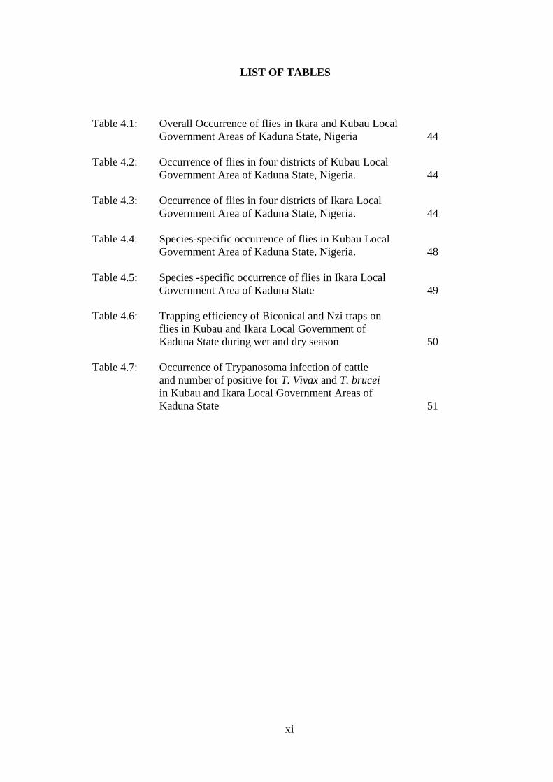
LIST OF TABLES
Table 4.1: Overall Occurrence of flies in Ikara and Kubau Local
Government Areas of Kaduna State, Nigeria 44
Table 4.2: Occurrence of flies in four districts of Kubau Local
Government Area of Kaduna State, Nigeria. 44
Table 4.3: Occurrence of flies in four districts of Ikara Local
Government Area of Kaduna State, Nigeria. 44
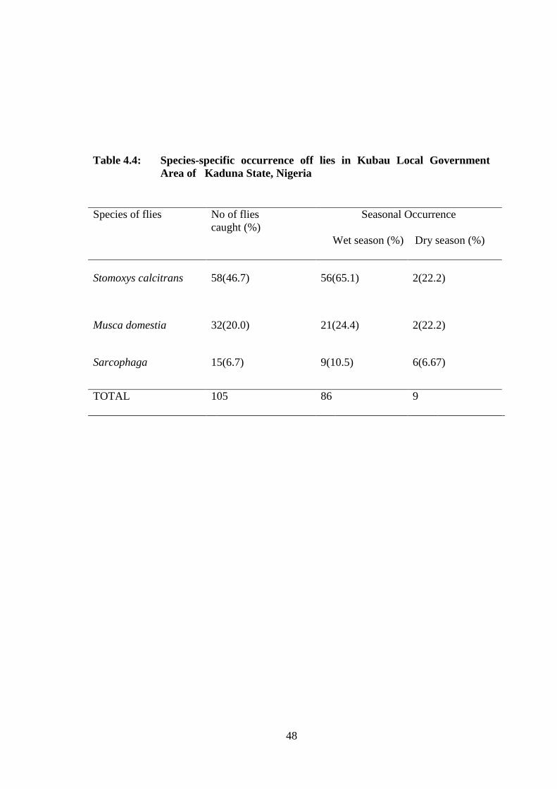
Table 4.4: Species-specific occurrence of flies in Kubau Local
Government Area of Kaduna State, Nigeria. 48
Table 4.5: Species -specific occurrence of flies in Ikara Local
Government Area of Kaduna State 49
Table 4.6: Trapping efficiency of Biconical and Nzi traps on
flies in Kubau and Ikara Local Government of
Kaduna State during wet and dry season 50
Table 4.7: Occurrence of Trypanosoma infection of cattle
and number of positive for T. Vivax and T. brucei
in Kubau and Ikara Local Government Areas of
Kaduna State 51
Page 13
xii
LIST OF PLATES
Plate 1: Installation of the Biconical trap in the study areas 52
Plate II: Installation of the Nzi trap in the study areas 53
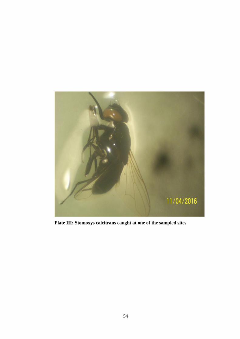
Plate III: Stomoxys calcitrans caught at one of the sampled sites 54
Plate IV: Tabanus sp caught at one of the sampled sites 55
Plate V: Trypanosoma brucei in sampled blood smear 56
Page 14
xiii
LIST OF FIGURE
Figure 3.1: Map of Kaduna State showing the study area s 38
Page 15
1
CHAPTER ONE
1.0 INTRODUCTION
1.1 Background of the Study
Hematophagous flies belong to the families Glossinidae and Tabanidae and to the genus
Stomoxys. They play an important role in both human and domestic animals health,
because many species of these groups are vectors of organisms responsible for several
human and animal diseases (Stephen, 1989). Moreover, several vector-transmitted
diseases are considered as emergent due to their recent evolution and propagation. The
preponderant role of the species belonging to the Glossinidae family in the transmission
of African Human Trypanosomosis (AHT), or sleeping sickness and AAT has
historically hidden the potential role of other hematophagous flies, like those belonging
to the genus Stomoxys and the family Tabanidae in trypanosome transmission and the
transmission of other pathogens. For example, it is now recognized that several species
of the genus Stomoxys are mechanical vectors of parasites, such as Trypanosoma
species (Phelps et al, 1978) and various viruses, such as the Capripox-viruses
responsible for lumpy skin disease in sheep and goats. The Tabanidae are also
mechanical or biological vectors of many human and animal pathogens and an
analogous pattern of Trypanosoma transmission has been documented for several
tabanids of the Atylotus genus. Species of the Chrysops genus are involved in the
cyclical transmission of Loa loa filariasis (Morlais et al.1998).
The feeding action of these hematophagous flies opens a channel for contamination of
the host species with disease causing agents. Thus, many animal and human infectious
diseases are transmitted by hematophagous species, such as the bubonic plague, chagas
disease, dengue fever, eastern equine encephalitis, filariasis, leishmaniasis, Lyme
Page 16
2
disease, malaria, rabies, sleeping sickness, St. Louis encephalitis, tularemia, typhus,
Rocky Mountain spotted fever, west nile fever and many others. Insects and arachnids
of medical and veterinary importance that are hematophagous, at least in some species,
include the sand-fly, black fly , tsetse fly, bedbug, assassin bug, mosquito, tick, louse,
mite, midge, and flea (Maudlin,1970).
1.2 Statement of Research Problem
Hematophagous flies activity is highly seasonal. For example, most of the Glossina
species respond to seasonal patterns, and within a region, the populations of the
different species increase in the rainy season (Shaba et al., 2012). The Tabanus and
Stomoxys species are caught throughout the year but there is usually a seasonal rise in
abundance corresponding to the ends of the rains and the beginning of the dry season
(Dede et al., 2005). However, the basic seasonal pattern of the different groups is
influenced by local climatic parameters and species exhibit various patterns of
population fluctuation related to local climate, vegetation and host blood meal source
(Challier, 1982), which also coincides with the incidence of the diseases they transmit.
Trypanosomosis is the most economically important disease transmitted by these
hematophagous flies most importantly by Glossina species (Moiser, 1912).
Trypanosomosis lowers productivity in livestock, reduces cattle density by 70%, sale of
meat and milk by 50% and calving rates by 20% and increases calf mortality by 20%
(Swallow, 2000). Tsetse flies, through the cyclical transmission of trypanosomosis to
both humans and their animals, greatly influence food production, natural-resource
utilization and the pattern of human settlement throughout much of sub-Saharan Africa
(Kabayo, 2003). It is estimated that the annual direct production losses in cattle alone
Page 17
3
amount to between US$6billion and $12billion, while animal deaths may reach 3
million (FAO, 1992).
According to Lehane et al. (2003), tsetse flies transmit African trypanosomosis leading
to half a million human cases annually and that the disease also known as Nagana in
animals remains a massive break on African Agricultural development.
More than a third of the land area across Africa is infested with tsetse flies (8.7 million
km2), where at least 46 million cattle are exposed to the risk of contracting tsetse-borne
trypanosomosis, as are millions of sheep, goats, donkeys, camels and horses (Reid et
al., 1998). African livestock producers are administering an estimated 35 million
curative and preventive treatments annually (Geerts and Holmes, 1997).
Apart from disease transmission, biting flies can produce an array of symptoms to
animals including pain, itching, urticarial and cellulitis. Allergic response is the most
common which may be characterize by hives, and in some cases wheezing (Ripamonti,
et al., 2002). Tabanid bites are very painful with some individuals developing severe
lesion, fever and general disability. These allergic responses are due to the large amount
of saliva injected by the flies to prevent their blood meal from clotting. Stable flies bites
are quite painful and they produce small papules that quickly fade but are often itchy
(Maldonado, 1910).
The design of management strategies against the different species and of strategies that
minimize the risk of fly bites and transmission of diseases such as trypanosomosis
requires a full regional understanding of the species‟ phenology and ecology. However,
such information is scarce in Northern part of Kaduna State. Hence, the present study
was conducted to improve our understanding of the distribution, the abundance, and the
Page 18
4
phenology of hematophagous flies (Glossinidae, Tabanidae Muscidae) in the Ikara and
Kubau Local government areas of Kaduna State.
1.3 Justification
Trypanosomosis is one of the major animal health constraints to livestock production in
Sub-Saharan Africa (FAO, 2002). In fact, it is also an important blood parasitic disease
in humans (Shaba et al., 2012). Animals have a central role in most African societies,
and provide milk, meat, manure, hides and skins as well as valuable draught power.
Additionally, because livestock represent a means of accumulating and distributing
wealth, they have great social significance.
Control of trypansomosis is aimed against either on the tsetsefly or the trypanosome
and in the absence of adequate funds for large-scale tsetse control, trypanocides are
widely used. At the farmer's level trypanocides provide a way out for the individual to
take action to control the disease. With few exceptions throughout Africa, governments
have lacked the resources to continue to provide effective Veterinary services to control
trypanosomosis, among other diseases.
Survey of Glossina (tsetse fly) and other biting flies and the Trypanosoma species they
transmit is an essential tool for strategic control measures against the vectors and
etiologic agent of trypanosomosis. This will go a long way to improving animal
production in Nigeria. This study is likely going to help in reducing the economic waste
associated with treating the disease by livestock owners and appropriate control and
preventive measures will be recommended.
Ikara and Kubau Local Government Areas of Kaduna State are situated in the Northern
Guinea savannah area which is tsetse endemic and a favourable settlement area for
cattle pastoralist (Ohaeri, 2007). Therefore, there is need to undertake this study to
Page 19
5
ascertain the potentials of these areas in terms of animal productivity and suggestive
suitable tsetse control.
1.3.1 Aim of the study
To survey for hematophagous flies and Trypanosoma species in Ikara and Kubau Local
Government Areas, Kaduna State, Nigeria.
1.3.2 Objectives of the study
The objectives of this were to:
Investigate the distribution of hematophagous flies of veterinary importance in
Ikara and Kubau LGAs of Kaduna State using trapping.
Compare the catching efficiency of Biconical and Nzi traps in hematophagous
flies of veterinary importance in Ikara and Kubau LGAs of Kaduna State.
Determine the occurrence of Trypanosoma species in cattle in Ikara and Kubau
LGAs of Kaduna State.
1.3.3 Research Questions
Are there hematophagous flies of veterinary importance in Ikara and Kubau
Local Government Areas of Kaduna State?
Which trap between biconical and Nzi traps is more efficient in catching fly in
Ikara and Kubau LGAs of Kaduna State?
Are there Trypanosoma species in cattle in Ikara and Kubau Local Government
Areas of Kaduna State?
Page 20
6
CHAPTER TWO
2.0 LITERATURE REVIEW
2.1 Historical Perspective
It is believed that hematophagy arose independently at least six times among the
arthropods of the Jurassic and Cretaceous periods (145–65 million years ago)
(Balashov, 1984; Ribeiro, 1995).The very patchy nature of the insect fossil record
means that discussion of the evolution of the bloodsucking habit has until now relied
heavily on detective work, with the major clues lying in the diversity of forms and
lifestyles seen in modern-day insects, and in some cases in the details of their
relationships with vertebrates. From careful interpretation of this evidence quite
credible accounts of the likely evolution of the blood-sucking habit can be made. From
this starting point, it has been convincingly argued that the evolution of the blood-
sucking habit in insects has occurred on several occasions, in each case along one of
two main routes (Waage, 1979), and these are discussed below. Insect molecular
systematic is beginning to emerge from its „Tower of Babel‟ stage (Caterino et al.,
2000) and it will make a major contribution in defining the detail of the evolutionary
routes taken by hematophagousinsects (Esseghir et al., 1997; Hafner et al., 1994;
Lanzaro et al., 1998; Mans et al., 2002; Sallum et al., 2002). The proposed population
bottleneck suffered by phlebotomines in the late Pleistocene and the subsequent
radiation of the species out from the eastern Mediterranean sub-region is a good
example of what we can expect (Esseghir et al., 1997).
Hematophagy can be classified into obligatory and optional practice. Obligatory
hematophagous animals do not have any other type of food besides blood; one such
species is Rhodnius prolixus (an assassin bug from South America). This contrasts with
Page 21
7
optional hematophagous, like the many mosquitoes species, such as Aedes aegypti,
which may also feed on pollen, fruit juice, and other biological fluids. Sometimes only
the female of the species is a hematophous (this is essential for egg production and
reproduction).
Hematophagous flies, and among those, species belonging to the families Glossinidae
and Tabanidae and to the Stomoxys genus.
2.2.0 General description of Glossina
Glossina species have been widely studied because of their economic importance as
major transmitters of animal and human trypanosomosis (Ndams, 1987).
Tsetse flies are robust insects measuring about 6-15mm in length. They are readily
distinguished from other biting flies morphologically by the combinations of useful
features such as widely separated compound eyes which are dark brown, and able to
detect moving objects at 137meters (150yards) (Pollock, 1982), with forwardly
projecting proboscis which sticks out horizontally from the front of the head
(Jordan,1993). They differ from most biting flies and non cyclorrhaphous insects
because when at rest, the wings are held over the other such that they overlap like the
blade of a closed pair of scissors, thus revealing part of the abdomen (Davies, 1977).
They also possess a characteristic wing venation where the discal medial cell of the
wing is shaped like a butcher‟s cleaver (referred to as the „Hatchet cell‟) and a
distinctive row of branched hairs on the arista of the antenna (Service, 1980; Jordan,
1993).
Tsetse flies range in color from yellowish or grayish to dark or almost blackish-brown,
sometimes there is a slight pink or sandy red tinge (Davies, 1977). The dorsal surface
has a pattern of dark brown stripes and patches making the insect difficult to see when
Page 22
8
settled on tree bark, rock and soil. They have seven visible abdominal segments
Pollock, 1982).
2.2.1 Habitat and distribution of Glossina
The genus Glossina occurs over some 11 million km2of Africa. Its northern limit
extends across the continent from Senegal in the west to southern Somalia in the east.
This limit is at about 14oN but in Somalia it is only about 4
oN. The northern limit
corresponds closely to the southern edges of the Sahara and Somalia Deserts. The
southern limit is less well defined. In the south west it ranges between 10oand 20
oS,
corresponding closely to the northern edges of the Kalahari and Namibian Deserts,
whereas in the south east, it is generally at about 20oS but extends as far as 29
oS along
the east African littoral (Jordan, 1986), with the mid pint of infestation at about 7oS
(Davies, 1977). In Nigeria, tsetse flies cover about 75% of the landmass from latitude 4o
to 13oN to longitude 2
o to 15
oE, an area covering all the five agro-ecological zones of
the country (Okam, 1988; Onyiah, 1997).
There are twenty two known species of Glossina which can be arranged into three
distinct species groups based on habitat preference (Ford, 1970) and morphological
differences in the construction of the male genitalia (Pollock,1982). These species
groups are summarized below:
The Morsitans species group (Morsitans) these include seven species and subspecies
namely:- G. longipalpis; G. pallidipes; G. morsitans morsitans; G. morsitans
submorsitans; G. morsitans centralis; G. swynnertoni; G. austeni (Bossche et al., 1997).
The Palpalis groups (Nemorhina) consists of a total of nine species and subspecies
namely: - G. palpalis palpalis; G. palpalis gambiensis; G. fuscipes fuscipes; G. fuscipes
Page 23
9
martini; G. fuscipes quanzensis; G. tachinoides; G. pallicera pallicera; G. pallicera
newsteadi; G. calliginea (Grev, 2007).
The Fusca species group (Austenia) also consists of fourteen species and subspecies
namely: G. Fusca Fusca; G. Fusca congolensis; G. nigrofusca nigrofusca; G.
nigrofusca hopkinsi; G. fuscipleuris; G. haningtoni; G. schwetzi; G. tabaniformis; G.
nashi; G. vanhoofi; G. medicorum; G. severini; G.brevipalpis; G. longipennis (Rogers,
et al., 2000).
2.2.2 Habitat and distribution of the morsitans group
The three Glossina subspecies of the morsitans group are exceptionally good vectors of
trypanosomes; all the seven species are potential vectors of both human and animal
trypanosomosis. All species within this group inhabit the savanna woodlands that
surround the two major blocks of lowland rainforests in Africa, and as such are referred
to as „game or savanna flies‟ (Davis, 1977). The distribution of tsetse flies in this group
closely follows the distribution of wild animals and water sources. In wetter areas, the
flies are observed to disperse more widely over the woodland, but in drier areas their
movements are 28 restricted to the mesophytic vegetation of the watercourses,
particularly during the severe dry season (Nash, 1937; Jordan, 1986). In Eastern and
Southern Africa where Glossina morstans morsitans is the primary vector of human and
animal trypanosomosis, the „Miombo‟ woodland (Brachystegia–Jilbernardia) that
extends from Mozambique to Tanzania, as well as the „Mopane‟ woodlands
(Colophospermum mopane) in Zambia and Zimbabwe are typical habitats. Glossina
morsitans centralis dominate northwards from Botswana and Angola into southern
Uganda.
Page 24
10
Glossina morsitans submorsitans has an east to west distribution from Ethiopia to
Senegal in „doka‟ woodland where the vegetation is dominated by Isoberlinia doka
species, and can be sporadically found to occur in the southern Guinea savanna
vegetation zone as well as the drier Sudan zone (Jordan, 1986).Glossina swynnertoni is
restricted to a small area between Tanzania (Serengeti) and southern Kenya
(Masaimara) where Acacia- commiphora vegetation can be found, with abundant wild
life. Glossina longipalpis and Glossina pallidipes both have a much wider range of
possible habitats displaying versatility by existing in different vegetation types.
Glossina longipalpis occurs in the narrow savanna belt just north of the rainforest in
West Africa, from Guinea to Cameroon while Glossina pallidipes occurs in East Africa
from Mozambique to Ethiopia over a relatively wide range of climatic and vegetation
conditions. Finally, Glossina austeni occupies secondary shrub, thickets and islands of
forests along the East African coast from Mozambique to Somalia, are rarely found at
altitudes over 200m or more than 250km inland from the coast (Jordan, 1986). In
Nigeria, the two morsitans group species present are:-Glossina morsitans submorsitans
found in the north where annual rainfall is as low as 635mm (25in) with a dry season of
seven months and further south, an annual rainfall of 1400mm (55in) with a dry season
of four months. Glossina longipalpis is found in the southern guinea and derived
savanna zones in the west and a small localities in the east, with annual rainfall not less
than 1150mm 45in) or more than 2300mm (90in) (Davies, 1977).
2.2.3 Habitat and distribution of the palpalis group
Out of the nine species in the palpalis subgenera, only five palpalis and
fuscipes subspecies are vectors of both human and animal trypanosomosis. Although
flies in this group are continuously found in the lowland rainforest, some are known to
extend out of the savanna region particularly along rivers and streams. The habitat of
Page 25
11
the palpalis flies occurs mainly in the drainage systems leading to the Atlantic or the
Mediterranean ocean, extending from the wet mangrove and rainforests along the
coastal regions of West Africa to the drier savanna areas just north of the rainforests.
The flies of the palpalis group are less tolerant to the wide range of climatic conditions
of the savanna belt, and are therefore restricted to the ecoclimate of the water courses
from where they derived their label as the „riverine species‟. Many of the palpalis
species, such as the Glossina palpalis palpalis in Cote d‟lvoire prefer peri-domestic
conditions and have been observed to maintain close association with villages (Baldry,
1980). Similarly, it is thought that the advancement of Glossina tachinoides in Cote
d‟lvoire and Togo have been attributed to intense agricultural development and the
rapid human population growth around the plantation (Hendrick et al., 1997). In
general, most of the palpalis group flies are less suited to desiccating conditions, and
therefore survive in thick riverine forests with enough shelter from wind and heat. This
is especially the case for the three fuscipes subspecies which are confined to
hygrophytic habitats rarely far from open water lacustrine or riverine habitats. Glossina
tachinoides, although typically a riverine species, were found in Northern Nigeria to
extend into human-inhabited savanna woodlands during the wet season, displaying
strong adaptation to peri-domestic habitats (Kuzoe, 1985; Ahmed, 2004).
2.2.4 Habitat and distribution of the fusca group
With the exception of Glossina brevipalpis and Glossina longipennis all the tsetse flies
in the Fusca group are found in West African forests. None of the species in the Fucsa
group are vectors of human trypanosomosis; however both Glossina fusca and Glossina
medicorum are efficient vectors of trypanosomosis to livestock (mainly Trypanosoma
vivax) causing considerable economic burden. Distribution of the Fusca group depends
primarily on forest vegetation and climatic factors. With the exception of Glossina
Page 26
12
longipennis, most Fusca group species inhabit moist, evergreen habitats either in
riverine forests with savanna (such as Glossina medicorum) or in dense and wet
rainforests (Glossina tabaniformis and Glossina nigrofusca).In stark contrast to the rest,
the Glossina longipennis species lives in one of the driest habitats inhabited by tsetse
flies (Jordan, 1986). Due to its pupal adaptation to dry conditions, their primary habitats
– consisting of dry deciduous acacia bush- are discontinuously spread throughout East
Africa (Glasgow, 1963).
2.3.0 Reproduction of Glossina
Tsetse flies exhibit a form of reproduction known as adenotrophic viviparity (Hagan,
1951) because the egg and larva stages develop within the fly. The egg contains
sufficient yolk for embryonic development and the larva in the uterus is nourished by
special maternal organs. The consequences of viviparity are that only a small number of
fully developed larvae can be produced, a free-larval stage is practically eliminated and
both adults and immature stages are dependent upon the same source of food (blood)
(Saunders, 1960).
2.3.1 Mating
Female tsetse flies mate within a day or so after emergence from their pupal case;
mating which usually takes place near or on host animals (Pollock, 1982). Male and
female generally meet when the female is about to take the first blood meal or is in the
process of doing so (Jordan, 1986). Almost at the end of the period spent in copulo the
male jerks vigorously and it is at this time that sperms are ejaculated into the
spermatophore (Pollock, 1982). Within the next few hours, the sperms are slowly
released from the spermatophore and move into the spermathecae, a paired golden-
colored rigid structure connected by ducts to the anterior end of the uterus. Active and
Page 27
13
viable sperms can remain in the spermathecae throughout the life of the female
fertilizing each egg as it is produced from the ovaries, thus allowing the female to breed
throughout her life time (Davies, 1977). Female tsetse flies usually mate only once in
their lives but some may mate more than once, males can mate several times. Older
males are better able to mate successfully than very young ones (Pollock, 1982).
2.3.2 Egg stage
The female tsetse fly has two ovaries, each of which has two ovarioles; eggs develop
sequentially in the four ovarioles (Saunders, 1970) and are ovulated into the uterus at
intervals of about 9 – 10 days, the first ovulation occurring when the fly is about 9 days
old (Jordan, 1986). Each egg is fertilized immediately it enters the uterus by sperm from
the spermathecae coming into contact with and penetrating the anterior part of the egg.
The fertilized egg remains lying in the uterus for about four days, while development of
the first instar larva takes place inside (Pollock, 1982). The age of wild-caught females
can be determined through dissection technique involving ovaries examination 33 and
counting the number of ovulations that have occurred (Saunders, 1962; Challier, 1965).
2.3.3 Larval stages
The larva in Glossina passes through several stages or instars, as it grows. There are
three larval instars in Glossina: the first, second and third instars. The larva has a mouth
at the anterior end, and two posterior spiracles (Pollock, 1982). The intrauterine larva is
supplied with nutrients in the form of „milk‟ substance secreted from a modified
accessory gland (Attardo et al., 2006), grows rapidly and molts twice before
larviposition.
Page 28
14
2.3.3.1 First instar larvae
The first instar larva develops within the egg and breaks out of the chorion using a
sharp larvae tooth. It grows to 1.8mm (Glossina morsitans) and lasts for a day (Pollock,
1982).
2.3.3.2 Second Instar Larvae
The second instar larva grows and develops rapidly lasting for two days and can reach
the length of 4.5mm (Glossina morsitans). Each side of the posterior spiracles swells
with small spines in between.
2.3.3.3 Third instar larvae
This also grows and develops rapidly. The third instar larvae is white in color and has
two conspicuous black respiratory lobes which are white at first, and later become
black. The third instar lava last just over two days and grows to a length of 6.7mm
(Glossina morsitans) (Pollock, 1982). The larva when fully developed is deposited
(larviposited) on the ground at shady sites to prevent desiccation. The larva burrows
itself rapidly in the soil to a depth of 1- 5cm, depending on the species, the season and
the soil type (Grev, 2002). Within an hour, the larva contracts to form a barrel-shaped
puparium, darkens rapidly to black. After about four days, ecdysis occurs within the
shell of the puparium and the true pupa is formed (Jordan, 1986).
2.3.4 The pupa
The pupa is dark brown and rounded; at the posterior end are the polypneustic lobes, the
shape of which helps to distinguish the tsetse pupa from the pupae of other flies. The
pupa is slightly shorter than the larva. The pupal stage usually lasts about four to five
weeks, depending on the temperature. Higher temperatures shorten the pupal period,
Page 29
15
lower temperatures lengthen the pupal period. Too high or too low temperatures cause
death of the pupae (Pollock, 1982).
2.3.5 The adult fly
The adult fly emerges by expanding its ptilinum to burst open the end of the puparium,
pushing itself out of the soil after which the ptilinum sink back into the head of the fly
(Davies, 1977). At this stage, the body is very soft and the wings are small and
crumpled (Pollock, 1982). The wings flatten out into their normal shape within a few
minutes of birth by blood being forced into the veins, harden and is then capable of
flight. The sex ratio on emergence is normally 1:1 (Jordan, 1986). The young fly before
its first blood meal is called „Teneral fly‟, the abdomen which appears whitish and
semi-transparent with ptilinum everted when squeezed between fingers on the head.
The non-teneral fly in contrast, are flies that have taken blood meal. They appear
creamier yellow, the thorax is firmer and hard and the ptilinum is not easily everted
(Pollock, 1982).
2.4.0 General behavior of Glossina
The behavior, distribution pattern and density of a population of flies depends mainly
on climate (temperature, humidity, sun, rain etc.); vegetation (shade and shelter); wild
animals(food); soil(breeding sites); predators of tsetse and human population (Davies,
1977).
2.4.1 Movement and activity of tsetse flies
The movement and dispersal of tsetse flies are related to the climate, the hunger stage of
the fly and the sex of the fly (Grev, 2002). The flies are active during the day spending
only about 15 – 30 minutes each day in active flight. A single flight does not last longer
than about 1½ - 2 ½ minutes and the speed of flight may be 3 -6 m/sec (Pollock, 2000).
Page 30
16
They are usually inactive during low temperatures and dull days; some species have
been found flying in the moon light (Service, 1980). Dispersal is higher in the wet
period, during these periods they spread all over the savanna and have implications for
transmission of African trypanosomosis; such fly movement facilitate both the spread
of the disease to new areas and their reintroduction to areas where it was previously
under control. In drier periods, where unfavorable conditions prevail, they utilize places
with dense vegetation close to water where suitable climatic condition exist (Ndams,
1987; Grev, 2002). The activity of tsetse flies during the daytime is mostly early
morning or late afternoon. Females are only active for a few moments a day while
mature males can be active up to 30 minutes a day (Grev, 2002).
2.4.2 Resting sites
Resting sites may vary according to the time of the day or night, climate and season,
species of tsetse fly, vegetation and resting places of host animals. During the hottest
part of the day (usually early to mid-afternoon), the true resting sites are lowest down
on tree trunks, and on the underside of shaded, fallen logs. At cooler times of the day,
and in cooler seasons, the flies rest 37 higher up tree trunks, and on the underside of
branches. At night some flies go up into the canopy of trees and rest on the leaves or
twigs (Pollock, 2000).
2.4.3 Response to host animals
Response to host animals is usually by tsetse fly sense of smell and sight (Davies,
1977), from up to one hundred meters away, with larger host animals being more
attractive to tsetse fly than smaller host animals (Pollock, 2000). Flies moves up-wind
closer to the host animal when it smells the host. Tsetse fly land on a greater variety of
host animals; flies that show the most attraction to the host animals are usually the most
Page 31
17
hungry flies in the population, while the non-hungry flies, particularly the males make
up the „following swarm‟. A male in a following swarm may fly on to a virgin female
as she comes for her first meal, and mate with her (Pollock, 2000).
2.5.0 Tsetse fly population dynamics
Tsetse fly populations are influenced mainly by density-independent factors such as
temperature and humidity, which in turn depend on vegetation cover. Fly densities are
determined by factors such as the availability of hosts or suitable habitats, which in turn
are influenced by human activity. According to Pollock, (2000) the density of a tsetse
population in a given area is never very accurately known unless all the flies are caught,
this could only be done on an island or in a very isolated woodland or thicket; even then
the fact that more than half of the total population of tsetse fly in an area are present
below ground as pupae, makes the estimation of a tsetse population a difficult exercise.
In some parts of Africa the fly population per square mile has been calculated, but in
most of Nigeria, flies are not often evenly distributed over any area because of local
differences in vegetation and climate, it is therefore difficult to determine the
population this way (Davies, 1977). In estimating tsetse population the term apparent
density and true density are sometimes used. This does not necessarily give information
about the „true density‟ (the number of flies per unit area) which may be heavy in a
certain place indicating a hungry population rather than a dense one. The apparent
density on the other hand may differ from time to time according to the availability of
the flies, when conditions for catching are poor, the apparent density will be low even
though the true density may be high (Davies, 1977). When traps are used, apparent
density is defined as the number of flies per trap per day (F/T/D).
Page 32
18
2.6.0 Transmission of trypanosomosis
Tsetse flies transmit the protozoan parasite of the Genus Trypanosoma, the agents of
human and animal trypanosomosis in sub-Saharan Africa (Hu et al., 2006). In West
Africa, the human trypanosomosis caused by Trypanosoma brucei gambiense is
transmitted by Glossina palpalis and Glossina tachinoides, the disease is devastating
and chronic. It is described as Gambian sleeping sickness (Dutton, 1902; Abenga et al.,
2005), whereas animal trypanosomosis caused by Trypanosoma brucei brucei is
transmitted by Glossina submorsitans and Glossina longipalpis. In East Africa, both
the human and animal trypanosomosis caused by Trypanosoma brucei rhodesiense and
Trypanosoma brucei brucei respectively are transmitted by Glossina morsitans,
Glossina pallidipes and to some extent Glossina palpalis (Willet, 1970; Grev, 2002).
The East African form described as Rhodesian form (Stephen and Fantham, 1910) is a
more acute, lasting for weeks or months (Garcia et al., 2006).
Trypanosomes exist as trypomastigote in blood and lymph in infected animals (Pollock,
2000); they are slightly curved elongated protozoan measuring about 10 - 35µm with a
single nucleus. Each possesses a single flagellum which originates near the posterior
end of the body and extends forward to the body by an undulating membrane. Near the
base of the flagellum is the dark-staining kinetoplast. The size and shape of the body,
position of the nucleus and kinetoplast, length and form of the undulating membrane
and flagellum are the diagnostic characters of various species of Trypanosomes (Jordan,
1986).
The form of trypanosomes in the tsetse fly can be identified to subgenus level on
morphological grounds and on their sites of development within the fly. In the
subgenus Duttonella is the Trypanosoma (vivaxgroup) of trypanosomes with large
terminal kinetoplast, distinct free flagellum and an inconspicuous undulating
Page 33
19
membrane. Trypanosoma vivax is a large (18 - 26µm long) monomorphic organism that
is very active in wet – mount blood smears. Their development within the tsetse fly is
restricted to the proboscis (labrum or hypopharynx). In the subgenus: Nannomonas is
the Trypanosoma congolense which is a small trypanosome with medium – sized
marginal kinetoplast, no free flagellum, and a poorly developed undulating membrane.
Trypanosoma congolense are hematic trypanosome found only in the blood vessels of
the animal they infect. They develop within the proboscis (labrum or hypopharynx) and
midgut of the tsetse fly. In the subgenus: Trypanozoon is the Trypanosoma brucei
brucei; this group is an extremely polymorphic; while some trypanosome occur as
short, stumpy organisms without flagella, others are found to be long slender organisms
with distinct flagella, and there are intermediate forms that are usually flagellated
(Moulton, 1976). These trypanosomes develop within the proboscis, midgut and
salivary glands. However, when trypanosomes are found only in the labrum, they are
regarded as immature form of all the subgenus mentioned, whereas if they are found in
the labrum and midgut, it indicates immature form of the congolense group (Davies,
1977).
When trypanosomes are ingested by tsetse fly, they undergo a cycle of development
within the fly. In the gut, they transform into the trypomastigote form and move into the
mouthpart (labrum and/or hypopharynx) where they develop into the epimastigote
form; they later reproduce by binary fission to produce the metacyclic form which is the
infective form. This final binary fission takes place in the proboscis, midgut or salivary
gland depending on the species of trypanosome (Jordan, 1986). However, transmission
of trypanosomes can also occur from one mammalian host to the other in or on the
mouthparts of various species of biting flies e.g. Tabanus, Stomoxys, Chrysops etc
including tsetse; this process is known as mechanical transmission (Jordan, 1986). This
Page 34
20
cycle of development varies in duration depending on the species of trypanosome,
species of tsetse fly, temperature, reservoir host, age, sex. For Trypanosoma vivax,
Trypanosoma congolense and Trypanosoma brucei brucei, it varies from 5 – 13 days at
22ºC - 29ºC; 12 -53 days and 11 – 60 days respectively (Davies, 1977; Jordan, 1986).
Reports by various authors have showed that Trypanosoma vivax have the highest
infection rate, followed by Trypanosoma congolense and then Trypanosoma brucei
brucei (Onyiah, 1997; Omotainse et al., 2000).
2.6.1 Control of tsetse flies
Attempts to control tsetse and trypanosomosis date back nearly 100 years, employing a
range of methods and approach. This range of methods have been developed and put
into practice to keep the disease under control, some of them less good than others
(Grev, 2002). The control strategies over the years have been directed against both the
parasites and the vectors (Onyiah, 1997). The initial methods of tsetse control
comprised of Hand-catching (Glasgow, 1970), Bush clearing (Steiner, 1964; Ford et al.,
1970), Game destruction, Settlement of people (Davies, 1977) and the use of chemicals
as insecticides (Davies, 1964). More recently, are the use of traps and screens (Challier
and Laveissiere, 1973) and the Sterile Insect Technique (SIT). Other interventions
aimed at eliminating the parasites have been by chemotherapy, chemoprophylaxis and
promotion of trypanotolerant breeds of cattle (Grev, 2002).
a. Hand-catching
Hand-catching is the most ancient method of insect control and was first tried in about
1913 against G. palpalis in the Portuguese island Principe, and against G. palpalis (G.
fuscipes) by the Germans on an island, Riamugasire, on Lake Victoria. Hand-catching
is absolutely specific and has been found effective against some species, however they
Page 35
21
are more expensive than chemicals because it always require a large labour force
especially if large areas were to be attacked, thus it fell into disuse (Glasgow, 1970).
b. Bush burning/Clearing
This method was performed based on the knowledge of the biology of tsetse fly, by
cutting down dense vegetation, thus destroying both the adult fly and pupae due to
decrease in humidity (Nagel, 1995). Bush clearing can be total or ruthless when all
vegetation is totally cleared and partial when it involves the destruction of only a
portion of the vegetation to render the environment unsuitable for tsetse fly. Partial
clearing could be discriminate clearing when woody vegetation in known tsetse
concentration sites is destroyed or selective where only certain components of the
vegetation forming the fly habitat are removed, which may be removal of only the
under storey, leaving the tall trees untouched or removing particular species of shrubs
and trees (Jordan, 1986).
The history of the development and application of methods of partial clearing for tsetse
control have been extensively reviewed by a number of authorities (Buxton, 1955; Ford
et al., 1970). In Nigeria, the record of partial clearing was described by Moiser (1912)
in Geidam, Borno State when the population of Glossina tachinoides was controlled by
vegetation clearing. Significant control was also achieved by Nash, (1940) on Glossina
tachinoides at Gadau and Anchau in Kano State.
The major short-coming of these method lie in the limited size for which they can be
economically deployed relative to the total size of tsetse affected area (Agyemang,
2001) since it requires economically unacceptable destruction of vast area of bush and
forest. Bush clearing also results in soil degradation and deforestation and as such was
no longer in use around 1970 (Nagel, 1995).
Page 36
22
c. Game destruction
The concept of game destruction was developed following the great rinderpest epizootic
at the end of the nineteenth century, which resulted in the death of many game animals
and thus the disappearance of tsetse flies and trypanosomosis (Jordan, 1986). This
method was used many years in Zimbabwe Zambia, Mozambique, Botswana and
Uganda to eliminate a wide range of the population of savanna species of Glossina and
Trypanosomosis due to its close association with game animal. McLennan, (1981)
reported the ineffectiveness of game destruction on riverine species because they feed
on other hosts besides wild game e.g. man, domestic animals, crocodiles and reptiles. In
West Africa, savanna species of tsetse population thrives on very low game densities
and as such, this method of control against tsetse did not succeed in Nigeria. This
method ceased because, animals migrated to area once cleared from larger mammals,
making it possible for tsetse to recover, and also because it became unacceptable to
public opinion (Grev, 2002).
d. Trapping method
Trapping method for tsetse fly originated from the Island of Principe, where farm
workers wore on their back a dark – colored piece of cloth, covered with glue, to reduce
biting nuisance of the flies (Maldonado, 1910) alongside with vegetation clearing and
pig elimination to control tsetse on the Island (Da Costa et al., 1916). The first real
tsetse trap constructed by Harris was used to eliminate Glossina pallidipes from
Zululand (Harris, 1938), and other various modified versions were introduced such as
the Animal trap (Morris and Morris, 1949); Biconical trap (Challier and Laveissiere,
1973); Monoconical trap (Lancien, 1981); Nitse trap (Omoogun, 1994) etc. In Nigeria,
Biconical and Nitse traps have been extensively used for tsetse sampling, ecological
studies and control (Omoogun et al., 1994; Dede et al., 2005). Traps were initially
Page 37
23
made for control purposes but were later used for sampling and ecological studies due
to their poor performance (Omoogun et al., 1994).
The interest in controlling tsetse flies by trapping declined rapidly with the introduction
of synthetic insecticides, but was renewed in the seventies, mainly due to increased
public awareness of environmental pollution by excessive use of insecticide (Grev,
2002). In West Africa, traps were widely used to control riverine species and was
suggested to be better for community – based operations because local people can see
flies being caught and killed, and can also see the catch decline as the operation
progresses. Trapping method is simple, inexpensive, and harmless to the environment
and is currently being used as an integrated part of the arsenal of tsetse control method
where fly population is suppressed by trap and other conventional methods such as bait
and target. However, reinvasion remains a reoccurring problem (Hargrove, 2002).
e. Use of pheromones
Most work undertaken on pheromones (Langley et al., 1975; Huyton et al., 1980; Offor
et al., 1981; Carlson et al., 1984) was based on the identification of the sex recognition
pheromone, in the cuticle of female Glossina species which induces copulatory
behavior in males of the same species, but not of other species, upon contact. This
method aims at mass producing the compound(s) that constitute the sex recognition
pheromones for the purpose of attracting flies to impregnated traps or screens to thus
effect control, but because of the lack of volatility of these pheromones, they could not
be exploited as attractant (Jordan, 1985).
f. Bait technology
The potential of bait technology for the control of tsetse fly was appreciated in the first
half of the 20th
century. The strategy of the technology was to improve bait design (live
Page 38
24
or artificial) by the careful analysis of basic responses of tsetse to baits, and using the
knowledge to improve the design of devices used in the field (Vale and Torr, 2004).
Van den Bossche and De Deken, (2004) reported the use of artificial baits to control fly
and reduce trypanosomosis at lower cattle density and stressed that insecticide – treated
cattle are more effective than stationary bait in area with higher cattle densities.
The work of various authors (Ndams, 1987; Bossche, 1997; Brightwell et al., 2001;
Esterhuizen et al., 2006) has suggested the efficiency of this method for the control of
tsetse flies.
Even though improved traps and bait technology (targets) rapidly became the standard
control method throughout Africa in late 1980s and 1990s, bait technology had pitfalls
in its application which include tackling too small an area and the variability in costs
and benefits relating to community – based action (Vale and Torr, 2004).
g. Use of insecticides
The use of insecticides as control methods against tsetse fly commenced in the mid-
1940s and is still today a major technique used in large scale (Grev, 2002), and almost
all method now used depend on insecticide. Two chemical groups have been widely
used in tsetse control: The organochlorides (DDT, dieldrin, endosulfan):- DDT was the
first chemical insecticides used against tsetse fly, after which dieldrin became popular
because of its high lethal characteristics in more humid conditions. These two
chemicals were then displaced by endosulfan for its toxicity and better solubility in
spray solvent (Vreysen, 1995). DDT is cheap, has low mammalian toxicity, persists
long in the environment and is effective against tsetse fly, while dieldrin on the other
hand is expensive, has longer persistent rate than DDT (Davies, 1977). However, both
fall victim of international ban due to their environmental side effect (Allsopp and
Page 39
25
Hursey, 2004). The spray of endosulfan (being an organochlorine insecticide) against
tsetse fly was reported to cause significant fish mortality (Douthwaite et al., 1981).
The synthetic pyrethroids (deltamethrin, alpacypermathrin and betadyfluthrin)
(Mangwiro et al., 1999), are the most potent insecticides used against tsetse fly.
Deltamethrin have been widely used for both spraying and for Impregnating traps and
targets. The major disadvantages of the synthetic pyrethroids are their high cost
(Vreysen, 1995).
In Nigeria, the use of insecticides to control tsetse fly started in 1955 (Davies, 1964) at
Kamadugu Gana river system which later extended to Kiyawa-Jama‟are Katagun
system, both area lying within Kano, Borno and Bauchi States. Persistent used of
insecticides (DDT) were applied from the ground on the tsetse resting sited by means of
pneumatic knapsack pressure sprayers (Davies, 1964; Maclennam, 1967). Aerial
spraying started in 1971 in the northern guinea vegetation zone, extended to the
southern guineas savanna zone (Spielberger et al., 1977). Spraying and re-spraying
activities reclaimed large area of land of about 399,551km² in the Sudan and northern
guinea vegetation zones from tsetse fly (Bature, 1985). In Botswana, an area of
16,000km² in the Okavango Delta was reclaimed from Glossina morsitans centralis
using aerial spraying with deltamethrin applied at 0.26–0.3g/ha and 12,000 deltamethrin
treated targets (Kgori et al., 2006).
h. Sterile Insect Technique (SIT)
The concept of sterile insect technique involves the mass production, sterilization – by
exposure of males to short burst of gamma radiation from a cobalt-60 source (Okhoya,
2003) and sequential release of sterile males to the target species to compete with the
wild male population. Mating between released sterile males and the wild females
Page 40
26
produces unviable progeny and the population is reduced over several generations to
unsustainable levels (Abila et al., 2003).
The application of the sterile male technique received considerable attention in the
1980s and is one method that seems feasible for the eradication of tsetse flies from the
continent of Africa. Economic feasibility of which is grater in the area-wide approach
(Feldmann, 2004). This approach was applied on an area-wide basis to eradicate the
New World Screw worm Cochiiomyia hominivirax in the U.S.A, Mexico and Central
America. Since then very effective programmes integrating the SIT have been mounted
against tropical fruit flies (Klassen and Curtis, 2005) and some species of tsetse flies on
pilot trials at Lake Kariba, Zimbabwe (vs. Glossina morsitans morsitans), at Tanga,
Tanzania vs. Glossina morsitans morsitans), in Burkina Faso (vs. Glossina palpalis
gambiense, Glossina tachinoides and Glossina morsitans submorsitans) in Plateau
state, Nigeria (vs. Glossina palpalis palpalis) and in Zanzibar, Tanzania (vs. Glossina
austeni) (Feldmann, 2004). The success in Zanzibar demonstrated the technical
feasibility of fighting the disease through the sterile insect technique approach (Kabayo,
2003).
The obvious constraints of sterile insect technique are the high cons associated with
mass rearing, low competitiveness of released sterile males (Whitten and Mahon, 2005),
low reproductive rate and low rate of re-infestation (Feldmann, 2004). The technique
which can only be employed realistically when density of target population is low is
Impractical for use against high density population. It is therefore used only in an
integrated approach with other control methods such as traps and targets (Jordan, 1986).
However, sterile insect technique still remains an exceptionally promising pest control
method in terms of efficacy and environmental compatibility (Nagel and Peverling,
2005).
Page 41
27
2.7 Tabanidae
These insects commonly known as „horseflies‟ or breeze flies, they are large, robust
flies with wings and large eyes (Soulsby1982).The pain caused by their bites leads to
interrupts feeding, and as consequence, flies may feed on succession of hosts and are
therefore important in the mechanical transmission of pathogens such as trypanosomes
(Urquhart et al., 1996).
There are many genera of tabanids, but only three are of veterinary significance, namely
Tabanus, Haematopota and Chrysops. Since the three genera are closely related in
behavior and pathogenic significance they will be discussed as a group.
2.7.1 Distribution and host
Distribution is worldwide although certain genera are absent from large areas: example,
there are no Haematopota species in Australia or North and South America (Urquhart et
al., 1996). The tabanid flies have been recovered from localities at sea-level and at
altitude of up to 10,000 feet (Nnochiri, 1974).
The hosts are generally large domestic or wild animals and man, but some small
mammals and birds may also be attached (Urquhart et al., 1996).
2.7.2 Morphology
These are medium to large biting flies, up to 25cm in length, with wing spans of up to
6.5cm.They are generally dark coloured, but may have various stripes or patches of
colour in the abdomen or thorax which varies from brown and red to yellow.
They have large coloured compound eyes which occupy a wide area of an equally semi
lunar head. The eyes are dichoptic in the female and holoptic in the male. It may be
coloured, the wings are broad and are characterized by their marginal cells. The
Page 42
28
coloration of the wings is useful in differentiating the three major genera: thus Tabanus
has clear or brownish wings, while there are often dark bands across the wings in
Chrysops. In contrast, Haematopota has ruffled or speckled wings (Urquhart et al.,
1996) They have a short, stout, interiorly projecting antennae consisting of three
markedly differentiate segments (Georgi and Georgi, 1990).The first segment of
antennae is small, the second may be expanded, and the third is marked by annulations
that make tabanid antennae appear to consist of many more than three units.
The antennae are also useful in generic differentiation. In species of the genus chrysops
the first and second segments of the antennae are long: the third (terminal) segment has
four annulations. While in species of the genus Haematopota the first segment of the
antennae is large and the second segment narrower, while the terminal segment has
three annulations (Soulsby, 1982).
The mouthparts, which are adapted for slashing/sponging, are short and strong and
always pointed downwards. The labium is also expanded terminally as paired large
labella which carry tubes called pseudotrachea through which the blood or fluid from
wounds is aspirated, The biting fascicle, which create the wound, consist of six
elements, the upper sharp labrum, hypopharynx with its salivary ducts paired rasp like
maxillae on pared boad pointed mandibles. Male flies have no mandibles and therefore
cannot feed on blood (Urquhart et al., 1996).
2.7.3 Life cycle
After a blood meal the female lays batches of several hundred creamy-white or grayish
cigar-shaped eggs, 1.0-2.5mm long, on the underside of vegetation or on stones,
generally in muddy or marshy areas. The larvae hatch after four to seven days and drop
into the water, or mud, into which they disappear. They are maggot-like and the body
Page 43
29
has 11 segments, besides the cephalic portion or which is conspicuous. Each segment
has weight fleshy tubercle. The mouth parts are prehensile and masticatory; the larvae
are carnivorous. There are three jointed antennae and the large lateral tracheae open on
the penultimate segment, which also bears a retractile siphoned tube. The larvae feed on
small crustaceans, or even on one another, and grow for two or three months,
performing several ecdyses. Finally, they pass through a quiescent stage and then
pupate. The pupa is brown and sub cylindrical; the abdominal segments are movable
and in the anterior part the appendages of the Imago can be distinguished. These stages
last about 10-14 days. The whole life-cycle takes four to five months under favorable
conditions, but low temperature prolongs development and the larvae may hibernate
(Soulsby, 1982).
2.7.4 Feeding and habitat
Unlike the males which feed only on vegetable sugars (nectar), female tabanid flies
attack animals and in addition, feed on plant juices. Feeding occurs during day light
hours especially early in the morning and late in the afternoon. A blood meal is usually
necessary for the development of the ovaries. With the exception of Chrysops silacea
which feeds indoors, most tabanids are outdoor feeders (Nnochiri, 1974). Some feed
mainly on the underside of the abdomen around the navel or on the legs; others bite also
on the neck and withers (Soulsby, 1982).
The flies feed about every three days. After feeding they rest for two hours on the
under-side of leaves or on stones or trees.
2.7.5 Pathogenic significance
The bites of the tabanidae are painful and may give rise to weals in soft-skinned
animals. Horses and cattle are restless when troubled by these flies and may become
Page 44
30
unmanageable, these flies also act as an efficient mechanical vectors of the organism
responsible for diseases such as anthrax, pasteurellosis, trypanosomosis, anaplasmosis
and the human filarial disease, loasis (Urquhart et al., 1996).
2.7.6 Control
This poses a special problem since breeding places are both diffuse and difficult to
detect (Urquhart et al., 1996). Where drainage is possible the breeding place may be
destroyed by these methods. Since the flies have the habit of skimming over water and
occasionally dipping their bodies in to it, the practice of pouring kerosene into water
which kills the flies when they dip into it can be utilized. Animals should be kept away
from places where the flies abound during hot part of the day.
For general flies control insecticidal spray with a residual effect are used in animal
houses and on the animals themselves. There is also the possibility of dark panels with
sticky adhesive as drapes and there are a number of electrocution grids which may
prove useful in animal's houses (Urquhart et al., 1996).
2.8 Stomoxys
Stomoxys calcitrans is the commonest species of this genus and is known as the stable
fly or biting housefly. It occurs all over the world and the host includes most animals
and man.
2.8.1 Morphology
S. calcitrans resemble the housefly M. domestica, being similar in size and grey with
four longitudinal dark stripes on the thorax. Its abdomen however is shorter and broader
than Musca with three dark spots on the second and third abdominal segments. The
proboscis is conspicuous and forward projecting which differentiates it from Musca and
other genera of non-biting muscid-flies. Stable fly can be distinguished from biting
Page 45
31
muscid flies of the genus Haematobia by the bigger size and the shorter pulp of stable
flies (Urquhart et al., 1996).
Larvae of Musca and Stomoxys can be differentiated by examination of the posterior
spiracles.
2.8.2 Life cycle
Stomoxys sometimes lay the eggs in horse manure, but prefers decaying vegetable
matter like straw and hay, especially when contaminated with urine. The female lays
batches of 25-50 eggs, resembling those of house flies. Eggs hatch in 1-4 days, or
longer in cold weather, and the larva are mature in 6-30 days.
After emergence the adult female require several blood meals before the ovaries mature
and egg laying can start.
The complete life cycle from egg to adult fly may take 12-60days depending mainly on
temperature (Urquhart et al., 1996).
2.8.3 Feeding and habitat
When feeding, the proboscis swings downwards and skin penetration is achieved by the
rasping action of fine teeth on the end of the labium. Approximately three minutes is
required for a blood meal and feeding is often interrupted, thus allowing mechanical
transmission of micro-organisms.
The flies are most abundant in summer and autumn and live about a month under
natural conditions. They prefer a fairly strong light; they are not seen in dark stables or
houses. They enter buildings only in autumn or during rainy weather. They are swift
flies; but do not travel along distances (Soulsby, 1982).
Page 46
32
2.8.4 Pathogenic significance
Both males and females are blood-suckers, attacking man, horse, cattle and other
mammals, and even birds and reptiles.
Trypanosoma evansi (Surra of equines and dogs) and T. equinum (Mal de caderas of
equines, cattle, sheep and goats) are transmitted mechanically by stomoxys. The species
may also mechanically transmit T. gambiense and T. rhodesience, the causative agents
of human Trypanosomosis in Africa, and T. brucei and T. vivax, which cause nagana of
cattle, sheep, goats and equines of Africa. It also serves as intermediate host of the
nematode Habronema majus, a nematode parasite of the stomach of the horse.
The role of S. calcitrans in the transmission of equine infections anemia is still under
debate (Steelman, 1976). However, the fly is responsible for the mechanical
transmission of septicaemia infections such as anthrax. The importance of biting flies in
the transmission of disease and economic loss through 'fly worry' has been reviewed by
(Stork, 1979).
2.8.5 Control
The fly is most troublesome in localities where suitable breeding places are readily
found. Control measures should therefore be directed toward destroying breeding-
places by regular removal of most bedding, hay and faeces from stables and yards, and
food waste from feeding troughs, and by preventing the accumulation of heaps of
weeds, grass cuttings and vegetable refuse.
Regular application of pyrethrins, synergized pyrethins, pyrethroids, coumaphos,
stirofos, or dichlorvos is indicated.
Application of insecticides to areas where they habitually rest (Urquhart et al., 1996).
Page 47
33
2.9.0 Sarcophaga
Sarcophaga haemorrhoidalis, also known as the red-tailed flesh fly, is a fly in the
Sarcophagidae family. This fly often breeds in carrion and feces, making it a possible
vector for disease. The larvae of this species can cause myiasis, as well as accidental
myiasis. It is potentially useful in forensic entomology.
2.9.1 Distribution of Sarcophaga
Sarcophaga haemorrhoidalis is a common species of flesh flies that appear worldwide
in distribution and is commonly found in the United States. It can be found throughout
the year in the southern portion of the United States. The larvae are adaptable and can
live in moist semi-aquatic habits that are unsuitable for most other fly species. Overall,
S. haemorrhoidalis is most likely to be found in climates with higher temperatures and
will prefer high temperatures throughout its entire life cycle.
2.9.2 Morphology
Sarcophagidae is the dipteran family commonly known as flesh flies, comprising
approximately 2000 species. Many species of Sarcophagidae prefer to breed in carrion
over other mediums, but there are several species that breed in dung. A large number of
species are parasitoids or cleptoparasitoids and never breed in carrion. It is difficult to
identify the S. haemorrhoidalis species unless genitalia can be observed. Only males
can be identified and classified within the genus. Sarcophagids are rather large in size
ranging from 4 to 23 mm, (adults of S. haemorrhoidalis vary in size from 7 to 14 mm).
Distinguishing characteristics include a checkerboard like pattern on the abdomen,
stripes on the thorax and red eyes. Flesh flies are attracted to anything rotting, including
feces. Sarcophagidae are unimpeded by rain and fly in any weather. Because of this
trait, Sarcophagidae will often be the first flies to colonize a corpse after an extended
Page 48
34
period of rain. Flesh flies appear to prefer sunlight over shaded conditions. Sarcophaga
haemorrhoidalis (Bercaea cruentata) is the one of the most common species of
Sarcophagidae recovered from indoor crime scenes in the United States.
2.9.2 Distribution
All members of the family Sarcophagidae are larviparous or ovoviviparous.
Sarcophaga haemorrhoidalis (Bercaea cruentata) gives live birth to larvae with the
female retaining the egg case in her abdomen. Flesh flies are strongly attracted to
carrion or dry flesh. The female has a strong desire to lay larvae on the flesh and have
even been noted to larviposit on the sleeve of a garment that has been previously soiled
with blood Oldroyd states that the larvae of Sarcophaga spp are voracious and will take
anything of animal origin be it alive or dead. A larva is forced out of the larvipositor
usually head first and soon disappears into the food material. Once larvae are deposited
as 1st stage instars, rapid development follows with 3rd instars usually being achieved
by three to four days. Larviposition to adulthood generally takes around two weeks.
If the fly is forced to hibernate due to temperate climates, it will do so in the pupal
stage.
2.9.3 Importance of Sarcophaga
2.9.3.1 Medical importance
Due to its attraction to feces and carrion, S. haemorrhoidalis has been accounted for as
a dipteran species that may serve as a mechanical vector for disease, especially if it
intrudes into homes. The family Sarcophagidae is particularly attracted to human food
and filth. Bacteria can be transferred physically from the fly‟s body, legs, or proboscis,
to an animal, human food, or open sores. S. haemorrhoidalis has also been found to
carry poliovirus. During a 1914 polio epidemic, samples of the virus were collected
Page 49
35
from S. haemorrhoidalis, among other dipterans. The sample was used to infect a
monkey with polio, showing that it was an active virus. However, there is still no
conclusive evidence as to whether or not this species actually transmits diseases to
humans or animals.
The larvae of S. haemorrhoidalis may produce myiasis on necrotic or dead flesh. The
first case of auricular myiasis (on the outer ear) on a human was reported in Iran in
1974. Other myiasis cases have been recorded around the world in both humans and
animals. Examples range from aural myiasis caused by S. haemorrhoidalis in four
children in Israel (from 1990 to 1993) that produced symptoms of ear discharge, otalgia
and itching, to the infection of a schnauzer in Umbria, Italy in 1994 by S.
haemorrhoidalis maggots.
Accidental myiasis can also be caused by S. haemorrhoidalis larvae. When meat
contaminated with live larvae is eaten, the maggots can make their way into the
gastrointestinal tract and infest the intestines. The larvae are usually excreted with the
feces.
2.9.3.2 Forensic importance
S. haemorrhoidalis is hardly ever used in forensic investigations, due to its global
distribution and the fact that little is known about them. Usually, other more researched
flies and beetles, if present on the body, take precedence. The fly has a pupation time
ranging from 93 hours to 153 hours. Development from larvae to adult can range from
252 to 802 hours. Knowing the pupation and life cycle times of S. haemorrhoidalis and
taking into consideration that this species is ovoviviparous allows investigators to
calculate how long the fly has been on the corpse. If time of colonization of the corpse
by maggots is known, it can help determine the PMI, or interval. The larvae of S.
Page 50
36
haemorrhoidalis occur on carcasses in the early and advanced stages of decomposition.
The maggots can live in amphibious habitats in which many other fly species may not
be able to thrive or breed, making it possible for them to be the first dipterans on a
corpse in wet weather.
Page 51
37
CHAPTER THREE
3.0 MATERIALS AND METHODS
3.1 Study Area
The research was conducted in two (2) Local Government Areas – Ikara and Kubau
Local Government Areas of Kaduna state. (Fig 1) Four (4) Districts namely Paki, Pala,
Furana, Kurmin Kogi for Ikara LGA and Mai tyiga, Angwan Palami, Ruwan Sanuyi
and Doka for Kubau LGA were randomly selected as the study sites.
Ikara Local Government area is located some 30 kilometers north-east of the city of
Zaria. It has an area of 853km2
with a population of 194,723 (NPC 2006), its
headquarters is in the town of Ikara (geographically located at (11.38 degrees). Kubau
Local Government has an area of 2,505km2 and a population of 282,045 during the
(NPC 2006). The headquarter is located at Anchau on Longitude 0.80 61‟E, also of
Kaduna State, Nigeria. The vegetation of these two areas is typical of the Northern
Guinea woodland savannah. However, because of the effects of annual bush farming,
settlement patterns are mainly hamlets and farm compounds. There are more than ten
different ethnic groups in these areas, among them are Kurama, Hausa, Amarwa, Warsa
and Fulani and the general spoken language is Hausa. The main occupations of the
people are farming, fishing and trading. Mixed farming of crop cultivation and animal
production is the usual practice.
These two Local Governments areas are known for their maize production and supply
to several parts of Nigeria. (Onah., 1985)During the rainy season, many of the cattle
rearers go on transhumance to Plateau and Bauchi States with their animals and return
at the end of the rainy season to feed on farm residuals. Infrastructure development is
poor with some areas being inaccessible during the rains.
Page 52
38
Fig 3.1: Map of Kaduna State Showing the 23 LGAs
Source: www.kadunastate.gov.ng
Page 53
39
3.2 Study Design
3.2.1 Survey for hematophagous flies in Ikara and Kubau LGAs of Kaduna State
The study was conducted between the months of March (dry season) and August (rainy
season) 2015.
A total of twelve traps in each LGAs, 6 Biconical and 6 Nzi traps were deployed for the
purpose of the study. Traps were positioned at about 100 -150 meters apart for 48 hours
in the following districts surveyed at Ikara LGA; Paki district - 2 biconical traps; Pala
district- 2 biconical traps; Furana district- 2 biconical traps and 2 Nzi traps; Kurmin
Kogi district - 4 Nzi traps. In Kubau LGA, Doka district 4 biconical traps: Mai Tgiya
district 2 biconical traps: Angwan Palamin district 2 Nzi traps: Ruwan Sanuyi district 4
Nzi traps: were installed.
Flies caught were harvested after 48hrs and placed in a bottle or polythene bags andthe
blood samples collected from nomadic white Fulani cattle‟s in each LGA within March
and August 2015 were placed in a cool box and brought to Entomology and
protozoology laboratory respectively, Department of Veterinary Parasitology and
Entomology, A.B.U, Zaria for Zaria for flies‟ identification using the guide described
by Keith, (1990).
Page 54
40
3.2.2 Survey for Trypanosoma species in Ikara and Kubau Local Government
Areas of Kaduna State
3.2.2.1 Sample size
Sample size was determined using the formula described by Thrustfield (1997).
N=ZP2q/d2
Where
N= Sample size
Z= Standard normal deviation for 95% confidence interval (1.96)
P= Expected prevalence (15.8% by Ahmed, 2010)
D= desired absolute precision (0.05)
Q= 1-p
n= 1.962 x 0.157 x (1-0.157) 0.052 = 200
200 blood samples from 200 cattle were to be collected for this research, 100 from each
LGA.
3.2.2.2 Blood sampling
A random sampling was used to obtain a total of 200 blood samples from cattle of
different ages in the study areas.
Animals below one year were considered as young calves, and those from one year and
above were regarded as adults. Two millilitres of blood was taken from each of the
selected animal from the jugular vein into specimen bottles containing ethylene diamine
tetra acetic acid (EDTA) dispensed at one milligram powder per milliliter (ml) of blood
and conveyed in cold box with ice packs to the protozoology laboratory of the
Department of Veterinary Parasitology and Entomology A B U Zaria for analysis.
Page 55
41
3.3 Parasitological Analysis of the Blood Samples
The collected blood samples were analyzed for the presence of Trypanosoma species
using the thick blood smear, thin blood smears as well as HCT as described by Soulsby
(1986);
3.3.1 Thick blood smears
A large drop of blood of about 10μl was placed on the centre of a clean microscope
slide and swirled with a toothpick or the corner of another slide so that an area of
approximately 1.0–1.25 cm in diameter was covered. This was air-dried for 1 hour or
longer, while protecting it from flies. The unfixed smear was then stained with Giemsa
(one drop of commercial Giemsa + 1 ml of phosphate buffered saline (pH 7.2), for 25
minutes. After rinsing the slides with water, the slides were examined under a light
microscope at high magnification (x100) using oil immersion.
3.3.2 Thin blood smears
A drop of blood (3-5 μl) was placed 20 mm from one end of a clean microscope slide
and a thin film was drawn out. The film was air-dried briefly, fixed in methyl alcohol
for 2 minutes and allowed to dry. The smears were then stained with Giemsa (one drop
Giemsa + 1 ml PBS, pH 7.2) for 25 minutes. This preparation was then poured off and
the slide washed under running water, dried, and examined under x1000 objective lens
using immersion oil.
3.3.3 Haemotocrit centrifugation
Blood was collected (70 μl) into heparinised capillary tubes (75 x 1.5 mm), which were
sealed at the dry end and centrifuged, with sealed end down, at 3000 g for 10 minutes.
After centrifugation, two pieces of glass (25 x 10 x 1.2 mm) were glued to a slide and
the spun capillary tube was placed between them. A drop of oil immersion was placed
Page 56
42
on top at the level of the Buffy coat junction where the trypanosomes were concentrated
and the Buffy coat area were examined under the microscope at x100.
3.4 Data Analysis
Statistical Package for Social Sciences (SPSS) Version 20.0, IBM USA 2011 was used
for data analysis.
Prevalence was calculated and expressed as percentages with respect to LGAs.
Chi-square test was used to test for association between prevalence of flies and
difference in prevalence with respect to seasons
Fisher‟s Exact Test was used to calculate for association between the number of flies in
each LGAs and per season.
The level of significance was determined at p<0.05 at 95% confidence interval (C.I).
Page 57
43
CHAPTER FOUR
4.0 RESULTS
4.1 Survey for Hematophagous Flies in Ikara and Kubau LGAs
A total of 232 hematophagous flies were caught, during the studies in Kubau and Ikara
LGAs, with the highest number 127 (54.7%) being caught in Ikara Local Government
while 105(45.3) were caught in Kubau Local Government.
Of the total number of flies caught, 212(91.4%) and 20(8.6) were caught during rainy
and dry seasons respectively (Table 4.1) .The overall highest number of flies caught in
both seasons from Ikara LGA with seasonal distributions were 54.7% and 55% for the
rainy and dry seasons respectively (Table 4.1).
The occurrence of flies in Kubau LGA is shown in Table 4.2, Ruwan Sanuyi district
had the highest prevalence of flies 35(97.2) caught followed closely by Mai Tgiya
district 25(92.6) during the rainy season. However during the dry season, Doka district
had the highest prevalence of 3(20.0%) followed by Angwan Palami district with
3(11.1).There was no significant difference (P>0.05) between occurrence of the flies
and seasons in Kubau LGA.
Occurrence of flies in four district of Ikara LGA. Paki district had the highest
prevalence of 51(91.1%) while Furana district had the least prevalence of 7(63.6%)
during the wet season similarly in the dry season, Paki district had the highest
prevalence of 5(8.9%) while Pala had the least. There was statistically significant
different (P<0.05) between the occurrences of the flies in the districts (Table 4.3).
Page 58
44
Table 4.1: Overall Occurrence of flies in Ikara and Kubau Local Government
Areas of Kaduna State, Nigeria
LGA Total No of flies caught (%) Seasonal occurrence
Wet season (%) Dry season (%)
Ikara 127(54.7) 116(54.7) 11(55)
Kubau 105(45.3) 96(45.3) 9(45)
TOTAL 232 212(91.4) 20(8.6)
Page 59
45
Table 4.2: Occurrence of flies in four districts of Kubau Local Government
Area of Kaduna State, Nigeria
District Total No of flies
caught (%)
Seasonal occurrence
Wet season (%) Dry season (%)
Mai Tgiya 27 25(92.6) 2(7.4)
Angwan Palami 27 24(88.9) 3(11.1)
Ruwan Sanuyi 36 35(97.2) 1(2.8)
Doka 15 12(80.0) 3(20.0)
TOTAL 105 96(91.4) 9(8.6)
Fisher’s Exact Test=4.267, df =3, P-value=0.203
Page 60
46
Table 4.3: Occurrence of flies in four districts of Ikara Local Government Area
of Kaduna State, Nigeria
District Total No of flies
caught (%)
Seasonal Occurrence
Wet season (%) Dry season (%)
Paki 56 51(91.1) 5(8.9)
Pala 42 42(100) 0(0.0)
Furana 11 7(63.6) 4(36.4)
Kurmin Kogi 18 16(88.9) 2(11.1)
TOTAL 127 116(91.3) 11(8.7)
Fisher‟s Exact Test= 12.596,df=3,P-value=0.002
Page 61
47
The species specific occurrence of flies in Kubau LGA is shown in Table 4.4, with
Stomoxys calcitrans having the highest prevalence of 56(95.9) during the rainy season
followed by Musca domestica with a prevalence of 21(95.2) and Sarcophaga 15(6.7%).
However, during dry season, Sarcophaga was more abundant with occurrence rate
6.7%, Stomoxys calcitrans (22.2%) and Musca domestica (22.2%).
During the rainy season in Ikara LGA, Musca domestica had the highest prevalence of
48(92.0) followed by Stomoxys calcitrans with 43(87.8) which however were not
significantly different (P>0.05) between the species of the flies caught and seasons of
the year (Table 4.5).
Similarly the result of determining the efficiency of flies catch by the Biconical and Nzi
trap in both LGA during wet and dry season shows that Biconical had the highest
trapping efficiency of 118(55.7), 12(60) with Nzi trap having 94(44.3), 8(40)
respectively and this difference was statistically significant (P<0.002) between the
number of flies caught or trapping efficiency between the traps (Table 4.6).
The specific occurrence of Trypanosma infection of cattle in Kubau and Ikara LGA
with T. brucei having a prevalence of 1(100) and 0(0.0) respectively while T. vivax for
Kubau and Ikara had 3(60) and 2(40) respectively However, all the two positive
samples from Ikara were all T. vivax infections (Table 4.7).
Page 62
48
Table 4.4: Species-specific occurrence off lies in Kubau Local Government
Area of Kaduna State, Nigeria
Species of flies No of flies
caught (%)
Seasonal Occurrence
Wet season (%) Dry season (%)
Stomoxys calcitrans 58(46.7) 56(65.1) 2(22.2)
Musca domestia
Sarcophaga
32(20.0)
15(6.7)
21(24.4)
9(10.5)
2(22.2)
6(6.67)
TOTAL 105 86 9
Page 63
49
Table 4.5: Species -specific occurrence of flies in Ikara Local Government Area
of Kaduna State
Specie of flies Total No of flies
caught (%)
Seasonal Occurrence
Wet season (%) Dry season (%)
Stomoxys calcitrans 49(38.6) 43(37.1) 6(54.5)
Tabanusspecies 9(7.1) 9(7.76) 0(0.0)
Musca domestica 52(39.4) 48(41.4) 4(36.4)
Sarcophaga 9(3.1) 8(6.9) 1(9.1)
TOTAL 127 116 11
Fisher‟s Exact Test=1.229, df =1, P=0.576
Page 64
50
Table 4.6: Trapping efficiency of Biconical and Nzi traps on flies in Kubau and
Ikara Local Government of Kaduna State during wet and dry
season
LGA Wet season Dry season
Bioconical Nzi Total Bioconical Nzi Total
Kubau 60(62.5) 36(37.5) 96 5(55.6) 4(44.4) 9
Ikara 58(50.0) 58(50.0) 116 7(63.6) 4(36.4) 11
Total 118 (55.7) 94(44.3) 212 12(60) 8(40) 20
Page 65
51
Table 4.7: Occurrence of Trypanosoma infection of cattle and Number positive
for T. vivax and T. brucei in Kubau and Ikara Local Government
Areas of Kaduna State
LGA No. of Cattle
Sampled
No. of positive
Samples (%)
No of positive for (%)
T. vivax T. brucei
Kubau 100 4(20) 3(60) 1(100)
Ikara 100 2(33.3) 2(40) 0(0.0)
Total 200 6(3.0) 5(2.5) 1(0.5)
Page 66
52
Plate 1: Installation of the Biconical trap in the study areas
Page 67
53
Plate II: Installation of the Nzi trap in the study areas
Page 68
54
Plate III: Stomoxys calcitrans caught at one of the sampled sites
Page 69
55
Plate IV: Tabanus sp caught at one of the sampled sites
Page 70
56
Plate V: Trypanosoma brucei in sampled blood smear (arrowed)
Page 71
57
CHAPTER FIVE
5.0 DISCUSSION
The present results showed the population of flies with particular reference on Glossina
species and other flies of Veterinary importance in Ikara and Kubau LGAs. The
findings has shown that Stomoxys calcitrans and Tabanus species are the dominant
hematophagous flies of Veterinary importance in the study areas, as also reported by
Jegede et al., (2015) of the abundance of these flies in Abuja municipal Area council,
Nigeria. This perhaps indicates that Stomoxys and Tabanus are well adapted to the
savannah vegetation zones of Nigeria.
The abundance of Tabanus sp was far less than that of Stomoxys species in the study
area, notwithstanding, Tabanus sp is of considerable significance, which serving as the
mechanical transmitter of various diseases most especially trypanosomosis and causing
economic losses in meat and milk industries due to its painful bite (Chvala et al.,1972;
Foil,1989).
The apparent absence of Glossina species (tsteflies) in the study areas but the detection
of T. brucei indicate the ability of hematophagous flies (biting flies) notably
trypanosome species and as potential vectors of this species of trypanosome.
The vegetation in the study areas was dry and the woodland open in the dry season,
except some few residual forests and thickets along streams; this makes the habitat
unsuitable for tsetse flies and may probably result in the migration of the fly to more
suitable habitats. Even though the riverine and woodland vegetation are thick in the wet
season, it is possible that dispersal could have been responsible for the absence of the
fly due to high humidity. This observation is supported by the findings of Nash, (1937)
Page 72
58
who reported that G. m. submorsitans exhibits low density in the dry season due to high
temperature which has drastic effect on the population.
Other important non hematophagous dipteran flies recorded in the present study were
Musca domestica and Sarcophaga species. The presence and abundance of Musca
domestica in the study area is of great concern as they are efficient mechanical
transmitters of important human and animal diseases such as corynebacterium pseudo
tuberculosis (Addo, 1983). In the present study, the Biconical trap caught more flies
than the Nzi trap throughout the study. Although the differences in the number of flies
caught between the two traps was not statistically significant, the result may still
indicate that Biconical trap is a better trap for catching dipteran flies as also reported by
Jegede et al, (2015) who also made similar observation in their study on fly types and
use of these traps in Abuja FCT, Nigeria.
The occurrence of Trypanosoma infection of cattle in the study area was very low and
this agrees with the reports of Abenga et al., (2009) in northern parts of Kaduna State.
This could be attributed to the very low population or absence of Glossina species, the
biological vectors of African Animal trypanosomoses (AAT) in the study areas.
Page 73
59
CHAPTER SIX
6.0 SUMMARY, CONCLUSION AND RECOMMENDATIONS
6.1 Summary
The survey for hematophagous flies and trypanosoma species was conducted in Ikara
and Kubau LGAs of Kaduna State. The study was conducted between the months of
March (a dry season period) and August (a rainy season period) 2015. Twelve traps
(Biconical and Nzi) were deployed and positioned at about 100-150 meters apart for
48hours in Ikara and Kubau LGA and the trappings harvested. Blood samples collected
from pastoralists raised white Fulani cattle in each LGAs were analysed in the
Entomology and Protozoology laboratories respectively in the Department of
Veterinary Parasitology and Entomology, ABU Zaria.The study showed that Stomoxys,
calcitrans and Tabanus species were the major hematophagous flies of Veterinary
importance in the study areas and were more abundant in the rainy season than in the
dry season. The occurrence of Trypanosoma vivax and brucei species infection among
cattle in the study areas was very low probably due to the absence of the major
biological vector of the parasites in the study area - Glossina species (tsetse flies).
Page 74
60
6.2 Conclusions
i. The present study showed that Stomoxys calcitrans and Tabanus species were
the major hematophagous flies of Veterinary importance in the study areas and
were more abundant in the rainy season than in the dry season.
ii. Glossina species were not captured in the study areas despite being the main
targeted fly species.
iii. There was no difference in the flies trapping efficiency of biconical and Nzi
traps.
iv. T. vivax and T. brucei were isolated from blood samples with T. vivax being
more prevalent.
v. The occurrence of Trypanosome species infection among cattle in the study
areas was very low probably due to the absence of the major biological vector
of the parasite in the study area.
Page 75
61
6.3 Recommendations
Based on the survey of hematophagous flies and trypanosoma species in Ikara and
Kubau LGAs, the following recommendations were made:
i. Further investigation of the LGAs is suggested in order to understand the actual
status of the hematophagous lies in the LGAs, since the study area is in the
guinea savanna belt where tsetse flies should ordinarily be found.
ii. The presence of hematophagous flies and trypanosome species in the study area
may pose a health risk to animals in the LGAs. Therefore, control or eradication
of hematophagous flies is suggested.
Page 76
62
REFERENCES
Abenga, J. N. and Lawal, I. A. (2005). Implicating role of animal reservoir hosts in the
resurgence of Gambian trypanosomosis (88sleeping sickness). African Journal
of Biotechnology, 4(2):134 – 137.
Abila, P. P., Kiendrebeogo, M.; Mutika, G. N., Parker, A. G. and Robinson, A. S.
(2003). The effect of age on the mating competitiveness of male Glossina
fuscipes fuscipes and G. palpalis palpalis. Journal of Insect Science, 3(13): 1 -8.
Agyemang, K., (2001). The role of trypanotolerant livestock‟s in the content of the
methodological approach for tsetse – trypanosomosis control/eradication
strategies with particular reference to West Africa. Workshop on strategic
planning of area-wide tsetse and trypanosomosis control in West Africa
Ouagadougou, Burkina faso. pp.61.
Ahmed, A. B., Omoogun, G. A. and Shaida, S. S. (2000). Trypanosomes infection in
Glossina species at the Kainji Wild Life Park, Nigeria. Entomological Society of
Nigeria Occasional Publication, 32: 57 – 62.
Ahmed, A.B. (2004). A peridomestic population of the tsetse fly Glossina palpalis
palpalis ROBINEAU-DESVOIDY, 1830 (DIPTERA: GLOSSINIDAE) at
Kontagora Town, Niger State, Nigeria. Entomologia Vectorale, 11(4):599 – 610.
Allsopp, R. and Hursey, B. H. (2004). Insecticidal control of tsetse. In The
Trypanosomiasis (eds. I. Maudlin, P. H. Holmes and M. A. Miles) CADL
Publishing, 491 -507.
Anavhe, A. O. (1988). Tsetse ecological studies at Ekosodin locality, Benin, Bendel
state. Annual Scientific Report. Nigerian Institute for Trypanosomiasis
Resaearch, Kaduna, Nigeria. 12 – 20.
Anon., (2002). Report of workshop on harmonization of the Activities of PAAT and
PATTEC Held at the FAO Headquarters, Rome, Italy, in Tsetse and
Trypanosomiasis Information Quarterly, 25(2): 29.
Anon. (2004). ADF Seeks to Eradicate Tsetse. Press Release (No. SEGL3/F/ / 043).
Askoy, S, Gibson, W.C and Lehane, M.J. (2003). Interaction between Tsetse and
Trypanosomes with implications for the control of Trypanosomiasis. Advances
in Parasitology, 53:1. 83.
Atkins, J. (1978). Sleeping sickness. Tropical Medicine and Parasitology, Classic
Investigations. Cornell University Press. Ithaca, Newyork, 181-182.
Attardo, G. M.; Guz, N.; Strickler-Dinglasan, P. and Askoy, S. (2006). Molecular
aspects of viviparous reproductive biology of the tsetse fly (Glossinamorsitans
morsitans): Regulation of yolk and milk gland protein synthesis. Journal of
Insect Physiology, 52:1128 -1136.
Page 77
63
Baldry, D.A.T. (1969). Variation in the ecology of Glossina spp with special reference
to Nigerian populations of Glossina tachinoides. Bulletin of World Health
Organisation, 40: 859-869.
Baldry, D.A.T. (1980). Local distribution and ecology of Gossina palpalis and Glossina
tachinoides in forest foci of West African human trypanosomiasis, with special
reference to associations between peri – domestic tsetse and their hosts. Insect
Science and its Applications, 1:85 – 93.
Baldry, D.A.T & Vloet van der, A.M.V., (1982). A modified wing-fray analysis
technique Forage estimation of Glossina. Procceedings of Symposiumon
Radiation In: Insect Control, 225/56: 185-190.
Bature, F., (1985). Management problems in tsetse (Glossina) control: The Nigerian
Experience. Project written up for the advance management course of the
Administrative Staff College of Nigeria. Topo, Badagry.
Bossche, P. vanden. (1997). The control of Glossina morsitans morsitans (Diptera:
Glossinidae) in a settled area in Petauke District (Eastern Province, Zambia)
using odour – baited targets. Onderstepoort Journal of Veterinary Research, 64
(4): 251 -257.
Boulanger, N; Brun, R; Ehret-sabatier, L; Kunz, C and Bulet, P.(2002) Immunopeptides
in the Defense reactions of Glossina morsitans to bacterial and trypanosoma
brucei brucei infections. Insect Biochemistry and Molecular Biology, 32(4):
369-375.
Brightwell, B. Dransfield, B.; Maudlin, I. Stevenson, P. and Shaw, A. (2001). Reality
vs. rhetoric - a survey and evaluation of tsetse control in East Africa.
Agricultural and Human Values, 18: 219 – 233.
Carlson, D. A. Nelson, D. R. Langley, P. A. Coates, T. W. Davies, T.L. and Leegwater-
vander Linden, M. E., (1984). Contact sex pheromones in tsetse fly Glossina
pallidipes (Austen): identification and synthesis, Journal of Chemical Ecology,
10: 429 – 450.
Challier, A. (1965). A melioration de la méthode de détermination déliage
physiologique de glossines. Bulletin de la Société Pathologie Exotique, 58: 250.
Challier, A. and Larveissiere, C. (1973). UN nouveau piege pour la capture des Glos
(Glossina: Diptera, Muscidae) description et essays sur le terrain. Cah.
ORSTOM. Series Entomologie Medicale et Parasitologie, 11:251-262.
Chretien, J. P. L and Smoak, B. L. (2005). African trypanosomiasis. Changing
epidemiology and consequences. Current Infectious Disease Reports, 7(1): 54 -
60.
Coccossis, H. and Parpairis, A. (1995). “Assessing the Interaction between Heritage,
Environment and Tourism: Mykonos”, In: Coccossis, H. and Nijkamp, P. (eds)
Sustainable Tourism Development, Avebury, Hong Kong, pp 127-140.
Page 78
64
Conway-Klaassen, J. M; Wyrick-Glatzel, J. M. Neyrinck, N. Belair, P. A. (2002).
Sleeping sickness in a young American tourist Laboratory Medical, 33: 783-
788.
Dagnogo, M. Traore, G. and Souleymane, F. (2004). Determination of sleeping sickness
transmission risk areas from trypanosome infection rates of tsetse flies in Daloa,
Cote d‟Ivoire. International Journal of Tropical Insect Science, 24(2):170 –176.
Dankwa, D., Oddoye, E.O.K. and Mzamo, K. B.2000. Efficacy of three trap types for
trapping Glossina palpalis, G. tachinoides and G. morsitans in the Mole Game
Reserve in Ghana. Ghana Journal of Agricultural Science, 33 (2): 177-181.
Dede, P.M., Halid, I., Omoogun, G.A., Uzoigwe, N.R., Njoku, C.I., Daniel, A. D. and
Dadah, A.J. (2005). Current tsetse and trypanosomiasis situation on Jos
Dutton, J.E. (1902). „Preliminary note upon a trypanosome occurring in the blood of
man‟, Thomson Yates Lab. Repts., 4, 455. In.‘The Africa Trypanosomiasis’ eds.
Mulligan, H. W. Potts, W. H. Allen & Unwin, London.
FAO, (2002). Fighting tsetse – a scourge to African farmers.
www.fao.org/english/newsroom/nes/2002/4620-en-html.
Federal Ministry Agriculture,. (1981). A livestock production plan for Nigeria: A study
conducted for the Federal Ministry of Agriculture and the Green Revolution
Committee.
Feldmann, U., (2004). The sterile insect technique as a component of area-wide
integrated pest management of tsetse. In: The Trypanosomiasis (eds. I. Maudlin,
P. H. Holmes and M.A. Miles) CABI publishing, 565 – 582pp.
Ford, J., (1970). The geographical distribution of Glossina. In: The African
Trypanosomosis. Eds. Mulligan, H.W and Potts, W.H. 274 – 275. London, Allen
and Unwin.
Ford, J. (1971). The role of the trypanosomosis in African Ecology; a study of the tsetse
problem. Clarendon Press, Oxford 568.
Ford, J., Nash, T.A.M. and Welch, J.R. (1970). Control by clearing of vegetation, chap.
27, 534 – 56 in Mulligan, H.W. (ed.), and The African Trypanosomiases.
George Allen and Unwin, London.
Garcia, A. Courtin, D. Solano, P. Koffi, M. and Jamonneau, V. (2006). Human African
trypanosomiasis: connecting parasite and host genetics. Trends in Parasitology,
22(9): 405 -409.
Glasglow, J. P., (1963). The distribution and abundance of tsetse. International Series of
Monographs in Pure and Applied Biology, 20, Pergamon Press, Oxford, 241.
Glasglow, J. P., (1970). The Glossina community. In: „The Africa Trypanosomiasis’
eds. Mulligan, H. W., Potts, W. H. Allen & Unwin, London, 348 -381.
Page 79
65
Glasson, J., Godfrey, K., and Goodey, B. (1995). Towards Visitor Impact Management:
Visitor Impacts, Carrying Capacity and Management Responses in Europe‟s
Historic Towns and Cities. Avebury, England.
Grev, H. (2002). The tsetse fly – a disease spreader. SLU, Tropical Ecology and
Environmental Management, B10380, 01-15.
Hadis, M; Endeshaw, T.; Kebede, A.; Asfaw, T. and Tilahun, T., (1995). Decline of
Glossina morstians ugadensis in Gambella, Ethiopia. East African Medical
Journal, 72 (6): 363 – 369.
Hargrove, J. W. (1975). Some changes in the flight apparatus of tsetse flies, Glossina
morsitans and G. pallidipes, during maturation. Journal of Insect Physiology,
21:1485 – 1489.
Hendrickx, G. Napala, A. (1997). Le contróle dela trypanosome a lecarte: une approche
integree basee sur un systeme d‟information Geographique. Academis Royale ds
Sciences d‟ outhre Mer. Memoires classe scinces Naturellles and medicales. In
8th
Nouvelle serie, Tome 24 – 2 Bruxelles, 100.
Hu, C and Askoy, S. (2006). Innate immune responses regulate trypanosome parasite
infection of the tsetse fly Glossina morsitans morsitans. Molecular
Microbiology, 60(5): 1194.
Huyton, P. M. Langley, P. A. Carlson, D.A. and Schwarz, M., (1980). Specificity of
contact sex pheromones in tsetse fly Glossina spp, Physiology and Entomology.
5: 253 -264.
Itard, J. 1971. Chromosomes de Glossina Fusca congolenses Newstead et Evns,
(1921). (Diptera – Muscidae) c.r.hebd. Séance Acad Sci. Paris 272, 2561- 2564.
Jackson,C.H.N., (1948). The analysis of a tsetse population III, Annals of Eugen. Camb.
14: 19-108.
Jaenson, T. G. (1981). Ecology and behaviour of Glossina pallidipes Austen (Diptera:
Glossinidae) in Southern Kenya. Bulletin Entomological Research, 71:703 -715.
Jamonneau, V. Ravels, S. Koffi, M. Kaba, D. Zeze, D. G. Ndri, I. Sane, B. Coulibaly,
B.Cunn, G. & Solano, P. (2004). Mixed infections of trypanosomes in tsetse and
pigs and their epidemiological significance in a sleeping sickness focus of Cote
d‟Ivoire. Parasitology, 129(6): 693 702.
Jelinek, J. Bisoffi, Z. Bonazzi, L. VanTheil, P. Bronne, U. De frey, A. Gundersen, S. G.
McWhinney, P. Ripamonti, D. (2002). Cluster of African trypanosomiasis in
Travellers to Tanzania National Park. Emerging Infectious Disease, 8: 634-635.
Jordan, A.M. (1961). An assessment of the economic importance of the tsetse species of
Southern Nigeria and Southern Cameroon based on their trypanosome infection
rates and ecology. Bulletin Entomological Research, 52:431-441.
Page 80
66
Jordan, A. M. (1974). Recent development in the ecology and methods of control of
tsetse flies Glossina spp (Diptera: Glossinidae). Bulletin Entomological
Research, 63: 361-399.
Jordan, A. M. (1986). Trypanosomiasis Control and African Rural Development.
Longman, London and New York, 357.
Jordan, A. M. (1993). Tsetse flies (Glossinidae). In: Medical Insects and arachnids (eds.
R. P. Lane and R. W. Crowsskey). Chapman and Hall, London, 333 – 388.
Kabayo, J. P. (2002). Aiming to eliminate tsetse from Africa. Trends in Parasitology,
18(11): 473 – 475.
Kabayo, J. P. (2003). Eradicating tsetse flies from Africa. In: Africa Recovery, Vol. 17.
Kalu, A. U. and Olatunde, O. A. (1989). Tsetse and Trypanosomiasis on the Jos
Plateau, Nigeria. Annual Scientific Report, Nigerian Institute for
Trypanosomiasis Research, Kaduna, Nigeria. pp 33 – 35.
Kalu, A. U. (1991). An outbreak of trypanosomiasis on the Jos Plateau, Nigeria.
Tropical Animal Health Production, 23: 215 -216.
Kennedy, P.G. (2006) Human African Trypanosomiasis – neurological aspects. Journal
of Neurology, 253(4): 411 -416.
Kgori, P. M. Modo. S. and Torr, S.J. (2006). The use of aerial spraying to eliminate
tsetse from the Okawango Delta of Botswana. Acta Tropica, 99(2-3): 184 -199.
Mahama, C. I. Mohammed, H. A. Abavana, M.; Sidibe, I. Kone, A and Geerts, S.
(2003). Tsetse and trypanosomoses in Ghana in the twentieth century: a review.
Revue d’Elevage et de medicine veterinaire des pays Tropicaux, 56(1-2): 27 –
32.
Maldonado, B. (1910). (English abstract of Portuguese texts of 1906 and 1909),
sleeping sickness Bureau Bulletin, 2: 26.
Mangwiro, T.N. C. Torr, S.J. Cox, J. R. and Holloway, M. T. P. (1999). The efficacy of
various pyrethroid insecticides for use on odour-baited targets to control tsetse.
Medical and Veterinry Entomology, 13:315 - 323
Maudlin, J. (1970). Preliminary studies on the karyotypes of the species of Glossina.
Parasitology, 6: 71 – 74.
Mellanby, K. 1936. „Experimental work with the tsetse fly G. palpalis in Uganda,‟
Parasitology, 31, 193. In: The African Trypanosomiasis. (Mulligan, H.W.; Potts,
W.H, Ed) ODA, George Allen & Unwin. London, 950.
Mohammed-Ahmed, M. M. Abdulkarim, E. I. and Rahman, A. H. A. (1993).
Reproductive status, catch and age composition of a Natural population of
Glossina morsitans submorsitans in Bahr El Arab fly belt, Sudan. Insect Science
and its Application, 14(4): 445 -453.
Page 81
67
Mohammed-Ahmed, M. M. and Dairri, M.F. (1987). Trypanosome infection rate of
Glossina pallidipes during wet and dry seasons in Somalia. Tropical Animal
Health Production, 19:11 – 20.
Mohammed-Ahmed, M.M and Wynholds, Y. (1997). Effects of vegetation and weather
on trap catches of Glossina fudcipes near Lake Victoria, Kenya. Entomologia
experimentalis et applicata, 85(3):231-236.
Moiser, B. (1912). Notes on the haunts and habitats of Glossina tachinoides near
Geidam, Bornu Province, Northern Nigeria. Bulletin Entomological Research,
3:195 -202.
Moore, A. C. Ryan, E. T. & Waldron, M. A. (2002). A 37 years old man with fever,
hepatosplenomegaly, and a cutaneous foot lesion after a trip to Africa – East
African trypanosomiasis (Trypanosoma brucei rhodesiense infection). New
England Journal of Medicine, 346(26): 2069 – 2076.
Morlais, I. Grebaut, P.; Bodo, J.M. Djoha, S. Cuny, G.; Herder, B.(1998). Detection and
identification of trypanosomes by polymerase chain reaction in wild tsetse flies
in Cameroon. Acta tropica, 70(1) 109-117.
Morris, K.R.S and Morris, M.G. (1949). The use of traps against tsetse in West Africa.
Bulletin Entomological Research, 39:491-528.
Nagel, P. (1995). Environmental monitoring handbook for tsetse control operations.
Margraf Verlag, Germany, 323.
Nagel, P. and Perverling, R.(2005). Environment and the sterile insect technique. In:
Sterile Insect Technique: Principles and practice an Area-wide Integrated Pest
Management. Springer, the Netherlands, 499 – 524.
Nash, T.A.M.(1937). Climate, the vital factor in the ecology of Glossina. Bulletin
Entomological Research, 28, 75 – 127.
Nash, T. A. M., (1940). The effect upon Glossina of changing the climate in the true
habitat by partial clearing of vegetation. Bulletin Entomological Research, 31:69
- 84
Nash, T.A.M.(1969). Africa‟s Bane. The tsetse fly. Collins Press, London, 224.
Ndams, I.S.(1987). Attraction of tsetse flies to experimental targets. M.Sc Thesis.
Ahmadu Bello University, Zaria.
Offor, I. I.Carlson, D. A. Gadzama, N. M. Bozimo, H. T.(1981). Sex recognition
pheromones in the West African tsetse fly, Glossina palpalis palpalis
(Robineau-Desvoidy). Insect Science and its Application, 1:417- 420.
Okhoya, N.(2003). Eradicating tsetse flies from Africa. African Recovery, 7(1):17.
Omoogun, G.A. Dipeolu, O. O. Akinboade, O. A., (1991). The decline of a Glossina
morsitans submorsitans belt in the Egbe area of the derived savanna zone,
Kwara State, Nigeria. Medical and Veterinary Entomology, 5: 43 – 50.
Page 82
68
Omoogun, G.A.(1994). The design and construction of the Nitse trap. Insect Science
and its Application, 15: 535- 539.
Omotainse, S. O. Edeghere, H. Omoogun, G. A. Elhassan, E. O. Thompson, G. Igweh,
C.A. Ukah, J. A. C. Halid, M. A.(2000). The prevalence of Animal
Trypanosomosis in Konshisha Government Area of Benue State, Nigeria. Isreal
Journal of Veterinary Medicine, 55: 4.
Onah, J. A., Onyeka, J.O.A. and Tenabe, S. O. (1985). Studies on trypanosome
infection rates of Glossina (Diptera: Glossinidae) in Pandam Wildlife Park,
Nigeria. Nigerian Journal of Entomology, 6:53-59.
Onyiah, J. A.(1995). Tsetse distribution in 30 states and the Federal Capital Territory,
N.I.T.R. publication series.
Onyiah, J.A.(1997). African Animal Trypanosomiasis: An overview of the current
status in Nigeria. Tropical Veterinary, 15:111 – 116.
Owaga, M. L. A.(1981). Trypanosome infection rate in the tsetse species, Glossina
pallidipes Austen, in a rural situation in Kenya. Insect Science and its
Application, 1(4):411 – 416.
Phelps, R.J. and Vale, G.A.(1978). Studies on populations of Glossina
morsitansmorsitans and G. pallidipes (Diptera: Glossinidae) in Rhodesia.
Journal of Applied Ecology, 15:743-760.
Pieter de Raadt.(2002). “The First Millenia”. Historical Perspectives on African
Trypanosomosis. http//www. Wuhmo.ox.ac.uk/docs/africantrypan abs.html.
Pollock, J. N. 1982. Training manual for tsetse control personnel. Vol. 1. Editor: FAO
Publ. No.M/P5178/E.280.
Pollock, J. N.(2000). Training manual for tsetse control personnel. Vol. II. Editor:
FAO publ: No M/P 5444/E1.82/1.99.
Potts, W.H.(1970). Miscellaneous techniques used in study of Glossina. In: The African
Trypanosomiasis. (Mulligan, H.W.; Potts, W.H, Ed) ODA, George Allen &
Unwin. London, 950.
Potts, W. H.(1973). Glossinidae (Tsetse flies). In: Insects and other arthropods of
medical importance eds by Smith, K. G. U. 209 – 248.
Rogers, D. J. and Randolph, S. E., (2002). A response to the aim of eradicating tsetse
from Africa. Trends in Parasitology, 18(12): 534 – 536.
Randolf, S. E. and Rogers, D. J., (1981). Physiological correlates of the availability of
Glossina morsitans centralis Machado to different sampling methods. Ecology
and Entomology, 6:63 – 77.
Sabbah, P. Brosset, P. Imbert, G. Bonardel, P. Jean del, J. Briant, F.(1997). Human
African Trypanosomiasis: Neuroradiology, 39:708-710.
Page 83
69
Saunders, D.S.(1962). Age determination for female tsetse flies and the age
composition of samples of Glossina pallidipes Aust., G. palpalis fuscipes
Newst.and G. brevipalis Newst. Bulletin Entomological Research, 53:579-595.
Service, M.W.(1980). A Guide to Medical Etomology. Macmillan International College
Edition. The Macmillan Press Ltd. 95 – 101.
Spielberger, U., Na‟isa, B.K. and Abdurrrahim, U.(1977). Tsetse (Diptera: Glossinidae)
eradication by aerial (helicopter) spraying of persistent insecticides in Nigeria,
Bulletin Entomological Research, 67: 589 -598.
Statistical Package for Social Sciences (SPSS) (2011).Version 20.0, IBM USA
Stephen, A.A.(1989). Intraspecific morphological variation in Glossina pallidipes
(Diptera: Glossinidae) over its Geographical Range. Postgraduate Thesis.
Steiner, J. O.(1964). Tsetse control in Rural Health Report, 1963, Min. of Health,
Northern Nigeria, Kaduna.
Turner, D. A.(1987). The populationecology of Glossina pallidipes Austen (Diptera:
Glossinidae) in the Lambwe valley, Kenya. I. feeding behaviour and activity
patterns. Bulletin Entomological Research, 77:317 -333.
Truc, P.(2003). About trypanosome brucei, gambiense, the causative agent of the
chronic form of Human African Trypanosomiasis: some findings and proposal.
African Journal of Biotechnology, 2(12):657 – 661.
Vale, G. A. and Torr, S. J.(2004). Development of bait technology to control tsetse. In
the Trypanosomiasis (eds. I. Maudlin, P. H. Holmes & M. A. Miles) CABL
Publishing, 509 – 523.
Van den Bossche, P. and De Deken, R.(2004). The application of bait technology to
control tsetse. In the Trypanosomiasis (eds I. Maudlin, P. H. Holmes & M. A.
Miles) CABL Publishing, 525 -532.
Van Etten, J.(1981). Comparative studies on the relative efficiency of two trap types for
two allopatric populations of Glossina pallidipes in Kenya. Entomologia
Experimentalis Applicata, 29:209 – 217.
Vreysen Mark, J. B.(1995). Radiation induced sterility to control tsetse flies. Grafisch
bedrijf ponsen and Looijen B. V. the Netherlands, 282.
Wahab S., and Pigram, J.J. (1997). Tourism, Development and Growth: The Challenge
of sustainability, Routledge, London.
Whitten, M. and Mahon, R. (2005). Misconceptions and constraints. In: Sterile Insect
technique: Principles and Practice in Area-wide Integrated Pest management.
Springer, the Netherlands. 601.
Page 84
70
APPENDIX
I: Researchers on site during the collection of sample
Page 85
71
II:. Educating the villagers on the mission about the samplings
Page 86
72
III: Deworming of cattle as part of the incentives
Page 87
73
IV: Species-specific occurrence of other flies caught in Kubau Local Government
Area of Kaduna State
Species of flies Total No of flies
caught
Season Occurrence
Wet season (%)
Dry season (%)
Butterfly 11(10.5) 1(100) 0(0.0)
Bee 4(3.8) 1(25.0) 3(75.0)
Grasshopper 9(8.6) 9(100) 0(0.0)
Wasp 4(3.8) 2(50.0) 2(50.0)
TOTAL 24 13 5
Page 88
74
V: Species-specific occurrence of other flies caught in Ikara Local Government
Area of Kaduna State
Species of flies Total No of flies
caught
Season occurrence
Wet season (%)
Dry season (%)
Culex mosquitoes 3(2.4) 3(100) 0(0.0)
W asp 5(3.9) 5(100) 0(0.0)
Grasshopper 2(1.8) 2(100) 0(0.0)
Butterfly 3(2.4) 3(100) 0(0.0)
Bee 2(1.8) 2(100) 0(0.0)
TOTAL 15 15 0
Page 89
75
VI. Percentage rate of other blood parasites in the samples collected in both LGA
Kubau LGA Ikara LGA
Parasite Prevalence No. Positive %Prevalence Parasite
Prevalence
No.
Positive
%Prevalence
T. vivax 1 0.5 T. vivax 2 1
T. brucei 3 1.5 T. brucei - -
Total Samples
Collected
100 5% Total Samples
Collected
100 3.5%
Page 90
76
VII: The result of thin and thick blood smear examination of blood samples collected
from 200 cattle in Ikara and Kubau LGAs
IKARA LGA BLOOD SAMPLE COLLECTION 17 MAY 15
S/N TAG NO THIN BLOOD SMEAR THICK BLOOD SMEAR
1 C2 NPF NPF
2 C24 NPF NPF
3 C1 NPF NPF
4 C26 NPF NPF
5 C27 T.vivax NPF
6 C13 NPF NPF
7 C12 NPF NPF
8 C22 NPF NPF
9 C14 NPF NPF
10 C5 NPF NPF
11 C6 NPF NPF
12 C9 NPF NPF
13 C18 T.vivax NPF
14 C7 NPF NPF
15 C11 NPF NPF
16 C31 NPF NPF
17 C16 NPF NPF
18 C17 NPF NPF
19 C35 NPF NPF
20 17 NPF NPF
Page 91
77
21 C16 NPF NPF
22 C31 NPF NPF
23 C11 NPF NPF
25 C7 NPF NPF
26 C8 NPF NPF
27 C18 NPF NPF
28 C9 NPF NPF
29 C6 NPF NPF
30 C5 NPF NPF
31 C14 NPF NPF
32 C22 NPF NPF
33 C12 NPF NPF
34 C13 NPF NPF
35 C27 NPF NPF
36 C26 NPF NPF
Page 92
78
VIII: KUBAU LGA BLOOD SAMPLE COLLECTION 09 AUG 15 (WET SEASON)
S/N TAG
NO
PCV
(%)
WET MOUNT THIN BLOOD
SMEAR
THICK BLOOD
SMEAR
1 C90 38 NPF NPF NPF
2 C60 42 NPF NPF NPF
3 C78 29 NPF NPF NPF
4 C82 32 NPF NPF NPF
5 C21 41 NPF NPF NPF
6 C92 39 NPF Theilera mutans NPF
7 C22 27 NPF NPF NPF
8 C86 38 NPF NPF NPF
9 C56 39 NPF NPF NPF
10 C97 43 NPF NPF NPF
11 C61 51 NPF NPF NPF
12 C83 47 NPF NPF NPF
13 C50 36 NPF NPF NPF
14 C89 39 NPF NPF NPF
15 C49 46 NPF NPF NPF
16 C84 41 NPF NPF NPF
17 C88 50 NPF NPF NPF
18 C35 49 NPF NPF NPF
19 C52 36 NPF NPF NPF
20 C79 38 Positive T.brucei NPF
21 C18 43 Positive T.brucei NPF
22 C76 36 NPF NPF NPF
Page 93
79
IX KUBAU LGA BLOOD SAMPLING (WET SEASON)
S/N TAG
NO
PCV
(%)
WET MOUNT THIN BLOOD
SMEAR
THICK BLOOD
SMEAR
23 C8 38 NPF Anaplasma bovis NPF
24 C99 31 NPF NPF NPF
25 C11 29 NPF NPF NPF
26 C68 40 NPF NPF NPF
27 C12 43 NPF NPF NPF
28 C71 46 NPF NPF NPF
29 C75 57 NPF NPF NPF
30 C23 45 Positive T.brucei NPF
31 C1 29 NPF NPF NPF
32 C47 42 NPF NPF NPF
33 C85 28 Positive T.vivax T.vivax
34 C25 41 NPF NPF NPF
35 C28 37 NPF NPF NPF
36 C58 47 NPF NPF NPF
37 C58 45 NPF NPF NPF
38 C40 40 NPF NPF NPF
39 C17 38 NPF NPF NPF
40 C77 57 NPF NPF NPF
41 C87 39 NPF NPF NPF
42 C2 30 NPF NPF NPF
43 C27 37 NPF NPF NPF
44 C69 40 NPF NPF NPF
Page 94
80
X IKARA LGA BLOOD SAMPLING 23 AUG 15 (WET SEASON)
S/N TAG NO PCV (%) WET
MOUNT
THIN BLOOD
SMEAR
THICK BLOOD
SMEAR
45 C96 46 NPF Theilera mutans NPF
46 C3 41 NPF NPF NPF
47 39 41 NPF NPF NPF
48 C95 50 NPF NPF NPF
49 C98 46 NPF NPF NPF
50 C65 43 NPF NPF NPF
51 C54 39 NPF NPF NPF
52 C74 40 NPF NPF NPF
53 C26 51 NPF NPF NPF
54 C64 46 NPF NPF NPF
55 C14 38 NPF NPF NPF
56 C91 36 NPF NPF NPF
57 C57 40 NPF NPF NPF
58 C39 46 NPF NPF NPF
59 C93 50 NPF NPF NPF
60 C36 37 NPF NPF NPF
61 C55 31 NPF NPF NPF
62 C53 36 NPF NPF NPF
63 C94 44 NPF NPF NPF
64 C5 40 NPF NPF NPF
65 C80 36 NPF NPF NPF
66 C63 41 NPF Anaplasma bovis NPF
Page 95
81
67 C70 39 NPF Anaplasma bovis NPF
68 C13 42 NPF Theileria mutans NPF
69 C100 48 NPF NPF NPF
70 C81 38 NPF NPF NPF
71 C44 39 NPF NPF NPF
72 C32 43 NPF NPF NPF
73 C27 52 NPF NPF NPF
74 C30 57 NPF NPF NPF
75 C37 41 NPF NPF NPF
76 C45 31 NPF NPF NPF
77 C41 40 NPF NPF NPF
78 C15 50 NPF NPF NPF
79 C46 46 NPF NPF NPF
80 C16 33 NPF Anaplasma bovis NPF
81 C57 43 NPF NPF NPF
82 C31 48 NPF NPF NPF
83 C38 32 NPF NPF NPF
84 C10 34 NPF NPF NPF
85 C42 45 NPF NPF NPF
86 C34 36 NPF NPF NPF
87 C7 31 NPF NPF NPF
88 C6 49 NPF NPF NPF
89 C4 40 NPF NPF NPF
90 C72 32 NPF NPF NPF
91 C48 30 NPF NPF NPF
Page 96
82
92 C73 41 NPF NPF NPF
93 C33 52 NPF NPF NPF
94 C68 38 NPF NPF NPF
95 C9 42 NPF NPF NPF
96 C82 46 NPF NPF NPF
97 C20 50 NPF NPF NPF
98 C43 38 NPF NPF NPF
99 C67 36 NPF NPF NPF
100 C24 31 NPF NPF NPF
Page 97
83
S/NO DATE TAG NO THIN BLOOD
SMEAR
THICK BLOOD
SMEAR
1 15 May 15 C35 NPF NPF
2 C54 NPF NPF
3 C46 NPF NPF
4 C58 NPF NPF
5 C43 NPF NPF
6 C32 NPF NPF
7 C33 NPF NPF
8 C27 NPF NPF
9 C16 NPF NPF
10 C52 NPF NPF
11 C11 Babesia bigemina NPF
12 C30 NPF NPF
13 C41 NPF NPF
14 C22 NPF NPF
15 C6 NPF NPF
16 C15 NPF NPF
17 C13 NPF NPF
18 C31 NPF NPF
19 C38 NPF NPF
20 C42 NPF NPF
21 C57 NPF NPF
Page 98
84
22 C18 NPF NPF
23 C4 NPF NPF
24 C14 Theleria mutans NPF
25 C53 NPF NPF
26 C31 NPF NPF
27 C8 NPF NPF
28 C7 NPF NPF
29 C5 NPF NPF
30 C34 NPF NPF
31 C48 NPF NPF
32 C50 NPF NPF
33 C29 NPF NPF
34 C21 NPF NPF
35 C45 NPF NPF
36 C40 Theilera mutans NPF
37 C31 NPF NPF
38 C19 NPF NPF
39 C20 NPF NPF
40 C2 NPF NPF
41 C9 NPF NPF
42 C12 NPF NPF
43 C25 NPF NPF
44 C26 NPF NPF
45 C3 NPF NPF
46 C36 NPF NPF
Page 99
85
47 C56 NPF NPF
48 C55 NPF NPF
49 C39 Theleria mutans NPF
50 C44 NPF NPF
51 C11 NPF NPF
52 C47 NPF NPF
53 C23 NPF NPF
54 C24 NPF NPF
55 C10 NPF NPF
56 C49 NPF NPF
57 C28
NPF NPF
Page 100
86
S/N TAG NO PVC % HCT WET MOUNT
1 C1 28% NPF NPF
2 C2 31% NPF NPF
3 C3 26% NPF NPF
4 C4 34% NPF NPF
5 C5 25% NPF NPF
6 C6 38% NPF NPF
7 C7 31% NPF NPF
8 C8 24% NPF NPF
9 C9 37% NPF NPF
10 C10 22% NPF NPF
11 C11 29% NPF NPF
12 C12 23% NPF NPF
13 C13 32% NPF NPF
14 C14 40% NPF NPF
15 C15 28% NPF NPF
16 C16 30% NPF NPF
17 C17 23% NPF NPF
18 C18 27% NPF NPF
19 C19 30% NPF NPF
20 C20 21% NPF NPF
21 C21 33% NPF NPF
22 C22 42% NPF NPF
23 C23 41% NPF NPF
Page 101
87
24 C24 32% NPF NPF
25 C25 29% NPF NPF
26 C26 36% NPF NPF
27 C27 26% NPF NPF
28 C28 30% NPF NPF
29 C29 34% NPF NPF
30 C30 39% NPF NPF
31 C31 24% NPF NPF
32 C32 39% NPF NPF
33 C33 32% NPF NPF
34 C34 41% NPF NPF
35 C35 28% NPF NPF
36 C36 30% NPF NPF