Vol. 90: 139-148.1992 MARINE ECOLOGY PROGRESS SERIES Mar. Ecol. Prog. Ser. Published December 22 Survey of ultraviolet radiation-absorbing mycosporine-like amino acids in organs of coral reef holothuroids* J. Malcolm Shick112, Walter C. ~unlap~, Bruce E. chalker2, Anastazia T. Banaszak3, Teresa K. ~osenzweig 41* 'Departments of Zoology and Oceanography, and Center for Marine Studies, University of Maine, 5751 Murray Hall. Orono. Maine 04469-5751, USA '~ustralian Institute of Marine Science, PMB No. 3, Townsville M. C.. Queensland 4810, Australia 3Department of Biological Sciences, University of California. Santa Barbara, California 93106. USA "he Flinders University of South Australia, Bedford Park, South Australia 5061, Australia ABSTRACT: Twelve deposit-feeding species of tropical holothuroid echinoderms (families Holo- thuriidae and Stichopodidae) were surveyed for the presence of UV-absorbing mycosporine-like amino acids (MAAs) during Austral summer at Hicks Reef, Great Barrier Reef (GBR). An additional species belonging to the Synaptidae was collected in Austral winter from Shrimp Reef, GBR. Tissues of all spe- cies contained MAAs, including mycosporine-glycine, shinorine, porphyra-334, mycosporine-2 gly- cine, palythine, asterina-330 and palythinol. The MAAs in holothuroids occur predominantly in their epidermis, which suggests a photoprotective function, since the highest concentrations of these UV- absorbing compounds would be expected in tissues directly exposed to sunlight. Concentrations of total MAAs in dorsal epidermal tissues ranged from 21.9 nmol mg-' protein (Stichopus variegatus) to 2053 nmol mg-' protein (Pearsonothuria graeffei). Taxonomic, tissue-specific and ecological relation- ships in the occurrence of MAAs in tropical deposit-feeding holothuroids are discussed. INTRODUCTION Owing to the high transparency of tropical ocean waters, shallow-dwelling organisms there are exposed to high fluxes of UV-A (320 to 400 nm) and UV-B (280 to 320 nm) radiation (Jerlov 1950, Smith & Baker 1979, Fleischmann 1989). Biochemical defenses against this include blocking potentially harmful wavelengths with UV-absorbing compounds, 1 group of which has ab- sorption maxima centered around 320 nm. These 'S-320 compounds' were first detected in several reef- building corals and a blue-green alga by Shibata (1969). Since identified as a family of compounds known as mycosporine-like amino acids (MAAs), they have been found in a wide variety of marine organ- Contribution number 627 from the Australian Institute of Marine Science ' ' Present address: Coherent Scientific, 138 Greenhill Road, Untley. South Australia 5061. Australia isms, including dinoflagellates (Carreto et al. 1990), macroalgae (Sivalingam et al. 1974, Wood 1987), di- verse corals, zoanthids, sea anemones and other antho- zoans (Takano et al. 1978, Hirata et al. 1979, Dunlap & Chalker 1986, Shick et al. 1991), and an assortment of Antarctic marine organisms (Karentz et al. 1991). Because MAAs consistently were found either in algae or in algal-invertebrate symbioses, and given that the pathway of their biosynthesis seems to be re- stricted to algae, bacteria and fungi (Yoshida 1969, Floss 1979, Favre-Bonvin et al. 1987), it has been inferred that the compounds present in symbiotic coelenterates are localized in their zooxanthellae, or translocated from these algae to the host (e.g. Dunlap & Chalker 1986). However, MAAs also occur in non- symbiotic animals, including bivalve molluscs (Chioc- cara et al. 1986a). echinoderms (Nakarnura et al. 1981, Chioccara et al. 1986b) and fishes (Dunlap et al. 1989). Alternative origins of the compounds in the animals, either dietary or de novo synthesis, therefore exist. 0 Inter-Research 1992
Transcript
Vol. 90: 139-148.1992 MARINE ECOLOGY PROGRESS SERIES Mar. Ecol. Prog. Ser.
Published December 22
Survey of ultraviolet radiation-absorbing mycosporine-like amino acids in organs
of coral reef holothuroids*
J. Malcolm Shick112, Walter C. ~ u n l a p ~ , Bruce E. chalker2, Anastazia T. Banaszak3, Teresa K. ~osenzweig 41*
'Departments of Zoology and Oceanography, and Center for Marine Studies, University of Maine, 5751 Murray Hall. Orono. Maine 04469-5751, USA
'~us t ra l i an Institute of Marine Science, PMB No. 3, Townsville M. C.. Queensland 4810, Australia 3Department of Biological Sciences, University of California. Santa Barbara, California 93106. USA
"he Flinders University of South Australia, Bedford Park, South Australia 5061, Australia
ABSTRACT: Twelve deposit-feeding species of tropical holothuroid echinoderms (families Holo- thuriidae and Stichopodidae) were surveyed for the presence of UV-absorbing mycosporine-like amino acids (MAAs) during Austral summer at Hicks Reef, Great Barrier Reef (GBR). An additional species belonging to the Synaptidae was collected in Austral winter from Shrimp Reef, GBR. Tissues of all spe- cies contained MAAs, including mycosporine-glycine, shinorine, porphyra-334, mycosporine-2 gly- cine, palythine, asterina-330 and palythinol. The MAAs in holothuroids occur predominantly in their epidermis, which suggests a photoprotective function, since the highest concentrations of these UV- absorbing compounds would be expected in tissues directly exposed to sunlight. Concentrations of total MAAs in dorsal epidermal tissues ranged from 21.9 nmol mg-' protein (Stichopus variegatus) to 2053 nmol mg-' protein (Pearsonothuria graeffei). Taxonomic, tissue-specific and ecological relation- ships in the occurrence of MAAs in tropical deposit-feeding holothuroids are discussed.
INTRODUCTION
Owing to the high transparency of tropical ocean waters, shallow-dwelling organisms there are exposed to high fluxes of UV-A (320 to 400 nm) and UV-B (280 to 320 nm) radiation (Jerlov 1950, Smith & Baker 1979, Fleischmann 1989). Biochemical defenses against this include blocking potentially harmful wavelengths with UV-absorbing compounds, 1 group of which has ab- sorption maxima centered around 320 nm. These 'S-320 compounds' were first detected in several reef- building corals and a blue-green alga by Shibata (1969). Since identified as a family of compounds known as mycosporine-like amino acids (MAAs), they have been found in a wide variety of marine organ-
Contribution number 627 from the Australian Institute of Marine Science
' ' Present address: Coherent Scientific, 138 Greenhill Road, Untley. South Australia 5061. Australia
isms, including dinoflagellates (Carreto et al. 1990), macroalgae (Sivalingam et al. 1974, Wood 1987), di- verse corals, zoanthids, sea anemones and other antho- zoans (Takano et al. 1978, Hirata et al. 1979, Dunlap & Chalker 1986, Shick et al. 1991), and an assortment of Antarctic marine organisms (Karentz et al. 1991).
Because MAAs consistently were found either in algae or in algal-invertebrate symbioses, and given that the pathway of their biosynthesis seems to be re- stricted to algae, bacteria and fungi (Yoshida 1969, Floss 1979, Favre-Bonvin et al. 1987), it has been inferred that the compounds present in symbiotic coelenterates are localized in their zooxanthellae, or translocated from these algae to the host (e.g. Dunlap & Chalker 1986). However, MAAs also occur in non- symbiotic animals, including bivalve molluscs (Chioc- cara et al. 1986a). echinoderms (Nakarnura et al. 1981, Chioccara et al. 1986b) and fishes (Dunlap et al. 1989). Alternative origins of the compounds in the animals, either dietary or de novo synthesis, therefore exist.
0 Inter-Research 1992
140 Mar Ecol. Prog. Ser. 90: 139-148, 1992
Holothuroid echinoderms of the order Aspidochi- rotida are the most important deposit-feeders on coral reef sediments (Bakus 1973, Moriarty et al. 1985, Feral & Cherbonn~er 1986). Their diet, culled from among the larger mass of sediment particles, includes bacte- ria, filamentous cyanobacteria, red algae and forami- niferans (Bakus 1968, Moriarty 1982). Although many of these holothuroids are cryptic or nocturnally active, many others are active foragers in shallow water in daylight (Bakus 1968, Hammond 1982, Cannon & Silver 1986, Feral & Cherbonnier 1986). Therefore they may require protection from UV radiation, and were
included in ongoing surveys of coral reef organisms for the presence of mycosporine-like amino acids. The morphological complexity of holothuroids presents the opportunity to examine organ-specific differences in MAA content that may be related to differences in U V exposure.
MATERIALS AND METHODS
Holothuroids were collected in December 1988 during daylight by scuba diving or snorkeling at depths of 1 to 20 m (usually 10 m or shallower) at Hicks
Table 1. Species of holothu,roids (Order Aspidochirotida) on Hicks Reef exam~ned for the presence of mycosporine-like amino acids, with notes on the habits and habitats of the species. A representatlve of Order Apodida from Shrimp Reef is also included Depth ranges are those reported for the species in New Caledonia by Feral & Cherbonnier (1986); depths in parentheses are those a t which specimens in the present study were collected. Notes are compiled from Cannon & Silver (1986), Feral & Cherbonnier
(1986) and our observations
Order/Family/Species Depth range Notes
Aspidochirotida Holothuriidae
Actinopyga echjnites (Jaeger)
Actinopyga lecanora (Jaeger)
Bohadschia argus Jaeger
Holothuria (Halodeima) atra Jaegex
Holothuria (Halode~ma) edulis Lesson
0-30 m On many substrates hard and sandy, on coral rub- ble and among living corals; withstands intertidal exposure; dorsal epiderm~s often covered with fine sand
0-20 m Mostly on hard substrates, often concealed among coral rubble; active only at night
2-40 m, Fully exposed on coral sand and rubble; readily usually 6-8 m (4 m) ejects Cuvierian tubules
0-3m Ubiquitous except on outer reef slope, fully exposed (1 m) in shallow water, often on sand flats; epidermis
~ ~ s u a l l y with a coating of sand
0-45 m In many reef habitats, exposed on var~ous sub- strates or among coral rubble, never with sand coating
Holothuria (Microlhele) fuscopunctata Jaeger 5-20 m In reef lagoon, on sand near seagrasses (12 m)
Holothuria (~Vicrothele) nobilis (Selenka) 0-40 m Exposed on rubble or seagrasses
Pearsonothuria graeffei" (Sernper)
Stichopodidae Stichopus chloronot~ls Brandt
Stichopus variegatus Semper
Thelenota ananas Jaeger
Thelenota anax H L Clark
5-30 m (1 m) Exposed on hard substrates encrusted with coral- l ~ n e algae, active diurnally and nocturnally
0-2 m, large On outer reef flat, often among coral debris speclmens to 15 m (1 m)
0-30 m On fine sediments of sand flats and lagoons 5 m)
2-30 m Exposed on large coral debris, on coral sand In reef (various) gutters
12-30 m Deep water on soft sediments; sometimes found with Holothurja fuscopunctata
Apodida Synaptidae
Synapta maculata (Chamisso and Eysenhardt) 2-25 m Exposed on cord1 sand and rubble, sometimes
(6 m) among sea grass
" Formerly known as Bohadschia graeffei; placed in new genus by Levin et al. (1984)
Shick et al . : UV-absorbing compounds in troplcal holothuroids
Reef, Great Barrier Reef (GBR), Australia (145" 29' E, 14" 27 ' s ) . Because many of the specimens were col- lected opportunistically by volunteer divers while on other missions, the actual collection depths are not known for some individuals. All specimens were col- lected from fully exposed, unshaded habitats. An addi- tional specimen of Holothuria nobilis and a specimen of Synapta maculata were collected in July 1991 at Shrimp Reef, GBR (148O05' E, 18O57'S). Table 1 presents a list of species collected, their depth ranges reported in Feral & Cherbonnier (1986), actual collec- tion depths where known, and natural history notes summarized from Feral & Cherbonnier (1986), Cannon & Silver (1986) and our observations. The nomencla- ture is from Feral & Cherbonnier (1986).
Shortly after specimens were returned to the RV 'Lady Basten', small pieces of various organs were ex- cised, minced and blotted free of adhering fluid. In particular, epidermal tissue was removed using a razor blade or scalpel, taking care to cut only as deep as the extent of the pigmented layer. The underlying body wall was sampled from the side of the perivisceral coelomic cavity, and included the inner epithelium as well as a larger mass of dermis; circular muscles were avoided when excising the body wall sample. Intestinal wall tissue was taken from the mid-region of that organ. Samples of respiratory trees and gonads were free of hemal tissue. Entire tentacles were sam- pled. Pieces of longitudinal muscle were separated from the body wall, and care was taken to remove cloacal tissues separately from the intestine.
Minced tissues were sequentially extracted 3 times for 20 min in 3 cm3 of 100 % methanol. The combined extract was clarified by centrifugation and analyzed aboard ship for mycosporine-like amino acids. Individual MAAs were separated by reverse-phase isocratic HPLC on a Brownlee RP-8 column (Spheri-5, 4.6 mm ID X 25 cm) protected with an RP-8 guard col- umn (Spheri-5, 4.6 mm ID X 5 cm), with an aqueous mobile phase containing 0.1 % acetic acid and 25 % methanol (v : v) at a flow rate of 0.8 cm3 min-l. A higher concentration of methanol (55 or 75 %) was required to resolve the extremely polar MAAs shinorine, por- phyra-334 and mycosporine-2 glycine (W. R. Stochaj, W. C. Dunlap & J. M. Shick unpubl.). Detection of peaks was by UV absorbance at 313 and 340 nm. Identities of peaks were confirmed by the wavelength method (ratio of peak areas detected at 313 nm/340 nm) and by CO-chromatography with standards of my- cosporine-glycine, palythine and palythinol from the zoanthid Palythoa tuberculosa (Hirata et al. 1979), por- phyra-334 and shinorine from the red alga Porphyra tenera ( 'non') (Takano et al. 1979, Tsujino et al. 1980), asterina-330 from the ocular lens of Plectropon~us leo- pardus (Dunlap et al. 1989), and mycosporine-2 gly-
cine from the sea anemone Anthopleura elegantissima (W. R. Stochaj, W. C. Dunlap & J. M. Shick unpubl.). Peaks were integrated on Hewlett-Packard or Spectra- Physics integrators, and quantification of individual MAAs was corrected for extraction efficiency as de- scribed in Dunlap & Chalker (1986) and Dunlap et al. (1989), using published molar extinction coefficients summarized by those authors and from Takano et al. (1979) and Tsujino et al. (1980). The molar extinction coefficient for mycosporine-2 glycine was assumed to be the same as that of the structurally simllar com- pound shinorine. Extraction coefficients ( E 3 ; 3 X 3 cm3 methanol) are: mycosporineglycine (0.980); shinorine (0.974); porphyra-334 (0.933); mycosporine-2 glycine (0.942); palythine (0.966); asterina-330 (0.968); and palythinol (0.968).
Methanol-extracted tissues were frozen and re- turned to the Australian Institute of Marine Science for determination of their protein content. Rehydrated tis- sues were digested in hot 1N NaOH, and after being cooled, neutralized and diluted, the mean protein con- tent of triplicate aliquots was measured by the method of Bradford (1976) using Coomassie Brilliant Blue with bovine gamma globulin standards (Bio-Rad Labora- tories).
RESULTS AND DISCUSSION
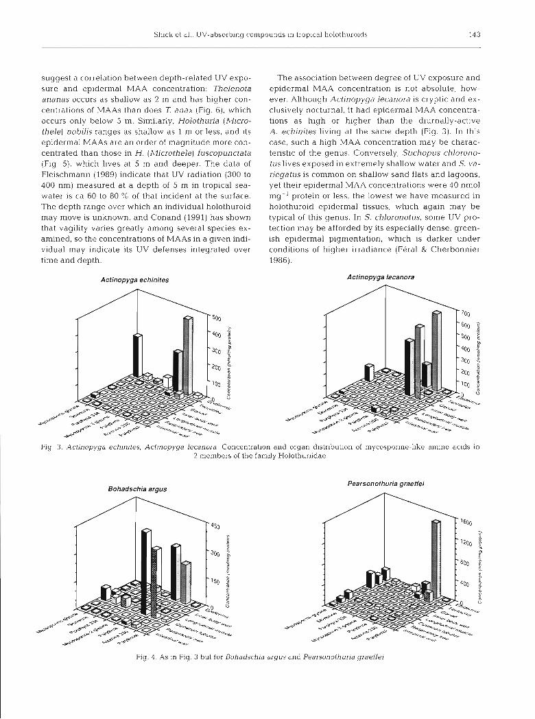
HPLC separations of the methanolic extracts of epi- dermal and gut tissues from Thelenota ananas are shown in Fig. 1. The chromatograms indicate the pres- ence of 7 mycosporine-like amino acids, the structures of which are given in Fig. 2. The taxonomic and tissue distributions of these MAAs in the species surveyed are shown in Figs. 3 to 7. Although most species are represented by a single specimen each, a measure of intraspecific varation is available for 1 ananas (Fig. 6) and indicates that organ- and species-specific differ- ences in MAAs can be discerned with some confi- dence. These results are discussed in several contexts: taxonomic and tissue distribution of MAAs, ecology of the species, and qualitative biochemistry.
The concentration of MAAs in the epidermis is gen- erally higher in members of the family Holothuriidae than in the Stichopodidae (cf. Figs. 3 to 5 with Fig. 6), and intermediate in the single specimen representing the apodid Synaptidae (Fig. 7). Within the Holothurii- dae, the predominant epidermal MAAs tend to be least concentrated in Holothuria (Halodeima) spp. (ca 60 to 90 nmol mg-' protein; Fig. 5), intermediate in concen- tration in Bohadschia argus (ca 250 nmol mg-' protein; Fig. 4), intermediate to high in Actinopyga spp. (ca 250 to 650 nmol mg-' protein; Fig. 3), and extremely con- centrated in Pearsonothuria (formerly Bohadschia)
142 Mar. Ecol. Prog. Ser. 90: 139-148, 1992
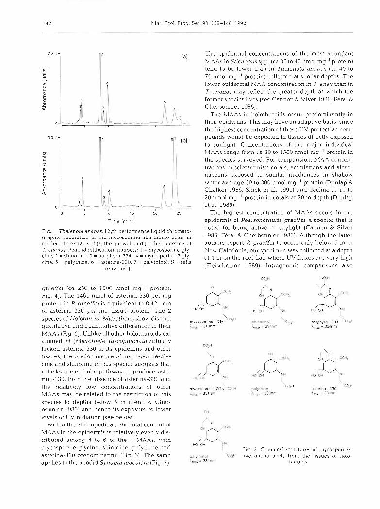
Fig. 1 Thelenota ananas. High performance liquid chromato- noted for being active in daylight (Cannon & Silver
graphic separation of the mycosporine-like amino acids in 1986, Feral & Cherbonnier 1986). Although the latter methanolic extracts of ( a ) the gut wall and (b) the epidermis of authors report F! graeffej to occur only below 5 m in Z ananas. Peak identification numbers. 1 = mycosporine-gly- New Caledonia, our specimen was collected at a depth cine; 2 = shinorine; 3 = porphyra-334 ; 4 = mycosporine-2 gly- of 1 m on the reef flat, where uv fluxes are very high cine; 5 = palythine; 6 = asterina-330; 7 = palythinol; S = salts
(refractive) (Fleischmann 1989). Intrageneric comparisons also
-2 The epidermal concentrations of the most abundant MAAs in Stjchopus spp. (ca 30 to 40 nmol mg-' protein) tend to be lower than in Thelenota ananas (ca 40 to 70 nmol mg- ' protein) collected at similar depths. The
4
I lower epidermal MAA concentration in T anax than in 7: ananas may reflect the greater depth at which the
5 former species lives (see Cannon & Silver 1986, Feral &
0.015-
- V) .- C 3
Q) 0 C m e a
graeffei (ca 250 to 1500 nmol mg- ' protein; Fig. 4). The 1461 nmol of asterina-330 per mg protein in F! graeffei is equivalent to 0.421 mg of asterina-330 per mg tissue protein. The 2 species of Holothuria (Microthele) show distinct qualitative and quantitative differences in their MAAs (Fig. 5). Unlike all other holothuroids ex- amined, H. (Microthele) Euscopunctata virtually lacked asterina-330 in its epidermis and other tissues; the predominance of mycosporine-gly- cine and shinorine in this species suggests that it lacks a metabolic pathway to produce aste- rina-330. Both the absence of asterina-330 and the relatively low concentrations of other MAAs may be related to the restriction of th.is species to depths below 5 m (Feral & Cher- bonnier 1986) and hence its exposure to lower levels of UV radiation (see below).
Within the Stichopodidae, the total content of MAAs in the epidermis is relatively evenly dis- tributed among 4 to 6 of the 7 MAAs, with mycosporine-glycine, shinorine, palythine and asterina-330 predominating (Fig. 61. The same applies to the apodid Synapta maculata (Fig. 7 ) .
l
mycosporlne . Gly hmar = 3lOnrn
2
shrnonne 'C4H l,,, = 334nm
6 Cherbonnier 1986). The MAAs in holothuroids occur predominantly in
palythme 'W X,, = 320nm
\ porphyra . 334 W h,,, = 334nm
0- I VL I I J their epidermis. This may have an adaptive basis, since the highest concentration of these UV-protective com-
astenna - 330 I,,. = 330nm
0.015-
V1 - .- C 3
a 0 c m
g V) D a
o -
( Fig. 2. Chemical structures of mycosporine- palythinol cqH like amino acids from the tissues of holo- i.,, = 3 ~ n m thuroids
o 5 1 0 15 20 25 The highest concentration of MAAs occurs in the Time (rnin) epidermis of Pearsonothuria graeffei, a species that is
1
- 2 pounds would be expected in tissues directly exposed
(b) to sunlight Concentrations of the major individual
4
- ;- ,L[ S
MAAs range from ca 30 to 1500 nmol mg-' protein in the species surveyed. For cornparision, MAA concen- trations in scleractinian corals, actiniarians and alcyo- naceans exposed to similar irradiances in shallow water average 50 to 300 nmol mg-' protein (Dunlap & Chalker 1986, Shick et al. 1991) arld cieclir~t: to 10 io 20 nmol mg-' protein in corals at 20 m depth (Dunlap et al. 1986).
1
Shick et a1 UV-absorbing compounds In trop~cal holothuroids 143
suggest a correlation between depth-related UV expo- sure and epidermal MAA concentration: Thelenota ananas occurs as shallow as 2 m and has higher con- centrations of MAAs than does 7: anax (Fig. 6), which occurs only below 5 m. Similarly, Holothuria (Micro- thele) nobilis ranges as shallow as 1 m or less, and its epidermal MAAs are an order of magnitude more con- centrated than those ln H. (Microthele) fuscopunctata (Flg. 5), which lives at 5 m and deeper. The data of Fleischmann (1989) indicate that UV radiation (300 to 400 nm) measured at a depth of 5 m in tropical sea- water is ca 60 to 80 % of that incident at the surface. The depth range over which a n individual holothuroid may move is unknown, and Conand (1991) has shown that vagility varies greatly among several species ex- amined, so the concentrations of MAAs in a given indi- vidual may indicate its UV defenses integrated over time and depth.
The association between degree of UV exposure and epidermal MAA concentration 1s not absolute, how- ever. Although Actinopyga lecanora is cryptic and ex- clusively nocturnal, it had epidermal MAA concentra- tions as hlgh or higher than the diurnally-active A. echinites living at the same depth (Fig. 3). In this case, such a high MAA concentration Inay be charac- teristic of the genus. Conversely, Stichopus chlorono- tuslives exposed In extremely shallow water and S. va- riegatus is common on shallow sand flats and lagoons, yet their epidermal MAA concentrations were 40 nmol mg-I protein or less, the lowest we have measured in holothuroid epidermal tissues, which again may be typical of this genus. In S. chloronotus, some UV pro- tection may be afforded by its especially dense, green- ish epidermal pigmentation, which is darker under conditions of higher irradiance (Feral & Cherbonnier 1986).
Actinopyga echinites Actinopyga lecanora
Fig 3 Actinopyga echlnltes, Actlnopyga lecanora. Concentration and organ distribution of mycospol-ine-like ainino acids in 2 members of the family Holothuriidae
Bohadschia argus Pearsonothuria graeffei
Fig. 4 . As in Fig. 3 but for Bohadsch~a argus and Pearsonothurja graeffei
- g"
2 5
0 cz 2
m Q
m
"" 2
0 F m
s Q
(n
S
Z
0 S 9
c 5. m Q
Con
con
trat
lon
(n
mo
Vm
g p
rote
in)
Shick et al.: UV-absorbing compounds in tropical holothuroids 145
Slichopus chloronotus Stichopus variegatus
Thelenota ananas Thelenota anax
Fig. 6. Stichopus chloronotus, S. variegatus. Thelenota ananas, 7: anax. Concentration and organ distribution of mycosporine-like amino acids in 4 members of the family Stichopodidae. For T ananas, numbers in parentheses indicate no. of specimens tested.
and vertical lines atop bars indicate +l SE where n = 4
The tentacles of these deposit-feeding holothuroids are also exposed to direct and reflected sunlight, and these tissues are relatively rich in MAAs in some spe- cies. Their ventral position presumably shades the ten- tacles somewhat, which may account for their lower concentration of MAAs than in the dorsal epidermis. Pearsonothuria graeffei and Synapta maculata, how- ever, extend their tentacles anteriorly when foraging (see photographs in Feral & Cherbonnier 1986) and the concentrations of the predominant MAAs in these spe- cies were the highest we have seen in holothuroid ten- tacles. Similarly, the 4 predominant MAAs were all more concentrated in dorsal than in ventral epidermis of 1 specimen of Holothuria (Halodeima) edulis, al- though only 2 of the 4 were more concentrated in the dorsal epidermis of a second specimen (Fig. 5). In Stichopus variegatus, all MAAs are more concentrated
in the ventral than in the dorsal epidermis, but the MAA concentrations (maximum <30 nmol mg-' pro- tein) in this specles of sea cucumber were the lowest of any that we measured.
With few exceptions, epidermal tissues contain the highest concentrations of MAAs, which is consistent with a postulated adaptive function of the compounds in UV photoprotection. This conclusion must be tem- pered by the finding that the MAA concentration in fish ocular lenses bears no apparent relationship to feeding depth or extent of diurnal activity (Dunlap et al. 1989). The pattern presented by the remaining or- gans in holothuroids is a mixed one, and it is clear that UV exposure is not the sole determinant of MAA con- centration. Other factors, including the embryological origin of the various tissues, apparently are involved.
Gonads contained detectable to large amounts of
Mar. Ecol. Prog. Ser. 90: 139-148, 1992
Synapta maculata
Fig. 7 Synapta maculata. Concentration and organ distribu- tion of mycosporine-like amino acids in 1 member of the
family Synaptidae
MAAs in 3 of the 4 species examined at Hicks Reef (Figs. 3 to 5); MAAs were essentially absent from the gonad of a single reproductively spent specimen of Actinopyga echinites (Fig. 3). Both the ripe testis and spawned sperm of a single specimen of Holofhuria nobilis collected at Shrimp Reef in July 1991 contained only small amounts (10 nmol mg- ' protein or less) of mycosporine-glycine as the only quantifiable MAA. Conversely, mycosporine-glycine was the principal MAA in the gonad (gender undetermined) of Pearso- nothuria graeffei, where it reached concentrations of >300 nmol mg-' protein (Fig. 4). and in the gonad (gender undetermined) of Synapta maculata, at a con- centration of 175 nmol mg-' protein (Fig. 7). Both this MAA and shinorine were present in the gonad of H. atra, but they were absent from other internal or- gans of this species (Fig. 5). Mycosporine-glycine and shinorine were present in the ripe gonad (gender un- determined) of A. lecanora, and palythine and aste- rina-330 also achieved very high concentrations in this organ (Fig. 3). Because spawned gametes would re- quire protection from solar UV, the generally high con- centration of MAAs in ripe gonads may be adaptive, although the low concentrations in H. nobilis testis and sperm confuse this issue. The low concentration of MAAs in H. nobilis sperm may reflect a sexual differ- ence, if the individuals of species having high MAA concentrations in the gonads were females; since spawned sperm would be exposed to solar UV for far less time than eggs or developing embryos and larvae, the sperm may not require such high concentrations of MAAs as the eggs. The absorption spectra of the 4 MAAs in the gonad span 310 to 334 nm, much of the range of biologically damaging solar UV.
The presence of high concentrations of MAAs in the gonad was in sharp contrast to the virtual absence of these compounds from longitudinal muscle; only in 2 of the 13 species examined (Holothuria fuscopunctata and Synapta maculata) were there appreciable amounts (ca 60 and 90 nmol mg-l protein, respec- tively) of mycosporine-glycine in the longitudinal muscle (Figs. 5 & 7). In the case of S. maculata, its very thin, translucent body wall may afford less UV protec- tion to the underlying longitudinal muscle and require endogenous defenses in that tissue. MAAs likewise were nearly absent from the inner body wall (consist- ing of inner epithelium and dermis, both of mesoder- mal origin) in 7 of the 12 species studied at Hicks Reef. The MAAs present in 4 species (H. fuscopundata, Stichopus variegatus, Thelenota ananas and 7: anax) were mycosporine-glycine and shinorine; concentra- tions were generally <20 nmol mg-' protein. These compounds were also present at similar leve!s in body wall of H. nobilis, which exceptionally contained 70 nmol astenna-330 mg-' protein (Fig. 5 ) . Apart from these few examples, the data suggest that internal, mesodermal tissues removed from UV exposure do not appreciably accumulate MAAs. The occurrence of mycosporine-glycine and shinorine in the inner body wall may indicate their transport from the gut to the epidermis via this route (see below).
Surprisingly high concentrations of mycosporine- glycine and shinorine were present in the cloaca and respiratory trees of many species in both the Holo- thuriidae and Stichopodidae, and in the Cuvierian tu- bules of Bohadschia argus and Pearsonothuria graef- fei. The Cuvierian tubules of B, argus also contained very large amounts of palythine and asterina-330, a pattern similar to the epidermis in this species (Fig. 4). A possible explanation of the seeming exception to the association between UV exposure and MAA concen- tration is that the cloaca is derived from ectoderm (Runnstrom 1927, Feral & Massin 1982); based on their continuity with the cloaca (as distinct from the lntes- tine, the digestive portion of which is endodermal in origin), and on the presence of T-shaped cells charac- teristic of integumental epithelia (J.-P. Feral pers. comm.), the respiratory trees and Cuvierian tubules also may be ectodermally-derived. Thus, the capacity to accumulate high concentrations of certain MAAs may be characteristic of ectodermal tissues in general, including cloaca, respiratory trees and Cuvienan tu- bules as well as integumental epidermis.
Intestinal tissue also contained measurable amounts of MAAs in most of the holothuroid species examined. Mycosporine-glycine and shinorine were most fre- quently encountered, although palythine and asterina- 330 were also concentrated in some species. Since holothuroids, like other metazoans, probably cannot
Shick et al.: UV-absorbing compounds in trop~cal holothuroids 147
synthesize the mycosporine base structure de novo (see 'Introduction'), the acquisition of MAAs in the diet might account for the presence of these compounds in the gut tissue. Most of the MAAs identified in holo- thuroid tissues are present in the coral sand (primarily in unicellular algae and cyanobacteria) that they in- gest, and appear to be removed from the gut contents as they move through the digestive tract (W. C. Dunlap, J. M. Shick & R. Larsen unpubl.).
Mycosporine-glycine is structurally the simplest MAA (Fig. 2) and may be the parent compound from which other MAAs are derived in secondary meta- bolism. It is the most widespread MAA among the holothuroids both taxonomically and in tissue distribu- tion, and it may be that most tissues in most species have some capacity to accumulate it following its transport from the gut. Holothuria fuscopunctata accu- mulates mycosporine-glycine and shinorine in a sirni- lar ratio in most of its tissues (Fig. 5). This species is un- usual in the virtual absence of other MAAs from its tissues, whereas Thelenota anax , which may occur in the same habitat, accumulates mycosporine-glycine and shinorine in a ratio similar to H. fuscopunctata, but also contains asterina-330 in its epidermis (Fig 6).
Very high concentrations of astenna-330, particu- larly in epidermis, may be a specific photoadaptation and reflect its selective accumulation in a tiss'ue that receives the highest amount of UV radiation. This is especially indicated because of the virtual absence of asterina-330 from the body wall tissue directly under- lying the epidermis, whereas in the latter tissue the concentration may reach several hundred nmol of aste- rina-330 per mg protein or higher. The epidermal con- centration of palythine tends to be high when that of asterina-330 is greatest, and this also may indicate a selective (UV-induced?) synthesis or accumulation of an MAA having an absorption maximum at a shorter wavelength (320 nm) than asterina-330, thus extend- ing the range of UV-protection. Palythinol is a minor component of the total MAA complement in holothur- oids. Its structural similarity to asterina-330, differing only in the methylation of the ethanolimine residue (Fig. 2), and the positive correlation between the epi- dermal concentrations of these MAAs (r2 = 0.45, n = 19, p = 0.0016) in the species examined, suggest that the 2 compounds are metabolically related.
In conclusion, taxonomic, tissue-specific and ecolog- ical trends can be discerned in the occurrence of mycosporine-like amino acids in tropical deposit-feed- ing holothuroids. Unlike coral reef anthozoan cnidar- ians, where MAAs may be synthesized by their symbi- otic dinoflagellates (Dunlap & Chalker 1986, Shick et al. 1991, see also Carreto et al. 1990), holothuroids do not harbor algal endosymbionts and their MAAs must have a different provenance. Uptake of dietary
MAAs is one possibility that is treated elsewhere (Dunlap et al. 1991, unpubl.). Also of interest are the metabolic interrelationships among algal-derived MAAs and their interconversion by microbes within the holothuroid gut. Whether subcuticular bacteria (see Holland & Nealson 1978, Feral 1980, Feral & Massin 1982) have any role in providing MAAs to the animal tissue in these and other echinoderms is un- known, but preliminary electron microscopic examlna- tions of Thelenota a n a n a s do reveal the presence of subcuticular bacteria in this species (K. C. Edwards, J. M. Shick & W. C. Dunlap unpubl. obs.). In the broad context, the mechanisms for the incorporation of MAAs in holothuroid tissues may provide important insight to a general biochemical pathway for the trophic accu- mulation of UV-absorbing MAAs in higher marine invertebrates and vertebrates.
Acknowledgements. We thank the masters and crew of RV 'Lady Basten' for logistical support, J. Wu Won for technical assistance, and R. Willis for critical comments on the manu- script. C. Conand, J.-P. Feral and M. Jangoux provided assist- ance with the literature and valuable insights from their own experience with echinoderms, and 3 anonymous reviewers supplied helpful comments. This research was conducted as part of the Reef Photobiology Program at AIMS. J.M.S was supported by National Science Foundation Grant DCB- 8509487 (Physiological Processes), and National Geograph~c Society research grants 3883-88 and 4532-91; his participa- tion in the research was facilitated by a sabbatical leave from the University of Maine and by the visiting investigator pro- gram at AIMS.
LITERATURE CITED
Bakus, G. J. (1968). Defensive mechanisms and ecology of some tropical holothurians. Mar. Biol. 2: 23-32
Bakus, G. J. (1973). The biology and ecology of tropical holo- thurians. In: Jones, 0. A., Endean, R. (eds.) Biology and geology of coral reefs, Vol. 2, Biology 1. Academic Press, New York, p. 325-367
Bradford, M. M. (1976). A rapid and sensitive method for the quantitation of microgram quantities of protein using the principle of protein-dye binding. Anal. Biochem. 72: 248-254
Cannon, L. R. G., Silver, H. (1986). Sea cucumbers of Northern Australia. Queensland Museum. Brisbane
Carreto, J. I.. Carignan. M. O., Daleo, D., Demarco, S. G. (1990). Occurrence of mycosporine-like amino acids in the red-tide dinoflagellate Alexandrium excavatum - UV- photoprotective compounds. J. Plankton Res. 12: 909-922
Chioccara, F., Misuraca, G., Novellino, E., Porta, G. (1986a). Occurrence of two new mycosporine-like amino acids my- tilins A and B in the edible mussel, Mytilus galloprovin- cialis. Tetrahedron Lett. 34: 3181-3182
Chioccara, F., Zeuli, L., Novellino, L. E. (198613). Occurrence of mycosporine related compounds in sea urchin eggs. Comp. Biochem. Physiol. 85B: 459-461
Conand, C. (1991). Long-term movements of some tropical sea-cucumbers monitored by tagging and recapture. In: Yanagisawa, T., Yasumasu, I . , Oguro, C., Suzukl, N., Motokawa. T. (eds.) Biology of Echinodermata. Pro-
Mar. Ecol. Prog. Ser. 90: 139-148, 1992
ceedings of the seventh international echinoderm confer- ence, Atami, Japan. A. A. Balkema, Rotterdam, p 169-175
Dunlap, W. C . , Banaszak, A. T., Rosenzweig, T T., Shick, J. M. (1991). Ultraviolet light-absorbing compounds In coral reef holothurians: organ distribution and possible sources. In: Yanagisawa, I., Yasumasu, I., Oguro, C., Suzuki, N.. Motokawa, T. (eds.) Biology of Echinodermata. Proceedings of the seventh international echinoderm con- ference, Atarni, Japan. A. A. Balkema, Rotterdam, p 560
Dunlap, W. C., Chalker, B. E . (1986). Identification and quan- titation of near-UV absorbing compounds (S-320) In a hermatypic scleractinian. Coral Reefs 5: 155-159
Dunlap, W. C., Chalker, B. E., Oliver, J. K. (1986). Bathyrnetric adaptations of reef-building corals at Davies Reef. Great Barrier Reef, Australia. 111. UV-B absorbing compounds. J . exp. mar. Blol. Ecol. 104: 239-248
Dunlap, W. C., W~lllarns, D. McB., Chalker, B. E. , Banaszak, A. T (1989). Biochemical photoadaptation in vis~on: UV- absorbing pigments in fish eye tissues. Comp. Biochem. Physiol. 93B: 601 -607
Favre-Bonvin, J . , Bernillon, J., Salain, N., Arpin, N. (1987). Biochemistry of mycosporines: rnycosporine glutaminol in Tnchothecium roseurn. Phytochemistry 26: 2509-2514
Feral, J.-P. (1980). Cuticule et bacteries associees des epidermes digestif et tegumentaire d e Leptosynapta gal- liennei (Herapath) (Holothuroidea: Apoda) - premieres donnees. In: Jangoux, M. (ed.) Echinoderms: present and past. A. A. Balkema, Rotterdam, p. 285-290
Feral, J.-P., Cherbonnier, G. (1986). Les holothurides. In: Guille, A . , Laboute, P,, Menou, J -L . (eds.) G'ulde des etoiles de mer, oursins et autres ech~nodermes du lagon de Nouvelle-Caledonie. ~ d i t i o n s de 1'ORSTOM. Institut Franqais de Recherche Scientifique pour le Develop- pement e n Cooperation, Paris, p. 55-107
Feral, J.-P,, Massin, C. (1982). Digestive systems: Holo- thuroidea. In: Jangoux, M,, Lawrence, J . M. (eds.) Echi- noderm nutrition A A. Balkema, Rotterdam, p. 191-212
Fle~schmann, E. M. (1989). The measurement and penetration of ultraviolet radiation into tropical marine water. Lirnnol. Oceanogr. 34: 1623-1629
Floss, H. G. (1979). The shikimate pathway. In: Swain. T., Harborne, J . B., Sumere, C. F. van (eds.) Recent advances in phytochemistry: biochemistry of plant phenolics. Plenum Press, New York, p. 59-89
Harnmond, L. S. (1982). Patterns of feeding and activity in de- posit-feeding holothurians and echinoids (Echinoderrnata) from a shallow back-reef lagoon, Discovery Bay, Jamaica. Bull. mar. Sci. 32: 549-571
Hirata, Y., Uemura, D., Ueda, K., Takano, S. (1979). Several compounds from Palythoa tuberc~~losa (Coelenterata). Pure appl. Chem 51. 1875-1883
Holland, N . D., Nealson. K. H. (1978). The f ~ n e structure of the echinoderm cuticle and the subcuticular bacteria of echi- noderms. Acta 2001. 59: 169-185
This article was submitted to the editor
Jerlov, N. G. (1950). Ultraviolet radiation in the sea. Nature 116: 111-112
Karentz, D., McEuen, F. S., Land, M. C , Dunlap, W. C. (1991). Survey of mycosporine-like amino acld compounds In Antarctic marine organisms: potential protection from ultraviolet exposure. Mar. Biol. 108: 157-166
Levin. V. S., Kalinin, V. I.. Stonik, V. A. (1984). Chemical char- acters and taxonomic revision of holothurian Bohadschia graeffej (Semper] as refer to erection of a new genus. B~ologia Morya 1984 (3): 33-38 (in Russian)
Moriarty, D. J . W. (1982) Feeding of Holothuria atra and Sfichopus chloronotus on bacteria, organic carbon and or- ganic nitrogen in sediments of the Great Barrier Reef. Aust. J . mar. freshwat. Res. 33: 255-263
Moriarty. D. J W., Pollard, P. C., Hunt, W. G.. Moriarty, C. M.. Wassenberg, T. J (1985). Productivity of bacteria and mi- croalgae and the effect of grazing by holothurians in sedi- rnents on a coral reef flat. Mar. Biol. 85. 293-300
Nakarnura, H., Kobayashi, J., Hirata, Y (1981). Isolation and structure of a 330-nm UV-absorbing substance. Asterina- 330 from the starfish Asterina pectinifera. Chem. Lett. 28: 1413-1414
Runnstrom, S. (1927). ~ b e r die Entwicklung von Lepto- synapta inhaerens. Rergens Mus. Arbok, Naturvid, rek., Bergen
Shibata, K. (1969). Pigments and a UV-absorbing substance in corals and a blue-green alga living in the Great Barrier Reef. Plant cell Physiol. 10: 325-335
Shick, J. M., Lesser, M. P., Stochaj, W. R. (1991). Ultraviolet radiation and photooxidative stress in zooxanthellate Anthozoa: the sea anemone Phyllod~scus semoni and the octocoral Clavularia sp. Symbiosis 10 145-173
Sivalingam. P. M., Ikawa. T., Yokohama, Y., Nisizawa, K. (1974). Distribution of a 334 UV-absorbing substance in algae, with special regard of its possible physiological roles. Bot. Mar. 17: 23-29
Smith, R C., Baker, K. G (1979). Penetration of UV-B and bi- ologically effective dose-rates in natural waters. Photochem. Photobiol. 29: 31 1-323
Takano, S., Nakanishi, A., Uernura, D., Hirata, Y (1979). Isolation and structure of a 334 nm UV-absorbing sub- stance, porphyra-334 from the red alga Porphyra Lenera Kjellman. Chem. Lett., p. 419-420
Takano, S , Uemura, D , Hirata, Y (1978). Isolation and structure of a new amino acid, palythine, from the zoanthid Palythoa Luberculosa. Tetrahedron Lett. 26: 2299-2300
Tsujino, I., Yabe, K., Sekekawa, I. (1980). Isolation and struc- ture of a new amino acid, shinorine, from the red algae, Chondrus yendoi Yarnada et Mikami. Bot. Mar. 23: 65-68
Wood, W F. (1987). Effect of solar ultra-violet radiation on the kelp Eklon~a radiata. Mar. Biol. 96: 143-150
Yoshida. S. (1969). Biosynthesis and conversion of aromatic amino acids in plants. A. Rev. plant Physiol. 20: 41-62
Manuscript first received. June 29, 1992 Revised verslon accepted November 5, 1992