SUSCEPTIBILITY OF MICRO-ORGANISMS TO CHLORAMPHENICOL (CHLOROMYCETIN) 1, 2 BY I. W. McLEAN, JR., J. L. SCHWAB, A. B. HILLEGAS, AND A. S. SCHLINGMAN ' (From the Research Laboratories, Parke, Davis & Co., Detroit 32, Mich.) The antibiotic, chloramphenicol, produced by Streptomyces venezuelae, was originally reported by Ehrlich et al. (1, 2) and independently de- scribed by Carter, Gottlieb et al. (3, 4). Since these original reports the active principle has been chemically isolated and characterized (5, 6), syn- thesized (7) and is at present commercially avail- able for clinical use. Originally (1) activity was described against several Gram-negative bacteria, in particular Shigella sonnei, and against Rickettsia prowazekii in chick embryos. Smadel et at. (8-11) have notably extended this spectrum to include other rickettsial agents as well as two viruses, lympho- granuloma venereum and psittacosis. Smith et al. ( 12) reported that chloramphenicol was active, un- der the conditions tested, against Gram-negative bacteria, R. prowazekii and Borellia recurrentis; moderately active against Gram-positive bacteria and Mycobacterium tuberculosis; and inactive in the concentrations used against yeasts, filamentous fungi, protozoa and certain viruses. Youmans et al. (13) tested strains of virulent human-type M. tuberculosis in vitro and concluded that chloram- phenicol was only moderately active when com- pared with streptomycin or para-amino salicylic acid. Clinically, confirmation of laboratory activity has been obtained for certain of the rickettsial dis- eases (14-18), typhoid fever (19), brucellosis (20) and in urinary infections caused by certain Gram-negative bacteria (21). With the drug now available in quantity for clinical trial, it seems op- portune at this time to collect the known informa- 1 Presented at the Second National Symposium on Recent Advances in Antibiotics Research held in Wash- ington, D. C., April 11-12, 1949, under the auspices of the Antibiotics Study Section, National Institutes of Health, Public Health Service, Federal Security Agency. 2 Parke, Davis & Company trade-name. ' Collaborators: F. A. Miller, J. S. Markey, A. Pucheu, M. C. Galbraith, M. E. Doles, M. F. Sheehan, G. Coffey, J. M. McCracken, B. D. Moore, II, M. C. Manning. Research Laboratories, Parke, Davis & Company. tion with respect to the microbiological spectrum of this new chemotherapeutic agent. METHODS Viruses and Rickettsiae Borreliota: Vaccinia virus (New York Public Health strain from Dr. S. K. Muckenfus, New York City Dept. of Public Health, N. Y., egg-adapted) was tested as a representative of this group of viruses. Two types of tests in chick embryos were used. The first consisted of mixing the chloramphenicol with the virus and inoculat- ing 0.1 ml. of the mixture onto the chorioallantoic mem- brane of 10-day chick embryos. After four days incuba- tion at 350 C. the membranes were examined and com- pared with controls for reduction in pock formation. The second method consisted of inoculating groups of treated, 10-day chick embryos via the yolk sac with a lethal dose of virus and repeating treatment on the fol- lowing day by the same route. The eggs were candied twice daily and delay in death time and seven-day sur- vivors recorded. Erro: St. Louis encephalitis virus (egg-adapted from Dr. Carl Duffy, Wayne University, Detroit, Michigan, and a mouse-adapted laboratory strain) was tested to represent the group of virus encephalitides. Tests were conducted in embryonated eggs and mice. For the egg test, nine-day embryos were infected via the allantoic sac and treated 30 minutes before infection and on the 1st day after infection. The eggs were candied twice daily and time of death and seven-day survivors noted. In the mouse test, groups of 10 to 12 Gm. white Swiss mice were infected intracerebrally and treated intraperitoneally before infection and twice daily thereafter for 10 doses. Mean death time was compared with that of a control group. Formido: The National Institutes of Health mouse- adapted strain of rabies virus was used, as well as a variant line of this strain adapted to the intramuscular route of infection. For the former, mice were injected intracerebrally with a lethal dose and daily treatments given intraperitoneally for four days. A similar proce- dure was used with the intramuscular strain except for the different route of virus administration. Time of paralysis and death of the treated mice was noted for comparison with control groups. Legio: The anti-poliomyelitis virus tests were con- ducted by Drs. J. L. Melnick and F. R. Corria (22) of the Yale University School of Medicine, New Haven, Conn. The Y-S.K and Lansing strains (mouse-adapted) were used as well as Theiler's intestinal virus of mice. 953
Transcript
SUSCEPTIBILITY OF MICRO-ORGANISMSTO CHLORAMPHENICOL(CHLOROMYCETIN)1, 2
BY I. W. McLEAN, JR., J. L. SCHWAB,A. B. HILLEGAS, ANDA. S. SCHLINGMAN'(From the Research Laboratories, Parke, Davis & Co., Detroit 32, Mich.)
The antibiotic, chloramphenicol, produced byStreptomyces venezuelae, was originally reportedby Ehrlich et al. (1, 2) and independently de-scribed by Carter, Gottlieb et al. (3, 4). Sincethese original reports the active principle has beenchemically isolated and characterized (5, 6), syn-thesized (7) and is at present commercially avail-able for clinical use.
Originally (1) activity was described againstseveral Gram-negative bacteria, in particularShigella sonnei, and against Rickettsia prowazekiiin chick embryos. Smadel et at. (8-11) havenotably extended this spectrum to include otherrickettsial agents as well as two viruses, lympho-granuloma venereum and psittacosis. Smith et al.( 12) reported that chloramphenicol was active, un-der the conditions tested, against Gram-negativebacteria, R. prowazekii and Borellia recurrentis;moderately active against Gram-positive bacteriaand Mycobacterium tuberculosis; and inactive inthe concentrations used against yeasts, filamentousfungi, protozoa and certain viruses. Youmans etal. (13) tested strains of virulent human-type M.tuberculosis in vitro and concluded that chloram-phenicol was only moderately active when com-pared with streptomycin or para-amino salicylicacid.
Clinically, confirmation of laboratory activityhas been obtained for certain of the rickettsial dis-eases (14-18), typhoid fever (19), brucellosis(20) and in urinary infections caused by certainGram-negative bacteria (21). With the drug nowavailable in quantity for clinical trial, it seems op-portune at this time to collect the known informa-
1 Presented at the Second National Symposium onRecent Advances in Antibiotics Research held in Wash-ington, D. C., April 11-12, 1949, under the auspices of theAntibiotics Study Section, National Institutes of Health,Public Health Service, Federal Security Agency.
2 Parke, Davis & Company trade-name.' Collaborators: F. A. Miller, J. S. Markey, A. Pucheu,
M. C. Galbraith, M. E. Doles, M. F. Sheehan, G. Coffey,J. M. McCracken, B. D. Moore, II, M. C. Manning.Research Laboratories, Parke, Davis & Company.
tion with respect to the microbiological spectrumof this new chemotherapeutic agent.
METHODS
Viruses and Rickettsiae
Borreliota: Vaccinia virus (New York Public Healthstrain from Dr. S. K. Muckenfus, New York City Dept.of Public Health, N. Y., egg-adapted) was tested as arepresentative of this group of viruses. Two types oftests in chick embryos were used. The first consisted ofmixing the chloramphenicol with the virus and inoculat-ing 0.1 ml. of the mixture onto the chorioallantoic mem-brane of 10-day chick embryos. After four days incuba-tion at 350 C. the membranes were examined and com-pared with controls for reduction in pock formation.The second method consisted of inoculating groups oftreated, 10-day chick embryos via the yolk sac with alethal dose of virus and repeating treatment on the fol-lowing day by the same route. The eggs were candiedtwice daily and delay in death time and seven-day sur-vivors recorded.
Erro: St. Louis encephalitis virus (egg-adapted fromDr. Carl Duffy, Wayne University, Detroit, Michigan,and a mouse-adapted laboratory strain) was tested torepresent the group of virus encephalitides. Tests wereconducted in embryonated eggs and mice. For the eggtest, nine-day embryos were infected via the allantoic sacand treated 30 minutes before infection and on the 1stday after infection. The eggs were candied twice dailyand time of death and seven-day survivors noted. In themouse test, groups of 10 to 12 Gm. white Swiss micewere infected intracerebrally and treated intraperitoneallybefore infection and twice daily thereafter for 10 doses.Mean death time was compared with that of a controlgroup.
Formido: The National Institutes of Health mouse-adapted strain of rabies virus was used, as well as avariant line of this strain adapted to the intramuscularroute of infection. For the former, mice were injectedintracerebrally with a lethal dose and daily treatmentsgiven intraperitoneally for four days. A similar proce-dure was used with the intramuscular strain except forthe different route of virus administration. Time ofparalysis and death of the treated mice was noted forcomparison with control groups.
Legio: The anti-poliomyelitis virus tests were con-ducted by Drs. J. L. Melnick and F. R. Corria (22) ofthe Yale University School of Medicine, New Haven,Conn. The Y-S.K and Lansing strains (mouse-adapted)were used as well as Theiler's intestinal virus of mice.
953
I. W. MCLEAN, JR., J. L. SCHWAB, A. B. HILLEGAS, AND A. S. SCHLINGMAN
Rabula: The egg-adapted mumps virus tested was ob-tained from Dr. J. E. Smadel of the Army MedicalSchool, Washington, D. C. The tests were conducted inseven-day embryonated eggs infected via the allantoic sac.Treatments were given 30 minutes before infection andon the second day. Allantoic fluid was harvested fromeach egg after seven-days incubation and tested for virusmultiplication by the addition of washed chicken red bloodcells. In addition a hemagglutination titration was-per-formed on an aliquot pool of allantoic fluid from thegroups of eggs. An untreated control group was in-cluded in the test for comparison of the number of posi-tive eggs and level of group hemagglutinin titer.
Tarpeia: Tests against three strains of type A, sevenstrains of type A' and two strains of type B influenzavirus have been conducted- in 11-day chick embryos. Thevirus was inoculated into the allantoic sac and treat-ments given by the same route 30 minutes before infectionand 60 minutes later. After 40 hours incubation at 350 C.the presence of infection-in each egg and the hemaggluti-nation titer of 'an aliquot pool of allantoic fluid weredetermined for each group as described above for mumpsvirus. Considerable work has been done using the mouse-adapted PR8 strain of type A in mice. In general,groups of 12 to 14 Gm. white Swiss mice were infectedintranasally and treated by various routes and at differenttimes with chloramphenicol. In the test reported heretreatment was given intraperitoneally for seven daysstarting at the time of infection.
One inconclusive test has been made with the Greenstrain of distemper virus (commercial distemperoid, ferret-adapted) in ferrets. Two ferrets were infected intra-nasally and one was treated intraperitoneally at the timeof infection and daily thereafter for three days. Severityof illness and time of development of fever were noted.The animals were sacrificed at the height of the disease;hence duration of illness could not be determined.
Tortor: Newcastle disease virus (California 14 fromDr. L. T. Giltner, Bureau of Animal Industry, Wash-ington, D. C.) was tested in 11-day embryonated eggsand white Leghorn pullets. The test in eggs was con-ducted in the same manner as the influenza virus testsdescribed above except that infection was determined bydeath or survival of the embryo. Comparisons of theaverage time of death of treated and untreated groupswere made. In chickens the virus was administered in-tramuscularly and treatments were given approximatelyevery 12 hours for four doses starting at the time ofinfection. Time of the appearance of symptoms and num-ber of survivors compared with a control group werenoted.
The chick bronchitis virus (obtained from Dr. C. H.Cunningham, Michigan State College, East Lansing,Michigan) was tested in chick embryos as describedabove for Newcastle disease.
Laryngo-tracheitis virus (commercial vaccine strain)was tested on the membrane of chick embryos using thetechnique described for vaccinia virus.
Rickettsia: Considerable work has been done with epi-demic typhus (R. frowazekii, Breinl strain) in chick
embryos. In the work reported here, groups of six-daychick embryos were infected via the yolk sac and treatedonce on the third day after infection by the same route.Average time of death and number of survivors werecompared with an untreated control group.
The results with murine typhus (R. typhi), scrubtyphus (R. tsutsugamushi) and rickettsialpox (R. akari)were taken from the work of Smadel et al. (8-11). Us-ing the method described above, their findings for murinetyphus in eggs have been confirmed in this laboratory.
Dermacentroxenus, Coxiella and Miyagawvanella: Thereports of Smadel et al. (8-11) are included in the tablefor Rocky Mountain spotted fever (D. rickettsi), Q fever(C. burnetii), lymphogranuloma venereum (M. lympho-granulomatis) and psittacosis (M. psittacii).
Bacteria
The bacterial species reported here were tested for theirsusceptibility to chloramphenicol by inoculating a speci-fied dilution or quantity of liquid culture into media con-taining varying concentrations of the drug, the end pointbeing taken as the lowest concentration which causedcomplete inhibition of growth for 18 hours. Except asindicated the original transplants for preparation of sus-pensions were made from cultures dried from the frozenstate.
The aerobes were tested by adding 1.0 ml. of a stand-ardized dilution of a tryptose broth culture to 1.0 ml.quantities of tryptose broth containing varying concen-tration of chloramphenicol. The final concentrations usedvaried from 0.1 to 200 jg/ml. The tubes were incubatedfor 18 hours and the results recorded as "growth" or "nogrowth."
It was necessary to utilize special media for some ofthe more fastidious organisms. However, the same tech-nique was used, replacing the tryptose broth with thespecial medium required. Brucella abortus was grownunder 10 per cent CO. for 48 hours (No. 1335).
The anaerobes were tested by adding varying concen-trations (ranging from 0.1 to 500 isg/ml.) of chloram-phenicol to melted agar medium cooled to 400 C. Toeach 'tube, 0.1 ml. of a 24-hour liquid culture of theorganism to be tested was added. The tubes were thenshaken thoroughly and allowed to gel. Growth was re-corded as positive or negative after 24 hours incubation.
Beef infusion agar (2 per cent) containing 2 per centglucose (pH 8.2) was used for Clostridium perfringens,novyi and septicum. Cl. feseri was tested in 2 per centliver infusion agar (pH 8.2) and the anaerobic agar rec-ommended by Records and Vawter (23) was utilized forCl. hemolyticum. Cl. tetani was grown in Taylor mediumcontaining 0.5 per cent agar and incubated in an atmos-phere of hydrogen and carbon dioxide.
Fungi
In determining the susceptibility of fungi to chloram-phenicol, a broth dilution method was employed. Serialdilutions of the drug in broth were dispensed in 1 ml.amounts in Wassermann tubes and to each tube was added
954
SUSCEPTIBILITY TO CHLORAMPHENICOL
1 ml. of inoculum in broth. The tubes were incubated at30° C. for one to seven days, depending upon the species,and then observed for inhibition of growth. The mini-mumconcentration of the drug causing complete inhibi-tion of growth was taken as the end point.
Modified Sabouraud's broth (dextrose 2 per cent, neopeptone 1 per cent) was used in preparing the dilutions ofthe drug and the fungus suspensions. In the case of theactinomycetes, Brain-Heart Infusion Broth (Difco) wasemployed.
Three types of inocula were used. 1) Spore suspen-sion: In heavily sporulating species suspensions wereprepared from either agar slant or agar plate culturesof the fungus. In the case of agar slants, broth was
added to the tube, the fungus stirred up with an inoculat-ing needle and the tube then shaken. When using agarplate cultures the mycelium was scraped from the surfaceof the plate, placed in broth and shaken. The suspensionswere then filtered through cotton and the spores countedon the hemocytometer. 2) Spores and mycelial frag-ments: Species having limited sporulation were removedfrom the surface of the culture medium, emulsified withbroth either in a Waring Blender, by shaking with glassbeads or by grinding in a mortar. The suspension wasfiltered through a loose cotton mat and in some instancesthe number of fragments and spores per ml. was deter-mined by means of the hemocytometer. 3) Mycelialsuspension: The mycelium of non-sporulating species was
BLE I
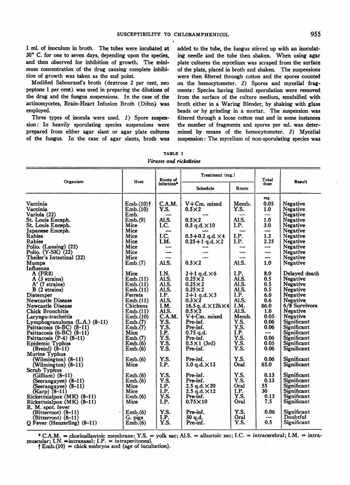
Viruses and rickettsiae
Treatment (mg.)Organism Host infection* Total Result
Schedule Route |_
VacciniaVacciniaVariola (22)St. Louis Enceph.St. Louis Enceph.Japanese Enceph.RabiesRabiesPolio. (Lansing) (22)Polio. (Y-SK) (22)Theiler's Intestinal (22)MumpsInfluenza
P. D. StockFrequent transferPUSUrine (B)Urine (B)Urine (B)B. A. I.B. A. I.College Station, Tex.A. T. C. C.Frequent transferFrequent transferDr. Huddlesn, M. S. C.Aborted bovine fetus N. I. H.Trachea of dogLungCampDetrickN. I. H.Dr. Huddleson, M. S. C.K. S. A. C.K. S. A C.Univ. Nev.Univ. Nev.Dr. Novy, U. of M.N. I. H.Dr. De-Kruif, U. of M.Dr. Reed, Kingston, Ont.Univ. of Calif.K. S. A. C.N. Y. Dept. of HealthMueller variantP. D. StockP. D. StockBovine uterusPurulent chest fluidP. D. StockN. I. H.Pyelitis, Rt. kidneyBlood cult. uterine infectionKidney infection, urineBlood cultureBlood cultureUrine Sp.Frequent transferP. D. StockUrine (F)Urine (0)Pre-.treatment sample of urinePure culture, U. of IowaDr. Kendrick, Mich.Clinical caseClinical casesSputumBlood cultureSpinal fluidSputumThroatSputumF.D. A.F.D. A.F.D. A.F.D. A.F.D. A.F.D. A.
Feunt transferFrqent transfer
1-20,000,0001-10,000,0001-10,000,0001-10,000,0001-10,000,0001-10,000,0001:201:201:101:201-10,000,0001-10,000,0001:201:201:201:201:201:201:200.1 cc. undiluted0.1 cc. undiluted0.1 cc. undiluted0.1 cc. undiluted0.1 cc. undiluted0.1 cc. undiluted0.1 cc. undiluted0.1 cc. undiluted0.1 cc. undiluted0.1 cc. undiluted0.1 cc. undiluted0.1 cc. undiluted1-10,000,0001-10,000,0001:101-10,000,0001-10,000,0001-5,000,0001-20,000,0001-20,000,0001-10,000,0001-10,000,0001-20,000,0001-20,000,0001-10,000,0001-10,000,0001-10,000,0001-10,000,0001-10,000,0001-10,000,000101 organisms10-1 organisms10, organisms1-10,000,0001-10,000,0001-10,000,0001-10,000,0001-10,000,0001-10,000,0001-500,0001-1,000,0001-10,000,0001-20,000,0001-20,000,0001-10,000,0001-20,000,0001-10,000,0001-10,000,000
Kiebsiella pneumoniae IIMalleomyces malleiMicrococcus citrusMicrococcus citrusMicrococcus pyogenes var. albusMicrococcus pyogenes var. albusMicrococcus pyogenes var. albusMicrococcus pyogenes var. albusMicrococcus pyogenes var. aureusMicrococcus pyogenes var. aureusMicrococcus pyogenes var. aureusMicrococcus pyogenes var. aureusMicrococcus Pyogenes var. aureusMicrococcus pyogenes var. aureusMicrococcus pyogenes var. aureusMicrococcus pyogenes var. aureusMicrococcus pyogenes var. aureusMicrococcus pyogenes var. aureusMicrococcus pyogenes var. aureusMicrococcus pyogenes var. aureusMicrococcus pyogenes var. aureusMycobacterium phlciMycobacterium.tuberculosis (13)
UrineP. D. StockBoil on headGonorrheaeFemale gonorrheaePusPustuleEar infection (M)F. D.A.F. D. A.U.S. D.A.U.S. D.A.N. I. H.F. D.A.Blood cultureBoilEar (M)Urine (Mc)Frequent transferFrequent transferFrequent transferP. D. StockFresh isolates
Trachea at autopsyTonsillar abscessCough plateN. I. H.Heart blood of chickenLiver of gooseHeart blood of chickenColorado Agric. Coll.Calf's lungBuffalo strainSinus of rabbitColorado Agric. Coll.P. D. StockN. I. H.N. I. H.N. I. H.UrineFrequent transfer
UrineEyeSwab from left armHorseAbdominal cavity of chickenTurkeyBovineFrequent transferStool cultureFrequent transferEyeStool cultureUrineUrine (Mc)Urine (C)Urine (Mc)Urine (Mel)Urine (Mel)Urine (S)Urine (L)P. D. StockP. D. StockP. D. StockChicken origin
TurkeyN. I. H.N. I. H.N. I. H.Frequent transferN. I. H.Rabbit typhoid 104RatHeart blood of lambP. D. StockN. I. H.N. I. H.N. I. H.N. T. C. C.Stool cultureTyphoid carrierFre uent transferTyphoid carrierTyphoid carrierTyphoid carrierTyphoid carrierTyphoid carrierTyphoid carrierTyphoid carrierTyphoid carrierTyphoid carrierTyphoid carrierTyphoid carrierTyphoid carrierTyphoid carrierTyphoid carrierTyphoid carrier (M.C.)*Typhoid carrier (M.C.)tTyphoid carrier (J.T.)tP. D. StockF. D. A.Frequent transferP. D. StockA. T. C. C.A. T. C. C.P. D. StockP. D. StockN. I. H.A. T. C. C.A. T. C. C.
A. T. C. C.P. D. StockP. D. StockA. T. C. C.P. D. StockP. D. StockP. D. StockP. D. StockP. D. StockP. D. StockP. D. StockFrequent transferAbscessP. D. StockGroup AFrequent transferFrequent transferPasteurized milkP. D. StockExtracted toothN. I. H.
processed in the manner described for preparing thesuspension of spores and mycelial fragments.
All of the fungi were tested first in a series of dilutionsranging from 50 to 1000 jg/ml. of the drug. The actino-mycetes, Actinomyces bovis and Nocardia asteroides, wereretested at lower concentrations and four species, Tricho-phyton interdigitale, T. mentagrophytes, T. rubrumn andMicrosporum cans, at higher concentrations (up to 2500.g/ml.) of chloramphenicol. In order to keep the drugin solution at the higher concentrations 1.8 ml. of thedrug-broth mixture and 0.2 ml. of inoculum were used ineach tube instead of 1 ml. of each as in the lower concen-tration series.
Spirochetes and Protozoa
The authors are indebted to Dr. Paul E. Thompson(24) and Dr. 0. M. Gruhzit (25) of the Parke, DavisResearch Laboratories for the data included in this section.
In Vitro Tests: Endamoeba histolytica (University ofChicago strain, with mixed bacterial flora) was tested intwo media, Egg-Locke diphasic and Balamuth (essen-tially protein free). Various concentrations of chloram-phenicol were incorporated in these media and amebicidalactivity determined by microscopic examination for mo-tility after 24 hours at 37° C.
Borrelia novyi and recurrentis and Trichomonas foetus
TABLE III
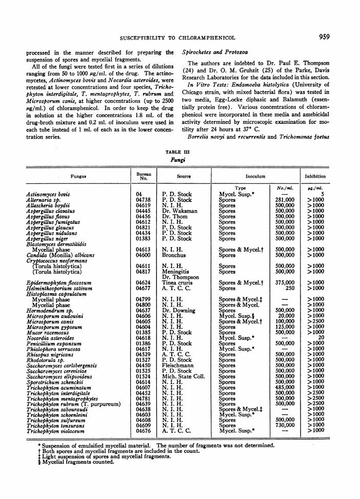
Fungi
Fungus Bureau Source | Inoculum Inhibition_________________________________________________________ No. iI
P. D. StockP. D. StockN. I. H.Dr. WaksmanDr. ThomN. I. H.P. D. StockP.: D. StockP. D. Stock
N. I. H.Bronchus
N. I. H.MeningitisDr. ThompsonTinea crurisA. T. C. C.
N. I. H.N. I. H.Dr. DowningN. I. H.N. I. H.N. I. H.P. D. StockN. I. H.P. D. StockN. I. H.A. T. C. C.P. D. StockFleischmannP. D. StockMich. State Coll.N. I. H.N. I. H.N. I. H.N. I. H.N. I. H.N. I. H.N. I. H.N. I. H.N. I. H.A. T. C. C.
* Suspension of emulsified mycelial material. The number of fragments was not determined.t Both spores and mycelial fragments are included in the count.t Light suspension of spores and mycelial fragments.§ Mycelial fragments counted.
I
I. W. MCLEAN, JR., J. L. SCHWAB, A. B. HILLEGAS, AND A. S. SCHLINGMAN
were suspended in a suitable menstruum containing vary-ing concentrations of chloramphenicol, as indicated inTable IV, and observed for immobilization. Observa-tions were made after two hours in the case of theBorreliae and after seven hours exposure for trichomonas.
In Vivo Tests: Plasmodium lophurae was tested inducks (intraperitoneal treatment) and chicks (oral treat-ment) and the results compared with the activity ofquinine.
Rats and dogs were used to evaluate the activity ofchloramphenicol against E. histolytica in vivo. Treat-ment was given orally and results expressed as numberof animals cleared of infection or degree of suppressionof infection.
B. novyi was tested in groups of mice treated by eitherthe intraperitoneal or oral route. Results are expressedas percentage suppression of spirochetemia.
Rabbits infected with Treponema pallidum were treatedwith various concentrations of chloramphenicol twicedaily for eight days. Rate of disappearance of trepo-
nemes and degree of healing of lesions under treatment
were noted. The animals were held to check for relapsesin the dosages at which healing under treatment was
noted.
RESULTS
Tables I to IV summarize the results obtainedwith the different micro-organisms that have beentested in the laboratory. Wherever possible theminimal inhibiting concentration of chloram-phenicol for the strain, under the conditions tested,is given. Otherwise, the maximum concentrationtested or the dose administered, in the case of insivo tests, is entered. The identifying numbersgiven for the bacteria and fungi are those assignedby the Parke, Davis and Company culture bureau,where a complete history of the strain is avail-able. Strains without assigned numbers are for themost part recent isolates from clinical cases priorto or during treatment with chloramphenicol.
The Emergence of Microbial Resistance
With the finding of considerable variation insusceptibility to chloramphenicol of different cul-tural lines of certain bacterial species, it became
TABLE IV
Spirochetes and protozoa
In vitro tests
Organism Medium or menstruum Time Conc. Result
0g./ml.Endamoeba histolytica * L. E. L. 24 hours 1000 NegativeEndamoeba histolytica * Balamuth 24 hours 250 SignificantBorrelia novyi * 50%horse serum 2 hours 10 to 50 ImmobilizationBorrdia recurrentis * 2.5% rat serum 2 hours 2.5 ImmobilizationTrichomonas foeus t 0.7% sodium chloride 7 hours 2000 NegativePelomyxa carolinensis Pace & Kimura buffer 48 hours 2500 NegativeTetrahymexa geleii 2%proteose peptone 48 hours 2500 Negative
5Thompson, P. E., Research Laboratories of Parke, Davis & Company, personal communication and (12).Reutner, T. F., Research Laboratories of Parke, Davis & Company and (12).Pace, D. M., and Russell, D., quoted (12).Gruhzit, 0. M., personal communication, Research Laboratories of Parke, Davis & Company.
I Quinine equivalent.
960
SUSCEPTIBILITY TO CHLORAMPHENICOL
TABLE V
Induced resistance to chloramphenicol in bacteria
P-D Transfers on Inhibiting conc. chloramphenicolOrganism Culture chloramphenicol pg./ml
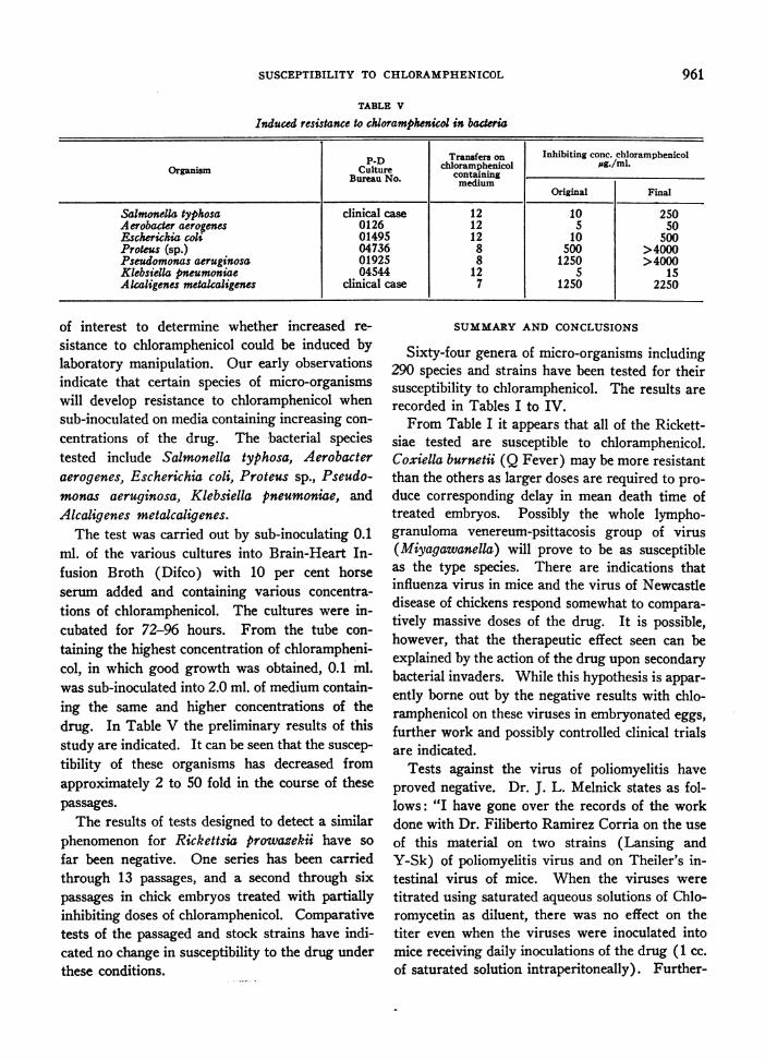
of interest to determine whether increased re-sistance to chloramphenicol could be induced bylaboratory manipulation. Our early observationsindicate that certain species of micro-organismswill develop resistance to chloramphenicol whensub-inoculated on media containing increasing con-centrations of the drug. The bacterial speciestested include Salmonella typhosa, Aerobacteraerogenes, Escherichia coli, Proteus sp., Pseudo-monas aeruginosa, Klebsiella pneumoniae, andAlcaligenes metalcaligenes.
The test was carried out by sub-inoculating 0.1ml. of the various cultures into Brain-Heart In-fusion Broth (Difco) with 10 per cent horseserum added and containing various concentra-tions of chloramphenicol. The cultures were in-cubated for 72-96 hours. From the tube con-taining the highest concentration of chlorampheni-col, in which good growth was obtained, 0.1 ml.was sub-inoculated into 2.0 ml. of medium contain-ing the same and higher concentrations of thedrug. In Table V the preliminary results of thisstudy are indicated. It can be seen that the suscep-tibility of these organisms has decreased fromapproximately 2 to 50 fold in the course of thesepassages.
The results of tests designed to detect a similarphenomenon for Rickettsia prowazekii have sofar been negative. One series has been carriedthrough 13 passages, and a second through sixpassages in chick embryos treated with partiallyinhibiting doses of chloramphenicol. Comparativetests of the passaged and stock strains have indi-cated no change in susceptibility to the drug underthese conditions.
SUMMARYAND CONCLUSIONS
Sixty-four genera of micro-organisms including290 species and strains have been tested for theirsusceptibility to chloramphenicol. The results arerecorded in Tables I to IV.
From Table I it appears that all of the Rickett-siae tested are susceptible to chloramphenicol.Coxiella burnetii (Q Fever) may be more resistantthan the others as larger doses are required to pro-duce corresponding delay in mean death time oftreated embryos. Possibly the whole lympho-granuloma venereum-psittacosis group of virus(Miyagawanella) will prove to be as susceptibleas the type species. There are indications thatinfluenza virus in mice and the virus of Newcastledisease of chickens respond somewhat to compara-tively massive doses of the drug. It is possible,however, that the therapeutic effect seen can beexplained by the action of the drug upon secondarybacterial invaders. While this hypothesis is appar-ently borne out by the negative results with chlo-ramphenicol on these viruses in embryonated eggs,further work and possibly controlled clinical trialsare indicated.
Tests against the virus of poliomyelitis haveproved negative. Dr. J. L. Melnick states as fol-lows: "I have gone over the records of the workdone with Dr. Filiberto Ramirez Corria on the useof this material on two strains (Lansing andY-Sk) of poliomyelitis virus and on Theiler's in-testinal virus of mice. When the viruses weretitrated using saturated aqueous solutions of Chlo-romycetin as diluent, there was no effect on thetiter even when the viruses were inoculated intomice receiving daily inoculations of the drug (1 cc.of saturated solution intraperitoneally). Further-
961
I. W. MCLEAN, JR., J. L. SCHWAB, A. B. HILLEGAS, AND A. S. SCHLINGMAN
more, daily oral administration of Chloromycetindid not appear to have any effect on the intestinalcarrier state of mice spontaneously harboringTheiler's virus of mouse encephalomyelitis" (22).
From Table II, which gives the results fromtests of over 200 bacterial- strains representing 25genera, it can be concluded that considerable vari-ation in susceptibility is encountered betweengenera as well as between species and even strainswithin the species. Under the conditions testedsusceptibility to concentrations of 10 pg/ml. orless, indicating possible clinical application, wasobserved for 21 of the genera. These includeAerobacter (2/2),4 Bacillus (6/6), Brucella(7/7), Corynebacterium (3/3), Diplococcus(3/3), Escherichia (11/11), Hemophilus (7/7),
Klebsiella (11/12), Micrococcus (19/19), Neis-seria (4/4), Pasteurella (8/8), Proteus (8/10),Pseudomonas (1/19), Salmonella (37/37), Sar-cina (3/3), Shigella (16/16), Streptococcus(8/8) and Vibrio (1/1). Pseudomonas, thoughfairly resistant, is included since it seems to besusceptible to chloramphenicol in concentrationsobtainable in the urine. The results given forAlcaligenes leave the status of this genus somewhatin doubt. A stock strain of A. faecalis was foundto be susceptible but three different isolates fromthe same clinical case of A. metalcaligenes infectionwere found to be highly resistant. A somewhatsimilar situation is observed for Proteus vulgariswhere two of ten strains tested required a some-what higher concentration (25 pg/ml.) for com-plete inhibition. An interesting finding was en-countered with the Clostridia. While this groupin general was of very low susceptibility, onestrain, the Mueller variant of Cl. tetani, was verysusceptible. This variant does not produce sporesin culture, which raises the speculation that pos-sibly the apparent resistance of this group is areflection of the resistance of the spores ratherthan of the vegetative forms.
While not within the scope of this paper, in vivolaboratory trials with certain of the bacterial agentshave been conducted. Gould et al. (26) reportencouraging results with the cholera vibrio in miceand Gruhzit (25) has found activity in mice
' Indicates number susceptible strains/number of strainstested.
against certain of the Gram-positive cocci and thesalmonella group. Sarber (27) has treated miceinfected with Hemophilus pertussis and foundchloramphenicol to be an effective chemothera-peutic agent. Clinical trials are in progressagainst many of these pathogens. To date clinicalconfirmation has been obtained in typhoid fever(19), brucellosis (20), and certain urinary infec-tions caused by Gram-negative species (21).
Of the forty species or strains of fungi tested forsusceptibility to chloramphenicol, Table III, onlytwo were completely inhibited by the drug at theconcentrations used in the test. These two spe-cies, Actinomyces bovis and Nocardia asteroides,both of which belong to the actinomycetes, wereinhibited by 5 ug/ml. and 20 pfg/ml. of the drug,respectively. Since blood levels of this magnitudeare easily attained, trial on clinical infections withthese agents is indicated.
Growth of four species of ringworm fungi,Trichophyton mentagrophytes, T. interdigitale, T.rubrum and Microsporum canis, was retarded by1000 ug/ml. of the drug but complete inhibitioncould not be obtained even when the drug concen-tration was increased to 2500 ,ug/ml.
With the possible exception of the spirocheteof relapsing fever (Borrelia recurrentis), the re-sults with the spirochetes and protozoa tested werenot promising. Doses of 12.5 mg/Kg. twice dailyfor eight days were ineffective on syphilitic lesionsin rabbits. Although similar treatment with 25 or50 mg/Kg. cleared the lesions, relapses occurredwhen treatment was stopped.
Attempts to induce increased resistance to chlo-ramphenicol by passage in the presence of thedrug have been successful in the case of sevenbacterial species, but not for R. prowazekii in theembryonated egg. It remains to be seen whether,under practical conditions in the clinic, this findingwill be of importance.
BIBLIOGRAPHY1. Ehrlich, J., Bartz, Q. R., Smith, R. M., Joslyn, D. A.,
and Burkholder, P. R., Chloromycetin, a new anti-biotic from a soil actinomycete. Science, 1947,106, 417.
2. Ehrlich, J., Gottlieb, D., Burkholder, P. R., Ander-son, L. E., and Pridham, T. G., Streptomycesvenezuelac n. sp. the source of Chloromycetin.J. Bact., 1948, 56, 467.
962
SUSCEPTIBILITY TO CHLORAMPHENICOL
3. Carter, H. E., Gottlieb, D., and Anderson, H. W.,Chloromycetin and streptothricin. Science, 1948,107, 113.
4. Gottlieb, D., Bhattacharyya, P. K., Anderson, H. W.,and Carter, H. E., Some properties of an anti-biotic obtained from a species of Streptomyces.J. Bact., 1948, 55, 409.
5. Bartz, Q. R., Isolation and characterization of Chlo-romycetin. J. Biol. Chem., 1948, 172, 445.
6. Bartz, Q. R., Isolation and chemistry of chloram-phenicol (Chloromycetin). J. Clin. Invest., 1949,28, 1051.
7. Controulis, J., Rebstock, M. C., and Crooks, H. M.,Jr., Chloramphenicol (Chloromycetin) v. synthe-sis. J. Am. Chem. Soc. In press.
8. Smadel, J. E., and Jackson, E. B., Chloromycetin, anantibiotic with chemotherapeutic activity in ex-perimental rickettsial and viral infections. Sci-ence, 1947, 106, 418.
9. Smadel, J. E., and Jackson, E. B., Effect of Chloro-mycetin on experimental infection with psittacosisand lymphogranuloma venereum viruses. Proc.Soc. Exp. Biol. & Med., 1948, 67, 478.
10. Smadel, J. E., Jackson, E. B., Ley, H. L., Jr., andLewthwaite, R., Comparison of synthetic and fer-mentation chloramphenicol (Chloromycetin) inrickettsial and viral infections. Proc. Soc. Exp.Biol. & Med., 1949, 70, 191.
11. Smadel, J. E., Jackson, E. B., and Cruise, A. B.,Chloromycetin in experimental rickettsial infec-tions. J. Immunol. In press.
12. Smith, R. M., Joslyn, D. A., Gruhzit, 0. M., McLean,I. W., Jr., Penner, M. A., and Ehrlich, J., Chloro-mycetin: biological studies. J. Bact., 1948, 55, 425.
13. Youmans, G. P., Youmans, A. S., and Osborne, R. R.,Tuberculostatic action of Chloromycetin in vitroand in vivo. Proc. Soc. Exp. Biol. & Med., 1948,67, 426.
14. Payne, E. H., Knaudt, J. A., and Palacios, S., Treat-ment of epidemic typhus with Chloromycetin. J.Trop. Med. & Hyg., 1948, 51, 68.
15. Smadel, J. E., Woodward, T. E., Ley, H. L., Jr.,Philip, C. B., Traub, R., Lewthwaite, R., andSavoor, S. R., Chloromycetin in the treatment ofscrub typhus. Science, 1948, 108, 160.
16. Smadel, J. E., Leon, A. P., Ley, H. L., Jr., andVarela, G., Chloromycetin in the treatment ofpatients with typhus fever. Proc. Soc. Exp. Biol.& Med., 1948, 68, 12.
17. Payne, E. H., Sharp, E. A., and Knaudt, J. A.,Treatment of epidemic typhus with Chloromycetin.Trans. Royal Soc. Trop. Med. Hyg., 1948, 42, 163.
18. Pincoffs, M. C., Guy, E. G., Lister, L. M., Wood-ward, T. E., and Smadel, J. E., The treatment ofRocky Mountain spotted fever with Chloromyce-tin. Ann. Int. Med., 1948, 29, 656.
19. Woodward, T. E., Smadel, J. E., Ley, H. E., Jr.,Green, R., and Mankikar, D. S., Preliminary re-port on the beneficial effect of Chloromycetin inthe treatment of typhoid fever. Ann. Int. Med.,1948, 29, 131.
20. Woodward, T. E., Smadel, J. E., Holbrook, W. A.,and Raby, W. R., The beneficial effect of Chloro-mycetin in brucellosis. J. Clin. Invest., 1949, 28,968.
21. Chittenden, G. E., Sharp, E. A., Glazko, A. J., andSchlingman, A. S., Chloromycetin in therapy ofbacillary urinary infection. J. Clin. Invest., 1949,28, 1052.
22. Melnick, J. L. Personal communication.23. Records, E., and Vawter, L. R., Tech. Bull. 173,
Univ. Nev. Agr. Exp. Sta. (June) 1945.24. Thompson, P. E. Personal communication.25. Gruhzit, 0. M. Personal communication.26. Gould, R. L., Schlingman, A. S., Jackson, E. B.,
Manning, M. C., Batson, H. C., and Campbell,C. C., Chloramphenicol (Chloromycetin) in ex-perimental cholera infections. J. Bact. In press.
27. Sarber, R. W., and Hemans, M. J., Chloramphenicol(Chloromycetin) in the chemotherapy of Hemo-philus Pertussis infection in mice. Presented at49th General Meeting Soc. American Bact., 1949,Cincinnati, Ohio.