SAND REPORT SAND2002-3870 Unlimited Release Printed December 2002 Switchable Hydrophobic-Hydrophilic Surfaces Bruce C. Bunker, Dale L. Huber, Michael S. Kent, Hyun Yim, John G. Curro, Ronald J. Manginell, James G. Kushmerick, Gabriel P. Lopez, and Sergio Mendez Prepared by Sandia National Laboratories Albuquerque, New Mexico 87185 and Livermore, California 94550 Sandia is a multiprogram laboratory operated by Sandia Corporation, a Lockheed Martin Company, for the United States Department of Energy’s National Nuclear Security Administration under Contract DE-AC04-94-AL85000. Approved for public release; further dissemination unlimited.
Transcript
SAND REPORTSAND2002-3870Unlimited ReleasePrinted December 2002
Switchable Hydrophobic-HydrophilicSurfaces
Bruce C. Bunker, Dale L. Huber, Michael S. Kent, Hyun Yim, John G. Curro, Ronald J.Manginell, James G. Kushmerick, Gabriel P. Lopez, and Sergio Mendez
Prepared bySandia National LaboratoriesAlbuquerque, New Mexico 87185 and Livermore, California 94550
Sandia is a multiprogram laboratory operated by Sandia Corporation,a Lockheed Martin Company, for the United States Department of Energy’sNational Nuclear Security Administration under Contract DE-AC04-94-AL85000.
Approved for public release; further dissemination unlimited.
Issued by Sandia National Laboratories, operated for the United States Department of Energy bySandia Corporation.
NOTICE: This report was prepared as an account of work sponsored by an agency of the UnitedStates Government. Neither the United States Government, nor any agency thereof, nor any oftheir employees, nor any of their contractors, subcontractors, or their employees, make anywarranty, express or implied, or assume any legal liability or responsibility for the accuracy,completeness, or usefulness of any information, apparatus, product, or process disclosed, orrepresent that its use would not infringe privately owned rights. Reference herein to any specificcommercial product, process, or service by trade name, trademark, manufacturer, or otherwise,does not necessarily constitute or imply its endorsement, recommendation, or favoring by theUnited States Government, any agency thereof, or any of their contractors or subcontractors. Theviews and opinions expressed herein do not necessarily state or reflect those of the United StatesGovernment, any agency thereof, or any of their contractors.
Printed in the United States of America. This report has been reproduced directly from the bestavailable copy.
Available to DOE and DOE contractors fromU.S. Department of EnergyOffice of Scientific and Technical InformationP.O. Box 62Oak Ridge, TN 37831
Bruce C. BunkerBiomolecular Materials and Interfaces
Dale L. HuberNanostructures and Advanced Materials Chemistry
Michael S. Kent and Hyun YimMicrosystems Materials, Tribology, and Technology
John G. CurroMaterials and Process Modeling
Ronald P. ManginellMicro-Total-Analytical Systems
Sandia National LaboratoriesP.O. Box 5800
Albuquerque, New Mexico 87185-1413
Gabriel P. Lopez and Sergio MendezUniversity of New Mexico
Albuquerque, New Mexico 87106
James G. KushmerickNaval Research Laboratory
Washington, DC 20375
4
Abstract
Tethered films of poly n-isopropylacrylamide (PNIPAM) films have beendeveloped as materials that can be used to switch the chemistry of a surface in responseto thermal activation. In water, PNIPAM exhibits a thermally-activated phase transitionthat is accompanied by significant changes in polymer volume, water contact angle, andprotein adsorption characteristics. New synthesis routes have been developed to preparePNIPAM films via in-situ polymerization on self-assembled monolayers. Swellingtransitions in tethered films have been characterized using a wide range of techniquesincluding surface plasmon resonance, attenuated total reflectance infrared spectroscopy,interfacial force microscopy, neutron reflectivity, and theoretical modeling. PNIPAMfilms have been deployed in integrated microfluidic systems. Switchable PNIPAM filmshave been investigated for a range of fluidic applications including fluid pumping viasurface energy switching and switchable protein traps for pre-concentrating andseparating proteins on microfluidic chips.
Acknowledgements
The authors would like to acknowledge the valuable support provided on theinterfacial force microscopy experiments by W. L. Smith and J. E. Houston and oninfrared experiments by Tim Dunbar.
5
ContentsPage
Introduction 6
Synthesis of Switchable Films 7
Characterization of Phase Transitions in Tethered Films 9Ellipsometry Results 9Surface Plasmon Resonance Experiments 10Infrared Results 10Interfacial Force Microscopy 11Neutron Reflection 12
Modeling of Polymer Phase Transitions 14
Switching of Surface Chemistry Using PNIPAM 15Hydrophilic-Hydrophobic Switching 15Switching of Protein Adsorption 16
Summary 18
Figure Captions 19
References 21
Figures
1. SPR resonance angle vs. temperature for PNIPAM film2. ATR-FTIR Spectra vs. Temperature for azo-initiated PNIPAM film3. IFM results vs. temperature for a “polymer brush” PNIPAM film4. Cloud point data for single PNIPAM chains dissolved in acetone5. NR data for grafted PNIPAM chains in D2O and d-acetone6. NR data for “polymer brush” PNIPAM film in D2O and d-acetone7. Spinodal curves for polymer-solvent mixture from PRISM calculations8. Density profiles from SCF calculations and NR experiments9. Advancing contact angle vs. temperature for azo-initiated PNIPAM film10. Advancing contact angle on PNIPAM vs. underlyingCOOH-CH3 content11. Human serum albumin adsorption vs. substrate functionalization12. UV-visible spectra of myoglobin desorption from PNIPAM films 13. Micro-hotplate device for reversible trapping of proteins14. UV-visible spectra of PNIPAM film exposed to albumin-myoglobin mix
6
Introduction
Extensive research is underway to develop microfluidic systems that can separate,purify, analyze, and deliver species such as biomolecules [1-3]. Applications for suchsystems include controlled drug delivery, proteomics, and the detection and analysis ofchemical and biological hazards. The ability to perform chemical and biological analyseson a microchip requires understanding how to transport, separate, and detect species innanoliter quantities of liquid confined in channels having dimensions of microns. Atsuch small length scales, surface effects play a dominant role. For example, although it isdifficult to force liquids through microchannels using pressure, it has been demonstratedthat fluids can be transported at cm/sec velocities by manipulating interfacial energies tocontrol whether channel walls attract or repel water [4]. Active control of interfacialenergies and exposed chemical functionality could also be used to control selectiveadsorption for advanced separations and sensors.
Most surfaces currently used in microfluidic systems are passive, having nointrinsic ability to modulate their interactions with fluids or dissolved species such asproteins. Interfacial interactions are often manipulated using self-assembled monolayers(SAMS) [5-7]. Surfaces can be made hydrophobic (or water repellant) by attachinglayers to surfaces that are terminated with simple hydrocarbons such as alkane chains. Ifthe alkane chains are terminated with different functional groups, such as hydroxylgroups or carboxylic acids, a hydrophilic (or wettable) surface results. Surfacemodifications can also control whether surfaces interact with solution species. Forexample, globular proteins tend to adsorb on hydrocarbon-terminated SAMS [6], whilepolyethylene oxide (PEO) termination results in an anti-fouling surface [5]. Whilepassive SAMS represent a powerful tool for surface modifications, our interest is in thedevelopment of active coatings that can be used to switch surface chemistry in responseto “on-chip” stimuli such as heat, light, or an applied voltage. The work described in thisreport represents the first such system that we have successfully synthesized,characterized, and deployed in a microfluidic system.
The active material forming the basis for our switchable surface is the polymerpoly (N-isopropylacrylamide) or PNIPAM [8-12]. In water, this polymer undergoes aphase transition at a lower critical solution temperature of 30-35oC. Below the phasetransition temperature (at room temperature), the polymer swells in water. Lightscattering studies conducted on polymer chains dissolved in water [11] (or tethered tolatex particles [12]) suggest that the swollen polymer occupies over ten times the volumeof the dry polymer, with a water content of 90 wt% (50 water molecules per NIPAMmonomer). Above the phase transition, the polymer collapses to a state having a muchlower volume (around 1.5 times that of dry polymer) and water content (25 wt% or 2water molecules per PNIPAM monomer). The net volume change in water is around afactor of 8-10 (or 2-3 in any given direction). The swollen polymer is hydrophilic.Above the transition temperature, the polymer surface becomes more hydrophobic. Forbulk gels, the contact angle for water can increase from 40o to as high as 90o in goingfrom the swollen to the collapsed state [10]. Since a change in contact angle of only 5-10o is sufficient to pump liquids through microchannels [4], PNIPAM was selected forstudy as a potential surface modification to promote thermally-programmable active
7
transport in microfluidic systems. In addition, extensive research on bio-fouling hasshown that water-soluble proteins tend to be repelled by hydrophilic surfaces, while suchproteins stick to hydrophobic surfaces [13]. Consistent with this trend, it has beendemonstrated that PNIPAM-functionalized particles and bulk hydrogels interact morestrongly with proteins above the LCST than below it [14,15]. Tethered PNIPAM couldpotentially activate a surface to grab or release proteins depending on whether thetemperature is above or below the transition temperature.
The ability to develop switchable films based on PNIPAM requires that wedevelop an in-depth understanding of polymeric phase transitions at interfaces. Whilefilm functions will be discussed, the major focus of this report is on the materials scienceissues associated with switchable polymer films. Facets of the materials research to bereported here include: 1) Synthesis of Switchable Films – The phase transition inPNIPAM could be sensitive to parameters such as molecular weight, grafting density,and crosslinking. A range of surface functionalization routes have been developed tovary some of these parameters to determine how material architectures influence filmperformance characteristics. 2) Characterization of Phase Transitions in Tethered Films –Once films are produced, we need to determine whether the tethered polymer isundergoing the desired phase transition and whether this transition induces desiredchanges in surface chemistry. Techniques used to characterize the tethered films includeellipsometry, surface plasmon resonance experiments, infrared spectroscopy, contactangle measurements, interfacial force microscopy, and neutron reflectivity. 3) Modelingof Polymer Phase Transitions – Monte Carlo and molecular dynamics simulations havebeen performed on both dissolved and tethered polymers to understand what factorscontrol the phase transition and to rationalize some of the experimental results. 4)Switching of Surface Chemistry Using PNIPAM – The wetting and protein adsorptionbehavior of tethered PNIPAM films have been investigated as a function of temperature.Films have been integrated into a micro-hotplate device and incorporated into amicrofluidic system. Experiments show that the integrated device can function as areversible protein trap in microfluidic systems. Below, each facet of the research isdescribed in greater detail.
Synthesis of Switchable Films
PNIPAM films must meet a stringent set of requirements for applications inmicrofluidic systems. For all applications, we must insure that the tethered polymer is ina configuration that supports a reversible thermally-activated phase transition. The filmsmust also be robust and strongly attached to the surface. However, surface interactionsmust not be so strong that they inhibit the desired transition. For applications involvingthe grabbing and release of proteins, the films must have an architecture that allows forrapid and reversible adsorption and desorption kinetics. In bulk gels, protein adsorptiontends to be irreversible [8], reflecting steric entrapment of the proteins within a tortuous,high molecular weight network. Due to uncertainties in desired grafting densities,molecular weights, and surface tethering strategies, we investigated a range of differentmethods for synthesizing thin PNIPAM films.
The simplest method for attaching PNIPAM surface involves grafting pre-polymerized chains to a surface that has been functionalized with a self-assembled
8
monolayer [16]. In this investigation, we prepared mixed self-assembled monolayers(SAMS) containing a range of relative concentrations of methyl and hydroxyl endgroups. Carboxylic acid terminated PNIPAM molecules with molecular weights rangingfrom 10,000 to 220,000 were then grafted onto the surface hydroxyl groups to formcovalent ester linkages. Unfortunately, ellipsometry and neutron reflectivitymeasurements showed that this grafting method is relatively inefficient, resulting ingrafting densities that appear to be too low for meaningful characterization or forpractical applications. For grafted chains having molecular weights from 10,000 to33,000 and –OH fractions in the underlying SAM (available for grafting) ranging from5% to 80%, no detectable differences in neutron reflectivity data were observed fortemperatures ranging from 20oC to 55oC. PNIPAM was barely detected above the SAMbackground. This means that grafting densities were low and/or that the PNIPAM wasswollen to such an extent that it was not detected. The only grafted films that weredetectable had a molecular weight of 200,000, and were more likely physisorbed thangrafted (see Neutron Reflection Data below).
A more sophisticated method for preparing PNIPAM films involved the in-situsynthesis of PNIPAM on substrates using free radical polymerization of n-isopropylacrylamide (NIPAM) [17,18]. In this route, an azo-initiator that can beactivated to produce free radicals was used as the reactive end group on a self-assembledmonolayer. The monolayers were tethered to oxide-terminated surfaces using silanecoupling agents. On gold surfaces, the azo-initiator was attached to carboxylic acidgroups in mixed SAMS with various concentrations of –COOH in a methyl-terminatedbackground. Exposure of the tethered initiator to heat or light promoted cleavage of theazo linkage to create free radicals at the surface. When in contact with a solutioncontaining NIPAM monomer, a surface-initiated free radical chain reaction took place.Films produced by the method (for the case of silane tethered material) werecharacterized using a combination of ellipsometry, light scattering, infrared spectroscopy,and the interfacial force microscope. Typical films were found to have high single chainmolecular weights (around 107 as determined by light scattering measurements ondetached chains) and low grafting densities (on the order of 4 x 10¹⁰ chains/cm2 based onellipsometric film thickness and known molecular weight), yielding chain-chainseparations of around 50 nm. Low grafting densities are produced because radicalswithin the monolayer tend to react with neighboring initiators before they have a chanceto react with NIPAM in solution.
In order to produce films with a high graft density, we developed an alternate in-situ polymerization scheme based on the use of a chain-transfer reaction on SAM chainsterminated with thiol groups [19]. Here, free radicals are produced in solution usingthermal activation of initiators such as azobis(isobutyronitrile) or AIBN) and aretransferred to the tethered thiols. In contrast to the conventional polymerization process,where chain transfer between surface bound initiators yields a radical and a dead initiator,chain transfer between thiol groups exchanges a radical for a hydrogen atom, yielding aradical and a regenerated thiol. High concentrations of surface radicals can therefore bemaintained, allowing for higher grafting densities. With tethered thiols, we can producefilms with grafting densities ranging from 1011 –1013 chains/cm2 (chains are separated by2-6 nm) with molecular weights ranging from 104-105. As will be shown below, this highdensity “polymer brush” configuration is critical for reversible protein traps.
9
A final synthesis method investigated involved the use of atom transfer radicalpolymerization (ATRP) from mixed SAM surfaces [20]. This method is a livingpolymerization process involving a metal complex that supports the existence of freeradicals on the end of the growing polymer. The living polymerization method isreported to form polymers with a much narrower polydispersity than can be achieved byother in-situ polymerization methods. Because it is a living polymerization, we cancontrol the polymer film thickness by varying the polymerization time. As with other in-situ methods, the polymer surface coverage can be controlled by varying the compositionof the mixed SAM. Although ATRP films have been produced, such films have yet to becharacterized. Below, we describe films produced via the other methods, which aredenoted as grafted polymers, azo-initated polymers, and “polymer brushes” (for thechain-transfer films).
Characterization of Phase Transitions in Tethered Films
Ellipsometry Results
For bulk gels, the phase transition in PNIPAM can be monitored by detecting thevolume change that accompanies the thermally-induced swelling or deswelling of thematerial. While detecting the order of magnitude shrinkage in bulk PNIPAM gels (afactor of two in any given direction) is easy, detecting volume changes for a thin (4-8 nmthick) PNIPAM film under water is much more difficult. The “standard” method formeasuring film thickness is ellipsometry. In ellipsometry, a plane-polarized light beam isreflected off of a PNIPAM-coated substrate. By measuring the reflection coefficients ofthe s- and p-polarization components of the reflected beam (which is ellipticallypolarized), the optical thickness (a convolution of actual thickness and index ofrefraction) is obtained. If the index of refraction is known, the film thickness can bedetermined. Assuming that n = 1.45 for bulk PNIPAM, we have been able to determinefilm thicknesses for materials prepared using all of the synthesis techniques describedabove. Optical thicknesses vary from sample to sample, but typically fall within therange of 4-8 nm. However, it is important to note that the optical thickness is calculatedassuming that the PNIPAM films are fully dense. As this assumption is rarely valid, theellipsometer really provides an estimate of the total mass deposited on the surface. Whenthe grafting density is low, actual film thickness can be much greater than the opticalthicknesses indicated by ellipsometry (see below).
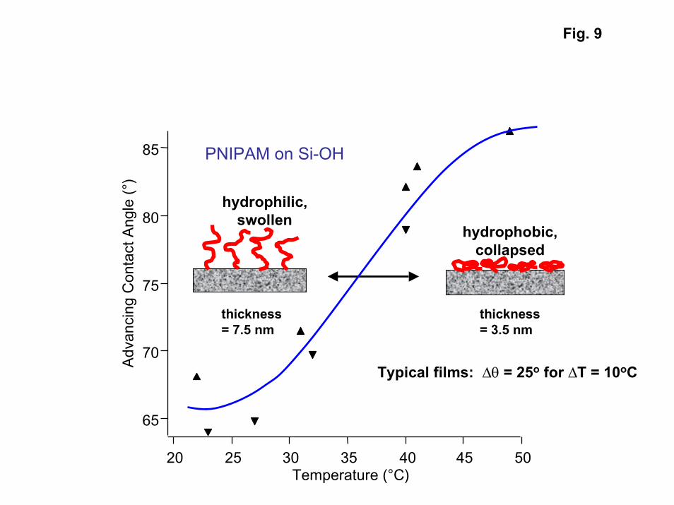
While ellipsometry measurement in air can be used to measure films that are onlya few nanometers thick, it has been difficult to make in-situ measurements of filmthickness under water. This is because the refractive indices of water (n = 1.33) andPNIPAM (n = 1.45) are similar. To try to observe the lower critical solution temperaturefor PNIPAM using our ellipsometry equipment, we were forced to perform ex-situellipsometry measurements. Samples were immersed in water at various temperatures,removed from solution, quickly blown dry, and placed in the ellipsometer. Using thistechnique, we found that a typical “polymer brush” film had an effective thickness of 7.5nm in its swollen state at room temperature. The same film had an effective thickness of3.5 nm at 50oC, indicating that the tethered PNIPAM film undergoes the expected factorof two change in volume on either side of the phase transition. However, it is hard to
10
blow-dry samples in such as way as to ensure that all surface water is eliminated withoutremoving water entrapped in the underlying PNIPAM gel. For this reason, all othermethods we have used to follow the phase transition have involved in-situ techniques.
Surface Plasmon Resonance Experiments
Surface plasmon resonance (SPR) techniques [21]were used to monitor the phasetransition in PNIPAM under water. In SPR, a light beam impinges on the “dry” side of athin gold layer that is functionalized on the “solution” side with PNIPAM. Most of thelight is reflected. However, a small part of the light propagates along the interfacebetween the metal and any dielectrics on the surface as a surface plasmon. The surfaceplasmon is an evanescent wave that decays exponentially into the dielectric. The fractionof light in the surface plasmon depends on the angle of incidence and the effective opticalthickness of the dielectric medium. In the SPR experiment, we monitor the angle ofincidence and look for the minimum in the reflected light intensity corresponding to theresonant condition required to produce the surface plasmon. The resonant angle is relatedto the dielectric constant of the region probed by the evanescent wave. For the PNIPAMfilms studied here, the effective dielectric constant depends on the dielectric constants ofthe PNIPAM, the water, and the distribution of the two materials as a function of distancefrom the interface.
Changes in the SPR resonance angle as a function of temperature are shown inFig. 1 [22]. The pronounced increase in resonance angle with temperature is consistentwith the collapse of a PNIPAM film as it goes through the transition. PNIPAM has ahigher dielectric constant than water. Collapse of the film would move the PNIPAMcloser to the surface, increasing the amount of higher dielectric constant material close tothe interface (in a region where the evanescent wave is more intense), and increasing theresonant angle. To date, we have not been able to quantitatively model the SPR data, assuch an analysis requires input regarding the effective dielectric constant as a function ofPNIPAM concentration, and simplifying assumptions involving how the PNIPAM isdistributed relative to position within the evanescent wave. However, qualitatively theresults are consistent with the collapse of a PNIPAM film that is on the order of 100 nmthick in room temperature water. The temperature dependence of the results indicatesthat while the transition is complete at 32oC (near the expected transition temperature),significant volume changes occur at temperatures well below the transition temperature.(Similar volume changes are apparent in light scattering experiments for single chains[11].) The SPR data indicate that a phase transition does occur in tethered films, but thatthe transition is not as sharp as the 5-10oC reported for bulk gels.
Infrared Results
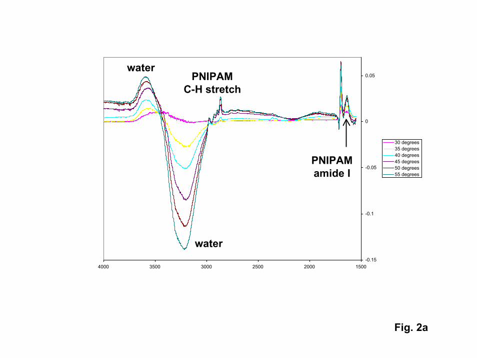
Infrared (IR) spectroscopy can be used to detect the vibrations of chemical bondswithin thin PNIPAM films. In simple reflectance or transmission modes, IR spectra canbe used to identify the chemical nature of the films and to detect the total quantity ofmaterial deposited. To monitor the swelling transition, we have used attenuated totalreflectance IR (ATR-IR). In ATR-IR, the PNIPAM film is grown on a Si prism. The IRbeam enters the prism and is reflected between prism surfaces multiple times. When the
11
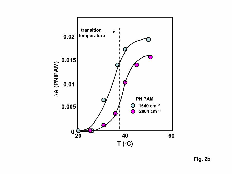
beam encounters the solution side of the interface, the IR beam penetrates the solutionwith an evanescent wave that exponentially decays with distance from the interface(analogous to the evanescent wave in the SPR technique described above). Althoughwater is an extremely good adsorber of IR radiation, precluding most in-situ IRmeasurements, in-situ experiments can be performed with ATR because the IR beaminteracts with less than a micron-thick layer of water near the surface (the calculatedpenetration depth into the water is 200 nm for 3600 cm-1 light). In contrast to SPR, theintensity of bands associated with PNIPAM vibrations depends only on how muchPNIPAM is present as a function of distance from the solution interface. If the PNIPAMfilm collapses, more PNIPAM is closer to the surface, increasing the intensity of allPNIPAM bands.
ATR-FTIR spectra of high molecular weight PNIPAM films produced via thestandard tethered azo initiator are shown in Fig. 2. As expected, the intensities of allPNIPAM bands increase with temperature as the transition temperature is approached,reaching constant values at high temperature. The transition temperature inferred for thisfilm (35oC) is slightly higher than that inferred from the gold-tethered film examined inthe SPR experiments. If one assumes that the tethered film collapses by a factor ofaround three above the transition temperature (consistent with the change inhydrodynamic radius (Rh)) measured for single chains in solution [11]), the observedchanges in IR band intensities are consistent with a PNIPAM film that extends around360 nm from the surface and collapses to around 120 nm.
Similar experiments were conducted on the “polymer brush” films produced viathe chain-transfer synthesis route. No phase transition was apparent in the ATR resultsfor this film (the IR intensities did not change appreciably with temperature). Webelieve that no transition was observed for the brush film because the film was muchthinner than the azo-initiated films. For thin films, the swelling transition should notmove the PNIPAM a sufficient distance relative to the decaying evanescent wave tocause appreciable intensity changes. However, an alternate explanation is that the tighterpacking of chains in the “brush” film is inhibiting the transition. To distinguish betweenthese two possibilities, we probed the thickness of a range of PNIPAM films both belowand above the phase transition using the interfacial force microscope.
Interfacial Force Microscopy
The swelling, collapse, and adhesive qualities of tethered PNIPAM films havebeen monitored as a function of temperature using an interfacial force microscope (IFM).The IFM is a scanning probe system developed by Jack Houston at Sandia NationalLaboratories to monitor the interactions between functionalized probe tips and substratesurfaces as a function of separation distance with a distance resolution of a few angstroms[23]. The IFM provides force profiles that are similar to those reported for PNIPAMfilms using the atomic force microscope (AFM) [16] and the surface forces apparatus(SFA) [24]. In experiments reported here, Si or fused silica substrates werefunctionalized with tethered PNIPAM films. The scanning probe tip was a fused silicarod drawn down and fire polished to produce a tip with a radius that is typically 1-5 �m.Experiments were conducted with bare tips (covered with surface –OH groups), or withtips coated with octadecyltrichlorosilane (-CH3 terminated). By moving the IFM tip in a
12
direction normal to the surface, forces experienced between the tip and the substrate weremeasured as a function of separation distance. As the tip approached the surface inwater, the sequence of events observed for typical tip-substrate interactions was from nointeraction at large distances, to detection of through-liquid forces (such as electricaldouble layer interactions and hydration forces), to measurements of the forces required todeform materials on the tip and substrate as the tip and substrate come into direct contact.The tip position was measured relative to a “zero separation distance” point at which themeasured forces are dominated by the mechanical deformation of the Si substrate and thefused silica tip. The IFM tip was also dithered parallel to the substrate to measurefrictional forces.
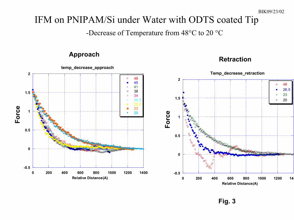
The IFM was used to examine both “polymer brush” films and high molecularweight films produced via azo-initiators. Experiments were performed as a function oftemperature (both increasing and decreasing) in deionized water. General features of theresulting force profiles are shown in Fig. 3. At room temperature, a long range repulsiveforce was detected as the tip approached the “polymer brush” film at a distance of around100 nm. The repulsion was similar regardless of tip functionalization, indicating that itwas not due to the presence of an electrical double layer. The repulsion did not dependon tip speed, indicating that it was not a repulsive hydration force arising from thepresence of an ordered water layer near the surface. On the azo-initiated PNIPAM filmwith a molecular weight of 107, the repulsion was detected at a distance of 320 nm, whichcorresponds almost exactly with the hydrodynamic radius of single PNIPAM chains atroom temperature [11]. We believe that the observed repulsion is due to physical contactbetween the tip and hydrated PNIPAM chains on the substrate and subsequentdeformation of the PNIPAM film by the tip. As the temperature was increased, there wasno apparent change in this repulsion until the temperature reached between 25-28.5oC.At this point, there was an abrupt change in the repulsion, which collapsed toward thesurface by about a factor of two and became stronger (as evidenced by a steeper slope).However, after the abrupt change, the repulsion stayed roughly constant up to at least50oC. We believe that the collapse of the repulsive force reflects the collapse of thePNIPAM film.
As the tip is retracted from the surface above the transition temperature, thePNIPAM film is attracted to the tip (the measured force drops below zero), indicatingthat the PNIPAM is undergoing adhesive interactions with materials it comes into contactwith. However, these adhesive interactions disappear below the transition temperature,and the PNIPAM surface repels all functionalized tips on retraction as well as onapproach. All of these finding are consistent with force profiles obtained elsewhere onPNIPAM films using an atomic force microscope [16] and a surfaces forces apparatus[24]. The IFM experiments suggest that materials such as dissolved proteins should berepelled by PNIPAM surfaces in water below the transition temperature, but can stick tothe PNIPAM above the transition. The IFM force profiles not only indicate that“polymer brush” films undergo a sharp phase transition, but that the transition might beuseful for applications such as a reversible protein trap.
Neutron Reflection
13
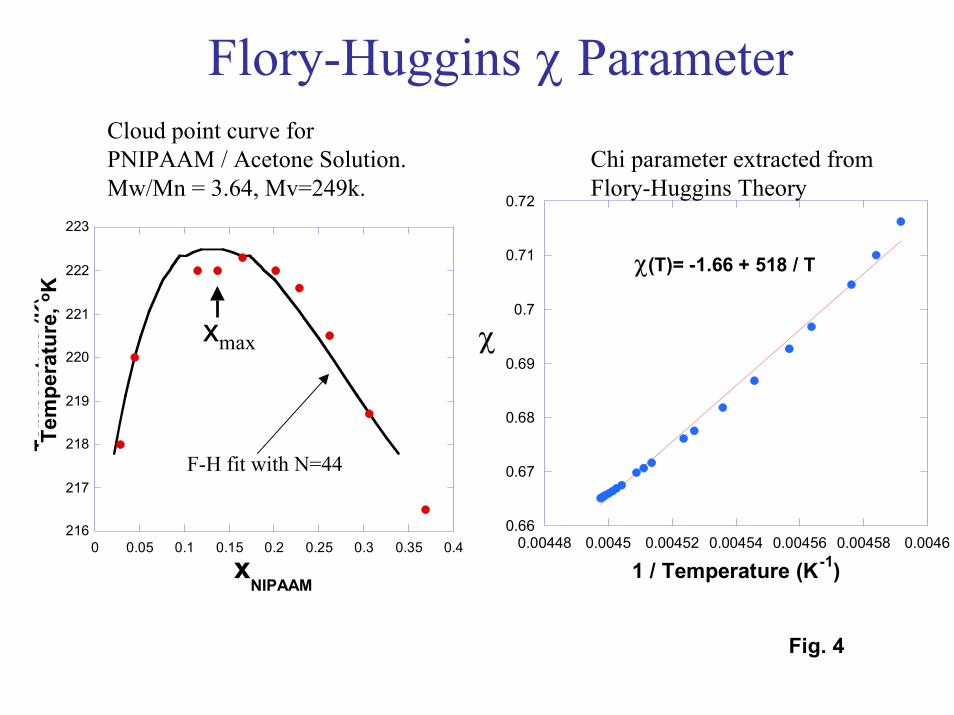
A final technique investigated for observing the phase transition in thinPNIPAM films was neutron reflection. Neutron reflectivity (NR) is a technique that canprovide density profiles for thin polymer films in the presence of deuterated solvents[25]. The solvents investigated in the NR experiments were D2O and d-acetone. Acetoneis of interest because experimental cloud point data (Fig. 4) show that PNIPAM exhibitsan upper critical solution temperature (UCST) in acetone. In contrast to its behavior inwater, PNIPAM films should be insoluble and collapsed below the phase transitiontemperature in acetone, and swollen (or soluble) above the transition temperature.However, since the transition temperature in acetone is well below room temperature,PNIPAM should be highly swollen in acetone at room temperature just as it is in ambientwater.
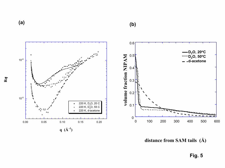
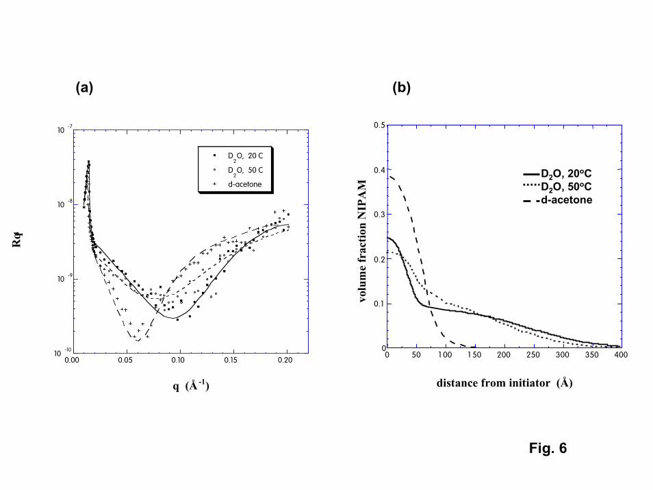
NR was able to detect the presence of PNIPAM for both the highest molecularweight (220,000) grafted samples as well as for “polymer brush” films (Fig. 5,6) [26]. Inacetone (at 20oC), both films exhibited a smoothly decaying single profile. The apparentvolume fractions occupied by PNIPAM at the immediate surface were 0.28 and 0.39 forthe grafted and polymer brush films, respectively. The effective thicknesses for thePNIPAM films (position where the density drops to half of its value at the immediatesurface) were 10 nm and 7.5 nm for the grafted and polymer brush films, respectively.For the polymer brush film, the NR thickness determined in acetone is an order ofmagnitude less than the thickness inferred from the IFM results described above. TheNR data suggest that although PNIPAM films swell in acetone, the extent of swelling isless than that reported for PNIPAM in cold water (swollen volume/dry volume = 2.6 and1.5 for the grafted and brush films, respectively, compared to 10 for PNIPAM dissolvedin water). The limited swelling may reflect the fact that attachment to the surface isinhibiting the ability of PNIPAM to expand, particularly at the high chain densitiesexpected for the polymer brush film. The swelling may also be influenced by attractiveinteractions between the polymer segments and the surface, particularly for the graftedchains. Model fits to the NR density profiles for PNIPAM in acetone suggest that suchsurface interactions are significant (see Modeling of Polymer Phase Transitions).
Although NR data have been obtained in water both below and above thetransition temperature (at 20oC and 55oC), the density profiles inferred from the NR dataare not consistent with any experimental or modeling results obtained on this project orreported in the literature (including the NR results in acetone). In water, bilayer profileswere required to fit the NR data for both film types. The NR results suggest that there isa thin layer of high PNIPAM concentration next to the immediate surface. Above thislayer is a second layer of much lower concentration that extends well into the water.Although each layer exhibits reversible changes with temperature, the changes are minor.Neither layer exhibits density changes that are consistent with a robust phase transition.The NR data suggest that for both grafted and polymer brush films, a relatively denselayer of PNIPAM exists at the immediate surface that does not swell in water at anytemperature. While polymerization from the surface in the “polymer brush” film couldgenerate chain densities near the surface that would be sufficient to inhibit swelling, it isunclear why this dense layer should be seen in room temperature water but not in roomtemperature acetone. (Perhaps chain segments are strongly physisorbed to the surface inwater but not in acetone.) The presence of a diffuse outer layer of PNIPAM couldaccount for the discrepancy in apparent film thickness between the NR and IFM results.
14
For the “polymer brush” film, the expected polydispersity in the tethered film could allowfor the presence of a less densely-packed outer layer whose swelling and collapse isapparent in the IFM data. However, it is unclear why there is no NR evidence for arobust phase transition in this outer layer unless the volume fraction in the outer zone issimply too low to generate meaningful NR results.
Modeling of Polymer Phase Transitions
While the experiments described above indicate that tethered PNIPAM exhibits alower critical solution temperature similar to that observed in bulk solutions, the varioustechniques yield different results regarding details such as the transition temperature, thesharpness of the transition, and the volume fraction occupied by PNIPAM in the swollenand collapsed phases. To develop further insight regarding the nature of polymerswelling transitions, we have developed computational models that allow us to study thephase behavior of bulk polymers as well as tethered chains immersed in a solvent.
The first phase of the modeling effort involved simulating the behavior of simplepolymers immersed in a simple solvent [27]. In the model, the polymer was representedby “strings of pearls” consisting of identical spherical units. The solvent was representedusing isolated spheres having the same diameter as the “monomers” in the polymer chain.All sites were allowed to interact with each other via repulsive Lennard-Jonesinteractions within a self-consistent polymer reference interaction site model, or PRISM,theory. PRISM is a liquid state theory in which generalized Ornstein-Zernike integralequations are solved numerically in a self-consistent manner to obtain the chainintermolecular structure and intermolecular radial distribution functions. Wth PRISM,we were able to explicitly include discrete solvent molecules. By using the random phaseapproximation together with direct correlation functions (the output of PRISM theory),we were able to estimate the phase diagram for a given polymer solution.
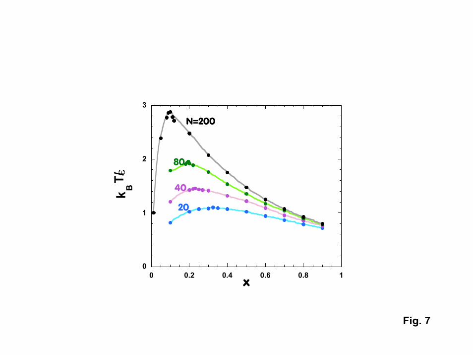
To date, PRISM theory has been used to estimate the spinodal curves for“normal” polymers in “normal” solvents (representative of PNIPAM in acetone). Forthe case where solvent-polymer interactions (�sp) are much less attractive than polymer-polymer (�pp) and solvent-solvent interactions (�ss) (�ss = �pp in the calculations), polymer-solvent mixtures exhibit an upper critical solution temperature (UCST) as predicted byFlory-Huggins theory. The polymer and solvent are immiscible at low temperature, withswelling or dissolution occurring above the spinodal decomposition temperature. Thetransition temperature Ts increases with the strength of polymer-polymer attraction andwith the molecular weight of the polymer (both of which make the polymer less soluble).Ts goes through a maximum as the mole fraction of polymer is increased, with themaximum moving to lower polymer mole fractions as the molecular weight increases.All of the above phenomena are in qualitative agreement with Flory-Huggins theory.However, PRISM calculations suggest that similar behavior should be observed evenwhen polymer-solvent interactions become significant. Fig. 7 shows the phase diagramcalculated for the case where � = �sp = �ss = �pp. Here, Flory-Huggins theory wouldpredict that such a mixture would be completely miscible. The fact that UCST behavioris still observed is driven by nonrandom mixing and compressibility effects that areincluded in PRISM, but are neglected in Flory Huggins theory (which treats the solventas a continuum rather than as discrete molecules).
15
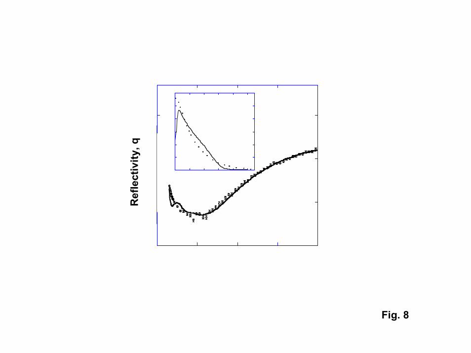
Tethered polymer brushes were modeled with self-consistent field (SCF) theory[28]. Here we simplified the problem of many tethered polymer chains surrounded bysolvent molecules to a problem of a single polymer that interacts with the surface througha potential field that must be solved for in a self-consistent manner with site volumefraction profiles. Our approach for modeling the phase behavior of the tethered brusheswas to determine the temperature-dependent � interaction parameter with Flory-Hugginstheory. The � parameter was extracted from experimental cloud point curves for polymersolutions of interest (Fig. 4). With SCF theory, we were able to generate polymer sitedensity profiles that could be compared directly to the experimental neutron reflectiondata (Fig. 8). In the calculated plot, the only parameter that was adjusted was theinteraction parameter between the polymer sites and the surface. The experimental andcalculated curves are in agreement except for a pronounced feature at low wave vector (q= 0.02 A-1). This feature disappears when one accounts for the polydispersity of thegrafted chains. Density profiles predicted by SCF theory are also in good agreement withthe profiles extracted from NR data. The SCF results indicate that it should now bepossible to model phase transitions for tethered polymers as the interactions betweencritical components (the polymer, solvent, and surface) are varied, aiding in thedevelopment of responsive films.
Switching of Surface Chemistry Using PNIPAM
Although the volume change associated with the phase transition in PNIPAMcould be of use for physical actuation of valve in microfluidic devices, the fluidicapplications of interest in this proposal rely on the use of the phase transition to modifythe chemistry of the surface. Below, we describe experiments that have been conductedto determine how the transition can be used to modify interfacial energies andinteractions with solution species such as proteins.
Water contact angle measurements provide a convenient measure of interfacialenergies as well as providing direct information regarding how hydrophilic orhydrophobic a surface is. Water contact angle measurements provide a direct measure ofthe forces that can be exploited for moving droplets in microfluidic systems based onactive switching of interfacial energies with a tethered PNIPAM film. The expressionrelating the velocity at which gradients in interfacial energy can be be used to pumpfluids through microchannels is given by [4]:
v = droplet velocity = (h/6�L[(�cos�)a – (�cos�)r)
where h is the height of the fluid channel, � is the viscosity of the droplet, L is the lengthof the droplet, � is the surface tension of the liquid, ��is the solid-liquid contact angle,and the subscripts a and r refer to the advancing and trailing sides of the droplet. Eq. 1suggests that for water, a difference in contact angle of as little as 10o should be sufficientto move droplets at sufficient velocities to be of interest for microfluidic systems.
16
Advancing contact angles on a pure azo-initiated PNIPAM film (Fig. 9) show thatthe thermally-activated phase transition induces pronounced changes in interfacialenergies. For tethered films, the contact angle typically increases by around 25o (from60o to 85o) as the temperature is increased from room temperature to above the transitiontemperature. The transition temperature inferred from the contact angle data is around35oC. Both the transition temperature and magnitude of the contact angle change areconsistent with values reported for PNIPAM films in the literature [10].
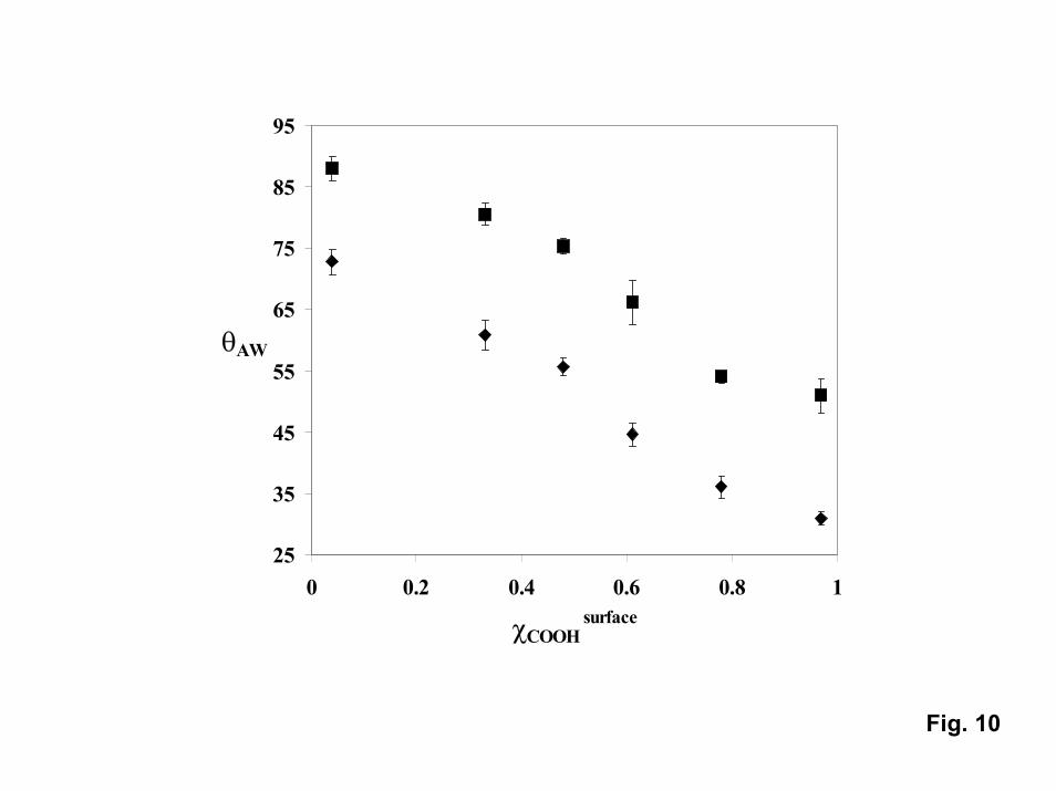
Not only can PNIPAM be used to switch contact angles, it can be used as acomponent in mixed monolayers to tune the contact angle [29]. In mixed-monolayerscontaining CH3 and COOH only (no PNIPAM), the advancing contact angle can be tunedfrom around 115o (pure CH3) down to around 30o (pure COOH). Once PNIPAM isgrown on mixed films by attaching azo-initiators to the COOH fraction, the contact anglemeasured for the “hydrophobic” state at 45�C exhibits a similar trend with CH3 molefraction (Fig. 10). However, in the presence of PNIPAM, the contact angle can beswitched from the “baseline” value to a value that is around 20o lower by lowering thetemperature to below the transition temperature (to 25oC) regardless of the COOH molefraction. The ability to both tune and modulate contact angles could be important for awide range of fluidic applications.
The significant changes in advancing contact angle observed for both pure andmixed monolayers containing PNIPAM were initially promising with regard toapplications involving the transport of liquids by switching interfacial energies.Unfortunately, all PNIPAM films exhibit a large hysteresis between the advancing(measured as the film is pushed into water) and receding (measured as the wet film isretracted from water) contact angles. For a pure PNIPAM film, measured values for thereceding contact angle are low (around 40o) and are independent of temperature. As thedriving force for propelling fluids in microchannels is based on the difference betweenthe advancing contact angle on the “hydrophilic” leading edge of a droplet and thereceding contact angle on the “hydrophobic” trailing edge, this contact angle hysteresisrepresents a serious concern for fluidics applications. Our speculation is that thishysteresis arises because there is a low barrier to rotation for functional groups at the endof the PNIPAM chains adjacent to the solution. In air, hydrophobic isopropyl groups arepresent at the surface, resulting in a surface that is relatively hydrophobic. However, inwater the lowest energy configuration is one in which more polar functional groups (suchas carbonyl groups) rotate to face the solution, while the isopropyl groups move awayfrom the immediate interface, dramatically lowering the contact angle. Strategies forminimizing the hysteresis (particularly above the transition temperature) could involvethe introduction of chain-end modifications (such as modified alkenes) to minimize freerotations at chain ends. Such modifications were not pursued because it was discoveredthat unmodified PNIPAM films exhibit the ability to switch interactions with solutionspecies such as proteins, opening up a completely new range of applications for PNIPAMin fluidic devices.
Switching of Protein Adsorption
Extensive research on biofouling has shown that water soluble proteins tend to berepelled by hydrophilic surfaces, while such proteins stick to hydrophobic surfaces [7].
17
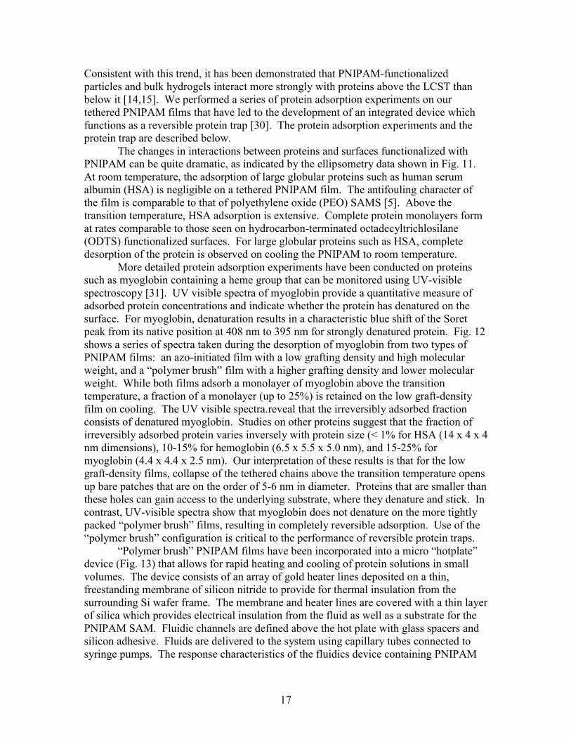
Consistent with this trend, it has been demonstrated that PNIPAM-functionalizedparticles and bulk hydrogels interact more strongly with proteins above the LCST thanbelow it [14,15]. We performed a series of protein adsorption experiments on ourtethered PNIPAM films that have led to the development of an integrated device whichfunctions as a reversible protein trap [30]. The protein adsorption experiments and theprotein trap are described below.
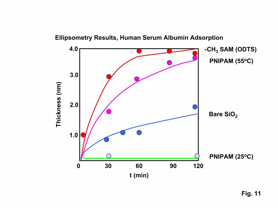
The changes in interactions between proteins and surfaces functionalized withPNIPAM can be quite dramatic, as indicated by the ellipsometry data shown in Fig. 11.At room temperature, the adsorption of large globular proteins such as human serumalbumin (HSA) is negligible on a tethered PNIPAM film. The antifouling character ofthe film is comparable to that of polyethylene oxide (PEO) SAMS [5]. Above thetransition temperature, HSA adsorption is extensive. Complete protein monolayers format rates comparable to those seen on hydrocarbon-terminated octadecyltrichlosilane(ODTS) functionalized surfaces. For large globular proteins such as HSA, completedesorption of the protein is observed on cooling the PNIPAM to room temperature.
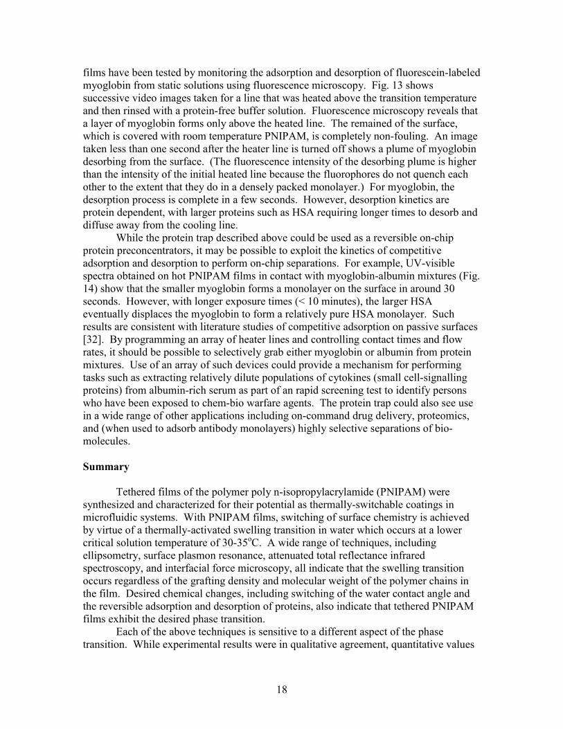
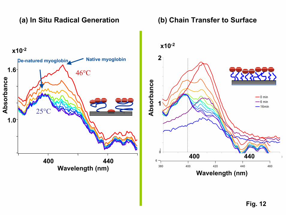
More detailed protein adsorption experiments have been conducted on proteinssuch as myoglobin containing a heme group that can be monitored using UV-visiblespectroscopy [31]. UV visible spectra of myoglobin provide a quantitative measure ofadsorbed protein concentrations and indicate whether the protein has denatured on thesurface. For myoglobin, denaturation results in a characteristic blue shift of the Soretpeak from its native position at 408 nm to 395 nm for strongly denatured protein. Fig. 12shows a series of spectra taken during the desorption of myoglobin from two types ofPNIPAM films: an azo-initiated film with a low grafting density and high molecularweight, and a “polymer brush” film with a higher grafting density and lower molecularweight. While both films adsorb a monolayer of myoglobin above the transitiontemperature, a fraction of a monolayer (up to 25%) is retained on the low graft-densityfilm on cooling. The UV visible spectra.reveal that the irreversibly adsorbed fractionconsists of denatured myoglobin. Studies on other proteins suggest that the fraction ofirreversibly adsorbed protein varies inversely with protein size (< 1% for HSA (14 x 4 x 4nm dimensions), 10-15% for hemoglobin (6.5 x 5.5 x 5.0 nm), and 15-25% formyoglobin (4.4 x 4.4 x 2.5 nm). Our interpretation of these results is that for the lowgraft-density films, collapse of the tethered chains above the transition temperature opensup bare patches that are on the order of 5-6 nm in diameter. Proteins that are smaller thanthese holes can gain access to the underlying substrate, where they denature and stick. Incontrast, UV-visible spectra show that myoglobin does not denature on the more tightlypacked “polymer brush” films, resulting in completely reversible adsorption. Use of the“polymer brush” configuration is critical to the performance of reversible protein traps.
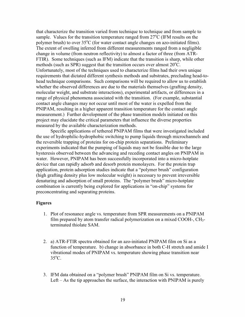
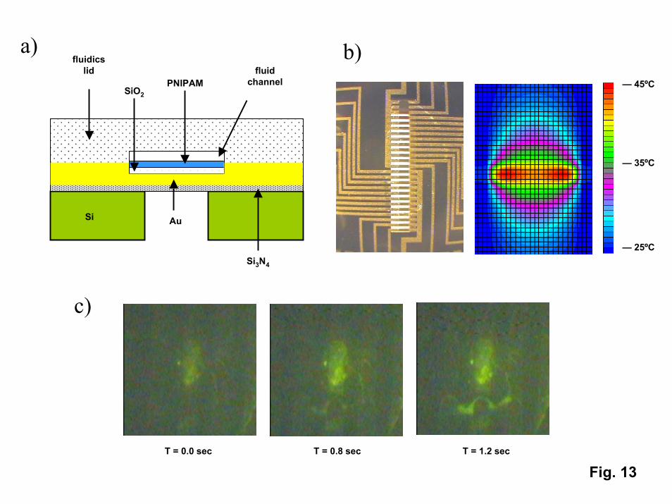
“Polymer brush” PNIPAM films have been incorporated into a micro “hotplate”device (Fig. 13) that allows for rapid heating and cooling of protein solutions in smallvolumes. The device consists of an array of gold heater lines deposited on a thin,freestanding membrane of silicon nitride to provide for thermal insulation from thesurrounding Si wafer frame. The membrane and heater lines are covered with a thin layerof silica which provides electrical insulation from the fluid as well as a substrate for thePNIPAM SAM. Fluidic channels are defined above the hot plate with glass spacers andsilicon adhesive. Fluids are delivered to the system using capillary tubes connected tosyringe pumps. The response characteristics of the fluidics device containing PNIPAM
18
films have been tested by monitoring the adsorption and desorption of fluorescein-labeledmyoglobin from static solutions using fluorescence microscopy. Fig. 13 showssuccessive video images taken for a line that was heated above the transition temperatureand then rinsed with a protein-free buffer solution. Fluorescence microscopy reveals thata layer of myoglobin forms only above the heated line. The remained of the surface,which is covered with room temperature PNIPAM, is completely non-fouling. An imagetaken less than one second after the heater line is turned off shows a plume of myoglobindesorbing from the surface. (The fluorescence intensity of the desorbing plume is higherthan the intensity of the initial heated line because the fluorophores do not quench eachother to the extent that they do in a densely packed monolayer.) For myoglobin, thedesorption process is complete in a few seconds. However, desorption kinetics areprotein dependent, with larger proteins such as HSA requiring longer times to desorb anddiffuse away from the cooling line.
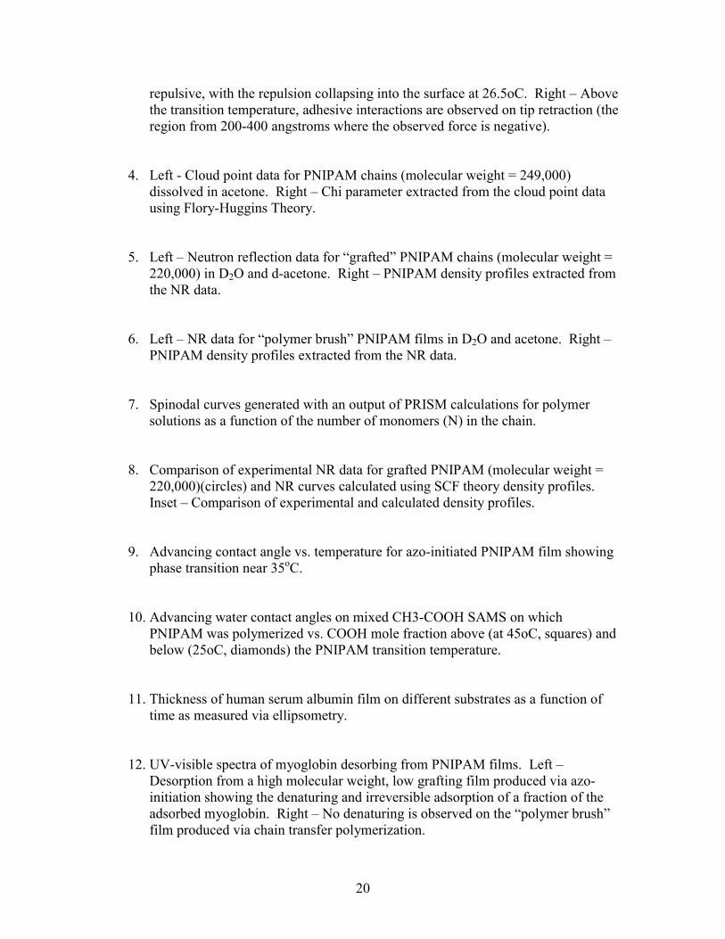
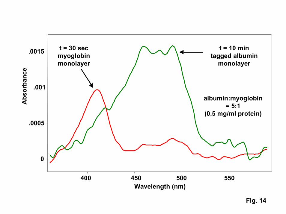
While the protein trap described above could be used as a reversible on-chipprotein preconcentrators, it may be possible to exploit the kinetics of competitiveadsorption and desorption to perform on-chip separations. For example, UV-visiblespectra obtained on hot PNIPAM films in contact with myoglobin-albumin mixtures (Fig.14) show that the smaller myoglobin forms a monolayer on the surface in around 30seconds. However, with longer exposure times (< 10 minutes), the larger HSAeventually displaces the myoglobin to form a relatively pure HSA monolayer. Suchresults are consistent with literature studies of competitive adsorption on passive surfaces[32]. By programming an array of heater lines and controlling contact times and flowrates, it should be possible to selectively grab either myoglobin or albumin from proteinmixtures. Use of an array of such devices could provide a mechanism for performingtasks such as extracting relatively dilute populations of cytokines (small cell-signallingproteins) from albumin-rich serum as part of an rapid screening test to identify personswho have been exposed to chem-bio warfare agents. The protein trap could also see usein a wide range of other applications including on-command drug delivery, proteomics,and (when used to adsorb antibody monolayers) highly selective separations of bio-molecules.
Summary
Tethered films of the polymer poly n-isopropylacrylamide (PNIPAM) weresynthesized and characterized for their potential as thermally-switchable coatings inmicrofluidic systems. With PNIPAM films, switching of surface chemistry is achievedby virtue of a thermally-activated swelling transition in water which occurs at a lowercritical solution temperature of 30-35oC. A wide range of techniques, includingellipsometry, surface plasmon resonance, attenuated total reflectance infraredspectroscopy, and interfacial force microscopy, all indicate that the swelling transitionoccurs regardless of the grafting density and molecular weight of the polymer chains inthe film. Desired chemical changes, including switching of the water contact angle andthe reversible adsorption and desorption of proteins, also indicate that tethered PNIPAMfilms exhibit the desired phase transition.
Each of the above techniques is sensitive to a different aspect of the phasetransition. While experimental results were in qualitative agreement, quantitative values
19
that characterize the transition varied from technique to technique and from sample tosample. Values for the transition temperature ranged from 27oC (IFM results on thepolymer brush) to over 35oC (for water contact angle changes on azo-initiated films).The extent of swelling inferred from different measurements ranged from a negligiblechange in volume (from neutron reflectivity) to almost a factor of three (from ATR-FTIR). Some techniques (such as IFM) indicate that the transition is sharp, while othermethods (such as SPR) suggest that the transition occurs over almost 20oC.Unfortunately, most of the techniques used to characterize films had their own uniquerequirements that dictated different synthesis methods and substrates, precluding head-to-head technique comparisons. Such comparisons will be required to allow us to establishwhether the observed differences are due to the materials themselves (grafting density,molecular weight, and substrate interactions), experimental artifacts, or differences in arange of physical phenomena associated with the transition. (For example, substantialcontact angle changes may not occur until most of the water is expelled from thePNIPAM, resulting in a higher apparent transition temperature for the contact anglemeasurement.) Further development of the phase transition models initiated on thisproject may elucidate the critical parameters that influence the diverse propertiesmeasured by the available characterization methods.
Specific applications of tethered PNIPAM films that were investigated includedthe use of hydrophilic-hydrophobic switching to pump liquids through microchannels andthe reversible trapping of proteins for on-chip protein separations. Preliminaryexperiments indicated that the pumping of liquids may not be feasible due to the largehysteresis observed between the advancing and receding contact angles on PNIPAM inwater. However, PNIPAM has been successfully incorporated into a micro-hotplatedevice that can rapidly adsorb and desorb protein monolayers. For the protein trapapplication, protein adsorption studies indicate that a “polymer brush” configuration(high grafting density plus low molecular weight) is necessary to prevent irreversibledenaturing and adsorption of small proteins. The “polymer brush” micro-hotplatecombination is currently being explored for applications in “on-chip” systems forpreconcentrating and separating proteins.
Figures
1. Plot of resonance angle vs. temperature from SPR measurements on a PNIPAMfilm prepared by atom transfer radical polymerization on a mixed COOH-, CH3-terminated thiolate SAM.
2. a) ATR-FTIR spectra obtained for an azo-initiated PNIPAM film on Si as afunction of temperature. b) change in absorbance in both C-H stretch and amide Ivibrational modes of PNIPAM vs. temperature showing phase transition near35oC.
3. IFM data obtained on a “polymer brush” PNIPAM film on Si vs. temperature.Left – As the tip approaches the surface, the interaction with PNIPAM is purely
20
repulsive, with the repulsion collapsing into the surface at 26.5oC. Right – Abovethe transition temperature, adhesive interactions are observed on tip retraction (theregion from 200-400 angstroms where the observed force is negative).
4. Left - Cloud point data for PNIPAM chains (molecular weight = 249,000)dissolved in acetone. Right – Chi parameter extracted from the cloud point datausing Flory-Huggins Theory.
5. Left – Neutron reflection data for “grafted” PNIPAM chains (molecular weight =220,000) in D2O and d-acetone. Right – PNIPAM density profiles extracted fromthe NR data.
6. Left – NR data for “polymer brush” PNIPAM films in D2O and acetone. Right –PNIPAM density profiles extracted from the NR data.
7. Spinodal curves generated with an output of PRISM calculations for polymersolutions as a function of the number of monomers (N) in the chain.
8. Comparison of experimental NR data for grafted PNIPAM (molecular weight =220,000)(circles) and NR curves calculated using SCF theory density profiles.Inset – Comparison of experimental and calculated density profiles.
9. Advancing contact angle vs. temperature for azo-initiated PNIPAM film showingphase transition near 35oC.
10. Advancing water contact angles on mixed CH3-COOH SAMS on whichPNIPAM was polymerized vs. COOH mole fraction above (at 45oC, squares) andbelow (25oC, diamonds) the PNIPAM transition temperature.
11. Thickness of human serum albumin film on different substrates as a function oftime as measured via ellipsometry.
12. UV-visible spectra of myoglobin desorbing from PNIPAM films. Left –Desorption from a high molecular weight, low grafting film produced via azo-initiation showing the denaturing and irreversible adsorption of a fraction of theadsorbed myoglobin. Right – No denaturing is observed on the “polymer brush”film produced via chain transfer polymerization.
21
13. a) Cross-section schematic of the micro-hotplate device showing the relativelocations of the Si substrate, the Si3N4 membrane, the gold heater lines, thePNIPAM film, and the fluidics. b) Left – photomicrograph of the top of thehotplate (the silicon nitride membrane (bright region in the center) is 200 mmwide. Right – Calculated thermal profiles around a single heated line. c)Fluorescence microscope images of PNIPAM desorbing from a single hot line asa function of time after the hot line is turned off.
14. UV-visible spectra of protein films adsorbed on PNIPAM above its transitiontemperature. Protein solutions containing both myoglobin and human serumalbumin result in initial adsorption of myoglobin (red curve) followed by thedisplacement of myoglobin by albumin at long contact times (green curve).
References
1. G. M. Whitesides and A. D. Strook, Phys. Today, 54, 42 (2001)2. D. Figeys and D. Pinto, Electrophoresis, 22, 208 (2001)3. J. C. McDonald, et al., Electrophoresis, 21, 27 (2000)4. M. A. Burns, et al., Proc. Nat. Acad. Sci., 93, 5556 (1996)5. P. Harder, M. Grunze, R. Dahint, G. M. Whitesides, and P. E. Laibinis, J. Phys.
Chem. B, 102, 426 (1998)6. E. Ostuni, L. Yan, and G. M. Whitesides, Colloid Surf. B, Biointerfaces, 15, 3
(1999)7. K. L. Prime and G. M. Whitesides, Science, 252, 1164 (1991)8. R. Pelton, Adv. Colloid Interface Sci., 85, 1 (2000)9. Y. G. Takei, et al., Macromolecules, 27, 6163, (1994)10. J. Zhang, R. Pelton, and Y. Deng, Langmuir, 11, 2301 (1995)11. C. Wu and S. Zhou, Macromolecules, 28, 8381 (1995)12. X. Wang, X. Qiu, and C. Wu, Macromolecules, 31, 2972 (1998)13. S. Oscarsson, J. Chromatography B, 699, 117 (1997)14. H. Kanazawa, et al., Anal. Chem., 68, 100 (1996)15. H. Lakhiari, et al., Biochim. Biophys. Acta General Subjects, 1379, 303 (1998)16. S. Kidoaki, S. Ohya, Y. Nakayama, and T. Matsuda, Langmuir, 17, 2402 (2001)17. L. Liang, X. D. Feng, J. Liu, P. C. Rieke, and G. E. Fryzell, Macromolecules, 31,
7845 (1998)18. L. K. Ista, S. Mendez, V. H. Perez-Luna, and G. P. Lopez, Langmuir, 17, 2552
(2001)19. A. Revillon, and D. Leroux, React. Funct. Polymers, 26, 105 (1995)20. T. E. Patten and K. Matyjaszewski, Acc. Chem. Res., 32, 895 (1999)21. L. S. Jung, C. T. Campbell, T. M. Chinowsky, M. N. Mar, and S. S. Yee,
Langmuir, 14, 5636 (1998)22. B. Subramanian, S. Mendez, S. Balamurugan, M. O. Obrien, and G. P. Lopez,
manuscript in preparation
22
23. S. A. Joyce and J. E. Houston, Rev. Scientific Instrum., 62, 710 (1991)24. F. –J. Schmitt, C. Park, J. Simon, H. Ringsdorf, and J. Israelachvili, Langmuir, 14,
2838 (1998)25. M. S. Kent, Macromol. Rapid Commun., 21, 243 (2000)26. H. Yim, M. S. Kent, D. L. Huber, S. Satija, J. Majewski, and G. S. Smith,
submitted to Macromolecules27. S. Mendez, J. G. Curro, M. Puetz, D. Bedrov, and G. D. Smith, J. Chem. Phys.,
115, 5669 (2001)28. J. D. McCoy, Y. Ye, and J. G. Curro, J. Chem. Phys., 117, 2975 (2002)29. S. Mendez, L. K. Ista, and G. P. Lopez, manuscript in preparation30. D. L. Huber, R. P. Manginell, M. A. Samara, and B. C. Bunker, submitted to
Science31. A. B. Anderson and C. R. Robertson, Biophys. J., 68, 2091 (1995)32. M. Kleijn and W. Norde, Hetero.Chem. Rev., 2, 157 (1995)