40
Syllabus
HEMATOLOGYCOMPILED ANDPUBLISHED AT
FIILISTfD THMMS SCHOOLLETTEIUWMI GEEIERfiL HOSPITAL
PUBLISHED JANUARY 1943REPUBLISHED JUNE 1943REPUBLISHED JAM. 1944


HEMATOLOGY
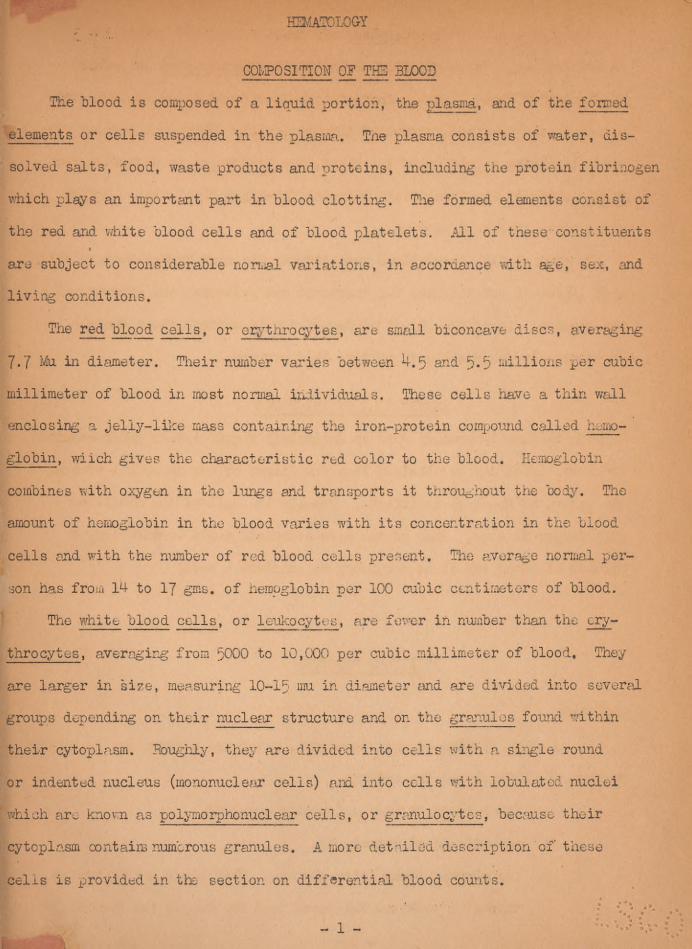
COMPOSITION OF THE BLOOD
The blood is composed of a liquid portion, the plasma, and of the formed
elements or cells suspended in the plasma. Tne plasma consists of water, dis-
solved salts, food, waste products and proteins, including the protein fibrinogen
which plays an important part in blood clotting. Tne formed elements consist of
the red and white blood cells and of blood platelets. All of these'constituents4
are subject to considerable normal variations, in accordance with age, sex, and
living conditions.
The red blood cells, or erytnrocytes, are small biconcave discs, averaging
7.7 Mu in diameter. Their number varies between U. 5 and 5.5 millions per cubic
millimeter of blood in most normal individuals. These cells have a thin wall
enclosing a jelly-like mass containing the iron-protein compound called hemo-
globin, wiich gives the characteristic red color to the blood. Hemoglobin
combines with oxygen in the lungs and transports it throughout the body. Tne
amount of hemoglobin in the blood varies with its concentration in the blood
cells and with the number of red blood cells present. The average normal per-
son has from 14 to 17 gins, of hemoglobin per 100 cubic centimeters of blood.
The white blood cells, or leukocytes, are fewer in number than the ery-
throcytes, averaging from 5000 to 10,000 per cubic millimeter of blood. They
are larger in size, measuring 10-15 mu in diameter and are divided into several
groups depending on their nuclear structure and on the granules found within
their cytoplasm. Roughly, they are divided into cells with a single round
or indented nucleus (mononuclear cells) and into cells with lobulated nuclei
’ which are known as polymorphonuclear cells, or granulocytes, because their
cytoplasm contains numerous granules. A more detailed description of these
cells is provided in the section on differential blood counts.
1
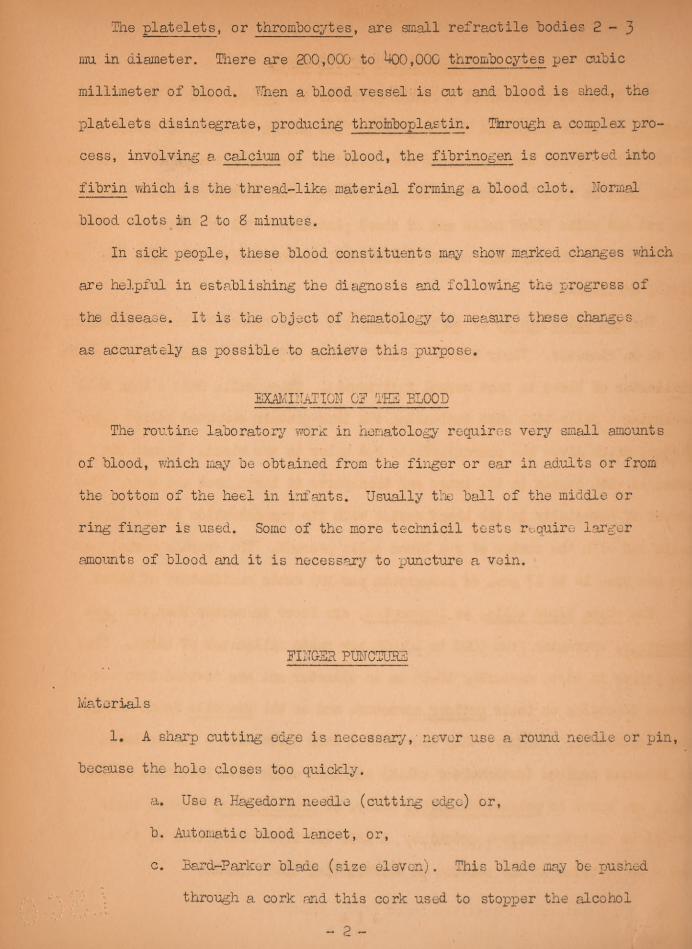
The platelets, or thrombocytes, are small refractile bodies 2 - 3mu in diameter. There are 200,000 to *400,000 thrombocytes per cubic
millimeter of blood. Then a blood vessel is cut and blood is shed, the
xslatelets disintegrate, producing throinboplastin. Through a complex jjro-
cess, involving a calcium of the blood, the fibrinogen is converted into
fibrin which is the thread-like material forming a blood clot. Normal
blood clots in 2 to 8 minutes.
In sick people, these blood constituents may show marked changes which
are helpful in establishing the diagnosis and following the. progress of
the disease. It is the object of hematology to measure these changes,
as accurately as possible to achieve this purpose.
EXAMINATION 0? THE BLOOD
The routine laboratory worm in hematology requires very small amounts
of blood, which may be obtained from the finger or ear in adults or from
the bottom of the heel in infants. Usually the ball of the middle or
ring finger is used. Some of the more technicil tests require larger
amounts of blood and it is necessary to puncture a vein.
DINGER PUNCTURE
Materials
1. A sharp cutting edge is necessary,• never use a round needle or pin,
because the hole closes too quickly.
a. Use a Hagedorn needle (cutting edge) or,
b. Automatic Hood lancet, or,
c. Bard-Parker blade (size eleven). This blade may be pushed
through a cork and this cork used to stopiDer the alcohol
2

"bottle. Keep all the cutting, blades as clean and shiny as possible.
2. Cotton
3. IQffc .Alcohol
k. Clean pipettes and chemically clean slides.
Procedure;
l. hub the patient’s fi iger briskly or place the hand in warm water topromote blood flow,
2. Clean the finger with the alcohol and dry. If the finger tip is wet the
blood will not form a round drop.
3. Hold the ball of the finger tightly between the operator’s thumb and
index finger, until the skin color is dark red. Puncture the finger with
a firm, quick stroke, deep enough so the blood will flow immediately. Do
not squeeze the finger after the puncture because this forces tissue
juices into the cut and dilutes the bicod.
k. Wipe off the first drojj with ary cotton.
5. Allow a sufficiently large drop form, before touching a blood pipette or
slide to the drop. Pill the pipettes and make smears as indicated.
BLEEDING TIKE
Bleeding time is the time that it takes the blood to stop flowing from
a measured cut in the finger or ear.
Materials;
1. finger puncture equipment
2. filter paper
3. Watch.
Procedure:
1. Puncture the finger or ear lobe.
2. Note time when blood begins to flow
3. Blot with the filter paper every >7 minute.3

U, The time, ’between the first drop and the last is the hi ceding time.
Normal bleeding time is from Ito 3 minutes, ■.'hen the bleeding
continues longer than 10 minutes the bleeding time is seriously
prolonged.
COAGULATION TIMEThis represents the length, of time it takes a specimen of
"blood to clot so that fibrinogen is converted into strands of fibrin.
Normal coagulation time is from 2 - 8 minutes.
I. Slide Method:
Material:
1. Finger puncture equipment
2. Clean Slide.
3. Needle
U. Watch
Procedure:
1. Cleai the finger with alcohol and make a. routine puncture.
2. Place a few drops of blood on the slide.
3. At minute intervals draw a needle slowly through the blood
drox:-. When a fine thread (fibrin) can be picked up by the
needle point, coagulation has begun.
U. The time between the flow of blood and the formation of fibrin
is coagulation time.
11. Capillary ’Tube Method;
Draw out soft glass tubing into a capillary pipette, over a wing
top bunsen burner. Aider finger puncture fill the tube, then at
minute intervals break off mm. of the tube. If the fibrin
strings out at the broken end coagulation has began. Normal co-
agulation time is from 2 to 8 minutes.
k

■tETmmmou of hmoglobiii
Hemoglobin' is measured in grams per hundred cubic centimeters of blood
or as a percentage of an arbitrary normal standard. These figures may be ob-
tained by comparing; the color of the blood to be examined with the color of
blood-whose hemoglobin concentration is known.. This principle is utilized in
the Tallquist method by placing a drop of the patient's blood on a of
special absorbent paper and comparing it with color plate reproductions of
blood containing various amounts of hemoglobin. The color plate which most near,
ly matches the patient's blood represents the percentage of, hemoglobin. In the
Tallquist scale a concentration of gm, of hemoglobin, per 100 cc of blood
represents 100$. The color plates axe arranged in descending gradations of
10$, but values higher than 15. S gm. can not be determined. Due to the diffi-
culty in matching colors this method requires daylight and is at best quite in-
accurate so that it is only used for rough estimations.
A more accurate method involves the dissolution of a. measured amount oft ,
blood with 1$ hydrochloric acid. The red cells are laked (dissolved) and the
hemoglobin is converted into a reddish brown conpound, arid,hemal in. The color
produced is proportional to the amount of hemoglobin present, and this may be
determined by comparison with either a known solution of a.cid hematin, or with
a standard cqlore! glass; or,the intensity of color may be measured by means
of a photo-electric cell. Instruments for such measurements are called hemo-
rneters or hemoglobinometers.
A. Tallquist Method: .
Materials;
1. Finger puncture equipment. ,
2. A Tallquist scale which is a sheet of pajjer with spots of red
color on it, graded- to represent hemoglobin content from 10%to 100%.
5
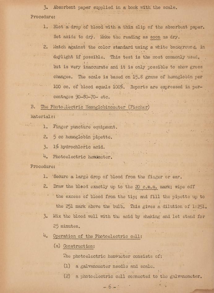
3. Absorbent paper supplied in a' “bonk with the scale.Procedure;
1. Blot a drop'oi ■’blood with a thin slip of the absorbent paper.
Set aside to dry. Make the. reading as soon as dry.
2. Match against the color standard using a white background, in
daylight if possible. 'This test is the most commonly used,
but is very inaccurate and it is only possible to show gross
changes. The scale is based on Ip.B grains of hemoglobin per
100 cc. of blood equals 100$. Reports are expressed in per-■ ' centages 90-80-70- etc.
B. The Photoelectric Hemoglohinometer (Pischer)
Material's;
1. linger puncture equipment.
2. scc hemoglobin xiipette,
3. Ijo hydrochloric acid.
U. Photoelectric hemoimeter.
Procedure:
1. ’Secure a large drop of blood from the finger or ear.
2. Draw the blood exactly up to the 20 c.m.m. mark; wipe off
the excess of blood from the tip; aid fill the pipette up to
the 251 mark above the bulb. This gives a dilution of 1:231.3. Mix the blood well with the acid by shaking and let stand for
2p minutes.Operation of the Photoelectric cell;
(a) Construction;
The photoelectric hemoueter consists of;
(1) a galvanometer needle and scale.(2) a photoelectric cell connected to the galvanometer.
6

FISH3R MCTKO-ESMCEETSR
Zero Kneb
Tube HoldersScale.
Test Hole
-Pointer Adjustment KnobPilot Light, Switch
6(a)

(3) a constant light shining on the photoelectric Cell.
(4) a tube holder between the light and the photoelectric cell.
(5) two standard test tubes, one sealed containing distilled
water, the other empty for the specimen of blood to be
examined.
(6) a control knob (marked "Adjust Zero") for setting the
galvanometer needle.(7) a rheostat control for the light located on the front panel.
("Pointer Adjustment Knob")
(b) Taking the reading
(1) the galvonometer needle is extremely sensitive and is easily
displaced and damaged by jarring. For this reason once
the hemometer is set up for operation it should not be re-
moved. If the galvanometer needle has been jarred from the
starting mark (A) it is brought back to the place by slowly
twisting the "adjust zero" knob before turning on the light:
(2) The current is then turned on and the instrument allowed to
"warm up" for 2 minutes because the photoelectric coll does
not reach its maximum efficiency until that time has
elaxssed.(3) The tube containing distilled water is now placed in the
holder between the light and the photoelectric cell. 3y
means of the rheostat knob the galvanometer needle is
brought to the line 3. The instrument is now ready for op-
eration, This adjustment must be made every time a reading
is made.
7

(U) The acid hematin solution in the pipette is expelled
into the specimen tube (allowing for full development
of color) and the specimen tube is placed in the reading
rack. The galvanometer needle will now swing from line
B toward line A and after several oscillations will
come to rest on the scale representing the hemoglobin
concentration. The reading is recorded and the specimen
tube is replaced by the distilled water tube which
should bring the needle back to line 3. The reading
is repeated.
(5) If the reading does not check within .2 gm. or the
needle does not return to line B, the hemoglobinometor
should be readjusted and new readings taken.
(b) NOTE; The direction given above are for the Fischer
photohemometer. Instruments made by other manufac-
turers work on a similar principle but have special
operating details. Be sure to read the instructions
before using these instruments. Also, all dirt and
finger marks must be wiped off the tubes before using,
(c) Reports;
The Fischer hemOmetcr should be read and reported di-
rectly in grains. The percentage scale corresponds to
15. b gm. as 100$ and percentages should be converted
to correspond to the standard of this laboratory in
which 16.6 Gm. is -100$.C. The Newcomer Hemoglobinometer.
Materials:
1. Finger Puncture equipment.
s;

2, 5 c,c, hemoglobin pipette.
3. 1$ hydrochloric acid.
U, Newcomer hemogloh inomet er,
Procedure:
1. Secure a drop of blood from the finger or ear.
2. Draw the blood exactly up to the 10 c.m.m. mark; wipe off the excess
of blood from the tip and fill the pipette up to the 251 mark above
the bulb. This gives a dilution of 1:501.3. Mix the blood well with the acid by shaking and let stand for 20
minutes.
h. Operation of the hemoglobinometer.
(a) Construction and operation;
The apparatus consists of a colorimeter arrangement in which
the standard crap is filled with distilled water and a color
disc is interposed between this cup and the eyepiece. Theunknown cup is filled by the acid hematin solution and the
colors matched by moving the cap up or down. The concentration
of hemoglobin in grams may then be directly read off the scale
on the adjustment knob,
5. The readings are made directly in grams and then expressed in per-
cent of the laboratory standard (1b,6 grams.)
Note; When 5 cc. hemoglobin pipettes are not available the blood may be
Dram with the 20 c.m.m. pipette and expelled into a test tube con-
tainirg exactly 10 c.c. of Vfo hydrochloric acid.
Red Blood Cell Counting:
A, Materials;
1. Lancet for puncture,
2. Cotton9

3. Wat er-> alcohol, .and ether for cleaning-pipette?.■ /.V.'
4. Microscope.
5. Diluting pipette for red blood cells. It often lias a red head
in the bulb to make it quickly recognizable. The Thoma pipette
is marked in graduated lines along the capillary bore. The
fifth graduation from the tip is marked 0. p, the tenth, 1.0;
above the bulb is a line marked 101. In this pipette, if blood
is drawn to the C. 5 mark and the diluting fluid to the 101 mark,
the dilution is 1 to 200,
b. Counting chamber. The Levy chamber with the iinproved Neubauer
ruling is the supply table item of issue. There are other
types of ruling and several kinds of chambers, all similarly
used. The chamber is a thick glass slide with two central
platforms; on the surface of each is engraved a series of ru-
lings. The side platforms on which the special cover glass fits
are exactly 0.1 mm higher than the central platforms, when the
cover slip is in place there is a space 0.1 mm deep, the ruled
areas, having a surface area of 9 sq. mm. The four large corner
squares outside the double ruled lines (marked 1,2,3»and 4)
are each subdivided into lb smaller squares. The central square
is divided by double lines .into 25 small squares each of which
contains l 6 smaller squares, making a tetal of ] 4OO squares (see
fig.-1) Each small square-then is 1/UOQ sq. mm.
Note; Tne method of blood counting is thoroughly explained here-
in. The number 1,2,3, and and- till lb surrounding squares
of each, indicate the parts of the slide used in counting white
blood cells. The letter A,3,C,D, and E, and the areas between
the double lines, indicate the areas used in counting red bloodcells.

Figure I,—lmproved Neubauer counting chamber.
Note: The method of "blood counting is thoroughly explained herein. The n05..1,2,3 and h, and the lb surrounding sou-res of each, indicate the parts of the slideused in counting white blood cells. The letters A,8,0,D, find A, and the areas"between the double lines, indicate the areas used in counting red blood chlls^
.7. Diluting fluid- Sayeras solution:
Sodium chloride 1.0 ga.
Sodium sulfate 5-0Mercuric cliloride. 0.5 dm *
Distilled water 200 cc.
B, Counting Chamber and pipette cleaning;
All pipettes and counting chambers should be clean and dry before using
and should be cleaned immediately after using. Avoid harsh rubbing or
strong solutions on the counting chamber.
11
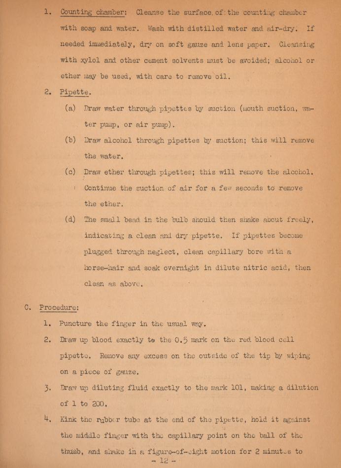
1. Counting chamber; Cleanse the surface, of : the counting chamber
with soap and water. Wash with distilled water and air-dry. If
needed immediately, dry on soft gauze and lens paper. Cleansing
with xylol and other cement solvents must he avoided; alcohol or
ether may he used, with care to remove oil.
2* Pipette.
(a) Draw water through pipettes hy suction (mouth suction, wa-
ter pump, or air pump).(h) Draw alcohol through pipettes by suction; this will remove
the water,
(c) Draw ether through pipettes; this will remove the alcohol,
i Continue the suction of air for a few seconds to remove
the ether.
(d) The small head in the hulh snould then shake about freely,
indicating a clean and dry pipette. If pipettes become
plugged through neglect, clean capillary bore with a
horse-hair and soak overnight in dilute nitric acid, then
clean as above.
C. Procedure;
1. Puncture the finger in the usual way,
2. Draw up hlood exactly to the o,p mark on the red hlood cell
pipette. Remove any excess on the outside of the tip hy wiping
on a piece of gauze.
3- Draw up diluting fluid exactly to the mark 101, making a dilution
of 1 to 200,
4, Kink the rubber tube at the end of the pipette, hold it against
the middle finger with the capillary point on the hall of the
thumb, and shake in a figure-of-eight motion for 2 minutos to

insure good mixing. '
5. Put the cover slip in place on the counting chamber.6. Blow out 3 drops, touch the tip of the pipette to the edge of the
platform, and allow a thin layer of fluid to flow under the cover
glass. If the fluid flows into the troughs, or there .are bubbles
under the cover glass, clean the counting chamber and try again.
7. Allow the cells to settle for 2 minutes.
8. Examine under the high-dry lens of a microscope.
D. Count ing:
Count all the cells in squares A,3,C,D, and E, as illustrated
in figure 1. In counting cells in each square (as Ain fig. 1) en-
closed by double lines, count .all cells touching the inner lines on
the right and top of the square. Do not count any cells touching the
lines on the left and bottom of the square. The difference betweenthe number of cells in any two blocks should not be more than Ip
cells. If this is the case, the mixing was not complete or the
chamber was dirty. From this count calculation is made of the number
of cells per cubic millimeter of blood.
E, Calculation Ixample;
1. Long method;
(a) Squares A,B, C,D, give counts 100, 9s, 10U and 100;
total, 500.(b) Therefore, SO small squares, v;hich occupy 5/25 or 1/5 sq.
mm. , contain 500 cells.
(c) One square millimeter would contain 5 x 500 “ 2,500 cells.
(d) As this cell layer is 0.1 mm thick, 1 cu ram would contain
10 x 2,500 ~ 25,000.
(e) As the blood was diluted 200 times: 200 x 25,000 = 5,000,000
13
cells per cubic millimeter of blood.
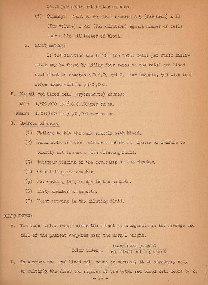
(f) Summary: Count of 80 small squares x 5 (for area) xlO
(for volume) x 200 (for dilution) equals number of cells
per cubic millimeter of blood.
2. Short method’;
If the dilution was 1:200, the total cells per cubic milli-
meter may be found by adding four zeros to the total red blood
cell count in squares A,3,C,D, and B. For example, 500 with four
zeros added will be 5*000,000.
F. Normal red blood cell (erythrocyte) counts:
Mri: 4,500,000 to 6,000,000 per cu mm.
Women: 4,000,000 to per cu mm.
Gr. Sources of error
(1) Failure to hit the mark exactly with blood.(2) Inaccurate dilution--either a bubble in pipette or failure to
exactly hit the mark with diluting fluid.
(3) Improper placing of the cover slip on the chamber,
Overfillirg- the chamber.
(5) hot shaking long enough in the pipette.
(6) Dirty chamber or pipette,
(7) Yeast growing in the diluting fluid.
COLOR INDEX:
A* The term n color index" means the amount of hemoglobin in the average red
cell of the patient compared with the normal amount.
hemoglobin percentColor index -
red blood cells percent
B. To express the red blood cell count as percent, it is necessary only
to multiply the first tT 'o figures of the total red blood cell count by 2.Ik

Example;
Red blood cells, 5»000,000.
Hemoglobin, 100 percent.Color index i50 x 2 = - 1 *
c. A normal color index ranges from 0.85 to 1.15.26. White blood cell (leucocyte) counting,
a. Materials
(1) Same as for red blood cell count except for the- pipette and
the diluting fluid.
(2) White hlood cell pipette is similar to the red cell pipette,
but has a smaller bulb which contains a small white bead and
gives less dilution to the blood. The fifth line on.the gra-
duated capillary tube is marked 0.5» the tenth line. 1.0, and
the above the bulb 11,(3) White blood cell diluting fluid:
Glacial acetic acid _______ 0.5 cc
Distilled Water - - - ~99.3 cc
This fluid may be tinted blue, for convenience in identifying it, by
addition of a drop of 1 percent gentian voilet. This solution should
be freshly prepared every two weeks.b, Procedure.
(1) Draw blood to the 0.5 mark.(2) Draw diluting fluid to the mark 11, making a dilution of 1;20.
(3) Shake as in red blood cell counting.
(h) Discard 3or k drops and fill the counting chamber,
(5) Allow the cells to settle,
(6) Examine under the low power of the microscope.
(7) When doing a complete blood count, shake the red blood pipette
in one hand, the white in the other, fill the counting cham-
bers, red on one side and white on the other.14a

Figure 3* —White "blood cell pipette.c. Calculation.
The white cells are counted in the four large corner squares labeled 1,
2,3»4 in figure 1. The difference between the largest and smallest num-
ber of cells in any two squares should not exceed 10. Each large square
contains l 6 smaller squares and represents a volume of 0.1 cu mu. The
4 squares are counted and divided by 4 to get the average per' 0.1 cu mm,
d. Example of calculation for white blood cells.
(l) Long method.
Square 1 3^Square 2
Square 3 —— 3®Square k Jo
Total for 0,4- cu mm diluted blood (4 squares
0.1 mm-thick) I^oTotal for O.fcu mm diluted Hood (ipG > M) 37*5Number of cells in Icu ram diluted blood (37*5 x 10) 375
To obtain the number in 1 cu ram -of undiluted blood multiply by 20 (as thedilution is Ito 20) or (20x375 =■ 7,500, : Sunuiary: Count of U squares,

divided by 4, times 10 (for area) times 10 (for volume) times 20 (for dilu-
tion) equals number of cells per cubic millimeter of blood.
0 Short method:
Multiply the total number of cells in cu mm by 50; 150 x s
7,500 white cells per cu mm.
e. Normal white blood cell (leucocyte) count.
Normally, this count is s>ooo to 10,000 per cu ram.
Many normal persons lave variable counts duo to activity, time of day,
etc. Daily counts on a patient snould be done at the same time every
In certain cases where the white count is very high, it may be necessary
to use a dilution of 1:100, using the red cell pipette, changing the calr
culation accordingly. In cases where the count is abnormally low, make
the dilution 1:10 by drawing bleed to the 1.0 mark instead of 0,5.
27. 'Glassware cleaning (for 11001 films).
A prerequisite in making a good "blood film is to have chemically cleanglassware.
a. New slides,(1) Wash in soapy water and rinse thoroughly with water,
(2) Place slides in a large "beaker of 95 percent alcohol.
(3) Polish with a soft lint-free cloth (not gauze).
(U) Flame over a bunsen burner.
(5) Place in box with clean slip of paper between each slide.
b. Dirty slides.(1) Boil in 5 percent • sodium bicarbonate solution.(2) Scrub with soap and water.
(3) Place in cleaning solution (potassium bichromate-sulfuric acid)
for 12 hours.
(*4) Then wash as for new slides.
(5) Discard all slides that are badly scratched or discolored.

The 'same- for- slides except, do not flame,. Careful wiping will
prevent much breakage. Do not use pressure.
28. Preparation of "blood films.a. Materials.
(1) Equipment for finger puncture...(2) Clean slides, free from grease.
b. Procedure.~(I)Puncture the finger.
(2) Place 1 small drop of "blood on the end of a slide and place
the slide on a flat surface,
(3) Hold a second slide "between the thumb-..and. third finger and
place one end at a 30° angle on the slide holding the drop ofblo’od; • :
(M) Pull the upper slide until it touches the drop of blood which
then spreads along the narrow end of the top slide (see fig 4).(The slide held with the left hand is supposed to be laid
flat on a smooth surface).
(5) Push the top slide with a firm, steady motion toward the oppo-site end of the bottom slide; the slower the movement, the thi-cker the film; the greater the angle, the thicker the film. A-
void all unnecessary pressure because of the fragility of thecells.
(6) Allow to air-dry. in areas where insects are abundant slides
must be protected or they will be ruined. A good film should be
smooth-and without waves. The edges should be even and the film
should not extend to the edges or end of the slide* Labelling
may be done by writing on the thicker end of the film with an
ordinary lead pencil when the film is dry. The slides should
be stained within 2h hours for best results.
29. Blood stains. - Wright’s stain.
This stain is used for routine Blood films and for other
laboratory purposes. : The prepared

stains purchased by the Army are easy to prepare if specific directions tire
followed: - ; ' ’ ■ -•v
Materials;* > t *.: ’ r -
1. Chemically clean glass ware;
2. Wright’s stain prepared as follows:
Wright's powder (supply table item)- - - 0.3 Gm
Glycerine - 3.0 cc
Methyl alcohol absolute (must be, acetone free- -
Put the powder in a dry mortar grind with a pestle, add. the
glycerine and grind. ‘Add the methyl, . alcohol • and mix. Allow
to stand overnight in a tightly stoppered flask, then filter
..and keep for a few days before use, age improves the stain.
3. Buffer solution:
Potassium phosphate (monobasic) - - 1,13 &m *
Dibasic sodium phosphate 3*20 Gm.
Distilled Water - - - - -1000.ee.Pro cedure; ; *■
‘ 4
1. Pill a Coplin jar with stain and place the smears in the
stain for 2 minutes. This fixes and stains the blood film.
2. Next, place the slides in another fcqplin jar filled with the
buffer solution- for ]4 minutes.
3. Place the-slides in a 3 Coplin jar filled with distilled
water and leave them until the blue color of the blood film ■commences to turn a pinkish color.. As soon as this decolc »
Ration process.....is complete, remove the .slides and dry them.They - are now ready to be examined.
NOTP; The color of the cells may- be varied by changing the stain or
buffer time. . The granules of the neutrophiles should stain a lilac

. colqiv the eosinophiles a lright, .fed, and the basophiles deep blue.
B. G-iemsa Stain; • r 4
Materials:
1. .Staining jars (Coplin jar)
2. Giemsa Stain(a) Stock solution
G-iemsa powder (supply table item) - - —-o*sGlycerine, dissolve powder in this 1 to 2 hours
33*0 cc.
Methyl alcohol absolute (acetone free) 33*00 cc *
(b) Dilute stain (ready for use)
1 cc. stock solution to 10 cc distilled water.
3 . .Methyl alcohol.
A. Procedure:(1) Fix smear with methyl alcohol 3 .5 minutes in
a Coplin jar. .
(2) Dry in air.
(3) Put in dilute stain for 20 to minutes (Coplin jar),
(U) Wash in distilled .water.
(5) Stand on end to dry.
(6) Examine under oil immersion.
This stain is excellent for protozoal staining but is more
time-consuming than the Wright’s stain method.
DIFFDRSWTIAL WHITB SLOQD CFLL COUNT
Materials; , .
1. Finger puncture aid staining equipment.
2. Wright’s stain.
Proce ure:
1. Prepare films.
2. St ain with W:r i ght l s stain. .- 18 -

3. Examine under oil immersion,- record each type of white cell seen.
Note: Survey the slide to determine the area that is neither too thick
nor too thin. This place will usually he towards the latter portion of
the slide. Start on one extreme side and cross the smear. Move one or
two fields and again cross the slide being careful to always count the
very edges.' Count 200 cells.
CriARACTSHISTICS 0? STAINED CELLS
Red Blood Cells;
Normal red blood cells (erythrocytes) are round, non-granular, norl-
andeat ed cells,'the centers of which arc less intensely and colored than
tiie borders. In the various diseases the blood may contain erythrocytes
showing the following abnormalities;
Achromia - Pale staining erythrocytes; decreased hemoglobin.
Polychromasia - Many of the erythrocytes take a "bluish rather than a
tan color.
Anisocytosis - A wide variation in the size of the cells.
Poikilocytosis - A wide variation in the shape of the cells.
Macrocytosis - The Average size of the cells is greater than the normal
7.5 mu.Microcytosis - The ■average size of the cells is smaller'than normal.
Stippling - The cells contain a fin©-dusting of "bluish black granules,as seen in lead poisoning.
Howell-Jolly Bodies- - The cells contain one or two small blue-black dots
Reticulocytes - These red cells have a. feathery, dark blue, irregular
. network within the cell when stained with Brilliant
Cresyl Blue Stain.
White Blood Cells;
Lymphocytes are always mononuclear. The magnitude of the nucleus in
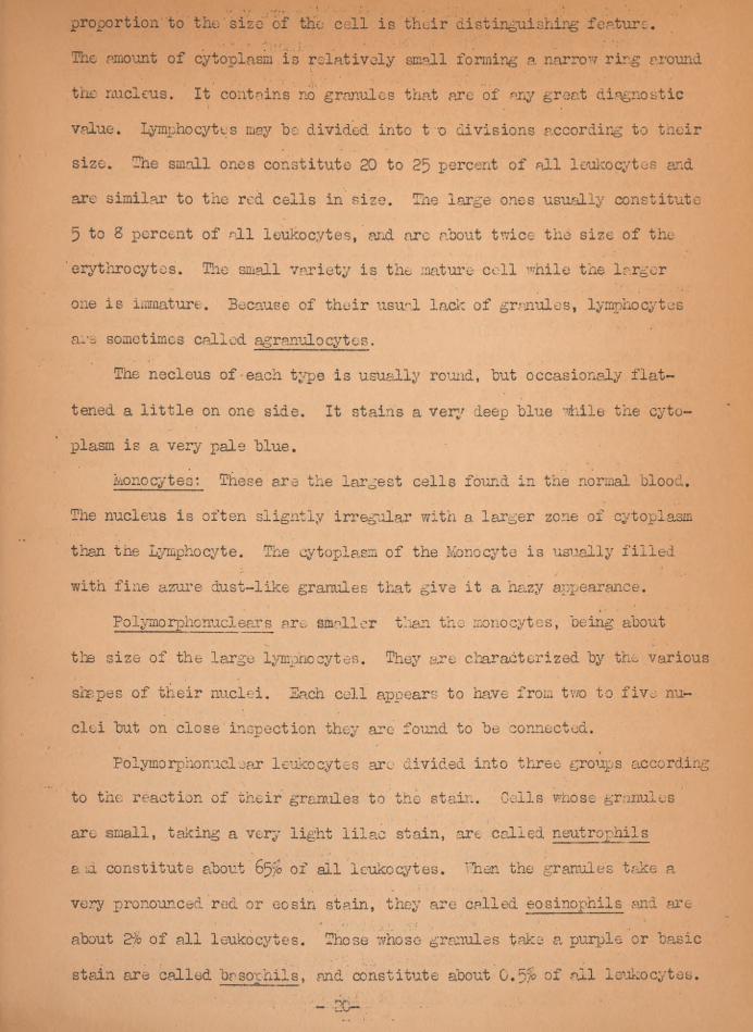
proportion to the'size’of the cell is their distinguishing feature,
'The amount of cytoplasm is relatively small forming a narrow ring around
the nucleus. It contains no granules that are of any great diagnostic
value. Lymphocytes may he divided into t o divisions according to their
size. The small ones constitute 20 to 25 percent of all leukocytes and
are similar to the red cells in size. The large ones usually constitute
5 to 8 percent of all leukocytes, arid arc about twice the size of the
erythrocytes. The small variety is the mature coll while the larger
one is immature. Because of their usual lack of granules, lymphocytes
are sometimes called agranulocytes.
The necleus of each type is usually round, hut occasionally flat-
tened a little on one side. It stains a very deep blue while the cyto-
plasm is a very pale blue.
Monocytes: These are the largest cells found in the normal blood.
The nucleus is often slightly irregular with a larger zone of cytoplasm
than tne Lymphocyte. The cytoplasm of the Monocyte is usually filled
with fine azure dust-like granules that give it a hazy appearance.
Folymorphenuclears are smaller than the monocytes, being about
the size of the large lymphocytes. They are characterized by the various
shapes of their nuclei. Each cell appears to have from two to five nu-
clei but on close inspection they are found to be connected.
Polymorphonuclear leukocytes arc divided into three groups according
to the reaction of their granules to the stain. Cells whose granules
are small, taking a very light lilac stain, are called neutrophils
aid constitute about 65$ of all leukocytes. Then the granules take a
very pronounced red or eosin stain, they are called eosinophils and are
about 2jb of all leukocytes. Those whose granules take a purple or basic
stain are called basophils, and constitute about 0.5$ of all leukocytes.

The nuclei of the neutrophils end eosinophils are very nearly the same
shape, hut in the basophils the nucleus, is almost round.
Because of the peculiar reaction of the granules to the stain, this
group is sometimes called granulocytes.
The abnormal forms of leukocytes are myeloblasts, myelocytes, juveniles,
lymphoblasts, monoblasts and a few atypical forms.
The myeloblasts are the grand-parents of the polymorphonuclears. They
range between 15 and l 6 microns in diameter. The cytoplasm forms a very
narrow rim which stains blue with a diffused effect, being free from gran-
ules. The nucleus is round and sharply defined. It contains several nucle-
oli. Occasionally, vacuoles are present in the cell. These cells are never
found in a healthy blood stream.The abnormal form of leukocytes most commonly seen is the myelocyte.
It is larger than any cell found in the normal blood. The nucleus occupies
about one-half the entire cell. It is usually flat or concave on one side
lying close to the cell wall. The granules vary in size rind react different-
ly to the stain. They can be classed as neutrophilic, eosinophilic or
basophilic. The myelocytes are direct descendants of the myeloblasts and
about the same size but differ since they have granules.
Juveniles are a group of cells emphasized by Schilling. 'They represent
a transitional stage between the; Myelocyte and the Polymorphonuclear
neutrophil. They are smaller than the Myelocyte and the nucleus is slight-
ly indented, like a. kidney bean. The cytoplasm of the Juvenile is neutro-
philic, Eosinophilic, or Basophilic.
Stabs are just a little more mature than are the Juveniles. The nu-
cleus of the Stab cell like a straight or curved rod and may bu
slightly constricted or bulged in different places, but never segmented.
As Juveniles, so also the cytoplasm of Stabs may be Neutrophilic, or
Basophilic. Normally about of all Leukocytes are Stabs.

The Lymphoblasts are the parent cells of lymphocytes and are never
found in normal blood. They are larger than the lymphocytes, but in other
respects, very similar. The cytoplasm forms a narrow band around a
large nucleus and contains no granules. The nucleus contains from one
to three nucleoli. The lymphoblasts are about the same size as the mye-
loblasts, making it difficult to differentiate between them.
monoblasts are the parent cells of monocytes and are never found in
the normal blood. Microscopically they cannot, as a. rule, be distinguished
from other cells.
RETICULOCYTE COUNTS
Materials;
1. Equipment for finger puncture.
2. Clean cover slips and slides.
3. I'p solution of brilliant cresyl blue in normal saline. FILTER
BEFORE USE.
9rocedurc:I', Into a clean centrifuge tube place equal quantities of fresh
blood and the dye solution and mix well. Two or three drops
ar e usually suf ficiexit
2, Lot stand 10 minutes.
3. Make smears so the rod cells will be almost touching each other,
but not piled up.
U, Examine under oil immersion 1 ns; or bettor, do a ‘right's stain
and than examine.
3. Coiliti number of Reticulocytes per 1000 red cells. Normal counts
will be .570 to 1.5^.NOTE: It often assists in counting reticulocytes to fit a piece of paper
covering tie lower lens of thw eye piece. In this cover cut a hole
about inch square. This greatly reduces tlx- visible area and is

mors resting to the eyes.
PLATELET COUNT
Materials:
1. Linger puncture equipment.
2. Red blood cell pipette
3. Haemacyt ometer
Diluting fhnid (Rees andEcker's).
Sodium Citrate (]>,&% acqueous solution) ICX}.O cc
Normal in 0.2 cc
Brilliant cresyl blue 0.1 gm
Procedure;
1. Rapid work is necessary to prevent clumping of platelets.
2. Draw Hood to the 0.5 mark and Rees and Ecker 1 s fluid to the 101
mark.
3. Shake immediately and thoroughly.
U. Fill the counting chamber as in making an erythrocyte count.
5. Allow to stand for 10 minutes so the platelets will settle out.
6. Examine small squares and multiply by 2000 to obtain the num-
ber of platelets per cubic millimeter of blood. Use IOX ocular
and U mm, objective.
DOTE: Platelets -are irregularly small, lightly staining, highly retrac-
tile bodies. Normal counts are around 350,000 per c mm.
SEDIMENTATION RATE;
There are so many methods now in use for performing this test that
results have not been comparable. Only two methods will be given here
with the normals for these methods only.
Materials;
1. Venipuncture equipment.
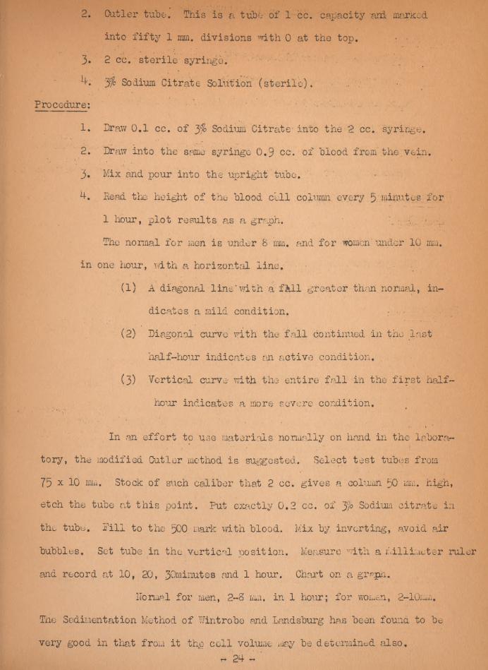
2. Cutler tube. This is a tube of 1 cc. capacity-and marked
into fifty 1 mm. divisions with 0 at tho top.
3* 2 cc. sterile syringe.
j/c Sodium Citrate Solution (sterile)
Procedure:
1. Draw 0,1 cc. of 3% Sodium Citrate- into the 2 cc. syringe,
2. Draw into the same syringe 0.9 cc. of blood from the vein.
3. Mix and pour into the upright tube.
Head the height of the blood cell column every 5 minutes for
1 hour, plot results as a graph. , g
The normal for men is under 8 mm, and for women under 10 mm.in one hour, with a horizontal line.
(1) A diagonal line'with a fAll greater than normal, in-
dicates a mild condition.(2) Diagonal curve with the fall continued in tho last
half-hour indicates an active condition.
(3) Vertical curve with the entire fall in the first half
hour indicates a more severe condition.
In .an effort to use materials normally on hand in the labora-
tory, the modified Cutler method is suggested. Select test tubes from
75 xlO mm. Stock of such caliber that 2 cc. gives a column 50 mm. high,
etch the tube at this point. Put exactly 0,2 cc. of Jjo Sodium citrate in
the tube. Pill to the 500 mark with blood. Mix by inverting, avoid air
bubbles. Set tube in the vertical position. Measure with a millimeter ruler
and record at 10, 20, 30minutes and 1 hour. Chart on a grape.
normal for men, 2-8 mm. in 1 hour; for women, 2-10mm,
The Sedimentation Method of Wintrobe and Landsburg has been found to be
very good in that from it the cell volume may be determined also.

This requires a Wintrohe and Landsburg tube.
Procedure;
1. Pill the tube to the 10. mark with well nixed oxalated blood.
2, Put the tube in exactly a verticle position, A cork with a hole
in it is excellent to put the tube in. Tube must not be moved or
jarred during period of observation.
3. Read at the end of 1 hour. It can be read at 13 minute intervals.
Normally the rate for males is 2 to 9 mm and for females 2 to 22 mm
Volume of the Red Blood Cells;
Equipment;
1. Hematocrit tube of Hintrobe and Landsburg.
2. Venipuncture equipment
3. Tube with Oxalate. USE 2 mg. OXALATE PLH cc BLOOD USED.
Procedure:
1. After the final reading for sedimentation rate centrifuge at 2pOQ
RPM for 30 minutes.
2. Stop centrifuge and take reading.
3. Centrifuge for another Ip minutes.
d. If the reading is lower than-at the end of JO minutes centrifugelize
again. If the reading remained constant, READ THE CELL VOLUME
DIRECTLY IN PERCENT.


