Università Degli Studi Di Parma Facoltà di Scienze MM.FF.NN Dottorato in Scienze Chimiche (XX ciclo) Synthesis and Applications of PNA and Modified PNA in Nanobiotechnology Relatori: Prof.ssa Rosangela Marchelli Prof. Roberto Corradini Coordinatore: Prof.ssa Marta Catellani Dottorando: Dott. Filbert Totsingan Triennio 2005-2007
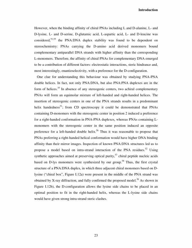
based on D-lys monomers were synthesized by our group.58 Thus, the first crystal
structure of a PNA:DNA duplex, in which three adjacent chiral monomers based on D-
lysine (“chiral box”, Figure I.12a) were present in the middle of the PNA strand was
obtained by X-ray diffraction, and fully confirmed the proposed model.36 As shown in
Figure I.12b), the D-configuration allows the lysine side chains to be placed in an
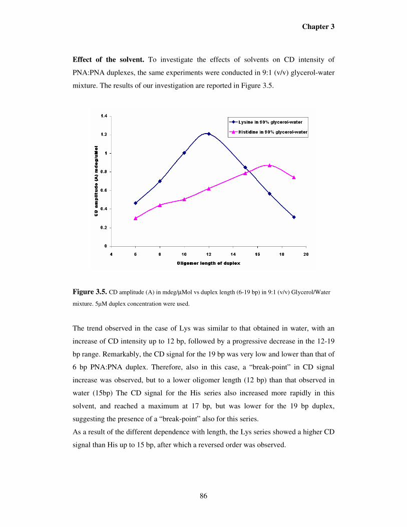
optimal position to fit in the right-handed helix, whereas the L-lysine side chains
would have given strong intra-strand steric clashes.
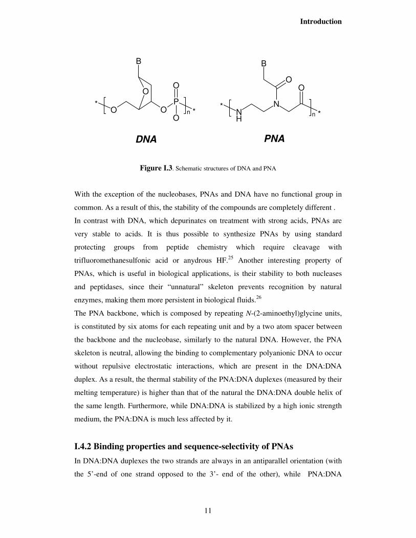
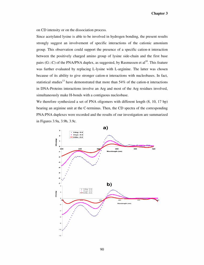
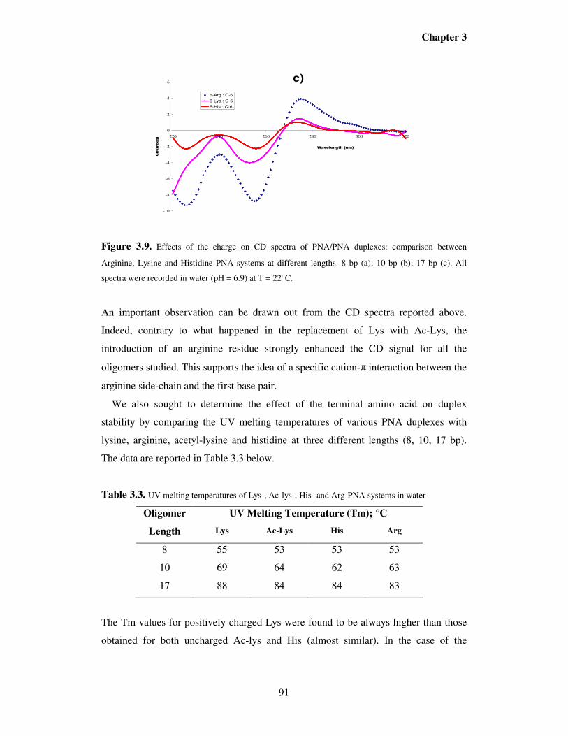
Introduction
24
Figure I.12. a) Crystal structure of the “chiral box” PNA:DNA duplex. b) Stereochemical model obtained from a monomer in the crystal structure, showing the effect of substituents derived from D- or L-amino acids either on the C2 or on the C5 carbon of the monomers.
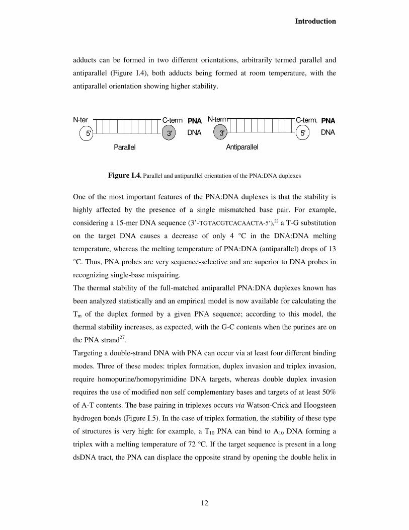
The structural data reported for the PNA:DNA duplexes and the model reported above
was used as a reference in order to predict the behaviour of substituents on the 5-
position. In fact, in this case the preferred stereochemistry would be that derived from
L-amino acids, since it allows the functional group to be placed in a less hindered
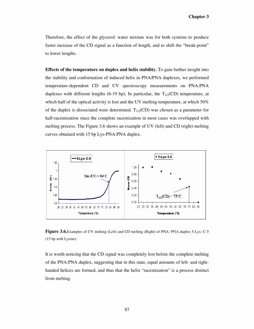
region. Using this design, Seitz et al. synthesized a PNA bearing at the N-terminus a
monomer with L-cysteine side chain at position 5 a allowing, in combination with
another PNA strand modified at the C-terminus as thioester, for PNA synthesis via
chemical ligation.59 Appella et al. synthesized a PNA bearing a fluorophore linked to a
L-lysine side chain in the same position.60 A more detailed study was performed by
our group by comparing chiral PNAs substituted with L- or D-lysine at either 2 or 5
position or at both position simultaneously, and it actually confirmed that, when
inserting a stereogenic center in position 5, the L-enantiomer gave rise to a PNA able
to bind to the complementary antiparallel DNA with increased stability.61 Recently, Ly
and co-workers have reported a detailed study on the effect of 5-substituted PNAs
bearing small side chains derived from alanine and serine on PNA helicity and on
DNA binding properties.62 Using NMR studies they could show that a single stranded
PNA dimer of this type derived from L-Ala have a right handed helical conformation,
similar to the PNA conformation in the PNA:DNA crystal structure reported in figure
I.12. Accordingly, PNAs made of 5-substituted monomers derived from L-Ser showed
Introduction
25
a very high degree of preorganization and hence very high DNA binding affinities,
with an increase of up to 19 °C of the melting temperature if compared to unmodified
PNAs. Also in this case, the proper use of chirality turned out to be a very powerful
tool for making this type of derivatives promising tools for drug development.
Furthermore, the comparison on the effect of substitution on 2 or 5 carbon of PNA
revealed that the latter is much more effective in determining both the helical
preference and the DNA binding ability.63
I.8. Applications of PNA in molecular biology and medicine The ability of PNAs to bind to specific RNA and DNA targets has provided new tools
to molecular biologists for studying nucleic acid recognition. Like antisense
oligonucleotides, PNAs have been used to block translation of mRNA into proteins.
PNA are much more selective than DNA oligonucleotides for point mutations
discrimination.64 Unlike oligonucleotides, PNAs have the ability of invading dsDNA,
thus allowing to interfere with gene expression at the DNA level.65 One example of
how powerful this strategy can be is illustrated in Figure I.13. The formation of a
triplex between T10 PNA and an A10 termination site has been used as a "roadblock"
for arresting the transcription by RNA polymerase III, which produces, among others,
tRNAs.66 This process allowed to isolate a truncated RNA transcript lacking ~25
bases, thus indicating the distance between the catalytic site and the front end of the
enzyme, an information which could be obtained in other experiments only by a much
more elaborated scheme.
Triplex forming PNAs have been used as "DNA openers". The efficiency of these
methods is higher when using "hairpin" PNAs in which two strands composed of
thymine and cytosine (in the Watson-Crick strand) and pseudoisocytosine (in the
Hoogsteen strand) are linked through an appropriate spacer. Labelling of plasmids by
triplex forming PNAs have also been described.67
Figure I.13. Triplex forming PNAs as “roadblocks” for RNA polymerase III. From ref. 66
AAAAAAAAAA
TTTTTTTTTT
TTTTTTTTTT
TTTTTTTTTT~ 25 bp
Pol IIIDNA
PNA
tRNA
Introduction
26
The availability of non self-complementary PNAs, containing the modified bases
thiouracyl and diaminopurine has allowed to target dsDNA in a more general way, not
restricted to polypyrimidine sequences, through double duplex invasion. The use of
PNA-DNA chimeras allowed new applications to be developed, in which the PNA
acts as a recognition element and the DNA part acts as a substrate for proteins
naturally interacting with DNA (nucleases, transcription factors).68,69
Due to their high specificity, chemical stability and resistance to nucleases and
peptidases26, PNA are also tested as drug candidates in antisense or antigene strategies
(Figure I.14)70 While sound evidence of antisense and antigene effects of PNAs has
been achieved in cell-free systems, the potential of these molecules as gene therapeutic
drugs have been hampered by the poor intrinsic uptake of PNAs by living cells.71
However, a variety of cellular delivery systems using either unmodified or modified
PNAs have been developed during the last few years. Although these systems have not
yet affored a general and easy-to –perform method for cellular delivery of PNAs, they
certainly provide clues for the eventual future of PNA drugs.72
A recent study has demonstrated that PNAs containing a lysine backbone are
internalized more than achiral PNAs.73
PNAs have recently been used for the inhibition of gene expression in vivo; these
results have been obtained in prokaryotes by direct permeation,74 indicating a possible
use of PNAs as antibiotics.75 Delivery of PNAs directed against galanine receptor
genes in eukaryotic cells was obtained by conjugation with “cargo” peptides, which
allowed the inhibition of gene expression in mice.76
Figure I.14. Antisense (a) and anti-gene (b) strategies.
Introduction
27
Antisense PNAs directed against the retinoic acid receptor (RAR) gene and bearing an
adamantyl group were used in combination with cationic liposomes. This strategy
allowed to increase the cellular uptake (5 fold) by promyelocytic leukemia cells,
leading to a 90% reduction of the expression of the targeted gene.77
Thanks to these promising examples, the use of PNAs as antisense agents has been
recently extended to other pathologies, such as the Alzheimer’s desease,78 with
positive results.
The interaction between the HIV trans-activating protein-TAT and its TAR RNA
target was recently inhibited by specific PNAs, leading to a 99% decrease of virus
production.79
An antisense PNA targeted against a unique sequence in terminus of the 5’-UTR of
oncogene MYCN mRNA, designed for selective inhibition of MYCN in
neuroblastoma cells has also been described. The probe, which determined MYCN-
translation inhibition , was tested in two human neuroblastoma cell lines.80
The ability of some PNAs to bind to dsDNA has also promoted attempts to use them
in an antigene approach (Figure I.14) in order to block transcription from DNA to
mRNA. Using a nuclear localization signal (NLS) peptide, a PNA directed against the
c-myc oncogene was delivered to the nucleus, and an antigene effect was shown to
occur, a mechanism rarely observed for other modified oligonucleotides.81 Coupling
with compounds able to interact with specific cellular receptors, such as
dihydrotestosterone, was shown to be an efficient method for cellular/nuclear delivery
for an antigene PNA, which was specifically targeted to prostatic carcinoma cells.82
After these seminal studies, other applications of the anti-gene strategy, for example
for the treatment of hypertension in vivo, have been described.83 A very effective
example has been described in the treatment of neuroblastoma cell lines with anti-gene
PNA targeted against the MYCN DNA.84
Previous interesting applications of PNAs in gene therapy have been reported:
hormone-PNAs conjugates have been used as non-covalent carriers for plasmid
vectors85 and PNA-DNA chimeras have been used for the reparing of mutated genes.86
The photochemical internalization of PNAs targeting the catalytic subunit of human
telomerase into the cytoplasm of DU145 prostate cancer cells has also been reported.87
After light exposure, cancer cells ,treated with the PNA probe and the photosensitizer
Introduction
28
TPPS2a, showed a marked inhibition of the telomerase activity and a reduced cell
survival, which was not observed after treatment with the PNA alone.
A PNA-based RNA-triggered drug-releasing system88, consisting of a PNA linked to a
coumarin ester (the prodrug component) and a PNA linked to a hystidine (the catalytic
component) complementary to the C loop of E.Coli 5S rRNA ( the triggering
component) has been reported. Upon binding the catalytic component to the RNA, a
prodrug-metabolizing enzyme is created which catalyzes a 60000 fold acceleration in
the rate of coumarin release from the prodrug.
I.9. PNA as tool for molecular devices and nanobiotechnology
I.9.1. PNA-based biosensors PNAs have been used for detecting specific gene sequences in connection with many
advanced diagnostic methods,89 such as PCR clamping,90 Real-time PCR,91 capillary
electrophoresis92, MALDI-TOF mass spectrometry,93 electrochemical biosensors,94,95
quartz crystal microbalance (QCM).96 Single-molecule detection of transgenic DNA
has also been performed by means of PNA probes and double wavelength
fluorescence analysis.97 Ultra fast detection and identification of microbial
contamination98 as well as measurements of the length of telomeres, the terminal part
of chromosomes, have been achieved by in situ hybridization techniques based on
fluorescence (FISH).99, 100
Recently, an analytical method based on ion-exchange HPLC for the detection of
PNA:DNA hybrids has been developed.101 The method was applied to DNA analysis
in food (in particular genetically modified organisms), allowing this type of analysis to
be performed on simple and widely available instrumentation within chemical
laboratories.
Surface-plasmon resonance (BIAcore) biosensors have been used for studying the
hybridization kinetics of PNA:DNA duplexes 102 and have been proposed as analytical
tools for the analysis of PCR products.103 PNA probes have also been used, for the
detection of a cystic fibrosis (W1282X) point mutation using BIAcore biosensors.104
More recently, a chiral PNA based on D-Lysine, containing a “chiral box” centered on
the mismatched base, was found to be much more selective when compared to achiral
Introduction
29
PNAs, allowing a better discrimination between homozygous and heterozygous
cases.105
Single nucleotide polymorphism of ssDNA has also been detected in solution by using
PNA probes in the presence of cyanine dyes, which change their colour at the
formation of a PNA:DNA duplexes51,106 and in PCR products with the combination of
single strand DNA nuclease and the dye.107
Electrochemical hybridization based on PNA probes has also been described. The
detection of PNA:DNA hybridization was accomplished on account of the oxidation
signal of guanine. Also with this technique it was possible to detect point mutations
containing DNA target sequences by the difference of the oxidation signals of the
guanine bases.108
Sequence-specific nucleic acid detection is critical for many medicinal and diagnostic
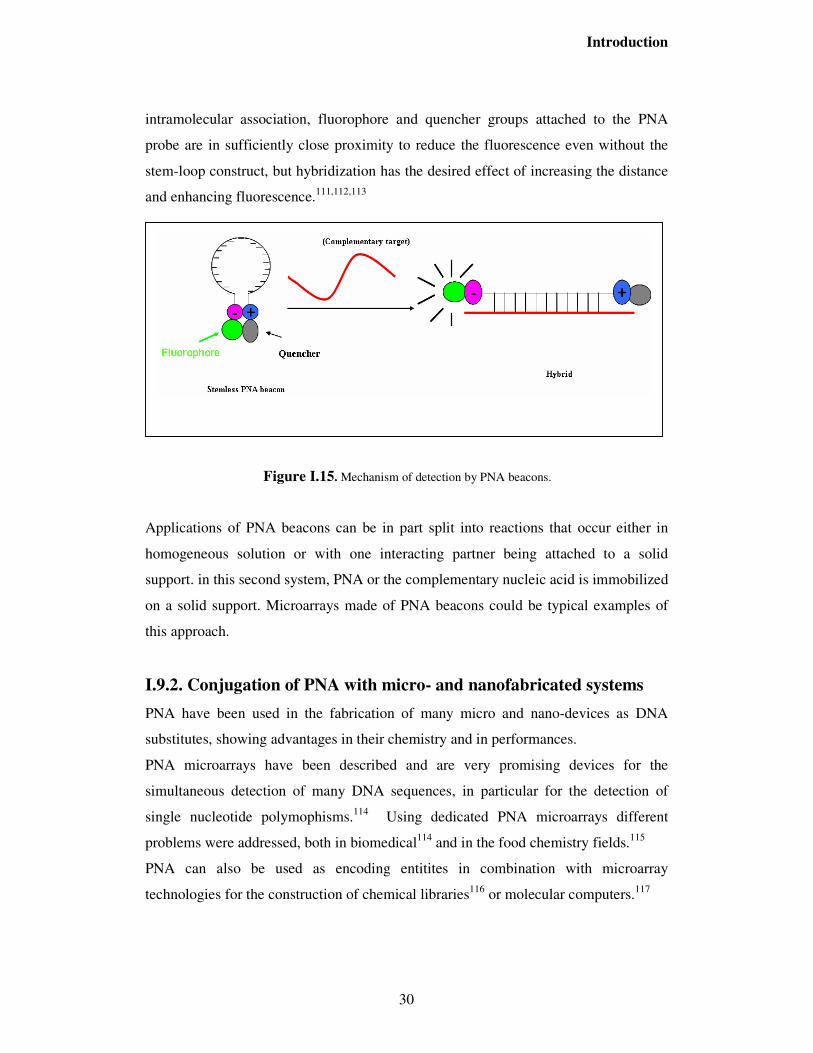
applications. In this area, molecular beacons (MBs) are popular tools for nucleic acids
detection. In these systems, a nucleic acid exhibits a fluorescent signal only in the
presence of the target oligonucleotide. Molecular beacons usually consist of a
fluorophore and a fluorescence-quencher attached at the termini of a nucleic acid
oligomer. When the termini are closed to one another, the fluorescence is quenched.
upon binding to the target oligonucleotide, separation of the termini is accompanied by
an increase in fluorescence. Previously, quencher-free molecular beacons have been
synthesized from DNA that utilize fluorophores quenched by nucleobases. With the
inception and continued study of PNA, molecular beacon strategies incorporating this
non natural oligoncleotide analogs have become increasingly popular.
The original design of DNA beacons placed the fluorophore and quencher on the ends
of hairpin-shaped molecules featuring a stem-loop structure. Stemless DNA beacons
in which the two ends of the sequence are non-complementary likely adopt extended
conformations at low salt concentration due to the polyanionic nature of the
backbone109. This reduces the amount of quenching in the unhybridized state, leading
to lower sensitivity for detection of DNA. In the case of PNA beacons, it was found
that a hairpin structure is not necessary. The lack of backbone charges allows single-
stranded PNA to collapse into a folded structure, most likely stabilized by a
combination of favorable intramolecular contacts as well as the hydrophobic effect.110
Moreover, PNAs are more likely to aggregate in solution. Due to this inter or
Introduction
30
intramolecular association, fluorophore and quencher groups attached to the PNA
probe are in sufficiently close proximity to reduce the fluorescence even without the
stem-loop construct, but hybridization has the desired effect of increasing the distance
and enhancing fluorescence.111,112,113
Figure I.15. Mechanism of detection by PNA beacons.
Applications of PNA beacons can be in part split into reactions that occur either in
homogeneous solution or with one interacting partner being attached to a solid
support. in this second system, PNA or the complementary nucleic acid is immobilized
on a solid support. Microarrays made of PNA beacons could be typical examples of
this approach.
I.9.2. Conjugation of PNA with micro- and nanofabricated systems
PNA have been used in the fabrication of many micro and nano-devices as DNA
substitutes, showing advantages in their chemistry and in performances.
PNA microarrays have been described and are very promising devices for the
simultaneous detection of many DNA sequences, in particular for the detection of
single nucleotide polymophisms.114 Using dedicated PNA microarrays different
problems were addressed, both in biomedical114 and in the food chemistry fields.115
PNA can also be used as encoding entitites in combination with microarray
technologies for the construction of chemical libraries116 or molecular computers.117
Introduction
31
Coupling of PNA with nanomaterials allows to produce very specific tools for
biomedical applications. Gold nanocrystal sensors modified with PNAs have been
prepared and applied to self-assembly and DNA sensing. In particular it was found
that base pair mismatch selectivity of PNAs is further enhanced on nanocrystals.118
PNAs have been combined with silicon nanowires for label-free detection of DNA.119
In these studies, highly sensitive, sequence-specific and label-free DNA sensors have
been developed by monitoring the electronic conductance of silicon nanowires
(SiNWs) with chemically bonded PNA probe molecules.
PNA have also been conjugated with single wall carbon nanotubes and with fullerene
to generate hybrid materials with special optical and electronic properties as
components of nanosystems.120
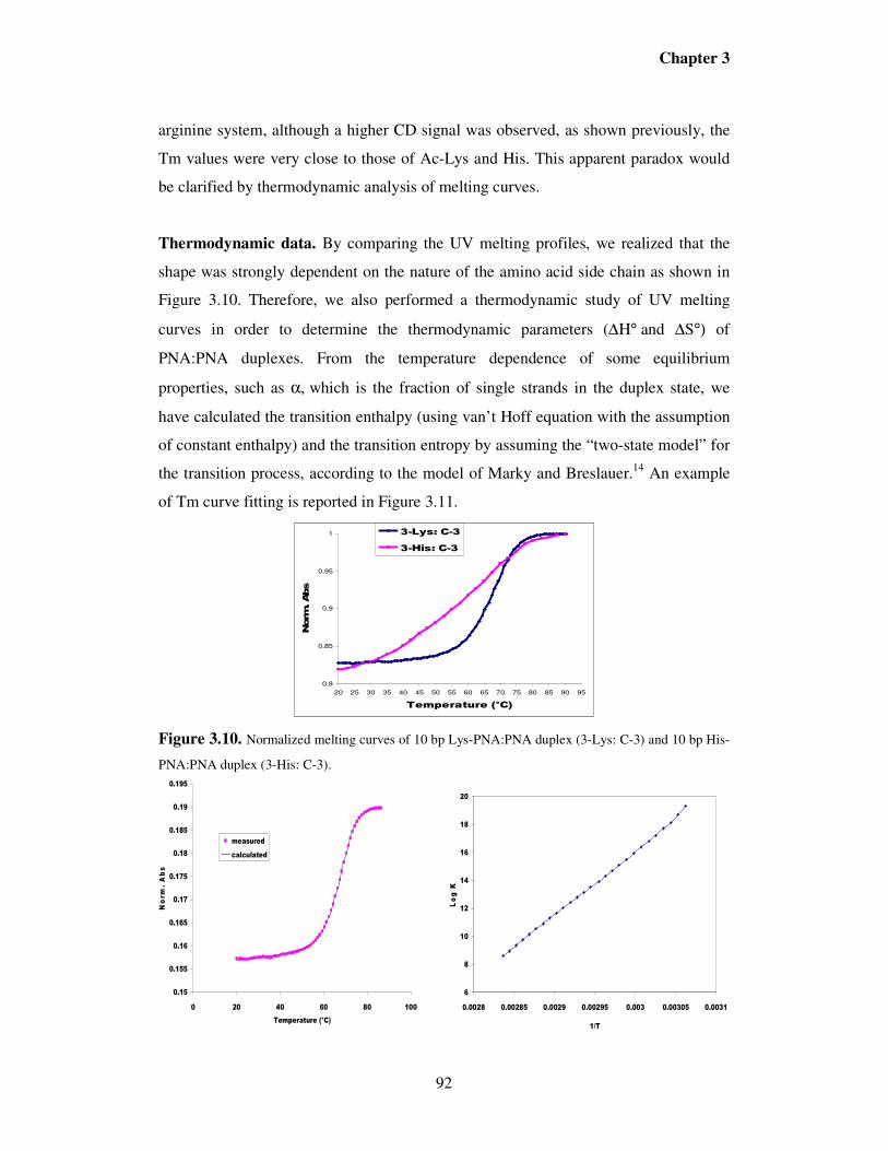
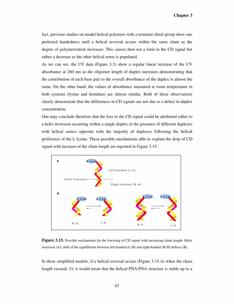
I.9.3. PNA:PNA duplexes as tunable nanomaterials: sergeant and soldiers behaviour. The helix is a very important conformational state, which exists widely in nature, be it
biological molecules like peptides, DNA, RNA, viruses or synthetic molecules like
polyisocyanates. Many internal and external factors have an effect on handedness of
the helix and are an interesting topic for scientists to study the origin of chirality and
evolution of biological molecules. Most biological polymers adopt a helical
conformation. This is clearly seen in the polynucleotide duplexes, the α- helix formed
by peptides and parts of protein structures. The presence of stabilizing soft interactions
in such biological systems gives rise to a barrier for inversion of helix handedness. In
the case of DNA (with certain base sequences), the B-form can invert to Z-form only
under drastic conditions of low humidity, high salt concentrations and certain base
sequences.
As mentioned earlier, two complementary PNA strands are able to form stable
PNA/PNA duplexes,31 both in parallel and in an antiparallel orientation. These
duplexes have no biological application, but can be considered as stable, highly
specific, programmable nanostructures, with higher chemical and biological stability
than DNA-based objects. One major difference among DNA- and PNA-based
duplexes is the possibility to control chirality and, through this, fine tuning helical
handedness and thus optical activity. The full control of these properties requires,
Introduction
32
however, the knowledge of factors able to induce and to propagate helicity in these
DNA-like structures, and the theoretical background in this field is still not complete.
Sound and experimentally proved models about helical propagation have been
developed in the polymer science. Based on the possibility of helix inversion, helical
polymers can be divided into two categories, helical polymers having high helix
inversion barriers and those with low helix inversion barriers. The polymers having
high helix inversion barriers can not easily be inverted from one helical sense to
another, as in case of the biological molecules. In recent years research has focused on
helical polymers with low helical inversion barriers. In these molecules the helical
domains with opposite handedness coexist and are interconvertible with reasonable
timescales. This makes it possible to use milder internal and external stimuli to
influence the helical conformation of the backbone.
Polyisocyanates fall in the second category of the helical polymers described above.
They are interesting in showing cooperative phenomenon in different situations and
give rise to chiral amplification121. The polymer backbone is found to be stiff and
helical due to the steric strain between the carbonyl oxygen and the nitrogen
substituent. The X-ray crystal structure of poly (butyl isocyanate) revealed a 8/3 -
helix. The backbones of achiral polyisocyanates composed of equal amounts of left
and right handed helices throughout the polymer chain, which are mirror images of
each other and dynamically interconvertible (similar to achiral PNAs), however with
small chiral perturbations in side groups, solvents or even circularly polarized light,
lead to the excess of one helical sense.
In case of short chain polyisocyanates, the whole chain can be composed of one single
helical sense consists of left or right handed. Thus the solution of short chain
polyisocyanates is a racemic mixture of the two helical senses. It was found that with
an increase of chain length, the single polymer chain is no longer composed of one
helical domain, but has multiple helical domains which are connected by helical
reversals. The free energy for a helical reversal in case of poly (n-hexyl isocyanate) in
hexane is about 3900cal/mol and varies somewhat with solvent. This energy
determines the length of the chain with a single helical sense. The 3900cal/mol free
energy corresponds to about 800 units at an ambient temperature, which is far larger
than the number of units in the persistence length. This is the source of cooperativity
Introduction
33
and the consequent effect of chiral amplification. This study was followed by the
synthesis of polyisocyanate copolymers consisting of varying ratios of chiral and
achiral monomers. The chiral residues impart preferential handedness to the helix
which tends to continue by the following achiral residues. The situation is similar to
soldiers following a sergeant and keeping in step with him. When small amounts of
chiral monomers (sergeant units) are introduced into polyisocyanates, which consist
predominately of achiral monomers (soldiers), it was found that the resulting
copolymer show high optical properties measured from the molar ellipticity values
obtained from CD at 254nm. The varying ratios of the chiral and achiral monomeric
units in the polymeric chains showed that a large CD signal appears even with the
incorporation of minute amounts of the chiral pendant group.122 The ellipticity
increased quickly and reached a saturation point with a proportion of only 2% of the
chiral monomer residue. It was evident that the preferential handedness in the helix
was controlled by very small portions of the chiral groups.
On the basis of the sergeant and soldiers experiment, it is reasonable to explore if this
kind of experiment could be applied to synthetic biopolymers such as PNA.
For example, the duplex formed by the PNA decamer H-G TAG ATC ACT- (L-Lys)-
NH2 and the complementary antiparallel sequence H- A GTG ATC TAC-(L-Lys)-NH2
melts at 67ºC. The corresponding antiparallel DNA-PNA duplex melts at 51ºC and the
DNA-DNA duplex melts at 33.5ºC. The antiparallel orientation is characteristically
more stable than the parallel duplex (45.5 ºC). It has been shown that, when achiral
strands of PNA are used for the formation of the duplex, no preferential helical sense
prevails. However attaching an amino acid at the carboxy terminus of one of the PNA
stands induces the formation of helices with preferential handedness (Figure I.16). The
kinetics of such a PNA-PNA duplex formation has been investigated by UV and CD
spectroscopy.31,123 The formation of a racemic mixture of the PNA/PNA duplex, as
estimated by UV measurements is a fast step followed by a relative slow conversion of
the double helix to one preferred helical sense as governed by the C-terminal amino
acid
Introduction
34
Figure I.16. Preferential helix handedness induced by C-terminal lysine in PNA/PNA duplexes
X-ray crystal structure analysis of a self-complementary PNA/PNA duplex (H-CGT
ACG-NH2), without the incorporation of chiral information, has been elucidiated.36,38
The duplex exists as both right-and left-handed helices, which are stacked alternately
in the crystal. As expected the base pairing is of Watson-Crick type and the bases lie
almost perpendicular to the helix axis with a propeller twist of about 5-9º. The helix
pitch was found to be 5.8nm and the rise per turn was equal to 32 Å. The base pairs
are displaced by 8.3Å relative to the helix axis, which gives a wide helix (28 Å) with
18 base pairs per turn. The helix has a very wide and deep major groove and a narrow
and shallow minor groove. The amide groups of the backbone are in the trans
conformation and carbonyl groups of the linkers point towards the C-terminus. This
type of helix is consistent with the P-form mentioned above. In DNA, the strong
circular dichroism arises from the helical stacking of the bases. The exciton coupling
between the transitions in nucleobases and the chiral deoxyribose backbone generates
strong chirality in duplexes and a strong CD spectrum. However, in case of PNAs the
backbone is completely achiral. Any electronic transitions between the majority of the
bases and the chiral C-terminal amino acid would be small. Thus any CD will arise
because of the chiral orientation of the base pairs relative to each other. As expected
the helices induced by D- and L- lysine were found to be of opposite helical sense.
NH
O
NH3+
NH2
ONH2H
3N+
NH3+
Achiral antiparallel
PNA duplex
Teminal AAL-Lys
Left-HandedRight-Handed
D-Lys
NH
O
NH3+
NH2
ONH2 NH
3+
H3N+
Introduction
35
I.10. PNA as models for prebiotic chemistry Due to their simple and chemically robust structure, PNA has also been considered as
a possible model for prebiotic chemistry. Many theories have been put forward which
lead to the current thinking that RNA may have been the first genetic material.
However the instability of the ribose and other sugars and the great difficulty of
prebiotic synthesis of the glycosidic bonds of nucleotides raised serious questions
about whether RNA could have been the first genetic material. In 1987, four years
before the discovery of PNA, Westheimer predicted that the backbone of the first
genetic material would be different from the ribose sugar backbone and N-(2-
aminoethylglycine) [AEG] could be one of the possibilities for the backbone of
prebiotic material. PNA thus seems to be one of the candidates for such a suggested
prebiotic material. It has been demonstrated124 that AEG and ethylenediamine are
produced directly in electric discharge from CH4, N2, H2 and H2O. Ethylenediamine is
also produced from NH4CN polymerization. The NH4CN polymerization in the
presence of glycine leads to adenine, cytosine and guanine-N9-acetic acid. Preliminary
experiments suggest that AEG may rapidly polymerize at 100ºC to give the
polypeptide backbone of PNA. The ease of synthesis of the components of PNA and
the possibility of polymerization of AEG reinforces that possibility that PNA may
have been the first genetic material.
An important number of theoretical and experimental studies has been performed in
order to support this hypothesis and gain further insight into the chemical evolution
and origin of life, in particular, of the RNA.
The origin of the RNA world is not easily understood, as effective prebiotic syntheses
of the components of RNA, the β-ribofuranoside-5’-phosphates are hard to envisage.
Recognition of this difficulty has led to the proposals that other genetic systems, the
components of which are more easily formed, may have preceded RNA. Among these,
PNA, which resembles RNA in its ability to form doubled-helices stabilized by
Watson-Crick H-bonding and bases stacking, has been investigated as model of a
potential genetic material that is free of phosphate. Based on these considerations,
several papers reported the use of PNA as possible precursor of RNA through
template-directed synthesis125, 126. For example, BÖhler et al126 suggested a new kind of
mechanism for genetic takeover, which demonstrates that PNA oligomers can act as a
Introduction
36
template for the regioselective oligomerization of RNA and vice versa. This means
that a transition between genetic systems can occur without loss of information.
However, a continuous transitions from one genetic system to another would be
possible if mixed molecules containing building block of both systems could be
formed. Koppitz et al.127 used PNA as template to form PNA/RNA (or DNA) chimeras
and investigated the role of the latter in a transition of information from PNA to RNA
or to DNA. They results provided evidence that a transition from PNA-like genetic
world to an DNA world is possible through multi-step process involving PNA-directed
PNA-DNA ligation. However, in the case of RNA transition, the information stored in
PNA could not necessary be utilized by RNA. Then, a sequence, that is, for example,
catalytically as PNA is unlikely to be active as RNA. Chimera formation, therefore
could not transfer useful information from PNA to RNA, but, could allow a transition
to a superior information-storing polymer. Therefore, RNA could first has evolved to
serve as a template to PNA synthesis, and only later evolved in sequences showing
independent catalytic function.
Although the RNA-world hypothesis which states that our biological life was preceded
by a prebiotic system in which RNA functioned both as genetic material and as
enzyme-like catalyst is widely accepted, this emphasizes the difficulty of forming and
replicating a homochiral nucleic acid in a solution of racemic nucleotides.128,129
Furthermore, prebiotic syntheses of chiral monomers always yield racemic mixtures.
Living systems use L-amino acids and D-nucleotide in their biopolymers. The
generation of optical asymmetry by selection and amplification in an autocatalytic
process is therefore an important element in many theory of the origin of the life.
Replication of polynucleotides in template-directed syntheses, is an obvious candidate
for such an amplification step for pre-“RNA world”130 A serious objection for this
suggestion is the observation that the efficiency of template-directed syntheses of
RNA is limited by enantiomeric cross-inhibition.131 PNA as model for a hypothetical,
nonachiral precursor of RNA in experiments re-examining enantiomeric cross-
inhibition has also been investigated and it was found that enantiomeric cross-
inhibition is as serious in the polymerization of nucleotides on PNA templates as it is
on a conventional RNA or DNA template.132 Since the influence of chiral substituents
such as amino acids on the distribution of left- and right-handed helices PNA has been
Introduction
37
investigated51,123 , one possible solution of this problem has been proposed by Kozlov
et al133, by using achiral PNA or PNA/RNA chimera as template through which a
chiral information induced by a terminal chiral unit can be propagated and amplified.
Their results especially suggested that the chirality induced by two nucleotides in a
template strand could be transmitted through normally achiral PNA and result in a
chirally selective template-directed remote elongation of a primer strand. This means
that the introduction of a short homochiral segment of DNA into a PNA helix could
have guaranteed that the next short segment of DNA to be incorporated would have
the same handedness. Once two segments of DNA were present, the probability that a
third segment would have the same handedness would increase and so on. This
scenario would allow the formation of a chiral oligonucleotide by processes that are
largely resistant to enantiomeric cross-inhibition.
Molecular Biology of the Gene, 1987, 4th ed. Benjamin/Cummings, Menlo park, CA. 7 Ptashne, M., Genetic Switch, 1987, Blackwell Scientific Publications, Palo Alto, CA. 8 Steitz, T.A., Q. Rev. Biophys., 1990, 23, 205-280. 9 Nielsen, P.E., Bioconjugate Chem., 1991, 2, 1-12. 10 Pyle, A.M. & Barton, J.K., Prog. Inorg. Chem., 1990, 38, 413-475. 11 b) Niemeyer, C. M.; Angew. Chem., 1997, 109, 603-606. 12 Seeman, N. C., Acc. Chem. Res., 1997, 30, 357-363. 13 a) Chen, J.; Seeman, N. C., Nature, 1991, 350, 631-633.
b) Brucale, M.; Zuccheri, G.; Samorì, B.; Trends Biotech., 2006, 24, 235-243. 14 Rothermund, P. W. K., Nature, 2006, 440, 297-302. 15 De Clerq E., Eckstein F., Sternbach H., Merigan T.C.; Virology, 1970, 42, 421. 16 Miller, P.S; Oligodeoxynucleotides. Antisense inhibitors of gene expression; 1989,
Macmillian Press, 79. 17 Manoharan M., Biochim. Biophys. Acta, 1999, 1489, 117. 18 Gryaznov, S. M., Biochim. Biophys. Acta 1999, 1489, 131. 19 Summerton, J., E., Biochim. Biophys. Acta 1999, 1489, 141. 20 Wengel, J., Acc. Chem. Res. 1999, 32, 301. 21 Nielsen P. E., Egholm M., Berg R. H., Buchardt O., Science, 1991, 1497.
Introduction
39
22 Egholm M., Buchardt O., Christensen R., Behrens C., Freier S. M., Driver D. A.,
Berg R. H., Kim S. K., Norden B., Nielsen P.E., Nature, 1993, 365, 566. 23 Buchardt O., Egholm M., Berg R. H., Nielsen P. E., Trends Biotechnol. 1993, 11,
384. 24 Nielsen P. E., Egholm M., Berg R. H., Buchardt O., Anti-Cancer Drug Des. 1993, 8,
53. 25 Koch, T.; Hansen, H.F., Andersen, P., Larsen, T.; Batz, H.G.; Otteson, K., Orum, H.
J. Pept. Res., 1997, 49, 80. 26 Demidov V.A., Potaman V.N., Frank-Kamenetskii M. D., Egholm M., Buchardt O.,
Chem. Lett., 1996, 6, 665. 44 Bergmann, F.; Bannwarth, W.; Tam S. Tertahedron Lett., 1995, 36, 6823 45 Casale R., Paul C.H., Jensen I.S., Moyer M.L, Kates S. A., Egholm M., in
Innovation in Solid Phase Synthesis and Combinatorial Libraries, Mayflower Science,
1998, 31, 139 46 Kovacs, G.; Timar, Z.; Kupihar, Z.; Kele, Z.; Kovacs, L. J. Chem. Soc.Perkin Trans.
1, 2002, 1266-1270. 47 Tedeschi, T.; Ph.D Thesis, University of Parma 2001-2003, pp 18 48 Hyunil, L.; Jae Hoon, J.; Jong Chan L.; Hoon C.; Yeohong Y.; Sung Kee, K., Org.
Lett., 2007, 9 (17), 3291-3293. 49 Ganesh, K. N.; Nielsen, P. E, Curr. Org. Chem., 2000, 4, 931-943. 50 a) Kumar, V. A., Eur. J. Org. Chem., 2002, 2021-2032.
8395-8399 62 Dragulescu-Andrasi, A.; Rapireddy, S.; Frezza, B. M.; Gayathri, C.; Gil, R. R.; Ly,
D. H., J. Am. Chem. Soc., 2006, 128, 10258-10267. 63 Sforza, S.; Tedeschi, T.; Corradini, R.; Marchelli, R., Eur. J. Org. Chem., 2007, 5879–5885. 64 Jensen K.K.; Orum, H.; Nielsen P.E.; Nordén, B. Biochemistry 1997, 36, 5072. 65 Nielsen P.E.; Egholm, M.; Buchardt, O. Gene 1994, 149, 139. 66 Dieci, G.; Corradini, R.; Sforza, S.; Marchelli, R.; Ottonello, S. J. Biol. Chem.
2001, 276, 5720 67 Zelphati, O.; Liang, X.W.; Hobart, P.; Felgner, P.L. Human Gene Ther.1999, 10, 15. 68 Romanelli, A.; Pedone,C.; Saviano, M., Bianchi, N.; Borgatti, M.; Mischiati, C.;
Gambari, R. Eur. J. Biochem. 2001; 268, 6066 69 Uhlmann, E. Biol. Chem. 1998, 379, 1045. 70 a) Braasch, D. A.; Corey, D. R, Biochemistry, 2002, 41, 4503-4510.
b) Xodo, L. E.; Cogoi, S.; Rapozzi, V., Curr. Pharm. Design, 2004, 10, 805-819. 71 Wittung P., Kajanus K., Edwards G., Haaima G., Nielsen P.E., Norden B.,
Malmstrom B.G., FEBS Lett., 1995, 375, 27 72 Koppelhus U., Nielsen P.E., Advanced Drug Delivery Reviews, 2003, 55, 267 73 Koppelhus, U.; Awasthi, S. K.; Holst, H. U.; Hebbesen, P.; Nielsen, P. E., Antisense
and Nucleic Acid Drug Develop, 2002, 12, 51. 74 Good, L.; Nielsen, P.E. Nature Biotech 1998, 16, 355. 75 Dryselius R., Nekhotiaeva N., Nielsen P.E., Good L.; BioTechniques, 2003, 35,
2000, 122, 7435. 118 Chakrabarti R., Klibanov M.; J. Am. Chem. Soc. 2003, 125, 12531. 119 a) Gao, Z.; Agarwal, A.; Trigg, A. D.; Singh, N.; Fang, C., Tung, C-H.; Fang, Y.;
Buddharaju, K. D.; Kong, J., Anal, Chem., 2007, 79, 3291-3297.
b) Li, Z.; Rajendran, B.; Kamins, T. I.; Li, X.; Cheng, Y.; Stanley Williams, R., Appl.
Phys. A., 2005, 80, 1257-1263. 120 Singh, K. V.; Panday, R. R.; Wang, X.; Lake, R.; Ozkan, C. S.; Wang, K.; Ozkan,
M., Carbon, 2006, 44, 1730-1739. 121 a) Green, M. M.; Park, J.-W.; Sato, T.; Teramoto, A.; Lifson, S.; Selinger, R. L. B.;
Selinger, J. V., Angew. Chem. Int. Ed. 1999, 38, 3138;
b) Green, M. M.; Cheon, K. S.; Yang, S. Y.; Park, J. W.; Swansburg, S.; Liu, W., Acc.
Chem. Res. 2001, 34, 672. 122 Jha, S.K.; Cheon, K. S.; Green M.M.; Selinger, J.V., J. Am. Chem. Soc. 1999,
121,1665. 123 Wittung, P.; Eriksson, M.; Reidar, L.; Nielsen P. E.; Nordén, B., J. Am, Chem.
Soc.1995, 117, (41), 10167. 124 Nelson, K.E.; Levy, M.; Miller, S.L., Proc. Nat. Acad. Sci. USA, 2000, 97, 3868.
Introduction
45
125 Miller, S. L. Nat. Struct. Biol., 1997, 4, 167-169. 126 Bohler, C.; Nielsen, P. E.; Orgel, L. E., Nature, 1995, 376, 578-581. 127 Koppitz, M.; Nielsen, P. E.; Orgel, L. E., J. Am. Chem. Soc., 1998, 120, 4563-4569. 128 Bolli, M.; Micura, R.; Eschenmoser, A, Chem. Biol., 1997, 4, 309-320. 129 Schwartz, A. W., Curr. Biol., 1997, 7, R477-R479. 130 Gestland, R.; Atkins, J. F., Eds; Cold Spring Habor Laboratory Press: Cold Spring
Habor, NY, 1993, Vol, Monograph 24. 131 Joyce, G. F.; Visser, G. M.; Van Boeckel, C. A. A.; Van Boom, J. H.; Orgel, L. E.;
Van Westrenen, J., Nature, 1984, 310, 602-604. 132 Schmidt, J. G.; Nielsen, P. E.; Orgel, L. E., J. Am. Chem. Soc, 1997, 119, 1494-
1495. 133 Kozlov, I. A.; Orgel, L. E.; Nielsen, P. E., Angew. Chem. Int. Ed., 2000, 39 (23),
4292-4295.
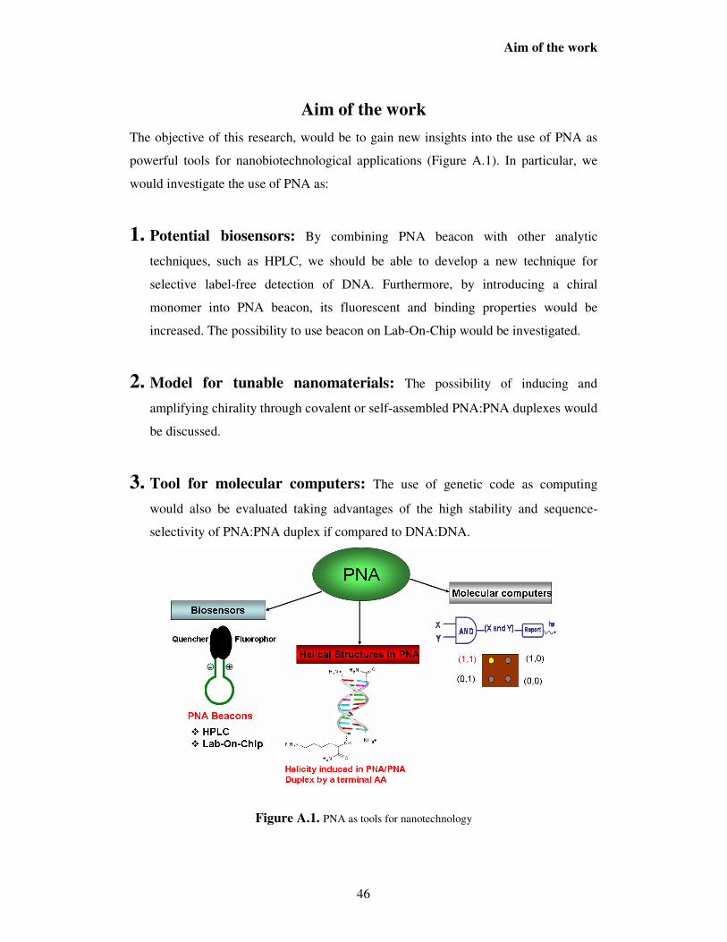
Aim of the work
46
Aim of the work
The objective of this research, would be to gain new insights into the use of PNA as
powerful tools for nanobiotechnological applications (Figure A.1). In particular, we
would investigate the use of PNA as:
1. Potential biosensors: By combining PNA beacon with other analytic
techniques, such as HPLC, we should be able to develop a new technique for
selective label-free detection of DNA. Furthermore, by introducing a chiral
monomer into PNA beacon, its fluorescent and binding properties would be
increased. The possibility to use beacon on Lab-On-Chip would be investigated.
2. Model for tunable nanomaterials: The possibility of inducing and
amplifying chirality through covalent or self-assembled PNA:PNA duplexes would
be discussed.
3. Tool for molecular computers: The use of genetic code as computing
would also be evaluated taking advantages of the high stability and sequence-
selectivity of PNA:PNA duplex if compared to DNA:DNA.
Figure A.1. PNA as tools for nanotechnology
Chapter 1
47
PNA Beacons in Label-Free Selective Detection of DNA by
Fluorimetry and by Ion Exchange HPLC.
1.1. Introduction
Genome-based technologies rely on the possibility to selectively recognize DNA
sequences of applicative interest. The quest of new and selective methods and
technologies for the detection of specific DNA tracts is gaining more and more
importance in diagnostics, from biomedical to more large scale items such as food and
feed1,2
One very important class of probes is represented by molecular beacons (MB), which
are composed of a sequence specific oligonucleotide coupled with a fluorophore and a
quencher (or a quenching surface) at each end, held together by a zipper DNA
sequence made of complementary antiparallel tracts; this structure allows to produce a
”switch-on” of fluorescence upon interaction with the target DNA sequence3,4. A
variety of applications to DNA or RNA detection have been proposed using MB
probes5,6; detection of single point mutations can be achieved by MB through careful
design of the sequence and selection of the detection temperature7,8. Combined
approaches using molecular beacons are also effective in mismatch detection9.
Peptide nucleic acids (PNAs) are efficient tools in diagnostics, since they can bind
DNA with high affinity and selectivity and are superior to oligonucleotide probes in
the recognition of single base mutations10,11. PNA-beacons12,13,14 and the related “light
up probes”15,16 have been recently described, displaying the advantages of higher
selectivity and simpler design, since their flexibility allows the fluorophore-quencher
interaction to occur even in the absence of a “zipper” sequence, thus allowing to
introduce possible interferences, and they are less sensitive to ionic strength changes17.
One of the major limitations in the use of PNA and other Molecular Beacons in
diagnostics is represented by the fluorescence background of the free (uncomplexed)
probe, which can be interfering with the signal obtained by the analyte sequence when
it is in low concentrations, especially for DNA amplified by complex biological
samples.
Chromatographic analysis has been performed on PCR products18. This type of
chromatography is simple and uses water solution at increasing ionic strength as
Chapter 1
48
eluents19, thus allowing the analysis to be performed under non-denaturing conditions;
however, the presence of a high number of interfering components, such as primers,
mononucleotide, or enzymes used in the PCR reaction, which are revealed by UV and
other type of detectors, makes the interpretation of data more difficult in real samples.
Furthermore, detection of small sequence differences such as single nucleotide
polymorphisms (SNPs) are very difficult using this approach. In previous works
carried out in our group it was demonstrated that ion-exchange (IE) HPLC can be used
for directly visualize the PNA:DNA interaction, since the latter shows retention times
different from those of PNA and DNA20,21. When the DNA to be analyzed is labelled
with fluorescent groups, the chromatographic profile is simpler, but interfering peaks
of primers and of unspecific amplified DNA can be present.
In the present work, we report the combined use of PNA-beacon and IE-HPLC
analysis for the label-free detection of DNA, taking advantage on one side from the
separation of the free probe from the complex, and on the other side from the very
specific signal generated by the PNA beacon, which allows to avoid unspecific peaks.
Furthermore, unlabelled PCR products can be detected with this method.
The PNA beacons and DNA sequences used are listed in Figure 1.1.
X = A; C; G; T
Figure 1.1. PNA beacons design. In the unhybridized state, the termini are close to one another, the
fluorescence is quenched. Upon binding to the target oligonucleotide, separation of the termini is
accompanied by an increase in fluorescence (switch-on).
Dabcyl was used as quencher, linked to the ε-amino group of lysine at the C-terminal
part and carboxyfluorescein was used as fluorophore, linked to the N-terminus of the
PNA molecule; two additional charges (a positive lysine side chain and a negative
glutamate unit) were inserted just before and after the PNA segment, following the
design described by Frank-Kamenetskii and co-workers.17
PNA
PNA 1 : GATTTCAATGC
PNA 2 : AGAGTCAGCTT
DNA 3-X 5’GCATTXAAATC3’
DNA 4-X 5’AAGCTGXCTCT3’
Glu -Lys-Lys-CONH2
Fluo DabcylN-terminal group N-Hε-amino group
H-N
Chapter 1
49
1.2. Results and Discussion
The beacon 1 was synthesized in a previous work and has a sequence for which a
relatively low melting temperature of the PNA:DNA duplex was observed (Table 2.1)
while the beacon 2 has the same sequence of a PNA previously utilized as a probe for
the detection of Roundup Ready soybean in HPLC, with higher PNA:DNA melting
temperature21.
Both PNA beacons were synthesized using solid phase synthesis on an automatic ABI
433A Synthesizer, according to the scheme reported in the introduction of this thesis.
Fmoc strategy was used with HBTU/DIEA as coupling agent. The fluorophore
(Carboxyfluorescein) and quencher (Dabcyl) were attached manually. The crude
products were purified by RP-HPLC and characterized with HPLC-MS. The
chromatogram profiles and ESI-MS spectra of the pure products are reported in
Figures 1.2 and 1.3.
Figure 1.2. Chromatogram profile and ESI-MS reconstructed spectra of PNA 1.
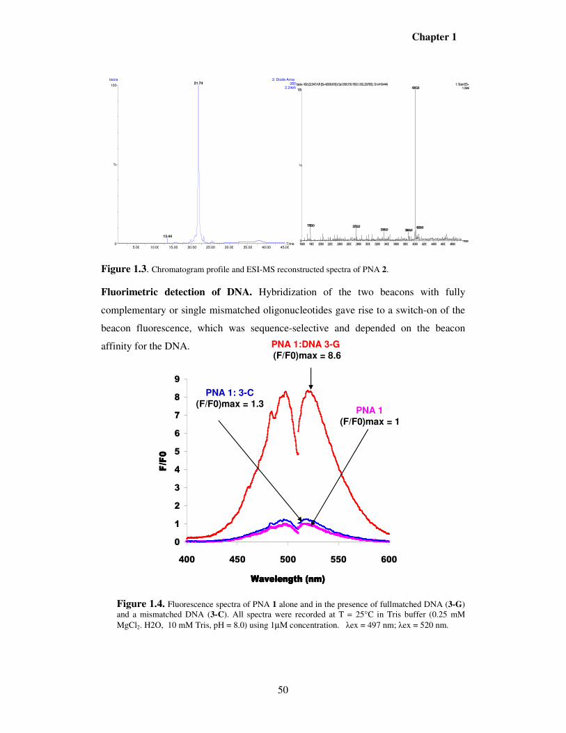
Figure 1.3. Chromatogram profile and ESI-MS reconstructed spectra of PNA 2.
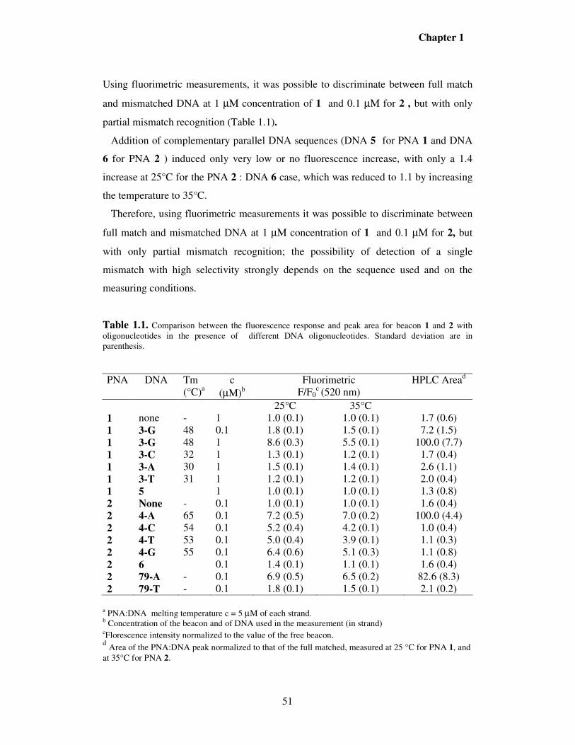
Fluorimetric detection of DNA. Hybridization of the two beacons with fully
complementary or single mismatched oligonucleotides gave rise to a switch-on of the
beacon fluorescence, which was sequence-selective and depended on the beacon
affinity for the DNA.
Figure 1.4. Fluorescence spectra of PNA 1 alone and in the presence of fullmatched DNA (3-G) and a mismatched DNA (3-C). All spectra were recorded at T = 25°C in Tris buffer (0.25 mM
MgCl2. H2O, 10 mM Tris, pH = 8.0) using 1µΜ concentration. λex = 497 nm; λex = 520 nm.
Using fluorimetric measurements, it was possible to discriminate between full match
and mismatched DNA at 1 µΜ concentration of 1 and 0.1 µΜ for 2 , but with only
partial mismatch recognition (Table 1.1).
Addition of complementary parallel DNA sequences (DNA 5 for PNA 1 and DNA
6 for PNA 2 ) induced only very low or no fluorescence increase, with only a 1.4
increase at 25°C for the PNA 2 : DNA 6 case, which was reduced to 1.1 by increasing
the temperature to 35°C.
Therefore, using fluorimetric measurements it was possible to discriminate between
full match and mismatched DNA at 1 µΜ concentration of 1 and 0.1 µΜ for 2, but
with only partial mismatch recognition; the possibility of detection of a single
mismatch with high selectivity strongly depends on the sequence used and on the
measuring conditions.
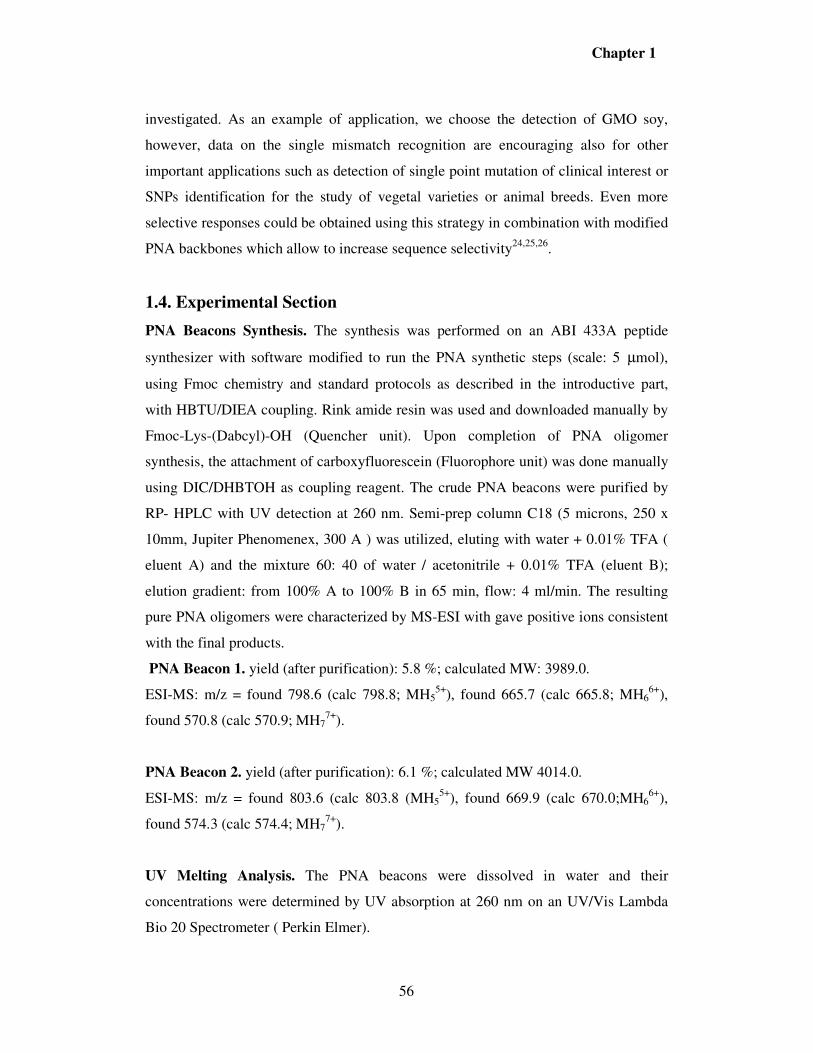
Table 1.1. Comparison between the fluorescence response and peak area for beacon 1 and 2 with oligonucleotides in the presence of different DNA oligonucleotides. Standard deviation are in parenthesis.
a PNA:DNA melting temperature c = 5 µM of each strand. b Concentration of the beacon and of DNA used in the measurement (in strand) cFlorescence intensity normalized to the value of the free beacon. d Area of the PNA:DNA peak normalized to that of the full matched, measured at 25 °C for PNA 1, and
a PNA:DNA melting temperature c = 5 µM of each strand. b Fluorescence intensity normalized to the value of the free beacon.
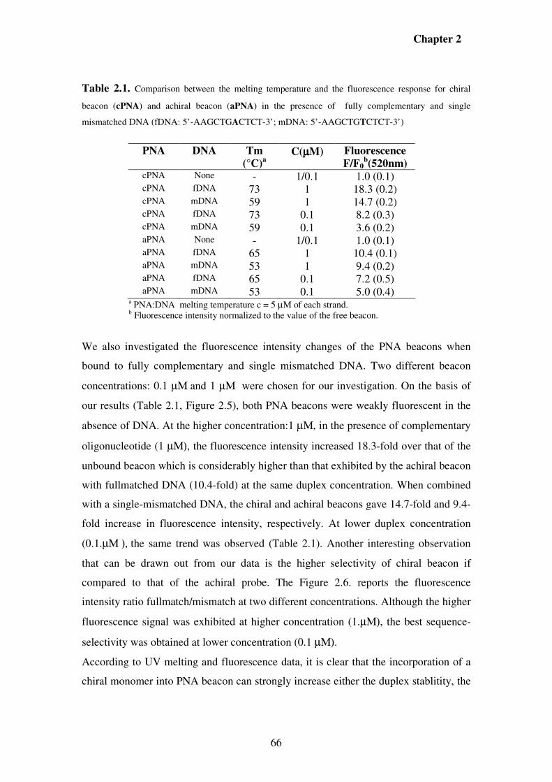
We also investigated the fluorescence intensity changes of the PNA beacons when
bound to fully complementary and single mismatched DNA. Two different beacon
concentrations: 0.1 µΜ and 1 µΜ were chosen for our investigation. On the basis of
our results (Table 2.1, Figure 2.5), both PNA beacons were weakly fluorescent in the
absence of DNA. At the higher concentration:1 µΜ, in the presence of complementary
oligonucleotide (1 µΜ), the fluorescence intensity increased 18.3-fold over that of the
unbound beacon which is considerably higher than that exhibited by the achiral beacon
with fullmatched DNA (10.4-fold) at the same duplex concentration. When combined
with a single-mismatched DNA, the chiral and achiral beacons gave 14.7-fold and 9.4-
fold increase in fluorescence intensity, respectively. At lower duplex concentration
(0.1.µΜ ), the same trend was observed (Table 2.1). Another interesting observation
that can be drawn out from our data is the higher selectivity of chiral beacon if
compared to that of the achiral probe. The Figure 2.6. reports the fluorescence
intensity ratio fullmatch/mismatch at two different concentrations. Although the higher
fluorescence signal was exhibited at higher concentration (1.µΜ), the best sequence-
selectivity was obtained at lower concentration (0.1 µΜ).
According to UV melting and fluorescence data, it is clear that the incorporation of a
chiral monomer into PNA beacon can strongly increase either the duplex stablitity, the
Chapter 2
67
sequence-selectivity or the fluorescent properties, allowing the use of chiral PNA
beacon as powerful biosensors for DNA detection and mismatch recognition.
Figure 2.5. Fluorescence spectra recorded at 25°C of chiral PNA beacon (cPNA) and achiral beacon
(aPNA) bound to a fully complementary DNA (fDNA) and a single-mismatched DNA (mDNA).
Excitation was monitored at 497 nm and Emission was measured at 520 nm. Strand concentration was
1µM in each component (PNA/DNA = 1:1).
1
1.2
1.4
1.6
1.8
2
2.2
2.4
Fullmatch/Mismatch
0.1 1
Concentration (uM)
Chiral
Achiral
Figure 2.6. Fluorescence intensity ratio between fullmatched and mismatched DNA hybridized with
chiral PNA beacon (Blu) and achiral beacon (Red) at two differents concentrations: 0.1.µΜ and 1.µΜ
0
2
4
6
8
10
12
14
16
18
20
400 450 500 550 600
Wavelength (nm)
F/F0
cPNA:fDNA
cPNA:mDNA
aPNA:mDNA
aPNA:fDNA
aPNA; cPNA
Chapter 2
68
Preliminary microarray studies. On the basis of the results obtained in solution, we
sought to determine if the higher performance of the chiral beacon, both in sequence-
selectivity and fluorescence properties can be further increased by attachment on a
solid support. Both PNA beacons were immobilized on the N-hydroxysuccinimide
(NHS) activated glass slide using microarray techniques protocols. The resulting spots
were hybridized with fully complementary DNA (fDNA) and a single-mismatched
DNA (mDNA). Despite a fluorescence signal was observed in the presence of the fully
complementary target sequence, no significant difference either in fluorescence
intensity or in sequence-selectivity was revealed between the two beacons (data not
shown). Then, the advantage of using a PNA containing the chiral monomer was not
as evident as in the solution study. Furthermore, the selectivities towards mismatch
were depending on the deposition conditions.
The major problem revealed in these experiments was the background fluorescence of
the beacon which was dependent on the hybridization conditions. This problem was
particularly severe when PCR products were directly hybridized on the microarray
(data not shown). In order to overcome this limitation, in collaboration with G. De
Bellis of the CNR (Milan), we performed a test using a new and powerful technology,
named “Lab-on-chip” (Figure 2.7) using the chiral beacon. This tool is a miniaturized
device (25x75.5 mm2) containing a thermostated chamber for PCR thermal cycles,
capillary electrophoresis unit and a microarray part, all arranged in sequence. The pre-
hybridization (PCR area) consists in PCR reaction step followed by separation of PCR
products; whereas, the chiral PNA beacon was immobilized in the hybridization area.
Although the study is still preliminary, it provides some important observations : The
presence of a fluorescence signal only in the case of specific PCR product suggesting
that this device could be use as a selective technology for detecting DNA tracts.
Futhermore, the time of analysis (45 min) is drastically reduced if compared to that of
traditional PNA microarray techniques. However, deposition conditions and
microarray set up has still to be improved. The advantage of this approach should be
the use of label-free DNA samples on a fully automated and miniaturized device.
Chapter 2
69
Figure 2.7. a) Lab-on-Chip device: showing the electrical contacts (left side) for interfacing with the TCS (temperature control system) instrument; the silicon chip (right side), showing four inlet holes, PCR area (center), and post-PCR analysis area. b) Results of detection by chiral PNA beacon of specific PCR product amplified on Lab-on-chip.device. PCR thermal cycling program: 40 cycles, 15s at 94 °C; 15s at 60 °C and 15s at 72 °C.The fluorescence signal deriving from the hybridization was acquired at λex = 497 nm and λex = 520 nm.
2.3. Conclusions
In this study, we developed a method to modify PNA backbone with side chain that
can be covalently attached to a solid support. Our modification shows an increase (in
solution) either in duplex stability and selectivity in mismatch recognition or in
fluorescence properties. Although the results obtained on microarray were not
satisfactory, the preliminary study performed on Lab-on-chip opens the possibility to
use the latter as a more selective and fast technology for DNA detection.
2.4. Experimental Section
General Information. Reagents were purchased from Sigma –Aldrich, Fluka, Applied
Biosystems, NovaBiochem and used without purification. DMF was dried over 4 Å
molecular sieves. THF was dried by distillation. TLC was run on Merck 5554 silica 60
aluminium sheets. Column chromatography was performed as flash chromatography
on Merck 9385 silica 60 ( 0.040- 0.063 mm). Reactions were carried out under
nitrogen.
NMR spectra were obtained on a Brucker 300 or Varian 600 MHZ spectrometer.
−δ values are in ppm relative to CDCl3 (7.29 ppm for proton and 76.9 for carbon) or
DMSO-d6 (2.50 ppm for proton and 39.5 for carbon).
b) Resultsa) Lab-On-Chip Device
PCR area Hybridization and
Detection areaElectric contacts
Inlet holesSpecific PCR
productAspecific products
And others
b) Resultsa) Lab-On-Chip Device
PCR area Hybridization and
Detection areaElectric contacts
Inlet holes
b) Resultsa) Lab-On-Chip Device
PCR area Hybridization and
Detection areaElectric contacts
b) Resultsa) Lab-On-Chip Device
PCR area Hybridization and
Detection areaElectric contacts
Inlet holesSpecific PCR
productAspecific products
And others
Chapter 2
70
Fmoc-Lys(Boc)–N(Me)OMe (1). Fmoc-Lys(Boc)-OH (2.01 g, 4.3 mmol) was
dissolved in dry DMF (20 ml) and HBTU ( 1.58 g, 4.13 mmol) was added to the
solution. the reaction mixture was then cooled to 0°C with an ice batch. after 15
minutes of stirring, DIEA (1.5 ml, 8.6 mmol) and N-methoxy-N-methylamine
hydrochloride salt (0.42 g, 4.3 mmol) were added slowly to the stirring solution. The
solution was allowed to stir for ten minutes at 0° C and the ice batch was removed.
The reaction mixture kept for 17 hours at room temperature and the DMF was then
evaporated. The residue was dissolved in EtOAc and washed with saturated potassium
hydrogen sulfate (2 times) and saturated sodium hydrogen carbonate (2 times). The
organic layer was dried over sodium sulfate, filtered and evaporated to afford 2.20 g
(99% yield) of the product as a colorless foam. 1H NMR (300 MHz, CDCl3): δ = 7.76 (d, 2H, J = 7.3 Hz, CH aromatic Fmoc), 7.60
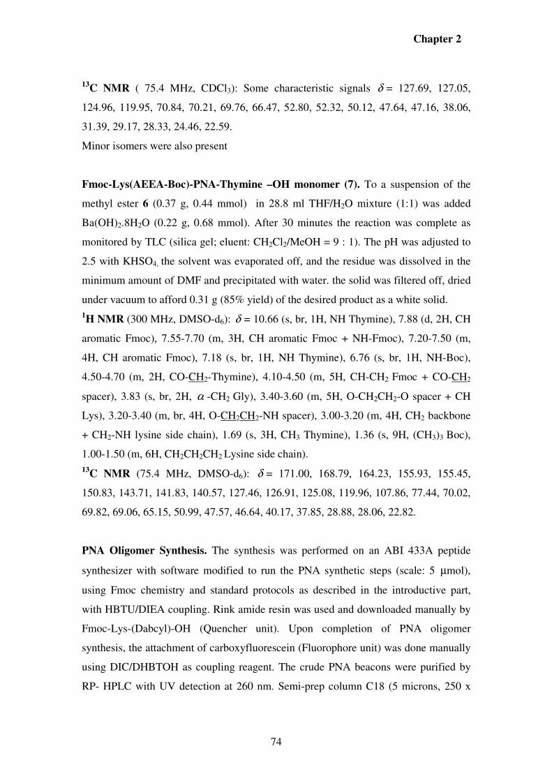
Fmoc-Lys(AEEA-Boc)-PNA-Thymine –OH monomer (7). To a suspension of the
methyl ester 6 (0.37 g, 0.44 mmol) in 28.8 ml THF/H2O mixture (1:1) was added
Ba(OH)2.8H2O (0.22 g, 0.68 mmol). After 30 minutes the reaction was complete as
monitored by TLC (silica gel; eluent: CH2Cl2/MeOH = 9 : 1). The pH was adjusted to
2.5 with KHSO4, the solvent was evaporated off, and the residue was dissolved in the
minimum amount of DMF and precipitated with water. the solid was filtered off, dried
under vacuum to afford 0.31 g (85% yield) of the desired product as a white solid. 1H NMR (300 MHz, DMSO-d6): δ = 10.66 (s, br, 1H, NH Thymine), 7.88 (d, 2H, CH
sulphate (SDS) was used as deposition buffer. Moreover, after every deposition, the
pin-and-ring system was purged with water for 10s and further washed with
acetonitrile/water (1:1), in order to avoid dragging of the probes in subsequent
depositions. The probes were coupled to the surface and the remaining reactive sites
were blocked by leaving the slides in a humid chamber (relative humidity 75%) at
room temperature for 12h, followed by immersion in a glass rack containing a 50mM
solution of ethanolamine, 0.1M TRIS, pH 9, prewarmed at 50°C, for 30min. The slides
were washed twice with bidistilled water at room temperature and then slowly shaken
for 30min in plastic tubes containing a 4× saline/sodium citrate (SSC) solution and a
0.1% SDS buffer prewarmed at 50°C. Each slide was then washed with bidistilled
water at room temperature and centrifuged in a plastic tube at 800rpm for 3min. Slides
were then ready to undergo the hybridization protocol.
DNA samples (1µM strand concentration) to be tested were prepared by diluting stock
solutions to a final volume of 65µl and a final concentration of 4× SSC and 0.1% SDS
buffer. Hybridization was performed by loading the samples to “in situ frame”
chambers and leaving the slides under slow shaking for 2h at 40°C. After the
hybridization step all the slides were treated individually to prevent cross
contamination. The slides were washed under slow shaking for 5min at 40°C with a 2×
SSC, 0.1% SDS buffer prewarmed at 40°C, followed by treatment for 1min with 0.2×
SSC and for 1min with 0.1× SSC at room temperature. The slides were then spin-dried
at 1000 rpm for 5min.
The fluorescent signal deriving from the hybridization was acquired using a GMS 418
Array Scanner (Genetic Microsystem) at λ ex=497nm and λ em=520nm.
PCR amplification and detection on Lab-On-Chip. The biochip was used to
amplify a long target DNA specific for Round Ready soy bean. The PCR
amplification was performed according to a protocol reported in a recent study.20 Then
the PCR products were transferred in the detection area where the chiral PNA beacon
was covalently linked. The fluorescence signal derived from hybridization was acquire
at λ ex=497nm and λ em=520nm.
Chapter 2
77
2.5. References 1Lipshutz, R. J., Fodor, S. P. A., Gingeras, T. R. & Lockart, D. J., Nat.Genet., 1999,
21, 20–24. 2 Wolcott, M. J., Clin. Microbiol., 1992, Rev. 5, 370–386. 3 Piatek, A. S., Tyagi, S., Pol, A. C., Telenti, A., Miller, L. P., Kramer, F. R. &Alland,
D., Nat. Biotechnol.,1998, 16, 359–363. 4 a) Ranade, K., Chang, M. S., Ting, C. T., Pei, D., Hsiao, C. F., Olivier, M., Pesich,
R., Hebert, J., Chen, Y. D., Dzau, V. J., Genome Res.,2001, 11, 1262–1268.
b) Wang, J., Nucleic Acids Res.,2000, 28, 3011–3016. 5 a) Service, R. F., Science, 1998, 282, 396–399.
b) Southern, E. M., Trends Genet, 1996, 12, 110–115. 6 Epstein, C. B. & Butow, R. A., Curr. Opin. Biotechnol., 2000, 11, 36–41. 7 Kozian, D.H.; Kirschbaum, B.J., Trends Biotechnol., 1999, 17, 73-78. 8 Debouck, C.; Goodfellow, P.N., Nat Genet., 1999, 21, 48-50. 9 Whitcombe, D.; Newton, C.R.; Little, S., Curr, Opin. Biotechnol., 1998, 9, 602-608. 10 Blais, B. W.; Phillippe L. M.; Vary, N., Biotechnol. Lett.,. 2002, 24 (17), 1407-1411. 11 Rudi, K.; Rud, I.; Holck, A., Nucleic Acid Res. 2003, 31 (11), e62/1-e62/8. 12 Weiler, J.; Gausepohl, H.; Hauser, N.; Jensen, O. N.; Hoeisel, J. D., Nucleic Acids
Res., 1997, 25, 2792. 13 Germini A, Mezzelani A, Lesignoli F, Corradini R, Marchelli R, Bordoni R,
Consolandi C, De Bellis G, J Agric Food Chem, 2004, 52(14), 4535–4540. 14 Germini A., Rossi S., Zanetti A., Corradini R., Fogher C., Marchelli R., J Agric
D.H. J. Am. Chem. Soc. 2006, 128, 10258-10267. 19 Englund E. A., Appella D. H., Org Lett 2005; 7: 3465-3467. 20 Consolandi, C.; Severgini, M.; Frosini, A; Caramenti, G.; De Fazio, M.; Ferrara, F.;
Zocco, A.; Fischetti, A.; Calmieri, M.; De Bellis, G.; Anal. Biochem, 2006, 353, 191-
197.
Chapter 3
79
Insights into the Propagation of Helicity in PNA:PNA
Duplexes as a Model for Nucleic Acid Cooperativity
3.1. Introduction
In recent years, the DNA molecule has been considered not only for its central role in
biological systems, but also as a special nanostructured programmable material which
allows fabrication of special structures through a self assembly process1.
The characteristics of DNA are well explored including the sense of the double helix
of the varying forms of DNA. The helical sense of a DNA duplex, as for example the
right handed conformation of B-DNA, is determined by the absolute configuration of
the D-deoxyribose sugar that attends every unit on each strand of the duplex. This
chiral influence is certainly over-determined considering that the structure of DNA is
highly cooperative.
In synthetic helical polymers the highly cooperative effect of monomers was shown
both theoretically and experimentally to allow far less chiral input than is found in
DNA to be capable of controlling helical sense2. For example, a helical polymer
constructed of very few chiral non-racemic chiral units disbursed among many achiral
units is adequate in a highly cooperative system to control the helical sense of large
portions of the chain (sergeant and soldiers experiment)3. In another approach, a
helical polymer constructed of a mixture of nearly racemic chiral units randomly
dispersed along the chain will take a helical sense of the units in the majority (majority
rule experiment)4. These studies have demonstrated that the higher the level of
cooperativity, the smaller is the chiral influence necessary to control helical sense. For
this reason these kinds of experiments yield a means to test the cooperative nature of a
polymer. However, this approach is not possible in DNA or for that matter in other
biological helical polymers because the chiral input is invariable and overwhelming in
enforcing one helical sense.
Peptide nucleic acids (PNA)5 and deoxyribonucleic acids (DNA)6 are similar to each
other in forming double stranded duplexes, which are maintained via base stacking and
Watson-Crick hydrogen bonding but differ from each other in the absence of charge
and in the absence of chiral information enforcing a preferred helical sense in the
duplex of PNA. These similarities and differences offer an opportunity to carry out
Chapter 3
80
experiments on PNA, which although relevant to the structure of DNA could not be
carried out directly on DNA.
PNA oligomers are controlled by parameters of fundamental biological interest5 while
yielding the possibility of addressing the question of cooperativity using the effect of
variable chiral input on helical sense excess .
Although unmodified PNAs are achiral molecules, they can form PNA:PNA duplex
showing helical structures in the solid state,7 with a peculiar structure called “P-helix”,
characterized by a rise in the base pair stacking similar to B-DNA (3.2 Å vs 3.4 Å of
B-DNA), but a much less pronounced twist angle (19.8° vs 36° of B-DNA), which
leads to a longer helix pitch (18 bp vs 10bp of B-DNA). In the structure of achiral
PNA:PNA duplexes, both helical handedness are observed in equal amounts with left-
and right-handed helices coaxially stacked. However, the presence of helical stuctures
indicates that the PNA naturally tend to adopt a chiral conformation in order to
maximize stabilizing interactions, in particular base stacking. It is also interesting to
note that in a PNA:DNA duplex described by our group,8 the conformation of the PNA
strand was conserved, and was similar to that of P-helix, while the structure of DNA
was distorted, being partly in A-type and partly in B-type conformations. This
suggests that the conformation assumed by the PNA is the more stabilized than that of
B-DNA.
The inventors of PNA, early understood the importance of chirality as a parameter
with a likely strong effect on the complexation of PNA with complementary sequences
of nucleic acids. The earliest experiments9 showed that terminal amino acids had a
significant effect on the helical sense character of PNA-PNA duplexes but with
restrictions of several factors including the amino acid used, the terminus of the PNA
strand holding the amino acid and the manner in which the two PNA strands were
associated with each other. They explored how the terminal chiral influence extended
through a series of oligomers from 8 to 12 units long. The result was reasonably
interpreted as arising from loss of influence on the helical sense of the PNA-PNA
duplex by the terminal amino acid after the 10-mer.
In a more recent paper10, the crystal strucutre of a PNA:PNA duplex bearing a C-
terminal lysine was described. Surprisingly, both right handed and left handed helices
were present again in equal amounts, while in solution an excess of one of the two was
Chapter 3
81
observed by the occurrence of a circular dichroism signal. It is interesting that a
similar difference between the solid crystal state and the solution state had been
observed in earlier studies by Pino and co-workers11 in Pisa on isotactic vinyl
polymers with pendant chiral side chains. That result was attributed to the crystal
forces favouring heterochiral packing of the helices, which then overwhelmed the
favouring of one helical sense by the side chain. In the PNA duplexes, through
molecular modelling, it was calculated that an excess of 96 to 4% left-handed to right-
handed helices should be present due to the differences in free energy, a result in line
with the observation of significant circular dichroism signals in the dissolved state.
Here we report on the properties of a series of oligomeric PNAs of closely related base
sequence to those studied previously9 but extended here from 6 to 19 bp, the latter
being the upper limit to what can be efficiently synthesized in fairly large quantitites.
This allowed us to have new insights in the propagation of helical structure far beyond
the previously reported limit. In an additional and related experiment, we have
evaluated the effect of helix propagation through self assembly of achiral PNA
segments using only one amino acid as inductor.The work reported here demonstrates
the complex cooperative issues involved in how chirality can be propagated in the
PNA duplexes.
This part of the work was carried out under the supervision of Prof. M. Green and in
collaboration with V. Jain at the Polytechnic University of New York.
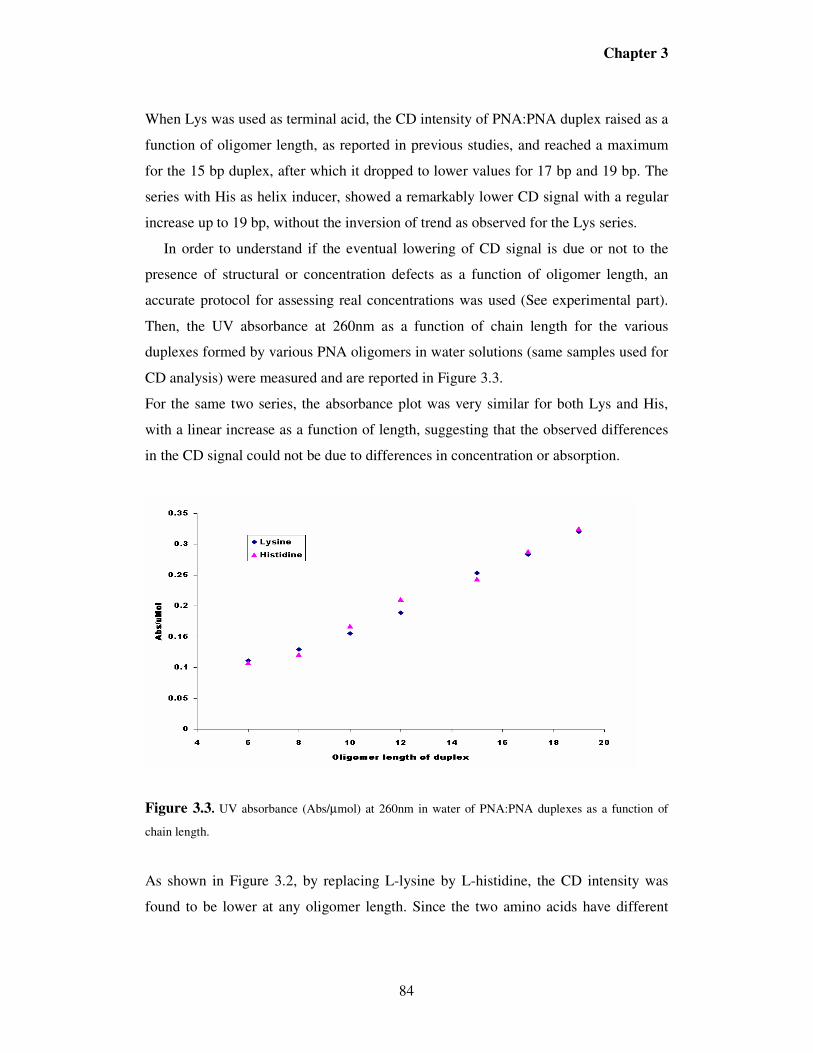
3.2. Results
PNA Sequences Design. In 1995, Nielsen, Nordén and co-workers reported that an
increase in the CD signal was observed in a PNA duplex with terminal L-lysine
moiety in going from an 8mer to a 10 bp duplex after which the CD is unchanged as
the chain length is increased to a 12 bp duplex. These data, which were taken at
constant molarity of the oligomer so that the number of base pairs increased as the
oligomer length increased, seemed to indicate that L-lysine extends its effect until a
maximum of 10 base pairs after which it looses its influence. As a consequence, the
energy barrier for helix inversion was calculated to be included between 5 to 6kJ/mol
per base pair.
Chapter 3
82
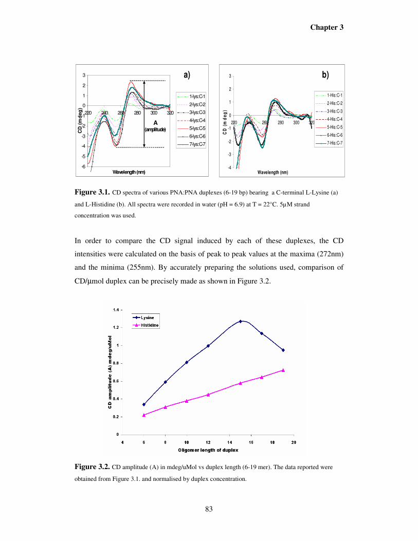
To verify if this hypothesis can still be valid beyond the 12mer limit, a complete set of
PNA oligomers bearing an L-Lys or L-His residue at the C-terminus (Table 3.1) was
synthesized with the shorter ones having the same sequences as those described
previously and the longer one designed in order to have a balanced presence of
nucleobases. The PNAs containing L-lysine as terminal residues were synthesized in a
previous PhD thesis12 by V. Jain at the Polytechnic University in New York. All other
PNAs were synthesized in this work.
Table 3.1. Various PNA sequences utilized in the present work.
∆∆∆∆G°°°° at a particular temperature can be calculated from:
000STHG ∆−∆=∆ (7)
Chapter 3
115
3.6. References
1 a) Mao, C.; Sun, W.; Seeman, N. C., J. Am. Chem. Soc, 1999, 121, 5437-5443.
b) Rothermond, P. W. K.; Nature, 2006, 440, 297-302.
c) Brucale, M.; Zuccheri, G.; Samorì, B.; Trends Biotech., 2006, 24, 235-243. 2 a) M.M. Green, N. C. Peterson, T. Sato, A. Teromoto, R. Cook and S. Lifson,
Science, 1995, 268, 1860.
b) Green, M.M.; Park, J. W.; Sato, T., Teromoto, A.; Lifson, S.; Selinger, L.B.R.;
1665. 4 Green, M.M.; Garetz, B. A; Munoz, B.; Chang, H.; Hoke, S.; Cook, R. G., J. Am.
Chem. Soc. 1995, 117, 4181. 5 Nielsen P. E., Egholm M., Berg R. H., Buchardt O., Science, 1991, 1497. 6 Watson, J.D. and Crick, F.H.C., Nature, 1953, 171, 737-738. 7 a) Rasmussen, H.; Kastrup, J. S.; Nielsen, J. E.; Nielsen, J. M.; Nielsen, P. E., Nat.
Struct. Biol., 1997, 4, 98-101.
b) Haaiama, G.; Rasmussen, H.; Schmidt, G.; Jensen, D. K.; Kastrup, J. S.; Wittung
Stafshede, P.; Nordén, B.; Buchardt, O.; Nielsen, P. E.; New J. Chem., 1999, 23, 833-
840. 8 a) Mendise, V.; De Simone, G.; Tedeschi, T.; Corradini, R.; Sforza, S.; Marchelli R.;
Papasso, D.; Saviano, M.; Pedone, C., Proc. Nat. Acad. Sci. USA., 2003, 21, 12021-
12026.
b) Mendise, V.; De Simone, G.; Corradini, R.; Sforza, S.; Sorrentino, N.; Romanelli,
A.; Saviano, M.; Pedone, C., Acta Cryst., 2002, D58, 553. 9 P. Wittung, M. Eriksson, L. Reidar, P. E. Nielsen and B. Nordén, J. Am, Chem.
Soc.1995, 117, (41), 10167. 10 Rasmussen, H.; Liljefors, T.; Peterson, B.; Nielsen, P. E.; Kastrup, J. S., Journal of
Biomolecular Structures & Dynamics, 2004, 21(4), 495-502 11 Pino, P.; Lorenzi, G. P., J. Am. Chem. Soc., 1960, 82, 4745-4747 12 Jain, V., Helical sense preference in peptide nucleic acids duplexes, Ph.D Thesis in
materials Chemistry, Polytechnic University of Brooklyn, NY, 2006.
Chapter 3
116
13 Wintjens, R.; Liévin, J.; Rooman, M.; Buisine, E.; J. Mol. Biol., 2000, 302, 395-410 14 a) Marky, L. A.; Breslauer, K. J., Biopolymers, 1987, 26, 1601-1620.
b) Applequist, J.; Damle, V., J. Am. Chem. Soc., 1965, 87, 1450-1458. 15 Wittung, P.; Nielsen, P.E; Buchardt, O; Egholm, M.; Nordén, B.; Nature, 1994, 368,
266-281. 19 Bonner, G., Klibanov, A. M., Biotechnology and Bioengineering, 2000, 68, 339. 20 Sorokin, V.A., Gladchenko, G. O.; Valeev, V.A.; Sysa, I.V.; Petrova, L.G.; Blagoi,
Y.P., J. Mol. Str. 1997, 408/409, 237. 21 Xie, G. ; Timasheff, S. N., Protien Science, 1997, 6, 211. 22 Arakawa, T.; Timasheff, S. N., Biophysics J. 1985, 47, 411. 23 Gerlsma, S. Y. J. Biol. Chem, 1968, 243, 9 24 J. Jarabak, A. E. Seeds and P. Talalay, Biochemistry, 1966, 5, 1269. 25 Gekko, K., Timasheff, S. N., Biochemistry ,1981, 20, 4667. 26 Pletneva, E.V.; Laederach, A.T.; Fulton, D.B.; Kostic, N.M.; J. Am. Chem. Soc.,
302, 691-699 30 Kumpf, R.A.; Dougherty, D.A.; Science, 1993, 261, 1708-1710 31Rooman, M.; Liévin, J.; Buisine, E.; Wintjens, R.; J. Mol. Biol., 2002, 309, 67-76 32 Gromiha, M.; Santhosh, C.; Ahmad, S.; Int. J. Biol. Macromol., 2004, 34(3), 203-
211. 33 Sen, A.; Nielsen, P. E., Nucleic Acids Res., 2007, 35, 3367-3374.
Chapter 4
117
PNA as tools for molecular computers
4.1. Introduction
The continuous and fast increase of the technology has strongly influenced our daily life.
For many years, humans have used manufactured devices to enhance their computational
abilities. The development of mechanical devices such as the adding machine and the
tabulating machine was an important advance that has further increased our computational
performance. Yet it was only with the advent of electronic devices and, in particular, the
electronic computer more than 60 years ago that a qualitative threshold seems to have
been passed and problem of considerable difficulty could be solved. The increasing role
that electronic devices play in our daily lives, as well as our constant need to pursue
superior technologies, have raised a wide interest in the development of molecular
systems mimicking the operation of electronic logic gates and circuits1,2,3
. Besides their
integration in the heart of digital computers, electronic logic circuits control the operation
of a variety of devices around us from calculators and store automation to video games
and music equipment.
One interesting possibility for improving computing devices is to use molecular
interactions, such as those occurring in the genetic code, for solving mathematical
problems. The pioneer of this idea was Adleman, who first solved a non-determinstic
problem using DNA molecules, in a study that can be considered as the first considerable
achievement of molecular-recognition based computing strategies.
Furthermore, many studies have reviewed the possibility to use supramolecular concepts
for building electronic gates at the molecular level. For example, more recently, an
interesting study4 has reported the development of an important electronic devices
mimicked at the molecular level, named “keypad lock” which differs from a simple logic
gate by the fact that its output signals are dependent not only on the proper combination of
inputs but also on the correct order by which these inputs are introduced. In other words,
one needs to know the exact password that opens this lock. This device could be used for
numerous applications in which access to an object or data is to be restricted to a limited
Chapter 4
118
number of persons; then represents a new approach for protecting information at the
molecular level.
In the few past years, considerable efforts have been focused on developing new
generation of molecular logic gates and molecular computers, based on DNA5,6,7,8,9
; a
biomolecule possessing well-regulated structures and the ability to store genetic
information. DNA computer has been intensively used for solving a class of intractable
computational problems7,10, such as 3-satisfiability (3-SAT) problems, in which the
computing time can grow exponentially with problem size.
The basis for the use of DNA:DNA duplex formation as computing tool is illustrated in
Figure 4.1. A set of DNA sequences can be used as variables and another set as possible
solutions, so that each solution forms a duplex only if it is a solution of a particular clause
containing the corresponding variable. Combining a set of molecular events of this type is
equivalent to performing parallel computing in normal calculators.
The most attractive advantage of DNA computation is its massive parallelism
computation power and huge memories. Up to now, many accomplishments have been
achieved to improve its performance and increase its reliability, 8,11
though improvement
in specificity of interaction and chemical stability are likely to be necessary to make these
devices robust enough.
Compared to DNA probes, PNAs12
have shown to be efficient tools in numerous
applications, since they can form duplexes more stable than double-stranded DNA and are
superior to oligonucleotide probes in the recognition of single base mutations13,14,15
. An
other interesting property of PNAs , which is useful in biological applications, is their
stability to both nucleases and peptidases, since their “unnatural” skeleton prevents
recognition by natural enzymes, making them more persistent in biological fluids16
.
In the present study, we investigated the possibility to use PNA:PNA interactions on a
PNA-based microarray system as a possible way for performing computational
experiments at the molecular level.
The advantages of using PNA:PNA interactions are: i) possibility to use the same scheme
as DNA computers; ii) higher stability of the duplex formed, which allows to use less base
Chapter 4
119
pairs (atom economy); iii) higher specifity of interaction, which increases the precision of
calculation iv) higher chemical and enzymatic stability of the components.
PNA probes (possible solutions) were immobilized on the surface and hybridized with
fluorescence labelling PNA targets (variables). Ideally, we expected that full signal (True)
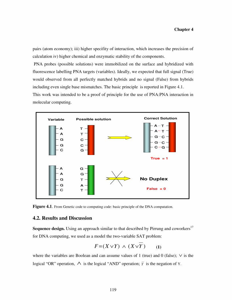
would observed from all perfectly matched hybrids and no signal (False) from hybrids
including even single base mismatches. The basic principle is reported in Figure 4.1.
This work was intended to be a proof of principle for the use of PNA:PNA interaction in
molecular computing.
Figure 4.1. From Genetic code to computing code: basic principle of the DNA computation.
4.2. Results and Discussion
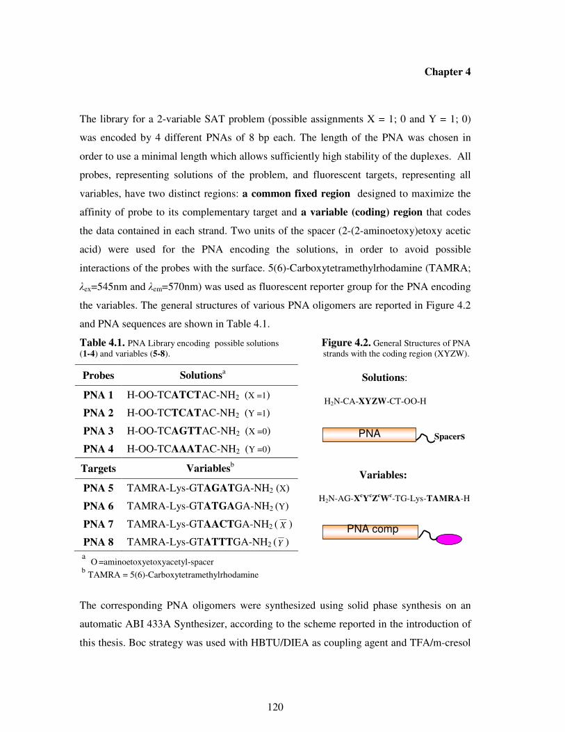
Sequence design. Using an approach similar to that described by Pirrung and coworkers17
for DNA computing, we used as a model the two-variable SAT problem:
)()( YXYXF ∨∧∨= (1)
where the variables are Boolean and can assume values of 1 (true) and 0 (false); ∨ is the
logical “OR” operation, ∧ is the logical “AND” operation; Y is the negation of Y.
A
A
G
G
C
T
T
C
C
G
A
A
G
G
C
T
T
C
C
G
A
A
G
G
C
T
T
C
C
G
A
A
G
G
C
T
T
C
C
G
True = 1
A
A
G
G
C
G
G
T
A
T
A
A
G
G
C
G
G
T
A
T
Variable Possible solution
No Duplex
Correct Solution
False = 0
Chapter 4
120
The library for a 2-variable SAT problem (possible assignments X = 1; 0 and Y = 1; 0)
was encoded by 4 different PNAs of 8 bp each. The length of the PNA was chosen in
order to use a minimal length which allows sufficiently high stability of the duplexes. All
probes, representing solutions of the problem, and fluorescent targets, representing all
variables, have two distinct regions: a common fixed region designed to maximize the
affinity of probe to its complementary target and a variable (coding) region that codes
the data contained in each strand. Two units of the spacer (2-(2-aminoetoxy)etoxy acetic
acid) were used for the PNA encoding the solutions, in order to avoid possible
interactions of the probes with the surface. 5(6)-Carboxytetramethylrhodamine (TAMRA;
λex=545nm and λem=570nm) was used as fluorescent reporter group for the PNA encoding
the variables. The general structures of various PNA oligomers are reported in Figure 4.2
and PNA sequences are shown in Table 4.1.
Table 4.1. PNA Library encoding possible solutions Figure 4.2. General Structures of PNA (1-4) and variables (5-8). strands with the coding region (XYZW).
Solutions:
H2N-CA-XYZW-CT-OO-H
Variables:
H2N-AG-X
cY
cZ
cW
c-TG-Lys-TAMRA-H
a
O =aminoetoxyetoxyacetyl-spacer
b TAMRA = 5(6)-Carboxytetramethylrhodamine
The corresponding PNA oligomers were synthesized using solid phase synthesis on an
automatic ABI 433A Synthesizer, according to the scheme reported in the introduction of
this thesis. Boc strategy was used with HBTU/DIEA as coupling agent and TFA/m-cresol
Probes Solutionsa
PNA 1 H-OO-TCATCTAC-NH2 (X =1)
PNA 2 H-OO-TCTCATAC-NH2 (Y =1)
PNA 3 H-OO-TCAGTTAC-NH2 (X =0)
PNA 4 H-OO-TCAAATAC-NH2 (Y =0)
Targets Variablesb
PNA 5 TAMRA-Lys-GTAGATGA-NH2 (X)
PNA 6 TAMRA-Lys-GTATGAGA-NH2 (Y)
PNA 7 TAMRA-Lys-GTAACTGA-NH2 ( X )
PNA 8 TAMRA-Lys-GTATTTGA-NH2 (Y )
PNA comp
Spacers PNA
Chapter 4
121
as deprotecting solution. The fluorophore (TAMRA) was attached manually. The crude
products were purified by RP-HPLC and characterized with HPLC-MS.
UV Melting Temperatures. The thermal stability of various duplexes and the sequence-
selectivity were initially evaluated by determining the melting temperatures (Tm) at 260
nm of full-matched and mismatched duplexes, in order to verify if the hybridization was
sufficiently strong and if aspecific binding of mismatched sequences could interfere with
the computing process. The Tm data are summarized in Table 4.2. The results show
almost the same melting temperatures for all perfectly matched duplexes (52.0-55.0 °C).
When two or three Watson-Crick base-pair mismatches were introduced at any position
in the code region (middle of the sequence), a large decrease in Tm (> 30°C) was
observed , thereby providing compelling evidence that the PNA:PNA interaction is highly
specific. According to these results, the subsequent computational study was performed at
T = 40 °C in order to avoid non-specific interactions.
Table 4.2. Melting temperatures Tm (°C) for various fullmatched and mismatched PNA:PNA duplexes.
Duplexes Number
of Mismatches
Tm
(°C)
PNA1:PNA5 0 55.0
PNA2:PNA6 0 54.0
PNA3:PNA7 0 54.0
PNA4:PNA8 0 52.0
PNA1:PNA8 2 25.0
PNA1:PNA7 2 < 20a
PNA2:PNA8 3 < 20a
a No melting transition was observed in the range 20-95°C
PNA Computing using microarray technology. PNA strands (1-4) representing possible
assignments were combinatorially deposited onto the surface (Figure 4.3) so that all
possible solutions were represented (For n variables, there are 2n possible solutions). Each
Chapter 4
122
spot contains two PNA strands, each strand representing the value of a different
variable:PNA1/PNA2 for 1/1, (i.e. x= 1 and y= 1); PNA1/PNA4 for 1/0 (x= 0, y= 0);
PNA3/PNA2 for 0/1 (x= 0, y = 1) and PNA3/PNA4 for 0/0 (x= 0, y =0). The array/PNA
probes were the same for all problems of n or fewer variables; equivalent to computing
hardware.
Moreover, the complementary fluorescent targets used depend of the logic expression to
be verified, for example PNA5/PNA6 for “ YORX ” or PNA5/PNA8 for “ YORX ”, and
were equivalent to software. The fluorescence signals indicate the zones containing
probes complementary to the targets used, and potentially encoding the solution of the
problem.
Figure 4.3.Computation protocol. (a) deposition: 30 µM PNA probes (1-4) representing all possible
solutions were spotted in quadruplicate on the microarray. Each spot contains two different PNA strands. (b)
Hybridization with fluorescent targets (i.e. PNA5 + PNA6; 1 µM strand concentration). The solutions of the
problem were read out using a scanner.
X/Y 1/1 1/0 0/1 0/0
X/Y 1/1 1/0 0/1 0/0
Microarray (Activated Slides)
(Possible Solutions)
Microarray/PNAs (Hardware)
(a)
X/Y 1/1 1/0 0/1 0/0
Microarray/PNAs (Hardware)
X/Y 1/1 1/0 0/1 0/0
Correct Solutions: 1/1; 1/0; 0/1.
Hybridization with
PNA5 (X) + PNA6 (Y)
(Software)
(b)
Deposition
Chapter 4
123
Logic Operation “X OR Y”. Using the protocol described above, we constructed a 2-
input logic gate “OR” that performs Boolean calculations; and a single-input gate
“Report” that releases a fluorescent output signal in response to specific single-stranded
PNA input sequences. The experimental study of this 2-variable problem used the above
described array, on which all possible solutions ( X/Y = 1/1; 1/0; 0/1; 0/0) were
immobilized; followed by hybridization with fluorescent PNA (PNA5 + PNA6)
representing variables X and Y, respectively. The output of the computation is reported in
Figure 4.4.
Figure 4.4. Diagrammatic representation of logic gate “X OR Y” wired to a fluorescent reporter (top).
Observed fluorescent output from execution of the calculation (bottom).
According to Watson-Crick base pairing interactions, upon hybridization with PNA5 +
PNA6, one set of spots (1/1) should contain two perfectly matched duplexes; two sets of
spots (1/0; 0/1) should contain one perfectly matched duplex each and the last set (0/0)
should contain no duplex. Then, after hybridization, only spots 1/1; 1/0; 0/1 should be
1/1 1/0 0/1 0/0
X/Y
Hybridization with PNA strands (X,Y)
0
10
20
30
40
50
60
70
80
90
100
Intensity
hעX
Y(X or Y)OR Report
hעX
Y(X or Y)OR Report
X
Y(X or Y)OR Report
1/11/0
0/1
0/0
X 1 1 0 0
Y 1 0 1 0
X or Y 1 1 1 0
Chapter 4
124
fluorescent. The results obtained are fully consistent with mathematical predictions and
provide solutions to the problem (X OR Y = 1) of (X,Y) = (1,1); (1,0); (0,1).
Logic Operation “X OR Y”. Analogously, when hybridization was performed using
PNA5 + PNA8 representing the variables X and (Not Y), respectively, two sets of spots
(1/1 and 0/0) should contain one fully matched duplex each; one set (1/0) should contain
two matched duplexes and the last one (0/1) should contain no duplex. Therefore, one
must expect only spots 1/1; 1/0; 0/0 containing PNA probes complementary to the targets
X and (Not Y) to be fluorescent. The experimental data reported in Figure 4.5 are fully
consistent to theoretical predictions, providing (X,Y) = (1,1); (1,0); (0,0) as solutions of
the problem.
Figure 4.5. Diagrammatic representation of logic gate “X OR NotY” wired to a fluorescent reporter
(top). Observed fluorescent output from execution of the calculation (bottom).
1/1 1/0 0/1 0/0
X/Y
Hybridization with PNA strands (X,Y)
0
10
20
30
40
50
60
70
80
90
100
Inte
nsity
1/1 1/0
0/1
0/0
X 1 1 0 0
Y 1 0 1 0
Y 0 1 0 1
X or Y 1 1 0 1
X
YX or (Not Y)OR
NOT
hעReport
X
YX or (Not Y)OR
NOT
hעReport
hעReport
Chapter 4
125
Combined Operation “(X OR Y) AND (X OR Y)”. Using the results obtained with the
two independent clauses, the combined “OR-AND-OR” gate was designed such that
outputs from single “OR” gates can serve as input for the further “AND” gate. In this
case, the reported fluorescence intensity (Figure 5.6.) is the product of the intensities of
signal exhibited by single gates in correspondence to the same set of spots (i.e. F(1/1) =
F1(1/1) x F2(1/1)). According to the data reported in Figure 4.6, the equation is true only if
(x,y) = (1,1); (1,0).
Figure 4.6. Diagrammatic representation of combined logic gate “(X OR Y)AND (X OR NotY)” (top).
Observed fluorescent output from execution of the calculation (bottom-right).
Although in this example, we performed a very simple calculation, the number of
calculation needed (two hybridization steps and product of intensities) is less than that of
the systematic test of all possible solutions, which is 2n
, in this case 4. This advantage is
X
Y(X or Y)OR
X
YX or (Not Y)OR
NOT
(X or Y) and (X or Not Y)AND Reporthע
1/1 1/0 0/1 0/0 1/1 1/0 0/1 0/0
X 0
10
20
30
40
50
60
70
80
90
100
Inte
nsity
1/1 1/0
0/10/0
X or Y 1 1 1 0
X or Y 1 1 0 1
F1 and F2 1 1 0 0
F1F2
Chapter 4
126
crucial when performing the same type of operations with a very high number of
variables.
4.3. Conclusions
The three examples described above demonstrate that our approach to Boolean control
based on PNA:PNA interactions is likely general, and that PNAs could be potential
candidates for integration into molecular circuits and computers. Although the prototype
of PNA computer constructed here can so far process a limited number of operations, it
inspires the imagination toward a new generation of processing devices based on PNA,
able to solve in short time, more complicated non deterministic problems. DNA-based
devices (computers and logic gates) have been intensively studied. Compared to DNA
probes, PNA form more stable duplexes and with higher specifity, and are more resistant
to enzymes and chemical agents. The current study, although still not suitable for practical
applications, demonstrate that it should be possible to employ PNA molecules in a broad
spectrum of nanobiotechnological applications, such as computers and logic gates.
For DNA computing, extension of simple technologies such as those described in the
present chapter has already led to the solution of a 20-variable SAT problem, which is a
computing challenge hardly addressable with normal computers. Therefore extension of
this protocol can be easily foreseen.
PNAs have also been successfully used as therapeutic drugs and it is generally believed
that the power of Boolean computing would find application in molecular agents able of
diagnosing biological disorders and produce the right signaling or even therapeutic
outputs18. Then, molecular devices based on biomolecules such as PNA capable of highly
discriminating between fullmatched and mismatched sequences, might have considerable
advantages over simple molecular logic gates.
4.4. Experimental Section
PNA Oligomer Synthesis. The synthesis was performed on an ABI 433A peptide
synthesizer with software modified to run the PNA synthetic steps (scale: 5 µmol), using
Chapter 4
127
Boc chemistry and standard protocols as described in the introductive part, with
HBTU/DIEA coupling. MBHA resin was used and downloaded manually by the first
monomer. In the case of PNA encoding the variables, upon completion of PNA oligomer
synthesis, the attachment of 5(6)-Carboxytetramethylrhodamine (Fluorophore unit) was
done manually using DIC/DHBTOH as coupling reagent. The crude PNA were purified
by RP- HPLC with UV detection at 260 nm. Semi-prep column C18 (5 microns, 250 x
10mm, Jupiter Phenomenex, 300 A ) was utilized, eluting with water + 0.01% TFA (
eluent A) and the mixture 60: 40 of water / acetonitrile + 0.01% TFA (eluent B); elution
gradient: from 100% A to 100% B in 50 min, flow: 4 ml/min. The resulting pure PNA
oligomer was characterized by MS-ESI with gave positive ions consistent
with the final products.
PNA 1. Calculated MW: 2410.4
ESI-MS: m/z = found 804.2 (calc 804.4; MH33+
), found 603.4 (calc 603.6; MH44+
), found
483.1 (calc 483.1; MH55+
).
PNA 2. Calculated MW: 2410.4
ESI-MS: m/z = found 804.4 (calc 804.4; MH33+
), found 603.9 (calc 603.6; MH44+
), found
483.2 (calc 483.1; MH55+
).
PNA 3. Calculated MW: 2450.4
ESI-MS: m/z = found 817.8 (calc 817.8; MH33+), found 613.3 (calc 613.6; MH4
4+), found
491.1 (calc 491.1; MH55+
).
PNA 4. Calculated MW: 2443.4
ESI-MS: m/z = found 815.4 (calc 815.5; MH33+), found 611.6 (calc 611.8; MH4
4+), found
489.7 (calc 489.7; MH55+
).
PNA 5. Calculated MW: 2790.0
Chapter 4
128
ESI-MS: m/z = found 930.5 (calc 931.0; MH33+
), found 698.4 (calc 698.5; MH44+
), found
559.0 (calc 559.0; MH55+), 466.0 (calc 466.0; MH6
6+).
PNA 6. Calculated MW: 2790.0
ESI-MS: m/z = found 930.5 (calc 931.0; MH33+
), found 698.5 (calc 698.5; MH44+
), found
559.0 (calc 559.0; MH55+
), 466.0 (calc 466.0; MH66+
).
PNA 7. Calculated MW: 2750.0
ESI-MS: m/z = found 917.4 (calc 917.6; MH33+
), found 688.4 (calc 688.5; MH44+
), found
550.8 (calc 551.0; MH55+
).
PNA 8. Calculated MW: 2756.0
ESI-MS: m/z = found 919.5 (calc 919.7; MH33+
), found 690.0 (calc 690.0; MH44+
), found
552.2 (calc 552.2; MH55+
).
UV Melting Analysis. Solutions of 1:1 PNA/PNA were prepared in pH = 7.0 Buffer
consisting of 100 mM NaCl, 10 mM NaH2PO4.H2O, 0.1 mM EDTA. Strand
concentrations were 5 µM in each component. Thermal denaturation profiles (Abs vs T)
of the hybrids were measured at 260 nm with an UV/Vis Lambda Bio 20 Spetrometer
equipped with a Peltier Temperature Programmer PTP6 which is interfaced to a personal
computer. For the temperature range 95°C to 20°C, UV absorbance was recorded at 260
nm every 0.5°C. A melting curve was recorded for each duplex. The melting temperature
(Tm) was determined from the maximum of the first derivative of the melting curves.
Array Preparation. Activated Slides were used as solid supports to which the amino-
terminal group of the PNA probes encoding all possible solutions were covalently linked.
The deposition of the probes was carried out using a SpotArrayTM24 (PerkinElmer) with a
pin-and-ring deposition system. The manufacturer's instructions for the deposition
protocol were slightly changed in order to comply with the special requirement of the
chemical structures of PNAs: in particular a 100mM carbonate buffer (pH 9.0) containing
Chapter 4
129
10% acetonitrile and 0.001% sodium dodecyl sulphate (SDS) was used as deposition
buffer. Moreover, after every deposition, the pin-and-ring system was purged with water
for 10s and further washed with acetonitrile/water (1:1), in order to avoid dragging of the
probes in subsequent depositions. The probes were coupled to the surface and the
remaining reactive sites were blocked by leaving the slides in a humid chamber (relative
humidity 75%) at room temperature for 12h, followed by immersion in a glass rack
containing a 50mM solution of ethanolamine, 0.1M TRIS, pH 9, prewarmed at 50°C, for
30min. The slides were washed twice with bidistilled water at room temperature and then
slowly shaken for 30min in plastic tubes containing a 4× saline/sodium citrate (SSC)
solution and a 0.1% SDS buffer prewarmed at 50°C. Each slide was then washed with
bidistilled water at room temperature and centrifuged in a plastic tube at 800rpm for 3min.
Slides were then ready to undergo the hybridization protocol.
Hybridization and Scanning Labeled PNA samples (1µM strand concentration)
encoding two variables were prepared by diluting stock solutions to a final volume of
65µl and a final concentration of 4× SSC and 0.1% SDS buffer. Hybridization was
performed by loading the samples to “in situ frame” chambers and leaving the slides
under slow shaking for 1h at 40°C. After the hybridization step, all the slides were treated
individually to prevent cross contamination. The slides were washed under slow shaking
for 5min at 40°C with a 2× SSC, 0.1% SDS buffer prewarmed at 40°C, followed by
treatment for 1min with 0.2× SSC and for 1min with 0.1× SSC at room temperature. The
slides were then spin-dried at 1000 rpm for 5min.
The fluorescent signal deriving from the hybridization was acquired using a ScanArray
Express (PerkinElmer) at λ ex=545nm and λ em=570nm.
Computational Protocol. The computational protocol was as follows: