Page 1
University of South Carolina University of South Carolina
Scholar Commons Scholar Commons
Theses and Dissertations
2020
Synthesis and Design of Novel Polymer Grafted Nanoparticles Synthesis and Design of Novel Polymer Grafted Nanoparticles
Relevant to Drug Delivery Vehicles for Biomedical Application Relevant to Drug Delivery Vehicles for Biomedical Application
Maan Abduldiyem Hassan Al-Ali
Follow this and additional works at: https://scholarcommons.sc.edu/etd
Part of the Chemistry Commons
Recommended Citation Recommended Citation Al-Ali, M. A.(2020). Synthesis and Design of Novel Polymer Grafted Nanoparticles Relevant to Drug Delivery Vehicles for Biomedical Application. (Doctoral dissertation). Retrieved from https://scholarcommons.sc.edu/etd/6088
This Open Access Dissertation is brought to you by Scholar Commons. It has been accepted for inclusion in Theses and Dissertations by an authorized administrator of Scholar Commons. For more information, please contact [email protected] .
Page 2
SYNTHESIS AND DESIGN OF NOVEL POLYMER GRAFTED
NANOPARTICLES RELEVANT TO DRUG DELIVERY VEHICLES
FOR BIOMEDICAL APPLICATIONS
by
Maan Abduldiyem Hassan Al-Ali
Bachelor of Science
University of Basrah, 2000
Master of Science
University of Basrah, 2007
Submitted in Partial Fulfillment of the Requirements
For the Degree of Doctor of Philosophy in
Chemistry
College of Arts and Sciences
University of South Carolina
2020
Accepted by:
Brian C. Benicewicz, Major Professor
Chuanbing Tang, Committee Member
Andrew B. Greytak, Committee Member
Alan W. Decho, Committee Member
Cheryl L. Addy, Vice Provost and Dean of the Graduate School
Page 3
ii
© Copyright by Maan Abduldiyem Hassan Al-Ali, 2020
All Rights Reserved.
Page 4
iii
DEDICATION
To the soul of my beloved father, my first ideal.
To the source of tenderness that illuminates my life, my dear mother.
To my soulmate who supported me and stood with me in hardness days, my wife,
Ayat.
To my adorable children, Nooran, Fatima, Sarah, and Yousuf.
To my brothers and sisters.
Page 5
iv
ACKNOWLEDGMENTS
First and foremost, it is a great honor and a big privilege to get my degree
mentored by my research advisor, Dr. Brian C. Benicewicz. I would like to express
my sincere gratitude to such an enthusiastic and knowledgeable person. Also, I
would like to thank him for all of his guidance and support for my research
projects. With his knowledge and enthusiasm for polymer science and chemistry,
I experienced a diverse, productive, and stimulating environment in my career. I
eagerly appreciate his advice and guidance, and I am sincerely grateful for all of
the opportunities that I have had while working in his group.
I would like to thank my doctoral committee members, Dr. Chuanbing
Tang, Dr. Andrew B. Greytak, and Dr. Alan W. Decho, for their contributions and
encouragement, and suggestions on my research. I want to thank Dr. Decho and
his student Savannah Chandler for their collaborations with our group.
I would like to thank the Benicewicz group members, both past and present,
for all of their help, suggestions, and friendship. Especially, I would like to thank
Dr. Kayley Hayat and Dr. Michel Bell for their help when I first joined the group.
Great thanks to Susan Hipp, Warren Steckle, for their help during my study in
Horizon building. Also, I would like to thank Dr. Julia Pribyl, Dr. Andrew
Page 6
v
Pingitore, Dr. Yucheng Huang, Dr. Mohammad Khani, Dr. Yang Zheng, Dr. Zaid
Alajeely, Laura Murdock, Dr. Fei Huang, Dr. Lihui Wang, Karl Golian, Caroline
Rohlfing, Richard Ly, and many others. Thanks for all your help and suggestions!
Next, I would like to thank my family, my wife, our daughters, and our son,
who, without their lovely emotional support and encouragement, I could not be
here. I would love to thank my brothers and sisters for always supporting and
encouraging me. Thanks and great gratitude for both my first inspirational and
teacher in my life, my dear father, and my dear mother, who will be more proud
of me than myself.
Finally, I would like to thank the Iraqi Ministry of Higher Education and
Scientific Research for funding me for the first five years in my Ph.D. program.
Page 7
vi
ABSTRACT
The modification of inorganic nanoparticles with organic polymer chains
has become a significant field of study for the engineering of advanced
nanocomposite materials. This dissertation presents the design, synthesis, and
characterization of novel polymer grafted silica nanoparticles as new strategies to
combat bacterial resistance. Described herein is the synthesis of monomers that
have been graft polymerized onto silica nanoparticles that can be used as a
delivery drug vehicle for biomedical applications. The polymerization of these
monomers was performed via reversible addition-fragmentation chain transfer
(RAFT) polymerization. The molecular design of the RAFT agents that are
attached to the surfaces of the nanoparticles has the main role in controlling the
molecular weight and dispersity of the polymer chains grafted to the surface of
the nanoparticles. The method of attachment of the RAFT agents additionally
controls the surface graft density. The important properties of nanocomposites can
be exploited in many different areas, such as biomedical applications.
In the first chapter of this work, the overall background of antimicrobial
polymers, the functionalization of nanoparticles using RAFT polymerization, and
Page 8
vii
the concept of the modification of silica nanoparticles to afford a bimodal brush
system is described. The second chapter focuses on designing a new type of
stimulus-responsive polymer that can work as antibiotic-delivery carriers in
biomedical applications. We reported pH-responsive “controlled release”
polymers that were grafted on silica nanoparticles using reversible addition-
fragmentation chain transfer (RAFT) polymerization. Two monomers 2-((2-
(propionyloxy) propanoyl)oxy)ethyl methacrylate (HEMA-LA) and 4-(2-
(methacryloyloxy)ethoxy)-4-oxobutanoic acid (HEMA-SA), containing
hydrolytically sensitive ester linkages were synthesized to functionalize on the
surface of silica nanoparticles. The degradation rate was monitored by attaching
dyes at the end of these monomers in each repeat unit to study the release rate,
thus assessing the use of these monomers as delivery vehicles for anti-bacterial
applications.
In the following chapter, bimodal polymer chains grafted on the surface of
silica nanoparticles was developed via RAFT polymerization to create water-
dispersible nanoparticles that have additional advantages as antibiotic-delivery
vehicles in biomedical applications. Two different polymer chains populations
were attached to silica nanoparticles; the first population is high graft density with
low molecular weight, which is a pH-responsive controlled release polymer
derived from two possible monomers (HEMA-LA) and (HEMA-SA), both
Page 9
viii
containing a hydrolytically sensitive ester linkage: the second population is a
water-dissolvable polymer of methacrylic acid (MAA) at low graft density with
high molecular weight. Fluorescent dyes were conjugated to the controlled release
polymers to monitor the nanoparticles in biological systems.
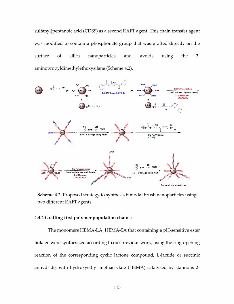
Finally, in the fourth chapter, we described a new approach using two
different RAFT agents, 4-cyano-4-(phenylcarbonothioylthio)pentanoic acid
(CPDB), and 4-cyano-4-[(dodecylsulfanylthiocarbonyl)sulfanyl]pentanoic acid
(CDSS) to create bimodal polymer brush grafted nanoparticle. These novel
bimodal brush silica nanoparticles were designed successfully to combat
antibiotic-resistant bacteria. The first population polymer brush is based on two
potential “controlled release” monomers 2-((2-((2-hydroxy propanoyl)oxy)
propanoyl)oxy) ethyl methacrylate (HEMA-LA), 2-(methacryloyloxy)ethyl
succinate (HEMA-SA) containing a hydrolytically sensitive ester linkage as a high
graft density, short brush to work as antibiotic-delivery carriers. However, the
second population polymer brush was based on a sugar-containing monomer, 2-
methacrylamido glucopyranose (MAG), as a low graft density, long brush to
enhance bacterial uptake of nanoparticles.
Page 10
ix
TABLE OF CONTENTS
DEDICATION ................................................................................................................. iii
ACKNOWLEDGEMENTS ............................................................................................. iv
ABSTRACT ...................................................................................................................... vi
LIST OF TABLES ............................................................................................................ xii
LIST OF FIGURES ......................................................................................................... xiii
LIST OF SCHEMES ..................................................................................................... xviii
LIST OF ABBREVIATIONS ......................................................................................... xxi
CHAPTER 1: INTRODUCTION .....................................................................................1
1.1 RAFT Polymerization ..................................................................................2
1.2 Mechanism of RAFT polymerization ........................................................3
1.3 Polymer Grafted Nanoparticles .................................................................5
1.4 Nanoparticles as delivery vehicles ............................................................9
1.5 Bimodal Nanocomposites .........................................................................10
1.6 References ....................................................................................................11
Page 11
x
CHAPTER 2: POLYMERIZATION OF “CONTROLLED RELEASE” MONOMERS CONTAINING A HYDROLYTICALLY SENSITIVE ESTER LINKAGE VIA RAFT POLYMERIZATION .................................................16
2.1 Abstract ........................................................................................................17
2.2 Introduction ................................................................................................18
2.3 Experimental ...............................................................................................20
2.4 Results and Discussion ..............................................................................31
2.5 Conclusions .................................................................................................49
2.6 References ...................................................................................................50
CHAPTER 3: ENGINEERING WATER-DISPERSIBLE
BIMODAL POLYMER GRAFTED SILICA NANOPARTICLES AS ANTIBIOTIC-CARRIERS ......................................54
3.1 Abstract............................................................................................................55
3.2 Introduction ....................................................................................................56
3.3 Materials and Methods..................................................................................58
3.4 Results and Discussion ..................................................................................72
3.5 Conclusions .....................................................................................................88
3.6 References .......................................................................................................89 CHAPTER 4: DESIGNING “SWEET-NANOPARTICLES”
AS A NOVEL STRATEGY TO COMBAT ANTIBIOTIC-RESISTANT BACTERIA .........................................................95
4.1 Abstract............................................................................................................96
4.2 Introduction ....................................................................................................97
4.3 Materials and Methods................................................................................100
Page 12
xi
4.4 Results and Discussion ................................................................................114
4.5 Conclusions ...................................................................................................134
4.6 References .....................................................................................................135
CHAPTER 5: CONCLUSION AND FUTURE WORK ............................................143
5.1 Conclusions ...................................................................................................144
5.2 Future Work ..................................................................................................147
BIBLIOGRAPHY ...........................................................................................................153
Page 13
xii
LIST OF TABLES
Table 2.1 Various molecular weights and chain densities of SiO2@P(HEMA-LA) and SiO2@P(HEMA-SA) using RAFT polymerization. ......................................................................................38
Table 3.1 Grafting densities and molecular weights of
bimodal nanoparticles, SiO2@P(HEMA-LA-dye)- PMAA, and SiO2@P(HEMA-SA-dye)-PMAA. ..............................................84
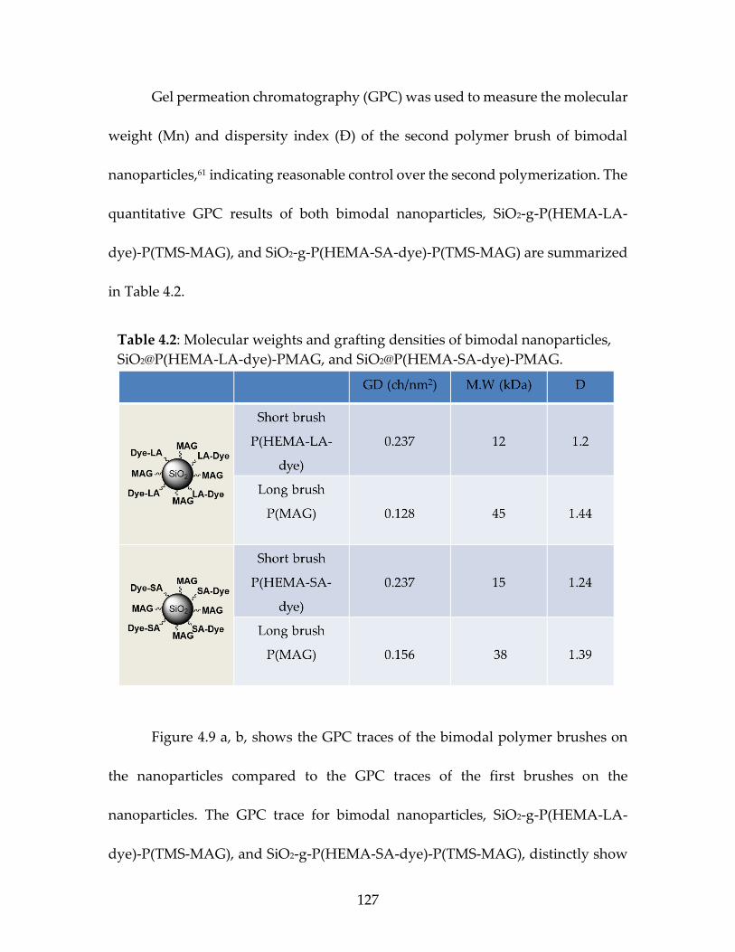
Table 4.1 Polymerization of the glycomonomer (MAG-TMS)
using CDSS as RAFT agent and AIBN as an initiator at 65oC ...............................................................................................................128
Table 4.2 Molecular weights and grafting densities of bimodal
nanoparticles, SiO2@P(HEMA-LA-dye)-PMAG, and SiO2@P(HEMA-SA-dye)-PMAG. ..................................................................129
Page 14
xiii
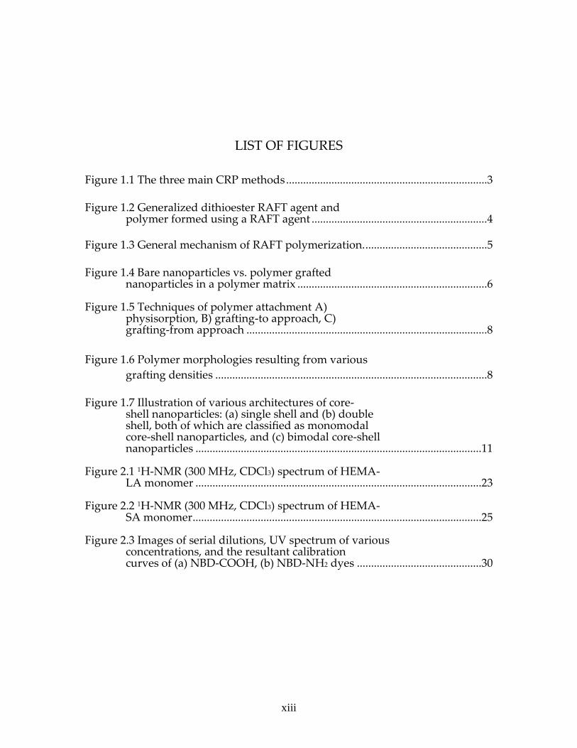
LIST OF FIGURES
Figure 1.1 The three main CRP methods .......................................................................3
Figure 1.2 Generalized dithioester RAFT agent and polymer formed using a RAFT agent ..............................................................4
Figure 1.3 General mechanism of RAFT polymerization. ...........................................5
Figure 1.4 Bare nanoparticles vs. polymer grafted nanoparticles in a polymer matrix ...................................................................6
Figure 1.5 Techniques of polymer attachment A)
physisorption, B) grafting-to approach, C) grafting-from approach .....................................................................................8
Figure 1.6 Polymer morphologies resulting from various
grafting densities ................................................................................................8
Figure 1.7 Illustration of various architectures of core-
shell nanoparticles: (a) single shell and (b) double shell, both of which are classified as monomodal core-shell nanoparticles, and (c) bimodal core-shell nanoparticles .....................................................................................................11
Figure 2.1 1H-NMR (300 MHz, CDCl3) spectrum of HEMA-
LA monomer .....................................................................................................23 Figure 2.2 1H-NMR (300 MHz, CDCl3) spectrum of HEMA-
SA monomer ......................................................................................................25 Figure 2.3 Images of serial dilutions, UV spectrum of various
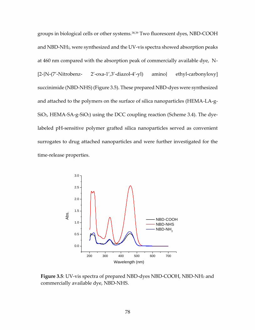
concentrations, and the resultant calibration curves of (a) NBD-COOH, (b) NBD-NH2 dyes ............................................30
Page 15
xiv
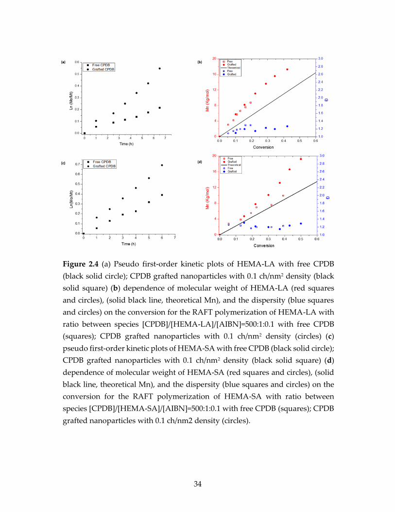
Figure 2.4 (a) Pseudo first-order kinetic plots of HEMA-LA
with free CPDB (black solid circle); CPDB grafted
nanoparticles with 0.1 ch./nm2 density (black solid
square) (b) dependence of molecular weight of
HEMA-LA (red squares and circles), (solid black line,
theoretical Mn), and the dispersity (blue squares and
circles) on the conversion for the RAFT
polymerization of HEMA-LA with ratio between
species [CPDB]/[HEMA-LA]/[AIBN]=500:1:0.1 with
free CPDB (squares); CPDB grafted nanoparticles with
0.1 ch./nm2 density (circles) (c) pseudo first-order
kinetic plots of HEMA-SA with free CPDB (black solid
circle); CPDB grafted nanoparticles with 0.1 ch./nm2
density (black solid square) (d) dependence of
molecular weight of HEMA-SA (red squares and
circles), (solid black line, theoretical Mn), and the
dispersity (blue squares and circles) on the conversion
for the RAFT polymerization of HEMA-SA with ratio
between species [CPDB] /[HEMA-
SA]/[AIBN]=500:1:0.1 with free CPDB (squares);
CPDB grafted nanoparticles with 0.1 ch./nm2 density
(circles) ...............................................................................................................34
Figure 2.5 GPC traces of (a) SiO2@P(HEMA-LA) and (b) SiO2@P
(HEMA-SA) in THF using ratio 500:1:0.1 of [monomer]: [CTA]:[initiator] at different times .................................................................38
Figure 2.6 UV-vis, FT-IR spectrums of SiO2-g-P(HEMA-LA)-
dye (red curve), and SiO2-g-P(HEMA-SA)-dye (black curve) ..................................................................................................................41
Figure 2.7 TGA trace of (a) SiO2-g-HEMA-LA-dye and (b) SiO2-
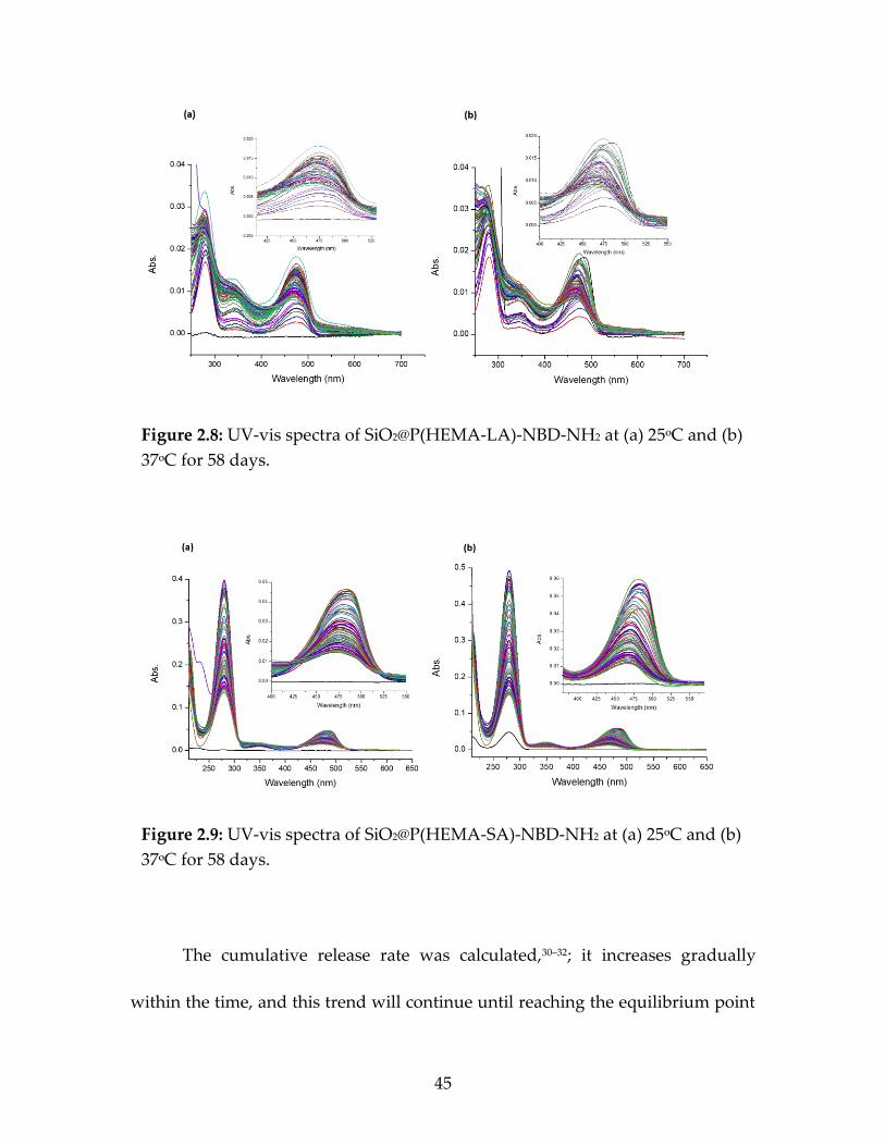
g-HEMA-SA-dye nanoparticles ......................................................................43 Figure 2.8 UV-vis spectra of SiO2@P(HEMA-LA)-NBD-COOH
at (a) 25ᵒC and (b) 37ᵒC for 58 days ...............................................................45 Figure 2.9 UV-vis spectra of SiO2@P(HEMA-SA)-NBD-NH2 at
(a) 25ᵒC and (b) 37ᵒC for 58 days ....................................................................45
Page 16
xv
Figure 2.10 Cumulative release rate of (a) SiO2@P(HEMA- LA-dye) and (b) SiO2@P(HEMA-SA-dye), at 25ᵒC and 37ᵒC for 58 days .........................................................................................46
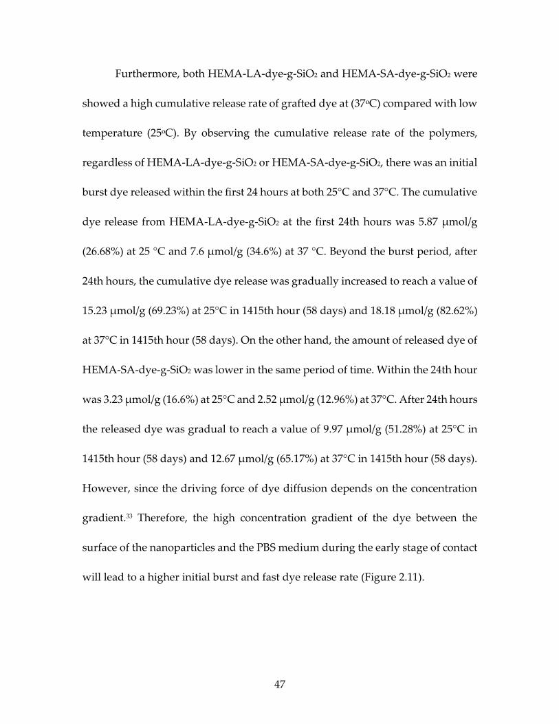
Figure 2.11 Cumulative release rate of SiO2@P(HEMA-LA
-dye) and SiO2@P(HEMA-SA-dye) at 25ᵒC and 37ᵒC for 58 days ................................................................................................48
Figure 3.1 1H NMR (300 MHz, CDCl3) spectrum of HEMA-
LA monomer .....................................................................................................62 Figure 3.2 1H NMR (300 MHz, CDCl3) spectrum of HEMA-
SA monomer ......................................................................................................63
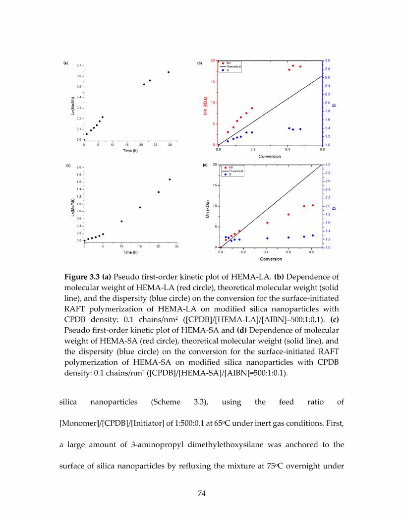
Figure 3.3 (a)Pseudo first-order kinetic plot of HEMA-LA. (b)
dependence of molecular weight of HEMA-LA (red
circle), theoretical molecular weight (solid line), and
the dispersity (blue circle) on the conversion for the
surface-initiated RAFT polymerization of HEMA-
LA on modified silica nanoparticles with CPDB
density: 0.1 chains/nm2 ([CPDB]/[HEMA-
LA]/[AIBN]=500:1:0.1). (c) Pseudo first-order kinetic
plot of HEMA-SA and (d) Dependence of molecular
weight of HEMA-SA (red circle), theoretical
molecular weight (solid line), and the dispersity
(blue circle) on the conversion for the surface-
initiated RAFT polymerization of HEMA-SA on
modified silica nanoparticles with CPDB density: 0.1
chains/nm2 ([CPDB]/[HEMA-SA]/[AIBN]=500:1:0.1) ..................................74
Figure 3.4 Thermogravimetric analysis of (a) SiO2-g-P(HEMA
-LA), and (b) SiO2-g-P(HEMA-SA) nanoparticles ........................................76 Figure 3.5 UV-vis spectra of prepared NBD-dyes NBD-COOH,
NBD-NH2 and commercially available dye, NBD-NHS ............................79 Figure 3.6 GPC analysis of bimodal grafted nanoparticles (a)
SiO2-g-P(HEMA-LA-dye)-PMAA, and (b) SiO2-g- P(HEMA-SA-dye)-PMAA ...............................................................................85
Page 17
xvi
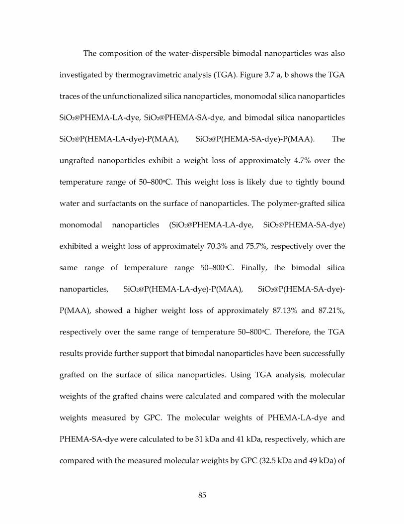
Figure 3.7 TGA analysis of (a) Bare SiO2, Monomdal NP’s
SiO2-g-P(HEMA-LA), and Bimodal NP’s SiO2-g-
P(HEMA-LA)-(PMAA), (b) Bare SiO2, Monomdal
NP’s SiO2-g-P(HEMA-SA), and Bimodal NP’s SiO2-
g-P(HEMA-SA)-(PMAA) .................................................................................87
Figure 4.1 UV absorption spectra of polymer grafted
nanoparticles with cleavedCDSS RAFT agent (red line), and with CDSS attached to the polymers on the surface of silica nanoparticles (black line)........................................................................................................119
Figure 4.2 UV-vis, FT-IR spectrums of MA 200: SiO2@P
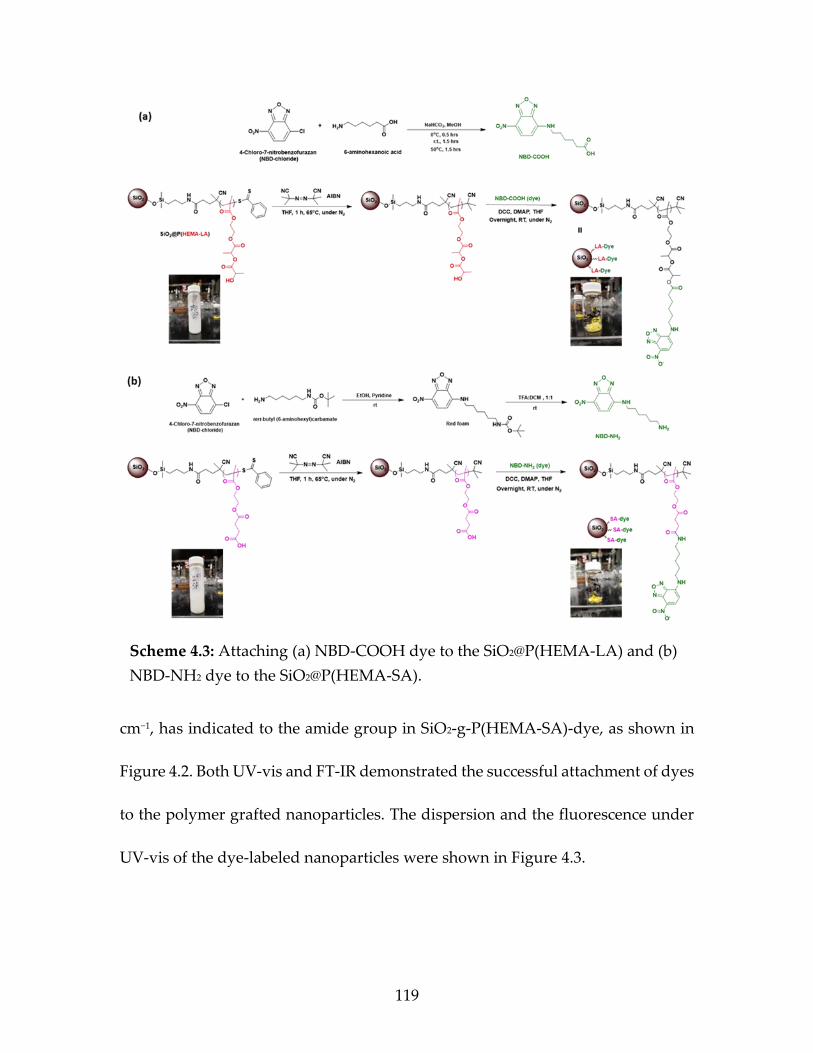
(HEMA-LA)-NBD-COOH, and MA 195: SiO2@P (HEMA-SA)-NBD-NH2 ..................................................................................122
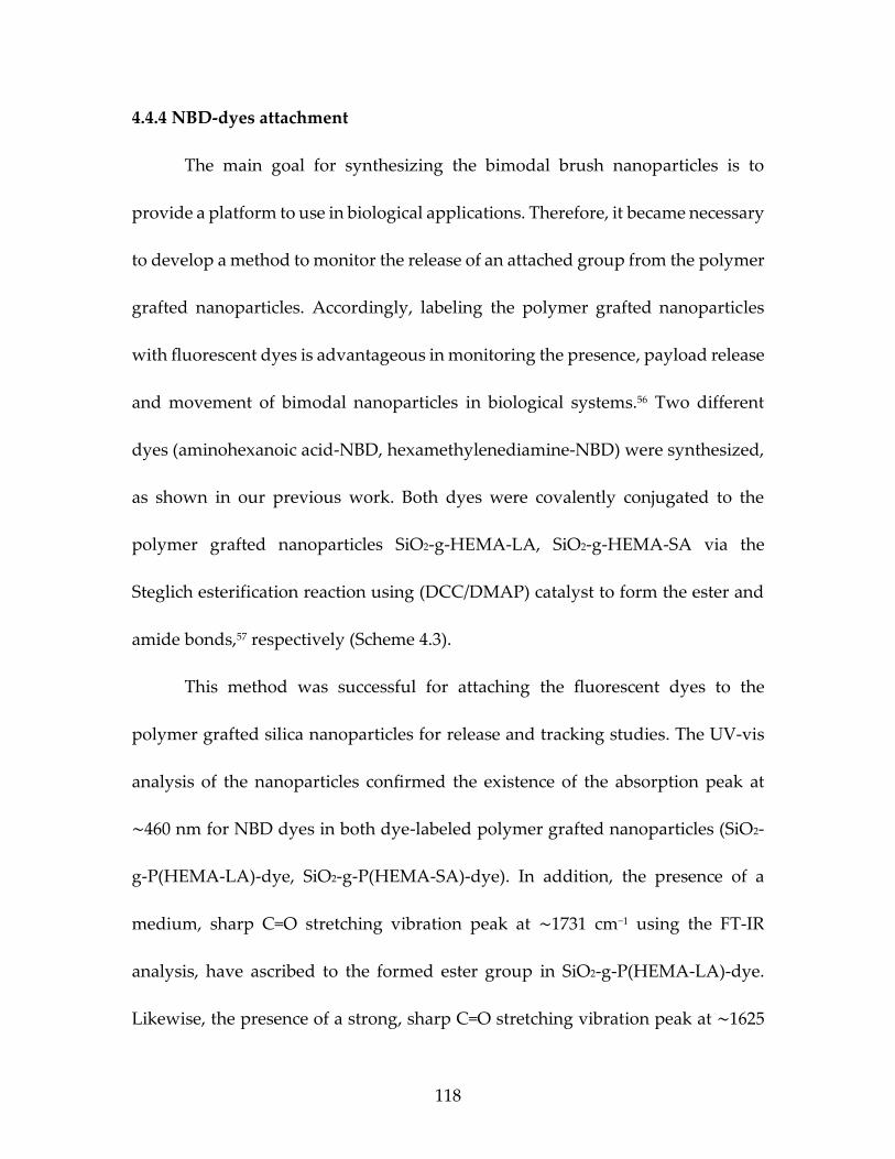
Figure 4.3 The dispersion of dye labeled polymer grafted
nanoparticles (SiO2-g-P(HEMA-SA)-dye) and the fluorescence under UV-vis light ...................................................................117
Figure 4.4 1H-NMR spectrums of CDSS RAFT agent, CDSS-
OH, and CDSS-Phosphate and (b) 31P-NMR spectrum of CDSS-Phosphate .......................................................................124
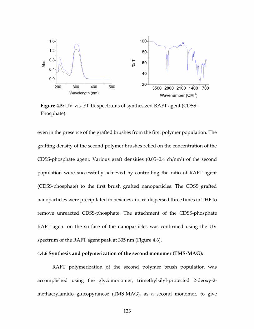
Figure 4.5 UV-vis, FT-IR spectrums of synthesized RAFT
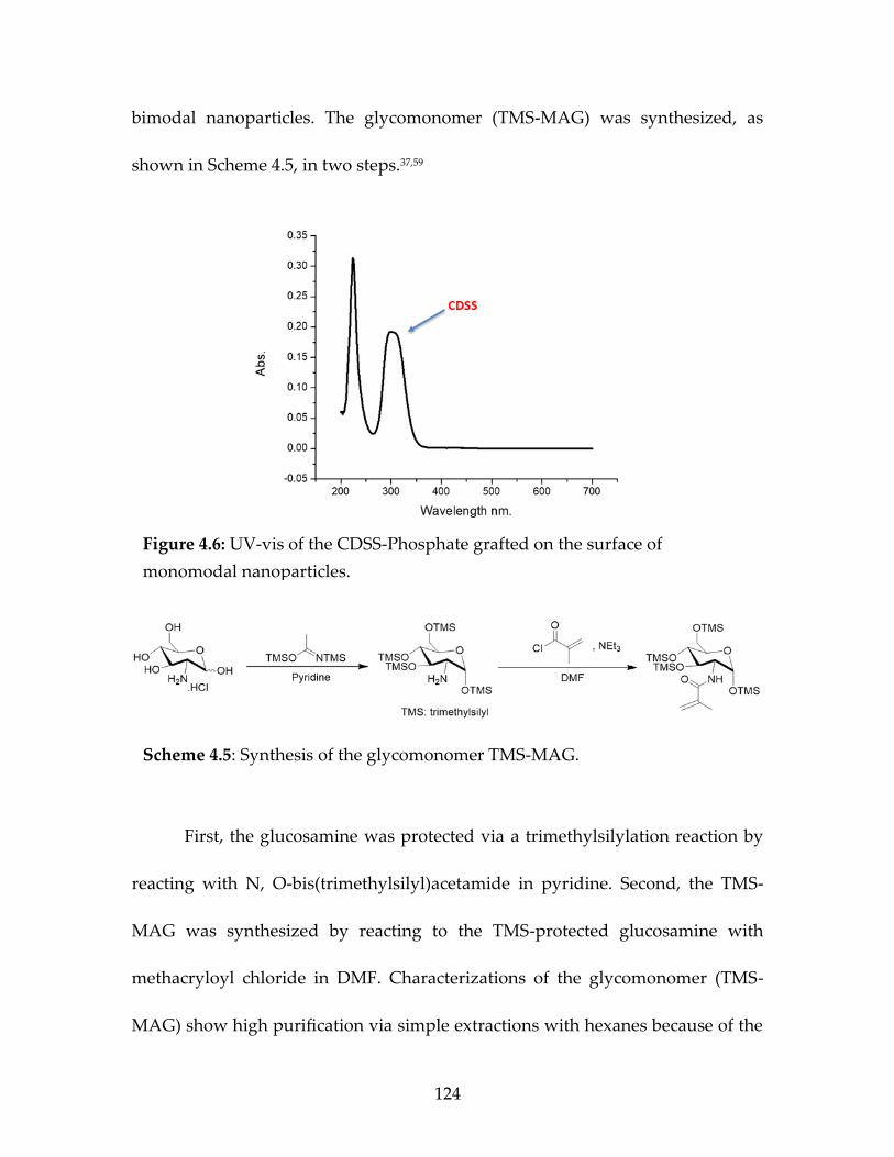
agent (CDSS-Phosphate) ................................................................................124 Figure 4.6 UV-vis of the CDSS-Phosphate grafted on the
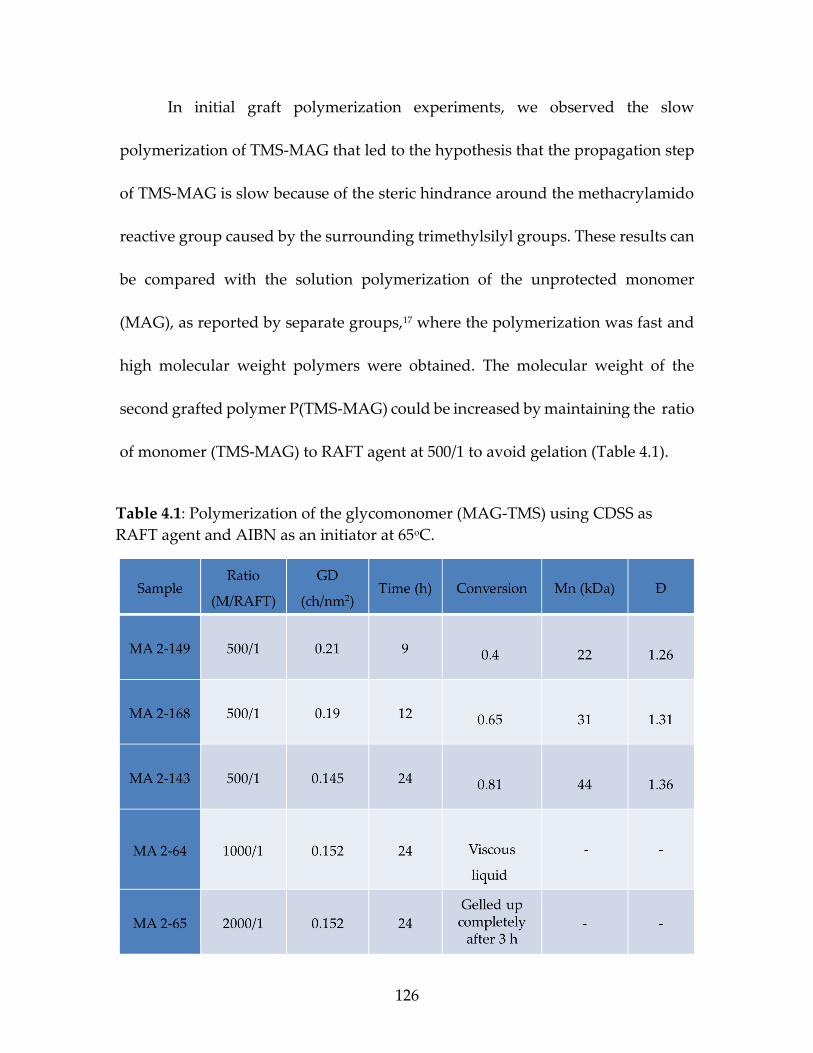
surface of monomodal nanoparticles...........................................................126 Figure 4.7 1H-NMR of the glucomonomer TMS-MAG ............................................127 Figure 4.8 13C-NMR of the glucomonomer TMS-MAG ...........................................127
Page 18
xvii
Figure 4.9 GPC traces of bimodal grafted silica nanoparticles
a) bimodal grafted nanoparticles SiO2-g-P(HEMA-
LA-dye)-P(TMS-MAG) (blue line), deconvoluted
peaks of monomodal nanoparticles SiO2-g-
P(HEMA-LA-dye) (green line), and SiO2-g-P(TMS-
MAG) (red line), measured monomodal grafted
nanoparticles SiO2-g-P(HEMA-LA) (black line). b)
bimodal grafted nanoparticles SiO2-g-P(HEMA-SA-
dye)-P(TMS-MAG) (blue line), deconvoluted peaks
of monomodal nanoparticles SiO2-g-P(HEMA-SA-
dye) (green line), and SiO2-g-P(TMS-MAG) (red
line), measured monomodal grafted nanoparticles
SiO2-g-P(HEMA-SA) (black line) ..................................................................130
Figure 4.10 The fluorescence under UV-vis of the “Sweet
Bimodal nanoparticles, (a)SiO2@P(HEMA-LA-dye)-PMAG, and (b) SiO2@P(HEMA-SA-dye)-PMAG .......................................142
Page 19
xviii
LIST OF SCHEMES
Scheme 2.1 Synthesis of HEMA-LA monomer ...........................................................23 Scheme 2.2 Synthesis of HEMA-SA monomer ...........................................................24 Scheme 2.3 Polymerization of (a) HEMA-LA and (b) HEMA-
SA mediated by free CPDB RAFT agent .......................................................33 Scheme 2.4 Synthetic scheme for the functionalization of
SiO2 nanoparticles with CPDB RAFT agents ................................................36 Scheme 2.5 Synthesis of the dyes, 6-aminohexanoic acid
(NBD-COOH), and NBD-hexamethylenediamine (NBD-NH2) ........................................................................................................39
Scheme 2.6 Synthesis of dye-labeled on SiO2@HEMA-LA,
and SiO2@HEMA-SA grafted-nanoparticles .................................................40 Scheme 3.1 Synthesis of HEMA-LA monomer ...........................................................61 Scheme 3.2 Synthesis of HEMA-SA monomer ...........................................................63 Scheme 3.3 Grafting-from Polymerization of HEMA-LA and
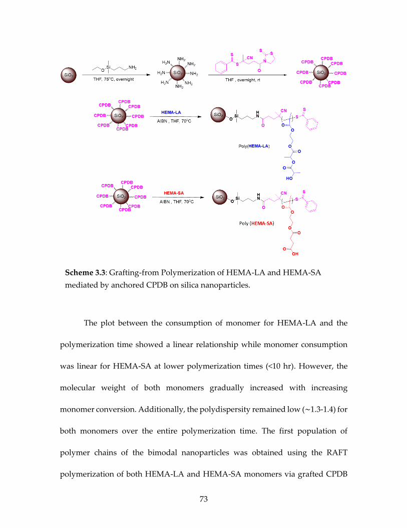
HEMA-SA mediated by anchored CPDB on silica nanoparticles .....................................................................................................73
Scheme 3.4 Synthesis and attachment of the fluorescence dyes
on silica nanoparticles ......................................................................................80 Scheme 3.5 The proposed approach to grafting the second
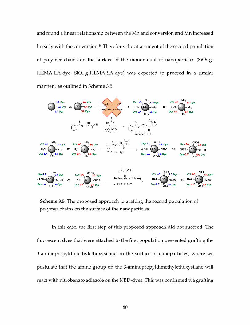
population of polymer chains on the surface of the nanoparticles .....................................................................................................81
Scheme 3.6 Modification of RAFT agent (CPDB) with a
phosphate group ...............................................................................................82 Scheme 3.7 Synthetic strategy for synthesizing the bimodal
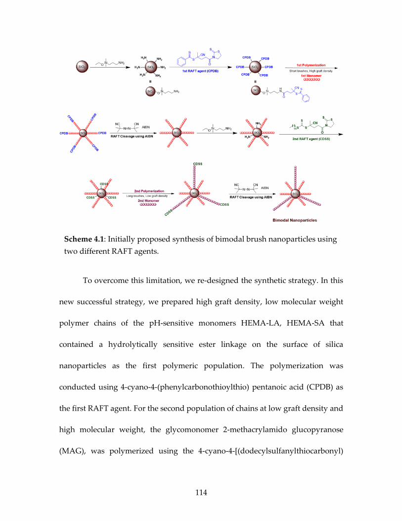
grafted nanoparticles .......................................................................................83 Scheme 4.1 Initially proposed synthesis of bimodal brush
nanoparticles using two different RAFT agents ........................................115 Scheme 4.2 Proposed strategy to synthesis bimodal brush
nanoparticles using two different RAFT agents ........................................117
Page 20
xix
Scheme 4.3 Attaching (a) NBD-COOH dye to the SiO2@ P(HEMA-LA) and (b)NBD-NH2 dye to the SiO2@ P(HEMA-SA) ...................................................................................................120
Scheme 4.4 Synthesis of phosphate-containing CDSS agent ..................................123 Scheme 4.5 Synthesis of the glecomonomer TMS-MAG .........................................126 Scheme 4.6 Total synthesis of bimodal “sweet nanoparticles”
SiO2-g-P(HEMA-LA-dye)-P(MAG) ..............................................................132 Scheme 4.7 Total synthesis of bimodal “sweet nanoparticles”
SiO2-g-P(HEMA-SA-dye)-P(MAG) ..............................................................133 Scheme 5.1 Synthesis various kinds of monomers HEMA-GL,
HEMA-DO, and HEMA-DA .........................................................................141 Scheme 5.2 The proposal new synthesis of bimodal
nanoparticles SiO2-g-P(MAA)-P(HEMA-LA-dye), and SiO2-g-P(MAA)-P(HEMA-SA-dye). .....................................................151
Page 21
xx
LIST OF ABBREVIATIONS
AIBN ............................................................................................. Azobisisobutyronitrile
ATRP ................................................................ Atom Transfer Radical Polymerization
CDSS .................. 4-cyano-4-[(dodecylsulfanylthiocarbonyl)sulfanyl]pentanoic acid
CPDB ................................................................. 4-cyanopentanoic acid dithiobenzoate
CRP .......................................................................... Controlled Radical Polymerization
CTA .................................................................................................. Chain transfer agent
DCC ............................................................................. N, N′-Dicyclohexylcarbodiimide
DDS ................................................................................................ Drug delivery system
DLS .......................................................................................... Dynamic Light Scattering
DMAP .................................................................................... 4-Dimethylaminopyridine
DSC ............................................................................ Differential scanning calorimetry
FT-IR ............................................................. Fourier-transform infrared spectroscopy
GPC ............................................................................ Gel permeation chromatography
HEMA ................................................................................. Hydroxyethyl Methacrylate
HEMA-LA .......................... 2-((2-(propionyloxy) propanoyl)oxy)ethyl methacrylate
HEMA-SA ................................... 4-(2-(methacryloyloxy)ethoxy)-4-oxobutanoic acid
HF .......................................................................................................... Hydrofluoric acid
MAA........................................................................................................ Methacrylic acid
MAG .......................................................................... 2-methacrylamido glucopyranose
Page 22
xxi
Mn ...................................................................... Number-average of molecular weight
NBD-Cl .......................................................... 4-Chloro-7-nitrobenzo-2-oxa-1,3-diazole
NBD-NHS ................ N-[2-{N-(7’-Nitrobenz- 2’-oxa-1’,3’-diazol-4’-yl) amino} ethyl-
carbonyloxy] succinimide
NMP ...................................................................... Nitroxide-Mediated Polymerization
NMR .................................................................................. Nuclear Magnetic Resonance
NP .................................................................................................................. Nanoparticle
PBS .......................................................................................... Phosphate-buffered saline
PDI .................................................................................................... Polydispersity index
RAFT .............. Reversible Addition-Fragmentation Chain Transfer Polymerization
SI-RAFT ....................................................................................... Surface-initiated RAFT
TEM .......................................................................... Transmission electron microscopy
TFA ..................................................................................................... Trifluoroacetic acid
Tg ...................................................................................... Glass Transition Temperature
TGA .................................................................................... Thermogravimetric Analysis
THF.......................................................................................................... Tetrahydrofuran
TMS-MAG ............................. 2-deoxy-2-methacrylamido tetra-(O-trimethylsilyl) D-
glucopyranose
UV-vis .......................................................................... Ultraviolet-visible spectroscopy
Page 23
1
CHAPTER 1
INTRODUCTION
Page 24
2
1.1 RAFT Polymerization:
Since 40 years ago, novel controlled polymerization techniques have been
discovered in polymer chemistry.1 Reversible addition-fragmentation chain
transfer (RAFT) polymerization is considered one of the controlled radical
polymerization (CRP) techniques that give living characteristics to free radical
polymerization.2–5 Living polymerization has emerged where the propagation of
polymerization is continued by all chains and its process in the absence of chain
termination.6 The RAFT polymerization technique can be used to improve the
properties of polymers such as precise control over polymer molecular weights
with narrow polydispersity and the abilities to create well-defined molecular
architectures.7 Controlled radical polymerization techniques are generally
classified by three major methods (Figure 1.1); nitroxide-mediated polymerization
(NMP)8 which requires high reaction temperature, atom transfer radical
polymerization (ATRP)9 that requires a metal catalyst, and reversible addition-
fragmentation chain transfer polymerization (RAFT).1,10 RAFT together with ATRP
are the most widely used CRP techniques to date. RAFT is often preferred for its
simplicity and versatility, usage with a wide range of monomers, lack of metal
catalyst, and low polymerization temperatures.7,11
Page 25
3
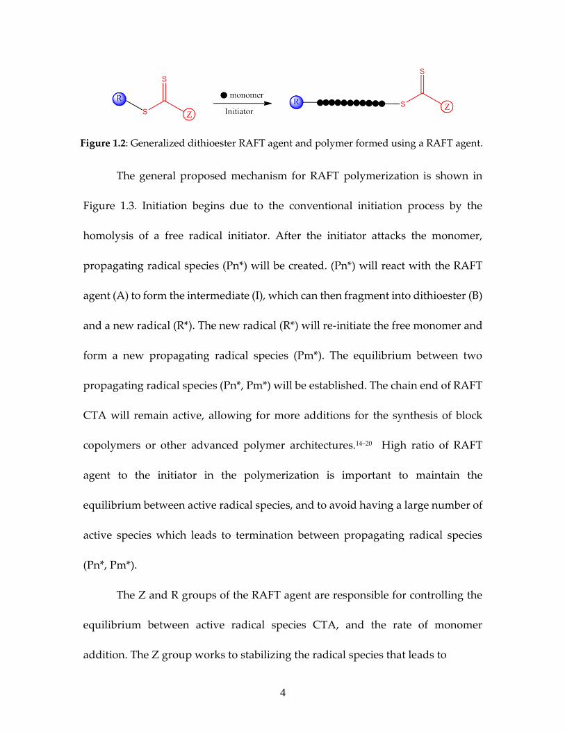
1.2 Mechanism of RAFT Polymerization:
The RAFT technique employs a chain transfer agent (CTA), which works to
control the polymerization due to its ability to create and participate in a chain
equilibrium. Common CTAs are dithioester, dithiocarbamate, or trithiocarbonate
compounds that referred to as RAFT agents and contain Z and R groups that are
responsible for controlling the polymerization (Figure 1.2). Monomer structure
and the structure of the R and Z group of the CTA are the main factors that affect
control of the polymerization.13
Figure 1.1: The three main CRP methods.12
Page 26
4
The general proposed mechanism for RAFT polymerization is shown in
Figure 1.3. Initiation begins due to the conventional initiation process by the
homolysis of a free radical initiator. After the initiator attacks the monomer,
propagating radical species (Pn*) will be created. (Pn*) will react with the RAFT
agent (A) to form the intermediate (I), which can then fragment into dithioester (B)
and a new radical (R*). The new radical (R*) will re-initiate the free monomer and
form a new propagating radical species (Pm*). The equilibrium between two
propagating radical species (Pn*, Pm*) will be established. The chain end of RAFT
CTA will remain active, allowing for more additions for the synthesis of block
copolymers or other advanced polymer architectures.14–20 High ratio of RAFT
agent to the initiator in the polymerization is important to maintain the
equilibrium between active radical species, and to avoid having a large number of
active species which leads to termination between propagating radical species
(Pn*, Pm*).
The Z and R groups of the RAFT agent are responsible for controlling the
equilibrium between active radical species CTA, and the rate of monomer
addition. The Z group works to stabilizing the radical species that leads to
Figure 1.2: Generalized dithioester RAFT agent and polymer formed using a RAFT agent.
Page 27
5
control of the reactivity of CTA while the R group acts as an excellent leaving
group with respect to (Pn*).10
1.3 Polymer Grafted Nanoparticles:
RAFT polymerization has a significant role in the development of
nanoparticles for polymer nanocomposites due to the surface modification of
nanoparticles.21 Properties of a polymer matrix can be significantly enhanced by
using nanoparticles as fillers. Usually, preventing the agglomeration of
nanoparticles is a necessary requirement for improving polymer nanocomposite
Figure 1.3: General mechanism of RAFT polymerization.
Page 28
6
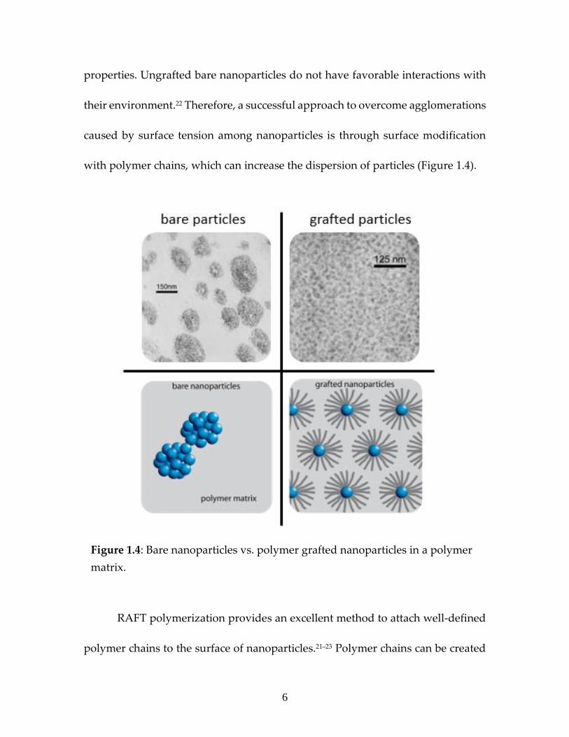
properties. Ungrafted bare nanoparticles do not have favorable interactions with
their environment.22 Therefore, a successful approach to overcome agglomerations
caused by surface tension among nanoparticles is through surface modification
with polymer chains, which can increase the dispersion of particles (Figure 1.4).
RAFT polymerization provides an excellent method to attach well-defined
polymer chains to the surface of nanoparticles.21–23 Polymer chains can be created
Figure 1.4: Bare nanoparticles vs. polymer grafted nanoparticles in a polymer
matrix.
Page 29
7
following two methods: (i) Non-covalent attachment (physisorption) and (ii)
Covalent attachment (chemisorption). Physisorption experimentally is very
straightforward, but it has several limitations, such as desorption or weak linkage,
that limits its applications. Furthermore, chemisorption attachment is more widely
used due to its strong attachment between introduced polymer chains and the
surface of nanoparticles.24
The covalent attachment of chains can be achieved by two main strategies:
grafting-to and grafting-from. The grafting-to technique covalently attaches
polymer chains which have reactive end groups to the surface of nanoparticles.
Grafting-to does not provide high graft density of polymer chains because of the
steric repulsions between them. Furthermore, the reaction between the end group
on polymer chains and the reactive group on the surface of the nanoparticles will
be less efficient with increasing the molecular weight of the polymer. On the other
hand, the grafting-from technique directly initiates the polymerization from
initiator functionalized surfaces, which are covalently linked to the surface.
Grafting-from is advantageous in that it achieves nanoparticles with higher graft
densities because steric interactions are avoided (Figure 1.5).24,25
The morphology of the polymer chains that are attached to the substrate
surface of nanoparticles depends on grafting density. Higher graft densities do not
allow for more distance between polymer chains. Therefore, steric hindrance leads
Page 30
8
to brushes with more extended chain conformations. In contrast, low graft
densities provide the polymer chain space to stretch back towards the substrate
surface of nanoparticles and adopt various conformations such as mushroom
structures (Figure 1.6). Therefore, graft density plays a significant role that affects
matrix interactions.26,27
Figure 1.5: Techniques of polymer attachment A) physisorption, B) grafting-to
approach, C) grafting-from approach.
Figure 1.6: Polymer morphologies resulting from various grafting densities.28
Page 31
9
1.4 Nanoparticles As Delivery Vehicles:
The widespread use of antibiotic drugs that become essential for many
medical interventions to reduce bacterial growth and to treat bacterial infections
specifically, combined with the adapt-ability of bacterial types, has led to
appearance a new phenomenon, antibacterial resistance, which has become a
global issue. Antibacterial resistance is one of the issues that has gathered
remarkable attention during the past three decades.29 Bacteria have developed
their abilities to become more resistant to traditional antibiotics. β-lactam
antibiotics are the common conventional antibiotics that have widely used and
have a long history. Bacteria gradually started to develop resistance against these
antibiotics by creating β-lactamase enzymes which work to deactivate antibiotics
through hydrolyzing the β-lactam ring efficiently.30 Therefore, one of the
significant approaches that are used to inhibit β-lactamase and overcome bacteria-
resistance is developing antibacterial nanoparticles where the antibiotic linkage to
the surface of nanoparticles will enhance their effectiveness against bacteria.31
Antibacterial nanoparticles have been developed and investigated as therapeutic
delivery vehicles. Nanoparticles can offer variable and structured surfaces having
various types and densities of antibiotics.32 Consequently, this will permit the
specificity of the quantities of antibiotic molecules that will be carried by
nanoparticles that will reach infectious bacterial cells. Overcoming bacterial
Page 32
10
resistance may occur by releasing antibiotics slowly into the system while
preserving an effective antibiotic concentration for extended times.33 One of the
synthetic strategies that can be used is the design of new monomers, containing an
ester linkage which can be easily hydrolyzed. The slow degradation of the side
chains will occur, resulting in the slow release of antibiotics from the surface of
nanoparticles.
1.5 Bimodal Nanocomposites:
A novel architecture of grafting bimodal polymer brushes on nanoparticles
can significantly improve the entanglement of nanoparticle fillers and matrix
polymers. Improving the properties of polymers/ matrices will lead to the wider
application of polymer nanocomposites. Extensive research has been done to
understand the relationship between the polymer brushes on the nanoparticles
and their matrices.34 However, controlling the graft densities of brushes and the
interface of the brush/matrix are significant issues that need to be addressed to
fully understand the structure-property relationship in polymer nanocomposites.
Typically, the aggregation process of monomodal brush grafted nanoparticles is
addressed by a delicate equilibrium between enthalpic and entropic interfacial
interactions.35,36 Therefore, using a bimodal polymer brush architecture on
nanoparticles is considered an improved approach currently to overcome the
aggregation of nanoparticles that occurs in polymer nanocomposites. A bimodal
Page 33
11
polymer brush is created by attaching two populations of polymer chains with
different lengths to the surface of nanoparticles (Figure 1.7).37 Both approaches,
grafting-from, and grafting-to are successfully used to prepare bimodal polymer
brushes.
1.6 References:
(1) Chiefari, J.; Chong, Y. K.; Ercole, F.; Krstina, J.; Jeffery, J.; Le, T. P. T.;
Mayadunne, R. T. A.; Meijs, G. F.; Moad, C. L.; Moad, G.; Rizzardo, E.;
Thang, S. H. Macromolecules 1998, 31 (16), 5559–5562.
(2) Ilgach, D. M.; Meleshko, T. K.; Yakimansky, A. V. Polym. Sci. Ser. C 2015, 57
(1), 3–19.
Figure 1.7: Illustration of various architectures of core-shell nanoparticles: (a)
single shell and (b) double shell, both of which are classified as monomodal
core-shell nanoparticles, and (c) bimodal core-shell nanoparticles.38
Page 34
12
(3) Moad, Graeme, Ezio Rizzardo, and S. H. T. Acc. Chem. Res. 2008, 41 (9),
1133–1142.
(4) Moad, G.; Rizzardo, E.; Thang, S. H. Aust. J. Chem. 2005, 58 (6), 379–410.
(5) Moad, G.; Rizzardo, E.; Thang, S. H. Aust. J. Chem. 2012, 65 (8), 985–1076.
(6) Vazaios, A.; Lohse, D. J.; Hadjichristidis, N. Macromolecules 2005, 38 (13),
5468–5474.
(7) Li, C.; Benicewicz, B. C. Macromolecules 2005, 38, 5929–5936.
(8) Hawker, C. J.; Bosman, A. W.; Harth, E. Chem. Rev. 2001, 101 (12), 3661–
3688.
(9) Wang, J. S.; Matyjaszewski, K. J. Am. Chem. Soc. 1995, 117 (20), 5614–5615.
(10) Moad, G.; Rizzardo, E.; Thang, S. H. Aust. J. Chem. 2006, 59 (10), 669–692.
(11) Perrier, S. Macromolecules 2017, 50 (19), 7433–7447.
(12) Spanswick, J.; Matyjaszewski, K.; Spanswick, J. Mater. Today 2005, 8 (3), 26–
33.
(13) Barner-Kowollik, C. Handbook of RAFT Polymerization; WILEY-VCH Verlag
GmbH & Co., 2008.
Page 35
13
(14) Keddie, D. J.; Moad, G.; Rizzardo, E.; Thang, S. H. Macromolecules 2012, 45
(13), 5321–5342.
(15) Harvison, M. A.; Roth, P. J.; Davis, T. P.; Lowe, A. B. Aust. J. Chem. 2011, 64
(8), 992–1006.
(16) Moad, G.; Rizzardo, E.; Thang, S. H. Polym. Int. 2011, 60 (1), 9–25.
(17) Gregory, A.; Stenzel, M. H. Prog. Polym. Sci. 2012, 37 (1), 38–105.
(18) Roth, P. J.; Boyer, C.; Lowe, A. B.; Davis, T. P. Macromol. Rapid Commun.
2011, 32 (15), 1123–1143.
(19) Moad, G.; Chong, Y. K.; Mulder, R.; Rizzardo, E.; Thang, S. H. ACS Symp.
Ser. 2009, 1024, 3–18.
(20) Moad, G. ACS Symp. Ser. 1187, 2015, 211–246.
(21) Li, C.; Han, J.; Ryu, C. Y.; Benicewicz, B. C. Macromolecules 2006, 39 (9),
3175–3183.
(22) Fu, S. Y.; Feng, X. Q.; Lauke, B.; Mai, Y. W. Compos. Part B Eng. 2008, 39 (6),
933–961.
(23) Ranjan, R.; Brittain, W. J. Macromolecules 2007, 40 (17), 6217–6223.
Page 36
14
(24) Barbey, R.; Lavanant, L.; Paripovic, D.; Schüwer, N.; Sugnaux, C.; Tugulu,
S.; Klok, H. A. Chem. Rev. 2009, 109 (11), 5437–5527.
(25) Dai, Xiao-Hui, D. C.-M. J. Polym. Sci. Part A Polym. Chem. 2008, 46
(September 2010), 817–829.
(26) Brittain, W. J.; Minko, S. J. Polym. Sci. Part A Polym. Chem. 2007, 45 (16),
3505–3512.
(27) Schadler, L. S.; Kumar, S. K.; Benicewicz, B. C.; Lewis, S. L.; Harton, S. E.
MRS Bull. 2007, 32 (4), 335–340.
(28) Dukes, D.; Li, Y.; Lewis, S.; Benicewicz, B.; Schadler, L.; Kumar, S. K.
Macromolecules 2010, 43 (3), 1564–1570.
(29) Deng, H.; McShan, D.; Zhang, Y.; Sinha, S. S.; Arslan, Z.; Ray, P. C.; Yu, H.
Environ. Sci. Technol. 2016, 50 (16), 8840–8848.
(30) Li, W.; Dong, K.; Ren, J.; Qu, X. Angew. Chemie - Int. Ed. 2016, 55 (28), 8049–
8053.
(31) Wang, L.; Chen, Y. P.; Miller, K. P.; Cash, B. M.; Jones, S.; Glenn, S.;
Benicewicz, B. C.; Decho, A. W. Chem. Commun. 2014, 50 (81), 12030–12033.
(32) Wang, L.; Benicewicz, B. C. ACS Macro Lett. 2013, 2 (2), 3–6.
Page 37
15
(33) Tamizharasi, S.; Rathi, V.; Rathi, J. C. Syst. Rev. Pharm. 2011, 2 (1), 19–29.
(34) Rungta, A.; Natarajan, B.; Neely, T.; Dukes, D.; Schadler, L. S.; Benicewicz,
B. C. Macromolecules 2012, 45 (23), 9303–9311.
(35) Meldal, M. Macromol. Rapid Commun. 2008, 29 (12–13), 1016–1051.
(36) Kumar, S. K.; Krishnamoorti, R. Annu. Rev. Chem. Biomol. Eng. 2010, 1 (1),
37–58.
(37) Skvortsov, A. M.; Gorbunov, A. A.; Leermakers, F. A. M.; Fleer, G. J.
Macromolecules 1999, 32 (6), 2004–2015.
(38) Qiao, Y.; Yin, X.; Wang, L.; Islam, M. S.; Benicewicz, B. C.; Ploehn, H. J.;
Tang, C. Macromolecules 2015, 48 (24), 8998–9006.
Page 38
16
CHAPTER 2
POLYMERIZATION OF “CONTROLLED RELEASE” MONOMERS
CONTAINING A HYDROLYTICALLY SENSITIVE ESTER
LINKAGE VIA RAFT POLYMERIZATION 1
1Al-Ali, M.A. and Benicewicz B. C. To be submitted to Journal of Polymer Science.
Page 39
17
2.1 Abstract:
The aim of this work was to develop a novel type of drug-delivery carrier
consisting of a pH-responsive “controlled release” polymer containing an
antibacterial drug grafted onto the surface of a nanoparticle. Herein, we describe
the first report of pH-responsive biodegradable polymers grafted from the surface
of silica nanoparticles. Grafted “controlled release” polymers containing a
hydrolytically sensitive ester linkage on silica nanoparticles were successfully
prepared via reversible addition-fragmentation chain transfer (RAFT)
polymerization. Two potential “controlled release” monomers, 2-((2-
(propionyloxy) propanoyl)oxy)ethyl methacrylate (HEMA-LA) and 4-(2-
(methacryloyloxy)ethoxy)-4-oxobutanoic acid (HEMA-SA), were synthesized by
the ring-opening reaction of L-lactide and succinic anhydride with 2-hydroxyethyl
methacrylate (HEMA), respectively. The polymerization of the methacrylate
monomers was carried out using 4-cyanopentanoic acid dithiobenzoate (CPDB) as
a RAFT agent. Both polymers poly(HEMA-LA) and poly(HEMA-SA) were
characterized by NMR spectroscopy and gel permeation chromatography (GPC).
The degradation rates of these two polymers were investigated using phosphate
buffer solution (PBS, pH = 7.4) at 25ᵒC and 37ᵒC as a function time using conjugated
dyes (NBD-aminohexanoic acid, NBD-hexamethylenediamine). The pH-
dependence of dye-loaded polymer grafted nanoparticles was confirmed by the
Page 40
18
evaluation of the cumulative release rate at two temperatures 25ᵒC, 37ᵒC. Such
polymer grafted nanoparticles are being developed for use as delivery vehicles for
antibacterial applications.
2.2. Introduction:
Drug delivery of pharmaceutical compounds is considered the key to
achieving a significant therapeutic effect, whether for humans or animals.1
Nanotechnology methods have more significant potential in drug delivery
systems (DDS) as the desired drug could be released using biodegradable
polymers.2 For the ideal drug delivery system(DDS), preserving the drug level
within a desired therapeutic range is the main aim because there is a toxic and
ineffective plasma level for each drug.3 The design of a “Controlled Release” drug
delivery technique using nanotechnology is one of the significant strategies to
overcome various diseases.4 Globally, different stimuli-sensitive polymeric
systems have attracted considerable attention in recent years that show a response
to an external stimulus such as pH, temperature, specific ion, and electric field.5
pH-sensitive nanopolymers, among the different types of stimuli-responsive
polymers, have been advanced and most widely used to develop sensitive nano-
systems in which the drug will release in different pH environments.6 The use of
polymers containing a pH-sensitive ester linkage on silica nanoparticles has
gained significant importance during recent decades.7
Page 41
19
Several strategies/ approaches of pH-responsive drug release have been
studied. For instance, one of the important strategies is to introduce ionizable
functional groups, such as esters, amides, phosphoric acids, and carboxylic acids
with nanomaterials. These ionizable functional groups are biodegradable, which
can result in the drug release through the mechanism of a pH-stimulus
environment.8 pH-sensitive polymers with ionizable groups that are considered a
class of polyelectrolytes that can be ionized and change their conformation.
Several pH-sensitive polymers have been developed by using acidic or basic
groups that accept or release protons in response to changes in the pH
environment. Esters linkages have been preferred when engineering polymeric
materials for controlled release compared to amides, carbonates, and carbamates
because of their relative ease of hydrolysis at physiological pH (7.4).9 At pH 7.4,
the esters groups that have a carbonyl adjacent to an ether linkage can be readily
hydrolyzed to alcohol and carboxylic acid derivatives.10
However, the current study is focused on the designing of pH-sensitive
polymers grafted onto silica nanoparticles (SiO2@HEMA-LA, SiO2@HEMA-SA).
Controlled release pH-responsive monomers containing ester linkage were
synthesized. The Grafting-from RAFT polymerization technique was used to
polymerize these controlled release monomers onto the surface of silica
nanoparticles to get controlled and high loading capacity.11 The controlled release
Page 42
20
study was investigated by attaching labeled-dyes to the pH-sensitive polymers to
monitor the degradation rate. Furthermore, loading drugs or antibiotics could be
attached to pH-sensitive polymers grafted on silica nanoparticles and study their
release rate.
2.3 Experimental:
2.3.1 Materials:
L-lactide (Sigma Aldrich, 95%) and succinic anhydride (Acros Organics,
99%) were used as received. 2-Hydroxyethyl methacrylate (HEMA, Sigma
Aldrich, 99%) was purified by passing through a column of basic aluminum oxide
(Alfa Aesar, 99%) to remove the inhibitor, methyl ether hydroquinone (MEHQ).
Colloidal silica nanoparticles (SiO2, spherical 14 ± 4 nm, 30 wt% in MEK) were
purchased from Nissan Chemical Co. The RAFT agent 4-cyanopentanoic acid
dithiobenzoate (CPDB) was purchased from Boron Molecular and used as
received. 3-Aminopropyldimethylethoxysilane and dimethylmethoxy-n-
octylsilane were purchased from Gelest, Inc. (95%) and used as received.
Azobisisobutyronitrile (AIBN) was used after purification by recrystallization in
methanol. The catalysts, tin (II) 2-ethylhexanoate and 4-dimethylaminopyridine
(DMAP), were purchased from Alfa Aesar and Chem-Impex Int'l Inc respectively.
All other reagents and solvents were used as received unless otherwise noted.
Page 43
21
2.3.2 Instrumentation:
1H-NMR spectra were recorded with a Bruker Avance III-HD spectrometer
(300 MHz) using CDCl3 as a solvent and measured with tetramethylsilane (TMS)
as an internal reference. Gel permeation chromatography (GPC) was used to
measure the molecular weights (Mn) and dispersity index (Đ) using a Varian 290-
LC pump, a Varian 390-LC refractive index detector, and three Styragel columns
(HR1, HR3 and HR4, molecular weight range of 100-5000, 500-30000, and 5000-
500000) calibrated with polystyrene and poly(methylmethacrylate) standards
obtained from Polymer Laboratories. Tetrahydrofuran (THF) was used as an
eluent at 30ᵒC and a flow rate of 1.0 mL/min. Thermogravimetric analysis (TA
Instruments Q5000) was used to obtain TGA characterization after preheating to
100°C for 10 min to remove residual solvents for all the samples. After cooling to
50°C, the samples were reheated to 800°C with a heating rate of 10°C/min under
nitrogen flow. FT-IR spectra were recorded using a BioRad Excalibur FTS 3000.
UV-vis absorption spectra were taken on a Shimadzu UV-2450 spectrophotometer.
2.3.2 Synthesis of “Controlled Release” Monomers:
Two methacrylate monomers were synthesized via the ring-opening
reaction of the corresponding cyclic lactone compound, L-lactide, or succinic
anhydride with hydroxyethyl methacrylate (HEMA) catalyzed by stannous 2-
ethylhexanoate and DMAP, respectively.
Page 44
22
2.3.2.1 Synthesis of 2-((2-(Propionyloxy) Propanoyl)oxy)ethyl
Methacrylate (HEMA-LA) (Scheme 2.1):
L-lactide (2.99 g, 20.7 mmol) was placed in round flask and dried overnight
under vacuum at rt. HEMA (2.8 mL, 23 mmol) and tin(II) 2-ethylhexanoate (52 μL,
0.16 mmol) were then added to the flask, and the reaction was deoxygenated by a
repeated vacuum nitrogen cycle. Subsequently, the mixture was heated to 115°C
under vacuum for 3 hours with stirring. The crude product was dissolved in
anhydrous chloroform and washed with 1 M HCl. Then, the organic phase was
washed with deionized water, isolated, and residual chloroform removed using a
rotary evaporator operating under vacuum. Yields varied from 70-75% based on
the added amount of L-lactide. 1H-NMR (300 MHz, CDCl3): δ=1.38–1.63 ppm (6H,
CH–CH3), δ= 1.94 ppm (3H, CH2=CCH3), δ= 2.79 ppm (1H, OH), δ= 4.26–4.39 ppm
(4H, OCH2–CH2), δ= 4.39–4.51 ppm (1H, CH-(OH)CH3), δ= 5.08–5.29 ppm (1H,
C(=O)–CH), δ= 5.58 ppm (1H, CH2=C), δ= 6.10 ppm (1H, CH2=C) (Figure 2.1).
HRMS (EI) (m/z) calcd for C12H18O7: 274.1149; found: 274.1167.12,13
Page 45
23
2.3.2.2 Synthesis of 4-(2-(Methacryloyloxy)ethoxy)-4-oxobutanoic Acid (HEMA-
SA) (Scheme 2.2):
2-Hydroxyethyl methacrylate (HEMA; 6.1 mL, 50 mmol) was dissolved in
anhydrous THF in a Schlenk flask (250 mL) at room temperature under nitrogen.
Scheme 2.1: Synthesis of HEMA-LA monomer.
Figure 2.1: 1H-NMR (300 MHz, CDCl3) spectrum of HEMA-LA monomer.
Page 46
24
Succinic anhydride (6 g, 0.06 mol), 12 mL of pyridine, and 4-
dimethylaminopyridine (0.49 g, 4.0 mmol) were added to the flask. Then, the
reaction mixture was stirred for 24 h at 40ᵒC under nitrogen. The reaction was
cooled to the room temperature, and the solvent was evaporated under vacuum.
The residue was dissolved in DCM, followed by washing three times with 0.1 M
HCl solution. The organic phase was dried over anhydrous magnesium sulfate
overnight and filtered. After evaporation of the solvent, the remaining HEMA-
COOH product was dried under vacuum at room temperature. A viscous liquid
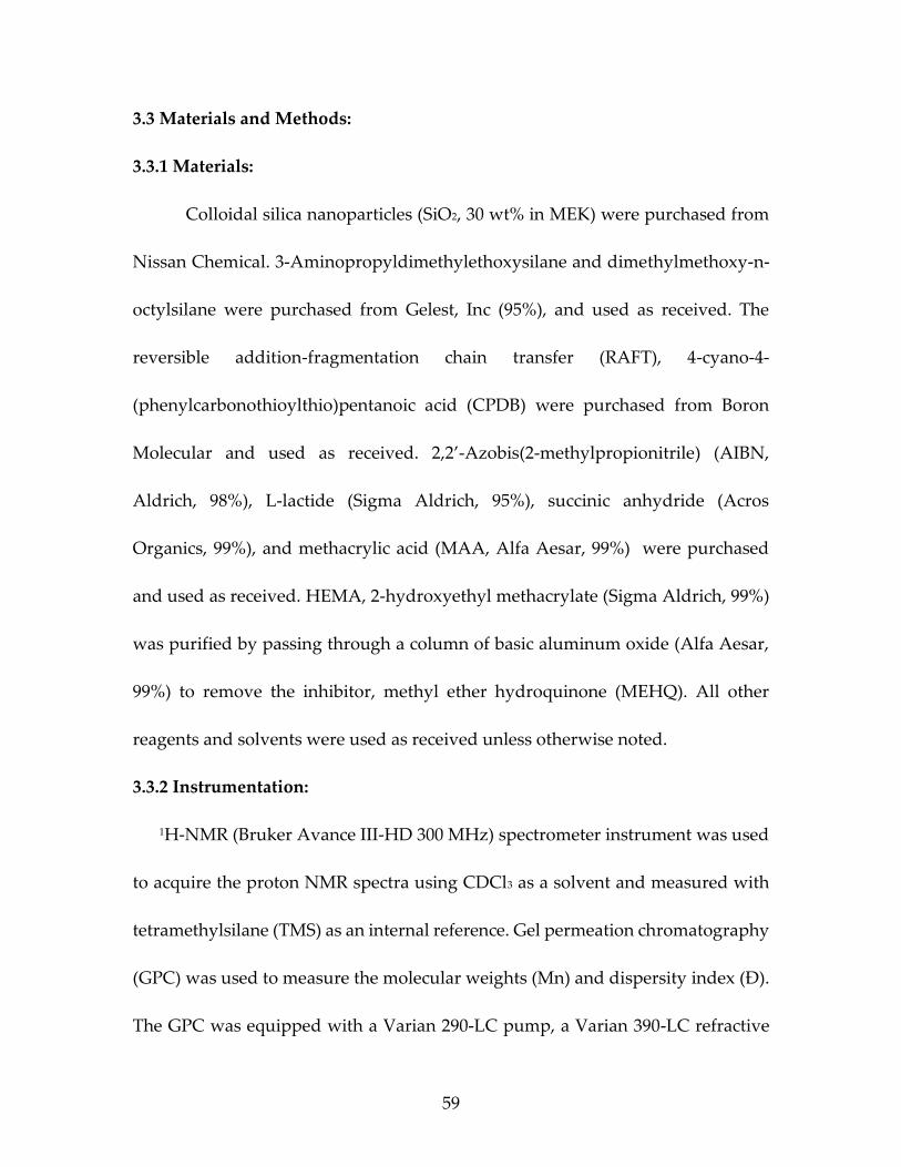
was obtained (yield 60%, 6.9 g). 1H NMR (300 MHz, CDCl3): δ = 6.13 (S, 1H,
HCH=C(CH3)-), 5.54 (S, 1H, HCH=C(CH3)-), 4.36 (t, 4H, -OOC(CH2)2 OCO-), 2.68
(t, 4H, HOOC(CH2)2COO-), 1.85 (S, 3H, H3CC(COO-)CH2) (Figure 2.2). HRMS (EI)
(m/z) calcd for C10H14O6: 230.0842; found: 230.0873.14,15
Scheme 2.2: Synthesis of HEMA-SA monomer.
Page 47
25
2.3.3 Activation of 4-cyano-4-(thiobenzoylthio)pentanoic acid (CPDB):
Dimethylamino pyridine (DMAP) (122 mg, 1.0 mmol) was added slowly to
the solution of CPDB (2.80 g, 10.0 mmol), 2-mercaptothiazoline (1.2 g, 10.0 mmol),
and dicyclohexylcarbodiimide (DCC) (2.5 g, 12.0 mmol) in 40 ml of
dichloromethane. Then, the solution was stirred (6 h) at room temperature. The
solids were removed from the solution by filtration. The solution was evaporated
to remove the solvent, and silica gel column chromatography (5:4 hexane: ethyl
acetate) was used to obtain activated CPDB as a red oil (80% yield, 4 g). 1H-NMR
(300 MHz, CDCl3): δ (ppm) 7.90 (d, 2H), 7.56 (t, 1H), 7.38 (t, 2H), 4.58 (t, 2H,
NCH2CH2S), 3.60-3.66 (m, 2H, (CN)C(CH3)-CH2CH2CON), 3.31 (t, 2H,
Figure 2.2: 1H-NMR (300 MHz, CDCl3) spectrum of HEMA-SA monomer.
Page 48
26
NCH2CH2S), 2.50-2.56 (m, 2H, (CN)C(CH3)CH2CH2CON), 1.95 (s, 3H,
(CH3)C(CN)S).16 HRMS (EI) (m/z) calcd for C16H16N2OS4: 380.0193; found: 380.0203.
2.3.4 Attachment of activated CPDB onto silica nanoparticles (SiO2@CPDB):
Silica nanoparticles (10.0 g, 30 wt % in MEK) were added to a round bottom
flask with 30 mL THF and 350 µL 3-aminopropyldimethylethoxy silane was
added. After purging with N2 for more than 30 min, the solution was refluxed in
a 75°C overnight. Then, the solution was cooled to r.t and precipitated into a large
amount of hexanes. The solution was centrifuged at 3,500 rpm for 8 minutes and
the solvent decanted. The precipitation-dissolution process was then repeated for
another two times. The amine-functionalized nanoparticles were dispersed in 30
mL of dry THF, which was added dropwise into a THF solution of 1.47 mL
activated CPDB (0.19 M) at r.t. and stirred for 6 hours. The solution was
precipitated into a large amount of hexane (approx. 500 ml), and the nanoparticles
were recollected by centrifugation at 3500 rpm for 8 min. This precipitation-
dissolution process was repeated until the supernatant solution was colorless. The
nanoparticles were dried under vacuum at r.t. The grafting density of CPDB
anchored silica nanoparticles (0.3 ch/nm2) was determined using a calibration
curve of made from standard solutions of free CPDB via UV-vis a spectrometer.17
Page 49
27
2.3.5 RAFT Polymerization of “Controlled Release” Monomers From CPDB
Functionalized Silica Nanoparticles:
CPDB-anchored silica nanoparticles (1g, 56.18 µmol/g) were dispersed in
THF (8 ml). HEMA-LA (7.7 g, 28.07 mmol) or HEMA-SA (6.5 g, 28.23 mmol), AIBN
(0.562 ml of 10 mM THF solution) were added to the Schlenk tube, where the ratio
between species of [CPDB]:[monomer]:[AIBN] was 1:500:0.1. The Schlenk tube
was degassed by three freeze−pump−thaw cycles, filled with nitrogen, and then
the Schlenk tube was placed in an oil bath at 65°C for the desired time. The Schlenk
tube was quenched in ice water to stop the polymerization. The polymer-grafted
silica nanoparticles were precipitated by pouring into 500 ml of hexanes and
centrifuged at 3500 rpm for 8 min. The nanoparticles were dispersed back into
THF. Polymer chains were cleaved from the nanoparticles by dissolving 50 mg of
polymer-grafted nanoparticles in 3 ml of THF and treating with 0.2 ml aqueous
HF (49%). The solution was stirred overnight, and the cleaved polymer chains
were analyzed by GPC.18
2.3.6 Cleavage of CPDB Agents From The Polymeric Chain Ends Of The Silica
Nanoparticles:
Polymer-grafted nanoparticles (1 g, SiO2-g-HEMA-LA, SiO2-g-HEMA-SA)
were dispersed in 40 ml THF, and solid AIBN (0.12 g) was added at the ratio of
([CTA]:[AIBN]= 1:20). The solution was heated under nitrogen at 65°C for 1 h. The
Page 50
28
solution was poured into 500 ml of hexanes and centrifuged at 3500 rpm for 8 min
to recover the nanoparticles.17
2.3.7 Preparation Of NBD-Labelled Amino Acid:
A solution of 6-aminohexanoic acid (1.2 eq, 3 mmol) and NaHCO3 (3 eq, 7.5
mmol) in MeOH (30 mL) were stirred at room temperature for 30 min and refluxed
at 65°C for 15 min. Then, 4-chloro-7-nitrobenzofurazan (NBD-Cl, 1 eq, 2.5 mmol)
was dissolved in MeOH (5 mL) and added dropwise to the solution. After two
hours, the reaction was cooled to room temperature and acidified to
approximately pH=2 with 1M HCl. Subsequently, the mixture was extracted three
times with EtOAc (20 mL), washed with brine, dried with MgSO4, filtered, and the
solvent removed using a rotary evaporator. The resultant NBD-labelled amino
acid was then recrystallized from aqueous MeOH.19 The prodect was yield as
bright orange crystals (yield: 80%, 0.59 g). Tm= 156-158°C, UV (MeOH) λmax: 335,
458. FT-IR νmax/cm-1 1700 (strong, sharp C=O). MS (EI+) m/z: [M]+ 294.
2.3.8 Preparation Of NBD-Labelled Hexamethylenediamine:
Hexamethylenediamine-NBD dye was synthesized in two steps, first
preparing N-Boc-hexamethylenediamine-NBD that was converted to the
hexamethylenediamine-NBD. A solution of 4-chloro-7-nitrobenzofurazan (NBD-
Cl) (1 eq, 2.5 mmol) and mono-Boc-hexamethylenediamine (1.1 eq, 2.76 mmol) was
prepared in ethanol (30 mL). Pyridine (catalytic, 260 μL) was added to the stirred
Page 51
29
solution and allowed to stir for 30 min. The mixture was concentrated and purified
by column chromatography (toluene: ethyl acetate 7:3) to obtain the product as a
red foam. Next, the Boc-protected dye was dissolved in a mixture of solvent (1:1
of trifluoroacetic acid (TFA): dichloromethane (DCM)) and then stirred for one
hour. The solution was concentrated and resuspended in acetonitrile. The final
product was obtained as golden crystals after the solution was precipitated into
cold diethyl ether (yield 81%, 0.6 g).20 UV (MeOH) λmax: 336, 460. FT-IR νmax/cm-1
3380 (medium, sharp N-H). HRMS (EI) (m/z) calcd for C12H17N5O3: 279.1382;
found: 279.3014.
2.3.9 Aminohexanoic Acid-NBD Conjugate On HEMA-LA-g-SiO2 And
Hexamethylenediamine-NBD Conjugate On HEMA-SA-g-SiO2:
Polymer-g-SiO2 (1 equiv.) (HEMA-LA-g-SiO2 or HEMA-SA-g-SiO2), dye-
labeled NBD (1 equiv.) (aminohexanoic acid-NBD or hexamethylenediamine-
NBD, respectively), and dicyclohexylcarbodiimide (DCC) (1.2 equiv.) were
dissolved in 30 mL of THF. (Dimethylamino) pyridine (DMAP) (0.1 equiv.) was
added slowly to the solution. Subsequently, the solution was stirred at r.t. for 6 h.
The solution was filtered, and the solvent was concentrated using a rotary
evaporator. The solution was then precipitated by pouring into hexane (400 ml)
and centrifuged at 3500 rpm for 8 min to recover the nanoparticles. The
Page 52
30
precipitation-dissolution process was repeated twice until the supernatant layer
after centrifugation was colorless to ensure the removal of free dyes.
2.3.10 In Vitro Quantification Of Dye:
The calibration curves for the dyes were achieved by preparing a standard
solution of dye using 27 mg of dye dissolved in 50 ml THF. Then, various
concentrations were prepared (13.6, 6.8, 4.3, 1.7, 0.8, 0.4) in 50 ml THF to obtain
serial dilutions and assayed at 457- 460 nm using UV spectrophotometry. The data
were plotted to obtain a straight lines for the quantification of the dyes (Figure 2.3
a,b).21
Figure 2.3: Images of serial dilutions, UV spectrum of various concentrations, and the
resultant calibration curves of (a) NBD-COOH, (b) NBD-NH2 dyes.
Page 53
31
2.3.11 Dye Release Rate Studies:
Dye release kinetics were determined using pH = 7.4 phosphate-buffered
saline solution (PBS) at pH = 7.4 at 25°C and 37°C for the dye attached polymer
grafted nanoparticles. HEMA-SA-dye-g-SiO2 or HEMA-LA-dye-g-SiO2 (200 mg)
were immersed in 250 ml of PBS solution at pH 7.4 using a dialysis membrane bag
(MWCO 3500, Fisherbrand), which was tied at the ends after filling with 5 ml of
the PBS buffer solution. The systems were incubated at different temperatures,
25°C and 37°C, and provided with gentle shaking at 40 rpm over the test periods.
PBS solution (5 ml) was sampled out and assayed for released dye at 480 nm using
a (Shimadzu UV-2450) spectrophotometer, at predetermined time points. The UV-
vis was measured for these withdrawn samples at 25°C and 37°C, at determined
intervals, and replaced with fresh buffer solution (PBS) following every sampling
point to keep the same concentration during the full release period. The study was
continued until the released amount reached an equilibrium.22
2.4 Results And Discussion:
2.4.1 Polymerization Of The "Controlled Release" (HEMA-LA, HEMA-SA)
Monomers Mediated By Free CPDB:
To praper for grafting HEMA-LA and HEMA-SA on the surface of silica
nanoparticles via RAFT polymerization, the polymerization behavior of both
monomers mediated by free CPDB RAFT agent was investigated. Generally, the
Page 54
32
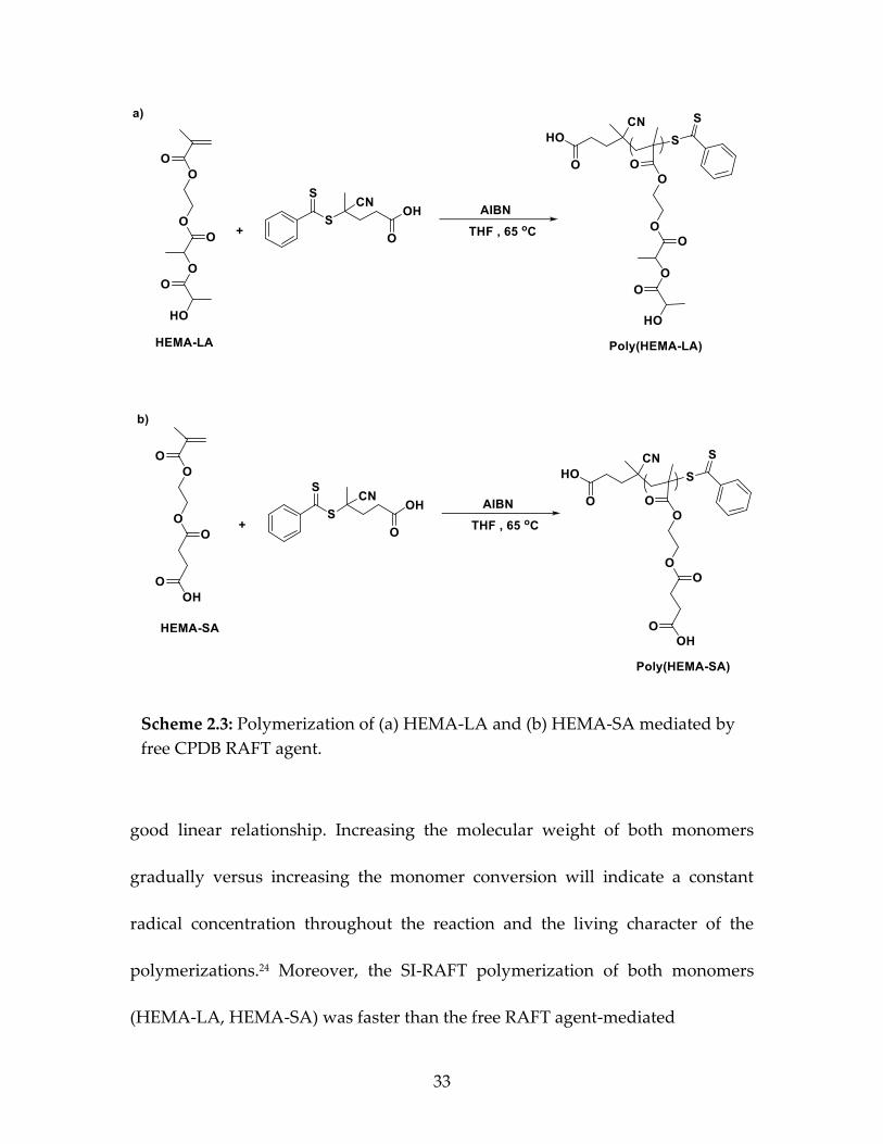
initial and significant aspect when using the RAFT technique is choosing a suitable
RAFT agent which is compatible with the monomer which will provide successful
control.23 In this work, we investigated two types of RAFT agents. A
trithiocarbonate derivative 4-cyano-4-[(dodecylsulfanylthiocarbonyl) sulfanyl]
pentanoic acid (CDSS) and a dithiobenzoate derivative 4-cyanopentanoic acid
dithiobenzoate (CPDB) were tested at 65ᵒC. Both monomers could be polymerized
with the trithiocarbonate derivative CDSS. However, the polymerizations resulted
in low monomer conversions and produced polymers with broad polydispersity.
However, we found that the dithiobenzoate derivative CPDB RAFT agent
provided a controlled polymerization, where was compatible with both
monomers HEMA-LA and HEMA-SA.
The synthetic procedure for the RAFT polymerization of both HEMA-LA
and HEMA-SA monomers via free CPDB in solution is shown in Scheme 2.3. The
feed ratio [CTA]/[Monomer]/[Initiator] of polymerization was 500: 1: 0.1 at 65ᵒC
under inert gas conditions. Figure 2.4 shows the results of the kinetic study for the
free RAFT polymerization and surface-initiated RAFT polymerization of both
monomers HEMA-LA and HEMA-SA. By observing the consumption ln(Mo/Mt)
of each monomer (HEMA-LA, HEMA-SA; individually), which increased
concurrently with the time and the conversion of the polymerizations, we found a
Page 55
33
good linear relationship. Increasing the molecular weight of both monomers
gradually versus increasing the monomer conversion will indicate a constant
radical concentration throughout the reaction and the living character of the
polymerizations.24 Moreover, the SI-RAFT polymerization of both monomers
(HEMA-LA, HEMA-SA) was faster than the free RAFT agent-mediated
Scheme 2.3: Polymerization of (a) HEMA-LA and (b) HEMA-SA mediated by
free CPDB RAFT agent.
Page 56
34
Figure 2.4 (a) Pseudo first-order kinetic plots of HEMA-LA with free CPDB
(black solid circle); CPDB grafted nanoparticles with 0.1 ch/nm2 density (black
solid square) (b) dependence of molecular weight of HEMA-LA (red squares
and circles), (solid black line, theoretical Mn), and the dispersity (blue squares
and circles) on the conversion for the RAFT polymerization of HEMA-LA with
ratio between species [CPDB]/[HEMA-LA]/[AIBN]=500:1:0.1 with free CPDB
(squares); CPDB grafted nanoparticles with 0.1 ch/nm2 density (circles) (c)
pseudo first-order kinetic plots of HEMA-SA with free CPDB (black solid circle);
CPDB grafted nanoparticles with 0.1 ch/nm2 density (black solid square) (d)
dependence of molecular weight of HEMA-SA (red squares and circles), (solid
black line, theoretical Mn), and the dispersity (blue squares and circles) on the
conversion for the RAFT polymerization of HEMA-SA with ratio between
species [CPDB]/[HEMA-SA]/[AIBN]=500:1:0.1 with free CPDB (squares); CPDB
grafted nanoparticles with 0.1 ch/nm2 density (circles).
Page 57
35
polymerization. The molecular weight distribution (Đ) of HEMA-LA was
generally narrow no more than (1.30) compared with the molecular weight
distribution (Đ) of the monomer HEMA-SA which is (1.31). At this time, the
reasons for these trends are unclear, although this study adds more data to
understand these relationships as new monomers are evaluated.
2.4.2 RAFT Polymerization of HEMA-LA and HEMA-SA from CPDB-
Functionalized On Silica Nanoparticles:
Both HEMA-LA and HEMA-SA polymer-grafted nanoparticles via RAFT
polymerization were prepared using the grafting-from approach using
nanoparticles having CPDB RAFT agents covalently attached to the surface of the
nanoparticles. The surface of the nanoparticles was modified by attachment of 3-
aminopropyl dimethylethoxysilane onto the surface. CPDB chain transfer agents
were anchored onto the surface of silica nanoparticles by reacting a
mercaptothiazoline activated-CPDB (4-cyano-4-(phenylcarbonylthioylthio)
pentanoate) with amine-functionalized silica nanoparticles (Scheme 2.4).25
Controlling the ratio of silica nanoparticles to 3-
aminopropyldimethylethoxysilane provides good control to prepare the CPDB-
grafted silica nanoparticles (CPDB-g-SiO2) with various graft densities from
0.01−0.7 chains/nm2.26 The grafting density of the RAFT agents attached to the
surface of silica nanoparticles was confirmed using UV-Vis spectrometry.
(a)
(b)
(c)
Page 58
36
Comparing the UV absorption at 302.5 nm of CPDB agents anchored onto
silica nanoparticles (SiO2-g-CPDB) to a standard absorption curve for known
amounts of free CPDB was performed to determine the amount of the RAFT
agents attached to the surface of nanoparticles before polymerization.17 RAFT
polymerization of "controlled release" monomers HEMA-LA and HEMA-SA was
studied in solution and on the surface of silica nanoparticles. Both polymers,
Poly(HEMA-LA) brush anchored silica nanoparticles (HEMA-LA-g-SiO2), and
Poly(HEMA-SA) brush anchored silica nanoparticles (HEMA-SA-g-SiO2), were
Scheme 2.4: Synthetic scheme for the functionalization of SiO2 nanoparticles
with CPDB RAFT agents.
Page 59
37
prepared via surface-initiated polymerization of HEMA-LA and HEMA-SA,
respectively, from the surface of CPDB-g-SiO2. In all RAFT polymerizations, we
used azobisisobutyronitrile (AIBN) as the initiator for the polymerization at a
molar ratio of [AIBN]/[CPDB] =1/10. An initiator to RAFT ratio of 0.1 was
maintained in all polymerizations. We observed that the graft polymerization of
HEMA-LA and HEMA-SA was affected by the ratio of [initiator]/[CTA]. When a
polymerization was conducted at a higher ratio of an initiator, e.g., 0.2 or 0.3,
partial and complete gelation of the polymerization solution, respectively, was
observed after 12 h when we used a molar ratio of ([Monomer]:[CPDB] =1000:1).
All polymerization reactions were carried out under similar conditions using
AIBN as the initiator at 65ᵒC and with the ratio of ([CTA]:[monomer]:[initiator]=
1:500:0.1). The molecular weight (Mn) and the dispersity (Đ) of HEMA-LA and
HEMA-SA polymeric chains were evaluated using the gel permeation
chromatography (GPC) analysis (Figure 2.5). HEMA-LA and HEMA-SA chains
were cleaved from the surface of silica nanoparticles (50 mg) by stirring overnight
in 4 mL of THF and 0.2 mL hydrofluoric acid.27 The GPC traces of both (HEMA-
LA, HEMA-SA) are shown from different polymerization times. All the curves are
unimodal and continuously shifted to lower elution times with increasing
polymerization time, which indicates an increase in the molecular weights. Table
1 summarizes some of the RAFT polymerizations that used to synthesize various
Page 60
38
Figure 2.5: GPC traces of (a) SiO2@P(HEMA-LA) and (b) SiO2@P(HEMA-SA)
in THF using ratio 500:1:0.1 of [monomer]:[CTA]:[initiator] at different times.
Table 2.1: Various molecular weights and chain densities of SiO2@P(HEMA-
LA) and SiO2@P(HEMA-SA) using RAFT polymerization.
Page 61
39
chain densities and molecular weights of polymer-grafted silica nanoparticles.
Both HEMA-LA and HEMA-SA grafted silica nanoparticles were prepared at a
constant RAFT/monomer ratio, (1:500) with targeted molecular weights less than
50 (kDa). Higher ratios and longer polymerization times will often resulted in
gelation of the polymerization solutions.
2.4.3 Dye Labelling On Polymer-g-Nanoparticles (HEMA-SA-g-SiO2, HEMA-
LA-g-SiO2):
Two different dyes (aminohexanoic acid-NBD, hexamethylenediamine-
NBD) were synthesized (Scheme 2.5) and conjugated to the polymer grafted
nanoparticles (HEMA-LA-g-SiO2, HEMA-SA-g-SiO2) (Scheme 2.6). After cleavage
Scheme 2.5: Synthesis of the dyes, 6-aminohexanoic acid (NBD-COOH), and
NBD-hexamethylenediamine (NBD-NH2).
Page 62
40
of the RAFT agent from the polymeric chain ends of the silica nanoparticles, both
dyes were conjugated to the polymers via the Steglich esterification reaction using
DCC/DMAP as the reagent and catalyst.28 The synthetic schemes show that two
different conjugation chemistries were used to link the dyes to the polymer on the
surface of nanoparticles. The conjugation of nanoparticles (SiO2-g-HEMA-LA,
SiO2-g-HEMA-SA) with dyes (aminohexanoic acid-NBD, hexyldiamine-NBD) was
made through the ester and amide bonds, respectively, and it was confirmed using
UV-vis and FT-IR spectroscopy (Figure 2.6). The UV-vis analysis of the NBD-dye
attached to polymer grafted nanoparticles was showed an absorption at 460 nm
Scheme 2.6: Synthesis of dye-labelled SiO2@HEMA-LA, and SiO2@HEMA-SA
grafted-nanoparticles.
Page 63
41
for both dyes that indicated the successful attachment. Moreover, the FT-IR
analysis for SiO2-g-P(HEMA-LA)-dye showed the ester group peak as a medium,
sharp C=O stretching vibration peak at ∼1731 cm−1. Additionally, the amide group
in SiO2-g-P(HEMA-SA)-dye appeared as a strong, sharp C=O stretching vibration
peak at ∼1625 cm−1.
The grafting density of the dye-attached polymer grafted nanoparticles
could be estimated by comparing the graft density of the nanoparticles prior to
and after attaching the dyes. The free dye showed an absorption at 460 nm. The
amount of NBD-dyes on the polymer grafted silica nanoparticles was determined
quantitatively by comparing the absorption at 480 nm for the dyes attached to
silica nanoparticles to a standard calibration curve made from the free NBD-dyes.
The amount of NBD-dye attached to the surface of the nanoparticles,
Figure 2.6: UV-vis, FT-IR spectrums of SiO2-g-P(HEMA-LA)-dye (red curve),
and SiO2-g-P(HEMA-SA)-dye (black curve).
Page 64
42
SiO2@PHEMA-LA, SiO2@PHEMA-SA (0.1 ch/nm2 as determined by the RAFT
agent) was calculated to be (22, 19.44 µmol/g, respectively) as determined by UV-
vis spectroscopy. The graft densities (0.093, 0.082 ch/nm2) of dye-attached polymer
grafted nanoparticles (SiO2@PHEMA-LA-NBD-COOH, SiO2@PHEMA-SA-NBD-
NH2, respectively) were comparable to that of polymer grafted nanoparticles
(SiO2@PHEMA-LA, SiO2@PHEMA-SA) (0.1 ch/nm2) as determined by the RAFT
agent measurement. The small differences may be due to the incomplete
conversion of the amine and acid groups into dye-labeled groups.
Figure 2.7 shows the TGA analysis of the SiO2-g-HEMA-LA-dye and SiO2-
g-HEMA-SA-dye nanoparticles, where the weight gain was observed after the
polymerization. Compared with the bare silica nanoparticles, the polymer-grafted
nanoparticles showed a higher weight loss of approximately (82.01%, 81.69%) for
HEMA-LA, and HEMA-SA, respectively. When measured over the temperature
range of 50–800ᵒC, we observed increasing weight loss related to the increase in
grafting organic materials on the surface of nanoparticles, such as
unfunctionalized nanoparticles, amino-functionalized silica nanoparticles, CPDB-
functionalized silica nanoparticles, polymer-grafted silica nanoparticles(PHEMA-
LA, PHEMA-SA). That was clear by attaching the NBD-dyes to the polymer-
grafted silica nanoparticles. Where the TGA traces were showed a higher weight
Page 65
43
loss of approximately (97.38%, 94.71%) for dye-labeled polymer-grafted silica
nanoparticles (PHEMA-LA-dye, PHEMA-SA-dye, respectively).
2.4.4 Releasing Of Loaded Dyes From Nanoparticles (HEMA-LA-dye-g-SiO2,
HEMA-SA-dye-g-SiO2):
To evaluate the controlled release properties of both polymer grafted silica
nanoparticles that could be used for applications in drug delivery, the cumulative
release rates of the dye-attached grafted nanoparticles were determined in-vitro.29
Dye release from both polymer grafted nanoparticles was studied in phosphate-
buffered saline (PBS) media with a pH value of 7.4 at two different temperatures,
25°C and 37°C, to evaluate the thermo-responsive nature of the polymers. The dye-
attached grafted nanoparticles(HEMA-LA-dye-g-SiO2, HEMA-SA-dye-g-SiO2)
were dispersed in 10 mL of dissolution media and placed in dialysis bags
Figure 2.7: TGA trace of (a) SiO2-g-HEMA-LA-dye and (b) SiO2-g-HEMA-SA-
dye nanoparticles.
Page 66
44
(molecular weight cutoff of 3500; Thermo Fisher Scientific, USA). The dissolution
media used in this study were 200 mL phosphate-buffered saline (PBS), pH 7.4,
under continuous stirring (100 rpm rotation speed) at 25ºC, 37ºC. The dye released
into the PBS buffer medium from HEMA-LA-dye-g-SiO2 and HEMA-SA-dye-g-
SiO2 was collected, and the medium was replaced by fresh PBS at pre-determined
time points. The dye released into the PBS buffer medium from HEMA-LA-dye-g-
SiO2 and HEMA-SA-dye-g-SiO2 was pursued and measured by UV/Vis
spectroscopy over 58 days until the amount of dye released reached an
equilibrium. The amount of released dye of both HEMA-LA-dye-g-SiO2 and
HEMA-SA-dye-g-SiO2 was determined by observing the absorbance of the
withdrawn samples at 25°C and 37°C, at predetermined intervals at 480 nm
wavelength (Figures 2.8, 2.9), respectively. As expected, the amount of released
dye of both polymers at 37°C was higher than at 25°C in the same period of time.
As well as the amount of released dye of HEMA-SA-dye-g-SiO2 was higher than
compared with the same amount (200 mg) of HEMA-LA-dye-g-SiO2 (Figure 2.10).
Page 67
45
The cumulative release rate was calculated,30–32; it increases gradually
within the time, and this trend will continue until reaching the equilibrium point
Figure 2.8: UV-vis spectra of SiO2@P(HEMA-LA)-NBD-NH2 at (a) 25ᵒC and (b)
37ᵒC for 58 days.
Figure 2.9: UV-vis spectra of SiO2@P(HEMA-SA)-NBD-NH2 at (a) 25ᵒC and (b)
37ᵒC for 58 days.
Page 68
46
of dye release at pH value 7.4. HEMA-LA-dye-g-SiO2 showed the maximal release
of grafted dye (69.23%) at 25ᵒC during the 1415 h (58 days) study period, compared
to the HEMA-SA-dye-g-SiO2 that have a maximal release (51.28%) the grafted dye
at the same period of time and temperature. In other words, HEMA-LA-dye-g-
SiO2, and HEMA-SA-dye-g-SiO2 provided a maximal amount of grafted NBD-dye
(15.23, 9.97 µmol/g, respectively) at 25ᵒC during the study period. On the other
hands, the cumulative release rate of HEMA-LA-dye-g-SiO2 showed the maximal
release (82.62%) at a higher temperature (37ᵒC) during the same period of study
(58 days), compared with the HEMA-SA-dye-g-SiO2 that showed a maximal
release (65.17%) of the dye at the same period and temperature (Figure 2.10 a, b).
Where the maximal amount of grafted NBD-dye that was released from HEMA-
LA-dye-g-SiO2, and HEMA-SA-dye-g-SiO2 was 18.18, 12.67 µmol/g, respectively
at 27ᵒC during the 1415 h (58 days) study period.
Figure 2.10: Cumulative release rate of (a) SiO2@P(HEMA-LA-dye) and (b)
SiO2@P(HEMA-SA-dye), at 25ᵒC and 37ᵒC for 58 days.
Page 69
47
Furthermore, both HEMA-LA-dye-g-SiO2 and HEMA-SA-dye-g-SiO2 were
showed a high cumulative release rate of grafted dye at (37ᵒC) compared with low
temperature (25ᵒC). By observing the cumulative release rate of the polymers,
regardless of HEMA-LA-dye-g-SiO2 or HEMA-SA-dye-g-SiO2, there was an initial
burst dye released within the first 24 hours at both 25°C and 37°C. The cumulative
dye release from HEMA-LA-dye-g-SiO2 at the first 24th hours was 5.87 µmol/g
(26.68%) at 25 °C and 7.6 µmol/g (34.6%) at 37 °C. Beyond the burst period, after
24th hours, the cumulative dye release was gradually increased to reach a value of
15.23 µmol/g (69.23%) at 25°C in 1415th hour (58 days) and 18.18 µmol/g (82.62%)
at 37°C in 1415th hour (58 days). On the other hand, the amount of released dye of
HEMA-SA-dye-g-SiO2 was lower in the same period of time. Within the 24th hour
was 3.23 µmol/g (16.6%) at 25°C and 2.52 µmol/g (12.96%) at 37°C. After 24th hours
the released dye was gradual to reach a value of 9.97 µmol/g (51.28%) at 25°C in
1415th hour (58 days) and 12.67 µmol/g (65.17%) at 37°C in 1415th hour (58 days).
However, since the driving force of dye diffusion depends on the concentration
gradient.33 Therefore, the high concentration gradient of the dye between the
surface of the nanoparticles and the PBS medium during the early stage of contact
will lead to a higher initial burst and fast dye release rate (Figure 2.11).
Page 70
48
One of the most attractive features of these polymers grafted on the
nanoparticles, containing a hydrolytically sensitive ester linkage as drug carriers,
is a thermos-responsive function to temperature changes. Overview, the
cumulative release patterns observed for both two polymers grafted on the surface
of nanoparticles showed a slow dye release during the first 20 days that could be
assigned to release dye molecules that are adsorbed onto the surface of
nanoparticles. Subsequently, the controlled release of released dye has occurred
over a period of time. The release rates and extents of both polymers are different,
although both exhibited very similar release profiles. The cumulative release rates
Figure 2.11: Cumulative release rate of SiO2@P(HEMA-LA-dye), and
SiO2@P(HEMA-SA-dye), at 25ᵒC and 37ᵒC for 58 days.
Page 71
49
at 37°C at the end of the study period 1415 h (58 days) were higher than those at
25°C for the polymers regardless of HEMA-LA-dye-g-SiO2 or HEMA-SA-dye-g-
SiO2, despite the cumulative release profiles at 37°C are the same as those at 25°C.
2.5 Conclusion:
In this research, the dye-loaded biodegradable PH-responsive polymers
grafted on silica nanoparticles (SiO2@PHEMA-LA-dye, SiO2@PHEMA-SA-dye)
were designed for usage in biomedical applications. These PH-responsive
polymers have contained a hydrolytically sensitive ester linkage that can use as a
drug delivery carrier. The polymers 2-((2-(propionyloxy) propanoyl)oxy)ethyl
methacrylate (HEMA-LA) and 4-(2-(methacryloyloxy)ethoxy)-4-oxobutanoic acid
(HEMA-SA) were successfully synthesized. Then, both were polymerized on the
surface of silica nanoparticles using the RAFT polymerization and 4-
cyanopentanoic acid dithiobenzoate (CPDB) as a chain transfer agent (CTA).
However, two kinds of dyes were prepared and attached to the polymer grafted
nanoparticles to investigate the controlled release rate of the polymer via usage in
drug delivery applications. The synthesized dyes (NBD-aminohexanoic acid and
NBD-hexylenediamine) were used as modal compounds to study the releasing
rate behavior from the surface of silica nanoparticles at two different temperatures
(25ᵒC and 37ᵒC) as a result of degradation of the polymers that containing a
hydrolytically sensitive ester linkage using phosphate buffer solution (PBS, pH =
Page 72
50
7.4). To achieve highly efficient targeting in specific sites, drug ligands (e.g.,
antibiotics) could be conjugated onto the surfaces of nanoparticles. PH-sensitivity
could be combined with other stimuli like temperature to develop nanomaterials
that have multifunctional drug delivery.
2.6 References:
(1) Tiwari, G.; Tiwari, R.; Bannerjee, S.; Bhati, L.; Pandey, S.; Pandey, P.;
Sriwastawa, B. Int. J. Pharm. Investig. 2012, 2 (1), 2.
(2) Freiberg, S.; Zhu, X. X. Int. J. Pharm. 2004, 282 (1–2), 1–18.
(3) Langer, R. 2003, 288 (4), 50–57.
(4) Risbud, M. V.; Hardikar, A. A.; Bhat, S. V.; Bhonde, R. R. J. Control. Release
2000, 68 (1), 23–30.
(5) Prabaharan, M.; Grailer, J. J.; Steeber, D. A.; Gong, S. Macromol. Biosci.
2008, 8 (9), 843–851.
(6) Liu, J.; Huang, Y.; Kumar, A.; Tan, A.; Jin, S.; Mozhi, A.; Liang, X. J.
Biotechnol. Adv. 2014, 32 (4), 693–710.
(7) Manatunga, D. C.; de Silva, R. M.; de Silva, K. M. N.; de Silva, N.;
Bhandari, S.; Yap, Y. K.; Costha, N. P. Eur. J. Pharm. Biopharm. 2017, 117,
29–38.
Page 73
51
(8) Stuart, M. A. C.; Huck, W. T. S.; Genzer, J.; Müller, M.; Ober, C.; Stamm,
M.; Sukhorukov, G. B.; Szleifer, I.; Tsukruk, V. V.; Urban, M.; Winnik, F.;
Zauscher, S.; Luzinov, I.; Minko, S. Nat. Mater. 2010, 9 (2), 101–113.
(9) D’Souza, A. J. M.; Topp, E. M. J. Pharm. Sci. 2004, 93 (8), 1962–1979.
(10) Thambi, T.; Deepagan, V. G.; Yoo, C. K.; Park, J. H. Polymer (Guildf). 2011,
52 (21), 4753–4759.
(11) Yang, H.; Wang, Q.; Huang, S.; Xiao, A.; Li, F.; Gan, L.; Yang, X. ACS Appl.
Mater. Interfaces 2016, 8 (12), 7729–7738.
(12) Smeets, N. M. B.; Patenaude, M.; Kinio, D.; Yavitt, F. M.; Bakaic, E.; Yang,
F.-C.; Rheinstädter, M.; Hoare, T. Polym. Chem. 2014, 5 (23), 6811–6823.
(13) Ishimoto, K.; Arimoto, M.; Ohara, H.; Kobayashi, S.; Ishii, M.; Morita, K.;
Yamashita, H.; Yabuuchi, N. Biomacromolecules 2009, 10 (10), 2719–2723.
(14) Hong, L.; Zhang, Z.; Zhang, Y.; Zhang, W. J. Polym. Sci. Part A Polym.
Chem. 2014, 52 (18), 2669–2683.
(15) Tous, E.; Ifkovits, J. L.; Koomalsingh, K. J.; Shuto, T.; Soeda, T.; Kondo, N.;
Iii, J. H. G.; Gorman, R. C.; Burdick, J. a. Biomacromolecules 2011, 12 (11),
4127–4135.
(16) Li, C.; Han, J.; Ryu, C. Y.; Benicewicz, B. C. Macromolecules 2006, 39 (9),
3175–3183.
Page 74
52
(17) Rungta, A.; Natarajan, B.; Neely, T.; Dukes, D.; Schadler, L. S.; Benicewicz,
B. C. Macromolecules 2012, 45 (23), 9303–9311.
(18) Li, J.; Wang, L.; Benicewicz, B. C. Langmuir 2013, 29 (37), 11547–11553.
(19) Woodland, J. G.; Hunter, R.; Smith, P. J.; Egan, T. J. Org. Biomol. Chem.
2017, 15 (3), 589–597.
(20) Borchmann, D. E.; Tarallo, R.; Avendano, S.; Falanga, A.; Carberry, T. P.;
Galdiero, S.; Weck, M. Macromolecules 2015, 48 (4), 942–949.
(21) Sanoj Rejinold, N.; Muthunarayanan, M.; Divyarani, V. V.; Sreerekha, P. R.;
Chennazhi, K. P.; Nair, S. V.; Tamura, H.; Jayakumar, R. J. Colloid Interface
Sci. 2011, 360 (1), 39–51.
(22) Manatunga, D. C.; de Silva, R. M.; de Silva, K. M. N.; de Silva, N.;
Bhandari, S.; Yap, Y. K.; Costha, N. P. Eur. J. Pharm. Biopharm. 2017, 117
(October), 29–38.
(23) Keddie, D. J.; Moad, G.; Rizzardo, E.; Thang, S. H. Macromolecules 2012, 45
(13), 5321–5342.
(24) Thomas, D. B.; Convertine, A. J.; Myrick, L. J.; Scales, C. W.; Smith, A. E.;
Lowe, A. B.; Vasilieva, Y. A.; Ayres, N.; McCormick, C. L. Macromolecules
2004, 37 (24), 8941–8950.
(25) Li, C.; Han, J.; Ryu, C. Y.; Benicewicz, B. C. Macromolecules 2006, 39 (9),
3175–3183.
Page 75
53
(26) Li, C.; Benicewicz, B. C. Macromolecules 2005, 38, 5929–5936.
(27) Ojang, L.; Zhang, L.; Zhang, Z.; Zhou, N.; Cheng, Z.; Xiulin, Z. J. Polym.
Sci. Part A Polym. Chem. 2010, 48 (9), 2006–2015.
(28) Giacomelli, C.; Schmidt, V.; Borsali, R. Macromolecules 2007, 40 (6), 2148–
2157.
(29) Thilanga Liyanage, A. D.; Chen, A. J.; Puleo, D. A. ACS Biomater. Sci. Eng.
2018, 4 (12), 4193–4199.
(30) Chandrasekaran, A. R.; Jia, C. Y.; Theng, C. S.; Muniandy, T.;
Muralidharan, S.; Dhanaraj, S. A. J. Appl. Pharm. Sci. 2011, 1 (5), 214–217.
(31) Leong, J.; Chin, W.; Ke, X.; Gao, S.; Kong, H.; Hedrick, J. L.; Yang, Y. Y.
Nanomedicine Nanotechnology, Biol. Med. 2018, 14 (8), 2666–2677.
(32) Liu, S. Q.; Wiradharma, N.; Gao, S. J.; Tong, Y. W.; Yang, Y. Y. Biomaterials
2007, 28 (7), 1423–1433.
(33) Kim, M.; Kim, T. Anal. Chem. 2010, 82 (22), 9401–9409.
Page 76
54
CHAPTER 3
ENGINEERING WATER-DISPERSIBLE BIMODAL POLYMER
GRAFTED SILICA NANOPARTICLES AS ANTIBIOTIC-CARRIERS 1
1Al-Ali, M.A. and Benicewicz B. C. To be submitted to Journal of Polymer Science.
Page 77
55
3.1 Abstract:
The growing global interest in bacterial resistance to conventional antibiotics
has attracted much attention in the pharmaceutical industry. Thus, novel
strategies to implement the efficient integration of antibiotics with nanomaterials
are required in the drug delivery systems. Bimodal polymer chains functionalized
on silica nanoparticles surface was designed using surface-initiated reversible
addition−fragmentation chain transfer (RAFT) polymerization. Two populations
of polymer chains were grafted to create water-dispersible nanoparticles that have
the advantage of serving as antibiotic-delivery vehicles in biomedical applications.
For the first chain population, a pH-responsive controlled release of two
monomers (HEMA-LA) and (HEMA-SA) containing a hydrolytically sensitive
ester linkage, were functionalized on silica nanoparticles at high graft density and
low molecular weight to use as antibiotic-delivery carriers. A low graft density of
the high molecular weight water-dispersible poly(methacrylic acid) (PMAA) was
grafted as the second population. Additionally, fluorescent dyes (NBD-X) were
conjugated to the ends of pH-sensitive polymers (HEMA-LA, HEMA-SA) via the
Steglich esterification reaction using (DCC/DMAP) catalyst, which is helpful to
monitor the nanoparticles in biological systems. Water-dispersible PMAA grafted
silica nanoparticles may provide an important platform for usage in biomedical
applications.
Page 78
56
3.2 Introduction:
Polymer grafted nanoparticles have gained much attention for a variety of
biomedical applications.1,2 In particular, silica nanoparticles have received wide
research attention because of their applications in drug delivery nanocomposites.3–
5 Maintaining good water-dispersibility of polymer grafted nanoparticles is still a
challenge for biomedical applications.6 One of the important applications of the
reversible addition−fragmentation chain transfer (RAFT) polymerization is
functionalizing different polymers on the surface of nanoparticles,7 such as acid-
containing monomers that have a significant advantage in biomedical fields such
as drug delivery.1 RAFT polymerization has many advantages, such as
engineering "bimodal nanoparticles" that can be used by grafting two different
polymeric chains, forming a nanocomposite that has new characteristics.8 One
particular approach is using a bimodal polymer brush that contains a high graft
density of short molecular weight homopolymer chains and the second set of
chains, which are grafted at low graft density high molecular weight.9 This
important approach, widely used and versatile, enables us to independently
control the molecular weights, synthesis, and graft densities of the individual
polymeric populations that are grafted on the surface of nanoparticles.10
Stimuli-responsive polymers are a significant class that has been used in
biomedical applications, such as poly(methacrylic acid) (PMAA) and other
Page 79
57
polymers made from acid-containing monomers.11 RAFT polymerization is a
useful technique that can be used to polymerize such monomers, while other CRP
techniques (e.g., ATRP) cannot be used due to catalyst poison issues.12 A few years
ago, several research groups reported the synthesis of PMAA on nanoparticle
surfaces using the RAFT polymerization. For example, Feng et al.13 synthesized a
quadruple-responsive nanocomposite that responds to temperature, pH, magnetic
field, and NIR by incorporating iron oxide nanoparticles and gold nanorods into
a dextran-based smart copolymer network that was prepared by sequential RAFT
polymerization of methacrylic acid (MAA) and N -isopropyl acrylamide. Yilmaz
et al.14 prepared a nanocomposite as a model anticancer drug via combined
doxorubicin (DOX) with polymethacrylic acid (PMAA) grafted on the gold
nanoparticles using the RAFT polymerization. Wang et al.15 engineered
polymethacrylic acid (PMAA) functionalized silica nanoparticles and used them
as a vehicle-delivery for antibiotics to bacterial cells.
In biomedical applications, specifically, grafted polymers on nanoparticles that
can be used as antibiotic carriers, the dispersibility of the polymer grafted
nanoparticles in water is considered a particular challenge.6,16 Therefore, the nature
of the polymer grafted on nanoparticles is a significant issue that affects the final
dispersibility of nanoparticles in the water. The dispersibility and biocompatibility
of the nanoparticles can be achieved by grafting polymers on the nanoparticle's
Page 80
58
surface.17 Therefore, designing water-dispersible polymer grafted nanoparticles
that work as an antibiotic-carriers are highly desirable in biomedical application.18
In this work, we report on research of water-dispersible bimodal silica
nanoparticles that consist of two different polymer populations prepared via
RAFT polymerization. One of these two populations is a polymer of pH-sensitive
antibiotic delivery carriers (HEMA-LA and HEMA-SA) grafted at a high graft
density and low molecular weight of the polymer. The second chain population is
the polymer of polymethacrylic acid (PMAA) grafted at a low graft density and
high molecular weight, which imports water dispersibility to the nanoparticles.19
We believe that these bimodal grafted silica nanoparticles have great potential for
bioapplications. Biocompatibility, controllable particle size, and an extensive
chemistry toolbox of surface functionalization are some of the important attributes
of these nanoparticles.20 Additionally, polymer chains containing carboxylic acid
moieties, such as poly(methacrylic acid), that are anchored on silica nanoparticles
possess an important role in dealing with bacterial infections and as antibiotic
delivery vehicles in the biomedical area.21,22
Page 81
59
3.3 Materials and Methods:
3.3.1 Materials:
Colloidal silica nanoparticles (SiO2, 30 wt% in MEK) were purchased from
Nissan Chemical. 3-Aminopropyldimethylethoxysilane and dimethylmethoxy-n-
octylsilane were purchased from Gelest, Inc (95%), and used as received. The
reversible addition-fragmentation chain transfer (RAFT), 4-cyano-4-
(phenylcarbonothioylthio)pentanoic acid (CPDB) were purchased from Boron
Molecular and used as received. 2,2’-Azobis(2-methylpropionitrile) (AIBN,
Aldrich, 98%), L-lactide (Sigma Aldrich, 95%), succinic anhydride (Acros
Organics, 99%), and methacrylic acid (MAA, Alfa Aesar, 99%) were purchased
and used as received. HEMA, 2-hydroxyethyl methacrylate (Sigma Aldrich, 99%)
was purified by passing through a column of basic aluminum oxide (Alfa Aesar,
99%) to remove the inhibitor, methyl ether hydroquinone (MEHQ). All other
reagents and solvents were used as received unless otherwise noted.
3.3.2 Instrumentation:
1H-NMR (Bruker Avance III-HD 300 MHz) spectrometer instrument was used
to acquire the proton NMR spectra using CDCl3 as a solvent and measured with
tetramethylsilane (TMS) as an internal reference. Gel permeation chromatography
(GPC) was used to measure the molecular weights (Mn) and dispersity index (Đ).
The GPC was equipped with a Varian 290-LC pump, a Varian 390-LC refractive
Page 82
60
index detector, and three Styragel columns (HR1, HR3 and HR4, molecular weight
range of 100-5000, 500-30000, and 5000-500000, respectively). Tetrahydrofuran
(THF) was used as eluent at 30ᵒC at a flow rate of 1.0 mL/min, calibrated with
polystyrene and poly(methylmethacrylate) standards obtained from Polymer
Laboratories. A thermogravimetric analyzer (TA) Instruments Q5000 was used to
obtain TGA characterization. Samples were preheated to 100°C and kept at this
temperature for 10 min to remove residual solvents for all the samples. After
cooling to 50°C, the samples were reheated to 800°C at a heating rate of 10°C/min
under nitrogen flow. FT-IR spectra were recorded using a BioRad Excalibur FTS
3000. UV-vis absorption spectra were taken on a Shimadzu UV-2450
spectrophotometer.
3.3.3 Methods:
3.3.3.1 Synthesis of “Controlled Release” Monomers:
Methacrylate monomers (HEMA-LA, HEMA-SA) were synthesized via
ring-opening reaction of the corresponding cyclic lactone compound, L-lactide, or
succinic anhydride, respectively. The hydroxyethyl methacrylate (HEMA) was
used as the initiator catalyzed by stannous 2-ethylhexanoate and DMAP,
respectively.
Page 83
61
3.3.3.1.1 Synthesis of 2-((2-(Propionyloxy) Propanoyl)oxy)ethyl Methacrylate
(HEMA-LA) (Scheme 3.1):
L-lactide (5.98 g, 41 mmol) was dried overnight under vacuum and placed in a
200 mL two-neck round bottom flask. Then, HEMA (5.6 mL, 46 mmol) and tin(II)
2-ethylhexanoate (105 μL, 0.32 mmol) were added to the flask. The reaction
mixture was deoxygenated by a repeated vacuum nitrogen cycle. The reaction was
heated to 110°C under a sealed vacuum for 3 hours with stirring. Anhydrous
chloroform (100 mL) was added to dissolve the crude product, which was washed
with 1 M HCl. The organic phase of the chloroform was isolated after washing
three times with deionized water. Finally, the residual chloroform was removed
using a rotary evaporator operating under a vacuum, and the product was
collected (yield: 75%, 8.55 g). 1H-NMR (300 MHz, CDCl3): δ = 1.38–1.63 ppm (6H,
CH–CH3)2, δ = 1.94 ppm (3H, CH2=CCH3), δ = 2.79 ppm (1H, C-OH), δ = 4.26–4.39
ppm (4H, OCH2–CH2), δ = 4.39–4.51 ppm (1H, CH-(OH)CH3), δ = 5.08–5.29 ppm
Scheme 3.1: Synthesis of HEMA-LA monomer.
Page 84
62
(q,1H), (C=O)CH(C-O), δ = 5.58 ppm (s,1H, CH2=C), δ = 6.10 ppm (s,1H, CH2=C)
(Figure 3.1). HRMS (EI) (m/z) calcd for C12H18O7: 274.1149; found: 274.1167.23,24
3.3.3.1.2 Synthesis of 4-(2-(Methacryloyloxy)ethoxy)-4-oxobutanoic acid
(HEMA-SA) (Scheme 3.2):
Anhydrous THF solution of 2-hydroxyethyl methacrylate (HEMA; 6.5 g, 50
mmol) was placed in a Schlenk flask (250 mL) with a magnetic stirring bar at room
temperature under nitrogen. Succinic anhydride (6 g, 60 mmol), 15 mL of pyridine,
and 4-dimethylaminopyridine (0.49 g, 4 mmol) were added to the Schlenk flask.
Then, the reaction mixture was stirred for 24 h at 40ᵒC under nitrogen. Thence, the
reaction was cooled down to room temperature, and the solvent was evaporated
under vacuum. DCM was added to dissolve the residue and washed three times
with 0.1 M HCl solution. The organic phase of DCM was dried over anhydrous
Figure 3.1: 1H NMR (300 MHz, CDCl3) spectrum of HEMA-LA monomer.
Page 85
63
magnesium sulfate overnight. MgSO4 was filtered out, and the solvent was
evaporated. The product (HEMA-COOH) was dried under vacuum at room
temperature. A viscous liquid was obtained (6.4 g, yield 65%). 1H NMR (300 MHz,
CDCl3): δ = 6.13 (S, 1H, HCH=C(CH3)-), 5.54 (S, 1H, HCH=C(CH3)-), 4.36 (t, 4H, -
OOC(CH2)2 OCO-), 2.68 (t, 4H, HOOC(CH2)2COO-), 1.85 (S, 3H, H3C-C(COO)CH2)
(Figure 3.2). HRMS (EI) (m/z) calcd for C10H14O6: 230.0842; found: 230.0873.25,26
Scheme 3.2: Synthesis of HEMA-SA monomer.
Figure 3.2: 1H-NMR (300 MHz, CDCl3) spectrum of HEMA-SA monomer.
Page 86
64
3.3.3.2 Activation of 4-cyano-4-(thiobenzoylthio)pentanoic acid (CPDB):
4-Cyano-4-(thiobenzoylthio)pentanoic acid (CPDB) (3 g, 10.74 mmol), 2-
mercapatothiazoline (1.54 g, 12.88 mmol), and dicyclohexylcarbodiimide (DCC)
(2.66 g, 12.88 mmol) were placed in a 250 ml round bottom flask and dissolved in
40 ml of dichloromethane. Then, dimethylamino pyridine (DMAP) (0.13 g, 1.1
mmol) was added slowly to the solution and stirred (6 h) at room temperature.
The solution was filtered, and the solids were removed. The solution was
evaporated to remove the solvent. The activated CPDB was obtained as a red oil
(3.1 g, 76% yield), which was purified via silica gel column chromatography (5:4
hexane: ethyl acetate).27 1H NMR (300 MHz, CDCl3): δ (ppm) 7.90 (d, 2H, aromatic
ring), 7.56 (t, 1H, aromatic ring), 7.38 (t, 2H, aromatic ring), 4.58 (t, 2H, NCH2CH2S),
3.60-3.66 (m, 2H, (CN)C(CH3)-CH2CH2CON), 3.31 (t, 2H, NCH2CH2S), 2.50-2.56 (m,
2H, (CN)C(CH3)CH2CH2CON), 1.95 (s, 3H, (CH3)C(CN)S). FT-IR: 1700 cm-1 (C=O),
1160 cm-1 (PhC=S), 1020 cm-1(NC=S). HRMS (EI) (m/z) calcd for C16H16N2OS4:
380.0193; found: 380.0203.
3.3.3.3 Attachment of Activated CPDB onto Silica Nanoparticles (SiO2@CPDB):
3-Aminopropyldimethylethoxy silane (500 µL) was added to the 35 ml THF
solution of 10.0 g silica nanoparticles. The solution was refluxed at 75°C overnight,
after purging with N2 more than 30 min. Then, the solution was cooled to the room
temperature and precipitated into a large amount of hexanes. The nanoparticles
Page 87
65
were recovered by centrifugation at 3,500 rpm for 8 minutes, and the solvent was
decanted. Then, the precipitation-dissolution process was repeated for another
two times and dispersed in 30 mL of dry THF. Next, the THF solution of the amine-
functionalized nanoparticles was added dropwise into a THF solution of the
activated CPDB at room temperature. Subsequently, the solution was stirred for 6
hours at r.t. Then, the solution was poured into (500 ml) of hexane, and the
nanoparticles were collected using the centrifugation at 4000 rpm for 7 min. This
precipitation-dissolution process was repeated until the supernatant solution was
colorless. After that, the CPDB anchored nanoparticles were dried using the
vacuum at r.t for 24 h. The grafting density (0.35 ch/nm2) of CPDB anchored silica
nanoparticles was determined using the calibration curve of the standard
solutions of free CPDB via UV-vis spectrometry.9
3.3.3.4 RAFT Polymerization of “Controlled Release” Monomers from CPDB
Functionalized Silica Nanoparticles:
A THF solution (10 ml) of monomer (HEMA-LA, or HEMA-SA), CPDB-
anchored silica nanoparticles with desired graft density, AIBN (10 mM) was
prepared in a dried Schlenk tube. The molar ratio of [CPDB]:[monomer]:[AIBN]
was 1:500:0.1. The solution was degassed via three cycles of freeze−pump−thaw,
then filled with nitrogen. The Schlenk tube was placed in an oil bath at 65°C for
the desired time. Later, the polymerization was stopped by quenching the Schlenk
Page 88
66
tube in ice water. The polymer-grafted silica nanoparticles were precipitated by
pouring into 400 ml of hexanes and centrifuged at 4000 rpm for 7 min. The
nanoparticles were dispersed back into 40 ml of THF. The molecular weight and
polydispersity index of the polymers grafted onto silica nanoparticles were
evaluated using GPC by dissolving (50 mg) of the nanoparticles in (3 ml) of THF
and treating with (0.2 ml) aqueous HF (49%). Then, the solution was stirred
overnight, and the cleaved polymer chains were analyzed by GPC.28
3.3.3.5 Cleavage of CPDB agents from the polymeric chain ends of the Silica
Nanoparticles:
The THF (40 ml) solution of dispersed polymer-grafted nanoparticles (SiO2-
g-HEMA-LA, SiO2-g-HEMA-SA) was placed in a round flask. The initiator AIBN
was added at the ratio 1:20 of ([CTA]:[AIBN]). The solution was heated at 65°C
under nitrogen for 1 h. Then, the nanoparticles were precipitated by pouring the
solution into 500 ml of hexane, and the nanoparticles were recovered by
centrifuging at 3500 rpm for 8 min.9
3.3.3.6 Preparation of NBD-labelled Amino Acids (NBD-COOH):
6-Aminohexanoic acid (1.2 eq, 4.5 mmol) and NaHCO3 (3 eq, 11.27 mmol)
were dissolved in MeOH (30 mL) and stirred for 30 min at room temperature. A
methanol solution (5 ml) of 4-chloro-7-nitrobenzofurazan (NBD-Cl; 1 eq, 3.76
mmol) was added dropwise to the solution of 6-aminohexanoic acid, which was
Page 89
67
refluxed to 65°C. After two hours, the reaction mixture was cooled to room
temperature and acidified to approximately pH 2 with 1M HCl. Subsequently, the
mixture was extracted three times with EtOAc (25 mL). The organic layer of EtOAc
was washed with brine solution and dried with MgSO4 for two hours. The solution
was filtered, and the solvent was removed out using a rotary evaporator. Then,
the resultant NBD-labelled amino acid was recrystallized using an aqueous
MeOH.29 The prodect was yield as bright orange crystals (yield: 77%, 0.85 g). Tm=
156-158°C, UV (MeOH) λmax: 335, 458. FT-IR νmax/cm-1 1700 (strong, sharp C=O). MS
(EI+) m/z: [M]+ 294.
3.3.3.7 Preparation of NBD-labelled hexamethylenediamine (NBD-NH2):
There are two steps for the synthesis of hexamethylenediamine-NBD dye.
First, N-Boc-hexamethylenediamine-NBD was prepared, which was converted to
the hexylenediamine-NBD. 4-Chloro-7-nitrobenzofurazan (NBD-Cl) (1 eq, 1 g, 5
mmol) and mono-Boc hexamethylenediamine (1.1 eq, 1.19 g, 5.5 mmol) was
dissolved in ethanol (30 mL). Pyridine was added (catalytic, 450 μL) and the
solution was stirred for 30 min. The solution was concentrated and purified using
column chromatography (toluene: ethyl acetate 7:3) to obtain the Boc-protected
dye as a red foam. In the second step, the Boc-protected dye was dissolved in a 1:1
solution of trifluoroacetic acid (TFA): dichloromethane (DCM) and stirred for one
hour at the room temperature. Subsequently, the solution was concentrated and
Page 90
68
resuspended in acetonitrile. The final product was obtained as golden crystals
after the solution was precipitated into cold diethyl ether (1.1 g, yield 78%).30 UV
(MeOH) λmax: 336, 460. FT-IR νmax/cm-1 3380 (medium, sharp N-H). HRMS (EI)
(m/z) calcd for C12H17N5O3: 279.1382; found: 279.3014.
3.3.3.8 Aminohexanoic acid-NBD conjugate on SiO2-g-HEMA-LA and
hexamethylenediamine-NBD Conjugate on SiO2-g-HEMA-SA:
Polymer-g-silica nanoparticles (1 eq, 0.5 g) (HEMA-LA-g-SiO2 or HEMA-
SA-g-SiO2) were dissolved in THF (50 mL) and placed in a 250 mL round flask.
Then, dye-labeled (1.1 eq) (aminohexanoic acid-NBD, 0.59 g, 2 mmol or
hexamethylenediamine-NBD, 0.66 g, 2.39 mmol), and dicyclohexylcarbodiimide
(DCC) (1.3 equiv.) were dissolved and added to the flask. The mixture was stirred
at room temperature for 9 h. The solution was filtered, and the solvent was poured
into hexane (500 mL) to precipitate the nanoparticles. NP's were recovered via
centrifugation at 4000 rpm for 7 min. Then, the precipitation-dispersion process
was repeated until the supernatant layer after centrifugation was colorless to make
sure there are no more free dyes.
3.3.3.9 Modification of CPDB RAFT agent with phosphate group:
Two synthetic steps were used to synthesize the CPDB-phosphate. 4-
Cyano-4-(thiobenzoylthio)pentanoic acid (CPDB) (5 g, 17.89 mmol), 1,6-
hexanediol (12.7 g, 107.38 mmol), and N, N′-dicyclohexylcarbodiimide (DCC) (4 g,
Page 91
69
19.68 mmol) were placed in a 500 ml round bottom flask and dissolved in 100 ml
of THF. The mixture was cooled to 0°C and flushed with N2 for 15 min. A solution
of 4-dimethylaminopyridine (DMAP) (0.1 g, 0.89 mmol) in 15 ml THF was added
dropwise over 30 min. The solution was stirred overnight and then allowed to
warm to room temperature. Next, the solids formed during the reaction were
filtered off and the solution was concentrated by removing the solvent using a
rotary evaporator. The product residue was dissolved in 100 ml DCM and washed
three times with DI water. The DCM layer was isolated and dried with MgSO4 for
2 hours. MgSO4 was filtered off and the solvent was removed under a rotary
vacuum. Then the residue was subjected to silica column chromatography (5:4,
hexanes: ethyl acetate) and the product was recovered as a yellow oil (5.65 g, 83 %
yield). 1H NMR (300 MHz, CDCl3): δ (ppm) 7.46 (d, 1H), 7.40 (t, 2H), 7.31 (t, 2H)
(aromatic protons), 4.7 (s,1H), (CH2)OH, 4.20 (t, 2H), 4.11 (t,2H), (C=O)CH2(CH2),
3.60 (t,2H) (CH2)CH2(OH), 2.60 (t, 2H), (CN)C(CH3)CH2(CH2CO), 2.34 (t, 2H),
(CN)C(CH3)(CH2)CH2(CO) 1.65–1.40 (O=CCH2)(CH2)4(CH2OH). FT-IR: 1700 cm-1
sharp (C=O), 3500 cm-1 broad (O-H).
The previous product (CPDB-OH) (4.5 g, 11.85 mmol) and triethylamine
(1.44 g, 14.23 mmol) were dissolved in 50 ml of dry THF in a 250 ml round bottom
flask. The solution was flushed with dry N2 for 30 min, cooled to 0°C, and then
phosphoryl chloride (6.36 g, 41.5 mmol) was added dropwise over one hour. The
Page 92
70
solution was allowed to warm to room temperature and stirred overnight under
an N2 atmosphere. Then, DI water (100 ml) was added to the solution and stirred
for two hours. Using a separatory funnel, the solution was transferred to an
organic layer by adding DCM (100 ml) which was isolated and washed with three
portions of DI water. The organic layer was isolated and dried with MgSO4. The
MgSO4 was filtered off and the DCM solvent was removed under reduced
pressure. The product was recovered as a thick pink to a red oil (3.9 g, 72% yield).
1H NMR (300 MHz, CDCl3): δ (ppm) 7.46 (d, 1H), 7.40 (t, 2H), 7.31 (t, 2H) (aromatic
protons), 4.2 (s, 2H), (P=O)(OH)2, 4.14 (t, 2H), (C=O)CH2(CH2), 4.05 (t, 2H)
(CH2)CH2(O-P=O), 2.61 (t, 2H), (CN)C(CH3)CH2(CH2CO), 2.33 (t, 2H),
(CN)C(CH3)(CH2)CH2(CO) 1.72 (s, 3H) (CN)C(CH3), (1.70–1.43
(O=CCH2)(CH2)4(CH2OH). 31P NMR (300 MHz, CDCl3): δ (ppm) 1.71, IR: 1700 cm-1
sharp (C=O), 1200 cm-1 (P=O). HRMS (ESI) [M+H] Calcd for C19H26NO6PS2:
459.0923; found 459.1031.
3.3.3.10 Functionalization of nanoparticles SiO2-g-HEMA-LA-dye and SiO2-g-
HEMA-SA-dye with the second RAFT Agent (modified CPDB):
The modified CPDB-phosphate agent was attached to the surface of
monomodal silica nanoparticles, which was synthesized previously. CPDB-
phosphate was functionalized directly on the nanoparticles in a process similar to
the one described for the first chain functionalization. THF solution (50 mL) of
Page 93
71
monomodal nanoparticles (1 g) was placed in a two-necked round bottom flask.
Then (0.37 g, 0.82 mmol, 5 ml) of CPDB-phosphate was added, and the solution
was refluxed at 70°C overnight under nitrogen protection. Next, the reaction was
cooled down to r.t. and poured into hexanes (500 ml). The nanoparticles were then
recovered by centrifugation (3500 rpm for 7 min.). This redisperse−precipitation
procedure was repeated two times until the supernatant layer after centrifugation
was colorless. The second chains of the CPDB-anchored silica nanoparticles were
dried and analyzed using UV-vis analysis to determine the graft density.
3.3.3.11 Graft Polymerization of Methacrylic acid (MAA) from SiO2-g-(HEMA-
LA-dye, CPDB) and SiO2-g-(HEMA-SA-dye, CPDB) to synthesize the Second
Brush:
The nanoparticles (SiO2-g-(HEMA-LA-dye, CPDB) and SiO2-g-(HEMA-SA-
dye, CPDB)) (0.5 g by weight of silica) were dispersed in 20 mL THF and added to
a Schlenk flask along with a predetermined amount of methacrylic acid (MAA)
and AIBN (0.2 mL of 0.001 M THF solution). The Schlenk flask was degassed by
three freeze-pump−thaw cycles, backfilled with nitrogen, and then placed in an oil
bath at 65°C for the predetermined time, after which the polymerization was
quenched in ice water. The nanoparticles were recovered by precipitating into
hexanes and centrifugation at 4000 rpm for 7 minutes.
Page 94
72
3.4 Results and Discussion:
Water-dispersible materials were designed using the bimodal brush
approach implemented using the RAFT polymerization technique. The bimodal
nanoparticles consisted of two polymer chain populations that were grafted on the
surface of the nanoparticles. The first polymer population was a short brush, high
graft density of the pH-responsive monomers (HEMA-LA, HEMA-SA). The
second polymer population was a long brush, low graft density of a water-
dissolvable polymer, which was methacrylic acid (MAA) in this study. The pH-
responsive monomers (HEMA-LA, or HEMA-SA) were synthesized, as described
previously. Briefly, both methacrylate monomers were synthesized by reacting
hydroxyethyl methacrylate (HEMA) with the corresponding cyclic lactone
compounds, L-lactide, or succinic anhydride, via ring-opening reaction in the
presence of the catalysts, stannous 2-ethyl hexanoate,31 and DMAP,25 respectively
(Schemes 3.1, 3.2). Using the grafting-from approach and controlled radical RAFT
polymerization technique, both monomers were grafted on the surface of silica
nanoparticles (Scheme 3.3).
A kinetic study was conducted to test the compatibility of the grafted RAFT
agent with the two monomers (HEMA-LA, HEMA-SA). Both monomers were
easily polymerized with the grafted dithiobenzoate derivative RAFT agent, 4-
cyanopentanoic acid dithiobenzoate (CPDB), at 65ᵒC (Figure 3.3).
Page 95
73
The plot between the consumption of monomer for HEMA-LA and the
polymerization time showed a linear relationship while monomer consumption
was linear for HEMA-SA at lower polymerization times (<10 hr). However, the
molecular weight of both monomers gradually increased with increasing
monomer conversion. Additionally, the polydispersity remained low (∼1.3-1.4) for
both monomers over the entire polymerization time. The first population of
polymer chains of the bimodal nanoparticles was obtained using the RAFT
polymerization of both HEMA-LA and HEMA-SA monomers via grafted CPDB
Scheme 3.3: Grafting-from Polymerization of HEMA-LA and HEMA-SA
mediated by anchored CPDB on silica nanoparticles.
Page 96
74
silica nanoparticles (Scheme 3.3), using the feed ratio of
[Monomer]/[CPDB]/[Initiator] of 1:500:0.1 at 65ᵒC under inert gas conditions. First,
a large amount of 3-aminopropyl dimethylethoxysilane was anchored to the
surface of silica nanoparticles by refluxing the mixture at 75ᵒC overnight under
Figure 3.3 (a) Pseudo first-order kinetic plot of HEMA-LA. (b) Dependence of
molecular weight of HEMA-LA (red circle), theoretical molecular weight (solid
line), and the dispersity (blue circle) on the conversion for the surface-initiated
RAFT polymerization of HEMA-LA on modified silica nanoparticles with
CPDB density: 0.1 chains/nm2 ([CPDB]/[HEMA-LA]/[AIBN]=500:1:0.1). (c)
Pseudo first-order kinetic plot of HEMA-SA and (d) Dependence of molecular
weight of HEMA-SA (red circle), theoretical molecular weight (solid line), and
the dispersity (blue circle) on the conversion for the surface-initiated RAFT
polymerization of HEMA-SA on modified silica nanoparticles with CPDB
density: 0.1 chains/nm2 ([CPDB]/[HEMA-SA]/[AIBN]=500:1:0.1).
Page 97
75
nitrogen. Next, the surface anchored amine groups were reacted with an excess of
mercaptothiazoline activated-CPDB (4-cyano-4-(phenylcarbonylthioylthio)
pentanoate) to obtain CPDB grafted silica nanoparticles.
Figure (3.4 a,b) shows the TGA traces of unmodified silica nanoparticles,
CPDB-functionalized silica nanoparticles, and both polymer-grafted silica
nanoparticles(PHEMA-LA, PHEMA-SA). The unfunctionalized nanoparticles
exhibit a weight loss of approximately 4.7% over the temperature range of 50–
800ᵒC. Compared with the unmodified silica nanoparticles, the CPDB-anchor
nanoparticles showed a slightly higher weight loss (5.2%) with temperature. This
of course is due to the presence of organic material on the surface of nanoparticles.
Finally, the polymer-grafted silica nanoparticles (PHEMA-LA, PHEMA-SA)
exhibited a weight loss of approximately 70.3% and 75.7%, respectively over the
same temperature range of 50–800ᵒC. Thus, the TGA results provide further
support that P(HEMA-LA) and P(HEMA-SA) had been successfully grafted on the
surface of silica nanoparticles. From the measured graft density of the starting
nanoparticles (0.237 ch/nm2) and the measured weight gain measured by TGA, it
is possible to calculate the molecular weight of the grafted chains. The molecular
weights of PHEMA-LA and PHEMA-SA were calculated to be 31 kDa and 41 kDa,
respectively, which are only slightly different from the measured molecular
weights by GPC (32.5 kDa and 49 kDa) of the starting nanoparticles (0.237 ch/nm2).
Page 98
76
Controlling the graft density of the RAFT agent on the surface of silica
nanoparticles will depend on the ratio of silica nanoparticles to 3-
aminopropyldimethylethoxysilane in the initial grafting process.32–35 After the
exhaustive conversion of the amine groups to the RAFT agent, the graft density of
the RAFT agent on the surface of the nanoparticles prior to polymerization was
measured accurately using the UV absorption at 302.5 nm of the CPDB agent.
Then, it was compared to a standard calibration curve of free CPDB to determine
the concentration of the attached CPDB on the surface of silica nanoparticles before
polymerization. Azobisisobutyronitrile (AIBN) was the initiator of all RAFT
polymerizations used at a molar ratio (0.1) of initiator to RAFT (CPDB).
Controlling the graft polymerization of the monomers HEMA-LA and HEMA-SA
was dependent on both ratios [Initiator]/[CTA] and ([Monomer]:[CPDB]. Gelation
Page 99
77
was observed when the molar ratio of initiator to the CPDB was >0.1, as well as
molar ratios of monomer to RAFT agent greater than 1000:1 when polymerization
times were greater than 12 h. The polymer chains of HEMA-LA and HEMA-SA
were cleaved from the surface of silica nanoparticles using 0.2 ml HF,36 followed
by GPC analysis to measure the molecular weight and polydispersity.
After functionalizing the first polymer chains on the surface of the
nanoparticles using the RAFT polymerization, it was necessary to cleave the RAFT
(chain transfer) agent,37 which remained as an end group on the grafted chains
prior to grafting the second population of polymer chains on the surface of the
nanoparticles. The chain transfer agent was cleaved using the initiator (AIBN) via
a radical cross-coupling mechanism. The efficient ratio for the cleavage reaction
between AIBN: RAFT was 15:1 to 20:1. A color change was observed from pink to
white polymer-coated nanoparticles when the reaction was complete and the
particles were easily dispersed in THF. The cleavage reaction was confirmed by
observing the disappearance of the CPDB peak using UV spectroscopy.
A major objective of this research was designing water-dispersible polymer
grafted nanoparticles that could work as antibiotic-carriers, which is highly
desirable in biomedical applications. Thus, labeling the end of repeat units of the
polymers of HEMA-LA and HEMA-SA on the surface of the nanoparticles with
fluorescent dyes is valuable in monitoring the hydrolysis of the pH-responsive
Page 100
78
groups in biological cells or other systems.38,39 Two fluorescent dyes, NBD-COOH
and NBD-NH2, were synthesized and the UV-vis spectra showed absorption peaks
at 460 nm compared with the absorption peak of commercially available dye, N-
[2-{N-(7’-Nitrobenz- 2’-oxa-1’,3’-diazol-4’-yl) amino} ethyl-carbonyloxy]
succinimide (NBD-NHS) (Figure 3.5). These prepared NBD-dyes were synthesized
and attached to the polymers on the surface of silica nanoparticles (HEMA-LA-g-
SiO2, HEMA-SA-g-SiO2) using the DCC coupling reaction (Scheme 3.4). The dye-
labeled pH-sensitive polymer grafted silica nanoparticles served as convenient
surrogates to drug attached nanoparticles and were further investigated for the
time-release properties.
200 300 400 500 600 700
0.0
0.5
1.0
1.5
2.0
2.5
3.0
Abs.
Wavelength (nm)
NBD-COOH
NBD-NHS
NBD-NH2
Figure 3.5: UV-vis spectra of prepared NBD-dyes NBD-COOH, NBD-NH2 and
commercially available dye, NBD-NHS.
Page 101
79
The next step after synthesizing the fluorescent-labeled monomodal silica
nanoparticles was grafting the second population of polymer chains on the surface
of nanoparticles to create the bimodal polymer brush architecture. Herein, the
polymer of the methacrylic acid (MAA) was used in bimodal nanoparticles as a
second polymer population to supply water solubility. The RAFT agent that is
compatible with MAA is 4-cyano-4-(thiobenzoylthio) pentanoic acid (CPDB),40 the
same that was used in the grafting of the first population of chains. Pelet and
Putnam have studied the kinetics for the polymerization of MAA with the CPDB
Scheme 3.4: Synthesis and attachment of the fluorescence dyes on silica
nanoparticles.
Page 102
80
and found a linear relationship between the Mn and conversion and Mn increased
linearly with the conversion.19 Therefore, the attachment of the second population
of polymer chains on the surface of the monomodal of nanoparticles (SiO2-g-
HEMA-LA-dye, SiO2-g-HEMA-SA-dye) was expected to proceed in a similar
manner,9 as outlined in Scheme 3.5.
In this case, the first step of this proposed approach did not succeed. The
fluorescent dyes that were attached to the first population prevented grafting the
3-aminopropyldimethylethoxysilane on the surface of nanoparticles, where we
postulate that the amine group on the 3-aminopropyldimethylethoxysilane will
react with nitrobenzoxadiazole on the NBD-dyes. This was confirmed via grafting
Scheme 3.5: The proposed approach to grafting the second population of
polymer chains on the surface of the nanoparticles.
Page 103
81
the 3-aminopropyldimethylethoxysilane as a second chain on the surface of non-
fluorescent nanoparticles (SiO2-g-HEMA-LA, SiO2-g-HEMA-SA). Therefore, this
proposed approach could not be followed.
An alternative strategy for functionalizing the second population of
polymeric chains on the surface of nanoparticles is grafting the RAFT agent
directly on the surface of monomodal nanoparticles without using 3-
aminopropyldimethylethoxysilane. This was accomplished via modification of the
RAFT agent (Scheme 3.6), using a phosphate containing RAFT agent where the M-
O-P bridges are more stable than M-O-Si bonds.41 CPDB was modified by reacting
with 1,6-hexanediol to produce CPDB with a hydroxyl end group that was
subsequently reacted with phosphorus oxychloride to obtain the modified RAFT
agent. As with silane coupling agents control of the graft density of polymer chains
on the surface of silica nanoparticles was achieved using various ratios of the
modified CPDB to the SiO2 nanoparticles. Increasing the feed ratio of modified
CPDB will lead to increased graft density on the surface of silica nanoparticles.
The direct functionalization of CPDB on the surface of the polymer grafted-
silica nanoparticles of (HEMA-LA, HEMA-SA) was more straightforward.
Scheme 3.6: Modification of RAFT agent (CPDB) with a phosphate group.
Page 104
82
Furthermore, the modified RAFT agent was strongly attached to hydroxylated
silica nanoparticles to produce CPDB anchored on the surface of silica
nanoparticles42,43, as shown in Scheme 3.7. Despite the grafting of the first polymer
chain population, the modified CPDB easily diffused to the surface of the
nanoparticles. These CPDB functionalized nanoparticles were washed several
times by precipitation in hexane and redispersed in THF to remove unreacted
modified CPDB. The grafting density of the second RAFT chains was determined
as in the first polymer chains using UV-Vis spectrometry.44 The polymerization of
methacrylic acid (MAA) onto the nanoparticles was conducted using surface-
initiated RAFT polymerization to form water-dispersible bimodal brush silica
Scheme 3.7: Synthetic strategy for synthesizing the bimodal grafted
nanoparticles.
Page 105
83
nanoparticles. Azobisisobutyronitrile (AIBN) was used as an initiator to start the
polymerization using a ratio 1:250:0.1 [M]/ [CPDB]/ [AIBN]. Using GPC, the
molecular weight and polydispersity of the second population of polymethacrylic
acid (PMAA) were measured, as shown in Table 3.1.
Bimodal polymer grafted nanoparticles were synthesized using either
HEMA-LA or HEMA-SA monomers combined with MAA, where the polymer
composition of the short, dense brush was different, but the second brush
population was the same polymer. For bimodal nanoparticles using HEMA-LA
monomer, a short, dense brush was polymerized at 0.237 ch/nm2 under controlled
radical polymerization conditions with a molecular weight of 32.5 kDa and PDI of
Table 3.1: Grafting densities and molecular weights of bimodal nanoparticles,
SiO2@P(HEMA-LA-dye)-PMAA, and SiO2@P(HEMA-SA-dye)-PMAA.
Page 106
84
1.18. A second long, dense brush population of MAA was polymerized at a density
of 0.2 ch/nm2 with a molecular weight of 90 kDa and polydispersity of 1.35. Also,
a short, dense brush of HEMA-SA monomer was polymerized at 0.237 ch./nm2
with a molecular weight of 49 kDa and PDI of 1.21. A second long, dense brush
was polymerized using controlled radical polymerization at 0.161 ch/nm2 with a
molecular weight of 103 kDa and PDI of 1.29.
Bimodal polymer brushes were synthesized while preserving independent
control over grafting density, molecular weight, and polydispersity using
sequential RAFT polymerizations. GPC of the cleaved polymer chains showed two
separate peaks representing each population of grafted polymer chains and
confirmed the bimodal polymer brush composition (Figure 3.6).45
Figure 3.6: GPC analysis of bimodal grafted nanoparticles (a) SiO2-g-
P(HEMA-LA-dye)-PMAA, and (b) SiO2-g-P(HEMA-SA-dye)-PMAA.
Page 107
85
The composition of the water-dispersible bimodal nanoparticles was also
investigated by thermogravimetric analysis (TGA). Figure 3.7 a, b shows the TGA
traces of the unfunctionalized silica nanoparticles, monomodal silica nanoparticles
SiO2@PHEMA-LA-dye, SiO2@PHEMA-SA-dye, and bimodal silica nanoparticles
SiO2@P(HEMA-LA-dye)-P(MAA), SiO2@P(HEMA-SA-dye)-P(MAA). The
ungrafted nanoparticles exhibit a weight loss of approximately 4.7% over the
temperature range of 50–800ᵒC. This weight loss is likely due to tightly bound
water and surfactants on the surface of nanoparticles. The polymer-grafted silica
monomodal nanoparticles (SiO2@PHEMA-LA-dye, SiO2@PHEMA-SA-dye)
exhibited a weight loss of approximately 70.3% and 75.7%, respectively over the
same range of temperature range 50–800ᵒC. Finally, the bimodal silica
nanoparticles, SiO2@P(HEMA-LA-dye)-P(MAA), SiO2@P(HEMA-SA-dye)-
P(MAA), showed a higher weight loss of approximately 87.13% and 87.21%,
respectively over the same range of temperature 50–800ᵒC. Therefore, the TGA
results provide further support that bimodal nanoparticles have been successfully
grafted on the surface of silica nanoparticles. Using TGA analysis, molecular
weights of the grafted chains were calculated and compared with the molecular
weights measured by GPC. The molecular weights of PHEMA-LA-dye and
PHEMA-SA-dye were calculated to be 31 kDa and 41 kDa, respectively, which are
compared with the measured molecular weights by GPC (32.5 kDa and 49 kDa) of
Page 108
86
the starting nanoparticles (0.237 ch/nm2). The second population of polymeric
chains (PMAA) grafted on the surface of silica nanoparticles SiO2@P(HEMA-LA-
dye)-P(MAA), SiO2@P(HEMA-SA-dye)-P(MAA), exhibited a weight loss of
approximately 17.1% and 11.5%, respectively different from the first grafted
polymer of population chains P(HEMA-LA-dye), P(HEMA-SA-dye) over the same
range of temperature range 50–800ᵒC. Using TGA analysis, the molecular weights
of the second grafted polymer of population chains were calculated to be
approximately 105 kDa and 132 kDa, respectively which are slightly different
compared with the measured molecular weights (90 kDa and 103 kDa) of
nanoparticles using the GPC analysis in which the measured graft densities were
(0.201 ch/nm2 and 0.161 ch/nm2) using the UV-vis absorption of the CPDB RAFT
agents.
Figure 3.7: TGA analysis of (a) Bare SiO2 , Monomdal NP’s SiO2-g-P(HEMA-
LA), and Bimodal NP’s SiO2-g-P(HEMA-LA)-(PMAA), (b) Bare SiO2 ,
Monomdal NP’s SiO2-g-P(HEMA-SA), and Bimodal NP’s SiO2-g-P(HEMA-
SA)-(PMAA).
Page 109
87
The direct grafting approach described herein avoided the side reactions
that occurred in the initial synthetic strategy when attempting to attach a second
population via amino silane attachment of the RAFT agent.9 The successful
grafting of MAA on dye-labeled polymer coated-silica nanoparticles, which are
particularly prone to agglomeration,46,47 resulted in well-dispersed nanoparticles
in both THF and water and were stable for a long time. Thus, this approach of
preparing bimodal nanoparticles presents a good platform for synthesizing
bimodal water-dispersible polymer grafted silica nanoparticles that should allow
for broad use in biomedical applications.
3.5 Conclusion:
We demonstrated a new technique using RAFT polymerization to
synthesize water-dispersible bimodal polymer grafted nanoparticles. Bimodal
silica nanoparticles were prepared by grafting two different populations of
polymer chains on the surface of silica nanoparticles. Short, dense polymer
brushes of pH-responsive monomers HEMA-LA, HEMA-SA were grafted on
silica nanoparticles using surface-initiated RAFT polymerization for the first
population. These polymer brushes could be used as antibiotic-delivery carriers.
Prior to grafting the second polymer population, the activity of the RAFT agent at
the polymer ends of the first population was removed via a radical cleavage
reaction. The second RAFT agent was attached directly to the surface of silica
Page 110
88
nanoparticles using a modified chain transfer agent that contained a phosphate
group. This was necessary to avoid a side reaction between the amino silane
coupling agent and the dye containing grafted polymer chains of the first
population. Then, the water-dissolvable monomer MAA was polymerized to a
high molecular weight at a graft density different than the first chain population.
This approach also allowed for independent control of the molecular weight and
the chemical composition of each chain population. The bimodal brush
architecture was confirmed by GPC analysis of the cleaved polymer chains, which
showed two separate peaks. The first large peak was assigned to the high
molecular weight of the low graft density PMAA, and the second smaller peak
was assigned to the lower molecular weight (HEMA-LA, HEMA-SA) at high graft
density. This GPC analysis confirmed the composition of bimodal polymer grafted
nanoparticles. The water-dispersible bimodal polymer grafted silica nanoparticles
provide a platform to synthesize bio-nanoparticles as antibiotic vehicle carriers
that could be used in bioapplications.
3.6 References:
(1) Mout R.; Moyano D.; Rana S.; Rotello V. Chem. Soc. Rev. 2012, 41 (7), 2539–
2544.
(2) Danhier, F.; Ansorena, E.; Silva, J. M.; Coco, R.; Le Breton, A.; Préat, V. J.
Control. Release 2012, 161 (2), 505–522.
Page 111
89
(3) Zhao, B.; Zhu, L. Macromolecules 2009, 42 (24), 9369–9383.
(4) Fischer, S.; Salcher, A.; Kornowski, A.; Weller, H.; Förster, S. Angew.
Chemie - Int. Ed. 2011, 50 (34), 7811–7814.
(5) Mackay, M. E.; Tuteja, A.; Duxbury, P. M.; Hawker, C. J.; Van Horn, B.;
Guan, Z.; Chen, G.; Krishnan, R. S. Science (80-. ). 2006, 311 (5768), 1740–
1743.
(6) Wang, L.; Cole, M.; Li, J.; Zheng, Y.; Chen, Y. P.; Miller, K. P.; Decho, A. W.;
Benicewicz, B. C. Polym. Chem. 2015, 6 (2), 248–255.
(7) Wang, L.; Benicewicz, B. C. ACS Macro Lett. 2013, 2 (2), 173–176.
(8) Vaia, R. A.; Maguire, J. F. Chem. Mater. 2007, 19, 2736–2751.
(9) Rungta, A.; Natarajan, B.; Neely, T.; Dukes, D.; Schadler, L. S.; Benicewicz,
B. C. Macromolecules 2012, 45 (23), 9303–9311.
(10) Zhao, B.; He, T. Macromolecules 2003, 36 (23), 8599–8602.
(11) Chen, H.; Hsieh, Y. Lo. Biotechnol. Bioeng. 2005, 90 (4), 405–413.
(12) Matyjaszewski, K.; Xia, J. Chem. Rev. 2001, 101 (9), 2921–2990.
(13) Feng, W.; Lv, W.; Qi, J.; Zhang, G.; Zhang, F.; Fan, X. Macromol. Rapid
Commun. 2012, 33, 133–139.
(14) Yilmaz, G.; Demir, B.; Timur, S.; Becer, C. R. Biomacromolecules 2016, 17
(9), 2901–2911.
Page 112
90
(15) Wang, L.; Chen, Y. P.; Miller, K. P.; Cash, B. M.; Jones, S.; Glenn, S.;
Benicewicz, B. C.; Decho, A. W. Chem. Commun. 2014, 50 (81), 12030–
12033.
(16) Chen, H.; Xia, L.; Fu, W.; Yang, Z.; Li, Z. Chem. Commun. 2013, 49 (13),
1300–1302.
(17) Yuan, J.; Lu, Y.; Schacher, F.; Lunkenbein, T.; Weiss, S.; Schmalz, H.;
Müller, A. H. E. Chem. Mater. 2009, 21 (18), 4146–4154.
(18) Qu, H.; Caruntu, D.; Liu, H.; O’Connor, C. J. Langmuir 2011, 27 (6), 2271–
2278.
(19) Pelet, J. M.; Putnam, D. Macromolecules 2009, 42 (5), 1494–1499.
(20) De, M.; Ghosh, P. S.; Rotello, V. M. Adv. Mater. 2008, 20 (22), 4225–4241.
(21) Cash, B. M.; Wang, L.; Benicewicz, B. C. J. Polym. Sci. Part A Polym. Chem.
2012, 50 (13), 2533–2540.
(22) Pothayee, N.; Pothayee, N.; Jain, N.; Hu, N.; Balasubramaniam, S.; Johnson,
L. M.; Davis, R. M.; Sriranganathan, N.; Riffle, J. S. Chem. Mater. 2012, 24
(11), 2056–2063.
(23) Smeets, N. M. B.; Patenaude, M.; Kinio, D.; Yavitt, F. M.; Bakaic, E.; Yang,
F.-C.; Rheinstädter, M.; Hoare, T. Polym. Chem. 2014, 5 (23), 6811–6823.
(24) Ishimoto, K.; Arimoto, M.; Ohara, H.; Kobayashi, S.; Ishii, M.; Morita, K.;
Yamashita, H.; Yabuuchi, N. Biomacromolecules 2009, 10 (10), 2719–2723.
Page 113
91
(25) Hong, L.; Zhang, Z.; Zhang, Y.; Zhang, W. J. Polym. Sci. Part A Polym.
Chem. 2014, 52 (18), 2669–2683.
(26) Tous, E.; Ifkovits, J. L.; Koomalsingh, K. J.; Shuto, T.; Soeda, T.; Kondo, N.;
Iii, J. H. G.; Gorman, R. C.; Burdick, J. a. Biomacromolecules 2011, 12 (11),
4127–4135.
(27) Li, C.; Han, J.; Ryu, C. Y.; Benicewicz, B. C. Macromolecules 2006, 39 (9),
3175–3183.
(28) Li, J.; Wang, L.; Benicewicz, B. C. Langmuir 2013, 29 (37), 11547–11553.
(29) Woodland, J. G.; Hunter, R.; Smith, P. J.; Egan, T. J. Org. Biomol. Chem.
2017, 15 (3), 589–597.
(30) Borchmann, D. E.; Tarallo, R.; Avendano, S.; Falanga, A.; Carberry, T. P.;
Galdiero, S.; Weck, M. Macromolecules 2015, 48 (4), 942–949.
(31) Smeets, N. M. B.; Patenaude, M.; Kinio, D.; Yavitt, F. M.; Bakaic, E.; Yang,
F. C.; Rheinstädter, M.; Hoare, T. Polym. Chem. 2014, 5 (23), 6811–6823.
(32) Li, C.; Han, J.; Ryu, C. Y.; Benicewicz, B. C. Macromolecules 2006, 39 (9),
3175–3183.
(33) Li, Y.; Benicewicz, B. C. Macromolecules 2008, 41 (21), 7986–7992.
(34) Dukes, D.; Li, Y.; Lewis, S.; Benicewicz, B.; Schadler, L.; Kumar, S. K.
Macromolecules 2010, 43 (3), 1564–1570.
Page 114
92
(35) Maillard, D.; Kumar, S. K.; Fragneaud, B.; Kysar, J. W.; Rungta, A.;
Benicewicz, B. C.; Deng, H.; Brinson, L. C.; Douglas, J. F. Nano Lett. 2012,
12 (8), 3909–3914.
(36) Ojang, L.; Zhang, L.; Zhang, Z.; Zhou, N.; Cheng, Z.; Xiulin, Z. J. Polym.
Sci. Part A Polym. Chem. 2010, 48 (9), 2006–2015.
(37) Chong, Y. K.; Moad, G.; Rizzardo, E.; Thang, S. H. Macromolecules 2007, 40
(13), 4446–4455.
(38) Sahoo, Y.; Goodarzi, A.; Swihart, M. T.; Ohulchanskyy, T. Y.; Kaur, N.;
Furlani, E. P.; Prasad, P. N. J. Phys. Chem. B 2005, 109 (9), 3879–3885.
(39) Peng, H. S.; Chiu, D. T. Chem. Soc. Rev. 2015, 44 (14), 4699–4722.
(40) Canning, S. L.; Neal, T. J.; Armes, S. P. Macromolecules 2017, 50 (16), 6108–
6116.
(41) Evans, G.; Duong, G. V.; Ingleson, M. J.; Xu, Z.; Jones, J. T. A.; Khimyak, Y.
Z.; Claridge, J. B.; Rosseinsky, M. J. Adv. Funct. Mater. 2010, 20 (2), 231–
238.
(42) Li, Y.; Tao, P.; Viswanath, A.; Benicewicz, B. C.; Schadler, L. S. Langmuir
2013, 29, 1211–1220.
(43) Li, J.; Khanchaitit, P.; Han, K.; Wang, Q. Chem. Mater. 2010, 22 (18), 5350–
5357.
Page 115
93
(44) Zheng, Y.; Abbas, Z. M.; Sarkar, A.; Marsh, Z.; Stefik, M.; Benicewicz, B. C.
Polymer (Guildf). 2018, 135, 193–199.
(45) Burdyńska, J.; Daniel, W.; Li, Y.; Robertson, B.; Sheiko, S. S.;
Matyjaszewski, K. Macromolecules 2015, 48 (14), 4813–4822.
(46) Tadano, T.; Zhu, R.; Muroga, Y.; Hoshi, T.; Sasaki, D.; Yano, S.; Sawaguchi,
T. Polym. J. 2014, 46 (6), 342–348.
(47) Wang, L.; Benicewicz, B. C. ACS Macro Lett. 2013, 2 (2), 3–6.
Page 116
94
CHAPTER 4
DESIGNING “SWEET-NANOPARTICLES” AS A NOVEL
STRATEGY TO COMBAT ANTIBIOTIC-RESISTANT BACTERIA1
1Al-Ali, M.A. and Benicewicz B. C. To be submitted to Journal of Polymer Science.
Page 117
95
4.1 Abstract:
The bacterial resistance of antibiotics has become one of the most important
medical issues that pose a public health threat and, thus, needs urgent intervention
around the world because of the widespread infectious diseases. In this work, we
investigate a novel design of grafted nanoparticles that may be used to combat
antibiotic-resistant bacteria. Herein, we explore the concept of “sweet-
nanoparticles” via grafting bimodal polymer brushes on nanoparticles using
reversible addition-fragmentation chain transfer (RAFT) polymerization. A sugar-
containing monomer, 2-methacrylamido glucopyranose (MAG) was grafted as a
low graft density, long brush on silica nanoparticles using 4-cyano-4-
[(dodecylsulfanylthiocarbonyl)sulfanyl]pentanoic acid (CDSS) as a chain transfer
agent. Two potential “controlled release” monomers 2-((2-((2-
hydroxypropanoyl)oxy)propanoyl)oxy)ethyl methacrylate (HEMA-LA) and 2-
(methacryloyloxy)ethyl succinate (HEMA-SA) containing potentially
hydrolytically sensitive ester linkages were grafted individually as a high graft
density, short brush using a different RAFT agent (4-cyano-4-
(phenylcarbonothioylthio)pentanoic acid, CPDB). Conceptually, the addition of
sugar-containing monomers in the long brushes should enhance bacterial uptake
while delivering concentrated amounts of drugs via the short, controlled release
monomers. The polymerization kinetics of the bimodal populations will be
Page 118
96
described, and the structural characterization will be reported by 1H-NMR
spectroscopy and gel permeation chromatography (GPC). Then, the antimicrobial
activities of these polymers will be investigating against clinically relevant Gram-
positive and Gram-negative bacteria. Bimodal polymer grafted nanoparticles are
envisioned for use as more efficient delivery vehicles for anti-bacterial
applications.
4.2 Introduction:
Scientists describe the situation between antibiotics and bacteria as a global
and ongoing medical condition.1 Whenever new antibiotics emerge that kill some
types of bacteria, the bacteria evolve to acquire new immunity that enables them
to resist these antibiotics.2 Bacterial resistance of β-lactam antibiotics has been
widely spread around the world via antibiotic overuse and misuse.3 However,
globally, it has become necessary to discover novel techniques to prevent antibiotic
resistance in bacteria,4 since the global consumption of antibiotics is increasing
incessantly.5,6 Therefore, the search for and development of new strategies and
methods to try to eliminate bacteria that are resistant to traditional antibiotics has
become imperative and urgent to avoid a global medical disaster. The use of
nanoparticles has become one of the most promising strategies to combat bacterial
resistance.7 Thus, designing polymer grafted nanoparticles for use as more
efficient delivery vehicles for anti-bacterial applications is considered as the main
Page 119
97
goal of this work. In particular, we propose to explore the use of bimodal polymer
grafted brushes that contain two different polymer chain populations.8 One
population would consist of a high graft density of short brushes that contain pH-
responsive “controlled release” monomers to work as antibiotic-delivery carriers,9
and a second population of low graft density of long brushes that consist of sugar-
containing monomer that work to enhance bacterial uptake for these "sweet"
nanoparticles.
During the last 20 years, interest in carbohydrates (sugars) grafted on
nanoparticles, which are referred to as glyconanoparticles,10,11 has increased
dramatically due to their importance and expanded uses in the biomedical field.12
Moreover, glyconanoparticles possess many properties, such as hydrophilicity,
stability, and biodegradability, which make them attractive for a wide range of
biomedical applications.13 Much research has been done using glyconanoparticles
in biomedical applications. Cerisy et al. explained the mechanisms by which
bacteria uptake and translocate sugars across the cell membrane.14 Disney et al.
used carbohydrates to detect pathogens through the use of carbohydrate
functionalized polymers as a detection method for bacteria.15 Disney et al. also
developed an efficient bacterium capturing system by designing specific
glyconanoparticle materials. Their results showed that the reusable antimicrobial
magnetic glyconanoparticles have high efficiency and excellent performance
Page 120
98
(more than 98%) for effective bacterial removal from water solutions by increasing
bacterial capturing efficiencies.16
As part of the current approach, we considered the preparation of grafted
glycopolymers on the surface of nanoparticles using one of the controlled
polymerization techniques such as controlled radical,17-20, and ring-opening
metathesis polymerization.21 Glycomonomers have been synthesized by
incorporating many olefinic groups, such as (meth)acrylates,18,22–24
(meth)acrylamides,17,25,26, and styrene derivatives27 with monosaccharides such as
glucose,17,18,20–23,28 galactose,24,29 and mannose,19,30 as well as disaccharides such as
lactose.27,31,32 Reversible addition−fragmentation chain transfer (RAFT)
polymerization is a desirable technique used to control the polymerization of
many monomers that have relatively fast polymerization rates, such as
(meth)acrylates and (meth)acrylamides and can be performed in many solvents
without the use of metal catalysts.17,19,20,23,25,26,30
This research is the first report that incorporates glycopolymers grafted on
nanoparticles that serve as antibiotic-delivery carriers in a bimodal grafted silica
nanoparticle architecture. Sugar-containing polymers grafted on the surface of
silica nanoparticles were used to increase the bacterial uptake as compared with
that of non-glyconanoparticles. Specifically, we investigated the polymerization of
the trimethylsilyl (TMS)-protected monomer, α-2-deoxy-2-methacrylamido-
Page 121
99
1,3,4,6-tetra(O-trimethylsilyl)D-glucopyranose (TMS-MAG) as a glycomonomer
grafted on the surface of silica nanoparticles. This was combined with a second
population of polymeric chains nanoparticles with stimuli-sensitive moieties that
work as antibiotic delivery carriers to create a bimodal architecture exhibiting both
controlled release and enhanced bacterial uptake properties.
4.3 Materials and Methods:
4.3.1 Materials:
Unless otherwise noted, all chemicals were purchased from Fischer and
used as received. Colloidal silica nanoparticles (SiO2, 30 wt% in MEK) were
purchased from Nissan Chemical. 2,2’-Azobis(2-methylpropionitrile) (AIBN, 98%)
was obtained from Aldrich. 2-Hydroxyethyl methacrylate (HEMA, Sigma Aldrich,
99%) was purified by passing through a column of basic aluminum oxide (Alfa
Aesar, 99%) to remove the inhibitor, methyl ether hydroquinone (MEHQ). L-
lactide (Sigma Aldrich, 95%), succinic anhydride (Acros Organics, 99%), D-
glucosamine hydrochloride (Acros, 98+%), N,O-bis(trimethylsilyl)acetamide
(BSA, Acros, 95%), methacryloyl chloride (Acros, 95%), and triethylamine (TEA,
Acros, 99.7%) were used as received. Two different radical addition-fragmentation
transfer (RAFT) chain transfer agents (CTA), 4-cyano-4-
(phenylcarbonothioylthio)pentanoic acid (CPDB) and 4-cyano-4-
[(dodecylsulfanylthiocarbonyl)sulfanyl]pentanoic acid (CDSS) were purchased
Page 122
100
from Boron Molecular and used as received. 3-Aminopropyldimethylethoxysilane
(95%) and dimethylmethoxy-n-octylsilane (95%) were purchased from Gelest, Inc,
and used as received. Hydrochloric acid (Sigma-Aldrich, 37%) was diluted with
DI water to a solution of 1.3 M before use.
4.3.2 Instrumentation:
1H NMR spectra were recorded using a Bruker Avance III-HD 300 MHz
spectrometer instrument using CDCl3 as a solvent and measured with
tetramethylsilane (TMS) as an internal reference. Gel permeation chromatography
(GPC) was used to measure the molecular weights (Mn) and polydispersity index
(Đ). The GPC was comprised of a Varian 290-LC pump, a Varian 390-LC refractive
index detector, and three Styragel columns (HR1, HR3 and HR4, molecular weight
range of 100-5000, 500-30000, and 5000-500000, respectively). Tetrahydrofuran
(THF) was used as eluent at 30ᵒC and a flow rate of 1.0 mL/min and calibrated
with polystyrene or poly(methylmethacrylate) standards obtained from Polymer
Laboratories. A Thermogravimetric Analyzer (TA) Instruments Q5000 was used
to obtain TGA characterization. Samples were preheated to 100°C and kept at this
temperature for 10 min to remove residual water and solvents for all the samples.
After cooling to 50°C, the samples were reheated to 800°C at a heating rate of
10°C/min under nitrogen flow. FT-IR spectra were recorded using a BioRad
Page 123
101
Excalibur FTS 3000. UV-vis absorption spectra were taken on a Shimadzu UV-2450
spectrophotometer.
4.3.3 Synthesis of “controlled release” monomers:
(HEMA-LA, HEMA-SA) monomers were synthesized using the ring-
opening reaction of the symmetrical cyclic lactone compound, L-lactide, or
succinic anhydride with hydroxyethyl methacrylate (HEMA) catalyzed by
stannous 2-ethylhexanoate and DMAP, respectively.
4.3.3.1 Synthesis of 2-((2-(propionyloxy) propanoyl)oxy)ethyl methacrylate
(HEMA-LA):
Hydroxyethyl methacrylate (HEMA, 5 g, 38.42 mmol) and tin(II) 2-
ethylhexanoate (0.1 g, 0.27 mmol) were placed in a 100 mL one-neck round bottom
flask. L-lactide (4.98 g, 34.57 mmol), dried overnight under vacuum, was added to
the flask, and the mixture was deoxygenated by a repeated vacuum nitrogen cycle.
Subsequently, the reaction mixture was stirred and heated to 110°C under vacuum
for 3 hours. The crude product was dissolved in anhydrous chloroform (100 mL)
and washed with 1 M HCl. Then, the organic phase was washed with deionized
water, isolated, and residual chloroform removed using a rotary evaporator
operating under vacuum. The colorless viscous liquid product, L-lactide, was
obtained (yield: 85%, 8.95 g). 1H-NMR (300 MHz, CDCl3): δ = 1.38–1.63 ppm (t,6H)
(CH–CH3)2, δ = 1.94 ppm (s,3H) (CH2=CCH3), δ = 2.79 ppm (s,1H) (C-OH), δ = 4.26–
Page 124
102
4.39 ppm (t,4H) (OCH2–CH2), δ = 4.39–4.51 ppm (q,1H) (CH-(OH)CH3), δ = 5.08–
5.29 ppm (q,1H) (C=O)CH(C-O), δ = 5.58 ppm (s,1H) (CH2=C), δ = 6.10 ppm (s,1H)
(CH2=C). HRMS (EI) (m/z) calcd for C12H18O7: 274.1149; found: 274.1167.33,34
4.3.3.2 Synthesis of 4-(2-(methacryloyloxy)ethoxy)-4-oxobutanoic acid (HEMA-
SA):
2-Hydroxyethyl methacrylate (HEMA; 5.00 g, 38.42 mmol) was placed in a
250 mL Schlenk flask and dissolved in anhydrous THF (100 mL) with a magnetic
stirring bar at room temperature under nitrogen. Succinic anhydride (4.6 g, 46.1
mmol), pyridine (15 mL), and 4-dimethylamiopryidine (0.375 g, 3 mmol) were
added. Then, the reaction mixture was stirred for 24 h at 40ᵒC under nitrogen.
Next, the solvent was evaporated under vacuum after cooling the reaction to room
temperature. The residue was dissolved in DCM (100 mL) followed by washing
three times with 0.1M HCl solution. The organic phase was dried over anhydrous
magnesium sulfate overnight and filtered. After evaporation of the solvent, the
remaining HEMA-SA product was dried under vacuum at room temperature. A
viscous liquid was obtained (yield 84%, 7.45 g). 1H NMR (300 MHz, CDCl3): δ =
6.13 (S, 1H, HCH=C(CH3)-), 5.54 (S, 1H, HCH=C(CH3)-), 4.36 (t, 4H, -OOC(CH2)2
OCO-), 2.68 (t, 4H, HOOC(CH2)2COO-), 1.85 (S, 3H, H3CC(COO-)CH2). HRMS (EI)
(m/z) calcd for C10H14O6: 230.0842; found: 230.0873.35,36
Page 125
103
4.3.4 Synthesis of α-2-deoxy-2-methacrylamido 1,3,4,6-tetra-(O-trimethylsilyl)
D-glucopyranose (TMS-MAG):
The glycomonomer (TMS-MAG) was synthesized via two synthetic steps.
Glucosamine hydrochloride (10.1 g, 46.84 mmol) was placed in a two neck flask
(500 ml) and flushed with nitrogen, then 200 mL of dry pyridine was added.
Bis(trimethylsilyl)acetamide (41.7 g, 205 mmol) was added using a funnel or a
syringe over 10 minutes, and the mixture was stirred at rt for 12 hours.
Subsequently, the mixture was poured into 1.5 L of ice-cold 0.1 M solution of
K2HPO4 cooled to 0°C using an ice bath. Next, the solution was allowed to warm
to rt, and the white solids that formed were filtered off. The white solids were
collected and redissolved in 400 mL of DCM, after which the DCM solution was
washed with water and brine solution, and finally, the solution was dried over
MgSO4 for 2 hours at rt. A colorless oil was obtained after DCM evaporation.
In the second step, the colorless oil was dissolved in DMF (400 mL), and 10
mL (70 mmol) of triethylamine (TEA) was added after the solution was cooled to
0°C using an ice bath. A solution of methacryloyl chloride (5.4 g, 51.65 mmol) in
50 mL of dry DCM was added to the previous solution of the colorless oil and TEA
over 20 minutes. The mixture was stirred for 1 hour in an ice bath (0°C) followed
by 3 hours at 25°C. The DCM solvent was evaporated under vacuum, and the
remaining solution in DMF was cooled to 0°C before being poured into 1.5L of an
Page 126
104
ice-cold 0.1M solution of K2HPO4. The aqueous mixture was extracted with 3×200
mL of hexanes after it was allowed to reach rt. The organic layer in hexane was
collected and washed with water and brine solution two times and finally dried
over MgSO4 overnight. The product was obtained after the MgSO4 was filtered off,
and the hexanes were evaporated to yield 7.8 g (91%) of an off-white crystalline
material and which re-crystallized in cold hexanes.37 1H NMR (300 MHz, CDCl3):
δ (ppm): 5.85 (d, 1H, Hk), 5.65 (s, 1H, Hj), 5.40 (s,1H, Hi), 5.10 (d, 1H, Hh), 4.10 (td,
1H, Hg), 3.60 – 3.75 (m, 5H, Hc,d,e,f,f'), 2.00 (s, 3H, Hb), 0.17 (s, 9H, TMS), 0.14 (s,
9H, TMS), 0.10 (s, 18H, TMS) (Figure 4.7). 13C NMR (300 MHz, CDCl3): δ (ppm):
168 (C=O, C11), 140 ((C–C(=C)–C),C10), 120 ((H2C=),C9), 93 (C8), 74 (C7), 72.5,
72(C6, C5), 62(C4), 55 (C3), 19 ((CH3),C2), 1.09, 0.83, –0.10, –0.28 (((CH3)3Si),C1)
(Figure 4.8). HRMS (EI) (m/z) calcd for C22H49NO6Si4: 535.2638; found: 535.2692.
4.3.5 Activation of 4-cyano-4-(thiobenzoylthio)pentanoic acid (CPDB):
Dichloromethane solution of CPDB (2 g, 7.16 mmol), 2-mercapatothiazoline
(0.94 g, 7.87 mmol), and dicyclohexylcarbodiimide (DCC) (1.77 g, 8.59 mmol) were
placed in a 250 mL two-neck round bottom flask. Then, dimethylamino pyridine
(DMAP) (0.087 g, 0.716 mmol) was added slowly to the solution. The solution was
stirred for 6 h at room temperature. The solution was filtered to remove the solids.
The solution was evaporated to remove the solvent and after silica gel column
chromatography (5:4 hexane: ethyl acetate), activated CPDB was obtained as a red
Page 127
105
oil (2.3 g, 84% yield). 1H NMR (300 MHz, CDCl3): δ (ppm) 7.90 (d, 2H, aromatic
ring), 7.56 (t, 1H, aromatic ring), 7.38 (t, 2H, aromatic ring), 4.58 (t, 2H, NCH2CH2S),
3.60-3.66 (m, 2H, (CN)C(CH3)-CH2CH2CON), 3.31 (t, 2H, NCH2CH2S), 2.50-2.56 (m,
2H, (CN)C(CH3)CH2CH2CON), 1.95 (s, 3H, (CH3)C(CN)S). FT-IR: 1700 cm-1 (C=O),
1160 cm-1 (PhC=S), 1020 cm-1(NC=S). HRMS (EI) (m/z) calcd for C16H16N2OS4:
380.0193; found: 380.0203.38
4.3.6 Attachment of activated CPDB onto silica nanoparticles (SiO2@CPDB):
Silica nanoparticles (15.0 g, 30 wt % in MEK) were dispersed in 50 mL THF
and placed in a round bottom flask, and 3-aminopropyldimethylethoxy silane 400
µL was added. The solution was purged with N2 for 1 h, and then the solution was
refluxed in a 70ᵒC oil bath overnight. The solution was then cooled to r.t and
precipitated into hexanes (500 mL). The solution was centrifuged at 4000 rpm for
7 minutes and the solvent decanted. The precipitation-dissolution process was
repeated for another two times. After that, the amine-functionalized nanoparticles
were dispersed in 50 mL of dry THF and were added dropwise into a THF solution
of activated CPDB (0.14 g, 0.185 M) at r.t and stirred for 6 hours. The solution was
precipitated into a large amount of hexane (approx. 400 ml), and the nanoparticles
were recollected by centrifugation at 4000 rpm for 7 min. This precipitation-
dissolution process was repeated until the supernatant solution was colorless.
Then, the nanoparticles were placed in a vacuum at r.t. The grafting density of
Page 128
106
CPDB anchored silica nanoparticles (0.35 ch/nm2) was determined using the UV-
vis calibration curve made from standard solutions of free CPDB.8
4.3.7 RAFT polymerization of “controlled release” monomers from CPDB
functionalized silica nanoparticles:
A THF solution (10 mL) of HEMA-LA or HEMA-SA, CPDB-anchored silica
nanoparticles (1g) with desired graft density, was placed in a 50 ml dried Schlenk
tube. AIBN (10 mM THF solution) was added to the Schlenk tube using a ratio
between species of [CPDB]:[monomer]:[AIBN] =1:500:0.1. The solution was
degassed by three freeze−pump−thaw cycles and filled with nitrogen. Then the
Schlenk tube was placed in an oil bath at 65ᵒC for the desired time. The
polymerization was stopped by quenching the Schlenk tube in ice water. The
polymer-grafted silica nanoparticles were precipitated by pouring into 400 ml of
hexanes and centrifuged at 4000 rpm for 7 min, and the particles were dispersed
back into THF. The polymer chains were cleaved by dissolving 50 mg of polymer-
grafted nanoparticles in 3 mL of THF and treating with 0.2 ml aqueous HF (49%).
The solution was stirred overnight and the cleaved polymer chains were analyzed
by GPC.39
Page 129
107
4.3.8 Cleavage of CPDB agents from the polymeric chain ends of the Silica
nanoparticles:
Polymer-grafted nanoparticles (1 g) of HEMA-LA-g-SiO2 or HEMA-SA-g-
SiO2 were dispersed in 40 ml THF and solid AIBN (20 eq, 0.12 g) was added at the
ratio of ([CTA]:[AIBN]= 1:20). The solution was heated at 65ᵒC under nitrogen for
1 hr. The solution was poured into 400 ml of hexanes and centrifuged at 4000 rpm
for 7 min to recover the nanoparticles.8
4.3.9 Preparation of NBD-labelled amino acids (NBD-COOH):
A solution of 6-aminohexanoic acid (1.2 eq, 0.39 g, 3 mmol) and NaHCO3 (3
eq, 0.63 g, 7.5 mmol) in MeOH (40 mL) were stirred at room temperature for 30
min and then refluxed in a 65°C oil bath. Then, the 4-chloro-7-nitrobenzofurazan
(NBD-Cl, 1 eq, 0.5 g, 2.5 mmol) was dissolved in MeOH (5 mL) and added
dropwise to the solution. After two hours, the reaction mixture was cooled to room
temperature and acidified to approximately pH 2 with 1M HCl. Subsequently, the
mixture was extracted three times with EtOAc (25 mL), washed with brine, dried
with MgSO4 filtered, and the solvent removed using a rotary evaporator. The
resultant NBD-labelled amino acid was then recrystallized from aqueous MeOH.40
The product was isolated as bright orange crystals (yield: 82%, 0.6 g). Tm= 156-
158°C, UV (MeOH) λmax: 335, 458. FT-IR νmax/cm-1 1700 (strong, sharp C=O). MS
(EI+) m/z: [M]+ 294.
Page 130
108
4.3.10 Preparation of NBD-labelled hexamethylenediamine (NBD-NH2):
Hexamethylenediamine-NBD dye was synthesized in two steps, first
preparing N-Boc-hexamethylenediamine-NBD that was converted to the desired
product. Preparation of N-Boc-hexamethylenediamine-NBD: A solution of 4-
chloro-7-nitrobenzofurazan (NBD-Cl) (1 eq, 0.75 g, 3.76 mmol) and mono-Boc-
hexamethylenediamine (1 eq, 0.89 g, 4.31 mmol) was prepared in 40 mL ethanol.
Pyridine (catalytic, 3400 μL) was added to the stirred solution and allowed to stir
for 30 min. The mixture was concentrated and purified by column
chromatography (toluene: ethyl acetate 7:3) to obtain the product as a red foam.
Preparation of hexamethylenediamine-NBD: At room temperature, the Boc-
protected dye was dissolved in a 1:1 solution of trifluoroacetic acid (TFA):
dichloromethane (DCM) and stirred for 1 h. Subsequently, the solution was
concentrated and resuspended in acetonitrile. The final product was obtained as
golden crystals (Tm= 149-152°C) after the solution was precipitated into cold
diethyl ether (0.84 g, yield 80%).41 UV (MeOH) λmax: 336, 460. FT-IR νmax/cm-1 3380
(medium, sharp N-H). HRMS (EI) (m/z) calcd for C12H17N5O3: 279.1382; found:
279.3014.
Page 131
109
4.3.11 Aminohexanoic acid-NBD Conjugate on HEMA-LA-g-SiO2 and
Hexamethylenediamine-NBD conjugate on HEMA-SA-g-SiO2:
Polymer-g-SiO2 (1 eq.) (HEMA-LA-g-SiO2, 0.53 g, 1.94 mmol or HEMA-SA-
g-SiO2, 0.5 g, 2.13 mmol), dye-labeled (1.1 eq) (aminohexanoic acid-NBD, 0.63 g,
2.13 mmol or hexamethylenediamine-NBD, 0.66 g, 2.39 mmol, respectively), and
dicyclohexylcarbodiimide (DCC) (1.2 eq, 0.48 g or 0.53 g respectively) were
dissolved in 40 mL of THF. (Dimethylamino) pyridine (DMAP) (0.1 eq, 0.194
mmole, or 0.217 mmole, respectively) was added slowly to the solution.
Subsequently, the solution was stirred at room temperature for 6 h. Then, the
solution was filtered, and the solvent was concentrated using a rotary evaporator.
The solution was precipitated by pouring into 400 mL of hexane and centrifuged
at 4000 rpm for 7 min to recover the nanoparticles. The precipitation-dissolution
process was repeated twice until the supernatant layer after centrifugation was
colorless to ensure the removal of free dyes.
4.3.12 Modification of CDSS RAFT agent with phosphate group:
Two synthetic steps were used to synthesize the CDSS-phosphate. 4-Cyano-
4-(dodecylsulfanylthiocarbonyl) sulfanyl pentanoic acid (CDSS) (4 g, 9.9 mmol),
1,6-hexanediol (7 g, 59.45 mmol), and N, N′-dicyclohexylcarbodiimide (DCC) (2.25
g, 10.9 mmol) were placed in 500 ml round bottom flask and dissolved in 100 ml
of THF. The mixture was cooled to 0°C and flushed with N2 for 15 min. A solution
Page 132
110
of 4-dimethylaminopyridine (DMAP) (0.06 g, 0.49 mmol) in 15 ml THF was added
dropwise over 30 min. The solution was stirred overnight and then allowed to
warm to room temperature. Next, the solids formed during the reaction were
filtered off and the solution was concentrated by removing the solvent using a
rotary evaporator. The product residue was dissolved in 100 ml DCM and washed
three times with DI water. The DCM layer was isolated and dried with MgSO4 for
2 hours. MgSO4 was filtered off and the solvent was removed under a rotary
vacuum. Then, the residue was subjected to silica column chromatography (5:4,
hexanes: ethyl acetate). The product was recovered as a yellow oil (4.2 g, 84%
yield). 1H NMR (300 MHz, CDCl3): δ (ppm) 4.70 (s, 1H) (CH2OH), 4.12 (t, 2H)
(O=CCH2CH2), 3.65 (t, 2H) (CH2CH2OH), 3.33 (t, 2H) (CH2CH2S), 2.64–2.60 (t, 2H)
(CN-CCH2CH2), 2.38–2.33 (t, 2H) (CN-CCH2CH2C=O), 2.0-1.9 (t, 2H)
(CH2CH2CH2S), 1.80 (s, 3H) (CH3C-CN), 1.65–1.40 (m, 8H) (OCH2(CH2)4CH2OH),
1.25-1.29 (s, 18H), 0.88 (t, 3H) (CH3CH2CH2). 13C NMR (300 MHz, CDCl3): δ (ppm)
217, 171.6, 119, 65.1, 62.7, 46.4, 37, 33.9, 32.6, 31.9, 29.8, 29.6, 29.5, 29.4, 29.3, 29.1,
28.9, 28.5, 27.7, 25.7, 25.4, 24.9, 22.7. FT-IR: 1700 cm-1 sharp (C=O), 3500 cm-1 broad
(O-H). HRMS (EI) [M+H] Calcd for C25H45NO3S3: 503.2562; found 503.2573.
The second step of synthesizing CDSS-phosphate was accomplished using
the following procedure. The previous product (CDSS-OH) (4 g, 7.94 mmol) and
triethylamine (0.96 g, 9.52 mmol) were dissolved in 50 ml of dry THF using a 250
Page 133
111
ml round bottom flask. The solution was flushed with dry N2 for 30 min and cooled
to 0°C, followed by the dropwise addition of phosphoryl chloride (4.26 g, 27.79
mmol) over one hour. The solution was allowed to warm to room temperature and
stirred overnight under an N2 atmosphere. Then, DI water (100 ml) was added to
the solution and stirred for two hours. Using the separatory funnel, the solution
was transferred to an organic layer by adding DCM (100 mL) which was washed
with three portions of DI water. The organic layer was isolated and dried with
MgSO4. The MgSO4 was filtered off and the DCM solvent was removed under
reduced pressure. The product was recovered as a thick yellow to a brown oil (3.57
g, 77% yield). 1H NMR (300 MHz, CDCl3): δ (ppm) 6.29 (s, 2H) O-P(OH)2, 4.12 (t,
2H) (O=CCH2CH2), 4.01 (t, 2H) (CH2CH2O-P), 3.33 (t, 2H) (CH2CH2S), 2.64–2.60 (t,
2H) (CN-CCH2CH2), 2.38–2.33 (t, 2H) (CN-CCH2CH2C=O), 2.0-1.9 (t, 2H)
(CH2CH2CH2S), 1.80 (s, 3H) (CH3C-CN), 1.65–1.40 (m, 8H) (OCH2(CH2)4CH2OH),
1.25-1.29 (s, 18H), 0.88 (t, 3H) (CH3CH2CH2). 13C NMR (300 MHz, CDCl3): δ (ppm)
217, 171.7, 119, 67.4, 65.1, 46.4, 37.1, 33.9, 31.9, 29.8, 29.6, 29.4, 29.3, 29.1, 29, 28.4,
27.7, 25.4, 25, 24.8, 22.7. 13P NMR (300 MHz, CDCl3): δ (ppm) 1.71. FT-IR: 1700 cm-
1 sharp (C=O), 1195 cm-1 (P=O). HRMS (EI) [M+H] Calcd for C25H46NO6PS3:
584.2300; found 584.2298.
Page 134
112
4.3.13 Functionalization of monomodal nanoparticles SiO2-g-HEMA-LA-dye
and SiO2-g-HEMA-SA-dye by the second modified CDSS RAFT Agent:
The second modified CDSS-phosphate agent was attached to the surface of
monomodal silica nanoparticles, as previously described. CDSS-phosphate was
functionalized directly on the nanoparticles in a process similar to the one
described for the first chain functionalization. THF solution (50 mL) of (1 g)
monomodal nanoparticles was placed in a two-necked round bottom flask. Then,
CDSS-phosphate (0.124 g, 0.21 mmol, 5 mL) was added, and the solution was
refluxed at 70°C overnight under nitrogen protection. Next, the reaction was
cooled to r.t. and poured into hexanes (500 mL). The nanoparticles were recovered
by centrifugation (3500 rpm for 7 min.). This redispersion−precipitation procedure
was repeated two times until the supernatant layer after centrifugation was
colorless. The second chains of the CDSS-anchored silica nanoparticles were dried
and analyzed using UV-vis analysis to determine the graft density.
4.3.14 Graft Polymerization of glycomonomer (TMS-MAG) from SiO2-g-
(HEMA-LA-dye, CDSS) and SiO2-g-(HEMA-SA-dye, CDSS) to synthesize the
second brush:
The nanoparticles (SiO2-g-(HEMA-LA-dye, CDSS) and SiO2-g-(HEMA-SA-
dye, CDSS)) (0.5 g by weight of silica) were dispersed in 20 mL THF and added to
a Schlenk flask along with predetermined the glycomonomer (TMS-MAG) and
Page 135
113
AIBN (0.2 mL of 0.001 M THF solution). The mixture was degassed by three freeze-
pump−thaw cycles, backfilled with nitrogen, and then placed in an oil bath at 65°C
for 24 h after which the polymerization was quenched in ice water. The
nanoparticles were recovered by precipitating into hexanes and centrifugation at
4000 rpm for 7 minutes.
4.4 Results and discussion:
4.4.1 Synthesis of bimodal nanoparticles:
Designing bimodal nanoparticles using the RAFT agent technique for
various monomers has been investigated for less than 10 years.8,42,43 Using the
RAFT polymerization technique and the grafting-from approach, several
researchers have previously shown this to be an effective method of synthesizing
bimodal polymer brushes on various surfaces of nanoparticles.44–46 In the current
work, we initially attempted to use a strategy shown in Scheme 4.1. This was
modeled after our previous work on bimodal grafted nanoparticles.8,44,47 However,
the attachment of the second round of aminosilane was not successful. We
hypothesize that during the addition of the amine containing silane, the amine
group also attached the ester groups in the first population of HEMA-LA and
HEMA-SA grafted chains, leading to side reactions that prevented the grafting of
the second population of polymer chains.
Page 136
114
To overcome this limitation, we re-designed the synthetic strategy. In this
new successful strategy, we prepared high graft density, low molecular weight
polymer chains of the pH-sensitive monomers HEMA-LA, HEMA-SA that
contained a hydrolytically sensitive ester linkage on the surface of silica
nanoparticles as the first polymeric population. The polymerization was
conducted using 4-cyano-4-(phenylcarbonothioylthio) pentanoic acid (CPDB) as
the first RAFT agent. For the second population of chains at low graft density and
high molecular weight, the glycomonomer 2-methacrylamido glucopyranose
(MAG), was polymerized using the 4-cyano-4-[(dodecylsulfanylthiocarbonyl)
Scheme 4.1: Initially proposed synthesis of bimodal brush nanoparticles using
two different RAFT agents.
Page 137
115
sulfanyl]pentanoic acid (CDSS) as a second RAFT agent. This chain transfer agent
was modified to contain a phosphonate group that was grafted directly on the
surface of silica nanoparticles and avoids using the 3-
aminopropyldimethylethoxysilane (Scheme 4.2).
4.4.2 Grafting first polymer population chains:
The monomers HEMA-LA, HEMA-SA that containing a pH-sensitive ester
linkage were synthesized according to our previous work, using the ring-opening
reaction of the corresponding cyclic lactone compound, L-lactide or succinic
anhydride, with hydroxyethyl methacrylate (HEMA) catalyzed by stannous 2-
Scheme 4.2: Proposed strategy to synthesis bimodal brush nanoparticles using
two different RAFT agents.
Page 138
116
ethylhexanoate and DMAP, respectively. The graft densities of polymer chains on
the surface of silica nanoparticles were controlled by varying the ratio of silica
nanoparticles to 3-aminopropyldimethylethoxysilane to prepare SiO2-g-CPDB
nanoparticles.38,48–50 As reported earlier, the 2-(2-cyanopropanyl dithiobenzoate)
(CPDB) RAFT agent provided a controlled polymerization with both monomers
HEMA-LA and HEMA-SA. Both have compatibly polymerized with CPDB using
1/500 as the feed ratio [CPDB]/[Monomer] for the polymerization under the
standard conditions of 65ᵒC and inert gas. The grafting density of the chains
attached to the surface of silica nanoparticles prior to polymerization was
measured using UV-Vis spectrometry. Azobisisobutyronitrile (AIBN) was used as
the initiator for the polymerization with a ratio of 10:1 [CPDB]/[AIBN] for all
polymerizations of both monomers (HEMA-LA, HEMA-SA). The molecular
weights of the various P(HEMA-LA) and P(HEMA-SA) were evaluated using GPC
by cleavage the polymeric chains from the surface of silica nanoparticles using
hydrofluoric acid (HF).
4.4.3 CPDB RAFT agent cleavage:
After completing the first RAFT polymerization, the CPDB agent remains
active on the chain ends and could be further polymerized upon the attempts to
prepare a second chain population. Therefore, prior to the attachment of the
second polymer population, it was necessary to cleave the first RAFT agent end
Page 139
117
group from the first polymer population.51 Therefore, the cleavage reaction was
achieved using a high ratio of the AIBN (monomer/initiator; 1/20) via a radical
cross-coupling mechanism.52–54 As shown in Figure 4.1 for HEMA-LA and HEMA-
SA grafted silica nanoparticles, UV spectroscopy was used to detect the CPDB
peaks before and after the cleavage reaction.55 Prior to cleavage, the nanoparticles
were pink in color and showed absorbance at 302.5 nm. This peak disappeared
from the UV spectrum after the cleavage reaction, and the color of the
nanoparticles changed to white polymer-coated nanoparticles. The disappearance
of the 302.5 nm absorbance peak provided evidence to the nearly quantitative
removal of RAFT moieties.
Figure 4.1: UV absorption spectra of polymer grafted nanoparticles with
cleaved CDSS RAFT agent (red line), and with CDSS attached to the polymers
on the surface of silica nanoparticles (black line).
Page 140
118
4.4.4 NBD-dyes attachment
The main goal for synthesizing the bimodal brush nanoparticles is to
provide a platform to use in biological applications. Therefore, it became necessary
to develop a method to monitor the release of an attached group from the polymer
grafted nanoparticles. Accordingly, labeling the polymer grafted nanoparticles
with fluorescent dyes is advantageous in monitoring the presence, payload release
and movement of bimodal nanoparticles in biological systems.56 Two different
dyes (aminohexanoic acid-NBD, hexamethylenediamine-NBD) were synthesized,
as shown in our previous work. Both dyes were covalently conjugated to the
polymer grafted nanoparticles SiO2-g-HEMA-LA, SiO2-g-HEMA-SA via the
Steglich esterification reaction using (DCC/DMAP) catalyst to form the ester and
amide bonds,57 respectively (Scheme 4.3).
This method was successful for attaching the fluorescent dyes to the
polymer grafted silica nanoparticles for release and tracking studies. The UV-vis
analysis of the nanoparticles confirmed the existence of the absorption peak at
∼460 nm for NBD dyes in both dye-labeled polymer grafted nanoparticles (SiO2-
g-P(HEMA-LA)-dye, SiO2-g-P(HEMA-SA)-dye). In addition, the presence of a
medium, sharp C=O stretching vibration peak at ∼1731 cm−1 using the FT-IR
analysis, have ascribed to the formed ester group in SiO2-g-P(HEMA-LA)-dye.
Likewise, the presence of a strong, sharp C=O stretching vibration peak at ∼1625
Page 141
119
cm−1, has indicated to the amide group in SiO2-g-P(HEMA-SA)-dye, as shown in
Figure 4.2. Both UV-vis and FT-IR demonstrated the successful attachment of dyes
to the polymer grafted nanoparticles. The dispersion and the fluorescence under
UV-vis of the dye-labeled nanoparticles were shown in Figure 4.3.
Scheme 4.3: Attaching (a) NBD-COOH dye to the SiO2@P(HEMA-LA) and (b)
NBD-NH2 dye to the SiO2@P(HEMA-SA).
Page 142
120
4.4.5 Grafting the second RAFT agent (CDSS-phosphate):
After attachment of the first population of polymer chains, the removal of
the RAFT agent and the attachment of the dyes, the second population was
synthesized by attachment of the second RAFT agent, 4-cyano-4-
((dodecylsulfanylthiocarbonyl) sulfanyl)pentanoic acid (CDSS), which is different
from the first chain transfer agent, 4-cyano-4-(phenylcarbonothioylthio)pentanoic
acid (CPDB) that was used to synthesize the first polymer brush-grafted silica
Figure 4.2: UV-vis, FT-IR spectrums of SiO2-g-P(HEMA-LA)-dye (red curve),
and SiO2-g-P(HEMA-SA)-dye (black curve).
Figure 4.3: The dispersion of dye labeled polymer grafted nanoparticles (SiO2-
g-P(HEMA-SA)-dye) and the fluorescence under UV-vis light.
Page 143
121
nanoparticles. The modification of a novel phosphate-containing CTA agent for a
RAFT polymerization was previously explored in our group.58 The modified
phosphate-CDSS was used to avoid using 3-aminopropyldimethylethoxysiline,
which apparently reacted with the ester groups of dye-labeled side chains of the
first population of bimodal nanoparticles.
Phosphate-containing CDSS agents are quite useful as a method for robust
attachment on the surface of silica nanoparticles. The resultant Si-O-P bond is
readily formed and hydrolytically stable. Synthesis of the phosphate-containing
CDSS agent was performed in two synthetic steps (Scheme 4.4). In the first step,
the esterification of the acid-containing CDSS RAFT agent was achieved using an
excess of 1,6-hexanediol in dilute solution to prevent the formation of difunctional
CDSS agents. In the second step, the unreacted alcohol moiety that resulted from
the first step was converted to the phosphate using phosphoryl chloride (POCl3).
The conversion of the alcohol to the phosphate moiety was confirmed via
1H-NMR and 31P-NMR analysis. Clearly, we could observe the difference among
1H-NMR spectrums of CDSS, CDSS-OH, and CDSS-phosphate. The chemical
peaks of adjacent protons at -(C=O)-O-CH2-(CH2)5 (δ = 4.1 ppm), (CH2)5-CH2-OH
Scheme 4.4: Synthesis of phosphate-containing CDSS agent.
Page 144
122
(δ = 3.6 ppm), were shifted to the downfield when going from alcohol functionality
to phosphate moiety -(C=O)-O-CH2-(CH2)5, (CH2)5-CH2-O-P (δ ∼4.2-4.0 ppm), as
shown in Figure 4.4. Also, the presence of the phosphorus was confirmed by 31P-
NMR which showed a peak at δ ∼1.7 ppm. Furthermore, the successful conversion
of the carboxylic acid in the RAFT agent (CDSS) to the phosphate moiety in the
modified RAFT agent (CDSS-phosphate), was confirmed using the UV-vis
analysis (no degradation of the trithiocarbonate moiety occurred), and FT-IR
analysis (medium sharp peak at ∼1200 cm-1 for P=O) (Figure 4.5).
Next, the attachment of the second RAFT agent (CDSS-phosphate) on the
surface of SiO2@P(HEMA-LA) or SiO2@P(HEMA-SA) was achieved using a similar
approach as employed for the first RAFT polymerization. The synthetic RAFT
agent CDSS-phosphate was able to diffuse to the surface of the silica nanoparticles
Figure 4.4: (a) 1H-NMR spectrums of CDSS RAFT agent, CDSS-OH, and
CDSS-Phosphate and (b) 31P-NMR spectrum of CDSS-Phosphate.
Page 145
123
even in the presence of the grafted brushes from the first polymer population. The
grafting density of the second polymer brushes relied on the concentration of the
CDSS-phosphate agent. Various graft densities (0.05−0.4 ch/nm2) of the second
population were successfully achieved by controlling the ratio of RAFT agent
(CDSS-phosphate) to the first brush grafted nanoparticles. The CDSS grafted
nanoparticles were precipitated in hexanes and re-dispersed three times in THF to
remove unreacted CDSS-phosphate. The attachment of the CDSS-phosphate
RAFT agent on the surface of the nanoparticles was confirmed using the UV
spectrum of the RAFT agent peak at 305 nm (Figure 4.6).
4.4.6 Synthesis and polymerization of the second monomer (TMS-MAG):
RAFT polymerization of the second polymer brush population was
accomplished using the glycomonomer, trimethylsilyl-protected 2-deoxy-2-
methacrylamido glucopyranose (TMS-MAG), as a second monomer, to give
Figure 4.5: UV-vis, FT-IR spectrums of synthesized RAFT agent (CDSS-
Phosphate).
Page 146
124
bimodal nanoparticles. The glycomonomer (TMS-MAG) was synthesized, as
shown in Scheme 4.5, in two steps.37,59
First, the glucosamine was protected via a trimethylsilylation reaction by
reacting with N, O-bis(trimethylsilyl)acetamide in pyridine. Second, the TMS-
MAG was synthesized by reacting to the TMS-protected glucosamine with
methacryloyl chloride in DMF. Characterizations of the glycomonomer (TMS-
MAG) show high purification via simple extractions with hexanes because of the
Figure 4.6: UV-vis of the CDSS-Phosphate grafted on the surface of
monomodal nanoparticles.
Scheme 4.5: Synthesis of the glycomonomer TMS-MAG.
Page 147
125
nonpolar nature of the product. The chemical shifts of Hh (doublet, 5.1 ppm) and
Ca (93 ppm), as shown in the 1H-NMR and 13C NMR spectra of TMS-MAG in
(Figures 4.7 and 4.8, respectively) confirmed the chemical structure.60
Figure 4.7: 1H-NMR of the glycomonomer TMS-MAG.
Figure 4.8: 13C-NMR of the glycomonomer TMS-MAG.
Page 148
126
In initial graft polymerization experiments, we observed the slow
polymerization of TMS-MAG that led to the hypothesis that the propagation step
of TMS-MAG is slow because of the steric hindrance around the methacrylamido
reactive group caused by the surrounding trimethylsilyl groups. These results can
be compared with the solution polymerization of the unprotected monomer
(MAG), as reported by separate groups,17 where the polymerization was fast and
high molecular weight polymers were obtained. The molecular weight of the
second grafted polymer P(TMS-MAG) could be increased by maintaining the ratio
of monomer (TMS-MAG) to RAFT agent at 500/1 to avoid gelation (Table 4.1).
Table 4.1: Polymerization of the glycomonomer (MAG-TMS) using CDSS as
RAFT agent and AIBN as an initiator at 65oC.
Page 149
127
Gel permeation chromatography (GPC) was used to measure the molecular
weight (Mn) and dispersity index (Đ) of the second polymer brush of bimodal
nanoparticles,61 indicating reasonable control over the second polymerization. The
quantitative GPC results of both bimodal nanoparticles, SiO2-g-P(HEMA-LA-
dye)-P(TMS-MAG), and SiO2-g-P(HEMA-SA-dye)-P(TMS-MAG) are summarized
in Table 4.2.
Figure 4.9 a, b, shows the GPC traces of the bimodal polymer brushes on
the nanoparticles compared to the GPC traces of the first brushes on the
nanoparticles. The GPC trace for bimodal nanoparticles, SiO2-g-P(HEMA-LA-
dye)-P(TMS-MAG), and SiO2-g-P(HEMA-SA-dye)-P(TMS-MAG), distinctly show
Table 4.2: Molecular weights and grafting densities of bimodal nanoparticles,
SiO2@P(HEMA-LA-dye)-PMAG, and SiO2@P(HEMA-SA-dye)-PMAG.
Page 150
128
that the cleaved polymers have a binary distribution of molecular weight. The
appearance of these two distributions has confirmed the composition of bimodal
nanoparticles.62 In both bimodal grafted nanoparticles, the higher peak of
molecular weight distribution indicates the long polymer brushes on the surface
of nanoparticles that have a short elution time. A short, dense brush of SiO2-g-
P(HEMA-LA-dye) was polymerized under controlled radical polymerization
conditions at 0.237 ch/nm2 with a molecular weight distribution of 12 kDa and Đ
of (1.2). As well, the low distribution peak has the same average molecular weight
Figure 4.9: GPC traces of bimodal grafted silica nanoparticles a) bimodal
grafted nanoparticles SiO2-g-P(HEMA-LA-dye)-P(TMS-MAG) (blue line),
deconvoluted peaks of monomodal nanoparticles SiO2-g-P(HEMA-LA-dye)
(green line), and SiO2-g-P(TMS-MAG) (red line), measured monomodal grafted
nanoparticles SiO2-g-P(HEMA-LA) (black line). b) bimodal grafted
nanoparticles SiO2-g-P(HEMA-SA-dye)-P(TMS-MAG) (blue line),
deconvoluted peaks of monomodal nanoparticles SiO2-g-P(HEMA-SA-dye)
(green line), and SiO2-g-P(TMS-MAG) (red line), measured monomodal grafted
nanoparticles SiO2-g-P(HEMA-SA) (black line).
Page 151
129
as the distribution in monomodal nanoparticles that have a long elution time. The
second population of TMS-MAG was polymerized at a density of 0.128 ch/nm2
with a molecular weight of 45 kDa and dispersity (Đ) 1.44. On the other hand, the
short, dense brush of SiO2-g-P(HEMA-SA-dye) was polymerized at 0.237 ch/nm2
with a molecular weight of 15 kDa and Đ of 1.24. While a long, dense brush was
polymerized at 0.156 ch/nm2 under controlled radical polymerization with a
molecular weight of 38 kDa and Đ of 1.39.
As the last step, deprotection of the glycopolymer was performed to
remove the TMS groups from TMS-MAG polymer using the acid-catalyzed
method in THF; 2 min of reaction at 25°C produced a quantitative and nearly
instantaneous removal (>99%) of the TMS groups as confirmed by 1H-NMR
spectroscopy. On the basis of this scheme, we have successfully achieved the
synthesis of bimodal sugar-monomer containing (or “sweet”) grafted
nanoparticles containing two different sets of polymer chains. Bimodal grafted
nanoparticles of high graft density, low molecular weight of HEMA-LA and
HEMA-SA and low graft density, high molecular weight of a sugar-containing
monomer (MAG) were synthesized.
The entire strategy for the synthesizing of “Bimodal Sweet Nanoparticles”
using two different RAFT agents (CPDB, CDSS), via grafting-from approach, is
described in Schemes 4.6, 4.7.
Page 152
130
The bimodal RAFT polymerization grafting-from approach described
above could be used to prepare several different types of bimodal polymer brush-
anchored silica nanoparticles using different RAFT agents. Additionally, labeling
the bimodal grafted nanoparticles with fluorescent dyes will be valuable to
monitor the possible uptake by bacteria in biological systems. Figure 4.10 shows
the fluorescence of the nanoparticles under UV light demonstrating that the
particles retain this property even in the presence of other functionalities attached
to the nanoparticle surface. The novel strategy described here opens up the
Scheme 4.6: Total synthesis of bimodal “sweet nanoparticles” SiO2-g-
P(HEMA-LA-dye)-P(MAG).
Page 153
131
Scheme 4.7: Total synthesis of bimodal “sweet nanoparticles” SiO2-g-
P(HEMA-SA-dye)-P(MAG).
Figure 4.10: The fluorescence under UV light of the “Sweet Bimodal
nanoparticles, (a)SiO2@P(HEMA-LA-dye)-PMAG, and (b) SiO2@P(HEMA-SA-
dye)-PMAG.
Page 154
132
opportunity for designing a wide range of multifunctional nanoparticles and
advanced polymer nanocomposites that could be used in various applications.
4.5 Conclusion:
We describe a novel synthesis of bimodal brush grafted nanoparticles that
use two different RAFT agents (CPDB, CDSS) on the same nanoparticle. All of the
previous reports of bimodal grafted nanoparticles were synthesized by grafting
the same or different monomers using one RAFT agent, but in this research, we
were able to polymerize two very different monomers using the grafting-from
technique and two different chain transfer agents (CTA’s), further expanding the
types of functionality that can be installed on the surface of nanoparticles. These
bimodal polymer grafted nanoparticles are envisioned for use as antibiotic
delivery vehicles for biomedical applications. High graft density, low molecular
weight of one of the “controlled release” monomers 2-((2-((2-
hydroxypropanoyl)oxy)propanoyl)oxy)ethyl methacrylate (HEMA-LA) or 2-
(methacryloyloxy)ethyl succinate (HEMA-SA) that contained hydrolytically
sensitive ester linkages were initially grafted on the surface of the silica
nanoparticles using 4-cyano-4-(phenylcarbonothioylthio) pentanoic acid (CPDB)
as the first RAFT agent. The second polymer population of low graft density, high
molecular weight chains made from a glycomonomer were grafted on the surface
of silica nanoparticles using 4-cyano-4-[(dodecylsulfanylthiocarbonyl)
Page 155
133
sulfanyl]pentanoic acid (CDSS) as the second RAFT agent. The bimodal grafted
architecture was confirmed by GPC that showed two different peaks representing
the two different polymer chain populations.
4.6 References:
(1) Jansen, K.; Anderson, A. Human Vaccines & Immunotherapeutic. 2018, 14,
9, 2142-2149.
(2) Hu, Y.; Wang, J.; Shen, Y. J. Hazard. Mater. 2020, 384 (September 2019),
121335.
(3) Masadeh, M. M.; Mhaidat, N. M.; Alzoubi, K. H.; Hussein, E. I.; Al-Trad, E.
I. Infect. Drug Resist. 2013, 6, 27–32.
(4) Makarov, V.; Manina, G.; Mikusova, K.; Möllmann, U.; Ryabova, O.; Saint-
Joanis, B.; Dhar, N.; Pasca, M. R.; Buroni, S.; Lucarelli, A. P.; Milano, A.; De
Rossi, E.; Belanova, M.; Bobovska, A.; Dianiskova, P.; Kordulakova, J.; Sala,
C.; Fullam, E.; Schneider, P.; McKinney, J. D.; Brodin, P.; Christophe, T.;
Waddell, S.; Butcher, P.; Albrethsen, J.; Rosenkrands, I.; Brosch, R.; Nandi,
V.; Bharath, S.; Gaonkar, S.; Shandil, R. K.; Balasubramanian, V.;
Balganesh, T.; Tyagi, S.; Grosset, J.; Riccardi, G.; Cole, S. T. Science (80-. ).
2009, 324 (5928), 801–804.
Page 156
134
(5) Klein, E. Y.; Van Boeckel, T. P.; Martinez, E. M.; Pant, S.; Gandra, S.; Levin,
S. A.; Goossens, H.; Laxminarayan, R. Proc. Natl. Acad. Sci. U. S. A. 2018,
115 (15), E3463–E3470.
(6) Lu, C.; Quan, G.; Su, M.; Nimmagadda, A.; Chen, W.; Pan, M.; Teng, P.; Yu,
F.; Liu, X.; Jiang, L.; Du, W.; Hu, W.; Yao, F.; Pan, X.; Wu, C.; Liu, D.; Cai, J.
Adv. Ther. 2019, 2, 1900147.
(7) Pelgrift, R. Y.; Friedman, A. J. Adv. Drug Deliv. Rev. 2013, 65 (13–14), 1803–
1815.
(8) Rungta, A.; Natarajan, B.; Neely, T.; Dukes, D.; Schadler, L. S.; Benicewicz,
B. C. Macromolecules 2012, 45 (23), 9303–9311.
(9) Abtew, E.; Ezra, A. F.; Basu, A.; Domb, A. J. Biomacromolecules 2019, 20
(8), 2934–2941.
(10) Bernardi, A.; Jiménez-Barbero, J.; Casnati, A.; De Castro, C.; Darbre, T.;
Fieschi, F.; Finne, J.; Funken, H.; Jaeger, K. E.; Lahmann, M.; Lindhorst, T.
K.; Marradi, M.; Messner, P.; Molinaro, A.; Murphy, P. V.; Nativi, C.;
Oscarson, S.; Penadés, S.; Peri, F.; Pieters, R. J.; Renaudet, O.; Reymond, J.
L.; Richichi, B.; Rojo, J.; Sansone, F.; Schäffer, C.; Turnbull, W. B.; Velasco-
Torrijos, T.; Vidal, S.; Vincent, S.; Wennekes, T.; Zuilhof, H.; Imberty, A.
Chem. Soc. Rev. 2013, 42 (11), 4709–4727.
Page 157
135
(11) Marradi, M.; Chiodo, F.; García, I.; Penadés, S. Chem. Soc. Rev. 2013, 42
(11), 4728–4745.
(12) de la Fuente, J. M.; Penadés, S. Biochim. Biophys. Acta - Gen. Subj. 2006,
1760 (4), 636–651.
(13) Veerapandian, M.; Lim, S. K.; Nam, H. M.; Kuppannan, G.; Yun, K. S. Anal.
Bioanal. Chem. 2010, 398 (2), 867–876.
(14) Cerisy, T.; Iglesias, A.; Rostain, W.; Boutard, M.; Pelle, C.; Perret, A.;
Salanoubat, M.; Fierobe, H. P.; Tolonen, A. C. J. Bacteriol. 2019, 201 (15), 1–
13.
(15) Disney, M. D.; Zheng, J.; Swager, T. M.; Seeberger, P. H. J. Am. Chem. Soc.
2004, 126 (41), 13343–13346.
(16) Malakootikhah, J.; Rezayan, A. H.; Negahdari, B.; Nasseri, S.; Rastegar, H.
Carbohydr. Polym. 2017, 170, 190–197.
(17) Smith, A. E.; Sizovs, A.; Grandinetti, G.; Xue, L.; Reineke, T. M.
Biomacromolecules 2011, 12 (8), 3015–3022.
(18) Muñoz-Bonilla, A.; Heuts, J. P. A.; Fernández-García, M. Soft Matter 2011,
7 (6), 2493–2499.
(19) Hetzer, M.; Chen, G.; Barner-Kowollik, C.; Stenzel, M. H. Macromol. Biosci.
2010, 10 (2), 119–126.
Page 158
136
(20) Albertin, L.; Kohlert, C.; Stenzel, M.; Foster, J. R.; Davis, T. P.
Biomacromolecules 2004, 5 (2), 255–260.
(21) Fraser, C.; Grubbs, R. H. Macromolecules 1995, 28 (21), 7248–7255.
(22) Dai, X. H.; Dong, C. M.; Yan, D. J. Phys. Chem. B 2008, 112 (12), 3644–3652.
(23) Liu, L. I.; Zhang, J.; Wenhui, L. V.; Luo, Y. A. N.; Wang, X. J. Polym. Sci.
Part A Polym. Chem. 2010, 48 (15), 3350–3361.
(24) Suriano Fabian, CoulembIier Olivier, D. P. J. Polym. Sci. Part A Polym.
Chem. 2010, 48, 3271–3280.
(25) Bernard, J.; Hao, X.; Davis, T. P.; Barner-Kowollik, C.; Stenzel, M. H.
Biomacromolecules 2006, 7 (1), 232–238.
(26) Pearson Samuel, Allen Nathan, S. M. J. Polym. Sci. Part A Polym. Chem.
2009, 47, 1706–1723.
(27) Ohno, K.; Tsujii, Y.; Miyamoto, T.; Fukuda, T.; Goto, M.; Kobayashi, K.;
Akaike, T. Macromolecules 1998, 31 (4), 1064–1069.
(28) Dai, Xiao-Hui, D. C.-M. J. Polym. Sci. Part A Polym. Chem. 2008, 46
(September 2010), 817–829.
(29) Ting, S. R. S.; Min, E. H.; Escalé, P.; Save, M.; Billon, L.; Stenzel, M. H.
Macromolecules 2009, 42 (24), 9422–9434.
(30) Ting, S. R. S.; Granville, A. M.; Quémener, D.; Davis, T. P.; Stenzel, M. H.;
Barner-Kowollik, C. Aust. J. Chem. 2007, 60 (6), 405–409.
Page 159
137
(31) Dong, C. M.; Chaikof, E. L. Colloid Polym. Sci. 2005, 283 (12), 1366–1370.
(32) Dong, C. M.; Faucher, K. M.; Chaikof, E. L. J. Polym. Sci. Part A Polym.
Chem. 2004, 42 (22), 5754–5765.
(33) Smeets, N. M. B.; Patenaude, M.; Kinio, D.; Yavitt, F. M.; Bakaic, E.; Yang,
F. C.; Rheinstädter, M.; Hoare, T. Polym. Chem. 2014, 5 (23), 6811–6823.
(34) Ishimoto, K.; Arimoto, M.; Ohara, H.; Kobayashi, S.; Ishii, M.; Morita, K.;
Yamashita, H.; Yabuuchi, N. Biomacromolecules 2009, 10 (10), 2719–2723.
(35) Hong, L.; Zhang, Z.; Zhang, Y.; Zhang, W. J. Polym. Sci. Part A Polym.
Chem. 2014, 52 (18), 2669–2683.
(36) Tous, E.; Ifkovits, J. L.; Koomalsingh, K. J.; Shuto, T.; Soeda, T.; Kondo, N.;
Iii, J. H. G.; Gorman, R. C.; Burdick, J. a. Biomacromolecules 2011, 12 (11),
4127–4135.
(37) Yin, L.; Dalsin, M. C.; Sizovs, A.; Reineke, T. M.; Hillmyer, M. A.
Macromolecules 2012, 45 (10), 4322–4332.
(38) Li, C.; Han, J.; Ryu, C. Y.; Benicewicz, B. C. Macromolecules 2006, 39 (9),
3175–3183.
(39) Li, J.; Wang, L.; Benicewicz, B. C. Langmuir 2013, 29 (37), 11547–11553.
(40) Woodland, J. G.; Hunter, R.; Smith, P. J.; Egan, T. J. Org. Biomol. Chem.
2017, 15 (3), 589–597.
Page 160
138
(41) Borchmann, D. E.; Tarallo, R.; Avendano, S.; Falanga, A.; Carberry, T. P.;
Galdiero, S.; Weck, M. Macromolecules 2015, 48 (4), 942–949.
(42) Natarajan, B.; Neely, T.; Rungta, A.; Benicewicz, B. C.; Schadler, L. S.
Macromolecules 2013, 46, 4909–4918.
(43) Zheng, Y.; Huang, Y.; Abbas, Z. M.; Benicewicz, B. C. Polym. Chem. 2016, 7
(34), 5347–5350.
(44) Qiao, Y.; Yin, X.; Wang, L.; Islam, M. S.; Benicewicz, B. C.; Ploehn, H. J.;
Tang, C. Macromolecules 2015, 48 (24), 8998–9006.
(45) Virtanen, S.; Krentz, T.; Nelson, J.; Schadler, L.; Bell, M.; Benicewicz, B.;
Hillborg, H.; Zhao, S. IEEE Trans. Dielectr. Electr. Insul. 2014, 21 (2), 563–
570.
(46) Li, Y.; Tao, P.; Viswanath, A.; Benicewicz, B. C.; Schadler, L. S. Langmuir
2013, 29, 1211–1220.
(47) Feng, J.; Haasch, R. T.; Dyer, D. J. Macromolecules 2004, 37, 9525–9537.
(48) Li, Y.; Benicewicz, B. C. Macromolecules 2008, 41 (21), 7986–7992.
(49) Dukes, D.; Li, Y.; Lewis, S.; Benicewicz, B.; Schadler, L.; Kumar, S. K.
Macromolecules 2010, 43 (3), 1564–1570.
(50) Maillard, D.; Kumar, S. K.; Rungta, A.; Benicewicz, B. C.; Prud’homme, R.
E. Nano Lett. 2011, 11, 4569–4573.
Page 161
139
(51) Chong, Y. K.; Moad, G.; Rizzardo, E.; Thang, S. H. Macromolecules 2007, 40
(13), 4446–4455.
(52) Spaccini, R.; Pastori, N.; Clerici, A.; Punta, C.; Porta, O. J. Am. Chem. Soc.
2008, 130 (52), 18018–18024.
(53) Liu, Z.; Zhang, J.; Chen, S.; Shi, E.; Xu, Y.; Wan, X. Angew. Chemie 2012,
124 (13), 3285–3289.
(54) Moad, G.; Rizzardo, E.; Thang, S. H. Aust. J. Chem. 2012, 65 (8), 985–1076.
(55) Lewis, R. W.; Evans, R. A.; Malic, N.; Saito, K.; Cameron, N. R. Polym.
Chem. 2017, 8 (24), 3702–3711.
(56) Reisch, A.; Klymchenko, A. S. Small 2016, 12 (15), 1968–1992.
(57) Bhat, M. Int. J. Curr. Eng. Technol. 2014, 4 (4), 2711–2715.
(58) Bell, M. H. Ligand Engineering for Advanced Functional Composite
Materials, University of South Carolina, 2016.
(59) Swapnil R Tale, Ligeng Yin, T. M. R. Polym. Chem. 2014, 5, 5160–5167.
(60) Pearson, Samuel, Allen Nathan, S. M. H. J. Polym. Sci. Part A Polym.
Chem. 2009, 47, 1706–1723.
(61) Zheng, Y.; Huang, Y.; Abbas, Z. M.; Benicewicz, B. C. Polym. Chem. 2016, 7
(34), 5347–5350.
(62) Bracchi, M. E.; Dura, G.; Fulton, D. A. Polym. Chem. 2019, 10 (10), 1258–
1267.
Page 162
140
CHAPTER 5
CONCLUSION AND FUTURE WORK
Page 163
141
5.1 Conclusion:
The reversible addition-fragmentation chain transfer (RAFT)
polymerization technique was used to functionalize polymer chains on the surface
of silica nanoparticles. Monomodal and bimodal grafted silica nanoparticles were
designed, synthesized and characterized to create an important platform for
synthesizing nanoparticles for medical applications, especially novel strategies to
combat bacterial resistance. Polymer grafted silica nanoparticles were synthesized
and characterized with the goal to demonstrate new properties or functionality
including: polymerization of “controlled release” monomers containing a
hydrolytically sensitive ester linkage, engineering water-dispersible bimodal
brush grafted silica nanoparticles as potentially antibiotic-carriers, and designing
“Sweet-Nanoparticles” to enhance cell uptake of nanoparticles as part of a larger
strategy to combat antibiotic-resistant bacteria.
Controlled release of polymer grafted silica nanoparticles containing a
hydrolytically sensitive ester linkage was studied. Two monomers were
synthesized and polymerized, 2-((2-(propionyloxy) propanoyl)oxy)ethyl
methacrylate (HEMA-LA), and 4-(2-(methacryloyloxy)ethoxy)-4-oxobutanoic acid
(HEMA-SA). We found the RAFT agent, 4-cyanopentanoic acid dithiobenzoate
(CPDB), is compatible with the controlled radical polymerization of these
monomers (HEMA-LA, HEMA-SA), with control of the molecular weight and
Page 164
142
polydispersity. Labeled-dyes were synthesized and attached to the polymer
grafted nanoparticles to monitor the release rate from the polymers. The release
rates were investigated using phosphate buffer solution (PBS, pH = 7.4) at two
different temperatures, 25ᵒC and 37ᵒC. The cumulative release rates of dye-loaded
polymer grafted nanoparticles were studied over 58 days. SiO2-g-P(HEMA-LA-
dye) nanoparticles showed a dye release rate of approximately 69.23% at 25ᵒC
during the 58 days, which is an average of 18% greater than the release rate of SiO2-
g-P(HEMA-SA-dye) (51.28%). Additionally, SiO2-g-P(HEMA-LA-dye) showed a
higher release rate of approximately 82.62% at 37ᵒC during the same period (58
days), compared with SiO2-g-P(HEMA-SA)-dye) nanoparticles that showed a
65.17% release rate. Overall, the cumulative release rates of SiO2-g-P(HEMA-LA-
dye) nanoparticles were higher than the release rates of SiO2-g-P(HEMA-SA-dye)
at both temperatures. We ascribe these differences to the higher hydrolytic
susceptibility of the lactide ester linkage compared to the “normal” esters of the
succinic ester linkages.
Water-dispersible bimodal brush grafted silica nanoparticles were
designed as a platform that could be used in biomedical applications as antibiotic-
carriers. Bimodal grafted silica nanoparticles were synthesized using RAFT
polymerization via grafting two different polymer chain populations. The first
population, HEMA-LA or HEMA-SA, were functionalized on silica nanoparticles
Page 165
143
at a high graft density and low molecular weight to use as antibiotic-delivery
carriers. Subsequently, a water-dispersible monomer (methacrylic acid, MAA)
was polymerized on the surface of nanoparticles at low graft density and high
molecular weight for the second polymer chain population. GPC analysis
confirmed the composition of the bimodal silica nanoparticle architecture. Two
GPC peaks were observed, a large peak which appeared at low elution time that
indicated the presence of high molecular weight and low graft density of P(MAA)
chains. A second smaller peak appeared at higher elution time which represented
the short brush, high graft density of the P(HEMA-SA-dye) or P(HEMA-LA-dye)
chains.
Bimodal “sweet-nanoparticles” were synthesized using the grafting-from
RAFT polymerization technique. This strategy created a novel platform to prepare
various bimodal nanoparticles that could be used to enhance nanoparticle uptake
and combat antibiotic-resistant bacteria. Additionally, we believe this is the first
example of a synthesis of bimodal grafted nanoparticles using two different RAFT
agents on the surface of silica nanoparticles. The first polymer chain population
was polymerized on the surface of the silica nanoparticles using 4-cyano-4-
(phenylcarbonothioylthio)pentanoic acid (CPDB) as a RAFT agent. Cyano-4-
[(dodecylsulfanyl thiocarbonyl)sulfanyl]pentanoic acid (CDSS) was used as the
second RAFT agent also anchored on the surface of the silica nanoparticles. The
Page 166
144
first population was the high graft density and low molecular weight of P(HEMA-
LA) or P(HEMA-SA). The second polymer chain population was the low graft
density and high molecular weight polymer made from the glycomonomer, α-2-
deoxy-2-methacrylamido 1,3,4,6-tetra-(O-trimethylsilyl) D-glucopyranose (TMS-
MAG).
Furthermore, the composition of the bimodal brush grafted silica
nanoparticle architecture was confirmed via GPC and TGA analysis. In the GPC
analysis, the first peak at the low elution time was ascribed to PMAG chains of
high molecular weight and low graft density. The second brush population
indicated by the second GPC peak at higher elution times, was attributed to the
P(HEMA-LA-dye) or P(HEMA-SA-dye) chains. TGA weight loss data correlated
reasonably well with the GPC findings.
5.2 Future Work:
This thesis focused on the design, synthesis, and characterization of
polymer grafted silica nanoparticles that are considered as an important platform
towards designing functionalized nanoparticles that could be used in biomedical
applications to combat bacterial resistance.
In this work, we polymerized two types of "controlled release" monomers
(HEMA-LA, HEMA-SA) that have a reasonable release rate over 58 days. One
suggestion for future research is the synthesis of different kinds of monomers that
Page 167
145
will have a higher release rate over shorter times and to investigate their RAFT
polymerization on the surface of silica nanoparticles. For instance, HEMA-GL,
HEMA-DO, and HEMA-DA are derivatives formed by reacting
hydroxyethylmethacrylate (HEMA) with different ring-opening compounds such
as glycolide, p-dioxanone, and diglycolic anhydride, respectively (Scheme 5.1).
Scheme 5.1: Synthesis various kinds of monomers HEMA-GL, HEMA-DO,
and HEMA-DA.
Page 168
146
In each of these cases, the less sterically hindered and the glycolic acid
linking structures are expected to increase the rate of ester hydrolysis. Comparing
the cumulative release rate of HEMA-GL, HEMA-DO, and HEMA-DA with the
release rate of HEMA-LA, HEMA-SA would provide further insights on tuning
release rates useful for antibiotic-delivery vehicles in biomedical applications. In
addition, antibiotics (instead of dyes) could be attached to the polymer (PHEMA-
LA, or PHEMA-SA) that are grafted on the surface of silica nanoparticles. Once
nanoparticles are grafted with polymer brushes that have antibiotics in each repeat
unit, these samples will be used to study the effectiveness against bacteria using
the "controlled release monomers" concept. Locally, the concentration and release
of antibiotics should be much higher than many other types of drug delivery. With
changes in pH conditions of the environment, it becomes possible to cleave the
antibiotic from the polymer brush that is selective to the pH of the type of tissue.
One of the essential and significant roles behind synthesizing bimodal
grafted nanoparticles and modifying the surface with various polymer chains is
controlling the interface between the organic polymer matrix and the inorganic
filler core. Moreover, achieving better properties of the polymer matrix will
depend on the polymer chain populations that are grafted on the surface of
nanoparticles. In many applications, water-dispersible bimodal brush grafted
nanoparticles are needed where the grafting density and molecular weight of both
Page 169
147
polymer chain populations are clearly different to enhance the various properties
and dispersion in a polymer matrix. In the current work, we outlined the problem
of using an aminosilane for the grafting of a second population when the first
population had a sensitive ester group linkage. This was solved by using a
modified RAFT agent, CPDB-phosphate group, to polymerize the methacrylic acid
(MAA) as a second polymer population. In the future, another approach could be
used to design water-dispersible bimodal nanoparticles by grafting the high
molecular weight, low grafting density of MAA as a first polymer population and
then grafting the low molecular weight, high grafting density of (HEMA-LA-dye,
HEMA-SA-dye) as a second polymer population of the water-dispersible bimodal
grafted nanoparticles as outlined in Scheme 5.2.
Bimodal polymer grafted silica nanoparticles were successfully synthesized
via grafting two different RAFT agents (CPDB, CDSS). HEMA-LA and HEMA-SA
were polymerized using CPDB as RAFT agent of the first, short brush polymer
population while the second, long polymer population was made from the
glycomonomer using CDSS as a RAFT agent. Thus, one of the important future
works would be an investigation of the bacterial uptake of the sweet nanoparticles
as the sugar coating could enhance the uptake of the nanoparticles.
Overall, this novel strategy of designing bimodal grafted nanoparticles
could establish an exciting synthetic platform for various applications. Although
Page 170
148
we demonstrated a system where one of the polymer brushes of the bimodal
grafted nanoparticles were dye-labeled polymers, antibiotics could be attached to
the grafted-polymer instead of the dyes. This will lead to a design of bimodal
grafted nanoparticles that could be used as more efficient delivery vehicles for
anti-bacterial applications.
Scheme 5.2: The proposal new synthesis of bimodal nanoparticles SiO2-g-
P(MAA)-P(HEMA-LA-dye), and SiO2-g-P(MAA)-P(HEMA-SA-dye).
Page 171
149
BIBLIOGRAPHY
(1) Chiefari, J.; Chong, Y. K.; Ercole, F.; Krstina, J.; Jeffery, J.; Le, T. P. T.;
Mayadunne, R. T. A.; Meijs, G. F.; Moad, C. L.; Moad, G.; Rizzardo, E.;
Thang, S. H. Macromolecules 1998, 31 (16), 5559–5562.
(2) Ilgach, D. M.; Meleshko, T. K.; Yakimansky, A. V. Polym. Sci. Ser. C 2015, 57
(1), 3–19.
(3) Moad, Graeme, Ezio Rizzardo, and S. H. T. Acc. Chem. Res. 2008, 41 (9),
1133–1142.
(4) Moad, G.; Rizzardo, E.; Thang, S. H. Aust. J. Chem. 2005, 58 (6), 379–410.
(5) Moad, G.; Rizzardo, E.; Thang, S. H. Aust. J. Chem. 2012, 65 (8), 985–1076.
(6) Vazaios, A.; Lohse, D. J.; Hadjichristidis, N. Macromolecules 2005, 38 (13),
5468–5474.
(7) Li, C.; Benicewicz, B. C. Macromolecules 2005, 38, 5929–5936.
(8) Hawker, C. J.; Bosman, A. W.; Harth, E. Chem. Rev. 2001, 101 (12), 3661–
3688.
(9) Wang, J. S.; Matyjaszewski, K. J. Am. Chem. Soc. 1995, 117 (20), 5614–5615.
(10) Moad, G.; Rizzardo, E.; Thang, S. H. Aust. J. Chem. 2006, 59 (10), 669–692.
Page 172
150
(11) Perrier, S. Macromolecules 2017, 50 (19), 7433–7447.
(12) Barner-Kowollik, C. Handbook of RAFT Polymerization; WILEY-VCH Verlag
GmbH & Co., 2008.
(13) Keddie, D. J.; Moad, G.; Rizzardo, E.; Thang, S. H. Macromolecules 2012, 45
(13), 5321–5342.
(14) Harvison, M. A.; Roth, P. J.; Davis, T. P.; Lowe, A. B. Aust. J. Chem. 2011, 64
(8), 992–1006.
(15) Moad, G.; Rizzardo, E.; Thang, S. H. Polym. Int. 2011, 60 (1), 9–25.
(16) Gregory, A.; Stenzel, M. H. Prog. Polym. Sci. 2012, 37 (1), 38–105.
(17) Roth, P. J.; Boyer, C.; Lowe, A. B.; Davis, T. P. Macromol. Rapid Commun.
2011, 32 (15), 1123–1143.
(18) Moad, G.; Chong, Y. K.; Mulder, R.; Rizzardo, E.; Thang, S. H. ACS Symp.
Ser. 2009, 1024, 3–18.
(19) Moad, G. ACS Symp. Ser. 1187, 2015, 211–246.
(20) Li, C.; Han, J.; Ryu, C. Y.; Benicewicz, B. C. Macromolecules 2006, 39 (9),
3175–3183.
(21) Fu, S. Y.; Feng, X. Q.; Lauke, B.; Mai, Y. W. Compos. Part B Eng. 2008, 39 (6),
933–961.
(22) Ranjan, R.; Brittain, W. J. Macromolecules 2007, 40 (17), 6217–6223.
Page 173
151
(23) Barbey, R.; Lavanant, L.; Paripovic, D.; Schüwer, N.; Sugnaux, C.; Tugulu,
S.; Klok, H. A. Chem. Rev. 2009, 109 (11), 5437–5527.
(24) Dai, Xiao-Hui, D. C.-M. J. Polym. Sci. Part A Polym. Chem. 2008, 46
(September 2010), 817–829.
(25) Brittain, W. J.; Minko, S. J. Polym. Sci. Part A Polym. Chem. 2007, 45 (16),
3505–3512.
(26) Schadler, L. S.; Kumar, S. K.; Benicewicz, B. C.; Lewis, S. L.; Harton, S. E.
MRS Bull. 2007, 32 (4), 335–340.
(27) Deng, H.; McShan, D.; Zhang, Y.; Sinha, S. S.; Arslan, Z.; Ray, P. C.; Yu, H.
Environ. Sci. Technol. 2016, 50 (16), 8840–8848.
(28) Li, W.; Dong, K.; Ren, J.; Qu, X. Angew. Chemie - Int. Ed. 2016, 55 (28), 8049–
8053.
(29) Wang, L.; Chen, Y. P.; Miller, K. P.; Cash, B. M.; Jones, S.; Glenn, S.;
Benicewicz, B. C.; Decho, A. W. Chem. Commun. 2014, 50 (81), 12030–12033.
(30) Wang, L.; Benicewicz, B. C. ACS Macro Lett. 2013, 2 (2), 3–6.
(31) Tamizharasi, S.; Rathi, V.; Rathi, J. C. Syst. Rev. Pharm. 2011, 2 (1), 19–29.
(32) Rungta, A.; Natarajan, B.; Neely, T.; Dukes, D.; Schadler, L. S.; Benicewicz,
B. C. Macromolecules 2012, 45 (23), 9303–9311.
(33) Meldal, M. Macromol. Rapid Commun. 2008, 29 (12–13), 1016–1051.
Page 174
152
(34) Kumar, S. K.; Krishnamoorti, R. Annu. Rev. Chem. Biomol. Eng. 2010, 1 (1),
37–58.
(35) Skvortsov, A. M.; Gorbunov, A. A.; Leermakers, F. A. M.; Fleer, G. J.
Macromolecules 1999, 32 (6), 2004–2015.
(36) Tiwari, G.; Tiwari, R.; Bannerjee, S.; Bhati, L.; Pandey, S.; Pandey, P.;
Sriwastawa, B. Int. J. Pharm. Investig. 2012, 2 (1), 2.
(37) Freiberg, S.; Zhu, X. X. Int. J. Pharm. 2004, 282 (1–2), 1–18.
(38) Langer, R. 2003, 288 (4), 50–57.
(39) Risbud, M. V.; Hardikar, A. A.; Bhat, S. V.; Bhonde, R. R. J. Control. Release
2000, 68 (1), 23–30.
(40) Prabaharan, M.; Grailer, J. J.; Steeber, D. A.; Gong, S. Macromol. Biosci. 2008,
8 (9), 843–851.
(41) Liu, J.; Huang, Y.; Kumar, A.; Tan, A.; Jin, S.; Mozhi, A.; Liang, X. J.
Biotechnol. Adv. 2014, 32 (4), 693–710.
(42) Manatunga, D. C.; de Silva, R. M.; de Silva, K. M. N.; de Silva, N.;
Bhandari, S.; Yap, Y. K.; Costha, N. P. Eur. J. Pharm. Biopharm. 2017, 117,
29–38.
(43) Stuart, M. A. C.; Huck, W. T. S.; Genzer, J.; Müller, M.; Ober, C.; Stamm,
M.; Sukhorukov, G. B.; Szleifer, I.; Tsukruk, V. V.; Urban, M.; Winnik, F.;
Zauscher, S.; Luzinov, I.; Minko, S. Nat. Mater. 2010, 9 (2), 101–113.
Page 175
153
(44) D’Souza, A. J. M.; Topp, E. M. J. Pharm. Sci. 2004, 93 (8), 1962–1979.
(45) Thambi, T.; Deepagan, V. G.; Yoo, C. K.; Park, J. H. Polymer (Guildf). 2011,
52 (21), 4753–4759.
(46) Yang, H.; Wang, Q.; Huang, S.; Xiao, A.; Li, F.; Gan, L.; Yang, X. ACS Appl.
Mater. Interfaces 2016, 8 (12), 7729–7738.
(47) Smeets, N. M. B.; Patenaude, M.; Kinio, D.; Yavitt, F. M.; Bakaic, E.; Yang,
F.-C.; Rheinstädter, M.; Hoare, T. Polym. Chem. 2014, 5 (23), 6811–6823.
(48) Ishimoto, K.; Arimoto, M.; Ohara, H.; Kobayashi, S.; Ishii, M.; Morita, K.;
Yamashita, H.; Yabuuchi, N. Biomacromolecules 2009, 10 (10), 2719–2723.
(49) Hong, L.; Zhang, Z.; Zhang, Y.; Zhang, W. J. Polym. Sci. Part A Polym. Chem.
2014, 52 (18), 2669–2683.
(50) Tous, E.; Ifkovits, J. L.; Koomalsingh, K. J.; Shuto, T.; Soeda, T.; Kondo, N.;
Iii, J. H. G.; Gorman, R. C.; Burdick, J. a. Biomacromolecules 2011, 12 (11),
4127–4135.
(51) Li, C.; Han, J.; Ryu, C. Y.; Benicewicz, B. C. Macromolecules 2006, 39 (9),
3175–3183.
(52) Li, J.; Wang, L.; Benicewicz, B. C. Langmuir 2013, 29 (37), 11547–11553.
(53) Woodland, J. G.; Hunter, R.; Smith, P. J.; Egan, T. J. Org. Biomol. Chem. 2017,
15 (3), 589–597.
Page 176
154
(54) Borchmann, D. E.; Tarallo, R.; Avendano, S.; Falanga, A.; Carberry, T. P.;
Galdiero, S.; Weck, M. Macromolecules 2015, 48 (4), 942–949.
(55) Sanoj Rejinold, N.; Muthunarayanan, M.; Divyarani, V. V.; Sreerekha, P. R.;
Chennazhi, K. P.; Nair, S. V.; Tamura, H.; Jayakumar, R. J. Colloid Interface
Sci. 2011, 360 (1), 39–51.
(56) Manatunga, D. C.; de Silva, R. M.; de Silva, K. M. N.; de Silva, N.;
Bhandari, S.; Yap, Y. K.; Costha, N. P. Eur. J. Pharm. Biopharm. 2017, 117
(October), 29–38.
(57) Thomas, D. B.; Convertine, A. J.; Myrick, L. J.; Scales, C. W.; Smith, A. E.;
Lowe, A. B.; Vasilieva, Y. A.; Ayres, N.; McCormick, C. L. Macromolecules
2004, 37 (24), 8941–8950.
(58) Ojang, L.; Zhang, L.; Zhang, Z.; Zhou, N.; Cheng, Z.; Xiulin, Z. J. Polym. Sci.
Part A Polym. Chem. 2010, 48 (9), 2006–2015.
(59) Giacomelli, C.; Schmidt, V.; Borsali, R. Macromolecules 2007, 40 (6), 2148–
2157.
(60) Thilanga Liyanage, A. D.; Chen, A. J.; Puleo, D. A. ACS Biomater. Sci. Eng.
2018, 4 (12), 4193–4199.
(61) Chandrasekaran, A. R.; Jia, C. Y.; Theng, C. S.; Muniandy, T.;
Muralidharan, S.; Dhanaraj, S. A. J. Appl. Pharm. Sci. 2011, 1 (5), 214–217.
Page 177
155
(62) Leong, J.; Chin, W.; Ke, X.; Gao, S.; Kong, H.; Hedrick, J. L.; Yang, Y. Y.
Nanomedicine Nanotechnology, Biol. Med. 2018, 14 (8), 2666–2677.
(63) Liu, S. Q.; Wiradharma, N.; Gao, S. J.; Tong, Y. W.; Yang, Y. Y. Biomaterials
2007, 28 (7), 1423–1433.
(64) Kim, M.; Kim, T. Anal. Chem. 2010, 82 (22), 9401–9409.
(65) Mout Rubul, Moyano Daniel, R. V. Chem Soc Rev 2012, 41 (7), 2539–2544.
(66) Danhier, F.; Ansorena, E.; Silva, J. M.; Coco, R.; Le Breton, A.; Préat, V. J.
Control. Release 2012, 161 (2), 505–522.
(67) Zhao, B.; Zhu, L. Macromolecules 2009, 42 (24), 9369–9383.
(68) Fischer, S.; Salcher, A.; Kornowski, A.; Weller, H.; Förster, S. Angew. Chemie
- Int. Ed. 2011, 50 (34), 7811–7814.
(69) Mackay, M. E.; Tuteja, A.; Duxbury, P. M.; Hawker, C. J.; Van Horn, B.;
Guan, Z.; Chen, G.; Krishnan, R. S. Science (80-. ). 2006, 311 (5768), 1740–
1743.
(70) Wang, L.; Cole, M.; Li, J.; Zheng, Y.; Chen, Y. P.; Miller, K. P.; Decho, A. W.;
Benicewicz, B. C. Polym. Chem. 2015, 6 (2), 248–255.
(71) Wang, L.; Benicewicz, B. C. ACS Macro Lett. 2013, 2 (2), 173–176.
(72) Vaia, R. A.; Maguire, J. F. Chem. Mater. 2007, 19, 2736–2751.
(73) Zhao, B.; He, T. Macromolecules 2003, 36 (23), 8599–8602.
(74) Chen, H.; Hsieh, Y. Lo. Biotechnol. Bioeng. 2005, 90 (4), 405–413.
Page 178
156
(75) Matyjaszewski, K.; Xia, J. Chem. Rev. 2001, 101 (9), 2921–2990.
(76) Feng, W.; Lv, W.; Qi, J.; Zhang, G.; Zhang, F.; Fan, X. Macromol. Rapid
Commun. 2012, 33, 133–139.
(77) Yilmaz, G.; Demir, B.; Timur, S.; Becer, C. R. Biomacromolecules 2016, 17 (9),
2901–2911.
(78) Wang, L.; Chen, Y. P.; Miller, K. P.; Cash, B. M.; Jones, S.; Glenn, S.;
Benicewicz, B. C.; Decho, A. W. Chem. Commun. 2014, 50 (81), 12030–12033.
(79) Chen, H.; Xia, L.; Fu, W.; Yang, Z.; Li, Z. Chem. Commun. 2013, 49 (13),
1300–1302.
(80) Yuan, J.; Lu, Y.; Schacher, F.; Lunkenbein, T.; Weiss, S.; Schmalz, H.;
Müller, A. H. E. Chem. Mater. 2009, 21 (18), 4146–4154.
(81) Qu, H.; Caruntu, D.; Liu, H.; O’Connor, C. J. Langmuir 2011, 27 (6), 2271–
2278.
(82) Pelet, J. M.; Putnam, D. Macromolecules 2009, 42 (5), 1494–1499.
(83) De, M.; Ghosh, P. S.; Rotello, V. M. Adv. Mater. 2008, 20 (22), 4225–4241.
(84) Cash, B. M.; Wang, L.; Benicewicz, B. C. J. Polym. Sci. Part A Polym. Chem.
2012, 50 (13), 2533–2540.
(85) Pothayee, N.; Pothayee, N.; Jain, N.; Hu, N.; Balasubramaniam, S.; Johnson,
L. M.; Davis, R. M.; Sriranganathan, N.; Riffle, J. S. Chem. Mater. 2012, 24
(11), 2056–2063.
Page 179
157
(86) Smeets, N. M. B.; Patenaude, M.; Kinio, D.; Yavitt, F. M.; Bakaic, E.; Yang,
F. C.; Rheinstädter, M.; Hoare, T. Polym. Chem. 2014, 5 (23), 6811–6823.
(87) Li, Y.; Benicewicz, B. C. Macromolecules 2008, 41 (21), 7986–7992.
(88) Dukes, D.; Li, Y.; Lewis, S.; Benicewicz, B.; Schadler, L.; Kumar, S. K.
Macromolecules 2010, 43 (3), 1564–1570.
(89) Maillard, D.; Kumar, S. K.; Fragneaud, B.; Kysar, J. W.; Rungta, A.;
Benicewicz, B. C.; Deng, H.; Brinson, L. C.; Douglas, J. F. Nano Lett. 2012, 12
(8), 3909–3914.
(90) Chong, Y. K.; Moad, G.; Rizzardo, E.; Thang, S. H. Macromolecules 2007, 40
(13), 4446–4455.
(91) Sahoo, Y.; Goodarzi, A.; Swihart, M. T.; Ohulchanskyy, T. Y.; Kaur, N.;
Furlani, E. P.; Prasad, P. N. J. Phys. Chem. B 2005, 109 (9), 3879–3885.
(92) Peng, H. S.; Chiu, D. T. Chem. Soc. Rev. 2015, 44 (14), 4699–4722.
(93) Canning, S. L.; Neal, T. J.; Armes, S. P. Macromolecules 2017, 50 (16), 6108–
6116.
(94) Evans, G.; Duong, G. V.; Ingleson, M. J.; Xu, Z.; Jones, J. T. A.; Khimyak, Y.
Z.; Claridge, J. B.; Rosseinsky, M. J. Adv. Funct. Mater. 2010, 20 (2), 231–238.
(95) Li, Y.; Tao, P.; Viswanath, A.; Benicewicz, B. C.; Schadler, L. S. Langmuir
2013, 29, 1211–1220.
Page 180
158
(96) Li, J.; Khanchaitit, P.; Han, K.; Wang, Q. Chem. Mater. 2010, 22 (18), 5350–
5357.
(97) Zheng, Y.; Abbas, Z. M.; Sarkar, A.; Marsh, Z.; Stefik, M.; Benicewicz, B. C.
Polymer (Guildf). 2018, 135, 193–199.
(98) Burdyńska, J.; Daniel, W.; Li, Y.; Robertson, B.; Sheiko, S. S.;
Matyjaszewski, K. Macromolecules 2015, 48 (14), 4813–4822.
(99) Tadano, T.; Zhu, R.; Muroga, Y.; Hoshi, T.; Sasaki, D.; Yano, S.; Sawaguchi,
T. Polym. J. 2014, 46 (6), 342–348.
(100) Hudson, J. A. Fighting antimicrobial resistance; 2018; Vol. 32.
(101) Hu, Y.; Wang, J.; Shen, Y. J. Hazard. Mater. 2020, 384 (September 2019),
121335.
(102) Masadeh, M. M.; Mhaidat, N. M.; Alzoubi, K. H.; Hussein, E. I.; Al-Trad, E.
I. Infect. Drug Resist. 2013, 6, 27–32.
(103) Makarov, V.; Manina, G.; Mikusova, K.; Möllmann, U.; Ryabova, O.; Saint-
Joanis, B.; Dhar, N.; Pasca, M. R.; Buroni, S.; Lucarelli, A. P.; Milano, A.; De
Rossi, E.; Belanova, M.; Bobovska, A.; Dianiskova, P.; Kordulakova, J.; Sala,
C.; Brodin, P.; Christophe, T.; Waddell, S.; Butcher, P.; Albrethsen, J.;
Rosenkrands, I.; Brosch, R.; Nandi, V.; Bharath, S.; Gaonkar, S.; Shandil, R.
K.; Balasubramanian, V.; Balganesh, T.; Tyagi, S.; Grosset, J.; Riccardi, G.;
Cole, S. T. Science (80-. ). 2009, 324 (5928), 801–804.
Page 181
159
(104) Klein, E. Y.; Van Boeckel, T. P.; Martinez, E. M.; Pant, S.; Gandra, S.; Levin,
S. A.; Goossens, H.; Laxminarayan, R. Proc. Natl. Acad. Sci. U. S. A. 2018,
115 (15), E3463–E3470.
(105) Lu, C.; Quan, G.; Su, M.; Nimmagadda, A.; Chen, W.; Pan, M.; Teng, P.; Yu,
F.; Liu, X.; Jiang, L.; Du, W.; Hu, W.; Yao, F.; Pan, X.; Wu, C.; Liu, D.; Cai, J.
Adv. Ther. 2019, 1900147, 1900147.
(106) Pelgrift, R. Y.; Friedman, A. J. Adv. Drug Deliv. Rev. 2013, 65 (13–14), 1803–
1815.
(107) Abtew, E.; Ezra, A. F.; Basu, A.; Domb, A. J. Biomacromolecules 2019, 20 (8),
2934–2941.
(108) Bernardi, A.; Jiménez-Barbero, J.; Casnati, A.; De Castro, C.; Darbre, T.;
Fieschi, F.; Finne, J.; Funken, H.; Jaeger, K. E.; Lahmann, M.; Lindhorst, T.
K.; Marradi, M.; Messner, P.; Molinaro, A.; Murphy, P. V.; Nativi, C.;
Oscarson, S.; Penadés, S.; Peri, F.; Pieters, R. J.; Renaudet, O.; Reymond, J.
L.; Richichi, B.; Rojo, J.; Sansone, F.; Schäffer, C.; Turnbull, W. B.; Velasco-
Torrijos, T.; Vidal, S.; Vincent, S.; Wennekes, T.; Zuilhof, H.; Imberty, A.
Chem. Soc. Rev. 2013, 42 (11), 4709–4727.
(109) Marradi, M.; Chiodo, F.; García, I.; Penadés, S. Chem. Soc. Rev. 2013, 42 (11),
4728–4745.
Page 182
160
(110) de la Fuente, J. M.; Penadés, S. Biochim. Biophys. Acta - Gen. Subj. 2006, 1760
(4), 636–651.
(111) Veerapandian, M.; Lim, S. K.; Nam, H. M.; Kuppannan, G.; Yun, K. S. Anal.
Bioanal. Chem. 2010, 398 (2), 867–876.
(112) Cerisy, T.; Iglesias, A.; Rostain, W.; Boutard, M.; Pelle, C.; Perret, A.;
Salanoubat, M.; Fierobe, H. P.; Tolonen, A. C. J. Bacteriol. 2019, 201 (15), 1–
13.
(113) Disney, M. D.; Zheng, J.; Swager, T. M.; Seeberger, P. H. J. Am. Chem. Soc.
2004, 126 (41), 13343–13346.
(114) Malakootikhah, J.; Rezayan, A. H.; Negahdari, B.; Nasseri, S.; Rastegar, H.
Carbohydr. Polym. 2017, 170, 190–197.
(115) Smith, A. E.; Sizovs, A.; Grandinetti, G.; Xue, L.; Reineke, T. M.
Biomacromolecules 2011, 12 (8), 3015–3022.
(116) Muñoz-Bonilla, A.; Heuts, J. P. A.; Fernández-García, M. Soft Matter 2011, 7
(6), 2493–2499.
(117) Hetzer, M.; Chen, G.; Barner-Kowollik, C.; Stenzel, M. H. Macromol. Biosci.
2010, 10 (2), 119–126.
(118) Albertin, L.; Kohlert, C.; Stenzel, M.; Foster, J. R.; Davis, T. P.
Biomacromolecules 2004, 5 (2), 255–260.
(119) Fraser, C.; Grubbs, R. H. Macromolecules 1995, 28 (21), 7248–7255.
Page 183
161
(120) Dai, X. H.; Dong, C. M.; Yan, D. J. Phys. Chem. B 2008, 112 (12), 3644–3652.
(121) Liu, L. I.; Zhang, J.; Wenhui, L. V.; Luo, Y. A. N.; Wang, X. J. Polym. Sci.
Part A Polym. Chem. 2010, 48 (15), 3350–3361.
(122) Suriano Fabian, CoulembIier Olivier, D. P. J. Polym. Sci. Part A Polym.
Chem. 2010, 48, 3271–3280.
(123) Bernard, J.; Hao, X.; Davis, T. P.; Barner-Kowollik, C.; Stenzel, M. H.
Biomacromolecules 2006, 7 (1), 232–238.
(124) Pearson Samuel, Allen Nathan, S. M. J. Polym. Sci. Part A Polym. Chem.
2009, 47, 1706–1723.
(125) Ohno, K.; Tsujii, Y.; Miyamoto, T.; Fukuda, T.; Goto, M.; Kobayashi, K.;
Akaike, T. Macromolecules 1998, 31 (4), 1064–1069.
(126) Ting, S. R. S.; Min, E. H.; Escalé, P.; Save, M.; Billon, L.; Stenzel, M. H.
Macromolecules 2009, 42 (24), 9422–9434.
(127) Ting, S. R. S.; Granville, A. M.; Quémener, D.; Davis, T. P.; Stenzel, M. H.;
Barner-Kowollik, C. Aust. J. Chem. 2007, 60 (6), 405–409.
(128) Dong, C. M.; Chaikof, E. L. Colloid Polym. Sci. 2005, 283 (12), 1366–1370.
(129) Dong, C. M.; Faucher, K. M.; Chaikof, E. L. J. Polym. Sci. Part A Polym.
Chem. 2004, 42 (22), 5754–5765.
(130) Yin, L.; Dalsin, M. C.; Sizovs, A.; Reineke, T. M.; Hillmyer, M. A.
Macromolecules 2012, 45 (10), 4322–4332.
Page 184
162
(131) Li, C.; Han, J.; Ryu, C. Y.; Benicewicz, B. C. Macromolecules 2006, 39 (9),
3175–3183.
(132) Natarajan, B.; Neely, T.; Rungta, A.; Benicewicz, B. C.; Schadler, L. S.
Macromolecules 2013, 46, 4909–4918.
(133) Zheng, Y.; Huang, Y.; Abbas, Z. M.; Benicewicz, B. C. Polym. Chem. 2016, 7
(34), 5347–5350.
(134) Qiao, Y.; Yin, X.; Wang, L.; Islam, M. S.; Benicewicz, B. C.; Ploehn, H. J.;
Tang, C. Macromolecules 2015, 48 (24), 8998–9006.
(135) Virtanen, S.; Krentz, T.; Nelson, J.; Schadler, L.; Bell, M.; Benicewicz, B.;
Hillborg, H.; Zhao, S. IEEE Trans. Dielectr. Electr. Insul. 2014, 21 (2), 563–
570.
(136) Feng, J.; Haasch, R. T.; Dyer, D. J. Macromolecules 2004, 37, 9525–9537.
(137) Maillard, D.; Kumar, S. K.; Rungta, A.; Benicewicz, B. C.; Prud’homme, R.
E. Nano Lett. 2011, 11, 4569–4573.
(138) Spaccini, R.; Pastori, N.; Clerici, A.; Punta, C.; Porta, O. J. Am. Chem. Soc.
2008, 130 (52), 18018–18024.
(139) Liu, Z.; Zhang, J.; Chen, S.; Shi, E.; Xu, Y.; Wan, X. Angew. Chemie 2012, 124
(13), 3285–3289.
(140) Lewis, R. W.; Evans, R. A.; Malic, N.; Saito, K.; Cameron, N. R. Polym.
Chem. 2017, 8 (24), 3702–3711.
Page 185
163
(141) Reisch, A.; Klymchenko, A. S. Small 2016, 12 (15), 1968–1992.
(142) Bhat, M. Int. J. Curr. Eng. Technol. 2014, 4 (4), 2711–2715.
(143) Bell, M. H. Ligand Engineering for Advanced Functional Composite
Materials, University of South Carolina, 2016.
(144) Swapnil R Tale, Ligeng Yin, T. M. R. Polym. Chem. 2014, 5, 5160–5167.
(145) Pearson, Samuel, Allen Nathan, S. M. H. J. Polym. Sci. Part A Polym. Chem.
2009, 47, 1706–1723.
(146) Zheng, Y.; Huang, Y.; Abbas, Z. M.; Benicewicz, B. C. Polym. Chem. 2016, 7
(34), 5347–5350.
(147) Bracchi, M. E.; Dura, G.; Fulton, D. A. Polym. Chem. 2019, 10 (10), 1258–
1267.
(148) Spanswick, J.; Matyjaszewski, K.; Spanswick, J. Mater. Today 2005, 8 (3), 26–
33.