Page 1
1
Synthesis and radioactive labeling of biologically active peptides, peptide
and protein fragments
Ph.D. Thesis
Erzsébet Szemenyei
Institute of Biochemistry Biological Research Centre of the Hungarian Academy of Sciences
Szeged 2008
Page 2
2
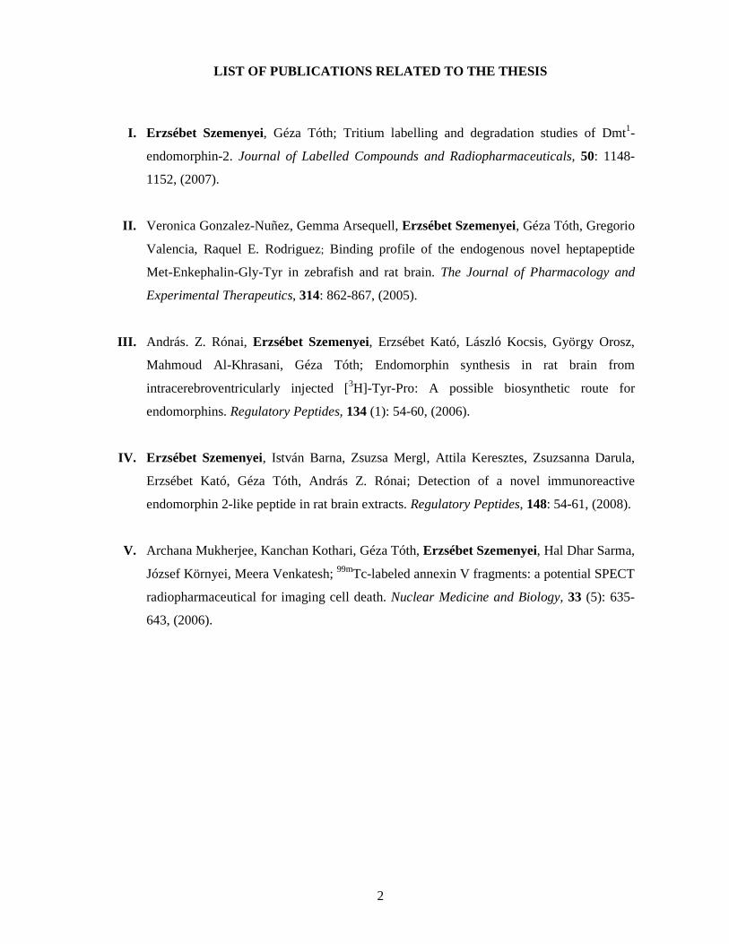
LIST OF PUBLICATIONS RELATED TO THE THESIS
I. Erzsébet Szemenyei, Géza Tóth; Tritium labelling and degradation studies of Dmt1-
endomorphin-2. Journal of Labelled Compounds and Radiopharmaceuticals, 50: 1148-
1152, (2007).
II. Veronica Gonzalez-Nuñez, Gemma Arsequell, Erzsébet Szemenyei, Géza Tóth, Gregorio
Valencia, Raquel E. Rodriguez; Binding profile of the endogenous novel heptapeptide
Met-Enkephalin-Gly-Tyr in zebrafish and rat brain. The Journal of Pharmacology and
Experimental Therapeutics, 314: 862-867, (2005).
III. András. Z. Rónai, Erzsébet Szemenyei, Erzsébet Kató, László Kocsis, György Orosz,
Mahmoud Al-Khrasani, Géza Tóth; Endomorphin synthesis in rat brain from
intracerebroventricularly injected [3H]-Tyr-Pro: A possible biosynthetic route for
endomorphins. Regulatory Peptides, 134 (1): 54-60, (2006).
IV. Erzsébet Szemenyei, István Barna, Zsuzsa Mergl, Attila Keresztes, Zsuzsanna Darula,
Erzsébet Kató, Géza Tóth, András Z. Rónai; Detection of a novel immunoreactive
endomorphin 2-like peptide in rat brain extracts. Regulatory Peptides, 148: 54-61, (2008).
V. Archana Mukherjee, Kanchan Kothari, Géza Tóth, Erzsébet Szemenyei, Hal Dhar Sarma,
József Környei, Meera Venkatesh; 99mTc-labeled annexin V fragments: a potential SPECT
radiopharmaceutical for imaging cell death. Nuclear Medicine and Biology, 33 (5): 635-
643, (2006).
Page 3
3
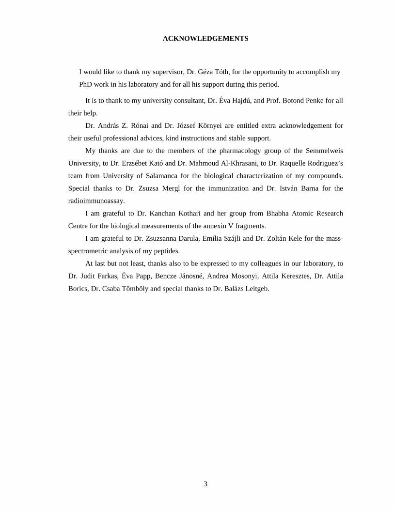
ACKNOWLEDGEMENTS
I would like to thank my supervisor, Dr. Géza Tóth, for the opportunity to accomplish my
PhD work in his laboratory and for all his support during this period.
It is to thank to my university consultant, Dr. Éva Hajdú, and Prof. Botond Penke for all
their help.
Dr. András Z. Rónai and Dr. József Környei are entitled extra acknowledgement for
their useful professional advices, kind instructions and stable support.
My thanks are due to the members of the pharmacology group of the Semmelweis
University, to Dr. Erzsébet Kató and Dr. Mahmoud Al-Khrasani, to Dr. Raquelle Rodriguez’s
team from University of Salamanca for the biological characterization of my compounds.
Special thanks to Dr. Zsuzsa Mergl for the immunization and Dr. István Barna for the
radioimmunoassay.
I am grateful to Dr. Kanchan Kothari and her group from Bhabha Atomic Research
Centre for the biological measurements of the annexin V fragments.
I am grateful to Dr. Zsuzsanna Darula, Emília Szájli and Dr. Zoltán Kele for the mass-
spectrometric analysis of my peptides.
At last but not least, thanks also to be expressed to my colleagues in our laboratory, to
Dr. Judit Farkas, Éva Papp, Bencze Jánosné, Andrea Mosonyi, Attila Keresztes, Dr. Attila
Borics, Dr. Csaba Tömböly and special thanks to Dr. Balázs Leitgeb.
Page 4
4
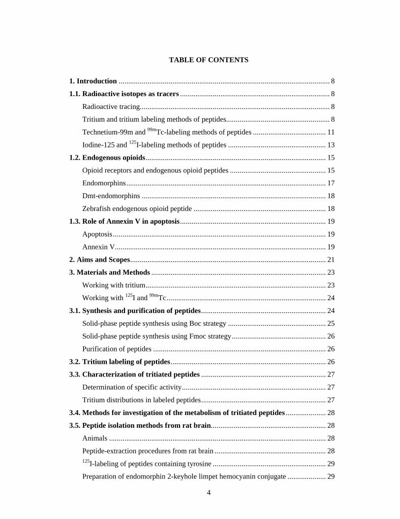
TABLE OF CONTENTS
1. Introduction .............................................................................................................. 8
1.1. Radioactive isotopes as tracers .............................................................................. 8
Radioactive tracing................................................................................................... 8
Tritium and tritium labeling methods of peptides...................................................... 8
Technetium-99m and 99mTc-labeling methods of peptides ...................................... 11
Iodine-125 and 125I-labeling methods of peptides ................................................... 13
1.2. Endogenous opioids .............................................................................................. 15
Opioid receptors and endogenous opioid peptides .................................................. 15
Endomorphins ........................................................................................................ 17
Dmt-endomorphins ................................................................................................ 18
Zebrafish endogenous opioid peptide ..................................................................... 18
1.3. Role of Annexin V in apoptosis ............................................................................ 19
Apoptosis ............................................................................................................... 19
Annexin V.............................................................................................................. 19
2. Aims and Scopes ...................................................................................................... 21
3. Materials and Methods ........................................................................................... 23
Working with tritium .............................................................................................. 23
Working with 125I and 99mTc ................................................................................... 24
3.1. Synthesis and purification of peptides ................................................................. 24
Solid-phase peptide synthesis using Boc strategy ................................................... 25
Solid-phase peptide synthesis using Fmoc strategy ................................................. 26
Purification of peptides .......................................................................................... 26
3.2. Tritium labeling of peptides ................................................................................. 26
3.3. Characterization of tritiated peptides ................................................................. 27
Determination of specific activity ........................................................................... 27
Tritium distributions in labeled peptides ................................................................. 27
3.4. Methods for investigation of the metabolism of tritiated peptides ..................... 28
3.5. Peptide isolation methods from rat brain............................................................ 28
Animals ................................................................................................................. 28
Peptide-extraction procedures from rat brain .......................................................... 28
125I-labeling of peptides containing tyrosine ........................................................... 29
Preparation of endomorphin 2-keyhole limpet hemocyanin conjugate .................... 29
Page 5
5
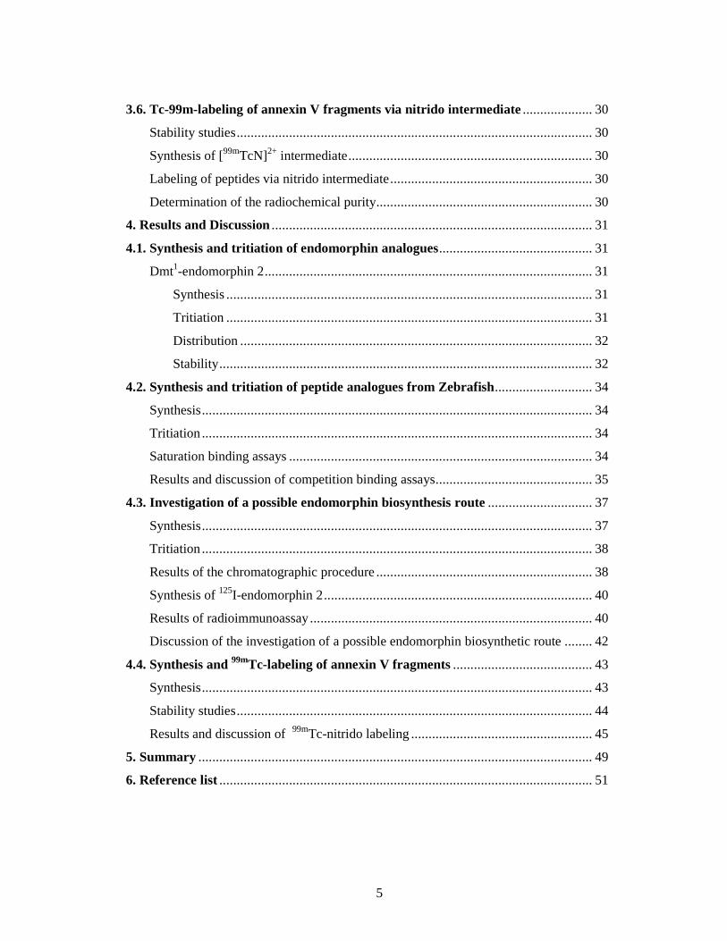
3.6. Tc-99m-labeling of annexin V fragments via nitrido intermediate .................... 30
Stability studies ...................................................................................................... 30
Synthesis of [99mTcN]2+ intermediate ...................................................................... 30
Labeling of peptides via nitrido intermediate .......................................................... 30
Determination of the radiochemical purity .............................................................. 30
4. Results and Discussion ............................................................................................ 31
4.1. Synthesis and tritiation of endomorphin analogues ............................................ 31
Dmt1-endomorphin 2 .............................................................................................. 31
Synthesis ......................................................................................................... 31
Tritiation ......................................................................................................... 31
Distribution ..................................................................................................... 32
Stability ........................................................................................................... 32
4.2. Synthesis and tritiation of peptide analogues from Zebrafish ............................ 34
Synthesis ................................................................................................................ 34
Tritiation ................................................................................................................ 34
Saturation binding assays ....................................................................................... 34
Results and discussion of competition binding assays ............................................. 35
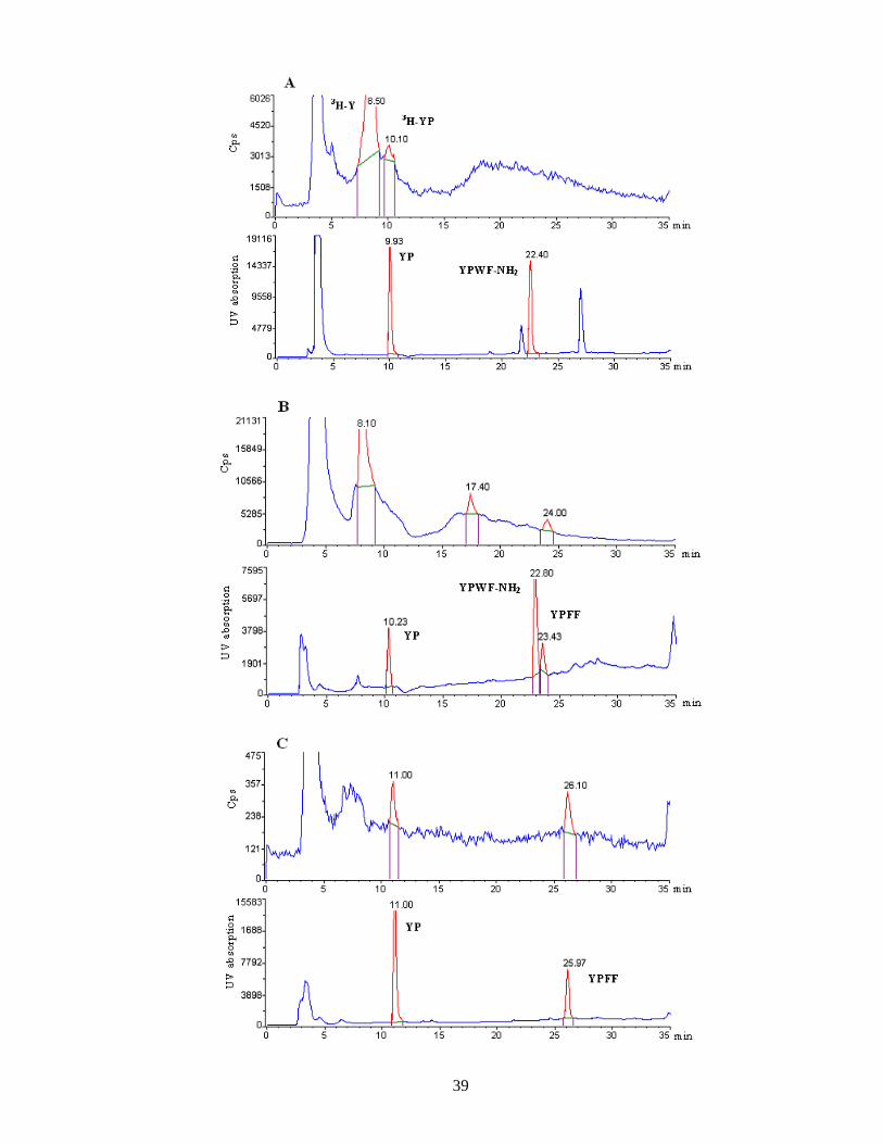
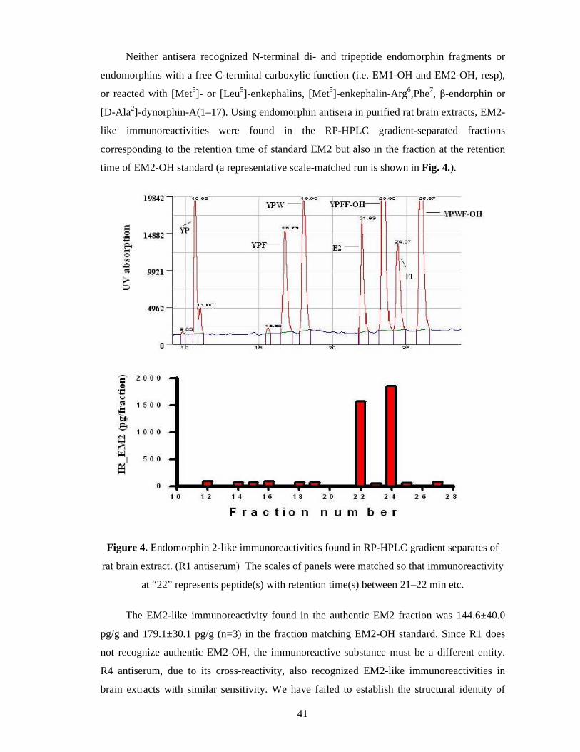
4.3. Investigation of a possible endomorphin biosynthesis route .............................. 37
Synthesis ................................................................................................................ 37
Tritiation ................................................................................................................ 38
Results of the chromatographic procedure .............................................................. 38
Synthesis of 125I-endomorphin 2 ............................................................................. 40
Results of radioimmunoassay ................................................................................. 40
Discussion of the investigation of a possible endomorphin biosynthetic route ........ 42
4.4. Synthesis and 99mTc-labeling of annexin V fragments ........................................ 43
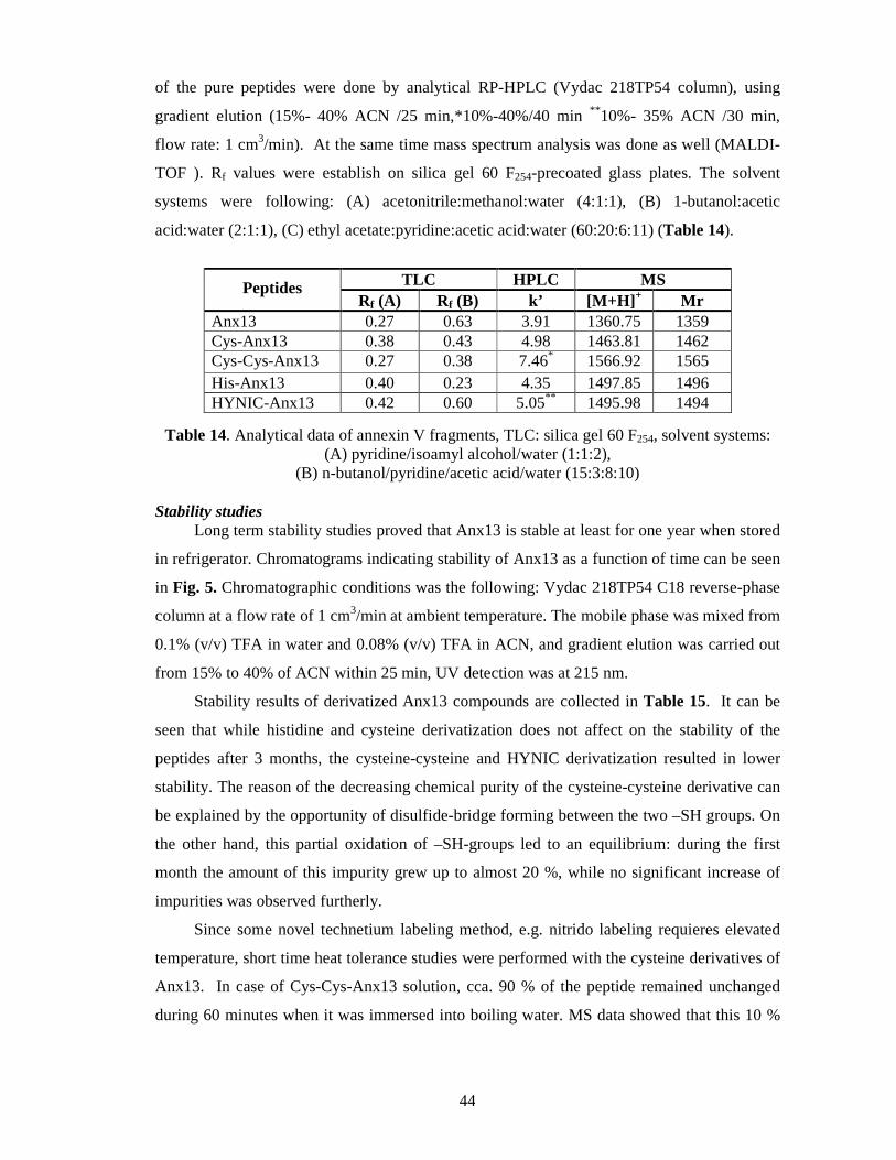
Synthesis ................................................................................................................ 43
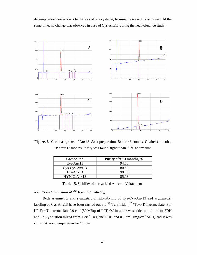
Stability studies ...................................................................................................... 44
Results and discussion of 99mTc-nitrido labeling .................................................... 45
5. Summary ................................................................................................................. 49
6. Reference list ........................................................................................................... 51
Page 6
6
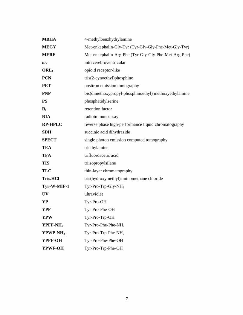
LIST OF ABBREVIATIONS
AcOH acetic acid
ACN acetonitrile
Anx V annexin V
Anx13 Ala-Gln-Val-Leu-Arg-Gly-Thr-Val-Thr-Asp-Phe-Pro-Gly
Bmax maximal number of binding sites
Boc tert-butyloxylcarbonyl
cDNA complementary DNA
CT computed tomography
DCC N,N’-dicyclohexylcarbodiimide
DCM dichloromethane
DIC N,N’-diisopropylcarbodiimide
DIEA N,N-diisopropylethylamine
DMF dimethylformamide
DMS dimethylsufide
Dmt 2’,6’-dimethyl-tyrosine
DTT 1,4-dithio-DL-threitol
EDTA ethylenediaminetetraacetic acid
EM2 endomorphin-2 (Tyr-Pro-Phe-Phe-NH2)
Dmt1-EM2 Dmt-endomorphin-2 (Dmt-Pro-Phe-Phe-NH2)
ESI electrospray ionization
EtOH ethanol
Fmoc 9-fluorenylmethoxycarbonyl
HBTU O-benzotriazol-1-yl-tetramethyl-uronium hexafluorophosphate
HF hydrogen fluoride
HOBt N-hydroxybenzotriazole
HYNIC 2-hydrazinonicotinic acid
k’ capacity factor
K D dissotiation constant
K i equilibrium inhibition constant
LC-MS liquid chromatography-mass spectrometry
LSC liquid scintillation counting
MALDI-TOF matrix-assisted laser desorption/ionization-time of flight
Page 7
7
MBHA 4-methylbenzhydrylamine
MEGY Met-enkephalin-Gly-Tyr (Tyr-Gly-Gly-Phe-Met-Gly-Tyr)
MERF Met-enkephalin-Arg-Phe (Tyr-Gly-Gly-Phe-Met-Arg-Phe)
icv intracerebroventricular
ORL 1 opioid receptor-like
PCN tris(2-cynoethyl)phosphine
PET positron emission tomography
PNP bis(dimethoxypropyl-phosphinoethyl) methoxyethylamine
PS phosphatidylserine
Rf retention factor
RIA radioimmunoassay
RP-HPLC reverse phase high-performance liquid chromatography
SDH succinic acid dihydrazide
SPECT single photon emission computed tomography
TEA triethylamine
TFA trifluoroacetic acid
TIS triisopropylsilane
TLC thin-layer chromatography
Tris.HCl tris(hydroxymethyl)aminomethane chloride
Tyr-W-MIF-1 Tyr-Pro-Trp-Gly-NH2
UV ultraviolet
YP Tyr-Pro-OH
YPF Tyr-Pro-Phe-OH
YPW Tyr-Pro-Trp-OH
YPFF-NH2 Tyr-Pro-Phe-Phe-NH2
YPWP-NH2 Tyr-Pro-Trp-Phe-NH2
YPFF-OH Tyr-Pro-Phe-Phe-OH
YPWF-OH Tyr-Pro-Trp-Phe-OH
Page 8
8
1. Introduction
1.1. Radioactive isotopes as tracers
Radioactive tracing
The first experiments with radioactive tracers were conducted in 1913 by György
Hevesy and Friedrich A. Paneth who determined the solubility of lead salts by using radium-
D, one of the naturally occuring radioactive isotopes of lead. For the development of the trace
method Hevesy was awarded the Nobel Prize in Chemistry in 1943. The tracer technique
came into common use after Word War II when relatively large amounts of cheap artificial
radionuclides became available through the use of nuclear reactors.
The largest field of application of radiotracers is in the life sciences. Using of the
labeled compounds is significant in the biochemical analysis such as in the autoradiography,
the immunoassay, the DNA-analysis and in the direct tracing. The one of the most important
user of radionuclides is medical sciences. Currently, the medical imaging techniques, which
use radionuclides (Transmission Tomography - CT, Emission Computed Tomography –
SPECT, PET) are widely applied diagnostic methods in the medicine. Radiotracers are also
used for therapy such as internal or external sources.
What are the advantages and the disadvantages of using radiotracers? The radioactive
isotopes are chemically identical with stable isotopes of the same element. The difference in
the mass of the nucleus between the various isotopes does cause some change in the chemical
and physical properties, but in most cases the isotope effect is rather small and difficult to
detect.
Apparently, the radiotracers are easy to detect and measure with high precision to
sensitivities of 10-16 to 10-6 g and the radioactivity is independent of temperature, pressure,
chemical and physical state. The radiotracers do not affect the system and can be used in
nondestructive techniques and if the tracer is radiochemically pure, interference from other
elements is of no concern. For most radioisotopes the radiation can be measured
independently of the matrix, eliminating the need for calibration curves (1).
Tritium and tritium labeling methods of peptides
Tritium was first predicted in the late 1920s by W. Russell, using his "spiral" periodic
table, then produced in 1934 from deuterium, another isotope of hydrogen, by E. Rutherford,
working with M. Oliphant and P. Harteck. Rutherford was unable to isolate the tritium, a job
that was left to L. Alvarez and R. Cornog (1939) (2) who correctly deduced that the substance
was radioactive. Upon the bombardment of deuterium with nuclei of deuterium (deuterons) in
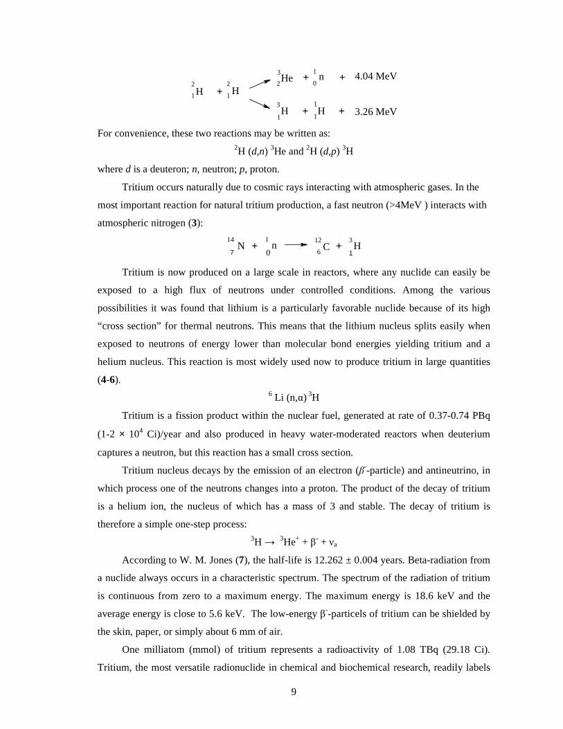
a cyclotron the following nuclear reactions occurred:
Page 9
9
H2
1 + H2
1
He3
2
H3
1
+
+
n1
0
H1
1
+
+
4.04 MeV
3.26 MeV
For convenience, these two reactions may be written as: 2H (d,n) 3He and 2H (d,p) 3H
where d is a deuteron; n, neutron; p, proton.
Tritium occurs naturally due to cosmic rays interacting with atmospheric gases. In the
most important reaction for natural tritium production, a fast neutron (>4MeV ) interacts with
atmospheric nitrogen (3):
7N
14+ n
1C
12
6+ H
3
10
Tritium is now produced on a large scale in reactors, where any nuclide can easily be
exposed to a high flux of neutrons under controlled conditions. Among the various
possibilities it was found that lithium is a particularly favorable nuclide because of its high
“cross section” for thermal neutrons. This means that the lithium nucleus splits easily when
exposed to neutrons of energy lower than molecular bond energies yielding tritium and a
helium nucleus. This reaction is most widely used now to produce tritium in large quantities
(4-6). 6 Li (n,α) 3H
Tritium is a fission product within the nuclear fuel, generated at rate of 0.37-0.74 PBq
(1-2 × 10⁴ Ci)/year and also produced in heavy water-moderated reactors when deuterium
captures a neutron, but this reaction has a small cross section.
Tritium nucleus decays by the emission of an electron (β--particle) and antineutrino, in
which process one of the neutrons changes into a proton. The product of the decay of tritium
is a helium ion, the nucleus of which has a mass of 3 and stable. The decay of tritium is
therefore a simple one-step process: 3H → 3He+ + β- + νa
According to W. M. Jones (7), the half-life is 12.262 ± 0.004 years. Beta-radiation from
a nuclide always occurs in a characteristic spectrum. The spectrum of the radiation of tritium
is continuous from zero to a maximum energy. The maximum energy is 18.6 keV and the
average energy is close to 5.6 keV. The low-energy β--particels of tritium can be shielded by
the skin, paper, or simply about 6 mm of air.
One milliatom (mmol) of tritium represents a radioactivity of 1.08 TBq (29.18 Ci).
Tritium, the most versatile radionuclide in chemical and biochemical research, readily labels
Page 10
10
complex organic and bioorganic molecules more so than any other radioisotope (8).
Several investigations, in vitro receptor studies, biochemical receptor analyses,
autoradiographic localization and distribution studies of the receptors and other
biodegradation assays are usually based on peptide labeled with tritium.
There are two basic methods for introducing tritium into organic molecules, exchange
methods and synthetic methods (9). The 3H/H isotope exchange reactions do not require
separate synthetic steps. The disadvantage of this method is that the compounds are randomly
labeled and the high percent of impurities are formed during radiolytic side reactions.
Synthetic methods, where tritium is directly and specifically inserted, yield high tritium
incorporation, but are limited by the chemistry required. The main methods for the tritium
labeling of neuropeptides (10) include above-mentioned isotope exchange reactions (β--
radiation induced (11), catalytic (12)) and methylation of peptides with 3H-methyl iodide (13)
or reductive methylation using tritiated metal hydrides (14), chemical or enzymatic synthesis
from precursor peptides or labeled amino acids. The synthesis of peptides from labeled amino
acids is advantegeous, the tritiated amino acids are characterized, the specific activity and
position of tritium atoms incorporated into amino acids are known. In case of synthesis of
tritiated peptides using precursor peptides, the most important chemical modification is the
iodination of peptides. Tyrosine and histidine residues can be iodinated using different
methods, for example using I2 solution in methanol, ICl, in situ generated iodine by the
reaction of HI and HIO3 under strong acidic conditions, reaction of chloramine-T with iodide,
enzymatic iodination with peroxidase, and reaction of Iodo-Gen® and Iodo-Beads® (15). In
most cases, the mono-, diiodinated and noniodinated peptides containing reaction mixture
should be purified by HPLC. For tritiation, the diiodo analogs are the favorable derivative.
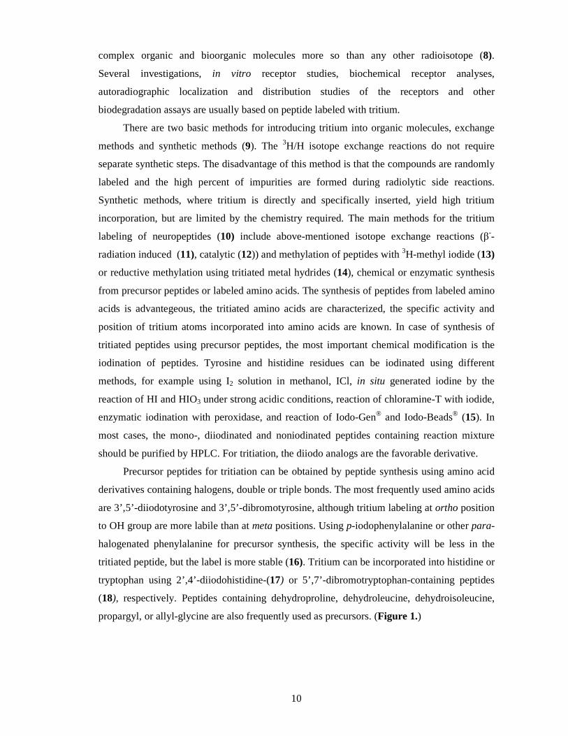
Precursor peptides for tritiation can be obtained by peptide synthesis using amino acid
derivatives containing halogens, double or triple bonds. The most frequently used amino acids
are 3’,5’-diiodotyrosine and 3’,5’-dibromotyrosine, although tritium labeling at ortho position
to OH group are more labile than at meta positions. Using p-iodophenylalanine or other para-
halogenated phenylalanine for precursor synthesis, the specific activity will be less in the
tritiated peptide, but the label is more stable (16). Tritium can be incorporated into histidine or
tryptophan using 2’,4’-diiodohistidine-(17) or 5’,7’-dibromotryptophan-containing peptides
(18), respectively. Peptides containing dehydroproline, dehydroleucine, dehydroisoleucine,
propargyl, or allyl-glycine are also frequently used as precursors. (Figure 1.)
Page 11
11
OH
XX
NH2
COOH
OH
NH2
COOH
BrBr
X
NH2
COOH
Br
BrNH
NH2
COOH
NH
N
I
I
NH2
COOH
NH
COOHCH2
CH2
COOHNH2
CH3
X=Br: 3',5'-DibromotyrosineX=I: 3',5'-Diiodotyrosine
2',6'-Dibromotyrosine X=Cl: p-ChlorophenylalanineX=Br: p-BromophenylalanineX=I: p-Iodophenylalnine
5',7'-Dibromotryptophan 2',4'-Diiodohistidine
3,4-Dehydroproline4,5-Dehydroleucine
Figure 1. Amino acid derivatives used for synthesis of precursor peptides for tritiation
Technetium-99m and 99mTc- labeling methods of peptides
C. Perrier and E. Segré in 1937 discovered the element of atomic number 43, isolated
by deuteron bombardment of molybdenum (19,20). The nuclear and chemical properties of
this missing element, eka-manganese, were predicted by D. Mendeleev. This element was
also isolable in larger amounts from the fission products of uranium (21). The name
technetium was coined by F. A. Paneth (22) from the Greek τεχνητός to denote that this was
the first artificial element made by man, and the chemical symbol was suggested to be Tc.
Nuclear isomerism of the element Tc and the existence of 99mTc were discovered by G. T.
Seaborg and E. Segré (23). 99mTc in some chemical form is used in more than 85% of the diagnostic scans done
each year in hospitals. The nuclear properties of 99mTc are virtually ideal for diagnostic
imaging. 99mTc emits a 140 keV γ-ray with 89% abundance which is close to optimum for
imaging with gamma cameras found in most hospitals. Its 6 h half-life is sufficiently long to
synthesize the 99mTc-labeled radiopharmaceuticals, assay them for purity, inject them into the
patient, and perform the imaging studies yet short enough to minimize the radiation dose to
Page 12
12
the patient. The metastable (a state where the nucleus is in an excited state) isotope, 99mTc is
produced as a fission product from the fission of uranium or plutonium in nuclear reactors,
but the vast majority of the 99mTc is formed from 99Mo which is formed by the neutron
activation of 98Mo. 99Mo has a half-life of 66 hours, so short-lived 99mTc, which results from
its decay, is being constantly produced (24). 99mTc decays to 99Tc.
Mo98
42+ n
1
0Mo
99
42+ gamma-ray
Mo99
42Tc
99m
43+ beta-particle
Tc99m
43 + gamma-rayTc43
99
MoO3 + H2O → H2MoO4 ( 99MoO4
-/ 99mTcO4-)
The inconvenience of purchasing a short half-life radionuclide was overcome by the
development of the 99Mo-99mTc generator ("technetium cow," also occasionally called a
molybdenum cow) (25), which takes advantage of the transient equilibrium between the
parent radionuclide 99Mo (66 h half-life) and the daughter radionuclide 99mTc (6 h half-life).
The separation of 99mTc from 99Mo is accomplished by the selective elution of 99mTcO4¯ with
sterile saline from alumina column containing 99MoO4¯. The transient equilibrium results in
optimum isolation of maximum 99mTc activity with minimal 99Tc buildup every 23-24 h. The
development of the 99Mo-99mTc generator allowed this radionulide to become both routinely
available and economical.
Klaus Schwochau's book Technetium lists 31 radiopharmaceuticals, based on 99mTc for
imaging, functional studies of the brain, myocardium, thyroid, lungs, liver, gall bladder,
kidneys, skeleton, blood and tumors. 99mTc-labeling is still an attractive approach for radiolabeling peptides for nuclear
medicine imaging due to its ideal physical characteristics for SPECT and readily availability
from a generator. Technetium exhibits a rich and diverse redox chemistry because of its
capability of existing in 8 different oxidation states ranging from -1 to +7 (26). The Tc(VII) in 99mTcO4
- has to be reduced to a lower oxidation state in order to produce a stable 99mTc-
peptide complex or to a reactive intermediate complex. When 99mTcO4- is reduced, the
oxidation state of technetium depends on the nature of the reducing agent, the chelator, and
reaction conditions.
Peptides contain a number of possible active side chains such as the ω-amino group
from lysine, phenol moiety from tyrosine, thiol group from cysteine and carboxylate group
Page 13
13
from aspartic or glutamic acid. These reactive groups can serve as “handles” for the
attachment of a bifuntional coupling agent (27).
Abrams and co-workers first reported the use of 99mTc-HYNIC (2-hydrazinonicotinic
acid) core for the 99mTc-labeling (28). Since then, the 99mTc-HYNIC core has been used for 99mTc-labeling of chemotactic peptides (29). The HYNIC can only occupy one or two
coordination sites the square pyramidal or octahedral coordination sphere of the technetium.
The advantage of using HYNIC as the bifuntional coupling agent is its high labeling
efficiency and the choice of various coligands. The a N atom of HYNIC is coordinated to Tc,
forming a –HN-N=Tc bond. The octahedral geometry of the complex is built 4 chelating
groups coming from a tetradentate ligand. Several modifications of the HYNIC core were also
accomplished by using a tridentate ligand such as tricine as the coligand and a monodentate
ancillary ligand (30). 99mTc complexes containing a terminal Tc≡N multiple bond are
currently easily produced at tracer level, after the advent of improved chemical methods for
obtaining the [99mTc≡N]2+ core in high radiochemical purity. A key advantage of using this
type of complexes for obtaining novel classes of diagnostic agents comes from their intrinsic
structural robustness (31). This method is based on the reaction depicted in Equation 1, where
D is a donor of the nitride nitrogen atoms (N3-), belonging to the class of derivatives of
dithiocarbazic acid (H2N−NH–CS2H) or, in general, of derivatives containing the –N–N–
functional moiety, and R is a reducing agent such as SnCl2 or a tertiary phosphine and HCl
(32).
[99mTcO4]- + D + R → [99mTc≡N] (1)
Through Reaction 1, pertechnetate is quantitatively converted into a mixture of
complexes, all of which contain the Tc≡N group. Subsequent addition of a suitable ligand to
this mixture leads to the high-yield formation of a single compound in which the terminal
metal-nitrogen multiple bond is retained. The high affinity of the Tc≡N core for sulfur donors
makes it particularly suitable for linking to peptides having S- as donor atoms. Experiments
conducted with short peptide sequences having a cysteine residue placed in a terminal
position of the amino acid chain, indicate that two peptide ligands can bind efficiently around
the Tc≡N group via the cysteine, through the thiolate sulfur atom and the amine nitrogen
atom. The resulting complexes have the expected square-pyramidal geometry.
Iodine-125 and 125I- labeling methods of peptides
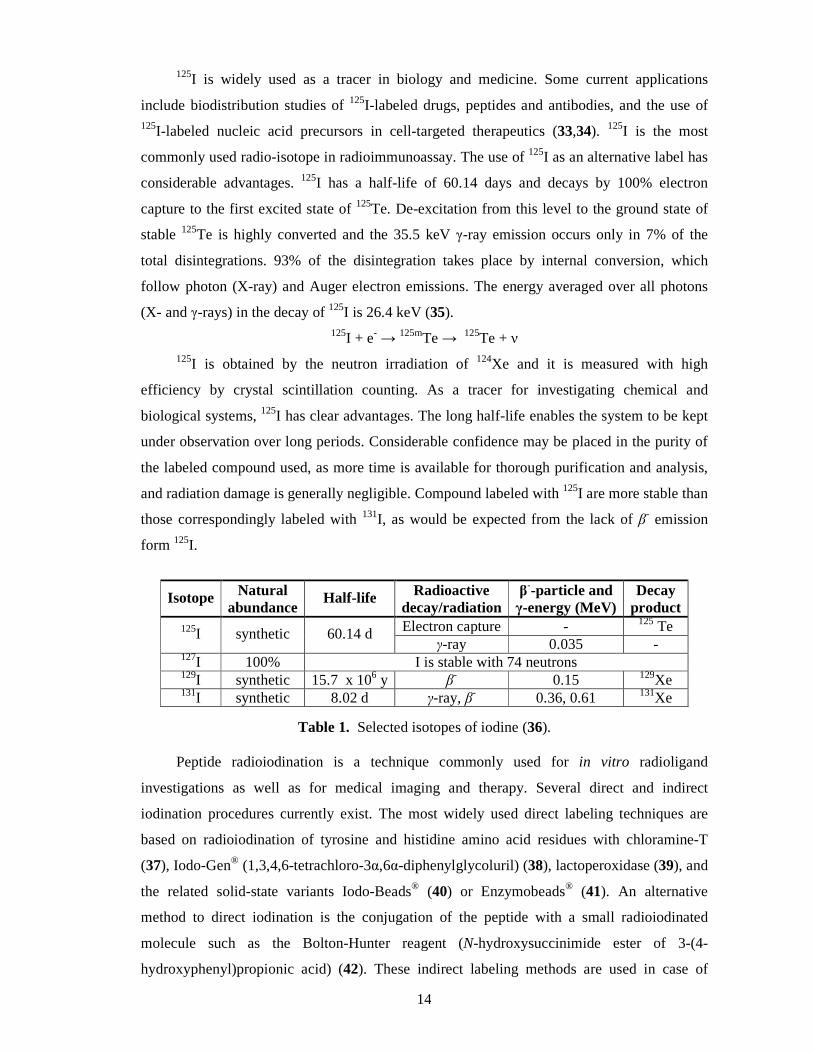
Iodine is an essential trace element; its only known roles in biology are as constituents
of the thyroid hormones, thyroxine (T4) and triiodothyronine (T3). There are 37 isotopes of
iodine and only one, 127I, is stable (Some selected isotopes of iodine can be seen in Table 1.).
Page 14
14
125I is widely used as a tracer in biology and medicine. Some current applications
include biodistribution studies of 125I-labeled drugs, peptides and antibodies, and the use of 125I-labeled nucleic acid precursors in cell-targeted therapeutics (33,34). 125I is the most
commonly used radio-isotope in radioimmunoassay. The use of 125I as an alternative label has
considerable advantages. 125I has a half-life of 60.14 days and decays by 100% electron
capture to the first excited state of 125Te. De-excitation from this level to the ground state of
stable 125Te is highly converted and the 35.5 keV γ-ray emission occurs only in 7% of the
total disintegrations. 93% of the disintegration takes place by internal conversion, which
follow photon (X-ray) and Auger electron emissions. The energy averaged over all photons
(X- and γ-rays) in the decay of 125I is 26.4 keV (35). 125I + e- → 125mTe → 125Te + ν
125I is obtained by the neutron irradiation of 124Xe and it is measured with high
efficiency by crystal scintillation counting. As a tracer for investigating chemical and
biological systems, 125I has clear advantages. The long half-life enables the system to be kept
under observation over long periods. Considerable confidence may be placed in the purity of
the labeled compound used, as more time is available for thorough purification and analysis,
and radiation damage is generally negligible. Compound labeled with 125I are more stable than
those correspondingly labeled with 131I, as would be expected from the lack of β- emission
form 125I.
Isotope Natural abundance
Half-life Radioactive decay/radiation
β--particle and
γ-energy (MeV) Decay
product 125I synthetic 60.14 d Electron capture - 125 Te
γ-ray 0.035 - 127I 100% I is stable with 74 neutrons 129I synthetic 15.7 x 10⁶ y β
- 0.15 129Xe 131I synthetic 8.02 d γ-ray, β- 0.36, 0.61 131Xe
Table 1. Selected isotopes of iodine (36).
Peptide radioiodination is a technique commonly used for in vitro radioligand
investigations as well as for medical imaging and therapy. Several direct and indirect
iodination procedures currently exist. The most widely used direct labeling techniques are
based on radioiodination of tyrosine and histidine amino acid residues with chloramine-T
(37), Iodo-Gen® (1,3,4,6-tetrachloro-3α,6α-diphenylglycoluril) (38), lactoperoxidase (39), and
the related solid-state variants Iodo-Beads® (40) or Enzymobeads® (41). An alternative
method to direct iodination is the conjugation of the peptide with a small radioiodinated
molecule such as the Bolton-Hunter reagent (N-hydroxysuccinimide ester of 3-(4-
hydroxyphenyl)propionic acid) (42). These indirect labeling methods are used in case of
Page 15
15
absence of tyrosine and histidine residues or when these amino acids are necessary for the
peptide activity.
1.2. Endogenous opioids
Opioid receptors and endogenous opioid peptides
The opium, derived from the poppy plant, papaver somniferum, has been used for many
hundreds of years to relieve pain. In 1803, Sertürner isolated a crystaline sample of the main
constituent alkaloid of the crude opium, morphine, which was named after the Greek god of
dreams, Morpheus. The structure of morphine was predicted by Gulland and Robinson and
the Robinson`s structure was confimed by chemical synthesis (43).
Opioids are the most powerful analgesic drugs available, and are the treatment of choice
for the management of moderate to severe pain (44). The rigid structural and stereochemical
requirements essential for the analgesic effect of morphine and related opioids led to the
theory that they produce their effects by interacting with a specific receptor (45). Side effects,
including respiratory depression, nausea, and constipation, impact their use and protracted
opioid therapy leads to drug tolerance and physical dependence.
The opioid receptors displayed heterogenous properties, and at least three types of
opioid receptors existed, classified as µ-, δ- and κ-receptors (46), and these receptors have
been confirmed by molecular cloning (47-50). All of the cloned opioid receptors possess the
same general structure of an extracellular N-terminal region, seven transmembrane domains
and intracellular C-terminal tail structure. There is pharmacological evidence for subtypes of
each receptor and other types of novel, less well-characterised opioid receptors, ε, λ, ι, ζ. More
recently, an „orphan” receptor was identified which has a high degree of homology to the
„classical” opioid receptors; on structural grounds this receptor is an opioid receptor and has
been named ORL1 (opioid receptor –like) (51).
Brain opioid peptide systems are known to play an important role in motivation,
emotion, attachment behaviour, the response to stress and pain, and the control of food intake.
In mammalian the endogenous opioid peptides are mainly derived from four precursors:
pro-opiomelanocortin, pro-enkephalin, pro-dynorphin and pro-nociceptin/orphanin FQ (52-
55). (Table 2.) β-endorphin is equiactive at µ-and δ-receptors with much lower affinity for κ-
receptors (56). [Met]-and [Leu] enkephalin have high affinities for δ-receptors, ten-fold lower
affinities for µ-receptors, but Metorphamide, which is a [Met]-enkephalin derivative
displaying highest affinity for the µ-receptor (57). The opioid fragments of pro-dynorphin,
perticularly dynorphin A and dynorphin B, have high affinity for κ-receptors but also have
significant affinity for µ-and δ-receptors (57). Nociceptin/OrphaninFQ is the endogenous
Page 16
16
ligand for the ORL1-receptor; it has little affinity for the µ-, δ- and κ-receptors (55,58).
Endomorphins, endomorphin 1 (Tyr-Pro-Trp-Phe-NH2) and endomorphin 2 (Tyr-Pro-Phe-
Phe-NH2) are two endogenous opioid tetrapeptides with the highest known affinity and
specificity for the µ-opioid receptor (59). It is assumed that endomorphins are the cleavage
products of a larger precursor, but this polypeptide or protein has not yet been identified.
Precursor Protein Opioid peptide Amino acid sequence Receptor selectivity
Pro-opiomelanocortin
α-Endorphin β-Endorphin γ-Endorphin
YGGFMTSEKSQTPLVT YGGFMTSEKSQTPLVTL
FKNAIIKNAYKKGE YGGFMTSEKSQTPLVTL
µ > δ >> κ
Pro-enkephalin [Met]enkephalin [Leu]enkephalin [Met]enkephalin-
Arg6-Phe7 [Met]enkephalin-
Arg6-Gly7-Leu8 Metorphamide
YGGFM YGGFL YGGFMRF YGGFMRGL YGGFMRRV-NH2
µ ~ δ >> κ δ > µ >> κ
κ2 κ µ
Prodynorphin Dynorphin A (1-8) Dynorphin A (1-13) Dynorphin A Dynorphin B α-neoendorphin β-neoendorphin
YGGFLRRI YGGFLRRIRPKLK YGGFLRRIRPKLKWDNQ YGGFLRRQFKVVT YGGFLRKYPK YGGFLRKYP
κ > δ ~ µ κ > δ ~ µ
κ κ
Pronociceptin/OFQ Nociceptin FGGFTGARKSARKLANQ ORL1 - Endomorphin 1
Endomorphin 2 YPWF-NH2
YPFF-NH2 µ µ
Others Tyr-MIF-1 Tyr-W-MIF-1 Deltorphin I Deltorphin II Dermenkephalin Dermorphin
YPLG-NH2 YPWG-NH2
YaFDVVG-NH2
YaFEVVG-NH2 YmFHLMD-NH2
YaFGYPS-NH2
µ µ δ δ δ µ
A: Ala, D: Asp, E: Glu, F: Phe, G: Gly, I: Ile, K: Lys, L: Leu, M: Met, N: Asn, P: Pro, Q: Gln, R: Arg, S: Ser, T: Thr, V: Val, W: Trp, Y: Tyr, a: D-Ala, m: D-Met
Table 2. Endogenous opioid peptides
Proteolysis of some functional proteins in vitro leads to the generation of peptides
exhibiting an opioid like activity when they have a Tyr-Pro N-terminus sequence. Thus β-
casomorphins are released from β-casein (60), and hemorphins from hemoglobin (61) by in
vitro peptic hydrolysis. β-casomorphins are a specific group of milk peptides with biological
activity of µ-δ- and κ-opioid receptor agonists (62,63). The members of the hemorphin family
include peptides from 4 to 10 amino acids, which are generated by proteolytic degradation of
Page 17
17
the [32-41] segment of human hemoglobin β-chain or [31-40] segment of the bovine
hemoglobin β-chain (64,65,66). (Table 3.)
In 1980, two opioid peptides, dermorphin (Tyr-D-Ala-Phe-Gly-Tyr-Pro-Ser-NH2) and
Hyp6-dermorphin were isolated from the skin of several amphibians (67). These heptapeptides
were shown to have high affinity and selectivity for the µ-receptors (68,69). On a molar basis,
the analgesic potency of dermorphin is about 1000 times greater than that of morphine (70).
Precursor Endogenous peptide Amino acid sequence
β-casein (bovine) β-Casomorphin 5 β-Casomorphin 7 Morphiceptin
YPFPG YPFPGPI YPFP-NH2
β-casein (human) β-Casomorphin 5 β-Casomorphin 7
YPFV YPFVEPI
Hemoglobin LVV-Hemorphin-7 VV-Hemorphin-7 Hemorphin-7 VV-Hemorphin-6 VV-Hemorphin-5 Hemorphin-5 Hemorphin-4
LVVYPWTQRF VVYPWTQRF YPWTQRF VVYPWTQR VVYPWTQ YPWTQ YPWT
Table 3. Other mammalian opioid peptides
Two distinct isoforms (prepro-xendorphin A and B) of an opioid propeptide were
isolated from a Xenopus laevis brain cDNA library (71). Two potential mature peptides,
xendorphin-1A and -1B, showed opioid agonist activity and xendorphin 1B (Tyr-Gly-Gly-
Phe-Ile-Arg-Lys-Pro-Asp-Lys-Tyr-Lys-Phe-Leu-Asn-Ala) binds with high affinity and
specificity to κ-receptors (72).
Endomorphins
In 1997, J. E. Zadina and coworkers synthesized a number of Tyr-W-MIF-1 analogs,
containing all possible natural substitutions at position 4, which were subsequently screened
for the opioid receptor binding. A biologically potent sequence, Tyr-Pro-Trp-Phe-NH2 was
discovered and then identified in the bovine brain and human cortex (59,73). This peptide
named endomorphin 1. a second peptide, Tyr-Pro-Phe-Phe-NH2, named endomorphin 2,
which differs by one amino acid from endomorphin 1 was also isolated. Endomorphins were
the first peptides isolated from brain that bind to the µ-opioid receptor with high affinity and
selectivity and therefore were proposed as endogenous µ-opioid receptor ligands (59).
However, the biosynthetic pathways for other vertebrate opioid peptides have already been
clarified, their precursor(s) or the possible biosynthetic route(s) still remain(s) unidentified.
Endomorphin 1 is widely and densely distributed throughout the brain and upper brainstem
and is particularly abundant in the nucleus accumbens (Nac), the cortex, the amygdala, the
Page 18
18
thalamus, the hypothalamus, the striatum, and the dorsal root ganglia (74,75). In contrast,
endomorphin 2 is more prevalent in the spinal cord and lower brainstem, endomorphin 2-
immunoreactive cell bodies were most prominent in the hypothalamus and the nucleus of the
solitary tract (NTS), whereas endomorphin 2-immunoreactive varicose fibers were mainly
observed in the substantia gelatinosa of the medulla and the spinal cord dorsal horn (74,76).
The studies in vivo showed that there are two groups of enzymes mainly responsible for
the degradation of endomorphins: dipeptidyl-aminopeptidase IV (DPP IV), which triggers the
process, and aminopeptidases, which are involved in secondary cleavage (77,78).
Endomorphins are degraded by similar pathways.
The first step in their catabolism is the cleavage of Pro2–Trp3 and Pro2–Phe3 peptide
bonds, respectively, and the dipeptides formed are then hydrolyzed into amino acids (79,80).
However, the degradation of endomorphin 1 contains an additional minor route: the Tyr1–Pro2
peptide bond might also be cleaved in the first step of the enzymatic degradation pathway.
There is another degradation pathway of endomorphins, when carboxypeptidase Y and
proteinase A hydrolyze endomorphins into peptide acids, releasing ammonia, and then cleave
off the C-terminal Phe (81,82).
Dmt-endomorphins
Endomorphins exhibited the highest affinity for the µ-opioid receptor and
extraordinarily high selectivity relative to the δ- and κ-opioid receptor systems of all known
opioid substances (59,83). Hitherto a great number of endomorphin analogues were
synthesized (84,85) Here only one analogue is paraphrazed from the populous endomorphin
derivatives. Studies on opioid peptides demonstrated that the introduction of 2’,6’-dimethyl-
tyrosine (Dmt) in lieu of the common N-terminal Tyr residue in opioid ligands resulted in an
exceptional improvement in receptor affinity and functional bioactivity in a wide variety of
opioid peptides (86-94). The substitution of Dmt for Tyr in endomorphin 2 (Dmt1-
endomorphin 2) resulted in one of the most active peptides among several analogues
containing an alkyl-modified aromatic ring of Tyr (95,96). Dmt increased µ-opioid receptor
affinity and µ-opioid receptor bioactivity of endomorphin 2 by 5- and 83-fold, respectively
The δ-affinity and bioactivity of Dmt1-endomorphin 2 increased 2 to 3 orders of magnitude,
thereby transforming once highly selective ligand into a bivalent or bifuntional opioid peptide
derivative (95,96).
By means of the building of Dmt into the first position, the increased hydrophobicity
and alteration in conformation might enhance receptor interaction through π–π stacking,
stabilization of hydrophobic interactions with aliphatic or aromatic side-chains in the receptor,
and strengthen hydrogen bonding capabilities of the hydroxyl group (97,98).
Page 19
19
Zebrafish endogenous opioid peptide
Zebrafish, Danio rerio is considered a model organism (99), not only for the study of
the biological functions of vertebrates but also as a tool to analyze the effects of some drugs
or toxic agents (100-102). Five zebrafish opioid precursor genes homologous to the
mammalian opioid propeptide genes have recently been identified (103-105). These
precursors contain novel opioid peptides that can display different pharmacological properties
than their counterparts in mammals. In particular, mammals present an enlarged form of Met-
enkephalin [Met-enkephalin-Arg-Phe (MERF)] that is different from its homolog in zebrafish,
the Met-enkephalin-Gly-Tyr (MEGY).
1.3. Role of Annexin V in apoptosis
Apoptosis
Apoptosis is a form of programmed cell death in multicellular organisms. It is one of
the main types of programmed cell death (PCD) (106), and involves a series of biochemical
events leading to a characteristic cell morphology and death: more specifically, a series of
biochemical events which lead to a variety of morphological changes, including blebbing,
changes to the cell membrane such as loss of membrane asymmetry and attachment, cell
shrinkage, nuclear fragmentation, chromatin condensation and chromosomal DNA
fragmentation (1-4). Processes of disposal of cellular debris whose results do not damage the
organism differentiates apoptosis from necrosis. Apoptosis (Greek: apo - from, ptosis -
falling) was distinguished from traumatic cell death by J. F. Kerr while he was studying
tissues using electron microscopy (107,108).
Apoptosis can occur when a cell is damaged beyond repair, infected with a virus, or
undergoing stress conditions such as starvation. DNA damage from ionizing radiation or toxic
chemicals can also induce apoptosis via the actions of the tumour-suppressing gene p53.
Apoptosis also plays a role in preventing cancer; if a cell is unable to undergo apoptosis, due
to mutation or biochemical inhibition, it can continue dividing and develop into a tumour.
Dying cells that undergo the final stages of apoptosis display phagocytotic molecules, such as
phosphatidylserine (PS), on their cell surface (109,110). PS is normally found on the cytosolic
surface of the plasma membrane, but is redistributed during apoptosis to the extracellular
surface by a hypothetical protein known as scramblase (111). These molecules mark the cell
for phagocytosis by cells possessing the appropriate receptors, such as macrophages (112).
Annexin V
Many drugs, such as cytostatic agents, induce a therapeutic effect through the activation
of programmed cell death in target cells (106). The detection and quantification of apoptosis
Page 20
20
in vivo are of significant clinical value for diagnosis and assessment of therapeutic efficacy.
In the last decade, a molecular imaging protocol was developed to measure the programmed
cell death in vitro and in vivo in animal modells and patients (113-116). This imaging protocol
is based on the facts that apoptotic cells externalize the negatively charged phospholipid (PS)
and that the human protein Annexin V binds to PS selectively and with a high affinity (117).
Anx V, a protein with a molecular weight of 36 kDa is a member of annexin family.
This is a multiprotein family of over 160 proteins that share the property of Ca2+-dependent
binding to negatively charged phospholipid surfaces (118). Annexins are located mainly
intracellularly, but AnxV can also be secreted and detected on the outer surface of plasma
membranes (119). AnxV consists of 319 amino acids and the molecule is arranged in planar
cyclic structure of 4 domains (120,121). The binding of AnxV to phospholipids is very rapid,
extremly dependent on the presence of Ca2+ and reversible in the presence of the ion chelator
EDTA. Studies with PS-containing liposomes found that the stoichiometry of AnxV binding
to PS ranges between 4 and 8 AnxV molecules per one PS molecule (122,123). The Anx V
affinity assay was further developed by labeling Anx V with biotin or with radionuclides to
enable various protocols for measuring apoptosis in vitro (124) and in vivo (125-127) animal
models.
Radionuclide imaging with radiolabeled annexin V is a highly specific technique that
enables delineation of apoptotic areas with good resolution (128). AnxV has been tagged with
several radionuclides such as 99mTc (129–131), 18F (132,133), 64Cu (134) and 123/124I (135–
138) to detect cell death in vivo by SPECT or PET imaging. 99mTc-HYNIC-Anx V has been
used to detect apoptosis in vivo with gamma camera imaging and SPECT (139,140).
Page 21
21
2. Aims and Scopes
Radioactive tracers have applications in medicine, research, indusrty, agriculture, and
many other fields of science and technology.
Tritium labeled biologically active peptides are valuable tools for biological
characterization of receptors, and binding sites. The metabolic pathway of tritium labeled
compounds is also easily traceable. Our aim was to acquire suitable tools for use in in vitro
and in vivo biological assays.
Two Dmt1-EM2 isotopomers were labeled with tritium in position 1 or position 2. The
isotopomers may become a useful ligands for direct radioreceptor binding and may serve as
important tools for degradation studies in rat brain homogenates.
We aimed to prepare an eligible tool for characterizing of the binding profile of MEGY
peptide in zebrafish, the organism in which this peptide is naturally present as an endogenous
opioid ligand. To achieve this objective, we have synthesized and labeled the MEGY peptide
and our biologist cooperators (Dr. Raquelle Rodriguez’s team from University of Salamanca)
performed binding assays. Two MEGY analogs were also synthesized: (D-Ala2)-MEGY (Y-
D-Ala-GFMGY) and (D-Ala2, Val5)-MEGY (Y-D-Ala-GFVGY). The change of a Gly by a
D-Ala may confer resistance against proteases such as dipeptidyl-aminopeptidases, which
remove the N-terminal dipeptide Tyr1-Gly2. In addition, the substitution of Met by Val may
help to determine the importance of the methionine residue for the specific opioid binding.
The biosynthetic pathways for other vertebrate opioid peptides have already been
clarified (it happens through a single- or multi-step cleavage from large molecular weight
precursors with or without additional post-translational modifications), the biosynthetic route
of endomorphins is still obscure. Based on the hypothesis that biosynthesis of an oligopeptide
may take place also from its fragments through a specific enzymatic route, we decided to
label Tyr-Pro dipeptide with tritium and test using HPLC combined with radiodetection the
probable incorporation of icv injected [3H]Tyr-Pro into endomorphin-related peptides in the
rat brain. In addition we aimed to develop a RIA to endomorphin 2, therefore we raised
antibodies to EM2-keyhole limpet hemocyanine conjugate in rabbits and labeled EM2 with 125I isotope.
Imaging apoptosis has many applications with new ones emerging with time. The most
widely used application is in cancer treatment for assessing tumor response to novel therapies
as tumor often respond to radiation as well as to chemotherapy by direct induction of
apoptosis. 99mTc labeled annexin V is considered as an useful tool for apoptosis detection but
it has many disadvantages from point of view of radiopharmaceutical kit formulation and
agents with faster urinary excretion are also required for routine clinical applications. We
Page 22
22
tried to focus on the development of phosphatidyl-serine specific small 99mTc-labeled annexin
V fragments for apoptosis imaging. Annexin type proteins generally possess their biospecific
sequences on the N-terminal and in case of Annexin V, the phosphatidyl-serine specific
sequency might be attributed to a chain on the N-terminal consisting of 13 amino acids. Based
on this concept, a peptide containing particular sequence of these 13 amino acids (Anx13) was
designed and derivatized with cisteine and two cisteine for novel 99mTc-nitrido labeling
method and additional two Anx13 derivatives was designed by attachment of histidine and
hydrazino nicotinic acid residues for tricarbonyl, HYNIC labeling approaches (respectively).
Page 23
23
3. Materials and Methods
Protected and unprotected amino acids and resins were purchased from Sigma-Aldrich,
Calbiochem-Novabiochem or Bachem. Coupling agents were from Fluka and Senn
Chemicals. Trifluoroacetic acid (TFA), catalyst, TLC plates (Silica gel 60 F254), and solvents
were fom Merck and Sigma-Aldrich. Na125I and 99mTcO4 were purchased from Institute of
Isotopes Co. Ltd. 3H2 was purchased from Technobexport, Russia.
Fluorenylmethyloxycarbonyl-hydrazinonicotinic acid (Fmoc-HYNIC) was synthesized in our
laboratory.
HF cleavage was performed using a standard apparatus from Peninsula Laboratories,
Inc. The following solvent systems were used for TLC analysis: acetonitrile:methanol:water
(4:1:1); 1-butanol-acetic acid-water (4:1:1); ethyl acetate:pyridine:acetic acid:water
(60:20:6:11,). Ninhydrin, UV light and iodine vapor were employed to detect the peptides and
amino acids on the thin layer.
RP-HPLC was performed on Merck-Hitachi or Merck-Lachrom RP-HPLC system,
utilizing Vydac 218TP1010 C18 (250 × 10 mm, 10 µm) semipreparative column for
preparative purposes, and Vydac 218TP54 C18 (250 × 4.6 mm, 5 µm) column for analytical
purposes. Peptides were detected by UV at 215 or 280 nm. The following solvents were used:
solvent A was 0.08 % TFA/acetonitrile and solvent B was 0.1% TFA/water.
Molar mass of the peptides were determined and LC-MS or MS analyses were carried
out by ESI mass spectrometry (Finnigan TSQ 7000 or Shimadzu QP 80000) and MALDI-
TOF mass spectrometry (Bruker Reflex III).
Working with tritium
In biological material, 50 % of the tritium β-particles are calculated to be absorbed
already by a layer only 0.3 µm thick, 80 % of the particles are calculated to be absorbed
within 1 µm from the source, and 99 % of the radiation does not reach beyond 2.5 µm.
Therefore the work with tritium does not require the using of the shielding. Tritium can be
absorbed easily through the skin or by inhalation. Lab coat and gloves provide efficient
protection in preventing skin contact with contaminated surfaces. After any potential skin
exposure the skin should be decontaminated as soon as possible in order to minimize
absorption into the body. Effective personal decontamination methods include rinsing the
affected part of the body with cold water and soap. Cold water keeps the pores of the skin
closed and reduced the transfer of 3HHO across the skin.
Tritiation reactions were carried out on our self-designed vacuum manifold described
earlier (10). Our lab has a Triton β-gas monitor (Johnston laboratories Inc.) for low level
detection and measurement of tritium gas in the air.
Page 24
24
Tritium-labeled materials were analysed and purifield on an RP-HPLC (Jasco)
instrument using Vydac 218TP54 C18 (0.46 × 25 cm, 5 µm) or Merck 50943 LiChroCART
(124-4 LiChrospher 100 RP-18 , 5 µm) column, detected by a Jasco UV-975 spectrometer and
a Canberra Packard 505 TR Flow Radiochromatography Detector. Radioactivity was counted
in toluene-Triton X-100 or Ultima Gold scintillation cocktail with a Packard TRI-CARB 2100
TR Liquid Scintillation Analyzer.
Working with 125I and 99mTc
Iodine-125 is an electron capture radionuclide emitting low energy X and gamma
radiation with 35.5 keV energy. Due to the volatile nature of iodine, the most significant
hazard is from inhalation, the critical organ for uptake is the thyroid. Iodine-labeled
compounds can penetrate surgical rubber gloves. Two pair should be worn, or polythene over
rubber. Direct handling of 125I is to be avoided, forceps, shielded syringes must be used. Low
activity RIA kits (< 370 kBq) may normally be handled on the open bench, but all other work
with iodine should be carried out in a fume cupboard. For work with higher activities special
transparent shielding made from lead impregnated acrylic to be necessary which has a lead
equivalence value of 0.5 mm. Sample pots containing 125I should be shielded with 1 mm of
lead. Solutions containing iodide ions should neither be made acidic nor stored frozen, both
lead to formation of volatile elemental iodine. An alkaline solution of 5% sodium thiosulphate
should be used to render the spill chemically stable, prior to decontamination by the normal
methods. In the case of an accident involving possible ingestion/inhalation of radioiodine may
be possible to block uptake to the thyroid by the administration of potassium iodide tablets.
(200 mg given two hours after ingestion will reduce uptake by 80%).
The gamma ray average energy of 99mTc is 140.5 keV and it also emits X-ray with 18
keV and 21 keV energy. Technetium-99m is a decay product of molybdenum-99 and is
obtained in solution form by eluting it from a molybdenum-99 “cow”. The recommended
protective clothing are lab coat (which must be monitored before leaving the laboratory),
disposable plastic, latex or rubber gloves, footwear covers. Waterproof gloves should be worn
during elution. The dispensing of the 99mTc should be carried out in a lead shielded box and
during the work wearing of lead apron is recommeded. Personal decontamination techniques
are following: washing of the contaminated part of the body with soap and water and after
monitoring of the skin. Decontamination of clothing and surfaces are covered under operating
and emergency procedures.
3.1. Synthesis and purification of peptides
Peptides were synthesized manually by solid phase-peptide synthesis using either Boc
or Fmoc chemistry.
Page 25
25
Solid-phase peptide synthesis using Boc strategy
Peptide synthesis using Boc protocol was carried out on 4-methylbenzhydrylamine
(MBHA) resin for peptide amides or on Merrifield resin for peptide acids. Attachment of the
first Boc-protected amino acid to the chloromethyl resin was performed by using Gisin
method (142). The synthesis protocol is summarized in Table 4.
Step Reagent Time
Washing DCM 3 × 1 min Deprotection 50% TFA, 2% anisole/DCM 1 × 2 min and 1 × 20 min
Washing DCM 3 × 1 min Neutralization 10% DIEA/DCM 2 × 2 min
Washing DCM 3 × 1 min Kaiser test Coupling 2 eq. Boc-amino acid,
2 eq. DCC, 2 eq. HOBt
60 min Kaiser test Washing DCM, EtOH 3 × 1 min, 3 ×1 min
Table 4. General schedule for peptide synthesis using Boc chemistry
For methionine or tryptophane containing peptides the deprotection mixture contained
0.5% DTT. Coupling reactions were carried out after the neutralization step with 2
equivalents of Boc-AA, HOBt and DCC in DCM until negative Kaiser test (143).
Hydroxybenzotriazole esters of protected amino acids are easily formed from DCC or
DIC/HOBt in situ. After the last washing step the peptide-resin was dried under vacuum.
Final deprotection and cleavage from the resin were carried out by HF.
In general, HF cleavage reactions were performed between -5°C – 0 °C, for 60 minutes.
The following cleavage mixtures have been used: for peptides containing cysteine:
HF/DMS/anisole/p-thiocresol (10:1:1:0.2) and for other peptides HF/DMS/anisole (10:1:1).
After completion of the cleavage reaction, HF was evaporated from the peptid-resin mixture.
To prevent side ractions during this process, it was important to maintain the temperature of
the reaction vessel between -5°C – 0 °C. After HF had been removed, diethyl ether was added
to the reaction mixture and the peptide-resin-scavenger mixture was stirred. The ether solution
was filtered and the resin was washed three times to remove the scavengers. The peptide was
extracted from the peptid-resin mixture by stirring the mixture in glacial acetic acid. This
procedure was repeated twice to ensure complete extraction of the peptide, using
approximately 30 cm3 of 30 % acetic acid per gram of peptide-resin each time. The peptide
solution was diluted with water to give a final concentration of AcOH less than 10% and
lyophilized.
Page 26
26
Solid-phase peptide synthesis using Fmoc strategy
Synthesis of the peptides by the Fmoc protocol were carried out on 2-chlorotrityl
chloride resin. The Fmoc protocol is summarized in Table 5.
Step Reagent Time
Washing DMF 3 × 1 min Kaiser test
Coupling
2 eq. Fmoc-amino acid 2 eq. HBTU and HOBt
4 eq. DIEA/DMF
30 min
Kaiser test Washing DMF 3 × 1 min
Deprotection 20% piperidine/DMF 1 × 2 min and 1 × 20 min Washing DMF 3 × 1 min
Table 5. General schedule for peptide synthesis using Fmoc chemistry
After the last coupling step, the peptide-resin was washed with DMF and EtOH and
then dried under vacuum. Final deprotection and cleavage from the resin was carried out by
TFA in the presence of scavengers. The TFA cleavage reaction was performed at room
temperature for 60 min, using the following mixture: 95% TFA, 2.5% water, 2.5% TIS. The
peptide was precipitated by ice-cold diethyl ether, the peptide-resin mixture filtered-off, then
the peptide was extracted by 10 % AcOH three times and finally lyophilized.
Purification of peptides
The crude peptides were purified by RP-HPLC on a semipreparative column (Vydac
218TP1010), applying gradient elution with the following eluents: A: 0.08% TFA/ACN, B:
0.1% TFA/water, flow rate was 4 cm3/min, detected at 215 nm by UV detector.
Purity control was performed on a Merck-Hitachi or Merck-LaChrom RP-HPLC
system, utilizing a Vydac 218TP54 C18 or Merck 50943 LiChroCART analytical column,
with gradient elution. Detection was as described above. Peptide purity was assessed also by
TLC on silica gel 60 F254-precoated glass plates, the solvent systems were the following: (A)
acetonitrile:methanol:water (4:1:1); (B) 1-butanol:acetic acid:water (4:1:1); (C) ethyl
acetate:pyridine:acetic acid:water (60:20:6:11). Molecular weight of the peptides were
determined by MALDI-TOF (Bruker Reflex III) or ESI-MS (Finnigan TSQ 7000 or
Shimadzu QP 80000).
3.2. Tritium labeling of peptides
Before the radioactive labeling procedure the reaction was carried out in inactive
circumstances, using hydrogen gas. This reaction helps to determine the proper reaction
condition such as catalyst, reaction time, solvent.
Page 27
27
The purified precursor peptide was dissolved in DMF and the catalyst (PdO/BaSO4)
was added. In most cases, an excess of TEA was added to neutralize HI formed during the
reaction and to help prevent poisoning the catalyst. The reaction was carried out in hydrogen
atmosphere at room temperature, while stirring continuously. The crude reaction mixture was
analyzed by RP-HPLC and in some cases by MS.
Tritium labeling of peptides was carried out in our in-house designed vacuum apparatus
(10) under a fume cupboard. The purified precursor peptide was dissolved in DMF and the
catalyst was suspended in the solution. The reaction vessel was connected to the tritiation
manifold frozen with liquid nitrogen and the air was removed by vacuum. The tritium gas was
liberated from uranium tritide by heating above 300 °C, and it was expanded into the reaction
vessel. The reaction mixture was agitated by the magnetic stirrer at room temperature. The
reaction was terminated by freezing the solution and absorbing the unreacted tritium on
pyrophoric uranium. The catalyst was removed by filtration through Whatman GF/C filters
and washed three times with ethanol. Labile tritium was removed by repeated evaporation of
protic solvent, such as EtOH/H2O mixture. The total activity of product was measured by
LSC. The crude tritiated peptide was analyzed by TLC and RP-HPLC. The purified labeled
peptide was dissolved in ethanol and stored in 2 cm3 aliquots under liquid nitrogen at a
radioactive concentration of 37 MBq/cm3.
3.3. Characterization of tritiated peptides
Determination of specific activity
The specific activity of the labeled peptides was determined by dividing the measured
activity by the amount of purified peptide. The quantity of the purified labeled material was
determined from its UV absorption spectrum, or by HPLC, using calibration curve.
Tritium distributions in labeled peptides
The labeled peptide was diluted by inactive material, 6M HCl was added, and the
mixture was incubated at 110 °C for 24 h under argon pressure in a sealed ampoule. The
solvent was then removed by evaporation. The formed amino acid mixture was analyzed by
TLC and subsequently to the formation of the Fmoc derivatives by RP-HPLC. Fmoc derivates
of amino acids were used as standards respectively (144).
The sample - amino acid or peptide hydrolysate - was dissolved in borate buffer (0.2 M,
pH: 7.7). Fmoc-Cl reagent was dissolved in aceton to give a concentration of 15 mM. The
sample and the reagent were mixed, and after 45 seconds the vial was filled with n-pentane
and the mixture was shaken to remove the excess reagent. The extraction was repeated twice
Page 28
28
and the pentane phases were discarded. Afterwards the sample had been acidified 10 µl of
acetic acid and then analyzed by RP-HPLC (145).
3.4. Methods for investigation of the metabolism of tritiated peptides
After preincubation of the rat brain homogenate, tritium labeled peptides were incubated
with it at 37 °C. Aliquots were withdrawn after incubation for 5, 15, 30 or 60 min, and
immediately acidified with 0.1 M HCl solution. Following centrifugation of the samples
(11,340 × g, 5 min, 25 °C) and the supernatant was analysed by radio-HPLC. For
determination of the rates of degradation of the peptides the following method was applied.
Aliquots of the 1 mM nonlabeled peptide stock solutions in 50 mM Tris–HCl buffer (pH =
7.4) were added to some rat brain homogenate, and the mixtures were incubated at 37 °C.
Aliquots were taken from these incubation mixtures and immediately acidified with 0.1 M
aqueous HCl solution. About 10 µl of each supernatant obtained after centrifugation of the
samples (11,340 × g, 5 min, 25 °C) was analysed by RP-HPLC. The degradation rate
constants (k) were obtained by least square linear regression analysis of the plots of
logarithmic peptide peak areas (ln(A/A0)) versus time, using a minimum of four points.
Degradation half-lives (t1/2) were calculated from the rate constants as ln(2/k).
3.5. Peptide isolation methods from rat brain
Animals
Animal care and experimental procedures were carried out according to the principles
set by EC Directive 86/609/EEC. Experimental protocols were approved also by the Ethical
Board controlling laboratory experiments at the Medical Faculty of Semmelweis University.
Male Wistar rats, weighing 110–150 g (analgesic measurements) or 170–220 g (brain
extracts), were used. Rats were kept in groups of 5, in temperature-controlled (22 ± 2 °C) unit
with 12 h light–dark cycle (08.00–20.00–08.00). Standard laboratory chow and tap water
were provided ad libitum.
Peptide-extraction procedures from rat brain
Rat brains were removed and powderized under liquid nitrogen, taken up with abs.
ethanol and stored at -80 °C until extraction. An extraction procedure deviced originally for
endomorphins (59), was used, except for sample boiling. In brief, the stored samples were
solubilized in eight-fold amount of 0.08% (w/w) Na2S2O5 solution then ACN was added to
yield 25% (v/v) final ACN concentration. They were mixed at room temperature overnight.
The mixtures were centrifuged at 29,000 × g for 20 min and the supernatants were extracted
by solid-phase method , using 70% (v/v) ACN in the final step. The extracts were evaporated
Page 29
29
to dryness and dissolved in 2% ACN/ 98% water (TFA 0.1% (v/v)) and the samples were
analyzed by RP-HPLC. Chromatographic conditions were the following: Vydac 218TP54
C18 reverse-phase column (250 × 4.6 mm, 5 µm) at a flow rate of 1 cm3/min at ambient
temperature. The mobile phase was mixed from 0.1% (v/v) TFA in water and 0.08% (v/v)
TFA in ACN, and gradient elution was carried out from 2% to 40% of ACN within 30 min. 125I-labeling of peptides containing tyrosine
RIA is based on the antigen-antibody reaction in which tracer amount of the radio-
labeled antigen competes with endogenous antigen for limited binding sites of the specific
antibody against the same antigen. Usually, high specific activity radio-labeled antigen is
prepared by iodination of the pure antigen on its tyrosine residue(s) by chloramine-T (146)
method and then separating the radio-labeled antigen from free-isotope by gel-filtration or
HPLC. The lyophilized peptide was dissolved in phosphate buffer (pH = 7.4). Iodinations was
performed by addition of chloramine-T solution to a polypropylene tube containing a mixture
of 1v/v% TFA solution, peptide and Na125I (in 0.04M NaOH) solution. The iodination
reaction was quenched after 1 min by addition sodium metabisulphite in water. 125I-peptide
was immediately purified by RP-HPLC. The fractions were collected at 12-sec intervalls. The
radioactivies in the peak fraction were measured by counting 2 µl aliquots on a LIN-LOG
NK-350 gamma counter and the radioactivities of the pure, collected 125I-EM2 were measured
by a TRI-CARB 2100TR liquid scintillation counter in a toulene-Triton X-100 cocktail. An
aliquot of the peak radioactive fraction was then rechromatrographed as above in the presence
of 5 nmol of unlabeled EM2 while simultaneously monitoring UV absorbance at 216 nm and
gamma emissions by using Canberra Packard Radiomatic 505TR Flow Radiochromatography
Detector with the Ultima-FloM scintillation cocktail.
Preparation of endomorphin 2-keyhole limpet hemocyanin conjugate
Based on an earlier method (147), approximately 10 mg of keyhole limpet hemocyanin
and 5 mg of endomorphin 2 were dissolved together in 10 cm3 of water. To this mixture was
added 0.5 cm3 of water containing 100 mg of freshly dissolved water soluble carbodiimide
reagent and it was incubated overnight at room temperature. The unadjusted pH of the
reaction mixture was 5–6. The reaction was terminated by dialysis against water for 24 h.
When precipitates formed, the granular and soluble materials were used together for
immunization.
Page 30
30
3.6. Tc-99m-labeling of annexin V fragments via nitrido intermediate
Stability studies
Before the labeling, a long run stability studies of the solid, non-radiolabeled Anx13
were carried out by storing them at –18 oC and at +5 oC. For short time heat tolerance
investigations, the aqueous/saline solution of the Cys-Cys-Anx13 were used by immersing
them into boiling water for 20 and 60 minutes.
Synthesis of [99mTc ≡ N]2+ intermediate
Nitrido intermedier was prepared at ambient temperature by using succinic acid
dihydrazide (SDH), SnCl2 in saline and freshly eluted 99mTcO4- (37-185 MBq). The mixture
was stirred at room temperature for 15 min.
Labeling of peptides via nitrido intermediate
Symmetric nitrido labeling were carried out by adding peptide in saline to the 99mTc-
nitrido intermediate. The reaction mixtures were heated in boiling water bath for 1 hour. For
asymmetric labeling peptide in saline, bis(dimethoxypropyl-phosphinoethyl)
methoxyethylamine (PNP) in ethanol or tris(2-cynoethyl)phosphine (PCN) in hydroxypropyl-
γ-cyclodextrin was reacted with the previously prepared 99mTc-nitrido intermediate at 100 oC
for 1 hour.
Determination of the radiochemical purity
Radiochemical purity of the nitrido labeled peptides was determined by HPLC and TLC
methods. For HPLC, Zorbax 300 SB C-18 column with both radioactivity and UV detectors
were used. Solutions A and B were prepared for gradient elution containing 0.1 % TFA in
water and 0.08% TFA in ACN, respectively. TLC was carried out by using Kieselgel 60
layers and ethanol-water 1:1 (v/v) as eluent or Gelman ITLC-SA leyers and n-buthanol
saturated with 0.3M HCl solution as eluent. For determination of free pertechnetate content,
Whatman ET-31 paper and acetone eluent were also used.
Page 31
31
4. Results and Discussion
4.1. Synthesis and tritiation of endomorphin analogues
Dmt1-endomorphin 2
Synthesis
Peptides (H-Dmt-Pro-Phe-Phe-NH2 and H-Dmt-3,4∆Pro-Phe-Phe-NH2 precursor
peptide) were synthetized manually on MBHA resin in 0.25 mmol scale by using the
Merrifield solid-phase method. Nα-t-Boc chemistry with HOBt and DCC as coupling agents
were employed for peptide elongation. The crude peptides were purified by RP-HPLC on a
Vydac 218TP1010 C18 column, using a linear gradient of from 20% to 50% of the organic
modifier within 25 min at a flow rate of 4 cm3/min with UV detection at 220 nm. Peptide
purity was assessed by TLC and HPLC, and the molecular weights of the peptides were
established by MALDI-TOF-MS. Rf values, capacity factor for a Vydac 218TP54 C18
column and the measured and calculated molecular weights of Dmt-EM2 analogues show in
the Table 6. Rf values were establish on silica gel 60 F254-precoated glass plates. The solvent
systems were following: (A) acetonitrile:methanol:water (4:1:1), (B) 1-butanol:acetic
acid:water (4:1:1), (C) ethyl acetate:pyridine:acetic acid:water (60:20:6:11).
Peptides TLC HPLC MS Rf (A) Rf (B) Rf (C) k’ [M+H] + Mr
Dmt-Pro-Phe-Phe-NH2 0.36 0.53 0.33 3.04 600.37 599 Dmt-∆Pro-Phe-Phe-NH2 0.39 0.53 0.33 3.01 598.33 597 Diiodo-Dmt-Pro-Phe-Phe-NH2 0.50 0.57 0.43 5.91 852.24 851
Table 6. Analytical data of Dmt-EM2 analogues
A precursore peptide, 3`, 5`-Diiodo-Dmt1-EM2 was synthetized by the chloramine T
method. Testing was performed to establish the optimum reaction conditions. Accordingly,
1.4 µmol of Dmt1-EM2 was dissolved in acetonitrile:water (1:1) and 5 equivalents of the
reagents (0.067 M NaI + 0.022 M chloramine T in phosphate buffer, pH = 7.4) was added to
the solution. The reaction was stopped after 30s by adding 0.053 M Na2S2O5. The reaction
mixture was analysed and purified by RP-HPLC (Vydac 218TP54 C18 RP column with a
gradient of 20% to 50% of organic modifier in 25 min, flow rate 1 cm3/min). The analytical
parameters of Diiodo-Dmt1-EM2 show in the Table 6.
Tritiation
2.4 mg (2.88 µmol) of 3`,5`-diiodo-Dmt1-EM2 and 2.8 mg (3.92 µmol) of 3,4∆Pro2-
Dmt1-EM2 were dissolved separately in 1 cm3 of dimethylformamide and labeled with tritium
gas. The reaction mixture contained 1.5 µl of triethylamine and 12 or 14 mg of PdO/BaSO4
catalyst, respectively. Tritium gas was liberated from uranium tritide by heating, and 555 GBq
Page 32
32
(15 Ci) of this gas was introduced into the reaction vessel (10,79). The reaction mixture was
stirred at room temperature for 1 or 2 h and the unreacted tritium gas was then adsorbed onto
pyrophoric uranium. The crude products were purified by HPLC to give a radioactive purity
of >95%. The quantitative analysis of the pure, labeled peptides was performed by HPLC
with a UV detector, using a calibration curve prepared with unlabeled Dmt1-EM2, and the
total activities of the products were measured by liquid scintillation counting. The specific
activity of [3H2]-Dmt1-EM2 were 2.88 TBq/mmol (77.8 Ci/mmol), and that of [3H2]Pro-Dmt1-
EM2 1.95 TBq/mmol (52.8 Ci/mmol) (Table 7). The pure, tritiated peptides were dissolved in
ethanol and were stored at a concentration of 37 MBq/cm3 under liquid nitrogen. The
stabilities of both tritiated endomorphin 2 analogues under these storage conditions were
really good. After 6 months, the purities were checked and proved to be >95%.
Peptides a TBq/mmol
TLC HPLC k’ Rf (A) Rf (B) Rf (C)
[3’,5’- 3H2]Dmt-Pro-Phe-Phe-NH2 2.88 0.36 0.53 0.33 3.26 Dmt-[3,4-3H2]Pro-Phe-Phe-NH2 1.95 0.36 0.53 0.33 3.26
Table 7. Radioanalytical data of tritium labeled Dmt-EM2 Distribution
The distributions of the tritium labels in [3H2]Dmt1-EM2 and [3H2]Pro2-Dmt1-EM2 were
determined after acidic hydrolysis and Fmoc derivatization by HPLC. The Dmt and Pro
contained tritium in >90% of the theoretical level. The a/amax is the ratio of the specific to
the theoretically maximum specific activity. The Phe residues in the peptides were also
partially labeled (Table 8). This phenomenon presumably caused the overall higher specific
activity of [3H2]Dmt1-EM2 than the theoretical level. The specific activity of [3H2]Pro2-Dmt1-
EM2 was >90 % of theoretical level, and the specificity of the label was satisfactory, resulting
in an appropriate radioligand for radioligand-binding experiments and metabolic studies.
Tritiated peptides a/amax HPLC
Fmoc-[3H2]Dmt Fmoc-[3H2]Pro Fmoc-[3H]Phe [3H2]Dmt1-EM2 133% 92% - 8% Dmt-[3H2]Pro2-EM2 92% 1% 95% 4%
Table 8. Tritium distributions in Dmt-EMs Stability
The stability of a radioligand in radioligand-binding studies is essential, and it is
therefore necessary to determine the degradation half-life of the ligand in the biological
matrix used. In our earlier investigations, EM1 and EM2 demonstrated long half-lives in the
Page 33
33
presence of a rat brain membrane preparation (0.3 mg/cm3 protein): 295 min and 230 min,
respectively, accordingly they do not degrade during binding assays (148). In the present
study, the degradation half-life of [3H2]Pro2-Dmt1-EM2 under the above conditions was 515
min. The kinetics of degradation of Dmt1-EM2 was studied in a rat brain homogenate, as
compared with the earlier published degradation kinetics of EM2 (79). The protein content of
the homogenate was 5.92 mg/cm3 and Dmt1-EM2 concentration was 100 µM. Figure 2.
shows the kinetics of degradation of EM2 and Dmt1-EM2 in the rat brain homogenate. After
12 min of incubation, only 24% of the parent EM2 remained in the samples, whereas 72% of
the initial Dmt1-EM2 concentration remained after 15 min of incubation.
0
20
40
60
80
100
120
0 50 100 150 200
Time (min)
% o
f in
itia
l pep
tid
e co
nce
ntr
atio
n
EM2
Dmt1-EM2
Figure 2. Degradation of EM2 and Dmt1-EM2 in rat brain homogenate
The logarithmic forms of these curves were analysed by linear regression, which
allowed calculation of the degradation half-lives of the EMs. As shown in Table 9, Dmt1-
EM2 broke down relatively slowly in the brain homogenate. Dmt1-EM2 had a half-life of
33.64 min, while EM2 was almost 6 times less resistant than Dmt1-EM2 to the peptidases: its
half-life was 5.88 min.
EM2 Dmt1-EM2
100 x k (min-1) t1/2 (min) 100 x k (min-1) t1/2 (min) 11.79 ± 0.75 5.88 ± 0.39 2.09 ± 0.39 33.64 ± 6.81
Table 9. Half-lives of EMs
Page 34
34
4.2. Synthesis and tritiation of peptide analogues from Zebrafish
Synthesis
The endogenous MEGY, (H-Tyr-Gly-Gly-Phe-Met-Gly-Tyr-OH) its two analogs, (D-
Ala2)-MEGY and (D-Ala2, Val5)-MEGY, and the precursor peptide for tritiation (3`, 5`-
Diiodo-Tyr1-MEGY) were synthesized by the above-mentioned solid-phase peptide synthesis
method using the Boc chemistry procedure. The crude peptides were purified by RP-HPLC
using a Vydac 218TP1010 column. Peptide purity was assessed by TLC and RP-HPLC, and
the molecular weights of the peptides were established by MALDI-TOF-MS. Rf values,
capacity factor for a Vydac 218TP54 C18 column and the measured and calculated molecular
weights of Dmt-EM2 analogues show in the Table 10. Rf values were establish on silica gel
60 F254-precoated glass plates. The solvent systems were following: (A)
acetonitrile:methanol:water (4:1:1), (B) 1-butanol:acetic acid:water (2:1:1), (C) ethyl
acetate:pyridine:acetic acid:water (60:20:6:11).
Peptides TLC HPLC MS Rf (A) Rf (B) Rf (C) k’ [M+H] + Mr
Tyr-Gly-Gly-Phe-Met-Gly-Tyr 0.63 0.62 0.14 2.06 794.25 793 Tyr-D-Ala-Gly-Phe-Met-Gly-Tyr 0.70 0.68 0.21 2.27 808.37 807 Tyr-D-Ala-Gly-Phe-Val-Gly-Tyr 0.60 0.68 0.14 1.75 776.31 775
Table 10. Analytical data of MEGY analogues Tritiation
[3’,5’- 3H2]MEGY was prepared by catalytic dehalogenation of the precursor peptide
using 555 GBq (15 Ci) 3H2 gas and 11.5 mg PdO/BaSO4 as the catalyst in the presence of
triethylamine. The crude tritiated peptide was purified by RP-HPLC as previously described.
The purity of the final product was established by analytical RP-HPLC and the degree of
purity observed was >95%. The total activity of the product was 3.51 GBq (95 mCi). The
specific radioactivity was 0.74 TBq/mmol (20 Ci/mmol). The purified peptide was stored in
ethanol under liquid nitrogen at a concentration of 37 MBq/cm3 (1 mCi/cm3).
Saturation binding assays
The ability of [3H]MEGY to bind opioid receptors from zebrafish, an organism in which
this peptide is naturally present (149), and from mammals, in which MERF is the
corresponding endogenous peptide, was measured using increasing concentrations of this
radioligand in zebrafish and rat brain membrane homogenates.
In zebrafish brain KD was 2.39 ± 0.29 nM and Bmax was 255.1 ± 10.84 fmol/ mg protein.
The same analysis is presented for rat brain membranes, KD was 3.8 ± 0.34 nM and Bmax was
308.1 ± 11.45 fmol/mg protein. MEGY can bind to the opioid receptors present in zebrafish
Page 35
35
and rat brain with high affinity and that this binding is reversed by naloxone. The binding
assays of [3H]MEGY obtained by us give similar saturation curves for zebrafish and rat brain
homogenates, although this peptide presents a significant higher affinity in zebrafish brain
(Results from Dr. Raquelle Rodriguez’s team).
Results and discussion of competition binding assays
To analyze the ability of MEGY to displace other conventional opioid compounds,
competition binding assays were performed using [3H]diprenorphine, a nonspecific
antagonist, as the radioligand and MEGY and its two analogs, (D-Ala2)-MEGY and (D-Ala2,
Val5)-MEGY, as unlabeled ligands at a concentration range of 0.3 nM to 10 µM (Results from
Dr. Raquelle Rodriguez’s team).
Ligand K i
Zebrafish Brain / nM
Displacement in Zebrafish
Brain %
K i Rat Brain
/ nM
Displacement in Rat
Brain %
MEGY K i1 = 1.17 ± 0.27 Ki2 = 673 ± 136
74.28 ± 4.20 22.10 ± 1.57 72.20 ± 7.15
(D-Ala2)-MEGY 12.40 ± 2.67 63.89 ± 2.69 29.90 ± 7.97 85.45 ± 2.23 (D-Ala2,Val5)-MEGY 137 ± 26.45 54.64 ± 2.36 72.20 ± 7.15 79.39 ± 3.30
Table 11. Ki values of MEGY and its two analogs in zebrafish and rat brain membranes obtained from competition binding assays using [3H]diprenorphine
[3H]diprenorphine presents two different binding sites in zebrafish brain (KD values
0.08 and 17 nM) (150), and in the further assays, MEGY shows a two-site displacement in
zebrafish brain, with high-affinity site and one site with lower affinity, which suggests that
this peptide may act on two or more different receptors with different affinities. Preliminary
work from R. E. Rodriguez’s laboratory proposes that MEGY binds to the δ receptors from
zebrafish (150).
The fact that a ligand shows a biphasic curve in competition binding assays has been
previously reported for other ligands, such as morphiceptin (151).
In the rat, the analog (D-Ala2)-MEGY shows a similar Ki and percentage of
displacement at 10 µM than MEGY, thus indicating that the change of Gly by a D-Ala does
not affect the ability of the ligand to bind to opioid sites. However, the (D-Ala2, Val5)-MEGY
analog displays a higher Ki, revealing that the change of Met by Val entails a loss in binding
affinity. These results can be taken into consideration for future opioid ligand design because
an effective peptidic ligand should have the methionine in the fifth position, whereas the Gly
can be replaced by another small residue that might confer resistance against protease
degradation.
Page 36
36
MEGY and its two analogs can displace almost all the diprenorphine binding in rat
brain, whereas in zebrafish brain, the native peptide displaces only up to 74%. This difference
can be explained if we postulate that diprenorphine may bind to the opioid sites present in
zebrafish brain in a different manner than to those present in mammalian brain. These results
are interesting because it may be inferred that diprenorphine can be considered as a good and
selective ligand to label opioid sites in mammalian brain, whereas in zebrafish diprenorphine
does not seem to show such selectivity and thus, other opioid ligands are not able to displace
up to 100% of its binding.
[3H]MEGY was also used as the radioligand in competition binding assays using the
unlabeled peptide MEGY and its two analogs as unlabeled ligands so that it was possible to
determine the influence of the structural changes in the binding ability. Heterologous
displacements with morphine (nonpeptidic opiate) and Met-enkephalin (an endogenous ligand
for both species) were also performed (Table 12.)
Ligand K i
Zebrafish Brain / nM
Displacement in Zebrafish
Brain %
K i Rat Brain
/ nM
Displacement in Rat
Brain % MEGY 2.89 ± 0.85 106.85 ± 1.40 4.21 ± 1.41 109.51 ± 1.19
(D-Ala2)-MEGY 7.65 ± 1.99 112.72 ± 4.90 4.45 ± 1.21 100.16 ± 1.48 (D-Ala2,Val5)-MEGY 25.93 ± 3.47 110.25 ± 2.46 21.06 ± 2.71 100.15 ± 0.92
Met-enkephalin 2.01 ± 0.59 108.43 ± 1.65 2.40 ± 0.29 102.55 ± 1.06 Morphine 14.15 ± 2.61 114.12 ± 2.69 7.23 ± 1.57 97.44 ± 3.78
Table 12. Ki values of several ligands in zebrafish and rat brain membranes obtained from competition binding assays using [3H]MEGY
In case of (D-Ala2,Val5)-MEGY, the changes in the peptide structure showed decreased
values in binding affinity in zebrafish brain. However, in rat brain, the substitution of Gly by
a D-Ala does not entail a change in the inhibition constant Ki, probably because this
modification does not cause a change in size or in charge of the ligand. Met-enkephalin shows
a similar pattern to the one observed for MEGY in zebrafish brain, whereas in rat brain it
displays a lower Ki value, possibly because it is an endogenous ligand for mammalian opioid
receptors, although to our knowledge the MEGY peptide is not naturally present in tetrapods.
All the ligands studied are able to present a higher displacement than naloxone, especially
when acting on zebrafish brain. This observation can be explained if we consider that the
[3H]MEGY peptide, apart from binding to naloxone-sensitive opioid sites, also binds to some
sites that are not recognized by naloxone. Sites recognized by naloxone should be considered
as classical opioid sites, and the naloxone-insensitive sites should be named as nonclassical
opioid sites. Therefore, the expression “nonopioid” could be used accurately when an opioid
Page 37
37
ligand acts on a different receptor than the opioid receptors (e.g., the Met-enkephalin and its
derivatives on the cytosolic opioid growth factor receptor) (152).
Present data point to the possibility that the antagonist naloxone does not bind to some
sites that are recognized by the agonists; hence, the MEGY peptide presents a higher
selectivity for recognizing the opioid binding sites in zebrafish. In conclusion, these results
prove that the MEGY peptide acts as a highly specific endogenous ligand for the zebrafish
opioid receptors and also binds with high affinity to their mammalian counterparts. Our work
reveals that the zebrafish opioid peptide MEGY presents a different binding profile than other
opioid agonists; therefore, this ligand can be used as a new tool to investigate the ligand-
receptor in- teractions in relation to the modulation of pain and drug addiction.
4.3. Investigation of a possible endomorphin biosynthesis route
Synthesis
Peptide amides (endomorphin 1 and 2) were prepared on MBHA resin, and peptide
acids (YP, 3’,5’I2-YP, YPW, YPF, YPWF, YPFF) in 0.25 mmol scale on choromethylated
resin (Merrifield resin). The crude peptides were purified by RP-HPLC on a Vydac
218TP1010 C18 column, using a linear gradient from 5% to 50% of the organic modifier
(ACN) within 30 min at a flow rate of 4 cm3/min, with UV detection at 220 nm. The purities
of peptides were assessed by TLC and analytical HPLC. The molecular weights of the
peptides were confirmed by ESI-MS or MALDI-TOF-MS. Rf values were establish on silica
gel 60 F254-precoated glass plates. The solvent systems were following: (A)
acetonitrile:methanol:water (4:1:1), (B) 1-butanol:acetic acid:water (4:1:1), (C) ethyl
acetate:pyridine:acetic acid:water (60:20:6:11). (Table 13.).
Peptides TLC HPLC MS Rf (A) Rf (B) Rf (C) k’ [M+H] + Mr
Tyr-Pro 0.41 0.44 0.13 1.05 279.17 278 H-Diiodo-Tyr-Pro 0.48 0.48 0.28 2.89 530.92 529.8 Tyr-Pro-Phe 0.44 0.47 0.31 2.70 426.40 425 Tyr-Pro-Trp 0.45 0.47 0.27 3.07 465.40 464 Tyr-Pro-Phe-Phe 0.53 0.54 0.33 4.43 573.60 572 Tyr-Pro-Trp-Phe 0.53 0.53 0.32 4.72 612.40 611 Tyr-Pro-Trp-Phe-NH2 0.45 0.56 0.34 4.26 611.60 610 Tyr-Pro-Phe-Phe-NH2 0.43 0.55 0.37 3.94 572.70 571
Table 13. Analytical data of EM fragments
Page 38
38
Tritiation
2.0 mg (3.76 µmol) of precursor peptide dissolved in 1 cm3 of DMF was tritiated with
tritium gas using tritium manifold. The reaction mixture contained 1.5 µl triethylamine and
12.2 mg PdO/BaSO4 catalyst. Tritium gas was liberated from uranium tritide by heating, and
555 GBq (15 Ci) of it was introduced into the reaction vessel. The crude product was purified
by RP-HPLC to give a radioactive purity of >95%, The total activity of the product was 4.44
GBq (120 mCi). The calculated specific activity was 1.85 TBq/mmol (49.9 Ci/mmol).
Results of the chromatographic procedure
In our experimental composition two series were designed. In the first series twelve
animals (1.1–1.12) were sacrificed, four at 15, four at 30 and the last four at 60 min after 0.74
MBq (20 µCi) of [3H2]Tyr-Pro intracerebroventricular (icv) injection. In the second series,
only four animals (2.1–2.4) were treated with 7.4 MBq (200 µCi) of [3H2]Tyr-Pro, all
sacrificed at 30 min. After the extraction procedures, RP-HPLC analysis of purified rat brain
extracts was carried out. Radiodetection (upper parts in panels A, B, C, D) and UV (at 220
nm) detection (lower parts in panels A, B, C, D) were used. In the UV chromatograms the co-
injected standards appear with the exact retention times.
The UV chromatographic profile of co-injected standard mixture outlined three regions.
In the first region appear Tyr and YP (around 8 and 11 min, respectively), in the second YPF
and YPW tripeptides (around 18 min) and the tetrapeptide cluster from 19.5 min, the order of
retention times being YPFF-NH2<YPFF-OH<YPWF-NH2<YPWF-OH.
In the ‘‘20 µCi’’ series, at 15 min, a radioactive peak corresponding to Tyr appeared in
all the three samples, both Tyr and YP in one (No. 1.2) and none in the tri- and tetrapeptide
region (Fig. 3., panel A). At 30 min, in two samples only minor active peaks at the position of
Tyr were found, and none in the other two regions. In extract No. 1.6, besides YP, YPFF-OH
could also be identified among the active peaks (Fig. 3., panel C). In one sample (No. 1.5),
besides Tyr, one peak which could be identified as YPFF-OH and one additional peak in the
tripeptide region, possibly YPF, appeared in the radiochromatogram (Fig. 3., panel B). Apart
from a minor active Tyr peak in one of four extracts, no activity could be detected in extracts
at 60 min (not shown). In the ‘‘200 µCi’’ series, although peaks appeared both in the tri- and
tetrapeptide regions in all samples, only one robust peak could be identified with safety as
YPFF-NH2 in sample No. 2.4 (Fig. 3., panel D).
Page 40
40
Figure 3. RP-HPLC analysis with online radiodetection of processed brain extracts
obtained from rats injected icv with 0.74 MBq (panel A, B, C)
or 7.4 MBq of [3H2]-Tyr-Pro (panel D)
Synthesis of 125I-endomorphin 2
The lyophilized EM2 was dissolved to a final concentration of 1.46 mM in 25 mM
sodium phosphate buffer (pH =7.4). Iodinations was performed by addition of 20 µl of a 0.25
mg/cm3 solution of Chloramine T to a polypropylene tube containing a mixture of 50 µl of
1v/v% trifluoroacetic acid solution, 7.3 nmol of EM2 and 37 MBq (1 mCi) of Na125I. The
iodination reaction was stopped after 1 min by addition of 20 µl of 0.5 mg/cm3 sodium
metabisulphite in water. The total activity of the pure labeled peptide was 16.83 MBq (455
µCi). The specific activity was approximately 72 TBq/mmol (2000 Ci/mmol).
Results of the radioimminoassay
Six male rabbits received a primary dosing and 3–6 booster injections of EM2-
hemocyanin conjugate. From the first booster, 14–16 days after the injection blood was drawn
and antibody production was determined. From the third booster, treatment was continued in
three rabbits (R1, R2 and R4). Antisera raised in R1 and R4 were used for further
experimentation.
R1 antiserum recognized EM2 with a median sensitivity of 65.5± 7.5 pg/tube (n=7), and
did not recognize EM1 even at 500 pg/tube. R4 antiserum recognized EM1 with a median
sensitivity of 113.5 ± 26,7 pg/tube (n=8) and also EM2 (46.3±11.3% (n=4) displacement at
500 pg/tube, although the displacement curve for the latter was rather shallow.
Page 41
41
Neither antisera recognized N-terminal di- and tripeptide endomorphin fragments or
endomorphins with a free C-terminal carboxylic function (i.e. EM1-OH and EM2-OH, resp),
or reacted with [Met5]- or [Leu5]-enkephalins, [Met5]-enkephalin-Arg6,Phe7, β-endorphin or
[D-Ala2]-dynorphin-A(1–17). Using endomorphin antisera in purified rat brain extracts, EM2-
like immunoreactivities were found in the RP-HPLC gradient-separated fractions
corresponding to the retention time of standard EM2 but also in the fraction at the retention
time of EM2-OH standard (a representative scale-matched run is shown in Fig. 4.).
Figure 4. Endomorphin 2-like immunoreactivities found in RP-HPLC gradient separates of
rat brain extract. (R1 antiserum) The scales of panels were matched so that immunoreactivity
at “22” represents peptide(s) with retention time(s) between 21–22 min etc.
The EM2-like immunoreactivity found in the authentic EM2 fraction was 144.6±40.0
pg/g and 179.1±30.1 pg/g (n=3) in the fraction matching EM2-OH standard. Since R1 does
not recognize authentic EM2-OH, the immunoreactive substance must be a different entity.
R4 antiserum, due to its cross-reactivity, also recognized EM2-like immunoreactivities in
brain extracts with similar sensitivity. We have failed to establish the structural identity of
Page 42
42
novel immunoreactive species by MS for technical reasons. Furthermore, we did not detect
immunoreactivities by R4 antiserum in the region where EM1-related standards appeared in
the chromatogram.
Discussion of the investigation of a possible endomorphin biosynthetic route
According to doctrine, endogenous peptides should be generated from large molecular
weight precursors by post-translational processing (141). In this case, appearance of labeled
tyrosine in the end-products would entail first the hydrolysis of [3H]Tyr-Pro dipeptide to yield
[3H]Tyr, the ribosomal incorporation of labeled Tyr into precursor, followed by processing.
Unless the time course of presumed, still unidentified endomorphin precursor production and
post-translational processing is entirely different from the ones described e.g. for pro-
opiomelanocortin, insulin- or parathyroid hormone precursors/end-products (153-156), the
presently found time course of incorporation of label into endomorphin 2-related tetrapeptides
rules out the route via precursor generation and processing. As it was stated, ‘‘labeled amino
acids do not begin to appear in the final products of biosynthetic pathway until about 45 min
after the beginning of labeling” (154). In our experiments, incorporation of label into
endomorphin 2-related tetrapeptides was found in brain extracts at 30 but not 60 min after icv
injection of 3H-Tyr-Pro. The time course of tyrosine generation from Tyr-Pro dipeptide
depends on whether the process takes place extra- or intracellularly.
The half-life of dipeptide in crude rat brain membrane preparation is 19.95 min (79,82);
the rate of biodegradation in the cytoplasm is considerably faster (Tóth, Szemenyei, Rónai,
unpublished) and unknown in the cerebrospinal fluid. This time factor should also be taken
into consideration when calculating the time course of label incorporation into presumed
precursor and the post-translational processing. Furthermore, if the incorporation of labeled
tyrosine into large molecular weight precursor did take place, the appearance of labeled end-
products should have increased by 60 min; in contrast, no significant radioactive peak was
found in the extracts after 60 min even at very high detector amplification (in 4 out of 4
experiments). Taken together, weighing the data in the literature against the time course of
incorporation of label into endomorphin-related end-products as found in the present series of
experiments argues against the end-product generation via large molecular weight precursor.
Whereas, in the developed RIA measurement system to EM2 by us, R1 antiserum did
not recognize natural or synthetic opioid peptide sequences unrelated to endomorhins. As
judged from the recognition profile, it had good differentiation power for the C-terminal
endomorphin motifs. Relevantly to the second EM2 immunoreactive peak, it did not react
with synthetic EM2-OH. Therefore, the detected species must be a distinct, though,
considering the similar retention pattern, structurally not an entirely unrelated one. We have
Page 43
43
mentioned previously that in rats treated icv with [3H]Tyr-Pro-OH, there was an
incorporation of label into a peptide species with the retention time of authentic endomorphin
2 in RP-HPLC gradient separates and also at the retention time corresponding to EM2-OH. It
is possible – though it is by no means proven – that the incorporation has happened into the
novel immunoreactive species and not into authentic EM2-OH. Using commercial polyclonal
antibody raised against EM2, Terskiy et al. (163) have reported four major immunoreactive
protein bands in mouse brain lysates and three in human cell lines. The molecular size of
these immunoreactive proteins (25–117 kDa range) renders it unlikely that any of these
immunoreactive species would be even remotely related to the ones appearing in our RP-
HPLC gradient separates.
The other possibility is de novo synthetic route, utilizing Tyr-Pro dipeptide precursor.
According to the principles of enzymology, hydrolases – among them, peptidehydrolases –
can theoretically operate both ways (i.e. as synthases as well) (157-160).
In the context of apparent presence of free carboxylic acid form of endomorphin 2 in
brain extracts, one should recall that the major biotransformation route of endomorphins is a
DPP-IV mediated cleavage between Tyr-Pro and Trp/Phe (79,82). Although
carboxypeptidaseY and proteinase A can desaminate endomorphins into peptide acids, this is
not a favoured biodegradation pathway, neither is any other carboxypeptidase action (79,82).
Thus, the free carboxylic form (YPFF-OH) might be even a synthetic product on its own
right. If there is an endogenous biosynthetic pathway for endomorphins utilizing Tyr-Pro
substrate, it is likely to take place in a compartment where a peptidyl-glycine intermediate
(141,161,162) formation can happen, with an amidated tetrapeptide as end-product.
4.4. Synthesis and 99mTc-labeling of annexin V fragments
Synthesis
Anx13, a peptide containing 13 amino acids was synthetized, corresponding to the
sequency of the N-terminal of the Annexin V protein (Anx13: H-Ala-Gln-Val-Leu-Arg-Gly-
Thr-Val-Thr-Asp-Phe-Pro-Gly-OH). The novel 99mTc-labeling methods require additional
functional groups, hence derivatization of Anx13 on the N-terminal were carried out by
attaching one or two cysteine (Cys-Anx13, Cys-Cys-Anx13), histidine (His-Anx13) or
hydrasinonicotinic acid (HYNIC-Anx13).
Synthesis of annexin V fragment and its derivatives were performed by using solid
phase peptide synthesis method on Merrifield resin, with Boc-chemistry. HYNIC-Anx13 was
synthetized on 2-Chlorotrityl chloride resin using Fmoc strategy. The crude peptides were
purified by RP-HPLC, using Vydac 218TP1010 semipreparative column. The identification
Page 44
44
of the pure peptides were done by analytical RP-HPLC (Vydac 218TP54 column), using
gradient elution (15%- 40% ACN /25 min,*10%-40%/40 min ** 10%- 35% ACN /30 min,
flow rate: 1 cm3/min). At the same time mass spectrum analysis was done as well (MALDI-
TOF ). Rf values were establish on silica gel 60 F254-precoated glass plates. The solvent
systems were following: (A) acetonitrile:methanol:water (4:1:1), (B) 1-butanol:acetic
acid:water (2:1:1), (C) ethyl acetate:pyridine:acetic acid:water (60:20:6:11) (Table 14).
Peptides TLC HPLC MS Rf (A) Rf (B) k’ [M+H] + Mr
Anx13 0.27 0.63 3.91 1360.75 1359 Cys-Anx13 0.38 0.43 4.98 1463.81 1462 Cys-Cys-Anx13 0.27 0.38 7.46* 1566.92 1565 His-Anx13 0.40 0.23 4.35 1497.85 1496 HYNIC-Anx13 0.42 0.60 5.05** 1495.98 1494
Table 14. Analytical data of annexin V fragments, TLC: silica gel 60 F254, solvent systems: (A) pyridine/isoamyl alcohol/water (1:1:2),
(B) n-butanol/pyridine/acetic acid/water (15:3:8:10) Stability studies
Long term stability studies proved that Anx13 is stable at least for one year when stored
in refrigerator. Chromatograms indicating stability of Anx13 as a function of time can be seen
in Fig. 5. Chromatographic conditions was the following: Vydac 218TP54 C18 reverse-phase
column at a flow rate of 1 cm3/min at ambient temperature. The mobile phase was mixed from
0.1% (v/v) TFA in water and 0.08% (v/v) TFA in ACN, and gradient elution was carried out
from 15% to 40% of ACN within 25 min, UV detection was at 215 nm.
Stability results of derivatized Anx13 compounds are collected in Table 15. It can be
seen that while histidine and cysteine derivatization does not affect on the stability of the
peptides after 3 months, the cysteine-cysteine and HYNIC derivatization resulted in lower
stability. The reason of the decreasing chemical purity of the cysteine-cysteine derivative can
be explained by the opportunity of disulfide-bridge forming between the two –SH groups. On
the other hand, this partial oxidation of –SH-groups led to an equilibrium: during the first
month the amount of this impurity grew up to almost 20 %, while no significant increase of
impurities was observed furtherly.
Since some novel technetium labeling method, e.g. nitrido labeling requieres elevated
temperature, short time heat tolerance studies were performed with the cysteine derivatives of
Anx13. In case of Cys-Cys-Anx13 solution, cca. 90 % of the peptide remained unchanged
during 60 minutes when it was immersed into boiling water. MS data showed that this 10 %
Page 45
45
decomposition corresponds to the loss of one cysteine, forming Cys-Anx13 compound. At the
same time, no change was observed in case of Cys-Anx13 during the heat tolerance study.
A B
C D
Figure. 5. Chromatograms of Anx13 A: at preparation, B: after 3 months, C: after 6 months,
D: after 12 months. Purity was found higher than 96 % at any time
Compound Purity after 3 months, % Cys-Anx13 94.08
Cys-Cys-Anx13 80.80 His-Anx13 98.13
HYNIC-Anx13 85.13
Table 15. Stability of derivatized Annexin V fragments Results and discussion of 99mTc-nitrido labeling
Both asymmetric and symmetric nitrido-labeling of Cys-Cys-Anx13 and asymmetric
labeling of Cys-Anx13 have been carried out via 99mTc-nitrido ([99mTc≡N]) intermediate. For
[99mTc≡N] intermediate 0.9 cm3 (50 MBq) of 99mTcO4- in saline was added to 1.1 cm3 of SDH
and SnCl2 solution mixed from 1 cm3 1mg/cm3 SDH and 0.1 cm3 1mg/cm3 SnCl2 and it was
stirred at room temperature for 15 min.
Page 46
46
On analysis by paper chromatography using acetone as mobile phase and Whatman ET-
31 paper as support system, the free pertechnatate migrated with the solvent front (Rf ~1) the
[99mTc≡N] intermediate remained at the point of spotting (Rf ~0) and 99mTc-nitrido
intermediate could be prepared with >95% yields.
For symmetric labeling of Cys-Cys-Anx13, 0.25 mg of Cys-Cys-Anx13 in 0.5 cm3 of
saline was added to separate aliquots of 2 cm3 of 99mTc-nitrido intermediate. The reaction
mixtures were heated in boiling water bath for 1 h.
The Cys-Cys-Anx13 complexed with the 99mTc-nitrido intermediate in high yields
(>95%) to form a single species as revealed by HPLC analysis (Rt: 11.5 min, result of A.
Mukherjee). The probable structure of 99mTc-nitrido complex with Cys-Cys-Anx13 is given in
Fig. 6. No radiochemical decomposition occured during two hours after the labeling.
Figure 6. Probable sturcture of 99mTc-Cys-Cys-Anx13 (A), 99mTc-PCN-Cys-Cys-Anx13 (B),
99mTc-PNP-Cys-Anx13 (C)
For asymmetric labeling of Cys-Cys-Anx13, 0.25 mg of Cys-Cys-Anx13 in 0.5 cm3 of
saline, 0.5 mg of PCN in hydroxypropyl-γ-cyclodextrin (4 mg/cm3 conc. saline solution) was
reacted with the previously prepared 99mTc-nitrido intermediate at 100 oC for 1 hour.
S SCH 2
N H 2
O O
C H 2 N H
Tc
PR
R
R
N
ANX13
R = (CH2 )2 CN
S
O
CH 2
N H 2
TcN
N
P
P
R
RR
R
R '
ANX13
R = (CH2 ) 3 OCH3
R' = (CH2 ) 2 OCH3
SS
CH 2 N H 2
O O
CH 2 NH
TcN
ANX13
Page 47
47
Based on the TLC chromatograms it can be established that 99mTc-nitrido intermedier
(A, Kieselgel 60/EtOH-water 1:1) is completely reacted with Cys-Cys-Anx13 and PCN by
forming a single complex (B, Kieselgel 60/EtOH-water 1:1) and no free pertechnetate is
present (C, 99mTc-PCN-Cys-Cys-Anx13 in Whatman ET-31 paper / acetone) (Fig.7.).
A B
C
Figure 7. TLC chromatograms
For asymmetric labeling of Cys-Anx13, 0.01 cm3 of PNP (~10 mg) was dissolved in 1
cm3 ethanol in nitrogen atmosphere. 0.1 cm3 of PNP was added to 1 cm3 of 99mTc-nitrido
intermediate along with 0.1 mg of C-Anx13 in 0.1 cm3 of saline, and the reaction mixture was
heated at 100 °C for 1 h (in Kanchan Kothari’s laboratory).
The Cys-Anx13 reacted with 99mTc-nitrido intermediate in >95% yields. However,
HPLC analysis of 99mTc-Cys-Anx13 revealed the formation of two species with retention
times of 13.0 ± 0.2 and 14.3 ± 0.2 min. When attempts were made to prepare asymmetric
complex via 99mTc-(PNP)2+ fragment using diphosphine ligand, the complex 99mTc-PNP-Cys-
Anx13 could be formed in >95% yields. But in this case also, HPLC analysis of the complex
revealed the formation of two species with retention times of 18.0 ± 0.2 and 24 ± 0.2 min
(results of A. Mukherjee).
Of three different 99mTc-nitrido-Anx13 complexes were studied, although all were
formed in high yields of >95%, it was observed that 99mTc-Cys-Cys-Anx13 was formed as
Page 48
48
single species, while 99mTc-PCN-Cys-Cys-Anx13 and 99mTc-PNP-Cys-Anx13 were formed
with more than one species. Since complexes with multiple species are generally not
acceptable as radiopharmaceuticals, owing to the possibility of differences in their
pharmacokinetics (164, 165), these complexes were not evaluated for their biological
behavior. Hence, in vitro studies were carried out only with 99mTc-Cys-Cys-Anx13 and 99mTc-(CO)3-His-Anx13 (another 99mTc-Anx13 complex, synthesized via tricarbonyl
precursor in Kanchan Kothari’s laboratory) in apoptotic HL60 cells. Both 99mTc-Cys-Cys-
Anx13 and 99mTc-(CO)3-His-Anx13, which were proven to be single species, were found to
be stable at room temperature for 24 h. 99mTc-Cys-Cys-Anx13 and 99mTc-(CO)3-His-Anx13
were also stable and did not dissociate or transchelate when challenged with cysteine (0.1 M)
and histidine (0.1 M) in saline, indicating their potential for further evaluation as apoptosis
marker.
Among these, 99mTc-Cys-Cys-Anx13 exhibited specific uptake in apoptotic HL60 cells,
which could be inferred from poor uptake in untreated HL60 cells as well as in treated cells in
the presence of excess cold peptide Anx13. In the case of 99mTc-(CO)3-His-Anx13, the uptake
was higher but not found to be specific.
Among the four tested complexes, 99mTc-Cys-Cys-Anx13, which showed the best
characteristics, was further evaluated in vivo in tumor-bearing Swiss mice to study its
suitability to assess response to cancer therapy. 99mTc-Cys-Cys-Anx13 has shown reasonably
good tumor uptake. Apart from the above, large molecules do not clear from the body
quickly, often accompanied by soft tissue uptake of the radiopharmaceuticals.
In the case of 99mTc-HYNIC-annexin V too, earlier studies in normal Balb/c mice have
shown high soft tissue retention and 99mTc-HYNIC-annexin V has also shown high retention
of activity in the abdominal region in murine tumor model, making the product not suitable
for evaluation of apoptosis in abdominal regions (166). In our studies with 99mTc-Cys-Cys-
Anx13 in tumor-bearing Swiss mice, the retention in soft tissues is relatively low in
comparison to 99mTc-HYNIC-annexin V (167). Thus, 99mTc-Cys-Cys-Anx13 could be a
potential agent for apoptosis imaging.
Page 49
49
5. Summary
Our aims were multiple, preparing of suitable tritium and 125I-labeled peptides for in
vitro or in vivo biological assays, development and application of a tritium labeled radioactive
tracer for examination of a hypothesis, synthesis and derivatization of a putative biologically
active protein fragments for novel 99mTc-labeling approaches.
Two Dmt1-EM2 isotopomers were labeled with tritium in position 1 or position 2. Both
radioligands exhibited high specific radioactivity. Different rates of degradation of EM2 and
of Dmt1-EM2 were observed in the rat brain homogenate. Dmt1-EM2 was six times more
resistant than EM2 to peptidases, the half-lives being 33.64 and 5.88 min, respectively. The
half-life of [3H2]Pro2-Dmt1-EM2 proved almost 2.5 times longer than that of EM2. (I. )
To characterize the pharmacological properties of MEGY, a peptide in zebrafish, the
organism in which this peptide is naturally present as an endogenous opioid ligand, we have
labeled it with tritium ([3H]MEGY). In addition, we have also synthesized two analogs: (D-
Ala2)-MEGY (YaGFMGY) and (D-Ala2, Val5)-MEGY (YaGFVGY). The binding profile of
these three agents has been studied in zebrafish and rat brain membranes. [3H]MEGY presents
one binding site in zebrafish, as well as in rat brain membranes, although it shows a slight
higher affinity in zebrafish brain. Competition binding assays indicate that the methionine
residue is essential for high-affinity binding of MEGY and probably of other peptidic agonists
in zebrafish, whereas the change of a Gly for a D-Ala does not dramatically affect the ligand
affinity. (II. )
Since no genomic code has hitherto been found encoding for endomorphin precursor-
like proteins. We have sought the possible indication of an alternative, de novo biosynthetic
pathway, which is also an infrequent event in eukariotes (e.g. glutathione). Based on the
hypothesis that biosynthesis of an oligopeptide may take place also from its fragments
through a specific enzymatic route, we decided to test the probable incorporation of the
tritiated Tyr-Pro dipeptide into endomorphin-related peptides. We found that radioactive
peaks, matching the retention times of endomorphin-related tri- and tetrapeptides, are present
in rat brain extracts following icv injection of [3H]Tyr-Pro dipeptide. These labeled peptides
appeared at 30 min but not 15 or 60 min after icv treatment, whereas labeled Tyr and/or Tyr-
Pro could be detected both at 15 and 30 min. Radioactive peaks appeared with retention times
matching that of co-injected free carboxylic form of endomorphin 2 (YPFF-OH, EM2-OH) in
two extracts and a robust labeled peak in the position of endomorphin 2 standard in one
sample. Although labeled peaks were regularly present in the tripeptide region, their exact
matching with YPF or YPW could not be established. It might be of importance that no active
peaks corresponding to endomorphin 1-related tetrapeptide standards were found. (III. )
Page 50
50
By right of the total activity and specific activity of 125I labeled endomorfin-2, the tracer
proved to be suitable tool for radioimmunoassy measuring. Thus we developed a RIA
protocol to endomorphin 2, and, in the course of selectivity profiling and validation of
antisera in rat brain extracts, we found an EM2-immunoractive peak in the fraction
corresponding to the retention time of standard EM2-OH in the RP-HPLC gradient separation
system. Since the antiserum did not recognize authentic/standard EM2-OH, the
immunoreaktive peak must be attributed to the presence of an as yet unidentified new EM2-
like peptide.(IV. )
On the other hand, as it above mentioned, we found the incorporation of label into the
purportedly same peaks, following icv injection of tritiated Tyr-Pro dipeptide into the lateral
cerebral ventricle of rats. In both cases, identical extraction–purificationand RP-HPLC
gradient separation conditions were used.
The N terminal chain of annexin V consisting of 13 amino acids, which is considered to
bind to phosphatidylserine exposed on apoptotic cells, could be labeled with 99mTc using
novel chemical approaches in 99mTc radiopharmaceutical chemistry. This study is the first one
to report on the use of 99mTc-labeled annexin 13 fragment for apoptosis detection. Among the
four different complexes tested, 99mTc-Cys-Cys-Anx13 has shown specific uptake in apoptotic
human leukemia HL-60 cells in the in vitro studies. Biodistribution studies of 99mTc-Cys-Cys-
Anx13 in tumorbearing Swiss mice also yielded encouraging results in tumor uptake and
revealed superiority in soft tissue clearance in comparison with 99mTc-HYNIC annexin V.
Being a small peptide that can be synthesized in large quantities, 99mTc-Cys-Cys-Anx13
warrants further evaluation for imaging apoptosis.(V.)
Page 51
51
6. Reference list
1. G. Choppin, J.-O. Liljenzin, J. Rydberg; Radiochemistry and Nuclear Chemistry,
Butterworth-Heinemann, Woburn, (Third Edition), (2002).
2. L.W. Alvarez, R. Cornog; Phys. Rev., 56: 613 (1939).
3. L. E. Feinendegen; Tritium-Labeled Molecules in Biology and Medicine, Academic Press,
New York and London, (1967).
4. C. V. Cannon, E. M. Shapiro, G. H. Jenks, J. Boyle, L. T. McClinton, N. Elliott, C. J.
Borkowski, H. S. Pomerance, R. P. Metcalf; The Production and Purification of Tritium,
Clinton Laboratories, Oak Ridge, Tenn., (1957).
5. P. W. Frank; Determination of the Effective Neutron Flux for Prodution of Tritium from
LiOH in Primary Coolant of PWR, Westinghouse Electric Corp., Bettis Atomic Power
Div., Pittsburgh, (1958).
6. N. Chellew, J. McGuire, W. Olsen, A. H. Barnes; Tritium Production Process, Argonne
National Laboratory, Illinois, (1960).
7. W. M. Jones; Phys. Rev., 100: 124 (1955).
8. E. A. Evans; Atomic Energy, 24: 262 (1968).
9. E. A. Evans, D. C. Warrell, J. A. Elvidge, J. R. Jones; Handbook of Tritium NMR
Spectroscopy and Applications, John Wiley and Sons Inc, New York, (1985).
10. G. Tóth, S. Lovas, F. Ötvös; In Meth. in Mol. Biol., Neuropept. Prot., 73: 219 (1997).
11. K. E. Wilzbach; J. Am. Chem. Soc., 79: 1013 (1957).
12. J. Oehlke, E. Mittag, G. Tóth, M. Bienert, H. Niedrich; J. Labelled Compd. Radiopharm.,
24: 1483 (1987).
13. K. Nägren, H. M. Franzén, U. Ragnarsson, B. Längström; J. Labelled Compd.
Radiopharm., 25: 141 (1988).
14. D. Cooper, E. Reich; J. Biol. Chem., 247: 3008 (1972).
15. P. J. Franker, J. C. Speck; Biochem. Biophys. Res. Commun., 80: 849 (1978).
16. B. Búzás, G. Tóth, S. Cavagnero, V. J. Hruby, A. Borsodi; Life Sci., 50: 75 (1992).
17. A. Y. L. Shu, R. Heys; In Synth. and Appl. of Isotopically Labelled Compd., Elsevier,
Amsterdam, pp 85 (1991).
18. R. S. P. Hsi, W. T. Stolle, G. L. Bundy; J. Labelled Compd. Radiopharm., 24: 1175
(1994).
19. C. Perrier, E. Segré; Nature, 140: 193 (1937).
20. C. Perrier, E. Segré; J. Chem. Phys., 5: 712 (1937).
21. E. Segré, C. S. Wu; Phys. Rev., 57: 552 (1940).
Page 52
52
22. F. A. Paneth; Nature, 159: 8 (1947).
23. G. T. Seaborg, E. Segré; Phys. Rev., 55: 808 (1939).
24. Nature's Building Blocks, page 423, paragraph 2
25. V. J. Molinski; Int. J. Appl. Radiat. Isot., 33: 811 (1982).
26. E. A. Deutsch, K. Libson, S. Jurisson; Progress in Inorganic Chemistry, John Wiley and
Sons Inc, New York, pp 75 (1983).
27. S. Liu, D. S. Edwards; Chem. Rev., 99: 2235 (1999).
28. M. J. Abrams, M. Juwied, C. I. TenKate, D. Schwartz, M. M. Hauser, F. E. Gaul, A. J.
Fuccello, R. H. Rubin, H. W. Strau, A. J. Fischman; J. Nucl. Med., 31: 2022 (1990).
29. J. W Babich., H. Solomon, M. C. Pike, D. Kroon, W. Graham, M. J. Abrams, R. G.
Tompkins, R. H. Rubin, A. J. Fischman; J. Nucl. Med., 34: 964 (1993).
30. S. Banerjee, M. R. A. Pillai, N. Ramamoorthy; Semin. in Nucl. Med., 31: 260 (2001).
31. A. Duatti, A. Boschi, L. Uccelli; Braz. Arch. Biol. Tech., 45: 135 (2002).
32. A. Duatti, C. Bolzati, L. Uccelli, G. L. Zucchini; Transition Met. Chem., 22: 313 (1997).
33. F. Daghighian, E. Barendswaard, S. Welt, J. Humm, A. Scott, M. C. Willingham, E.
McGuffie, L. J. old, S. M. Larson; J. Nucl. Med., 37: 1052 (1996).
34. A. I. Kassis, K. S. R. Sastry, S. J. Adelstein; Radiat. Res., 109: 78 (1987).
35. P. V. Harper, W. D. Siemens, K. A. Lathrop, H. E. Brizel, R. W. Harrison; Iodine-125.
Proc. Japan Conf. Radioisotopes, (1961).
36. D. R. Lide, N. E. Holden; In CRC Handbook of Chemistry and Physics, 85th Edition.
CRC Press. Boca Raton, Florida, (2005).
37. W. M. Hunter, F. C. Greenwood; Nature 194: 495 (1962).
38. P. R. P. Salacinski, C. McLean, J. E. C. Sykes, V. V. Clement-Jones, P. J. Lowry; Anal.
Biochem., 117: 136 (1981).
39. J. J. Marchalonis; Biochem. J., 113: 299 (1969).
40. M. A. K. Markwell; Anal. Biochem., 125: 427 (1982).
41. J. B. Tatro, S. Reichlin; Endocrinology, 121: 1900 (1987).
42. A. E. Bolton, W. M. Hunter; Biochem J., 133: 529 (1973).
43. M. Gates, G. Tschudi; J. Am. Chem. Soc., 78: 1380 (1956).
44. H. McQuay; Lancet, 353: 2229 (1999).
45. A. H. Beckett, A. F. Casy; J. Pharm. Pharmacol., 6: 986 (1954).
46. A. D. Corbett, G . Henderson, A. T. McKnight, S. J. Paterson; Brit. J. Pharmacol., 147:
S153 (2006).
47. C. J. Evans, D. E. Keith, H. Morrison, K. Magendzo, R. H. Edwards; Science, 258: 1952
(1992).
Page 53
53
48. B. L. Kieffer, K. Befort, C. Gaveriaux-Ruff, C. G. Hirth; Proc. Natl. Acad. Sci. USA, 89:
12048 (1992).
49. Y. Chen, A. Mestek, J. Liu, J. A. Hurley, L. Yu; Mol. Pharmacol., 44: 8 (1993).
50. M. Minami, T. Toya, Y. Katao, K. Maekawa, S. Nakamura, T. Onogi, S. Kaneko, M.
Satoh; FEBS Lett., 329: 291 (1993).
51. C. Mollereau, M. Parmentier, P. Mailleux, JL. Butour, C. Moisand, P. Chalon, D. Caput,
G. Vassart, JC. Meunier; FEBS Lett., 341: 33 (1994).
52. S. Nakanishi, A. Inoue, T. Kita, A. Inoue, M. Nakamura, A. C. Y. Chang, S. N. Cohen, S.
Numa; Nature, 278: 423 (1979).
53. H. Kakidani, Y. Furutani, H. Takahashi, M. Noda, Y. Morimoto, T. Hirose, M. Asai, S.
Inayama, S. Nakanishi, S. Numa; Nature, 298: 245 (1982).
54. M. Noda, Y. Furutani, H. Takahashi, M. Toyosato, T. Hirose, S. Inayama, S. Nakanishi,
S. Numa; Nature, 295: 202 (1982).
55. J. C. Meunier, C. Mollereau, L. Toll, C. Suaudeau, C. Moisand, P. Alvinerie, J. L.
Butour, J. C. Guillemot, P. Ferrara, B. Monsarrat, H. Mazarguil, G. Vassart,
M. Parmentier, J. Costentin; Nature, 377: 532 (1995)
56. H. W. Kosterlitz , S. J. Paterson; Philos. Trans. R. Soc. Lond. B. Biol. Sci., 308: 291
(1985).
57. A. D. Corbett, S. J. Paterson, H. W. Kosterlitz; In: Handbook of Exp. Pharmacol., 104/1:
645 (1993).
58. R. K. Reinscheid, H. P. Nothacker, A. Bourson, A. Ardati, R. A. Henningsen, J. R.
Bunzow, D. K. Grandy, H. Langen, F. J. Monsma, Jr., O. Civelli; Science, 270: 792
(1995)
59. J. E. Zadina, L. Hackler, L. J. Ge, A. J. Kastin; Nature, 386: 499 (1997)
60. H. Teschemacher, G. Koch, V. Brantl; Biopolymers, 43: 99 (1997).
61. V. Brantl, C. Gramsch, F. Lottspeich, R.Mertz, K.-H. Jaeger, A. Herz; Eur. J.
Pharmacol., 125: 309 (1986).
62. E. Kostyra, E. Sienkiewicz-Szłapka, B. Jarmołowska, S. Krawczuk, H. Kostyra; Pol. J.
Food Nutr. Sci., 13/54: 25 (2004).
63. H. Meisel, R. J. FitzGerald; Br. J. Nutr., 84: 27 (2000).
64. F. Nyberg, K. Sanderson, E.L. Glämsta; Biopolymers, 43: 147 (1997).
65. S. Moisan, N. Harvey, G. Beaudry, P. Forzani, K.E. Burhop, G. Drapeau, F. Rioux;
Peptides, 19: 119 (1998).
66. E. Blishchenko, O. Sazonova, A. Surovoy, S. Khaidukov,Y. Sheikine, D. Sokolov, I.
Freidlin, M. Philippova, A. Vass, A. Karelin, V. Ivanov; J. Pept. Sci., 8: 438. (2002).
Page 54
54
67. V. Erspamer, P. Melchiorri; Trends Pharmacol. Sci., 1: 391 (1980).
68. V. Erspamer , P. Melchiorri, G. Falconieri-Erspamer, L. Negri, R. Corsi, C. Severini, D.
Barra, M. Simmaco, G. Kreil; Proc. Natl. Acad. Sci. USA, 86: 5188 (1989).
69. M. Amiche, A. Delfour, P. Nicolas; EXS, 85: 57 (1998).
70. M. Broccardo, V. Erspamer, G. Falconieri Erspamer,G. Improta, G. Linari, P. Melchiorri,
P. C. Montecucchi; Br. J. Pharmacol., 73: 625 (1981).
71. P. Pattee, A.-E. Ilie, S. Benyhe, G. Toth, A. Borsodi, S. R. Nagalla; J. Biol. Chem., 278:
53098 (2003).
72. C. W. Stevens, G. Toth, A. Borsodi, S. Benyhe; Brain Res. Bull., 71: 628 (2007).
73. L. Hackler, J. E. Zadina, L.-J. Ge, A. J. Kastin; Peptides, 18: 1635 (1997).
74. M. Schreff, S. Schulz, D. Wiborny, V. Höllt; Neuroreport, 9: 1031 (1998).
75. S. Martin-Schild, A. A. Gerall, A. J. Kastin, J. E. Zadina; J. Comp. Neurol., 405: 450
(1999).
76. T. L. Pierce, M. W. Wessendorf; J. Chem. Neuroanat., 18: 181 (2000).
77. R. Shane, S. Wilk, R. J. Bodnar; Brain Res., 815: 278 (1999).
78. C. Sakurada, S. Sakurada, T. Hayashi, S. Katsuyama, K. Tan-No, T. Sakurada; Biochem.
Pharmacol., 66: 653 (2003).
79. C. Tomboly, A. Peter, G. Toth; Peptides, 23:1573 (2002).
80. A. Janecka, R. Kruszynski, J. Fichna, P. Kosson, T. Janecki; Peptides, 27: 131 (2006).
81. P. F. Berne, J. M. Schmitter, S. Blanquet; J. Biol. Chem., 265: 19551 (1990).
82. A. Peter, G. Toth, C. Tomboly, G. Laus, D. Tourwe; J. Chrom. A., 846: 39 (1999).
83. J. Gong, J. A. Strong, S. Zhang, X. Yue, R. N. DeHaven, J. D. Daubert, j. A. Cassel, G.
Yu, E. Mansson, L. Yu; FEBS Lett., 439: 152 (1998).
84. A. Janecka, R. Staniszewska, J. Fichna; Curr. Med. Chem., 14: 1 (2007).
85. B. Leitgeb; Chem. Biodiv., 4: 2703 (2007).
86. J. D. W. Hansen, A. Stapelfeld, M. A. Savage, M. Reichman, D. L. Hammond, R. C.
Haaseth, H. I. Mosberg; J. Med. Chem., 35: 684 (1992).
87. S. Salvadori, A. Martti, G. Balboni, C. Bianchi, S. D. Bryant, O. Crescenzi, R. Guerrini,
D. Picone, T. Tancredi, P.A. Temussi, L. H. Lazarus; Mol. Med., 1: 678 (1995).
88. R. Guerrini, A. Capasso, L. Sorrentino, R. Anacardio, S. D. Bryant, L. H. Lazarus A.
Martti, S. Salvadori; Eur. J. Pharmacol. 302: 37 (1996).
89. S.D. Bryant, S. Salvadori, P. S. Cooper, L. H. Lazarus; Trends Pharmacol. Sci.,19: 42
(1998).
90. Y. Sasaki, T. Suto, A. Ambo, H. Ouchi, Y. Yamamoto; Chem. Pharm. Bull., 47: 1506
(1999).
Page 55
55
91. P. W. Schiller, T. M. Nguyen, I. Berezowska, S. Dupuis, G. Weltrowska, N. N. Chung, C.
Lemieux; Eur. J. Med. Chem., 35: 895 (2000).
92. G. Balboni, S. Salvadori, R. Guerrini, L. Negri, E. Giannini, Y. Jinsmaa, S.D. Bryant,
L.H. Lazarus; J. Med. Chem., 45: 5556 (2002).
93. D. Bryant, Y. Jinsmaa, S. Salvadori, Y. Okada, L. H. Lazarus; Biopolymers (Pept. Sci.),
71: 86 (2003).
94. Y. Okada, Y. Tsuda, Y. Fujita, T. Yokoi, Y. Sasaki, A. Ambo, R. Konishi, M. Nagata, S.
Salvadori, Y. Jinsmaa, S. D. Bryant, L. H. Lazarus; J. Med. Chem., 46: 3201 (2003a).
95. Y. Fujita, Y. Tsuda, T. Li, T. Motoyama, M. Takahashi, Y. Shimizu, T. Yokoi, Y.
Sasaki, A. Ambo, A. Kita, Y. Jinsmaa, S. D. Bryant, L. H. Lazarus, Y. Okada; J. Med.
Chem., 47: 3591 (2004).
96. Y. Okada, Y. Fujita, T. Motoyama, Y. Tsuda, T. Yokoi, T. Li, Y. Sasaki, A. Ambo, Y.
Jinsmaa, S. D. Bryant, L. H. Lazarus; 2003b. Bioorg. Med. Chem., 11: 1983 (2003b).
97. K. Befort, L. Tabbara, D. Kling, B. Maigret, B. L. Kieffer; J. Biol. Chem., 271: 10161
(1996).
98. I. Alkorta, G. H. Lowe; Protein Eng., 9: 573 (1996).
99. G. Golling, A. Amsterdam, Z. Sun, M. Antonelli, E. Maldonado, W. Chen, S.Burgess, M.
Haldi, K. Artzt, S. Farrington, S.-Y. Lin, R. M. Nissen, N. Hopkins; Nat. Genet., 31:135
(2002).
100. T. Darland, J. E. Dowling; Proc. Natl. Acad. Sci. USA, 98: 11691 (2001).
101. C. A. Dlugos, R. A. Rabin; Pharmacol. Biochem. Behav., 74: 471 (2003).
102. F. B. Pichler, S. Laurenson, L. C. Williams, A. Dodd, B. R. Copp,D. R. Love; Nat.
Biotechnol., 21: 879 (2003).
103. V. Gonzalez-Nunez, R. Gonzalez-Sarmiento, R. E. Rodriguez; Mol. Brain. Res., 114:
31 (2003a).
104. V. Gonzalez-Nunez, R. Gonzalez-Sarmiento, R. E. Rodriguez; BBA Gene Struct. Expr.,
1629: 114 (2003b).
105. V. Gonzalez-Nunez, R. Gonzalez-Sarmiento, R. E. Rodriguez; Mol. Brain. Res., 120: 1
(2003c).
106. M. Leist, M. Jaattela; Nat. Rev. Mol. Cell. Biol., 2: 589 (2001).
107. J. F. Kerr; J. Path. Bact., 90: 419 (1965).
108. J. F. Kerr, A. H. Wyllie, A. R. Currie; Br. J. Cancer, 26: 239 (1972).
109. V. A. Fadok, D. R. Voelker, P. A. Campbell, J. J. Cohen, D. L. Bratton, P. M. Henson;
J. Immunol., 148: 2207 (1992).
Page 56
56
110. M. O. Li, M. R. Sarkisian, W. Z. Mehal, P. Rakic, R. A. Flavell; Science, 302: 1560
(2003).
111. X. Wang, Y.-C. Wu, V. A. Fadok, M.-C. Lee, K. Gengyo-Ando, L.-C. Cheng, D.
Ledwich, P.-K. Hsu, J.-Y. Chen, B.-K. Chou, P. Henson, S. Mitani, D. Xue; Science,
302: 1563 (2003).
112. J. Savill, C. Gregory, C. Haslett; Science 302: 1516 (2003).
113. L. Hofstra, I. H. Liem, E. A. Dumont, H. H. Boersma, W. L. van Heerde, P.
A. Doevendans, E. DeMuinck, H. J. J. Wellens, G. J. Kemerink, C. P.
M. Reutelingsperger, G. A. Heidendal; Lancet, 356: 209 (2000).
114. E. A. Dumont, C. P. Reutelingsperger, J. F. Smits, M. J. A. P. Daemen, P. A. F.
Doevendans, H. J. J. Wellens, L. Hofstra; Nat. Med., 7: 1352 (2001).
115. E. A. Dumont, L. Hofstra, W. L. van Heerde, S. van den Eijnde, P. A. F. Doevendans,
E. DeMuinck, M. A. R. C. Daemen, J. F. M. Smits, P. Frederik, H. J. J. Wellens, M. J.
A. P. Daemen, C. P. M. Reutelingsperger; Circulation, 102: 1564 (2000).
116. F. G. Blankenberg, P. D. Katsikis, J. F. Tait, R. E. Davis, L. Naumovski, K. Ohtsuki, S.
Kopiwoda, M. J. Abrams, M. Darkes, R. C. Robbins, H. T. Maecker, H. W. Strauss;
Proc. Natl. Acad. Sci. USA, 95: 6349 (1998).
117. G. Koopman, C. P. Reutelingsperger, G. A. Kuijten, R. M. Keehnen, S. T. Pals, M. H.
van Oers; Blood, 84: 1415 (1994).
118. V. Gerke, S. E. Moss; Physiol. Rev., 82: 331 (2002).
119. S. E. Moss, R. O. Morgan; Genome Biol., 5: 219 (2004).
120. U. Grundmann, K.-J. Abel, H. Bohn, H. Lobermann, F. Lottspeich, H. Kupper; Proc.
Natl. Acad. Sci. USA, 85: 3708 (1988).
121. R. Huber, R. Berendes, A. Burger, M. Schneider, A. Karshikov, H. Luecke, J. Römisch,
E. Paques; J. Mol. Biol., 223: 683 (1992).
122. C. Pigault, A. Follenius-Wund, M. Schmutz, J.-M. Freyssinet, A. Brisson; J. Mol. Biol.,
236: 199 (1994).
123. P. Meers, T. Mealy; Biochemistry, 32: 11711 (1993).
124. M. van Engeland, H. J. Kuijpers, F. C. Ramaekers, C. P. Reutelingsperger, B. Schutte;
Exp. Cell. Res., 235: 421 (1997).
125. H. Kenis, H. van Genderen, A. Bennaghmouch, H. A. Rinia, P. Frederik, J. Narula, L.
Hofstra, C. P. M. Reutelingsperger; J. Biol. Chem., 279: 52623 (2004).
126. P. W. Vriens, F. G. Blankenberg, J. H. Stoot, K. Ohtsuki, G. J. Berry, J. F. Tait, H. W.
Strauss, R. C. Robbins; J. Thorac. Cardiovasc. Surg., 116: 844 (1998).
Page 57
57
127. A. M. Post, P. D. Katsikis, J. F. Tait, S. M. Geaghan, H. W. Strauss, F. G. Blankenberg;
J. Nucl. Med., 43: 1359 (2002).
128. M. Subbarayan, U. O. Hafeli, D. K. Feyes, J. Unnithan, S. N. Emancipator, H. Mukhtar;
J. Nucl. Med., 44: 650 (2003).
129. A. M.Green, N. D. Steinmetz; Cancer J., 8: 82 (2002).
130. A.M. Post, P. D. Katsikis, J. F. Tait, S. M. Geaghan, H. W. Strauss, F. G. Blankenberg;
J. Nucl. Med., 43:1359 (2002).
131. F. G. Blankenberg, L. Naumovski, J.F. Tait , A. M. Post, H. W. Strauss; J. Nucl. Med.,
42: 309 (2001).
132. S. Zijlstra, J. Gunawan, W. Burchert; Appl. Radiat. Isot., 58: 201 (2003).
133. J. Toretsky, A. Levenson, I. N. Weinberg, J. F. Tait, A. Uren, R. C. Mease; Nucl. Med.
Biol., 31: 747 (2004).
134. P. McQuade, L. A. Jones, J. L.Vanderheyden, M. J. Welch; J. Labelled Compd.
Radiopharm., 46: S1(2003).
135. B. Dekker, H. Keen, D. Shaw, L. Disley, D. Hastings, J. Hadfield; Nucl. Med. Biol., 32:
403 (2005).
136. M. Glaser, D. R. Collingridge, E.O. Aboagye, L. B. Hayes, O. C. Hutchinson, S. J.
Martin, Appl. Radiat. Isot., 58: 55 (2003).
137. C. Lahorte, G. Slegers, J. Philippe, C. van de Wiele, R. A. Dierckx; Biomol. Eng., 17:
51 (2001).
138. J. Russell, J. A. O’Donoghue, R. Finn, J. Koziorowski, S. Ruan, J. L. Humm; J. Nucl.
Med., 43: 671 (2002).
139. F. G. Blankenberg, P. D. Katsikis, J. F. Tait, R. E. Davis, L. Naumovski, K. Ohtsuki;
Proc. Natl. Acad. Sci. USA, 95: 6349 (1998).
140. L. Hoftstra, I. H. Liem, E. A. Dumont, H. H. Boersma, W. L. van Heerde, P. A.
Doevendans; Lancet, 356: 209 (2000).
141. Y. P. Loh, D. C. Parish; In: Neuropeptides and their peptidases. Chichester’ Ellis
Horwood Ltd.; 65–84 (1987).
142. B. F. Gisin; Helv. Chim. Acta., 56: 1476 (1973).
143. E. Kaiser, R. L. Colescott, C. D. Bossinger, P. I. Cook; Anal. Biochem., 34: 595 (1970).
144. S. Baba, H. Hasegawa, Y. Shinohara; J. Labelled Compd. Radiopharm., 27: 1359
(1989).
145. S. Einarsson, S. Folestad, B. Josefsson, S. Lagerkvist; Anal. Chem., 58: 1638 (1986).
146. A.J. Czernik, B. Petrack; J. Biol. Chem., 258: 5525 (1983).
147. T. L. Goodfriend, L. Levine, G. D. Fasman; Science, 144:1344(1964).
Page 58
58
148. C. Tomboly, R. Dixit, I. Lengyel, A. Borsodi, G.Tóth, J. Labelled Compd.
Radiopharm., 44: 355 (2001).
149. V. Gonzalez-Nunez, R. Gonzalez-Sarmiento, R. E. Rodriguez; Mol. Brain. Res., 114: 31
(2003).
150. V. Gonzalez-Nunez, A. Barrallo, J.R. Traynor, R. E. Rodriguez; J. Pharmacol. Exp.
Ther., 316: 900 (2005).
151. K. J. Chang, E. Hazum, P. Cuatrecasas; Proc. Natl. Acad. Sci. USA, 78: 4141(1981).
152. I. S. Zagon, M. F. Verderame, P.J. McLaughlin; Brain. Res. Rev., 38: 351 (2002).
153. R. E. Mains, B. A. Eipper; J. Biol. Chem., 253: 651 (1978).
154. R. E. Mains, B. A. Eipper; In: Endorphins ’78. Budapest’ Publishing House of
Hungarian Academy of Sciences; p. 79–120 (1979).
155. J. F. Habener, J. T. Potts, A. Rich Jr.; J. Biol. Chem., 251: 3893 (1976).
156. D. F. Steiner, W. Kemmler, H. S. Tager, J. D. Peterson; Fed. Proc., 33: 2105 (1974).
157. U. Eichhorn, A. S. Bommarius, K. Drauz, H. D. Jakubke; J. Pept. Sci., 3: 245 (1997).
158. A. Murphy, C. O Fagain; Essays Biochem., 31:61 (1996).
159. M. Schuster, A. Aaviksaar, M. Haga, U. Ullmann, H. D. Jakubke; Biomed. Biochim.
Acta., 50: S84 (1991).
160. H. Kleinkauf, H. von Dohren; Eur. J. Biochem., 192:1 (1990).
161. S. L. Milgram, S. T. Kho, G. V. Martin, R. E. Mains, B. A. Eipper; J. Cell. Sci., 110:
695 (1997).
162. R. Meskini, R. E. Mains, B. A. Eipper; Endocrinology, 141: 3020 (2000).
163. A. Terskiy, K. M. Wannemacher, P. N. Yadav, M. Tsai, B. Tian, R. D. Howells; Life
Sci., 81:1593 (2007).
164. R. Hom, J. Katzenellenbogen; Nucl. Med. Biol., 24: 485 (1997).
165. J. R. Dilworth, S. Parrot; Chem. Soc. Rev., 27: 43 (1998).
166. M. Subbarayan, U. O. Hafeli, D. K. Feyes, J. Unnithan, S. N. Emancipator, H. Mukhtar;
J. Nucl. Med., 44: 650 (2003).
167. F. G. Blankenberg, P. D.Katsikis, J. F. Tait, R. E. Davis, L. Naumovski, K. Ohtsuki, S.
Kopiwoda, M. J. Abrams, H. W. Strauss; J. Nucl. Med., 40:184 (1999).