• V I S I O N S • S C I E N C E • T E C H N O L O G Y • R E S E A R C H H I G H L I G H T S Dissertation 39 Synthesis of betulin derivatives against intracellular pathogens Sami Alakurtti OH HO
Transcript
•VIS
ION
S•SCIENCE•TEC
HN
OL
OG
Y•RESEARCHHIGHLI
GH
TS
Dissertation
39
Synthesis of betulin derivatives against intracellular pathogensSami Alakurtti
OH
HO
VTT SCIENCE 39
Synthesis of betulin derivativesagainst intracellular pathogens
Sami AlakurttiVTT Technical Research Centre of Finland
Division of Pharmaceutical Chemistry, Faculty of Pharmacy,University of Helsinki, Finland
ACADEMIC DISSERTATION
Thesis for the degree of Doctor of Philosophy to be presented with thepermission of Faculty of Pharmacy of the University of Helsinki for publicexamination and criticism in Auditorium 1041 at University of Helsinki,Biokeskus 2, Viikinkaari 5, Helsinki, on September 6, 2013 at 12 noon.
ISBN 978-951-38-8013-2 (Soft back ed.)ISBN 978-951-38-8014-9 (URL: http://www.vtt.fi/publications/index.jsp)
VTT Technical Research Centre of FinlandP.O. Box 1000 (Tekniikantie 4 A, Espoo)FI-02044 VTT, FinlandTel. +358 20 722 111, fax +358 20 722 7001
Kopijyvä Oy, Kuopio 2013
3
Synthesis of betulin derivatives against intracellular pathogens
Betuliinijohdannaisten syntetisointi solunsisäisiä patogeenejä vastaan. Sami Alakurtti.Espoo 2013. VTT Science 39. 99 p. + app. 43 p.
AbstractBirch (Betula spp.) is utilized in huge quantities in the forest industry throughoutthe Northern Hemisphere, and low-value side-stream birch bark is burnt for energy.Outer birch bark is rich in (up to 30% dry weight) triterpene betulin, which is readilyisolable by solvent extraction. Betulin can be used both in its raw form and as astarting material for more valuable products and fine chemicals.
The increasing drug resistance of numerous microbes and viruses is an issueof global concern, and new inexpensive therapeutic agents are urgently needed.In this study two sets of betulin derivatives were synthesized and screened asantiviral, antileishmanial and antibacterial agents. The first set includes relativelyeasily synthesizable betulin derivatives, such as esters and various oxidationproducts. The second set includes novel heterocyclic betulin derivatives, wherethe triazole ring is fused by the Diels-Alder reaction to the lupane skeleton of betulin.
Alphavirus Semliki Forest virus (SFV) is distributed by mosquitoes and infectsavian and mammalian hosts. Some alphaviruses may cause fatal encephalitis inhumans, although the number of cases is small. On the other hand, some alpha-viruses have caused millions of cases of serious illnesses characterized by fever,rash and painful arthralgia. There is currently no efficient medical treatmentagainst alphaviruses. In the antiviral assay, 18 betulin-derived compounds dis-played good activity against SFV with low-micromolar 50% inhibitory concentrationvalues combined with low cytotoxicity. In addition, three assayed potent and rep-resentative compounds displayed synergistic effect with modified nucleoside ana-logue against SFV, and similar good antiviral efficacy against another alphavirus,Sindbis virus.
The neglected tropical disease leishmaniasis is caused by protozoan parasitesbelonging to the genus Leishmania, and is transmitted to mammalian hosts bysandflies. It is estimated that around 12 million people are currently infected, mostlyin developing countries. The most severe form, visceral leishmaniasis, is fatal ifnot treated. There are currently several drugs marketed for the treatment of leish-maniasis. However, none of these are fully effective against Leishmania, andsevere side effects, often requiring hospitalization, are common. In addition, para-site resistance to drugs is a serious growing problem. In the present study, themost potent betulin derivatives displayed low-micromolar 50% growth inhibitionvalues against L. donovani amastigotes. Good inhibition activity was well retainedagainst L. donovani amastigotes growing inside macrophages. However, in somecases betulin derivatives also showed cytotoxicity to host macrophage cell line.
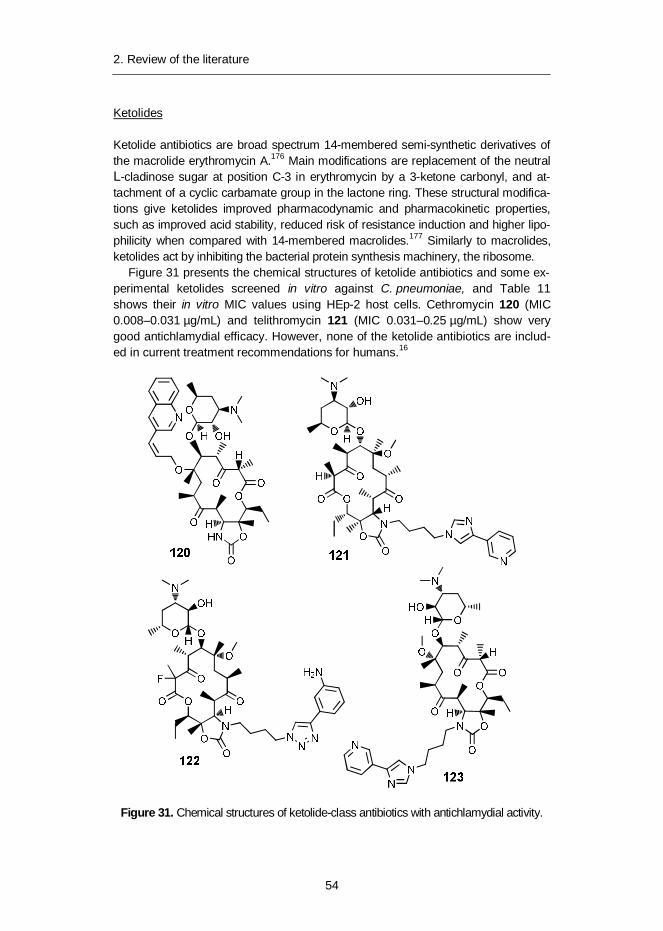
Chlamydia pneumoniae is a common Gram-negative human pathogen mainlycausing mild respiratory infections, which can lead to pneumonia or bronchitis.There is also strong evidence that associates C. pneumoniae with other severediseases, such as atherosclerotic cardiovascular diseases as well as some neuro-degenerative diseases, such as Alzheimer’s disease and multiple sclerosis.C. pneumoniae is susceptible to antibiotics that interfere with DNA and proteinsynthesis. However, its complex life cycle and its chlamydial persistence, whichcan last for years, as well as, importantly, the lack of specific diagnostic tests fordetection of the organism in clinical samples, make the current treatment regimensunsatisfactory. Out of 32 betulin derivatives, five betulin derivatives showed high(>70% growth inhibition) antichlamydial activity against C. pneumoniae at 1 µMconcentration. The most potent derivative displayed a remarkable 50% inhibitionat nanomolar concentration.
Synthesis of betulin derivatives against intracellular pathogens. Sami Alakurtti. Espoo 2013.VTT Science 39. 99 s. + liitt. 43 s.
TiivistelmäMetsäteollisuus käyttää koivua raaka-aineenaan suunnattomia määriä. Sivuottee-na syntyvä koivunkuori poltetaan energian tuotantoon. Koivun ulkokuori sisältäärunsaasti betuliini-nimistä triterpeeniä jopa 30 % kuivapainostaan. Betuliini voi-daan helposti eristää kuoresta liuotinuutolla. Betuliinia voidaan käyttää sellaise-naan tai lähtöaineena muille tuotteille ja hienokemikaaleille.
Useiden pieneliöiden ja virusten kasvava lääkeresistenssi on maailmanlaajuinenongelma, minkä takia on ilmennyt suuri tarve kehittää uusia lääkeaineita niitä vastaan.Tässä väitöskirjatyössä syntetisoitiin kaksi betuliinijohdannaisryhmää ja yhdistei-den tehokkuutta testattiin alfavirusten, Leishmania-suvun alkueläinten ja keuhko-klamydiabakteerin vastaisina yhdisteinä. Ensimmäinen ryhmä sisältää melko hel-posti valmistettavissa olevia johdannaisia, kuten betuliinin estereitä ja erilaisiahapetustuotteita. Toinen ryhmä sisältää uusia heterosyklisiä betuliinijohdannaisia,joissa triatsoli-rengas on fuusioitu betuliinin lupaanirakenteiseen hiilivetyrankaan.
Alfaviruksiin kuuluva Semliki Forest virus (SFV) leviää moskiittojen välityksellä,ja se infektoi lintuja ja nisäkkäitä. Jotkin alfavirukset voivat aiheuttaa tappavaaaivotulehdusta, mutta nämä tapaukset ovat hyvin harvinaisia. Useimmiten alfavi-rukset aiheuttavat sairauskohtauksia, joiden oireet ilmenevät usein kuumeena,allergisena ihottumana ja kivuliaana niveltulehduksena. Tällä hetkellä alfaviruksiavastaan ei ole tehokasta ja turvallista lääkitystä. Kaikkiaan 18 betuliinijohdannaistaosoitti alfavirusten vastaista aktiivisuutta mikromolaarisella konsentraatiolla. Lisäk-si kolmella potentiaalisella ja kemialliselta rakenteeltaan erilaisella betuliinijohdan-naisella oli synergistisiä alfaviruksen vastaisia vaikutuksia muokatun nukleosidi-johdannaisen kanssa. Lisäksi valitut kolme johdannaista olivat aktiivisia myöstoista alfavirusta, Sinbis virusta, vastaan.
Leishmaniaasi-tautia aiheuttavat Leishmania-sukuun kuuluvat alkueläimet. Tau-ti esiintyy nisäkkäissä etenkin tropiikissa, ja sitä levittävät perhossääsket. Arvioi-den mukaan 12 miljoonaa ihmistä sairastaa tällä hetkellä leishmaniaasia. Taudinvakavin muoto on sisäelinleishmaniaasi, joka on hoitamattomana tappava. Leish-maniaasia vastaan on käytössä useita lääkeaineita, mutta niiden tehokkuudessaja turvallisuudessa on toivomisen varaa. Ne aiheuttavat usein vakavia sivuvaiku-tuksia, ja niiden käyttö edellyttää sairaalahoitoa. Lisäksi lääkeresistenssi on vaka-va ja kasvava ongelma. Tässä tutkimuksessa lupaavimmat betuliinijohdannaisetosoittivat Leishmania donovanin vastaisia vaikutuksia mikromolaarisilla konsent-raatioilla. Johdannaisten estovaikutus pysyi hyvänä myös selvitettäessä niidenvaikutuksia syöjäsolujen sisällä kasvavia L. donovani -alkueläimiä vastaan. Valitet-
6
tavasti osa betuliinijohdannaisista osoitti sytotoksisuutta myös itse syöjäsolujavastaan.
Keuhkoklamydia (Chlamydia pneumoniae) on yleinen Gram-negatiivinen bak-teeri, joka aiheuttaa lieviä hengitystieinfektioita. Ne voivat pahimmillaan johtaakeuhkokuumeeseen tai keuhkoputkentulehdukseen. C. pneumoniaen aiheuttamillainfektiolla on myös osoitettu olevan yhteys useisiin muihin vakaviin sairauksiin,kuten sydän- ja verisuonisairauksiin, ja hermostoperäisiin sairauksiin, kuten Al-zheimerin tautiin ja MS-tautiin. C. pneumoniaen aiheuttamia infektioita voidaanhoitaa antibiooteilla, jotka vaikuttavat bakteerin DNA- tai proteiinisynteesiin. Vali-tettavasti bakteerin monimutkainen elämänkierto, taudin vaikea diagnosointi jabakteerille tyypillinen oireeton, jopa vuosia kestävä piilevänä sairautena pysyminentekevät taudin hoidosta ongelmallista. Testisarjasta viisi betuliinijohdannaista estihuomattavasti C. pneumoniae -bakteerin kasvua 1 µM konsentraatiolla. Parhaallajohdannaisella 50 %:n inhibitioarvo saavutettiin nanomolaarisella konsentraatiolla.
AcknowledgementsThis work was carried out at the University of Helsinki, Division of PharmaceuticalChemistry, during 2004–2007 and at VTT Technical Research Centre of Finlandwithin the Process chemistry centre during 2007–2013. Funding was provided byTekes – the Finnish Funding Agency for Technology and Innovation, the Founda-tion for Research of Natural Resources in Finland, the European Commission(Grant 227239) and the Academy of Finland (Grant 252308).
I am most thankful to my supervisor, Professor Jari Yli-Kauhaluoma, for hisnever-ending support, guidance and knowledge in organic chemistry. I am alsograteful for your patience and encouragement throughout the many years of ourjourney into the chemistry of wood extractives.
I thank my co-authors Pia Bergström and Tuomo Heiska at VTT; Dr. LeenaPohjala, Dr. Alexandros Kiriazis, Antti Siiskonen, Dr. Tero Ahola and Dr. PäiviTammela at the University of Helsinki; Dr. Olli Salin and Professor Pia Vuorela atÅbo Akademi University; Dr. Nina Sacerdoti-Sierra and Professor Charles L. Jaffeat the Hebrew University-Hadassah Medical School, Jerusalem, Israel; as well asDr. Viola Maass and Dr. Matthias Maass at PMU University Hospital Salzburg,Austria.
Warm thank to my colleagues in several joint projects concerning valorization ofbetulin and other wood extractives: Minni Pirttimaa, Pauliina Pitkänen, Dr. DaveThomas, Heimo Kanerva, Yukho Sok-sar, Juha Karttunen, Jukka Tulisalo, HarriHeikkinen, Anja Salakari and Janne Hulkko at VTT. In addition, warm thanks toex-colleagues at the Division of Pharmaceutical Chemistry (JYK group), especiallyto Raisa Haavikko, Dr. Mohanathas Rajaratman, Dr. Kirsi Harju, Dr. Gustav Boijeaf Gennäs, Dr. Vânia Moreira and Dr. Ingo Aumüller; and finally to the M.Sc. andother undergraduate students who have helped me with the chemistry of woodextractives: Kristian Meinander, Jaana Minkkinen, Niko Salminen, Maija-LiisaTuononen, Darin Al-Ramahi, Erkki Metsälä and Jukka Pernilä. Special thanks tomy mentor Dr. Salme Koskimies for taking me under her wing.
I wish to thank Professor Tapani Vuorinen of Aalto University and ProfessorDulcie Mulholland at the University of Surrey, UK, for kindly reviewing this thesis.
Finally, my heartfelt thanks to my family: my parents Matti and Annikki as wellas my siblings, Kirsi and Mikko for all the love and support; and lastly, my belovedpartner Sari for her patience and encouragement; and my precious daughter Lumifor always brightening my day and bringing a smile to my face.
8
Academic dissertation
Supervisor Professor Jari Yli-KauhaluomaDivision of Pharmaceutical ChemistryFaculty of PharmacyUniversity of HelsinkiFinland
Reviewers Professor Tapani VuorinenSchool of Chemical TechnologyDepartment of Forest Products TechnologyAalto UniversityFinland
Professor Dulcie MulhollandDivision of Chemical SciencesDepartment of ChemistryUniversity of SurreyUK
Opponent Professor Stefan WillförLaboratory of Wood and Paper ChemistryDepartment of Chemical EngineeringÅbo Akademi UniversityFinland
Custos Professor Jari Yli-Kauhaluoma
9
List of publicationsThis thesis is based on the following original publications which are referred to inthe text as I–IV. The publications are reproduced with kind permission from thepublishers.
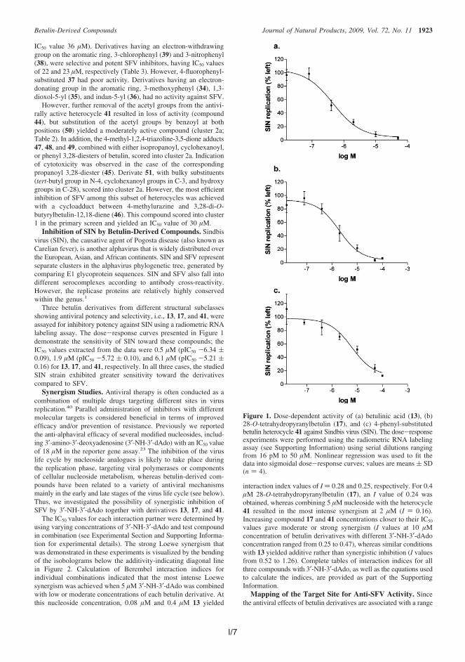
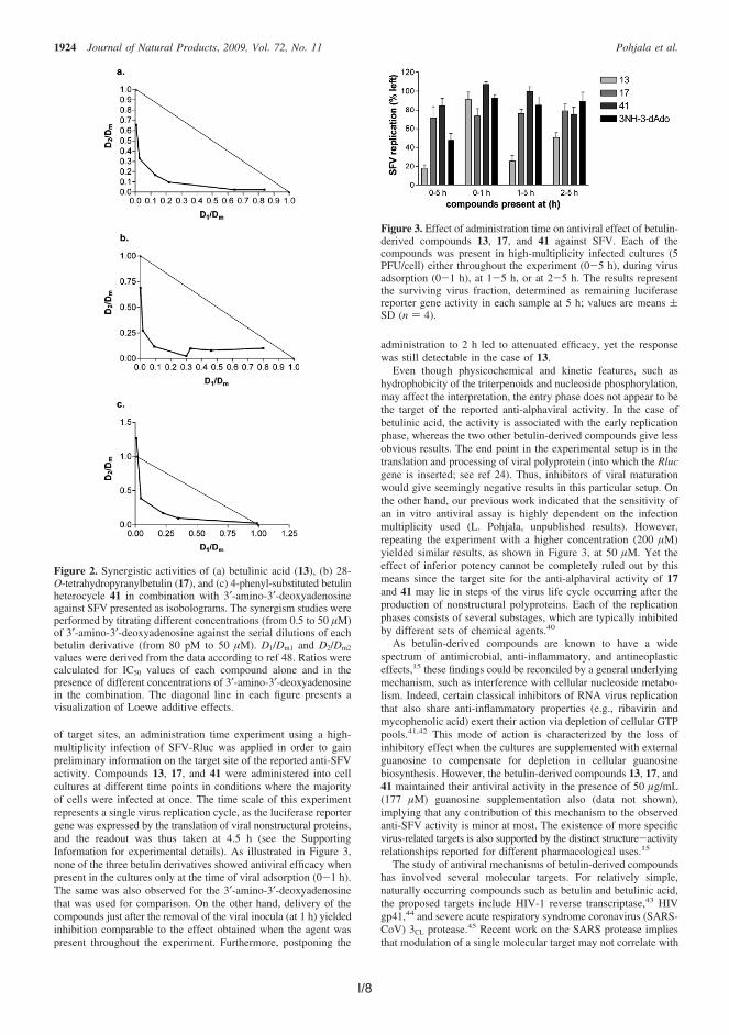
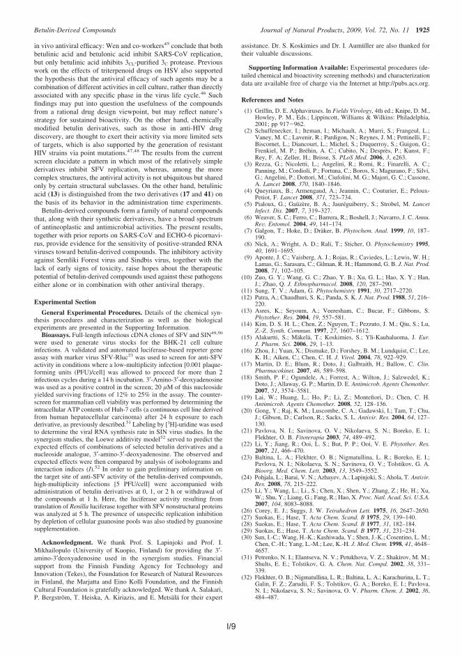
I Pohjala, L.*, Alakurtti, S.*, Ahola, T., Yli-Kauhaluoma, J. and Tammela, P.Betulin-derived compounds as inhibitors of alphavirus replication, J. Nat.Prod. 2009, 72, 1917.
II Alakurtti, S., Heiska T., Kiriazis, A., Sacerdoti-Sierra, N., Jaffe, C., Yli-Kauhaluoma, J. Synthesis and anti-leishmanial activity of heterocyclic betulinderivatives. Bioorg. Med. Chem. 2010, 8, 1573.
III Alakurtti, S., Bergström, P., Sacerdoti-Sierra, N., Jaffe, C., Yli-Kauhaluoma,J. Anti-leishmanial activity of betulin derivatives. J. Antibiot. 2010, 63, 123.
IV Salin, O.*, Alakurtti, S.*, Pohjala, L., Siiskonen, A., Maass, V., Maass, M.,Yli-Kauhaluoma, J., Vuorela P. Inhibitory effect of the natural product betulinand its derivatives against the intracellular bacterium Chlamydia pneumoniae.Biochem. Pharmacol. 2010, 80, 1141.
* Equal contribution
Related publications, not included in this thesis:
Alakurtti, S., Mäkelä, T., Koskimies, S., Yli-Kauhaluoma, J. Pharmacological prop-erties of the ubiquitous natural product betulin. Eur. J. Pharm. Sci. 2006, 29, 1.
Šilhár P., Alakurtti, S., Capková, K., Xiaochuan, F., Shoemaker, C. B., Yli-Kauhaluoma, J., Janda, K. Synthesis and evaluation of library of betulin deriva-tives against the botulinum neurotoxin A protease. Bioorg. Med. Chem. Lett. 2011,21, 2229.
Wert, L., Alakurtti, S., Corral, M., Sánchez-Fortún S., Yli-Kauhaluoma, J., Alunda, J.Toxicity of betulin derivatives and in vitro effect on promastigote and amastigotesof Leishmania infantum and L. donovani. J. Antibiotics. 2011, 64, 475.
10
Author’s contributions in the originalpublications
I The author synthesized and characterized almost all of the betulin deriva-tives (few derivatives were synthetized by Mrs. P. Bergström, A. Salakari,senior laboratory technician and undergraduate students T. Heiska and E.Metsälä under author’s supervision). The author wrote the manuscript to-gether with Dr. L. Pohjala with the aid of other co-authors. This publicationis included as one of the required publications in Dr. L. Pohjala’s academicdissertation as well.
II The author synthesized and characterized almost all of the betulin deriva-tives (few urazole intermediates were obtained from Dr. A. Kiriazis and fewbetulin derivatives were synthetized by undergraduate student T. Heiskaunder author’s supervision). Author wrote the article with the aid of otherco-authors.
III The author synthesized and characterized almost all of the betulin deriva-tives (few derivatives were synthetized by Mrs. P. Bergström, A. Salakari,senior laboratory technician and undergraduate student E. Metsälä underauthor’s supervision). The author wrote the article with aid of other co-authors.
IV The author synthesized and characterized almost all of the betulin deriva-tives (few derivatives were synthetized by Mrs. P. Bergström and under-graduate students T. Heiska and E. Metsälä under author’s supervision).The author wrote the manuscript together with Dr. O. Salin with the aid ofother co-authors. This publication is included as one of the required publi-cations in Dr. O. Salin’s academic dissertation as well.
1. Introduction ............................................................................................. 161.1 Birch bark ......................................................................................... 161.2 Alphaviruses..................................................................................... 161.3 Leishmania spp. ............................................................................... 171.4 Chlamydia pneumoniae .................................................................... 171.5 Origin of drugs during the last 25 years ............................................. 18
2. Review of the literature ........................................................................... 192.1 Betulin .............................................................................................. 19
2.1.1 Pharmacological properties of betulin derivatives .................... 192.2 Alphaviruses..................................................................................... 20
2.2.1 Species, taxonomy and lifecycle ............................................. 202.2.2 Chemotherapy against infections caused by alphaviruses ....... 22
2.3 Leishmania spp. ............................................................................... 282.3.1 Species, taxonomy and lifecycle ............................................. 282.3.2 Leishmania and HIV co-infection ............................................ 302.3.3 Chemotherapy against infections caused by Leishmania spp. . 30
2.4 Chlamydia pneumoniae .................................................................... 462.4.1 Species, taxonomy and life cycle ............................................ 462.4.2 Chlamydial persistence .......................................................... 482.4.3 Chemotherapy against infections caused by C. pneumoniae ... 48
12
3. Aims of the study .................................................................................... 58
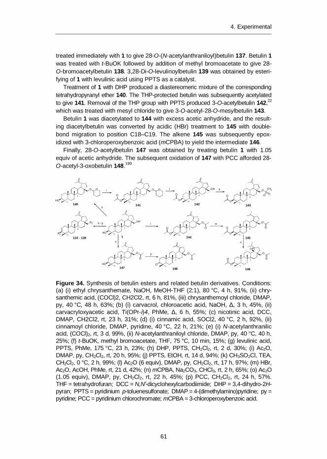
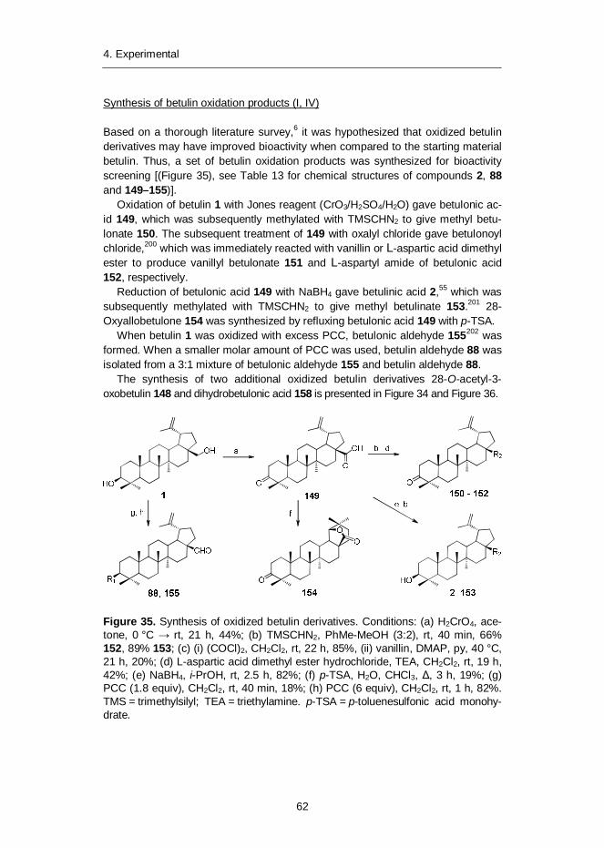
4. Experimental ........................................................................................... 594.1 Materials and methods ...................................................................... 594.2 Results and discussion ..................................................................... 59
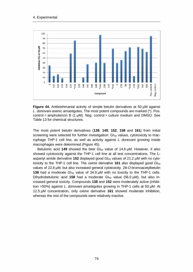
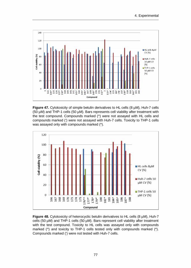
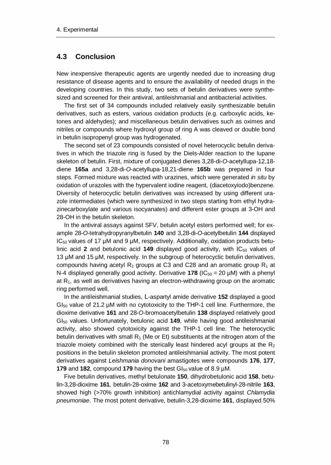
4.2.1 Synthesis of simple betulin derivatives (I, II, IV) ....................... 604.2.2 Synthesis of heterocyclic betulin derivatives (II) ...................... 664.2.3 SAR studies: Semliki Forest virus (I) ....................................... 694.2.4 SAR studies: Leishmania donovani (II) ................................... 714.2.5 SAR studies: Leishmania donovani (III) .................................. 734.2.6 SAR studies: Chlamydia pneumoniae (IV) .............................. 754.2.7 Cytotoxicity of betulin compounds (I, II, III, IV) ......................... 76
Publications II and IV of this publication are not included in the PDF version.
13
Abbreviations13C NMR Carbon-13 nuclear magnetic resonance1H NMR Hydrogen-1 nuclear magnetic resonance
AB Aberrant body
AIDS Acquired immunodeficiency syndrome
AT Adenine, thymine
ATP Adenosine triphosphate
AV Antiviral effect
BALB/c Bagg albino, laboratory-bred, genotype c/c
BHK Baby hamster kidney fibroblast cell line
BPQ Buparvaquone
BVM Bevirimat
CHIKV Chikungunya virus
CL Cutaneous leishmaniasis
CTP Cytidine triphosphate
CV Cell viability
DCC N,N'-Dicyclohexylcarbodiimide
DEAD Diethyl acetylenedicarboxylate
DHP 3,4-Dihydro-2H-pyran
DMAP 4-(Dimethylamino)pyridine
DMC 2’,6’-dihydroxy-4’-methoxychalcone
DMSO-d6 Deuterated dimethyl sulfoxide
DNA Deoxyribonucleic acid
14
DSB Dimethylsuccinyl betulinic acid
EB Elementary body
EC50 Half maximal effective concentration
ECHO Enteric cytopathic human orphan virus
ED50 50% effective dose
EEEV Eastern equine encephalitis virus
FPV Fowl plague virus
FTIR Fourier transform infrared spectroscopy
GETV Getah virus
GI50 50% growth inhibition
HeLa Cancer cell line taken from Henrietta Lacks
Hep Human epithelial cervix carcinoma cell line
HIV Human immunodeficiency virus
HL Human promyelocytic leukemia cell line
HPLC High-performance liquid chromatography
HPLC-MS High-performance liquid chromatography – mass spectrometry
HSV-1 Herpes simplex type 1
Huh-7 Human hepatocellular carcinoma cell line
I Interaction index
IC50 50% inhibitory concentration
ICTV International Committee on Taxonomy of Viruses
IMPDH Inosine-5 -monophosphate dehydrogenase
kDNA Kinetoplast DNA
LP Liposome
MB-III Maesabalide III
MCL Mucocutaneous leishmaniasis
mCPBA m-Chloroperbenzoic acid
MIC Minimum inhibitory concentration
NC Nucleocapsid
NSV Neuroadapted Sinbis virus
15
p-TSA p-Toluenesulfonic acid monohydrate
PCC Pyridinium chlorochromate
Pd/C Palladium on carbon
PLA Polylactic acid
PPTS Pyridinium p-toluenesulfonate
PS Phosphatidylserine
RB Reticulate body
RNA Ribonucleic acid
SAR Structure–activity relationships
SARS Severe acute respiratory syndrome
SFV Semliki Forest virus
SI Selectivity index
SINV Sindbis virus
spp Species
t-BuOK Potassium tert-butoxide
TEA Triethylamine
THF Tetrahydrofuran
THP Tetrahydropyran
THP-1 Human leukaemia monocyte cell line
TLC Thin layer chromatography
TMS Trimethylsilyl
tRNA Transfer RNA
UV Ultra violet
VEEV Venezuelan equine encephalitis virus
VL Visceral leishmaniasis
WEEV Western equine encephalitis virus
1. Introduction
16
1. Introduction
1.1 Birch bark
Birch is (Betula spp.) widespread throughout the Northern Hemisphere and har-vested in huge volumes. Birch bark is produced in considerable quantities as a by-product of the forest industry and its upgrading is almost totally neglected. Cur-rently, this low-value side stream is burnt for combined heat and power production.Birch bark could, however, find more valuable uses, for example as an additive inplastic composite materials.1 Birch bark consists of brown inner bark ~75% andwhite outer bark ~25%.2 The inner bark consists mainly of wood-like material suchas lignin, pentosans and hexosans. The outer bark contains, by dry weight, up to40% fats, fatty acids, resins and triterpenes, in particular betulin, at up to 30%. Inaddition, the outer bark contains up to 35% suberin. Valorization and upgrading ofthese compounds by using modern chemical technology opens up entirely newopportunities to produce new speciality chemicals from this low-value biomassstream. It has been estimated theoretically that a pulp mill with an annual produc-tion capacity of 200,000 tonnes of birch kraft pulp produces enough bark to pro-duce around 2,500 tonnes of betulin of around 95% purity and 4,000 tonnes ofsuberin acids per annum.3,4 Suberin polyester can be hydrolyzed by base treat-ment to multifunctional suberin acids, which are potential raw materials for paints,adhesives, lubricants and surface-active agents.5 New potential applications forbetulin or betulin derivatives include pharmaceuticals and cosmetic products aswell as agrochemicals.6
1.2 Alphaviruses
The Semliki Forest virus (SFV) belongs to the alphaviruses, which are small en-veloped viruses containing a single-stranded positive-sense RNA genome.7 Virus-es belonging to this genus are predominantly arthropod-borne viruses using mos-quitoes as vectors and have a very wide geographic distribution, with isolateshaving been reported from all continents except Antarctica and from many is-lands.8 Alphaviruses infect avian and mammalian hosts and are a serious or po-tential threat to human health. In North and South America some alphaviruses areknown to cause fatal encephalitis in humans, although the number of recorded
1. Introduction
17
fatalities is small.9 Alphaviruses have, however, caused millions of cases of seri-ous illness characterized by fever, rash and painful arthralgia.10 There is currentlyno efficient pharmacotherapy for alphavirus-borne diseases.
1.3 Leishmania spp.
Leishmaniasis is a disease caused by protozoan parasites belonging to the genusLeishmania. The disease is transmitted by sandflies and is present in all inhabitedcontinents.11 It is estimated that around 12 million people are currently infected.More than 350 million people live in risk areas for the disease, and 2 million newcases occur every year, especially in the developing countries. Leishmaniasis hasbeen designated as a neglected tropical disease by the World Health Organiza-tion. The most severe form, visceral leishmaniasis, in which parasites invade theliver, spleen and bone marrow, is fatal if not treated.12 Current treatment includespentavalent antimony compounds, pentamidine and amphotericin B.13 However,there are risks of severe side effects and all of these current drugs are adminis-tered by intravenous injection. Parasite resistance to pentavalent antimony drugshas become a serious problem and is present in approximately 65% of patientswith visceral leishmaniasis in India.14 New drugs, such as primaquine, allopurinol,imipramine, are being developed, however none of them are fully effective againstLeishmania.12 Recently, orally administrable miltefosine has shown promisingantileishmanial activity.15
1.4 Chlamydia pneumoniae
Chlamydia (Chlamydophila) pneumoniae is an important Gram-negative humanpathogen, mainly causing respiratory infections. It has been proposed that almost allhumans will become infected with C. pneumoniae during their life.16 C. pneumoniaefrequently causes community-acquired pneumonia in adults and children. Ofteninfections are asymptomatic and frequently of long duration, up to several years.There is also strong evidence that associates C. pneumoniae with other severediseases, such as atherosclerotic cardiovascular diseases,17 as well as someneurodegenerative diseases, such as Alzheimer’s disease and multiple sclerosis.18
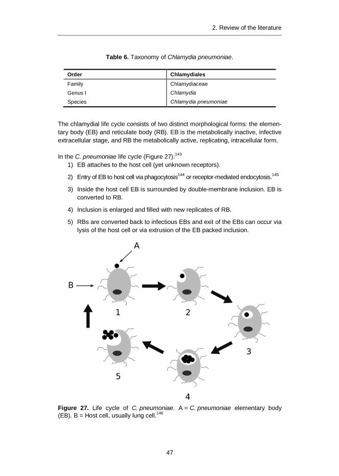
Interestingly, C. pneumoniae infection has also been reported in a wide range ofanimals, including other mammals such as horses and koalas, and frogs and otherreptiles.19 All chlamydial species are intracellular bacteria that infect and replicateinside a variety of human cells, including epithelial, endothelial, macrophages, andsmooth muscle cells.20 C. pneumoniae is classified as an obligate intracellularpathogen, as it has to infect another cell to reproduce. C. pneumoniae is suscepti-ble to antibiotics that interfere with DNA and protein synthesis, including tetracy-clines, macrolides and quinolones.16 However, its complex life cycle and its chla-mydial persistence, which can last for years, as well as, importantly, the lack ofspecific diagnostic tests for detection of the organism in clinical samples, make thecurrent treatment regimens unsatisfactory.
1. Introduction
18
1.5 Origin of drugs during the last 25 years
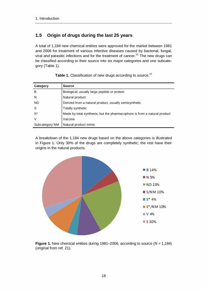
A total of 1,184 new chemical entities were approved for the market between 1981and 2006 for treatment of various infective diseases caused by bacterial, fungal,viral and parasitic infections and for the treatment of cancer.21 The new drugs canbe classified according to their source into six major categories and one subcate-gory (Table 1).
Table 1. Classification of new drugs according to source.21
Category Source
B Biological; usually large peptide or protein
N Natural product
ND Derived from a natural product, usually semisynthetic
S Totally synthetic
S* Made by total synthesis, but the pharmacophore is from a natural product
V Vaccine
Subcategory NM Natural product mimic
A breakdown of the 1,184 new drugs based on the above categories is illustratedin Figure 1. Only 30% of the drugs are completely synthetic; the rest have theirorigins in the natural products.
Figure 1. New chemical entities during 1981–2006, according to source (N = 1,184)(original from ref. 21).
2. Review of the literature
19
2. Review of the literature
2.1 Betulin
Betulin 1, lup-20(29)-ene-3 ,28-diol, also known as betulinol, betuline and betulin-ic alcohol (Figure 2), is a pentacyclic triterpene alcohol with a lupane skeleton.Common structural features of the lupane skeleton are its five-membered ring Eand isopropylidene group. Betulin 1 can be isolated (up to 30% dry weight) fromthe outer birch bark by extraction with high boiling hydrocarbon solvents or withwater azeotropes of alcohols.4
Betulin 1 can be used as a starting compound for other useful compounds thatpossess various interesting pharmacological properties. Betulin 1 has three posi-tions in its structure, namely a secondary hydroxy group at position C-3, a primaryhydroxy group at position C-28, and an alkene moiety at position C-20, wherechemical modifications can be easily performed to yield derivatives for structure–activity relationship (SAR) studies. It is clear from the chemical structure of betulinthat most of the derivatives presented in the experimental part of this thesis arelipophilic compounds and thus poorly soluble in water if no organic co-solvent isused. This may have important implications for the interpretation of the results ofthe bioactivity assays, which have been carried out predominantly in cell cultures.Observed differences in bioactivity between different modified betulin derivativesmay be explained, at least partly, by different water/lipid distribution constants betweenthese analogues.
2.1.1 Pharmacological properties of betulin derivatives
Betulin 1 is biologically a relatively inactive compound. However, betulin 1 can beoxidized to pharmacologically more active betulinic acid 2.22 Betulinic acid 2 and itsderivatives possess a wide spectrum of biological and pharmacological activity.6
Most notably, betulinic acid 2 and its derivatives have shown activity against humanimmunodeficiency virus (HIV) as well as cytotoxicity against a variety of tumour celllines comparable to some clinically used drugs.
A new mechanism of action has been confirmed for some of the most promis-ing anti-HIV derivatives, which makes them potentially useful additives to currentanti-HIV therapy. A43D23 3 and statine-derived IC956424 4 act as entry inhibitors
2. Review of the literature
20
and block HIV adsorption or membrane fusion. Furthermore, the dimethylsuccinylderivative of betulin, bevirimat 5 (DSB, BVM), acts as a virus maturation inhibitor.25
Bevirimat 5 was demonstrated to have dose-dependent anti-HIV potency in phaseI and phase II clinical studies.26 However, mutations in HIV cause resistance toBVM 5, in addition to which some patients have this polymorphism present, result-ing in lower BVM 5 anti-HIV efficacy.27 Development of BVM 5 has thus beenrecently halted.
Betulinic acid 2 is specifically cytotoxic to several tumour cell lines by directlytriggering mitochondrial membrane permeabilization and inducing apoptosis incells.28,29 Moreover, it is non-toxic up to 500 mg/kg body weight in mice.28 Currently,betulinic acid 2 is undergoing anti-cancer development with assistance from theRapid Access to Intervention Development Program of the National Cancer Insti-tute.30
Figure 2. Betulin 1 and potential anticancer agent betulinic acid 2, as well as po-tential anti-HIV agents A43D 3, IC9564 4 and bevirimat 5.
2.2 Alphaviruses
2.2.1 Species, taxonomy and lifecycle
Currently there are two main schemes used for the classification of viruses: theInternational Committee on Taxonomy of Viruses (ICTV) system and the Baltimoreclassification system. A universal system for classifying viruses, and a unifiedtaxonomy, is being established by the International Committee on Taxonomy ofViruses (ICTV). 31 The system makes use of a series of ranked taxons:
2. Review of the literature
21
Table 2. Taxonomy of Semliki Forest virus.
Order (-virales) Virus
Family (-viridae) Togaviridae
Subfamily (-virinae) -
Genus (-virus) Alphavirus
Species Semliki Forest virus
In the ICTV 2012 taxonomy classification, the Semliki Forest virus (SFV) (whichwas used as a target pathogen for betulin inhibition activity in publication I) spe-cies is classified under the family Togaviridae and the genus Alphavirus, whichcontains a total of 30 viruses, including human pathogens such as Rubella, Sind-bis and Chikungunya viruses (Table 2).31
In the Baltimore classification system, viruses are divided into one of sevengroups depending on a combination of their nucleic acid (DNA or RNA), stranded-ness (single- or double-stranded), sense, and method of replication (Table 3).32
Table 3. Virus groups according to the Baltimore classification system.
Group Classification
I Double-stranded DNA (dsDNA) viruses
II Single-stranded DNA (ssDNA) viruses (+)sense DNA
III Double-stranded RNA (dsRNA) viruses
IV Single-stranded RNA [(+)ssRNA] viruses (+)sense RNA
V Single-stranded RNA [(-)ssRNA] viruses ( )sense RNA
VI Single-stranded RNA (ssRNA-RT) viruses (+)sense RNA with replicationthrough a DNA intermediate
VII Double-stranded DNA (dsDNA-RT) viruses with replication through asingle-stranded RNA intermediate
Alphaviruses have a single-stranded, positive-sense RNA genome and are classi-fied under group IV according to the Baltimore classification.8 Group IV includesviruses from several ICTV orders: 1. Nidovirales including significant pathogenssuch as Coronavirus and Severe acute respiratory syndrome virus (SARS); 2.Picornavirales including significant pathogens such as Norwalk, Polio, the com-mon cold and Hepatitis A viruses; 3. Tymovirales; and unassigned virus familiesincluding significant human pathogens such as Yellow fever, West Nile, Hepatitis Cand Dengue fever.33
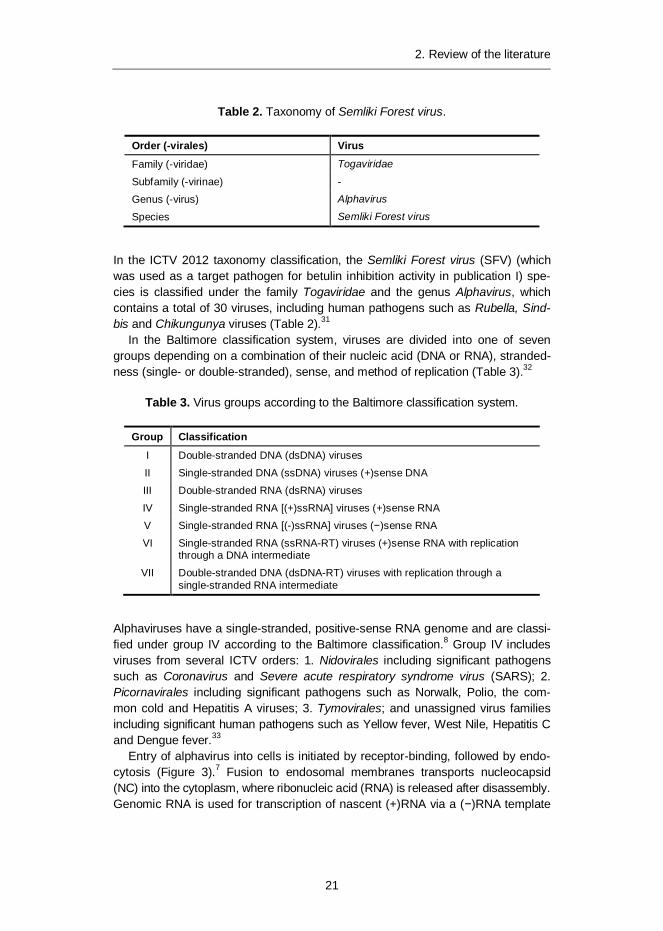
Entry of alphavirus into cells is initiated by receptor-binding, followed by endo-cytosis (Figure 3).7 Fusion to endosomal membranes transports nucleocapsid(NC) into the cytoplasm, where ribonucleic acid (RNA) is released after disassembly.Genomic RNA is used for transcription of nascent (+)RNA via a ( )RNA template
2. Review of the literature
22
and translation of proteins from genomic and subgenomic (26S) RNA. The struc-tural proteins translated from 26S RNA encapsidate nascent genomic RNA beforebudding from cells, and eventual release.
Figure 3. Life cycle of alphavirus (original from ref. 7).
2.2.2 Chemotherapy against infections caused by alphaviruses
This section focuses on small-molecule antiviral inhibitors found to be effectiveagainst viruses in the genus Alphavirus. The alphaviral species discussed in thissection include the Chikungunya virus (CHIKV), Eastern equine encephalitis virus(EEEV), Getah virus (GETV), Semliki Forest virus (SFV), Sindbis virus (SINV),Venezuelan equine encephalitis virus (VEEV) and Western equine encephalitisvirus (WEEV). As there are only very few reported in vivo alphavirus experiments,in vitro experiments are also discussed here.
Nucleoside or nucleotide analogues
Synthetic nucleoside or nucleotide analogues are prodrugs that are phosphory-lated to active triphosphate drugs by viral enzymes and used as normal buildingblocks for the DNA polymerase-catalyzed replication of viral DNA.34 However, as aconsequence of the modifications in the nucleobase or in the sugar moiety, viralDNA polymerase enzyme is deactivated or DNA chain formation is terminated.
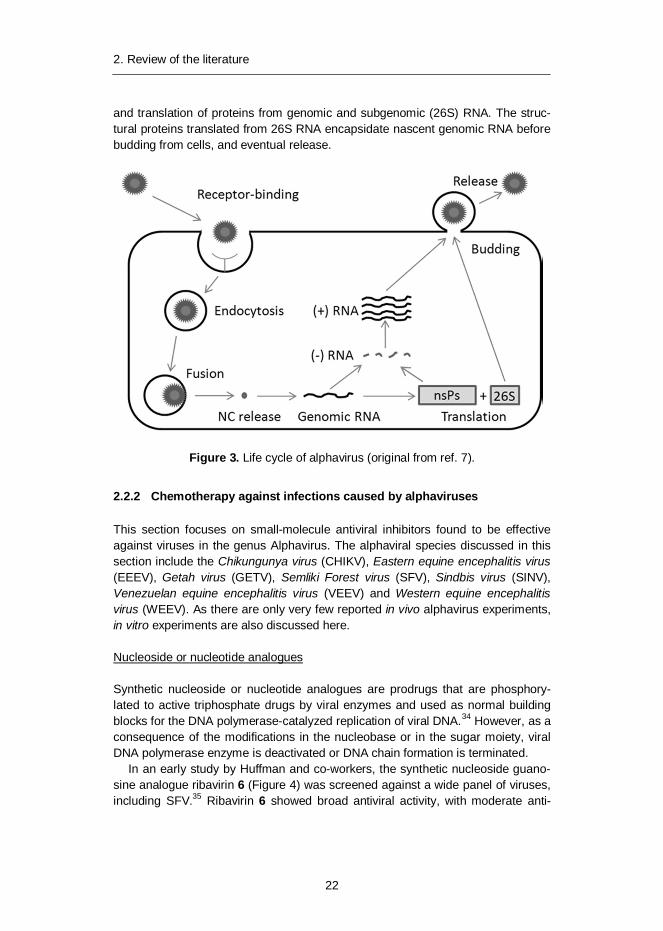
In an early study by Huffman and co-workers, the synthetic nucleoside guano-sine analogue ribavirin 6 (Figure 4) was screened against a wide panel of viruses,including SFV.35 Ribavirin 6 showed broad antiviral activity, with moderate anti-
2. Review of the literature
23
alphaviral activity and a minimum inhibitory concentration (MIC) value of 131 µM.Ribavirin 6 has since been used as a positive control in several assays.
Nucleotide analogue ribavirin 5’-sulphamate 7 displayed several-fold improvedantiviral activity against SFV with an IC50 value of 10 µM. The IC50 value repre-sents the concentration of drug required for 50% inhibition in vitro (50% inhibitoryconcentration). Ribavirin 6 displayed an IC50 value of <1 mm.36 In in vivo testingwith lethally SFV infected mice, ribavirin 5’-sulphamate 7 showed a clear protec-tive effect with survival rates of 92% and 83% when intraperitoneally administeredat doses of 20 and 40 mg/kg/day for seven days.
Selenazofurin 8 and tiazofurin were assayed alone or in combination with rib-avirin 6 against several viruses, including VEEV. Selenazofurin 8 displayed goodactivity against VEEV with a 50% effective dose (ED50) value of 0.5 µg/mL, whereastiazofurin was inactive.37 Combination of ribavirin and 6 selenazofurin 8 displayedsynergistic efficacy with an ED50 value of 0.1 µg/mL.
Figure 4. Nucleoside guanosine analogues with antiviral activity against alphavirus.
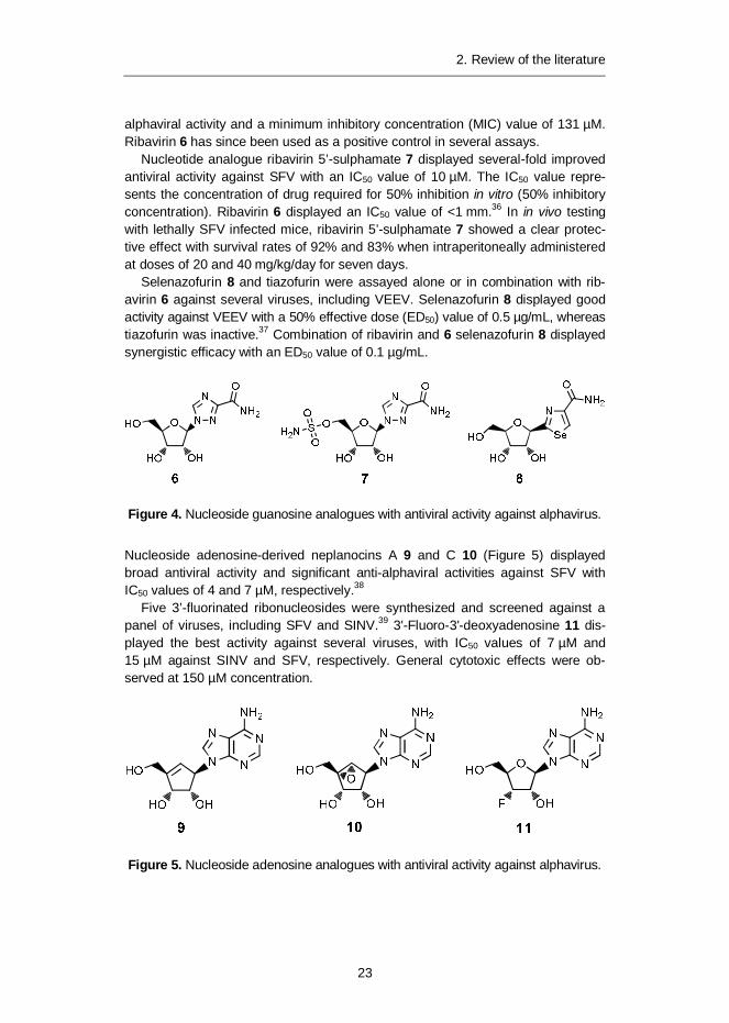
Nucleoside adenosine-derived neplanocins A 9 and C 10 (Figure 5) displayedbroad antiviral activity and significant anti-alphaviral activities against SFV withIC50 values of 4 and 7 µM, respectively.38
Five 3’-fluorinated ribonucleosides were synthesized and screened against apanel of viruses, including SFV and SINV.39 3'-Fluoro-3'-deoxyadenosine 11 dis-played the best activity against several viruses, with IC50 values of 7 µM and15 µM against SINV and SFV, respectively. General cytotoxic effects were ob-served at 150 µM concentration.
Figure 5. Nucleoside adenosine analogues with antiviral activity against alphavirus.
2. Review of the literature
24
In a study by de Clercq et al., antiviral activity of racemic cytidine analogue car-bodine 12 and 13 (Figure 6) was compared to known antiviral agents such ascarbocyclic 3-deazaadenosine (C-c3 Ado) and ribavirin 6.40 The racemic mixture ofcarbodine enantiomers 12 and 13 showed broad activity against most of the viralspecies tested by inhibition of cellular cytidine triphosphate (CTP) synthetase.Especially against SINV, carbodine showed an IC50 value of 3 µM, which wasalmost 60 times more potent than C-c3 Ado and over 200 times more potent thanribavirin 6. In addition, the carbodine racemate displayed good inhibition againstSFV with an IC50 value of 12 µM.
In a study by Julander et al., activity of the D-( )- and L-(+)-enantiomers of car-bodine was determined against VEEV in cell culture and in an in vivo mouse model.( )-Carbodine 13 showed good inhibition activity with an EC50 value of 0.8 µM,while (+)-carbodine 12 was not active (EC50 > 100 µM). Post-virus exposuretreatment with ( )-carbodine 13 was effective in significantly improving diseaseparameters in mice infected with VEEV when treatment was initiated as late as 4days post-virus installation, with a mouse survival rate of 90% (placebo 0%).
In another study by De Clercq et al., cyclopentenylcytosine (Ce-Cyd) 14 dis-played broad-spectrum antiviral activity against several viruses, including SFV andSINV with IC50 values of 0.4 and 0.2 µg/mL, respectively.41
Antiviral activity of eight commercially available compounds was estimatedagainst CHIKV and SFV.42 When comparing antiviral activities against CHIKV,positive control ribavirin 6 had an EC50 value of 83 µM and a selectivity index (SI)of 24. The two best test compounds, 6-azauridine 15 and sulfated polysaccharideiota-carrageenan, displayed significantly better EC50 values of 0.2 and 3.8 µM andSI values of 204 and >133, respectively. Against SFV, these compounds showedsimilar activities: 6-azauridine 15 and iota-carrageenan EC50 values of 0.4 and0.7 µM and SI values of 85 and >714, respectively, while ribavirin 6 showed amoderate EC50 value of 47 µM and an SI value of 109.
Figure 6. Nucleoside cytidine analogues with antiviral activity against alphavirus.
Non-nucleoside analogues
In a study by Pohjala et al.,43 a library of 356 compounds was screened in vitroagainst CHIKV and SFV. The library consisted of natural compounds (mainlyflavonoids, coumarins and other phenolic compounds) as well as clinically ap-proved drugs and their metabolites. Four natural 5,7-dihydroxyflavones, 16–19,
2. Review of the literature
25
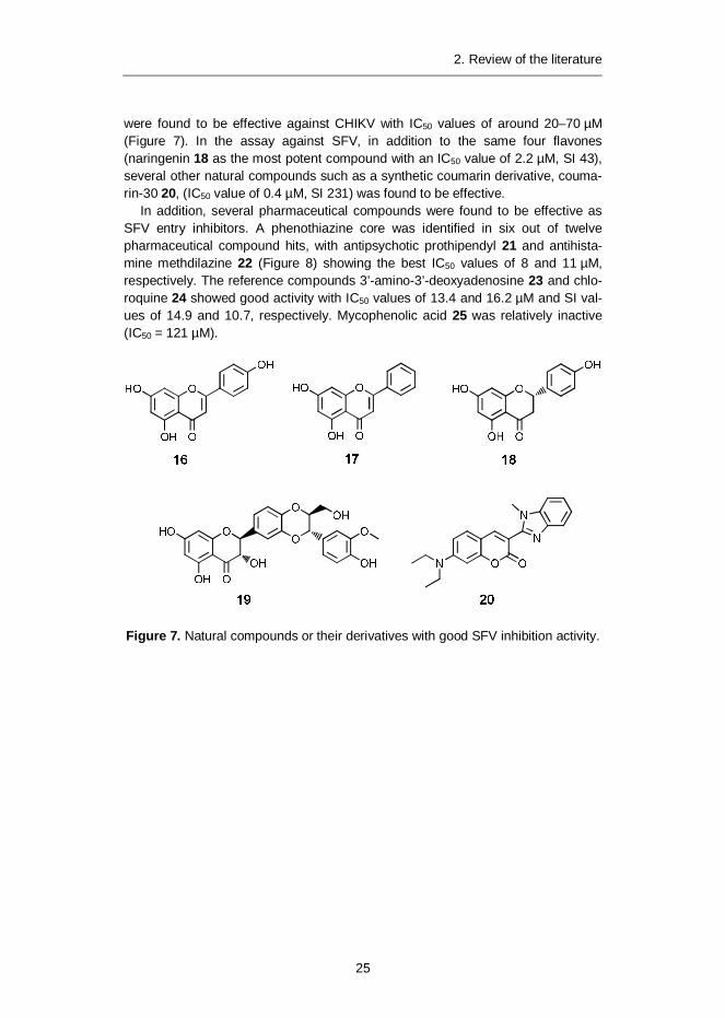
were found to be effective against CHIKV with IC50 values of around 20–70 µM(Figure 7). In the assay against SFV, in addition to the same four flavones(naringenin 18 as the most potent compound with an IC50 value of 2.2 µM, SI 43),several other natural compounds such as a synthetic coumarin derivative, couma-rin-30 20, (IC50 value of 0.4 µM, SI 231) was found to be effective.
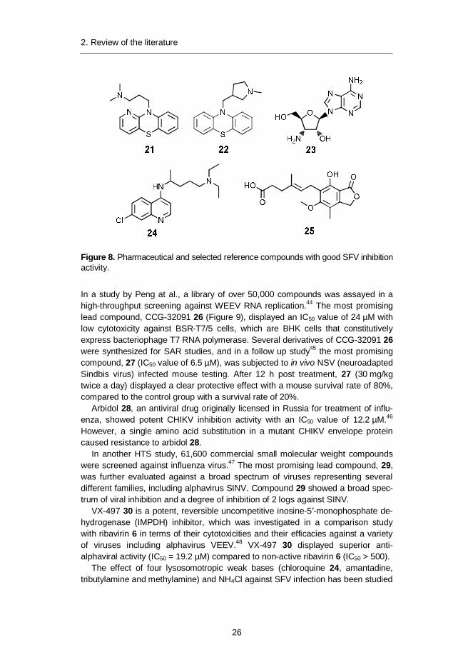
In addition, several pharmaceutical compounds were found to be effective asSFV entry inhibitors. A phenothiazine core was identified in six out of twelvepharmaceutical compound hits, with antipsychotic prothipendyl 21 and antihista-mine methdilazine 22 (Figure 8) showing the best IC50 values of 8 and 11 µM,respectively. The reference compounds 3’-amino-3’-deoxyadenosine 23 and chlo-roquine 24 showed good activity with IC50 values of 13.4 and 16.2 µM and SI val-ues of 14.9 and 10.7, respectively. Mycophenolic acid 25 was relatively inactive(IC50 = 121 µM).
Figure 7. Natural compounds or their derivatives with good SFV inhibition activity.
2. Review of the literature
26
Figure 8. Pharmaceutical and selected reference compounds with good SFV inhibitionactivity.
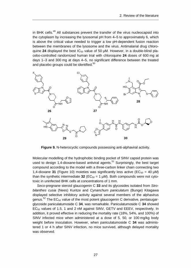
In a study by Peng at al., a library of over 50,000 compounds was assayed in ahigh-throughput screening against WEEV RNA replication.44 The most promisinglead compound, CCG-32091 26 (Figure 9), displayed an IC50 value of 24 µM withlow cytotoxicity against BSR-T7/5 cells, which are BHK cells that constitutivelyexpress bacteriophage T7 RNA polymerase. Several derivatives of CCG-32091 26were synthesized for SAR studies, and in a follow up study45 the most promisingcompound, 27 (IC50 value of 6.5 µM), was subjected to in vivo NSV (neuroadaptedSindbis virus) infected mouse testing. After 12 h post treatment, 27 (30 mg/kgtwice a day) displayed a clear protective effect with a mouse survival rate of 80%,compared to the control group with a survival rate of 20%.
Arbidol 28, an antiviral drug originally licensed in Russia for treatment of influ-enza, showed potent CHIKV inhibition activity with an IC50 value of 12.2 µM.46
However, a single amino acid substitution in a mutant CHIKV envelope proteincaused resistance to arbidol 28.
In another HTS study, 61,600 commercial small molecular weight compoundswere screened against influenza virus.47 The most promising lead compound, 29,was further evaluated against a broad spectrum of viruses representing severaldifferent families, including alphavirus SINV. Compound 29 showed a broad spec-trum of viral inhibition and a degree of inhibition of 2 logs against SINV.
VX-497 30 is a potent, reversible uncompetitive inosine-5 -monophosphate de-hydrogenase (IMPDH) inhibitor, which was investigated in a comparison studywith ribavirin 6 in terms of their cytotoxicities and their efficacies against a varietyof viruses including alphavirus VEEV.48 VX-497 30 displayed superior anti-alphaviral activity (IC50 = 19.2 µM) compared to non-active ribavirin 6 (IC50 > 500).
The effect of four lysosomotropic weak bases (chloroquine 24, amantadine,tributylamine and methylamine) and NH4Cl against SFV infection has been studied
2. Review of the literature
27
in BHK cells.49 All substances prevent the transfer of the virus nucleocapsid intothe cytoplasm by increasing the lysosomal pH from 4–5 to approximately 6, whichis above the critical value needed to trigger a low pH-dependent fusion reactionbetween the membranes of the lysosome and the virus. Antimalarial drug chloro-quine 24 displayed the best IC50 value of 50 µM. However, in a double-blind pla-cebo-controlled randomized human trial with chloroquine 24 doses of 600 mg atdays 1–3 and 300 mg at days 4–5, no significant difference between the treatedand placebo groups could be identified.50
Molecular modelling of the hydrophobic binding pocket of SINV capsid protein wasused to design 1,4-dioxane-based antiviral agents.51 Surprisingly, the best targetcompound according to the model with a three-carbon linker chain connecting two1,4-dioxane 31 (Figure 10) moieties was significantly less active (EC50 = 40 µM)than the synthetic intermediate 32 (EC50 = 1 µM). Both compounds were not cyto-toxic in uninfected BHK cells at concentrations of 1 mm.
Seco-pregnane steroid glaucogenin C 33 and its glycosides isolated from Stro-bilanthes cusia (Nees) Kuntze and Cynanchum paniculatum (Bunge) Kitagawadisplayed selective inhibitory activity against several members of the alphavirusgenus.52 The EC50 value of the most potent glaucogenin C derivative, pentasugar-glycoside paniculatumoside C 34, was remarkable. Paniculatumoside C 34 showedEC50 values of 1.5, 1 and 2 nM against SINV, GETV and EEEV, respectively. Inaddition, it proved effective in reducing the mortality rate (18%, 54%, and 100%) ofSINV infected mice when administered at a dose of 5, 50, or 100 mg/kg bodyweight before inoculation. However, when paniculatumoside C 34 was adminis-tered 1 or 4 h after SINV infection, no mice survived, although delayed mortalitywas observed.
2. Review of the literature
28
Figure 10. 1,4-Dioxane-based compounds 31 and 32, and seco-pregnane-derivedcompounds 33 and 34, R = [(O- -D-glucopyranosyl-(1 4)-O- -D-glucopyranosyl-(1 4)-O-2,6-dideoxy-3-O-methyl- -D-arabino-hexopyranosyl-(1 4)-O-2,6-dideoxy- -D-ribo-hexopyranosyl-(1 4)-2,6-dideoxy-3-O-methyl- -D-arabino-hexopyranosyl)oxy] with anti-alphaviral activity.
Betulin-derived compounds
Bevirimat 5 (Figure 2), a semisynthetic dimethylsuccinic acid derivative of naturalproduct betulinic acid53, blocks HIV maturation by inhibiting the final stage of HIVGag protein processing.54 It was initially considered as a possible first member ofthe HIV maturation inhibitors, and it successfully demonstrated potency in phase Iand phase II clinical studies.26 However, mutations in HIV cause resistance toBVM 5, in addition to which some patients also have this polymorphism present,resulting in lower anti-HIV efficacy.27 Thus, development of BVM 5 has been cur-rently halted. Bevirimat 5 was found to be inactive against herpes simplex type 1(HSV-1) and influenza virus.25
The antiviral activity of betulin 1 and betulinic acid 2 and their derivatives have alsobeen studied against influenza A, herpes simplex type 1 (HSV-1), influenza FPV/Rostockand ECHO-6 enterovirus, however their antiviral activities were weak. 55,56,57
2.3 Leishmania spp.
2.3.1 Species, taxonomy and lifecycle
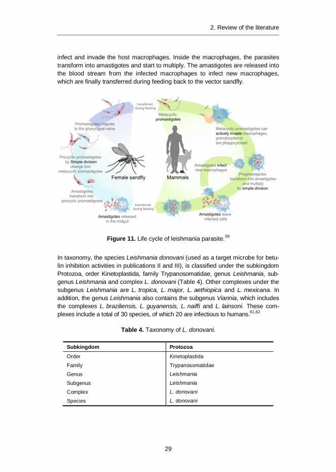
Protozoan parasites belonging to the genus Leishmania are transmitted to mam-malian hosts by female sandflies of the genera Phlebotomus and Lutzomyia in theOld and New World, respectively.58 The life cycle of the leishmanial parasite con-sists of a flagellated promastigote phase in the sandfly gut and a non-flagellatedintracellular amastigote phase in mammalian macrophages (Figure 11).59 After ablood meal from the infected host, amastigotes are released from the macrophagesinto the sandfly gut; these then transform through a multiplying non-infectiousprocyclic promastigote phase to infectious metacyclic promastigotes and migrateto the pharyngeal valve of the sandfly.60 The metacyclic promastigotes are trans-mitted during feeding to the mammalian host, where the promastigotes successfully
2. Review of the literature
29
infect and invade the host macrophages. Inside the macrophages, the parasitestransform into amastigotes and start to multiply. The amastigotes are released intothe blood stream from the infected macrophages to infect new macrophages,which are finally transferred during feeding back to the vector sandfly.
Figure 11. Life cycle of leishmania parasite.59
In taxonomy, the species Leishmania donovani (used as a target microbe for betu-lin inhibition activities in publications II and III), is classified under the subkingdomProtozoa, order Kinetoplastida, family Trypanosomatidae, genus Leishmania, sub-genus Leishmania and complex L. donovani (Table 4). Other complexes under thesubgenus Leishmania are L. tropica, L. major, L. aethiopica and L. mexicana. Inaddition, the genus Leishmania also contains the subgenus Viannia, which includesthe complexes L. braziliensis, L. guyanensis, L. naiffi and L. lainsoni. These com-plexes include a total of 30 species, of which 20 are infectious to humans.61,62
3) Visceral leishmaniasis (VL): devastates internal organs, especially liver, spleenand bone marrow and the untreated disease is usually fatal.
2.3.2 Leishmania and HIV co-infection
Interaction between leishmaniasis (VL) and HIV has been well established, mak-ing VL-HIV co-infection a serious worldwide concern.63 HIV-infected people areparticularly vulnerable to VL infection and the risk of developing active VL is in-creased dramatically. VL accelerates HIV replication and progression to AIDS. Inareas endemic for VL, many people have asymptomatic infection and patientsshould be considered as potential reservoirs of infection. In addition, all antileish-manial therapies are less effective with HIV-positive patients and the risk of treat-ment failure or relapse of VL is increased. There is a high mortality rate due toconcurrent illness, complications, and drug toxicity. VL-HIV co-infection decreaseshost humoral and cellular responses (specific antibodies), which limits the diag-nostic value of simple serological tests for co-infected patients.
2.3.3 Chemotherapy against infections caused by Leishmania spp.
This section focuses on small molecule antileishmanial drugs currently in use aswell as on compounds in clinical or preclinical trials, and experimental inhibitorsfound promising in in vivo activity in mouse trials. The results of preclinical humantrials are collected in Table 5.
Vaccines
The ideal antileishmanial treatment would be an effective vaccine. The Leishmaniaparasite has a relatively uncomplicated life cycle and patients that recover frominfection have resistance to subsequent infection. This indicates that a successfulvaccine could be produced. For example, vaccines comprising killed parasites,subunits such as parasite proteins, DNA, poly-protein, and peptides derived fromleishmanial antigens have shown promising results on animal models. However,these vaccines have been disappointing when tested in field trials and currentlythere is no effective vaccine available.64,65
2. Review of the literature
31
Currently approved drugs
The first-line drugs for treatment of leishmaniasis include pentavalent antimonycompounds such as stibogluconate (Pentostam®) 35 and meglumine antimonate(Glucantime®) 36 (Figure 12),66 which have been in use for over 70 years. How-ever, there are several drawbacks with these drugs. As pentavalent antimonydrugs are highly water soluble, they are not absorbed through the lipophilic intesti-nal barrier and must be administered by intravenous injection.67 In addition, drugshave severe toxic side effects, such as cardiotoxicity and hepatotoxicity, and theiruse requires clinical supervision or hospitalization. Moreover, resistance to anti-mony-based drugs is increasing, for example in India.68 Although antimonials havebeen used for decades for the treatment of leishmaniasis, their mode of action isnot fully known. Pentavalent antimony compounds are accumulated to pro-mastigotes and amastigotes and, in the most accepted model for the mechanismof action, Sb(V) acts as a prodrug and is reduced to the more biologically activeand toxic Sb(III).69 There is evidence that Sb(III) inhibits trypanothione reductaseand glutathione reductase70 or induces apoptosis71 of the parasite.
OH
HOOH
MeHN OSb+
O
O
MeHN
OH
OH
OHO
36
OSbO
OOH
SbO
O
COO-Na+
HOH
HO
COO-Na+
OHOH
HO-Na+
35
O O9 H2O
Figure 12. Traditional antimony-based drugs stibogluconate 35 and meglumineantimonate 36 used for treatment of leishmaniasis.
The second-line drugs for treatment of leishmaniasis include aromatic diaminepentamidine 37 and amphotericin B 38 (Figure 13). Pentamidine 37 binds to theadenine and thymine (AT) sequences of leishmanial kinetoplast DNA, leading tomitochondrial destruction and parasite death.72 Pentamidine 37 has severe toxicside effects leading to renal toxicity73 and cardiotoxicity74, in addition to whichresistance to pentamidine 37 has developed.75 The polyene macrolide compoundamphotericin B 38 increases parasite membrane permeability by acting with mem-brane sterols.76 This leads to release of cellular components, mainly potassium, thuskilling the parasite. Using relatively expensive lipid-based formulations of the drug,77
the occurrence of severe side effects, such as nephrotoxicity, is reduced.78
2. Review of the literature
32
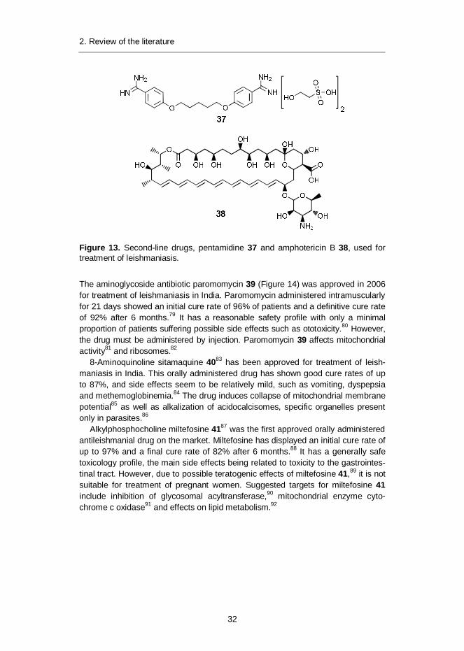
Figure 13. Second-line drugs, pentamidine 37 and amphotericin B 38, used fortreatment of leishmaniasis.
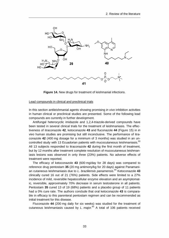
The aminoglycoside antibiotic paromomycin 39 (Figure 14) was approved in 2006for treatment of leishmaniasis in India. Paromomycin administered intramuscularlyfor 21 days showed an initial cure rate of 96% of patients and a definitive cure rateof 92% after 6 months.79 It has a reasonable safety profile with only a minimalproportion of patients suffering possible side effects such as ototoxicity.80 However,the drug must be administered by injection. Paromomycin 39 affects mitochondrialactivity81 and ribosomes.82
8-Aminoquinoline sitamaquine 4083 has been approved for treatment of leish-maniasis in India. This orally administered drug has shown good cure rates of upto 87%, and side effects seem to be relatively mild, such as vomiting, dyspepsiaand methemoglobinemia.84 The drug induces collapse of mitochondrial membranepotential85 as well as alkalization of acidocalcisomes, specific organelles presentonly in parasites.86
Alkylphosphocholine miltefosine 4187 was the first approved orally administeredantileishmanial drug on the market. Miltefosine has displayed an initial cure rate ofup to 97% and a final cure rate of 82% after 6 months.88 It has a generally safetoxicology profile, the main side effects being related to toxicity to the gastrointes-tinal tract. However, due to possible teratogenic effects of miltefosine 41,89 it is notsuitable for treatment of pregnant women. Suggested targets for miltefosine 41include inhibition of glycosomal acyltransferase,90 mitochondrial enzyme cyto-chrome c oxidase91 and effects on lipid metabolism.92
2. Review of the literature
33
Figure 14. New drugs for treatment of leishmanial infections.
Lead compounds in clinical and preclinical trials
In this section antileishmanial agents showing promising in vivo inhibition activitiesin human clinical or preclinical studies are presented. Some of the following leadcompounds are currently in further development.
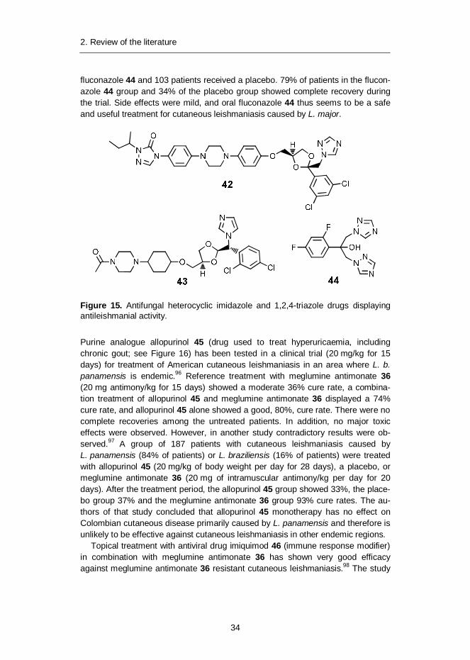
Antifungal heterocyclic imidazole and 1,2,4-triazole-derived compounds havebeen tested in several clinical trials for the treatment of leishmaniasis. The effec-tiveness of itraconazole 42, ketoconazole 43 and fluconazole 44 (Figure 15) in invivo human studies are promising but still inconclusive. The performance of itra-conazole 42 (400 mg dosage for a minimum of 3 months) was studied in an un-controlled study with 13 Ecuadorian patients with mucocutaneous leishmaniasis.93
All 13 subjects responded to itraconazole 42 during the first month of treatment,but by 12 months after treatment complete resolution of mucocutaneous leishman-iasis lesions was observed in only three (23%) patients. No adverse effects oftreatment were reported.
The efficacy of ketoconazole 43 (600 mg/day for 28 days) was compared toreference drug pentostam 35 (20 mg antimony/kg for 20 days) against Panamani-an cutaneous leishmaniasis due to L. braziliensis panamensis.94 Ketoconazole 43clinically cured 16 out of 21 (76%) patients. Side effects were limited to a 27%incidence of mild, reversible hepatocellular enzyme elevation and an asymptomat-ic, reversible, approximately 70% decrease in serum testosterone in all patients.Pentostam 35 cured 13 of 19 (68%) patients and a placebo group of 11 patientshad a 0% cure rate. The authors conclude that oral ketoconazole 43 is compara-ble in efficacy to this parenteral pentostam regimen and can be recommended asinitial treatment for this disease.
Fluconazole 44 (200 mg daily for six weeks) was studied for the treatment ofcutaneous leishmaniasis caused by L. major.95 A total of 106 patients received
2. Review of the literature
34
fluconazole 44 and 103 patients received a placebo. 79% of patients in the flucon-azole 44 group and 34% of the placebo group showed complete recovery duringthe trial. Side effects were mild, and oral fluconazole 44 thus seems to be a safeand useful treatment for cutaneous leishmaniasis caused by L. major.
Figure 15. Antifungal heterocyclic imidazole and 1,2,4-triazole drugs displayingantileishmanial activity.
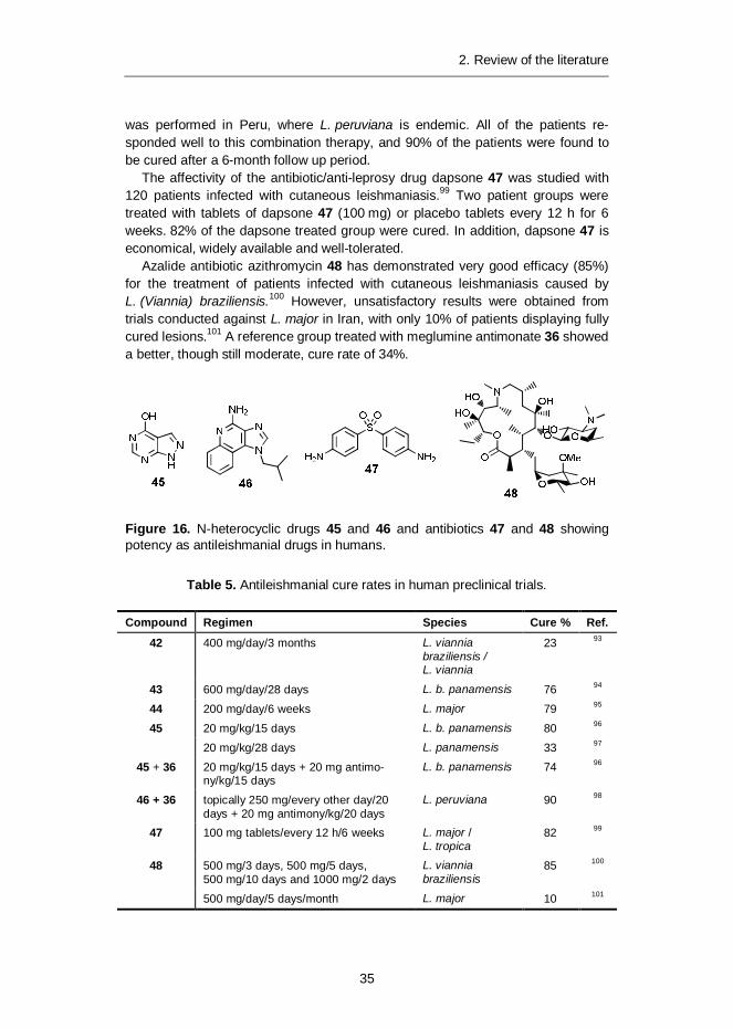
Purine analogue allopurinol 45 (drug used to treat hyperuricaemia, includingchronic gout; see Figure 16) has been tested in a clinical trial (20 mg/kg for 15days) for treatment of American cutaneous leishmaniasis in an area where L. b.panamensis is endemic.96 Reference treatment with meglumine antimonate 36(20 mg antimony/kg for 15 days) showed a moderate 36% cure rate, a combina-tion treatment of allopurinol 45 and meglumine antimonate 36 displayed a 74%cure rate, and allopurinol 45 alone showed a good, 80%, cure rate. There were nocomplete recoveries among the untreated patients. In addition, no major toxiceffects were observed. However, in another study contradictory results were ob-served.97 A group of 187 patients with cutaneous leishmaniasis caused byL. panamensis (84% of patients) or L. braziliensis (16% of patients) were treatedwith allopurinol 45 (20 mg/kg of body weight per day for 28 days), a placebo, ormeglumine antimonate 36 (20 mg of intramuscular antimony/kg per day for 20days). After the treatment period, the allopurinol 45 group showed 33%, the place-bo group 37% and the meglumine antimonate 36 group 93% cure rates. The au-thors of that study concluded that allopurinol 45 monotherapy has no effect onColombian cutaneous disease primarily caused by L. panamensis and therefore isunlikely to be effective against cutaneous leishmaniasis in other endemic regions.
Topical treatment with antiviral drug imiquimod 46 (immune response modifier)in combination with meglumine antimonate 36 has shown very good efficacyagainst meglumine antimonate 36 resistant cutaneous leishmaniasis.98 The study
2. Review of the literature
35
was performed in Peru, where L. peruviana is endemic. All of the patients re-sponded well to this combination therapy, and 90% of the patients were found tobe cured after a 6-month follow up period.
The affectivity of the antibiotic/anti-leprosy drug dapsone 47 was studied with120 patients infected with cutaneous leishmaniasis.99 Two patient groups weretreated with tablets of dapsone 47 (100 mg) or placebo tablets every 12 h for 6weeks. 82% of the dapsone treated group were cured. In addition, dapsone 47 iseconomical, widely available and well-tolerated.
Azalide antibiotic azithromycin 48 has demonstrated very good efficacy (85%)for the treatment of patients infected with cutaneous leishmaniasis caused byL. (Viannia) braziliensis.100 However, unsatisfactory results were obtained fromtrials conducted against L. major in Iran, with only 10% of patients displaying fullycured lesions.101 A reference group treated with meglumine antimonate 36 showeda better, though still moderate, cure rate of 34%.
Figure 16. N-heterocyclic drugs 45 and 46 and antibiotics 47 and 48 showingpotency as antileishmanial drugs in humans.
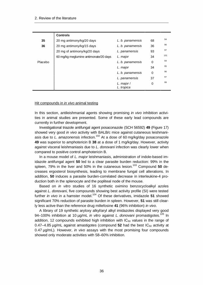
Table 5. Antileishmanial cure rates in human preclinical trials.
Compound Regimen Species Cure % Ref.
42 400 mg/day/3 months L. vianniabraziliensis /L. viannia
23 93
43 600 mg/day/28 days L. b. panamensis 76 94
44 200 mg/day/6 weeks L. major 79 95
45 20 mg/kg/15 days L. b. panamensis 80 96
20 mg/kg/28 days L. panamensis 33 97
45 + 36 20 mg/kg/15 days + 20 mg antimo-ny/kg/15 days
L. b. panamensis 74 96
46 + 36 topically 250 mg/every other day/20days + 20 mg antimony/kg/20 days
L. peruviana 90 98
47 100 mg tablets/every 12 h/6 weeks L. major /L. tropica
82 99
48 500 mg/3 days, 500 mg/5 days,500 mg/10 days and 1000 mg/2 days
L. vianniabraziliensis
85 100
500 mg/day/5 days/month L. major 10 101
2. Review of the literature
36
Controls35 20 mg antimony/kg/20 days L. b. panamensis 68 94
36 20 mg antimony/kg/15 days L. b. panamensis 36 96
20 mg of antimony/kg/20 days L. panamensis 93 97
60 mg/kg meglumine antimonate/20 days L. major 34 101
Placebo L. b. panamensis 0 94
L. major 34 95
L. b. panamensis 0 96
L. panamensis 37 97
L. major /L. tropica
0 99
Hit compounds in in vivo animal testing
In this section, antileishmanial agents showing promising in vivo inhibition activi-ties in animal studies are presented. Some of these early lead compounds arecurrently in further development.
Investigational triazole antifungal agent posaconazole (SCH 56592) 49 (Figure 17)showed very good in vivo activity with BALB/c mice against cutaneous leishmani-asis due to L. amazonensis infection.102 At a dose of 60 mg/kg/day posaconazole49 was superior to amphotericin B 38 at a dose of 1 mg/kg/day. However, activityagainst visceral leishmaniasis due to L. donovani infection was clearly lower whencompared to positive control amphotericin B.
In a mouse model of L. major leishmaniasis, administration of indole-based im-idazole antifungal agent 50 led to a clear parasite burden reduction: 99% in thespleen, 79% in the liver and 50% in the cutaneous lesion.103 Compound 50 de-creases ergosterol biosynthesis, leading to membrane fungal cell alterations. Inaddition, 50 induces a parasite burden-correlated decrease in interleukine-4 pro-duction both in the splenocyte and the popliteal node of the mouse.
Based on in vitro studies of 16 synthetic oximino benzocycloalkyl azolesagainst L. donovani, five compounds showing best activity profile (SI) were testedfurther in vivo in a hamster model.104 Of these derivatives, imidazole 51 showedsignificant 70% reduction of parasite burden in spleen. However, 51 was still clear-ly less active than the reference drug miltefosine 41 (96% inhibition) in vivo.
A library of 19 synthetic aryloxy alkyl/aryl alkyl imidazoles displayed very good94–100% inhibition at 10 µg/mL in vitro against L. donovani promastigotes.105 Inaddition, 12 compounds exhibited high inhibition with IC50 values in the range of0.47–4.85 µg/mL against amastigotes (compound 52 had the best IC50 activity at0.47 µg/mL). However, in vivo assays with the most promising four compoundsshowed only moderate activities with 58–60% inhibition.
2. Review of the literature
37
Figure 17. Triazole and imidazole-based antileishmanial compounds.
Pyrazinamide 53 (Figure 18), which is used in tuberculosis chemotherapy, showedgood efficacy in the treatment of L. major-infected mice with a 100-fold reductionin parasite burden, when compared to the control.106 In addition, mechanistic stud-ies suggest that pyrazinamide 53 enhances effective immune responses againstthe parasite and has an immunostimulatory effect. Treatment was non-toxic anddid not affect the growth of the experimental animals.
Two novel arylimidamide class antileishmanial drug candidates, DB745 and DB76654 showed exceptional activity against intracellular L. donovani, L. amazonensis, andL. major in vitro.107 In vivo orally given, DB766 54 produced a dose-dependentinhibition of liver parasitemia in two efficacy models, L. donovani-infected miceand hamsters, (71% and 89%, respectively). A marked reduction in parasitemia inthe spleen (79%) and bone marrow (92%) of hamsters was also observed. Fur-thermore, the compounds were well distributed in the liver and spleen target tis-sues, showed moderate oral bioavailability (up to 25%), and had a suitable elimi-nation half-life ranging from 1 to 2 days in mice. No toxic side effects to liver orkidney were observed, although mild hepatic cell eosinophilia, hypertrophy, andfatty changes were noted. The results demonstrated that arylimidamides are apromising class of compounds for preclinical development as an orally adminis-tered drug.
The conventional dihydropyridine antihypertensive drugs amlodipine 55 and la-cidipine 56 inhibited L. donovani infection in vitro and in mice when administeredorally.108 Amlodipine 55 and lacidipine 56 therapies led to significant reductions insplenic (85% and 75%) and liver (86% and 72%) parasite burdens, when com-pared to controls. The compounds functioned through dose-dependent inhibitionof oxygen consumption, triggering caspase 3-like activation-mediated programmedcell death of the parasites.
2. Review of the literature
38
A series of 2,4,6-trisubstituted pyrimidines and 1,3,5-triazines were synthesizedand screened for in vitro and in vivo antileishmanial activity against L. donovani.109
Three compounds, 57, 58 and 59, showed a good selectivity index (SI) in vitro,and these were screened for in vivo activity in golden hamsters infected withL. donovani. The compounds showed decent in vivo inhibition of 48–56% at adose of 50 mg/kg when administered intraperitoneally.
Figure 18. Six-membered nitrogen-containing heterocycles with in vivo antileish-manial activity.
A sitamaquine derivative of 8-aminoquinoline (±)NPC1161B 60 (Figure 19) and pure( )- and (+)-enantiomers were studied in vivo with L. donovani-infected mice.110 Allthree components (dose 10 mg/kg/day) showed very good and comparable activityto the parent compound sitamaquine, and cleared the parasites after a 5-day courseof treatment. ( )-Enantiomer was better tolerated and had an increased therapeuticwindow when compared to the racemate or (+)-enantiomer of the compound.
Oral administration of natural quinoline-based compound chimanine B 61 (twicea day 50 mg/kg) to mice infected with L. amazonensis or L. venezuelensis resultedin a decrease in lesion weight by 70% and parasite loads by 95% when comparedto the group of untreated mice.111 Injections of chimanine B 61 (five injections atfour-day intervals) and subcutaneous administration or intralesional injections ofreference drug meglumine antimonate 36 displayed similar and very good efficacy.In the follow up study, the efficacy of nine chimanine B derivatives was determined
2. Review of the literature
39
in a murine model.112 Activity was further improved by the hydroxy derivative 62,showing antileishmanial activity up to 90% in in vivo rat models.
A series of synthetic bis-quinolines showed excellent antileishmanial efficacyagainst L. donovani in both in vitro and in vivo studies.113 Compound 63 exhibited themost significant activity against visceral leishmaniasis in a mouse model without show-ing any toxic manifestation. Intraperitoneal treatment with compound 63 at 12.5 mg/kgbody weight led to significant reduction of parasite burden in spleen (95%) and liver(98%) compared to untreated controls. Ultrastructural studies of treated promastigotesdemonstrated membrane blebbing, chromatin condensation and vacuolization in theparasites, and flagellated parasites became round-shaped after treatment.
The quinazoline derivative peganine hydrochloride dihydrate 64 isolated fromPeganum harmala L. seeds showed only moderate in vitro activity against bothextracellular promastigotes as well as intracellular L. donovani amastigotes grow-ing inside macrophages.114 However, the alkaloid 64 administered by oral routeexhibited good in vivo activity with 80% reduction of L. donovani parasites in ham-ster spleen at a dose of 100 mg/kg. The reference drug miltefosine 41 resulted in96% inhibition of parasites at a dose of 40 mg/kg.
The quaternary isoquinoline alkaloid berberine 65 and several of its derivativeswere tested for efficacy against L. donovani and L. braziliensis panamensis in goldenhamsters.115 Tetrahydroberberine 66 was the most potent derivative against L. donovaniwith 50% suppression of parasite burden, but was not as potent as the reference drugmeglumine antimonate 36. Only berberine 65 and 8-cyanodihydroberberine 67 showedsignificant activity (>50% suppression of lesion size) against L. braziliensis pana-mensis.
Figure 19. Quinoline-derived compounds showing antileishmanial activity in vivo.
2. Review of the literature
40
Various synthetic rhodacyanine derivatives were studied for their antileishmanial invitro and in vivo activities.116 Among the derivatives, the fluorinated variant SJL-0168 (Figure 20) showed an excellent in vitro selectivity index of >15,000 and an IC50
value of 0.011 M against L. donovani. The fluorinated compound 68 displayed anexceptional 95% inhibition against L. donovani parasites in female mice by1.3 mg/kg intravenous administrations. Preliminary studies showed that no bioa-vailability was obtained by subcutaneous administration.
In vitro and in vivo (mice) activities of antiarrhythmic amiodarone 69 andmiltefosine 41 were investigated alone or in combination on L. mexicana.117 It wasfound that whereas both drugs given individually failed to cure lesions, a combina-tion of amiodarone 69 and miltefosine 41 had synergistic effects on the prolifera-tion of intracellular amastigotes growing inside macrophages and led to 90% para-sitological cures in a murine model. Amiodarone 69 disrupts intracellular Ca2+
homeostasis and inhibits the de novo sterol biosynthesis of the parasite.
Figure 20. Structures of potential antileishmanial compounds rhodacyanine deriv-atives 68 and 69.
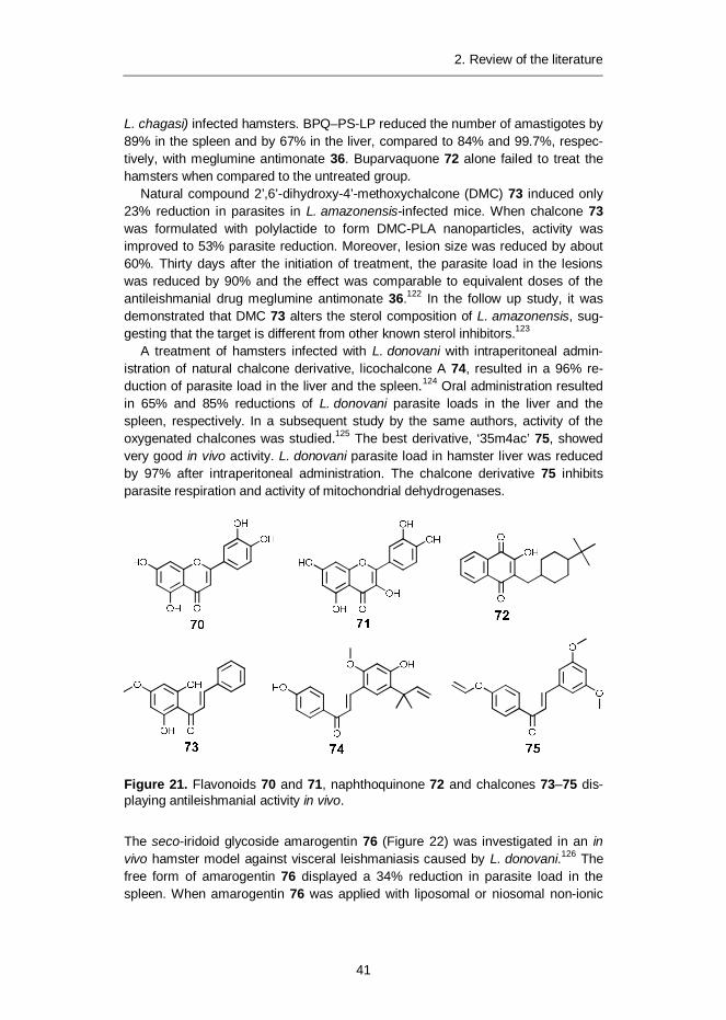
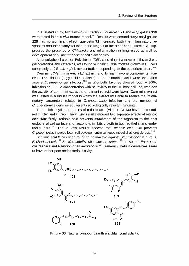
The common flavonoids luteolin 70 and quercetin 71 (Figure 21) were assayed invivo for visceral leishmaniasis against L. donovani-infected golden hamsters.118
Luteolin 70 showed good activity by an over 80% reduction in splenic parasites.Quercetin 71 reduced the splenic parasite load by 90% at four times higher con-centration. In vitro studies suggested that leishmanicidal activity was related toinhibition of promastigotes DNA synthesis and promotion of topoisomerase II-mediated linearization of kDNA minicircles. Quercetin 71 was almost equally po-tent to the standard antileishmanial drug sodium stibogluconate 35 in lowering theparasite load in the spleen of L. donovani-infected hamsters (reductions 77% and82%, respectively).119 Combination therapy with quercetin 71 and stibogluconate35 showed improved synergistic activity with a 93% reduction of parasites in ham-ster spleen.
Hydroxynaphthoquinone-based buparvaquone 72 showed moderate antileish-manial activity in L. donovani infected mice at very high concentration.120 Potencyof buparvaquone 72 was increased several-fold with the formulation containingbuparvaquone 72 (BPQ) and phosphatidylserine (PS) entrapped in liposomes(BPQ–PS-LP).121 BPQ–PS-LP was evaluated in vivo against L. infantum (syn.
2. Review of the literature
41
L. chagasi) infected hamsters. BPQ–PS-LP reduced the number of amastigotes by89% in the spleen and by 67% in the liver, compared to 84% and 99.7%, respec-tively, with meglumine antimonate 36. Buparvaquone 72 alone failed to treat thehamsters when compared to the untreated group.
Natural compound 2’,6’-dihydroxy-4’-methoxychalcone (DMC) 73 induced only23% reduction in parasites in L. amazonensis-infected mice. When chalcone 73was formulated with polylactide to form DMC-PLA nanoparticles, activity wasimproved to 53% parasite reduction. Moreover, lesion size was reduced by about60%. Thirty days after the initiation of treatment, the parasite load in the lesionswas reduced by 90% and the effect was comparable to equivalent doses of theantileishmanial drug meglumine antimonate 36.122 In the follow up study, it wasdemonstrated that DMC 73 alters the sterol composition of L. amazonensis, sug-gesting that the target is different from other known sterol inhibitors.123
A treatment of hamsters infected with L. donovani with intraperitoneal admin-istration of natural chalcone derivative, licochalcone A 74, resulted in a 96% re-duction of parasite load in the liver and the spleen.124 Oral administration resultedin 65% and 85% reductions of L. donovani parasite loads in the liver and thespleen, respectively. In a subsequent study by the same authors, activity of theoxygenated chalcones was studied.125 The best derivative, ‘35m4ac’ 75, showedvery good in vivo activity. L. donovani parasite load in hamster liver was reducedby 97% after intraperitoneal administration. The chalcone derivative 75 inhibitsparasite respiration and activity of mitochondrial dehydrogenases.
Figure 21. Flavonoids 70 and 71, naphthoquinone 72 and chalcones 73–75 dis-playing antileishmanial activity in vivo.
The seco-iridoid glycoside amarogentin 76 (Figure 22) was investigated in an invivo hamster model against visceral leishmaniasis caused by L. donovani.126 Thefree form of amarogentin 76 displayed a 34% reduction in parasite load in thespleen. When amarogentin 76 was applied with liposomal or niosomal non-ionic
2. Review of the literature
42
surfactant vesicles, its activity was improved considerably with a parasite loadreduction in the spleen of 90% and 69%, respectively.
Argentilactone 77 showed very good in vivo antileishmanial activity when ad-ministered to L. amazonensis-infected mice by oral or subcutaneous routes.127
Argentilactone showed the same efficacy as the reference drug meglumine anti-monate 36, reducing parasite load in lesions by 96% and in the spleen by 50%.
Figure 22. Lactone derivatives having antileishmanial activity in vivo.
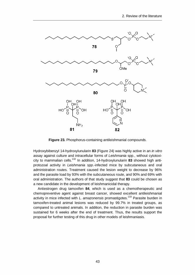
Four potential anticancer alkyl-lysophopholipids were assayed by oral administra-tion against two strains of L. donovani in a mouse model.128 The currently ap-proved antileishmanial drug miltefosine 41 displayed the best parasitic reduction inliver (97% and 99% inhibition against strain LV9 and Patna I) at a 30 mg/kg dose.Ilmofosine 78 (Figure 23) displayed good activity with 67% and 92% inhibition.Edelfosine 79 was moderately active. In a study by Cabrera-Serra et al., miltefo-sine-related derivatives edelfosine 79 and perifosine 80 were orally administeredto L. amazonensis-infected mice.129 The edelfosine-treated mouse group showed49% and perifosine 80 38% inhibition in footpad lesions. Biopsies obtained frommice treated with edelfosine 79 showed a 19% parasitic burden when compared tothe non-treated control. In perifosine-treated mice, the parasitic burden was only7% when compared to the control. The researchers concluded that perifosine 80should be studied further in preclinical studies.
In vivo intraperitoneal administration of the osteoporosis bisphosphonate drugpamidronate 81 to mice infected with cutaneous leishmaniasis caused byL. mexicana amazonensis resulted in long-term disappearance of lesions.130 Inanother study, pamidronate 81 and related bisphosphonate risedronate 82 werestudied against L. donovani.131 Intravenous administration of pamidronate 81 andrisedronate 82 was found to inhibit L. donovani amastigotes parasite burden inmouse liver by 92% and 99%, respectively. However, at high doses of risedronate82 toxicity was also observed.
Hydroxybibenzyl 14-hydroxylunularin 83 (Figure 24) was highly active in an in vitroassay against culture and intracellular forms of Leishmania spp., without cytotoxi-city to mammalian cells.132 In addition, 14-hydroxylunularin 83 showed high anti-protozoal activity in Leishmania spp.-infected mice by subcutaneous and oraladministration routes. Treatment caused the lesion weight to decrease by 96%and the parasite load by 93% with the subcutaneous route, and 90% and 69% withoral administration. The authors of that study suggest that 83 could be chosen asa new candidate in the development of leishmanicidal therapy.
Antiestrogen drug tamoxifen 84, which is used as a chemotherapeutic andchemopreventive agent against breast cancer, showed excellent antileishmanialactivity in mice infected with L. amazonensis promastigotes.133 Parasite burden intamoxifen-treated animal lesions was reduced by 99.7% in treated groups, ascompared to untreated animals. In addition, the reduction in parasite burden wassustained for 6 weeks after the end of treatment. Thus, the results support theproposal for further testing of this drug in other models of leishmaniasis.
2. Review of the literature
44
Figure 24. Antileishmanial compounds 14-hyroxylunularin 83 and antiestrogenagent tamoxifen 84.
Natural oleane triterpene saponin maesabalide III 85 (MB-III, PX-6518, Figure 25)demonstrated in vivo activity against L. donovani in golden hamsters.134 It wasconcluded that administration of a single dose of MB-III has efficacy comparable tothat of a single dose of liposomal amphotericin B 38. However, severe toxicity wasobserved as several animals died during the experiment series and further devel-opment is required. Activity of MB-III 85 was further evaluated in vivo with miceinfected with L. mexicana, L. panamensis or L. major.135 MP-III 85 completelyhealed L. mexicana and L. panamensis lesions, whereas L. major lesions werereduced by 50%, thus demonstrating broad-spectrum curative efficacy. In thefollow up study, the same authors synthesized several semisynthetic MB-III de-rivatives to study the structure-activity relationships, but in in vitro studies none ofthe derivatives showed increased activity compared to 85.136
Monoterpenoid cantharidin 86 is a natural poisonous terpenoid secreted bymale blister beetles (Lytta vesicatoria). The effect of different doses of cantharidin86 on L. major were investigated both in vitro (promastigote and amastigote viability)and in infected mice (skin lesions) using ointment or soluble cantharidin. Twoweeks of topical treatment with 0.1% cantharidin ointment was an effective methodfor treating cutaneous leishmaniasis in infected mice and skin lesions were totallyhealed. However, in the follow up study, after two months a relapse of lesions wasobserved for two mice out of eight.
The widely used antimalarial drug sesquiterpene lactone, which contains anunusual peroxide bridge, artemisinin 87, demonstrated only moderate antileish-manial activity against promastigotes of a wide range of Leishmania species,including strains responsible for visceral leishmaniasis (L. donovani, L. infantum),cutaneous leishmaniasis (L. tropica, L. mexicana) and mucocutaneous leishmani-asis (L. amazonensis, L. braziliensis) promastigotes, the IC50 value being approx.100 µM.137 However, in a mouse model of visceral leishmaniasis, orally adminis-tered artemisinin 87 (10 mg/kg and 25 mg/kg body weight) effectively reducedboth splenic weight and parasite burden by 83% and 86%, respectively. The refer-ence drug sodium stibogluconate 35 demonstrated a similar significant 85% de-crease in parasite burden.
2. Review of the literature
45
O
OO
OO
OH
OO
OO
O
O
O
O
O
O
HO
HOHOHOOC
OHHO
HOOH
HOHO
HOHO OH
OH
OH
85
O
O
O O
87
O
O
O
O
86
Figure 25. Terpenes and terpenoids possessing antileishmanial activity in vivo.
Betulin-derived compounds
In a study by Sauvain et al., betulinic aldehyde 88 (Figure 26), obtained from Doli-ocarpus dentatus (Aubl.) Stand. showed weak in vitro activity againstL. amazonensis amastigotes with a survival index of 12% at 136 µM and 42% at68 µM.138 However, at these doses 88 also showed toxicity against peritonealmacrophages with survival indices of 70% and 80%, respectively. At a concentra-tion of 34 µM, aldehyde 88 was ineffective against L. amazonensis as well as non-toxic to macrophages.
In another study, in which a library of natural compounds was screened for an-tileishmanial activity, betulinic acid 2 was found to be weakly active againstL. major with an IC50 value of 88 µM.139 It was also noted that in triterpenes havinga ursane, oleanane or lupane skeleton, a carboxy substituent was required forantileishmanial activity.
In a study by Chowdhury et al., dihydrobetulinic acid 89 was found to inhibitgrowth of L. donovani promastigotes and amastigotes with an IC50 value of 2.6and 4.1 µM, respectively.140 It acts by targeting DNA topoisomerase I and II, pre-venting DNA cleavage and formation of the enzyme-DNA complex, ultimatelyinducing apoptosis. Leishmanial parasitic burden in golden hamsters was reducedby 92% after 6-week treatment with dihydrobetulinic acid 89 (10 mg/kg bodyweight). In a subsequent study by the same authors, six betulin and six dihydrobetu-lin esters were assayed against sodium stibogluconate sensitive and resistant
2. Review of the literature
46
L. donovani strains in vitro.141 Derivatives having small ester groups were active againstboth L. donovani strains. 3,28-Disuccinoylbetulin 90, 3,28-disuccinoyldihydrobetulin 91and 3,28-diglutaroylbetulin 92 showed the best inhibition activity against amastigotegrowth inside macrophages, with EC50 values between 6.1 and 10.5 µM. Thesuggested biochemical mechanism of these betulin esters was similar to dihy-drobetulinic acid 89, as they inhibited in a reversible manner the relaxation activityof enzyme type IB topoisomerase of the parasite as well as topoisomerase I–DNAcomplex formation.
Figure 26. Betulin derivatives with antileishmanial activity.
2.4 Chlamydia pneumoniae
2.4.1 Species, taxonomy and life cycle
The order Chlamydiales comprises four families and 13 species, with theC. pneumoniae species belonging to Family I: Chlamydiaceae and Genus I:Chlamydia (Table 6).142 In addition to C. pneumoniae, C. trachomatis is the onlyother bacterial species in the order Chlamydiales that infects humans. C. tracho-matis causes urogenital infections and also trachoma, which causes roughening ofthe inner eyelids and can lead to blindness. The remaining species are infectiousto animals only, including insects, birds and other mammals.
2. Review of the literature
47
Table 6. Taxonomy of Chlamydia pneumoniae.
Order Chlamydiales
Family Chlamydiaceae
Genus I Chlamydia
Species Chlamydia pneumoniae
The chlamydial life cycle consists of two distinct morphological forms: the elemen-tary body (EB) and reticulate body (RB). EB is the metabolically inactive, infectiveextracellular stage, and RB the metabolically active, replicating, intracellular form.
In the C. pneumoniae life cycle (Figure 27):143
1) EB attaches to the host cell (yet unknown receptors).
2) Entry of EB to host cell via phagocytosis144 or receptor-mediated endocytosis.145
3) Inside the host cell EB is surrounded by double-membrane inclusion. EB isconverted to RB.
4) Inclusion is enlarged and filled with new replicates of RB.
5) RBs are converted back to infectious EBs and exit of the EBs can occur vialysis of the host cell or via extrusion of the EB packed inclusion.
Figure 27. Life cycle of C. pneumoniae. A = C. pneumoniae elementary body(EB). B = Host cell, usually lung cell.146
Chlamydial persistence can last for years and it is defined as a viable but non-cultivable growth stage inside the infected host cell.142 In the persistence stage,C. pneumoniae is not metabolically active and not replicating. Persistence iscaused by several stress factors that are unfavourable for bacterial growth, suchas insufficient nutrients (e.g. amino acids), iron depletion, non-lethal antibiotics,heat and growth in monocytes.147 In all cases, the developmental cycle is haltedand the RBs are not converted back to the infective extracellular EB stage. Insteadof the normal round shape of EBs, enlarged aberrant bodies (AB) are formed. Thisis clinically highly relevant, since a persistent culture cannot be subcultured fordiagnostic purposes. However, chlamydial persistence can be detected fromchlamydial DNA or marker proteins in the absence of viable Chlamydiae.148 It hasbeen suggested that C. pneumoniae can persist for many years after initial respir-atory infection.149
2.4.3 Chemotherapy against infections caused by C. pneumoniae
C. pneumoniae is susceptible to antibiotics that interfere with DNA and proteinsynthesis, including tetracyclines, macrolides, quinolones and rifamycins.16 Theseclasses of antibiotics are most widely studied and used for treatment of humaninfections. However, C. pneumoniae is resistant to some common antibiotic classes,such as trimethoprim, sulphonamides, aminoglycosides and glycopeptides.150
Clinical manifestation and treatment
Asymptomatic infection or unrecognized, mildly symptomatic illnesses, such asprolonged cough and mild fever, are very common symptoms of C. pneumoniaeinfection.151 Severe systemic infections with C. pneumoniae alone are not common.Even in mild cases, complete recovery is slow, despite appropriate antibiotic therapy,and cough and malaise may persist for many weeks after the acute illness. It hasbeen estimated that 10% of cases of pneumonia and approximately 5% of bron-chitis and sinusitis cases in adults have been attributed to this organism.152 Inaddition, there is strong evidence that C. pneumoniae is associated with othersevere diseases, such as atherosclerotic cardiovascular diseases as well as someneurodegenerative diseases, such as Alzheimer’s disease and multiple sclerosis.17,18
The intracellular location of C. pneumoniae limits and hampers the efficacy ofsome antibiotics. The antichlamydial agent must penetrate several biologicalmembranes in therapeutic concentrations: the host cell membrane, the inclusionmembrane, and, finally, the inner and outer membrane of the Gram-negative bac-terium itself. Thus, antimicrobial agents need good intracellular penetration proper-ties to be effective. The following regimen guidelines (Table 7) for adults can befollowed in clinical treatment of C. pneumoniae infection.16 However, some patientsmay need re-treatment.
2. Review of the literature
49
Table 7. Current treatment guidelines for the treatment of respiratory infection dueto C. pneumoniae.16
Antibiotic class Drug Dosing Regimen
Fluoroquinolone Levofloxacin 101 500 mg once a day Orally or intravenously7–14 days
Moxifloxacin 103 400 mg once a day Orally 10 days
Tetracycline Doxycycline 112 100 mg twice a day Orally 14–21 days
Tetracycline 114 250 mg four times a day Orally 14–21 days
Macrolide Azithromycin 115 1.5 g once a day Orally 5 days
Clarithromycin 116 500 mg twice a day Orally 10 days
Fluoroquinolones
Many of the commonly used fluoroquinolone antibiotics show very good in vitroantichlamydial activity with low micromolar MIC values. The fluoroquinolone classof antibiotics share a quinoline ring system with a fluorine atom at C-6. Piperazineor another ring system is attached to C-7. Fluoroquinolones block DNA replicationand repair by inhibiting topoisomerase II function.153
The chemical structures of fluoroquinolone antibiotics and some experimentalfluoroquinolones (and a few quinolones) screened in vitro against C. pneumoniaeare presented in Figure 28 and their in vitro MIC values in Table 8. In most of thecases, susceptibility of C. pneumoniae to antibiotics is tested using a HEp-2 cellline or, in some cases, a HeLa 229 cell line, as a host. Several antibiotics, such asgarenoxacin 97 (MIC 0.015–0.03 µg/mL), sitafloxacin 107 (MIC 0.031–0.0125 µg/mL)and experimental BMS-284756 94 (MIC 0.015–0.03 µg/mL) show very good invitro activities (Table 8). The MIC values from different studies are very consistent,and usually only small variations can be seen between HEp-2 or HeLa 229 hostcell lines. The current recommendation for human treatment is to use levofloxacin101 (MIC 0.25–1 µg/mL) or moxifloxacin 103 (MIC 0.06–1 µg/mL) as the first-choice antibiotic of this class.16
2. Review of the literature
50
Figure 28. Chemical structures of the fluoroquinolone and quinolone classes ofantibiotics with antichlamydial activity.
2. Review of the literature
51
Table 8. In vitro efficacy of fluoroquinolones and some quinolones againstC. pneumoniae.
Quinolone or fluoroquinolone MIC range µg/mL Cell line Ref.
BAY 12-8039 93 0.5–1 HEp-2 154
BMS-284756 94 0.015–0.03 HEp-2 155
0.004–0.008 HEp-2 156
Ciprofloxacin 95 1.0–4.0 HEp-2 157
0.25–4 HeLa 229, HEp-2 158
Fleroxacin 96 2–8 HeLa 229, HEp-2 158
Garenoxacin 97 0.008 HEp-2 159
0.015–0.03 HEp-2 160
Gatifloxacin (AM-1155) 98 0.125 HEp-2 159
0.125–0.25 HEp-2 161
0.063–0.125 HeLa 229 162
Gemifloxacin 99 0.125–0.25 HEp-2 163
Grepafloxacin 100 0.063–0.125 HeLa 229 162
Levofloxacin 101 0.5–1.0 HEp-2 155
0.25 HEp-2 159
0.5 HEp-2 156
Lomefloxacin 102 2 * 164
Moxifloxacin 103 0.125–1.0 HEp-2 155
0.06–0.12 HEp-2 156
0.063 HEp-2 159
Norfloxacin 104 16 * 164
Ofloxacin 105 0.5–2.0 HEp-2 157
0.5–2.0 HEp-2 165
0.5–1 HEp-2 156
L-Ofloxacin 0.125–0.5 HeLa 229, HEp-2 166
OPC-17116 106 0.25–0.5 HEp-2 165
Sitafloxacin (DU-6859a) 107 0.031–0.125 HEp-2 157
0.031–0.063 HeLa 229 162
Sparfloxacin 108 0.031–0.125 HEp-2 157
0.016 HEp-2 159
0.06–0.25 HEp-2 165
0.06–0.25 HeLa 229, HEp-2 158
Temafloxacin 109 0.125–1 HeLa 229, HEp-2 158
Tosulfloxacin 110 0.063–0.25 HEp-2 157
0.125 * 164
Trovafloxacin 111 0.5–1 HEp-2 167
* Cell line not mentioned in publication.
2. Review of the literature
52
Tetracyclines
The first tetracycline antibiotics, chlortetracycline and oxytetracycline, were dis-covered in the late 1940s and they are produced by the bacteria Streptomycesaureofaciens and S. rimosus, respectively.168 Tetracycline antibiotic moleculesconsist of four linearly fused rings, to which a variety of functional groups are at-tached. Tetracyclines inhibit bacterial protein synthesis by preventing the associa-tion of aminoacyl-tRNA with the bacterial ribosome.169
The chemical structures of tetracycline antibiotics, natural tetracycline 114, andsemisynthetic derivatives doxycycline 112 and minocycline 113, screened in vitroagainst C. pneumoniae, are presented in Figure 29, and their in vitro MIC valuesusing HEp-2 or HeLa 229 host cell lines are given in Table 9. All three compoundsdisplay good efficacy (MIC 0.05–0.25 µg/mL) against C. pneumoniae. Currently,doxycycline 112 and tetracycline 114 are recommended antibiotics for treatment ofchlamydial infection in humans.16
Figure 29. Chemical structures of tetracycline-class antibiotics with antichlamydialactivity.
Table 9. In vitro efficacy of tetracyclines against C. pneumoniae.
Tetracycline MIC range Cell line Ref.
Doxycycline 112 0.06–0.25 HEp-2 166
Minocycline 113 0.0625 HEp-2 170
Tetracycline 114 0.05–0.1 HeLa 229 171
Macrolides
Macrolide antibiotics are usually used to treat infections caused by Gram-positivebacteria.172 The macrolide antibiotics have a large, usually 14-, 15-, or 16-membered macrocyclic lactone ring with one or more deoxy sugars attached.Macrolides belong to the polyketide class of natural products, and they act byinhibiting the bacterial protein synthesis machinery, the ribosome.
The chemical structures of macrolide antibiotics and some experimental macro-lides screened in vitro against C. pneumoniae are presented in Figure 30, andtheir in vitro MIC values using a HEp-2 host cell line are given in Table 10.
2. Review of the literature
53
Azithromycin 115, clarithromycin 116 and erythromycin 118 display very goodantichlamydial in vitro inhibition activity (MIC 0.004–0.5 µg/mL), whereas dirithro-mycin and roxithromycin show slightly diminished activities. MIC results from dif-ferent studies are very consistent. The current recommendation for human treat-ment is to use azithromycin 115 (MIC 0.06–0.5 µg/mL) or clarithromycin 116 (MIC0.004–0.063 µg/mL) as the first-choice antibiotic of this class.16
Figure 30. Chemical structures of macrolide-class antibiotics with antichlamydial activity.
Table 10. In vitro efficacy of macrolides against C. pneumoniae.
Macrolide MIC range Cell line Ref.
Azithromycin 115 0.125–0.5 HEp-2 173
0.06–0.25 HEp-2 166
Clarithromycin 116 0.016–0.063 HEp-2 173
0.015–0.06 HEp-2 155
0.004–0.03 HEp-2 166
Dirithromycin 117 0.5–40 HEp-2 174
Erythromycin 118 0.063–0.25 HEp-2 173
0.016–0.5 HEp-2 174
Roxithromycin 119 0.0625–2 HEp-2 175
2. Review of the literature
54
Ketolides
Ketolide antibiotics are broad spectrum 14-membered semi-synthetic derivatives ofthe macrolide erythromycin A.176 Main modifications are replacement of the neutralL-cladinose sugar at position C-3 in erythromycin by a 3-ketone carbonyl, and at-tachment of a cyclic carbamate group in the lactone ring. These structural modifica-tions give ketolides improved pharmacodynamic and pharmacokinetic properties,such as improved acid stability, reduced risk of resistance induction and higher lipo-philicity when compared with 14-membered macrolides.177 Similarly to macrolides,ketolides act by inhibiting the bacterial protein synthesis machinery, the ribosome.
Figure 31 presents the chemical structures of ketolide antibiotics and some ex-perimental ketolides screened in vitro against C. pneumoniae, and Table 11shows their in vitro MIC values using HEp-2 host cells. Cethromycin 120 (MIC0.008–0.031 µg/mL) and telithromycin 121 (MIC 0.031–0.25 µg/mL) show verygood antichlamydial efficacy. However, none of the ketolide antibiotics are includ-ed in current treatment recommendations for humans.16
Figure 31. Chemical structures of ketolide-class antibiotics with antichlamydial activity.
2. Review of the literature
55
Table 11. In vitro efficacy of ketolides against C. pneumoniae.
The molecular structure of rifamycins consists of an aromatic moiety bridged atnonadjacent positions by an aliphatic chain.180 The aromatic moiety can be anaphthalene or naphthoquinone ring system, as in rifamycin, or a benzene orbenzoquinone ring. The rifamycins were first isolated by Sensi and co-workers in1959.181
The rifamycins display a broad spectrum of antibiotic activity, especially againstGram-positive bacteria.180 After evaluation of a large number of semisyntheticanalogues, rifampicin 126 with excellent oral bioavailability was developed. Rifam-picin 126 shows more pronounced activity against Gram-positive bacteria, in par-ticularly mycobacteria. In addition, activity against Gram-negative bacteria is im-proved. The antibacterial action of rifampicin 126 results from its inhibition of DNA-dependent RNA synthesis.182
The chemical structures of semisynthetic rifamycin antibiotic derivativesscreened in vitro against C. pneumoniae are presented in Figure 32, and their invitro MIC values using a HEp-2 host cell line are given in Table 12. All compoundsdisplay excellent efficacy (MIC 0.00125–0.02 µg/mL) against C. pneumoniae,however, rifamycins are currently not recommended for antichlamydial antibioticsin humans.16
2. Review of the literature
56
Figure 32. Chemical structures of rifamycin-class antibiotics with antichlamydial activity.
Table 12. In vitro efficacy of rifamycins against C. pneumoniae.
Rifamycins MIC range Cell line Ref.
ABI-1131 124 0.00125–0.0025 HEp-2 183
Rifalazil (ABI-1648) 125 0.00125–0.0025 HEp-2 183
Rifampicin 126 0.005 HEp-2 184
Rifapentin 127 0.04–0.02 HEp-2 185
Natural non-antibiotic-derived compounds
Little research has been conducted on the antichlamydial properties of non-antibiotic natural compounds.
In a study by Alvesalo et al., a library of 57 natural phenolic compounds or theirderivatives were tested against C. pneumoniae in vitro using HL cells.186 Thecompounds included flavones, flavonols, flavonones, isoflavonones, coumarins,catechins, phenolic acids, gallates, a stilbene or their semisynthetic derivatives.Active compounds were found in several compound groups, especially in gallates(dodecyl gallate 128, MIC = 18 µM), but also flavones (luteolin 70 MIC = 8.8 µM)and flavonols (myricetin 131, MIC = 29 µM) with no cytotoxicity to host cells(Figure 33).
2. Review of the literature
57
In a related study, two flavonoids luteolin 70, quercetin 71 and octyl gallate 129were tested in an in vivo mouse model.187 Results were contradictory: octyl gallate129 had no significant effect; quercetin 71 increased both the inflammatory re-sponses and the chlamydial load in the lungs. On the other hand, luteolin 70 sup-pressed the presence of Chlamydia and inflammation in lung tissue as well asdevelopment of C. pneumoniae-specific antibodies.
A tea polyphenol product “Polyphenon 70S”, consisting of a mixture of flavan-3-ols,gallocatechins and catechins, was found to inhibit C. pneumoniae growth in HL cellscompletely at 0.8–1.6 mg/mL concentration, depending on the bacterium strain.188
Corn mint (Mentha arvensis L.) extract, and its main flavone components, aca-cetin 132, linarin (diglycoside acacetin); and rosmarinic acid were evaluatedagainst C. pneumoniae infection.189 In vitro both flavones showed roughly 100%inhibition at 100 µM concentration with no toxicity to the HL host cell line, whereasthe activity of corn mint extract and rosmarinic acid were lower. Corn mint extractwas tested in a mouse model in which the extract was able to reduce the inflam-matory parameters related to C. pneumoniae infection and the number ofC. pneumoniae genome equivalents at biologically relevant amounts.
The antichlamydial properties of retinoic acid (Vitamin A) 130 have been stud-ied in vitro and in vivo. The in vitro results showed two separate effects of retinoicacid 130: firstly, retinoic acid prevents attachment of the organism to the hostendothelial cell surface and, secondly, inhibits growth in both epithelial and endo-thelial cells.190 The in vivo results showed that retinoic acid 130 preventsC. pneumoniae-induced foam cell development in a mouse model of atherosclerosis.191
Betulinic acid 2 has been found to be inactive against Staphylococcus aureus,Escherichia coli,192 Bacillus subtilis, Micrococcus luteus,193 as well as Enterococ-cus faecalis and Pseudomonas aeruginosa.194 Generally, betulin derivatives seemto have rather poor antibacterial activity.
Figure 33. Natural compounds with antichlamydial activity.
3. Aims of the study
58
3. Aims of the study
The aim of this study was to use the triterpene skeleton of an abundant birch barkextractive betulin 1 to synthesize a library of derivatives with various functionalgroups for bioactivity screening.
More specifically, the aims were: to prepare a library of betulin derivatives with diverse functional groups by
modifying reactive moieties of the betulin skeleton at 3-OH, 28-OH andisopropenyl group at C22–C29
to investigate the structure-activity relationships of betulin derivativesagainst intracellular pathogenic bacteria, protozoan parasites and viruses
to synthetize a library of novel heterocyclic betulin derivatives with morehydrophilic drug-like properties and optimize their bioactivities againstLeishmania donovani.
4. Experimental
59
4. Experimental
4.1 Materials and methods
Commercially available reagents were used without further purification and allsolvents were HPLC grade. Anhydrous solvents were purchased from Sigma-Aldrich. All reactions in anhydrous solvents were performed in oven-dried glass-ware under an inert atmosphere of anhydrous argon or nitrogen. Thin layer chro-matography (TLC) was performed on E. Merck Silica Gel 60 aluminium packedplates, with visualization accomplished by UV illumination and staining with 5%H2SO4 in MeOH. The 1H NMR spectra were measured on a Varian Mercury-VX300 MHz or a Chemagnetics CMX 400 MHz spectrometer with chemical shiftsreported as parts per million (in CDCl3 at 23 °C, solvent peak at 7.26 ppm as aninternal standard or in DMSO-d6 at 23 °C, solvent peak at 2.50 ppm as an internalstandard). The 13C NMR spectra were obtained on a Varian Mercury-VX 75 MHzor a Chemagnetics CMX 100 MHz spectrometer with chemical shifts reported asparts per million (in CDCl3 at 23 °C, solvent peak at 77.0 ppm as an internalstandard or in DMSO-d6 at 23 °C, solvent peak at 39.50 ppm as an internal stand-ard). HPLC-MS and elemental analyses were performed to determine the purity ofall tested compounds. The purity of all tested compounds was >95%. Mass spec-tra were measured on a Bruker Daltonik Esquire-HPLC spectrometer, with anXTerra MS RP18 column (4.6 × 30 mm, 2.5 µm) or on a JEOL JMS-AX505 spec-trometer with direct input and electron ionization (EI). Elemental analyses wereperformed by Robertson Microlit Laboratories, Madison, NJ, USA. Melting pointswere obtained with a Sanyo Gallenkamp apparatus without correction. Fouriertransform infrared (FTIR) spectra were recorded on a Bruker Vertex 70 spec-trometer with Pike MIRacle diamond crystal or with a Bruker Equinox 55 spec-trometer including IRScope II and diamond anvil.
Biological assays, cell lines and methodology are described in details in theoriginal publications I–IV.
4.2 Results and discussion
The overall objective in the synthesis work was to produce desired test compoundsfor bioactivity screening. Neither synthesis method development nor optimization
4. Experimental
60
of the reaction conditions was performed and in many cases yields are only mod-erate, but still sufficient to obtain enough pure material for bioactivity assays.
The first objective was to prepare a library of betulin derivatives with diversefunctional groups by modifying the reactive moieties of the betulin skeleton at 3-OH, 28-OH and isopropenyl group between C22–C29. Details of the chemicalsynthesis procedures and characterization data are presented in Publications I, IIand IV. The chemical structures of all synthesized simple betulin derivatives arepresented in Table 13.
The second objective of the study was to synthesize a library of novel hetero-cyclic betulin derivatives for bioactivity screening. Details of the chemical synthesisprocedures and characterization data are presented in Publication II. The chemicalstructures of all synthesized heterocyclic betulin derivatives are presented in Table 14.
4.2.1 Synthesis of simple betulin derivatives (I, II, IV)
Synthesis of betulin esters and related derivatives (I, II, IV)
Many of the natural terpenes or aromatic acids possess numerous bioactivities innature, such as pheromones and semiochemicals in insects.195 At least 30 hy-droxy- and polyhydroxybenzoic acids have been reported in the last 10 years tohave biological activities with potential use in pharmaceutical and agriculturalproducts to improve human health and nutrition.196 In addition, hydroxycinnamicacids are a large group of phenolic compounds (e.g. caffeic, p-coumaric and feru-lic acids), which have shown antioxidant and anticancer activities.197 Because bothbetulin 1 and its naturally occurring carboxylic acid derivatives have been shownto possess numerous bioactivities, it was hypothesized that it would be very inter-esting to try combining these kinds of moieties in a single compound. Thus, sever-al such carboxylic acids were chosen and esterified with betulin 1 to yield a set ofbetulin esters and related derivatives for bioactivity screening [(Figure 34), seeTable 13 for the chemical structures of the compounds 133–139)]. Some of thearomatic betulin derivatives were initially designed for cosmetic applications aspossible bioactive agents having UV-activity in a single molecule. These sub-stances were also included in the bioactivity assays.
Ethyl chrysanthemate was hydrolyzed to produce chrysanthemic acid, whichwas subsequently converted with oxalyl chloride to chrysanthemoyl chloride. Finally,chrysanthemoyl chloride was reacted with 1 to afford a 1:3 mixture of cis and trans28-O-chrysanthemoylbetulin 133. The treatment of carvacrol with chloroacetic acidin an aqueous solution of sodium hydroxide gave carvacryloxyacetic acid,198 whichwas esterified with 1 using titanium(IV) isopropoxide as a catalyst to produce betu-linyl 28-carboxymethoxycarvacrolate 134. Treatment of 1 with nicotinic acid usingDCC as a coupling reagent gave 28-O-nicotinoylbetulin 135. Cinnamic acid wasconverted to cinnamoyl chloride with thionyl chloride, which was subsequentlyesterified with 1 to give 28-O-cinnamoylbetulin 136. N-Acetylanthranilic acid wastreated with oxalyl chloride to produce N-acetylanthraniloyl chloride, which was
4. Experimental
61
treated immediately with 1 to give 28-O-(N-acetylanthraniloyl)betulin 137. Betulin 1was treated with t-BuOK followed by addition of methyl bromoacetate to give 28-O-bromoacetylbetulin 138. 3,28-Di-O-levulinoylbetulin 139 was obtained by esteri-fying of 1 with levulinic acid using PPTS as a catalyst.
Treatment of 1 with DHP produced a diastereomeric mixture of the correspondingtetrahydropyranyl ether 140. The THP-protected betulin was subsequently acetylatedto give 141. Removal of the THP group with PPTS produced 3-O-acetylbetulin 142,22
which was treated with mesyl chloride to give 3-O-acetyl-28-O-mesylbetulin 143.Betulin 1 was diacetylated to 144 with excess acetic anhydride, and the result-
ing diacetylbetulin was converted by acidic (HBr) treatment to 145 with double-bond migration to position C18–C19. The alkene 145 was subsequently epox-idized with 3-chloroperoxybenzoic acid (mCPBA) to yield the intermediate 146.
Finally, 28-O-acetylbetulin 147 was obtained by treating betulin 1 with 1.05equiv of acetic anhydride. The subsequent oxidation of 147 with PCC afforded 28-O-acetyl-3-oxobetulin 148.199
Based on a thorough literature survey,6 it was hypothesized that oxidized betulinderivatives may have improved bioactivity when compared to the starting materialbetulin. Thus, a set of betulin oxidation products was synthesized for bioactivityscreening [(Figure 35), see Table 13 for chemical structures of compounds 2, 88and 149–155)].
Oxidation of betulin 1 with Jones reagent (CrO3/H2SO4/H2O) gave betulonic ac-id 149, which was subsequently methylated with TMSCHN2 to give methyl betu-lonate 150. The subsequent treatment of 149 with oxalyl chloride gave betulonoylchloride,200 which was immediately reacted with vanillin or L-aspartic acid dimethylester to produce vanillyl betulonate 151 and L-aspartyl amide of betulonic acid152, respectively.
Reduction of betulonic acid 149 with NaBH4 gave betulinic acid 2,55 which wassubsequently methylated with TMSCHN2 to give methyl betulinate 153.201 28-Oxyallobetulone 154 was synthesized by refluxing betulonic acid 149 with p-TSA.
When betulin 1 was oxidized with excess PCC, betulonic aldehyde 155202 wasformed. When a smaller molar amount of PCC was used, betulin aldehyde 88 wasisolated from a 3:1 mixture of betulonic aldehyde 155 and betulin aldehyde 88.
The synthesis of two additional oxidized betulin derivatives 28-O-acetyl-3-oxobetulin 148 and dihydrobetulonic acid 158 is presented in Figure 34 and Figure 36.
Synthesis of miscellaneous betulin derivatives (I, IV)
To gain a more comprehensive understanding of the structure-activity relation-ships (SAR) of different betulin derivatives, a set of miscellaneous betulin deriva-tives with diverse functional groups were synthesized (Figure 36, Table 13).
Allobetulin 156 was synthesized by refluxing a mixture of betulin 1 and p-TSA inchloroform.203
Dihydrobetulin 157 was obtained by catalytic hydrogenation of 1 in the pres-ence of palladium on carbon as a catalyst. The subsequent oxidation of dihy-drobetulin 157 with Jones reagent produced dihydrobetulonic acid 158.
3-Deoxy-2,3-didehydrobetulin 159 was prepared by treating 1 with a mixture ofDEAD, PPh3 and 3,3-dimethylglutarimide.204 Subsequent acetylation of 159 gave3-deoxy-2,3-didehydro-28-O-acetylbetulin 160.
Betulin 1 was oxidized with PCC to a 3:1 mixture of betulonic aldehyde 155 andbetulin aldehyde 88,202 which was subsequently treated with excess hydroxyla-mine hydrochloride to produce oximes 161 and 162.205 Separate treatment of 161and 162 with neat acetic anhydride gave the nitriles 163 and 164, respectively.
Table 13. Summary and chemical structures of the simple betulin derivatives.
Compound R1 R2 R3
1 OH CH2OH CH3-C=CH2
133 OH CH3-C=CH2
134 OH CH3-C=CH2
135 OH N
O
O
CH3-C=CH2
136 OH CH3-C=CH2
137 OH
O
O HN
O
CH3-C=CH2
138 OH CH3-C=CH2
139 CH3-C=CH2
140 OH CH3-C=CH2
141 OAc CH3-C=CH2
142 OAc CH2OH CH3-C=CH2
143 OAc CH2OMs CH3-C=CH2
4. Experimental
66
144 OAc CH2OAc CH3-C=CH2
145 (vide supra) OAc CH2OAc CH3-CH-CH3
146 (vide supra) OAc CH2OAc CH3-CH-CH3
147 OH CH2OAc CH3-C=CH2
148 O= CH2OAc CH3-C=CH2
149 O= CO2H CH3-C=CH2
150 O= CO2Me CH3-C=CH2
151 O= CH3-C=CH2
152 O= CH3-C=CH2
2 OH CO2H CH3-C=CH2
153 OH CO2Me CH3-C=CH2
154 (vide supra) O= * *155 O= CHO CH3-C=CH2
88 OH CHO CH3-C=CH2
156 (vide supra) OH * *157 OH CH2OH CH3-CH-CH3
158 O= CO2H CH3-CH-CH3
159 - CH2OH CH3-C=CH2
160 - CH2OAc CH3-C=CH2
161 =NOH CH=NOH CH3-C=CH2
162 OH CH=NOH CH3-C=CH2
163 =NOAc CN CH3-C=CH2
164 OAc CN CH3-C=CH2
4.2.2 Synthesis of heterocyclic betulin derivatives (II)
It is clear from the chemical structure of betulin 1 that most of the betulin deriva-tives presented in Table 13 are lipophilic and thus poorly soluble in water. Thismay have important implications for the interpretation of the results of the bioactivityassays. Observed differences in bioactivity between different modified betulinderivatives may be explained at least partly by different partition coefficients (log P)of these compounds. This prompted us to convert these hydrophobic triterpenoidsto more soluble and drug-like compounds by introducing a fused heterocyclicsystem to the lupane skeleton.
A treatment of 146 (see Figure 34 for synthesis of 146) with pyridine p-toluenesulfonate (PPTS) gave a mixture (4:1) of conjugated dienes, 3,28-di-O-acetyllupa-12,18-diene 165a and 3,28-di-O-acetyllupa-18,21-diene 165b (Figure37).206 Heterocycloadducts 166–188 were synthesized by reaction of a mixture ofbetulin-derived dienes 165a/165b and urazines 195, which were generated in situ by
4. Experimental
67
oxidation of urazoles 194 with the hypervalent iodine reagent, (diacetoxyiodo)benzene(Figure 38).207 In addition, reactions of commercially available 4-phenyl- or 4-methyl-1,2,4-triazoline-3,5-dione with a mixture of dienes 165a/165b gave the correspondingheterocycles 178 and 179. The subsequent hydrolysis of heterocycles 178 and 179with NaOH provided the diols 180 and 181 as the hydrolysis products.
In order to synthesize heterocyclic derivatives with different acyl groups at C3and C28, the dienes 165a/165b were hydrolyzed with NaOH treatment and theobtained diols 189a/189b were subsequently acylated with various acyl chlorides.The resulting acylated dienes 190a/190b (see Table 14 for acyl R2 groups, betulinderivatives 182–188) were treated with 4-methyl- or 4-phenyl-1,2,4-triazoline-3,5-dione to give the corresponding heterocycles 182–187. In addition, heterocycload-duct 188, with bulky R1 and R2 groups, was synthesized by treating 190a/190bwith tert-butyl-substituted in situ-generated urazine 195. In all cases, the onlyisolable heterocyclic betulin derivatives 166–188 were those obtained from theDiels-Alder reaction between the sterically least hindered diene 165a and urazines195. The [4+2] cycloaddition reactions between the mixture of dienes 165a/165band other less reactive dienophiles, such as maleic anhydride, N-phenylmaleimide,diethyl azodicarboxylate and dimethyl acetylenedicarboxylate were unsuccessful.
The dienophilic urazines 195 and their corresponding urazole precursors 194 weresynthesized as follows (Figure 38).208 Reaction of a stoichiometric amount of ethylhydrazinecarboxylate 191 and isocyanates 192 yielded carbethoxysemicarbazides193. When the substituent R1 of the carbethoxysemicarbazides 193 was aromatic(see Table 14 for R1 groups, betulin derivatives 166–174), cyclization to the corre-sponding urazoles 194 was carried out by heating in aqueous KOH solution. Acidi-fication (HCl) of the basic solution afforded urazoles 194. When the substituent R1
was aliphatic (see Table 14 for R1 groups, betulin derivatives 175, 176 and 188),
4. Experimental
68
the cyclization reaction was carried out by sodium ethoxide in refluxing ethanol.Acidification (HCl) of the basic solution afforded urazoles 194.
HN NH
N OOR1
O
O
NH
NH
NH
OR1O N
HNH2
O
+
191 192 193 194N N
N OOR1
195
O=C=N-R1
a b or c d
Figure 38. Synthesis of urazines 195. Conditions: (a) PhMe, rt, 2 h to 80 °C, 2 h,90–99%; (b) (i) 4 M KOH in H2O, 70 °C, 90 min, (ii) 37% HCl (see Table 14 for R1
groups, betulin derivatives 166–174) 37–88%; (c) (i) Na, EtOH, 85 °C, 24 h, (ii)1.25 M HCl in EtOH (see Table 14 for R1 groups, betulin derivatives 175, 176 and 188),94–96%; (d) in situ PhI(OAc)2, CH2Cl2, THF.
Table 14. Summary and chemical structures of heterocyclic betulin derivatives.
Compound R1 R2
166 Bn Ac167 3-MeO-Ph Ac168 4-F-Ph Ac169 3-NO2-Ph Ac170 4-Cl-Ph Ac171 4-Ac-Ph Ac172 1-naphthyl Ac173 indan-5-yl Ac174 1,3-dioxol-5-yl Ac175 n-Bu Ac176 Et Ac177 H Ac178 Ph Ac179 Me Ac180 Ph H181 Me H182 Me COEt183 Me COPr184 Me COi-Pr185 Me COcHex186 Me COPh187 Ph COPh188 t-Bu COcHex
4. Experimental
69
4.2.3 SAR studies: Semliki Forest virus (I)
In the primary screen, the inhibition activity of 33 simple betulin derivatives and 18heterocyclic betulin derivatives was assayed against SFV at 50 M concentration.In addition, the cytotoxicity of the compounds to a Huh-7 cell line was determined(Figure 39, Figure 40). Based on the results from the primary screen, a total of 18(marked by * in Figure 39 and Figure 40) promising compounds having good anti-viral activity combined with low cytotoxicity to the Huh-7 cell line were selected fordetermination of IC50 values (Figure 41).
In the set of simple betulin derivatives, only three derivatives having an unmodi-fied hydroxy group at C3 showed good activity combined with low cytotoxicity. Thestarting material betulin 1 inhibited SFV replication with an IC50 value of 46 M,and betulinic acid 2 and 28-O-tetrahydropyranylbetulin 140 displayed improvedactivity with IC50 values 15 M and 17 M, respectively. Five compounds out ofseven having an acetyl group at C3 performed well, 3,28-di-O-acetylbetulin 144having the best SFV inhibition with an IC50 value of 9 M. Compounds having acarbonyl group at C3 also performed well, with four out of seven compoundsshowing good performance, of which betulonic acid 149 displayed best activitywith an IC50 value of 13 M. For comparison, a standard SFV inhibitor, ribavirin,has an IC50 value of 95 M.
Figure 39. Antiviral effect of simple betulin derivatives against SFV in the primaryscreen at 50 µM. For chemical structures, see Table 13. Compounds marked (*),yielding <20% (dashed blue line) remaining viral replication (AV) and >80%(dashed red line) cell viability (CV), were selected for determination of IC50 values.
In the subgroup of heterocyclic betulin derivatives, all compounds that displayedgood inhibition combined with low cytotoxicity had acetyl R2 groups at C3 and C28and a bulky R1 substituent at N-4, except for compound 183 (IC50 = 30 µM), whichhad propanoyl groups at C3 and C28 and methyl at N-4 (Figure 40). From thegroup of betulin derivatives having bulky aromatic R1 moiety at N-4, benzyl 166
4. Experimental
70
(IC50 = 36 µM), phenyl 178 (IC50 = 20 µM) as well derivatives having an electron-withdrawing group on the aromatic ring, performed well: 3-nitrophenyl 169(IC50 = 23 µM) and 3-chlorophenyl 170 (IC50 = 22 µM) showed good activity.
Figure 40. Antiviral effect of heterocyclic betulin derivatives against SFV in theprimary screen at 50 µM. For chemical structures of compounds see Table 14.Compounds marked (*), yielding <20% (dashed blue line) remaining viral replica-tion (AV) and >80% (dashed red line) cell viability (CV), were selected for IC50 de-termination.
Figure 41. IC50 values of betulin-derived compounds against Semliki Forest virus(SFV). Positive control = ribavirin.
4. Experimental
71
It is reported that antiviral inhibition of betulin-derived compounds takes placeduring the early or late stages of the virus life cycle.24,204 On the other hand, it canbe speculated that the inhibitory effect of the SFV inhibitor nucleoside analogue 3 -amino-3 -deoxyadenosine (3 -NH2-3 -dAdo) occurs during the replication phase.209
Thus, synergism of the three representative and potent betulin derivatives 28-O-tetrahydropyranylbetulin 140, betulinic acid 2 and heterocycloadduct 178 wasassayed in combination with (3 -NH2-3 -dAdo). The best synergism was achievedby combination of 5 M 3 -NH2-3 -dAdo and 2 M of the heterocycle 178, showingan interaction index (I) value of 0.16.
Finally, the three compounds 28-O-tetrahydropyranylbetulin 140, betulinic acid 2and heterocycloadduct 178 were assayed against another alphavirus Sindbis (SIN). Allthree compounds were also effective against SIN, with lower IC50 values when com-pared to SFV, betulinic acid 2 having the best activity at a IC50 value of 0.5 M.
4.2.4 SAR studies: Leishmania donovani (II)
Initial antileishmanial activity of heterocyclic compounds 166–181 was screenedagainst Leishmania donovani axenic amastigotes at 50 µM on a microplate assaywith alamarBlue as previously described.210 The screening results of the first batchwith derivatives having acetyl R2 groups at C3 and C28 showed that small R1
substituents at the nitrogen atom gave the best antileishmanial activity (Figure 42).Derivatives 176 (R1 = Et; inhibition = 88%), 177 (R1 = H; inhibition = 88%) and 179(R1 = Me; inhibition = 98%) displayed the best activity at 50 µM.
Therefore, a second batch of heterocyclic betulin derivatives 182–186 was syn-thesized such that the acyl groups at R2 were altered and the R1 group was keptas methyl. In addition, two cycloadducts 187 and 188 with bulky R1 and R2 groupswere synthesized. Also in this case, the screening results showed that small acylR2 groups at C3 and C28 (182 R2 = Et; inhibition = 93%) were more active thanderivatives with bulky acyl groups.
4. Experimental
72
Figure 42. Antileishmanial activity of heterocyclic betulin derivatives againstL. donovani axenic amastigotes. Determined at 50 µM concentration of betulinderivative with the exception of compounds 183 and 184, which were used at15 µM due to their poor solubility. The most potent compounds are marked (*).Pos. control = amphotericin B (1 µM). Neg. control = culture medium and DMSO.See Table 14 for chemical structures.
The most potent derivatives (176, 177, 179 and 182, Figure 42) with a small acylgroup at R2 and small substituent R1 (Me or Et) at the nitrogen on the initial micro-plate assay were selected for further investigation: determination of GI50 on axenicamastigotes, cytotoxicity for THP-1 macrophages, and antileishmanial activityagainst L. donovani amastigotes growing inside macrophages (Figure 43). Themethyl derivative 179 had the best GI50 value at 8.9 µM. However, it showed cyto-toxicity against macrophage cell line THP-1. Other compounds showed slightlyhigher but very similar GI50 values and displayed cytotoxicity only at highest 50 µMconcentration.
Finally, the compounds were tested for antileishmanial activity against L. donovaniamastigotes growing inside macrophages at concentrations that showed less than40% cytotoxicity to the macrophages. When compared to initial screening withaxenic amastigotes at 50 µM, compound 182 showed similar strong activity againstL. donovani amastigotes growing inside macrophages. At 25 µM, 176, 177 and182 still showed good activity and the relatively cytotoxic derivative 179 showedgood inhibition even at 12.5 µM concentration.
4. Experimental
73
Figure 43. GI50, cytotoxicity to THP-1 cells as well as antileishmanial activity of176, 177, 179 and 182 against L. donovani amastigotes growing in THP-1 cells atconcentrations showing <40% (dashed line) cytotoxicity to THP-1 cells.
4.2.5 SAR studies: Leishmania donovani (III)
Similarly to Publication II, the antileishmanial activities of simple betulin derivativesat 50 µM were screened using a fluorescent viability microplate assay withL. donovani axenic amastigotes (Figure 44).
Betulin 1 showed moderate antileishmanial activity against L. donovani axenicamastigotes, showing 35% inhibition at 50 µM in a microplate assay. Acetylation,esterification or etherification of the hydroxy groups at C3 or C28 in most casesretained antileishmanial activity. Only 28-O-(N-acetylanthraniloyl)betulin 137 and28-O-bromoacetylbetulin 138 showed improved leishmanicidal activity, comparedwith 1.
Oxidized betulin derivatives seem to have similar or increased antileishmanialactivity when compared to 1. Furthermore, 28-O-Acetyl-3-oxobetulin 148, methylbetulonate 150, betulonic aldehyde 155 and betulinic acid 2 all showed moderate40% inhibition at 50 µM. L-Aspartyl amide of betulonic acid 152, betulin aldehyde88 and dihydrobetulonic acid 158 displayed improved antileishmanial growth inhi-bition between 64–72%, and betulonic acid 149 had remarkable antileishmanialactivity with 98% inhibition at 50 µM.
In addition, oxime derivatives 161 and 162 and the betulin derivative 164 SAL-II-141 with a nitrile group at C28 showed increased inhibition (63–73%) at 50 µM.
4. Experimental
74
Figure 44. Antileishmanial activity of simple betulin derivatives at 50 µM againstL. donovani axenic amastigotes. The most potent compounds are marked (*). Pos.control = amphotericin B (1 µM). Neg. control = culture medium and DMSO. SeeTable 13 for chemical structures.
The most potent betulin derivatives (138, 149, 152, 158 and 161) from initialscreening were selected for further investigation: GI50 values, cytotoxicity to mac-rophage THP-1 cell line, as well as activity against L. donovani growing insidemacrophages were determined (Figure 45).
Betulonic acid 149 showed the best GI50 value of 14.6 µM. However, it alsoshowed cytotoxicity against the THP-1 cell line at all test concentrations. The L-aspartyl amide derivative 152 displayed good GI50 values of 21.2 µM with no cyto-toxicity to the THP-1 cell line. The oxime derivative 161 also displayed good GI50
values of 22.8 µM, but also increased general cytotoxicity. 28-O-bromoacetylbetulin138 had a moderate GI50 value of 34.9 µM with no toxicity to the THP-1 cells.Dihydrobetulonic acid 158 had a moderate GI50 value (56.0 µM), but also in-creased general toxicity. Compounds 138 and 152 were moderately active (inhibi-tion >50%) against L. donovani amastigotes growing in THP-1 cells at 50 µM. At12.5 µM concentration, only oxime derivative 161 showed moderate inhibition,whereas the rest of the compounds were relatively inactive.
4. Experimental
75
Figure 45. GI50, cytotoxicity to THP-1 cells, and antileishmanial activity of 138,149, 152, 158 and 161 against L. donovani amastigotes growing in THP-1 cells atconcentrations showing <40% (dashed line) cytotoxicity to THP-1 cells.
4.2.6 SAR studies: Chlamydia pneumoniae (IV)
A total of 32 betulin derivatives, including 5 heterocyclic adducts (compounds 176,178, 182, 185 and 187), were screened for activity against Chlamydia pneumoniae at1 µM concentration (Figure 46). In addition, the cytotoxicity of the compounds wasscreened at 1 and 8 µM concentration against HL cells (only the results of the 8 µMassay are presented in Figure 46). Five derivatives, methyl betulonate 150, dihy-drobetulonic acid 158, betulin-3,28-dioxime 161, betulin-28-oxime 162 and 3-acetoxy-mebetulinyl-28-nitrile 164 showed high (>70% growth inhibition) antichlamydial activity.
These five compounds were selected for dose–response experiments to de-termine MIC concentrations. Only compound 161 was able to completely eradicateC. pneumoniae from the HL cells, having a MIC value of 1 µM. Furthermore, a50% inhibition activity of betulin-3,28-dioxime 161 was achieved with a nanomolarconcentration of 290 nM or 125 nM depending on the assay method. For compari-son, one of the most potent antichlamydial compounds known, rifampicin 126,displayed a MIC value of 12 nM.
Chlamydia stimulates the production of host cytosolic phospholipase A2
(cPLA2). Preventing this enzyme from functioning is known to cause inhibition ofchlamydial growth.211 Thus, the effect of ten betulin derivatives was assayedagainst the PLA2 enzyme of HL cells. Betulin 1 showed PLA2 inhibition of 62.5% at0.5% (w/v) concentration. The potent antichlamydial derivatives betulin-3,28-dioxime161 and betulin-28-oxime 162 displayed the highest PLA2 inhibition of 75% at
4. Experimental
76
0.5% (w/v) concentration. Despite good PLA2 inhibition activity of compounds 161and 162, no clear correlation was observed between antichlamydial activity andPLA2 inhibition of the betulin derivatives.
Figure 46. Antichlamydial activity of betulin derivatives at 1 µM against Chlamydiapneumoniae and host cell viability at 8 µM. The most potent compounds aremarked (*). See Table 13 and Table 14 for chemical structures.
4.2.7 Cytotoxicity of betulin compounds (I, II, III, IV)
The cytotoxic effects of the test compounds to cell viability were evaluated with twomethods using three cell lines (Figure 47, Figure 48). The methods included an ATPassay using HL and Huh-7 cell lines, and an alamarBlue assay using an THP-1 cellline. In general, betulin compounds were well tolerated by the test cell lines at aconcentration of 8 µM or 50 µM. However, in some cases there were clear differ-ences between the cell line tolerances to certain betulin derivatives. For example,138 was found to be toxic to the Huh-7 cell line, but nontoxic to the other two celllines tested. Conversely, betulonic acid 149 and dihydrobetulonic acid 158 werenontoxic to the Huh-7 cell line, but relatively toxic to HL and THP-1 cells. Thesekinds of toxicity differences should be taken into an account when testing cytotoxicity,and several cell lines should be used to exclude possible false non-toxic compounds.
The cytotoxicity of the heterocyclic betulin derivatives was also relatively low(Figure 48). However, in this case, not all derivatives were tested extensively withall three cell lines. Unfortunately, the most active heterocyclic betulin derivativesagainst Leishmania donovani, i.e. 48 and 50, were also significantly toxic to theTHP-1 cell line.
4. Experimental
77
Figure 47. Cytotoxicity of simple betulin derivatives to HL cells (8 µM), Huh-7 cells(50 µM) and THP-1 cells (50 µM). Bars represents cell viability after treatment withthe test compound. Compounds marked (^) were not assayed with HL cells andcompounds marked (‘) were not assayed with Huh-7 cells. Toxicity to THP-1 cellswas assayed only with compounds marked (*).
Figure 48. Cytotoxicity of heterocyclic betulin derivatives to HL cells (8 µM), Huh-7cells (50 µM) and THP-1 cells (50 µM). Bars represent cell viability after treatmentwith the test compound. Toxicity to HL cells was assayed only with compoundsmarked (^) and toxicity to THP-1 cells tested only with compounds marked (*).Compounds marked (‘) were not tested with Huh-7 cells.
4. Experimental
78
4.3 Conclusion
New inexpensive therapeutic agents are urgently needed due to increasing drugresistance of disease agents and to ensure the availability of needed drugs in thedeveloping countries. In this study, two sets of betulin derivatives were synthe-sized and screened for their antiviral, antileishmanial and antibacterial activities.
The first set of 34 compounds included relatively easily synthesizable betulinderivatives, such as esters, various oxidation products (e.g. carboxylic acids, ke-tones and aldehydes); and miscellaneous betulin derivatives such as oximes andnitriles or compounds where hydroxyl group of ring A was cleaved or double bondin betulin isopropenyl group was hydrogenated.
The second set of 23 compounds consisted of novel heterocyclic betulin deriva-tives in which the triazole ring is fused by the Diels-Alder reaction to the lupaneskeleton of betulin. First, mixture of conjugated dienes 3,28-di-O-acetyllupa-12,18-diene 165a and 3,28-di-O-acetyllupa-18,21-diene 165b was prepared in foursteps. Formed mixture was reacted with urazines, which were generated in situ byoxidation of urazoles with the hypervalent iodine reagent, (diacetoxyiodo)benzene.Diversity of heterocyclic betulin derivatives was increased by using different ura-zole intermediates (which were synthesized in two steps starting from ethyl hydra-zinecarboxylate and various isocyanates) and different ester groups at 3-OH and28-OH in the betulin skeleton.
In the antiviral assays against SFV, betulin acetyl esters performed well; for ex-ample 28-O-tetrahydropyranylbetulin 140 and 3,28-di-O-acetylbetulin 144 displayedIC50 values of 17 M and 9 M, respectively. Additionally, oxidation products betu-linic acid 2 and betulonic acid 149 displayed good activity, with IC50 values of13 M and 15 M, respectively. In the subgroup of heterocyclic betulin derivatives,compounds having acetyl R2 groups at C3 and C28 and an aromatic group R1 atN-4 displayed generally good activity. Derivative 178 (IC50 = 20 µM) with a phenylat R1, as well as derivatives having an electron-withdrawing group on the aromaticring performed well.
In the antileishmanial studies, L-aspartyl amide derivative 152 displayed a goodGI50 value of 21.2 µM with no cytotoxicity to the THP-1 cell line. Furthermore, thedioxime derivative 161 and 28-O-bromoacetylbetulin 138 displayed relatively goodGI50 values. Unfortunately, betulonic acid 149, while having good antileishmanialactivity, also showed cytotoxicity against the THP-1 cell line. The heterocyclicbetulin derivatives with small R1 (Me or Et) substituents at the nitrogen atom of thetriazole moiety combined with the sterically least hindered acyl groups at the R2
positions in the betulin skeleton promoted antileishmanial activity. The most potentderivatives against Leishmania donovani amastigotes were compounds 176, 177,179 and 182, compound 179 having the best GI50 value of 8.9 µM.
Five betulin derivatives, methyl betulonate 150, dihydrobetulonic acid 158, betu-lin-3,28-dioxime 161, betulin-28-oxime 162 and 3-acetoxymebetulinyl-28-nitrile 163,showed high (>70% growth inhibition) antichlamydial activity against Chlamydiapneumoniae. The most potent derivative, betulin-3,28-dioxime 161, displayed 50%
4. Experimental
79
inhibition activity at a nanomolar concentration of 290 nM or 125 nM depending onthe assay method.
We have shown that, by simple chemical modifications, the antibacterial, anti-protozoal and antiviral activities of the ubiquitous naturally occurring triterpene,betulin, can be improved considerably. It is possible to derive potent antimicrobialcompounds with low micromolar or even nanomolar inhibition values. In mostcases, a single derivative showed very different activities at different bioassays.For example, several O-betulinyl acetates were active against SFV, but totallyinactive against L. donovani and C. pneumoniae. Thus, it is also quite impossibleto predict the general activity of a certain derivative against other microbial or viralstrains. For example, several potent antichlamydial compounds were totally inac-tive against other bacterial and fungal species such as Staphylococcus aureus,Escherichia coli, Pseudomonas aeruginosa, Enterobacter aerogenes, Enterococ-cus faecalis and Candida albicans (unpublished data, manuscript in preparation).
Further studies are needed to develop more potent betulin derivatives with an-timicrobial properties, improved water solubility, and zero cytotoxicity to host celllines. Moreover, thorough early ADME (absorption, distribution, metabolism andexcretion), biological mechanism and animal studies are needed to evaluate thepotency of betulin-derived compounds in vivo.
80
References
1 Immonen, K., Nurmi, A. Biopolymeerin lisäaine. 2011. FI20115239A.
2 Ukkonen, K., Erä, V. Birch bark extractives. Kemia-Kemi 1979, 5, 217.
3 Ekman, R. The suberin monomers and triterpenoids from the outer bark ofBetula verrucosa Ehrh. 1985, Holzforschung 1983, 37, 205.
4 Eckerman, C., Ekman, R. Comparison of solvents for extraction and crystalli-zation of betulinol from birch bark waste. Paperi ja puu 1985, 3, 100.
5 Koskimies, S., Hulkko, J., Pitkänen, P., Heiskanen, N., Yli-Kauhaluoma, J.,Wähälä, K. Method for the manufacture of oligo- and polyesters from a mix-ture of carboxylic acids obtained from suberin and/or cutin and the usethereof. 2007, WO2007045728.
6 Alakurtti, S., Mäkelä, T., Koskimies, S., Yli-Kauhaluoma, J. Pharmacologicalproperties of the ubiquitous natural product betulin. Eur. J. Pharm. Sci. 2006,29, 1.
7 Leung, J., Ng, M., Chu, J. Replication of alphaviruses: A review on the entryprocess of alphaviruses into cells. Adv. Virol. 2011, Article ID 249640.
8 Strauss, J., Strauss, E. The alphaviruses: Gene expression, replication, andevolution. Microbiol. Rev. 1994, 58, 491.
9 Weaver, S., Ferro, C., Barrera, R., Boshell, J., Navarro, J. Venezuelan eq-uine encephalitis. Annu. Rev. Entomol. 2004, 49, 141.
11 World Health Organization, WHO (who.int/health-topics/leishmaniasis.htm)(2000).
12 Saha, P., Mukhopadhyay, D., Chatterjee, M. Immunomodulation by chemo-therapeutic agents against leishmaniasis Int. Immunopharmacol. 2011, 11,1668.
13 Croft, S., Seifert, K., Yardley, V. Current scenario of drug development forleishmaniasis. Indian J. Med. Res. 2006, 123, 399.
81
14 Sundar, S. Drug resistance in Indian visceral leishmaniasis. Trop. Med. Int.Health 2001, 6, 849.
15 Jha, T., Sundar, S., Thakur, C., Bachmann, P., Karbwang, J., Fischer, C.,Voss, A., Berman, J. Miltefosine, an oral agent, for the treatment of Indianvisceral leishmaniasis. N. Engl. J. Med. 1999, 341, 1795.
16 Hammerschlag, M. Treatment of chlamydial infections. Expert Opin. Phar-macother. 2012, 13, 545.
17 Boman, J., Hammerschlag, M. Chlamydia pneumoniae and atherosclerosis:critical assessment of diagnostic methods and relevance to treatment stud-ies. Clin. Microbiol. Rev. 2002, 15, 1.
18 Stratton, C., Sriram, S. Association of Chlamydia pneumoniae with centralnervous system disease. Microbes Infect. 2003, 5, 1249.
19 Bodetti, T., Jacobson, E., Wan, C., Hafner, L., Pospischil, A., Rose, K.,Timms, P. Molecular evidence to support the expansion of the host range ofChlamydophila pneumoniae to include reptiles as well as humans, horses,koalas and amphibians. Syst. Appl. Microbiol. 2002, 25, 146.
20 Godzik, K., O’Brien, E., Wang, S., Kuo, C. In vitro susceptibility of humanvascular wall cells to infection with Chlamydia pneumoniae. J. Clin. Microbiol.1995, 33, 2411.
21 Newman, D., Cragg, G. Natural products as sources of new drugs over thelast 25 years. J. Nat. Prod. 2007, 70, 461.
22 Kim, D., Chen, Z., Van Tuyen, N., Pezzuto, J., Qiu, S., Lu, Z. A concisesemi-synthetic approach to betulinic acid from betulin. Synth. Commun.1997, 27, 1607.
23 Huang, L., Ho, P., Lee, K., Chen, C. Synthesis and anti-HIV activity of bi-functional betulinic acid derivatives. Bioorg. Med. Chem. 2006, 14, 2279.
24 Soler, F., Poujade, C., Evers, M., Carry, J., Henin, Y., Bousseau, A., Huet,T., Pauwels, R., De Clercq, E., Mayaux, J., Le Pecq, J., Dereu, N. Betulinicacid derivatives: A new class of specific inhibitors of human immunodeficien-cy virus type 1 entry. J. Med. Chem. 1996, 39, 1069.
25 Kanamoto, T., Kashiwada, Y., Kanbara, K., Gotoh, K., Yoshimori, M., Goto,T., Sano, K., Nakashima, H. Anti-human immunodeficiency virus activity of
82
YK-FH312 (a betulinic acid derivative), a novel compound blocking viral mat-uration. Antimicrob. Agents Chemother. 2001, 45, 1225.
26 Smith, P., Ogundele, A., Forrest, A., Wilton, J., Salzwedel, K., Doto, J.,Allaway, G., Martin, D. Phase I and II study of the safety, virologic effect, andpharmacokinetics/pharmacodynamics of single-dose 3-O-(30,30-dimethylsuccinyl) betulinic acid (bevirimat) against human immunodeficiencyvirus infection. Antimicrob Agents Chemother. 2007, 51, 3574.
27 Wainberg, A., Albert, J. Can the further clinical development of bevirimat bejustified? AIDS 2010, 24, 773.
28 Pisha, E., Chai, H., Lee, I., Chagwedera, T., Farnsworth, N., Cordell, G.,Beecher, C., Fong, H., Kinghorn, A., Brown, D., Wani, M., Wall, M., Hieken,T., Das Gupta, T., Pezzuto, J. Discovery of betulinic acid as a selective inhib-itor of human melanoma that functions by induction of apoptosis. Nat. Med.1995, 1, 1046.
29 Fulda, S., Friesen, C., Los, M., Scaffidi, C., Mier, W., Benedict, M., Nunez,G., Krammer, P., Peter, M., Debatin, K. Betulinic acid triggers CD95 (APO-1/Fas)- and p53-independent apoptosis via activation of caspases in neu-roectodermal tumors. Cancer Res. 1997, 57, 4956.
30 Tan, Y., Yu, R., Pezzuto, J. Betulinic acid-induced programmed cell death inhuman melanoma cells involves mitogen-activated protein kinase activation.Clin. Cancer Res. 2003, 9, 2866.
32 Baltimore, D. Expression of animal virus genomes. Microbiol. Mol. Biol. R.1971, 35, 235.
33 Prescott, L., Harley, J., Klein, D. Microbiology. 2. ed. 1993. Dubuque (IA):Wm. C. Brown Publishers 1993.
34 Ray, A. Intracellular interactions between nucleos(t)ide inhibitors of HIV re-verse transcriptase. AIDS Rev. 2005, 7, 113.
35 Huffman, J., Sidwell, R., Khare, G., Witkowski, J., Allen, L., Robins, R. Invitro effect of 1-beta-D-ribofuranosyl-1,2,4-triazole-3-carboxamide (virazole,ICN 1229) on deoxyribonucleic acid and ribonucleic acid viruses. Antimicrob.Agents Chemother. 1973, 3, 235.
83
36 Smee, D., Alaghamandan, H., Kini, G., Robins, R. Antiviral activity and modeof action of ribavirin 5'-sulfamate against Semliki Forest Virus. Antiviral Res.1988, 10, 253.
37 Huggins, J., Robins, R., Canonico, P. Synergistic antiviral effects of ribavirinand the C-nucleoside analogs tiazofurin and selenazofurin against Togavi-ruses, Bunyaviruses, and Arenaviruses. Antimicrob. Agents Chemother.1984, 26, 476.
38 De Clercq, E. Antiviral and antimetabolic activities of neplanocins. Antimi-crob. Agents Chemother. 1985, 28, 84.
39 Van Aerschot, A., Herdewijn, P., Janssen, G., Cools, M., De Clercq, E. Syn-thesis and antiviral activity evaluation of 3'-fluoro-3'-deoxyribonucleosides:Broad-spectrum antiviral activity of 3'-fluoro-3'-deoxyadenosine. AntiviralRes. 1989, 12, 133.
40 De Clercq, E., Bernaerts, R., Shealy, Y., Montgomery, J. Broad-spectrumantiviral activity of carbodine, the carbocyclic analogue of cytidine. Biochem.Pharmacol. 1990, 39, 319.
41 De Clercq, E., Murase, J., Marquez, V. Broad-spectrum antiviral and cyto-cidal activity of cyclopentenylcytosine, a carbocyclic nucleoside targeted atCTP synthetase. Biochem. Pharmacol. 1991, 41, 1821.
42 Briolant, S., Garin, D., Scaramozzino, N., Jouan, A., Crance, J. In vitro inhibi-tion of Chikungunya and Semliki Forest Viruses replication by antiviral com-pounds: Synergistic effect of interferon-alpha and ribavirin combination. Anti-viral Res. 2004, 61, 111.
43 Pohjala, L., Utt, A., Varjak, M., Lulla, A., Merits, A., Ahola, T., Tammela, P.Inhibitors of alphavirus entry and replication identified with a stableChikungunya replicon cell line and virus-based assays. PLoS One, 2011, 6,e28923.
44 Peng, W., Peltier, D., Larsen, M., Kirchhoff, P., Larsen, S., Neubig, R., Miller,D. Identification of thieno[3,2-b]pyrrole derivatives as novel small moleculeinhibitors of neurotropic alphaviruses. J. Infect. Dis. 2009, 199, 950.
45 Sindac, J., Yestrepsky, B., Barraza, S., Bolduc, K., Blakely, P., Keep, R.,Irani, D., Miller, D., Larsen, S. Novel inhibitors of neurotropic alphavirus repli-cation that improve host survival in a mouse model of acute viral Encephali-tis. J. Med. Chem. 2012, 55, 3535.
84
46 Delogu, I., Pastorino, B., Baronti, C., Nougairede, A., Bonnet, E., de Lam-ballerie, X. In vitro antiviral activity of arbidol against Chikungunya virus andcharacteristics of a selected resistant mutant. Antiviral Res. 2011, 90, 99.
47 Hoffmann, H-H., Kunz, A., Simona, V., Palese, P., Shaw, M. Broad-spectrumantiviral that interferes with de novo pyrimidine biosynthesis. Proc. Natl.Acad. Sci. U. S. A. 2011, 108, 5777.
48 Markland, W., McQuaid, T., Jain, J., Kwong, A. Broad-spectrum antiviralactivity of the IMP dehydrogenase inhibitor VX-497: A comparison with rib-avirin and demonstration of antiviral additivity with alpha interferon. Antimi-crob. Agents Chemother. 2000, 44, 859.
49 Helenius, A., Marsh, M., White, J. Inhibition of Semliki Forest Virus penetra-tion by lysosomotropic weak bases. J. Gen. Virol. 1982, 58, 47.
50 De Lamballerie, X., Boisson, V., Reynier, J., Enault, S., Charrel, R., Flahault,A., Roques, P., Le Grand, R. On Chikungunya acute infection and chloro-quine treatment. Vector Borne Zoonotic Dis. 2008, 8, 837.
51 Kim, H., Patkar, C., Warrier, R., Kuhn, R., Cushman, M. Design, synthesis,and evaluation of dioxane-based antiviral agents targeted against the sindbisvirus capsid protein. Bioorg. Med. Chem. Lett. 2005, 15, 3207.
52 Li, Y., Wang, L., Li, S., Chen, X., Shen, Y., Zhang, Z., He, H., Xu, W., Shu,Y., Liang, G., Fang, R., Hao, X. Seco-pregnane steroids target the subge-nomic RNA of alphavirus-like RNA viruses. Proc. Natl. Acad. Sci. U. S. A.2007, 104, 8083.
53 Kashiwada, Y., Hashimoto, F., Cosentino, L., Chen, C-H., Garrett, P., Lee, K-H. Betulinic acid and dihydrobetulinic acid derivatives as potent anti-HIVagent. J. Med. Chem. 1996, 39, 1016.
54 Li, F., Goila-Gaur, R., Salzwedel, K., Kilgore, N., Reddick, M, Matallana, C.,Castillo, A., Zoumplis, D., Martin, D., Orenstein, J., Allaway, G., Freed, E.,Wild, C. PA-457: A potent HIV inhibitor that disrupts core condensation bytargeting a late step in Gag processing. Proc. Natl. Acad. Sci. U. S. A. 2003,100, 13555.
55 Flekhter, O., Nigmatullina, L., Baltina, L., Karachurina, L., Galin, F., Zarudii,F., Tolstikov, G., Boreko, E., Pavlova, N., Nikolaeva, S., Savinova, O. Syn-thesis of betulinic acid from betulin extract and study of the antiviral and an-tiulcer activity of some related terpenoids. Pharm. Chem. J. 2002, 36, 484.
85
56 Baltina, L., Flekhter, O., Nigmatullina, L., Boreko, E., Pavlova, N., Nikolaeve,S., Savinova, O., Tolstikov, G. Lupane triterpenes and derivatives with antivi-ral activity. Bioorg. Med. Chem. Lett. 2003, 13, 3549.
57 Pavlova, N., Savinova, O., Nikolaeva, S., Boreko, E., Flekhter, O. Antiviralactivity of betulin, betulinic and betulonic acids against some enveloped andnon-enveloped viruses. Fitoterapia 2003, 74, 489.
58 Handman, E. Cell biology of Leishmania. Adv. Parasitol. 1999, 44, 1.
60 Alexander, J., Satoskar, A., Russell, D. Leishmania species: Models of intra-cellular parasitism. J. Cell Sci. 1999, 112, 2993.
61 WHO. Control of Leishmaniases. 1990. Geneva: World Health Organization.
62 Banuls, A-L., Hide, M., Prugnolle, F. Leishmania and the Leishmaniases: Aparasite genetic update and advances in taxonomy, epidemiology and path-ogenicity in humans. Adv. Parasitol. 2007, 64, 1.
63 Alvar, J., Aparicio, P., Aseffa, A., Den Boer, M., Canavate, C., Dedet, J-P.,Gradoni, L., Ter Horst, R., Lopez-Velez, R., Moreno, J. The Relationship be-tween leishmaniasis and AIDS: The Second 10 Years. Clin. Microbiol. Rev.2008, 21, 334.
65 Tabbara, K. Progress towards a Leishmania vaccine. Saudi Med. J. 2006,27, 942.
66 Croft, S., Coombs, G. Leishmaniasis - current chemotherapy and recentadvances in the search for novel drugs. Trends Parasitol. 2003, 19, 502.
67 Demicheli, C., Ochoa, R., da Silva, J., Falcao, C., Rossi-Bergmann, B., deMelo, A., Sinisterra, R., Frezard, F. Oral delivery of meglumine antimoniate-beta-cyclodextrin complex for treatment of leishmaniasis. Antimicrob. AgentsChemother. 2004, 48, 100.
68 Sundar, S., More, D., Singh, M., Singh, V., Sharma, S., Makharia, A., Kumar,P., Murray, H. Failure of pentavalent antimony in visceral leishmaniasis in In-dia: Report from the center of the Indian epidemic. Clin. Infect. Dis. 2000, 31,1104.
86
69 Burguera, J., Burguera, M., Petit de Pena, Y., Lugo, A., Anez, N. Selectivedetermination of antimony (III) and antimony (IV) in serum and urine and oftotal antimony in skin biopsies of patients with entaneous leishmaniasistreated with meglumine antimoniate. J. Trace Elem. Med. Biol. 1993, 10, 66.
70 Cunningham, M., Zvelebil, M., Fairlamb, A. Mechanism of inhibition of trypa-nothione reductase and glutathione reductase by trivalent organic arsenicals.Eur. J. Biochem. 1994, 221, 285.
71 Sudhandiran, G., Shaha, C. Antimonial-induced increase in intracellular Ca2+
through non-selective cation channels in the host and the parasite is respon-sible for apoptosis of intracellular Leishmania donovani amastigotes. J. Biol.Chem. 2003, 278, 25120.
72 Fox, K., Sansom, C., Stevens, M. Footprinting studies on the sequence-selective binding of pentamidine to DNA. FEBS Lett. 1990, 266, 150.
73 Assan, R., Perronne, C., Assan, D., Chotard, L., Mayaud, C., Matheron, S.,Zucman, D. Pentamidine-induced derangements of glucose homeostasis.Determinant roles of renal failure and drug accumulation. A study of 128 pa-tients. Diabetes Care, 1995, 18, 47.
74 Antoniou, T., Gough, K. Early-onset pentamidine-associated second-degreeheart block and sinus bradycardia: Case report and review of the literature.Pharmacotherapy, 2005, 25, 899.
75 Bray, P., Barrett, M., Ward, S., de Koning, H. Pentamidine uptake and re-sistance in pathogenic protozoa: Past, present and future. Trends Parasitol.2003, 19, 232.
76 Saha, A., Mukherjee, T., Bhaduri, A. Mechanism of action of amphotericin Bon Leishmania donovani promastigotes. Mol. Biochem. Parasitol. 1986, 19,195.
77 Hann, I., Prentice, H. Lipid-based amphotericin B: A review of the last 10years of use. Int. J. Antimicrob. Agents, 2001, 17, 161.
78 Ostrosky-Zeichner, L., Marr, K., Rex, J., Cohen, S. Amphotericin B: Time fora new ''gold standard''. Clin. Infect. Dis. 2003, 37, 415.
79 Sundar, S., Agrawal, N., Arora, R., Agarwal, D., Rai, M., Chakravarty, J.Short-course paromomycin treatment of visceral leishmaniasis in India: 14-Day vs 21-day treatment. Clin. Infect. Dis. 2009, 49, 914.
87
80 Sundar, S., Jha, T., Thakur, C., Sinha, P., Bhattacharya, S. Injectable paro-momycin for visceral leishmaniasis in India. N. Engl. J. Med. 2007, 356,2571.
81 Maarouf, M., de Kouchkovsky, Y., Brown, S., Petit, P., Robert-Gero, M. Invivo interference of paromomycin with mitochondrial activity of Leishmania.Exp. Cell. Res. 1997, 232, 339.
82 Maarouf, M., Lawrence, F., Croft, S., Robert-Gero, M. Ribosomes of Leish-mania are a target for the aminoglycosides. Parasitol. Res. 1995, 81, 421.
83 Yeates, C. Sitamaquine (GlaxoSmithKline/Walter Reed Army Institute). Curr.Opin. Investig. Drugs 2002, 3, 1446.
84 Jha, T., Sundar, S., Thakur, C., Felton, J., Sabin, A., Horton, J. A phase IIdose-ranging study of sitamaquine for the treatment of visceral leishmaniasisin India. Am. J. Trop. Med. Hyg. 2005, 73, 1005.
85 Vercesi, A., Docampo, R. Ca2+ transport by digitoninpermeabilized Leish-mania donovani. Effects of Ca2+, pentamidine and WR-6026 on mitochondrialmembrane potential in situ. Biochem. J. 1992, 284, 463.
86 Vercesi, A., Rodrigues, C., Catisti, R., Docampo, R. Presence of aNa(+)/H(+) exchanger in acidocalcisomes of Leishmania donovani and theiralkalization by anti-leishmanial drugs. FEBS Lett. 2000, 473, 203.
87 Croft, S., Engel, J. Miltefosine - discovery of the antileishmanial activity ofphospholipid derivatives. Trans. R. Soc. Trop. Med. Hyg. 2006, 100, S4.
88 Bhattacharya, S., Sinha, P., Sundar, S., Thakur, C., Jha, T., Pandey, K.,Das, V., Kumar, N., Lal, C., Verma, N., Singh, V., Ranjan, A., Verma, R., An-ders, G., Sindermann, H., Ganguly, N. Phase 4 trial of miltefosine for thetreatment of Indian visceral leishmaniasis. J. Infect. Dis. 2007, 196, 591.
89 Sindermann, H., Engel, J. Development of miltefosine as an oral treatmentfor leishmaniasis. Trans. R. Soc. Trop. Med. Hyg. 2006, 100, S17.
90 Lux, H., Heise, N., Klenner, T., Hart, D., Opperdoes, F. Ether - lipid (alkyl-phospholipid) metabolism and the mechanism of action of ether-lipid ana-logues in Leishmania. Mol. Biochem. Parasitol. 2000, 111, 1.
91 Luque-Ortega, J., Rivas, L. Miltefosine (hexadecylphosphocholine) inhibitscytochrome c oxidase in Leishmania donovani promastigotes. Antimicrob.Agents Chemother. 2007, 51, 1327.
88
92 Rakotomanga, M., Blanc, S., Gaudin, K., Chaminade, P., Loiseau, P.Miltefosine affects lipid metabolism in Leishmania donovani promastigotes.Antimicrob. Agents Chemother. 2007, 51, 1425.
93 Calvopina, M., Guevara, A., Armijos, R., Hashiguchi, Y., Davidson, R.,Cooper, P. Itraconazole in the treatment of new world mucocutaneous leish-maniasis. Int. J. Dermatol. 2004, 43, 659.
94 Saenz, R., Paz, H., Berman, J. Efficacy of ketoconazole against Leishmaniabraziliensis panamensis cutaneous leishmaniasis. Am. J. Med. 1990, 89,147.
95 Alrajhi, A., Ibrahim, E., Devol, E., Khairat, M., Faris, R., Maguire, J. Flucona-zole for the treatment of cutaneous leishmaniasis caused by Leishmania ma-jor. N. Engl. J. Med. 2002, 346, 891.
96 Martinez, S., Marr, J. Allopurinol in the treatment of American cutaneousleishmaniasis. N. Engl. J. Med. 1992, 326, 741.
97 Velez, I., Agudelo, S., Hendrickx, E., Puerta, J., Grogl, M., Modabber, F.,Berman, J. Inefficacy of allopurinol as monotherapy for Colombian cutaneousleishmaniasis: A randomized, controlled trial. Ann. Intern. Med. 1997, 126,232.
98 Arevalo, I., Ward, B., Miller, R., Meng, T., Najar, E., Alvarez, E. Matlash-ewski, G., Llanos-Cuentas, A. Successful treatment of drug-resistant cutane-ous leishmaniasis in humans by use of imiquimod, an immunomodulator.Clin. Infect. Dis. 2001, 33, 1847.
99 Dogra, J. A double-blind study on the efficacy of oral dapsone in cutaneousleishmaniasis. Trans. R. Soc. Trop. Med. Hyg. 1991, 85, 212.
100 Prata, A., Silva-Vergara, M., Costa, L., Rocha, A., Krolewiecki, A., Silva, J.,de Paula, E., Pimenta, F., Giraldo, L. Efficacy of azithromycin in the treat-ment of cutaneous leishmaniasis. Rev. Soc. Bras. Med. Trop. 2003, 36, 65.
101 Layegh, P., Yazdanpanah, M., Vosugh, E., Pezeshkpoor, F., Shakeri, M.,Moghiman, T. Efficacy of azithromycin versus systemic meglumine antimoni-ate (glucantime) in the treatment of cutaneous leishmaniasis. Am. J. Trop.Med. Hyg. 2007, 77, 99.
102 Al-Abdely, H., Graybill, J., Loebenberg, D., Melby, P. Efficacy of the triazoleSCH 56592 against Leishmania amazonensis and Leishmania donovani in
103 Pagniez, F., Abdala-Valencia, H., Marchand, P., Leborgne M., Le Baut, G.,Robert-Piessard, S., Le Pape, P. Antileishmanial activities and mechanismsof action of indole-based azoles. J. Enzyme Inhib. Med. Chem. 2006, 21,277.
104 Verma, A., Srivastava, S., Sane, S., Marrapu, V., Srinivas, N., Yadav, M.,Bhandari, K., Gupta S. Antileishmanial activity of benzocycloalkyl azoleoximino ethers: The conformationally constraint analogues of oxiconazole.Acta Trop, 2011, 117, 157.
105 Bhandari, K., Srinivas, N., Marrapu, V., Verma, A., Srivastava, S., Gupta, S.Synthesis of substituted aryloxy alkyl and aryloxy aryl alkyl imidazoles as an-tileishmanial agents. Bioorg. Med. Chem. Lett. 2010, 20, 291.
106 Mendez, S., Traslavina, R., Hinchman, M., Huang, L., Green, P., Cynamon,M., Welch, J. The antituberculosis drug pyrazinamide affects the course ofcutaneous leishmaniasis in vivo and increases activation of macrophagesand dendritic cells. Antimicrob. Agents Chemother. 2009, 53, 5114.
107 Wang, M., Zhu, X., Srivastava, A., Liu, Q., Sweat, M., Pandharkar, T., Ste-phens, C., Riccio, E., Parman, T., Munde, M., Mandal, S., Madhubala, R.,Tidwell, R., Wilson, D., Boykin, D., Hall, J., Kyle, D., Werbovetz, K. Novel ar-ylimidamides for treatment of visceral leishmaniasis. Antimicrob. AgentsChemother. 2010, 54, 2507.
108 Palit, P., Ali, N. Oral therapy with amlodipine and lacidipine, 1,4-dihydropyridine derivatives showing activity against experimental visceralleishmaniasis. Antimicrob. Agents Chemother. 2008, 52, 374.
109 Agarwal, K., Sharma, V., Shakya, N., Gupta, S. Design and synthesis ofnovel substituted quinazoline derivatives as antileishmanial agents. Bioorg.Med. Chem. Lett. 2009, 19, 5474.
110 Nanayakkara, N., Ager, A., Bartlett, M., Yardley, V., Croft, S., Khan, I.,McChesney, J., Walker, L. Antiparasitic activities and toxicities of individualenantiomers of the 8-aminoquinoline 8-[(4-amino-1-methylbutyl)amino]-6-methoxy-4-methyl-5-[3,4-dichlorophenoxy]quinoline succinate. Antimicrob.Agents Chemother. 2008, 52, 2130.
90
111 Fournet, A., Ferreira, M., Rojas De Arias, A., Torres De Ortiz, S., Fuentes,S., Nakayama, H., Schinini, A., Hocquemiller, R. In vivo efficacy of oral andintralesional administration of 2-substituted quinolines in experimental treat-ment of new world cutaneous leishmaniasis caused by Leishmania ama-zonensis. Antimicrob. Agents Chemother. 1996, 40, 2447.
112 Nakayama, H., Loiseau, P., Bories, C., Torres de Ortiz, S., Schinini, A.,Serna, E., Rojas de Arias, A., Fakhfakh, M., Franck, X., Figadere, B.,Hocquemiller, R., Fournet, A. Efficacy of orally administered 2-substitutedquinolines in experimental murine cutaneous and visceral leishmaniases. An-timicrob. Agents Chemother. 2005, 49, 4950.
113 Palit, P., Paira, P., Hazra, A., Banerjee, S., Gupta, A., Dastidar, S., Mondal,N. Phase transfer catalyzed synthesis of bisquinolines: Antileishmanial activi-ty in experimental visceral leishmaniasis and in vitro antibacterial evaluation.Eur. J. Med. Chem. 2009, 44, 845.
114 Khaliq, T., Misra, P., Gupta, S., Reddy, K., Kant, R., Maulik, P., Dube, A.,Narender, T. Peganine hydrochloride dihydrate an orally active antileishma-nial agent. Bioorg. Med. Chem. Lett. 2009, 19, 2585.
115 Vennerstrom, J., Lovelace, J., Waits, V., Hanson, W., Klayman, D. Berberinederivatives as antileishmanial drugs. Antimicrob. Agents Chemother. 1990,34, 918.
116 Yang, M., Arai, C., Bakar, A., Lu, J., Ge, J., Pudhom, K., Takasu, K., Kasai,K., Kaiser, M., Brun, R. Yardley, V., Itoh, I., Ihara, M. Fluorinated rhodacya-nine (SJL-01) possessing high efficacy for visceral leishmaniasis (VL). J.Med. Chem. 2010, 53, 368.
117 Serrano-Martin, X., Payares, G., De Lucca, M., Martinez, J., Mendoza-Leon,A., Benaim, G. Amiodarone and miltefosine act synergistically againstLeishmania mexicana and can induce parasitological cure in a murine modelof cutaneous leishmaniasis. Antimicrob. Agents Chemother. 2009, 53, 5108.
118 Mittra, B., Saha, A., Chowdhury, A., Pal, C., Mandal, S., Mukhopadhyay, S.,Bandyopadhyay, S., Majumder, H. Luteolin, an abundant dietary componentis a potent anti-leishmanial agent that acts by inducing topoisomerase II-mediated kinetoplast DNA cleavage leading to apoptosis. Mol. Med. 2000, 6,527.
91
119 Sen, G., Mandal, S., Roy, S., Mukhopadhyay, S., Biswas, T. Therapeutic useof quercetin in the control of infection and anemia associated with visceralleishmaniasis. Free Radical Biol. Med. 2005, 38, 1257.
120 Croft, S., Hogg, J., Gutteridge, W., Hudson, A., Randall, A. The activity ofhydroxynaphthoquinones against Leishmania donovani. J. Antimicrob.Chemother. 1992, 30, 827.
121 Reimao, J., Colombo, F., Pereira-Chioccola, V., Tempone, A. Effectivenessof liposomal buparvaquone in an experimental hamster model of Leishmania(L.) infantum chagasi. Exp. Parasitol. 2012, 130, 195.
122 Torres-Santos, E., Rodrigues, J., Moreira, D., Kaplan, M., Rossi-Bergmann,B. Improvement of In vitro and in vivo antileishmanial activities of 29,69-dihydroxy-49-methoxychalcone by entrapment in poly(D,L-lactide) nanoparti-cles. Antimicrob. Agents Chemother. 1999, 43, 1776.
123 Torres-Santos, E., Sampaio-Santos, M., Buckner, F., Yokoyama, K., Gelb,M., Urbina J., Rossi-Bergmann, B. Altered sterol profile induced in Leishma-nia amazonensis by a natural dihydroxymethoxylated chalcone. J. Antimi-crob. Chemother. 2009, 63, 469.
124 Chen, M., Christiensen, S., Theander, T., Kharasmi, A. Antileishmanial activi-ty of licochalcone A in mice infected with Leishmania major and in hamstersinfected with Leishmania donovani. Antimicrob. Agents Chemother. 1994,38, 1339.
125 Zhai, L., Chen, M., Blom, J., Theander, T., Christensen, S., Kharazmi, A. Theantileishmanial activity of novel oxygenated chalcones and their mechanismof action. J. Antimicrob. Chemother. 1999, 43, 793.
126 Medda, S., Mukhopadhyay, S., Basu, M. Evaluation of the in vivo activity andtoxicity of amarogentin, an antileishmanial agent, in both liposomal and nio-somal forms. J. Antimicrob. Chemother. 1999, 44, 791.
127 Waechter, A., Ferreira, M., Fournet, A., Rojas de Arias, A., Nakayama, H.,Torres, S., Hocquemiller, R., Cave, A. Experimental treatment of cutaneousleishmaniasis with argentilactone isolated from Annona haematantha. PlantaMed. 1997, 63, 433.
128 Croft, S., Snowdon, D., Yardley, V. The activities of four anticancer alkyllyso-phospholipids against Leishmania donovani, Trypanosoma cruzi and Trypa-nosoma brucei. J. Antimicrob. Chemother. 1996, 38, 1041.
92
129 Cabrera-Serra, M., Valladares, B., Pinero J. In vivo activity of perifosineagainst Leishmania amazonensis. Acta Trop. 2008, 108, 20.
130 Rodriguez, N., Bailey, B., Martin, M., Oldfield, E., Urbina, J., Docampo, R.Radical cure of experimental cutaneous leishmaniasis by the bisphospho-nate pamidronate. J. Infect. Dis. 2002, 186, 138.
131 Yardley, V., Khan, A., Martin, M., Slifer, T., Araujo, F., Moreno, S., Docampo,R., Croft, S., Oldfield, E. In vivo activities of farnesyl pyrophosphate synthaseinhibitors against Leishmania donovani and Toxoplasma gondii. Antimicrob.Agents Chemother. 2002, 46, 929.
132 Roldos, V., Nakayama, H., Rolo, M., Trucco, F., Torres, S., Vegan, C., Mar-rero-Ponce, Y., Heguaburu, V., Yaluff, G., Gomez-Barrio, A., Sanabria, L.,Ferreira, M., Rojas de Arias, A., Pandolfi, E. Activity of a hydroxybibenzylbryophyte constituent against Leishmania spp. and Trypanosoma cruzi: Insilico, in vitro and in vivo activity studies. Eur. J. Med. Chem. 2008, 43, 1797.
133 Miguel, D., Yokoyama-Yasunaka, J., Uliana, S. Tamoxifen is effective in thetreatment of Leishmania amazonensis infections in mice. PLoS Negl. Trop.Dis. 2008, 2, e249.
134 Maes, L., Germonprez, N., Quirijnen, L., Van Puyvelde, L., Cos, P., VandenBerghe, D. Comparative activities of the triterpene saponin maesabalide IIIand liposomal amphotericin B (AmBisome) against Leishmania donovani inhamsters. Antimicrob. Agents Chemother. 2004, 48, 2056.
135 Inocencio da Luz, R., Vermeersch, M., Deschacht, M., Hendrickx, S., VanAssche, T., Cos P., Maes L. In vitro and in vivo prophylactic and curative ac-tivity of the triterpene saponin PX-6518 against cutaneous Leishmania spe-cies. J. Antimicrob. Chemother. 2011, 66, 350.
136 Germonprez, N., Maes, L., Van Puyvelde, L., Van Tri, M., Tuan, D., DeKimpe, N. In vitro and in vivo anti-leishmanial activity of triterpenoid saponinsisolated from Maesa balansae and some chemical derivatives. J. Med.Chem. 2005, 48, 32.
137 Sen, R., Ganguly, S., Saha, P., Chatterjee, M. Efficacy of artemisinin in ex-perimental visceral leishmaniasis. Int. J. Antimicrob. Agents 2010, 36, 43.
138 Sauvain, M., Kunesch, N., Poisson, J., Gantier, J-C., Gayral, P., Dedet, J-P.Isolation of leishmanicidal triterpenes and lignans from the Amazonian lianaDoliocarpus dentatus (Dilleniaceae). J. Phytother. Res. 1996, 10, 1.
93
139 Takahashi, M., Fuchino, H., Sekita, S., Satake, M. In vitro leishmanicidalactivity of some scarce natural products. Phytother. Res. 2004, 18, 573.
140 Chowdhury, A., Mandal, S., Goswami A., Ghosh, M., Mandal, L.,Chakraborty, D., Ganguly, A., Tripathi, G., Mukhopadhyay, S., Bandyopadh-yay, S., Majumder, H. Dihydrobetulinic acid induces apoptosis in Leishmaniadonovani by targeting DNA topoisomerase I and II: Implications in antileish-manial therapy. Mol. Med. 2003, 9, 26.
141 Chowdhury, S., Mukherjee, T., Sengupta, S., Chowdhury, S., Mukhopadh-yay, S., Majumder, H. Novel betulin derivatives as antileishmanial agentswith mode of action targeting type IB DNA topoisomerase. Mol. Pharmacol.2011, 80, 694.
142 Beagley, K., Huston, W., Hansbro, P., Timms, P. Chlamydial infection ofimmune cells: Altered function and implications for disease. Crit. Rev. 2009,29, 275.
143 Beatty, W., Morrison, R., Byrne, G. Persistent Chlamydiae: From cell cultureto a paradigm for chlamydial pathogenesis. Microb. Rev. 1994, 58, 686.
144 Ward, M., Murray, A. Control mechanisms governing the infectivity of Chla-mydia trachomatis for HeLa cells: Mechanisms of endocytosis. J. Gen. Mi-crobiol. 1984, 130, 1765.
145 Hodinka, R., Davis, C., Choong, J., Wyrick, P. Ultrastructural study of endo-cytosis of Chlamydia trachomatis by McCoy cells. Infect. Immun. 1988, 56,1456.
147 Hogan, R., Mathews, S., Mukhopadhyay, S., Summersgill, J., Timms, P.Chlamydial persistence: Beyond the biphasic paradigm. Infect. Immun. 2004,72, 1843.
148 Patton, D., Askienazy-Elbhar, M., Henry-Suchet, J., Campbell, L., Cappuc-cio, A., Tannous, W., Wang, S., Kuo, C. Detection of Chlamydia trachomatisin fallopian tube tissue in women with postinfectious tubal infertility. Am. J.Obstet. Gynecol. 1994, 171, 95.
149 Hammerschlag, M., Chirgwin, K., Roblin, P., Gelling, M., Dumornay, W.,Mandel, L., Smith, P., Schachter, J. Persistent infection with Chlamydia
94
pneumoniae following acute respiratory illness. Clin. Infect. Dis. 1992, 14,178.
150 Kuo, C., Grayston, J. In vitro drug susceptibility of Chlamydia sp. strainTWAR. Antimicrob. Agents Chemother. 1988, 32, 257.
152 Grayston, J. Infections caused by Chlamydia pneumoniae strain TWAR. Clin.Infect. Dis. 1992, 15, 757.
153 De Sarro, A., De Sarro, G. Adverse reactions to fluoroquinolones. An over-view on mechanistic aspects. Curr. Med. Chem. 2001, 8, 371.
154 Roblin, P., Hammerschlag, M. In vitro activity of a new 8-methoxyquinolone,BAY 12-8039, against Chlamydia pneumoniae. Antimicrob. AgentsChemother. 1998, 42, 951.
155 Malay, S., Roblin, P., Reznik, T., Kutlin, A., Hammerschlag, M. In vitro activi-ties of BMS-284756 against Chlamydia trachomatis and recent clinical iso-lates of Chlamydia pneumoniae. Antimicrob. Agents Chemother. 2002, 46,517.
156 Fung-Tomc, J., Minassian, B., Huczko, B., Aleksunes, L., Stickle, T., Washo,T., Gradelski, E., Valera L., Bonner, D. Antibacterial spectrum of a novel des-fluoro(6) quinolone, BMS-284756. Antimicrob. Agents Chemother. 2000, 44,3351.
157 Miyashita, N., Niki, Y., Matsushima, T. In vitro and in vivo activities ofsitafloxacin against Chlamydia spp. Antimicrob. Agents Chemother. 2001,45, 3270.
158 Hammerschlag, M., Hyman, C., Roblin, P. In Vitro Activities of five quin-olones against Chlamydia pneumoniae. Antimicrob. Agents Chemother.1992, 36, 682.
159 Ameyama, S., Shinmura, Y., Takahata, M. Inhibitory Activities of quinolonesagainst DNA gyrase of Chlamydia pneumoniae. Antimicrob. AgentsChemother. 2003, 47, 2327.
160 Roblin, P., Reznik, T., Hammerschlag, M. In vitro activity of garenoxacinagainst recent clinical isolates of Chlamydia pneumoniae. Int. J. Antimicrob.Agents. 2003, 21, 578.
95
161 Roblin, P., Hammerschlag, M. In vitro activity of gatifloxacin against Chla-mydia trachomatis and Chlamydia pneumoniae. J. Antimicr. Chemother.1999, 44, 549.
162 Soejima, R., Niki, Y., Kishimoto, T., Miyashita, N., Kubota, Y., Nakata, K. Invitro and in vivo activities of sparfloxacin and reference drugs against Chla-mydia pneumonia. J. Infect. Chemother. 1995, 1, 107.
163 Roblin, P., Reznik, T., Kutlin, A., Hammerschlag, M. In vitro activities ofgemifloxacin (SB 265805, LB20304) against recent clinical isolates of Chla-mydia pneumoniae. Antimicrob. Agents Chemother. 1999, 43, 2806.
164 Yamaguchi, K. Evaluation of in vitro and in vivo antibacterial activity of to-sufloxacin tosilate. J. Infect. Chemother. 2001, 7, 205.
165 Roblin, P., Montalban, G., Hammerschlag, M. In vitro activities of OPC-17116, a new quinolone; ofloxacin; and sparfloxacin against Chlamydiapneumoniae. Antimicrob. Agents Chemother. 1994, 38, 1402.
166 Hammerschlag, M., Qumei, L., Roblin, P. In Vitro activities of azithromycin,clarithromycin, L-ofloxacin, and other antibiotics against Chlamydia pneu-moniae. Antimicrob. Agents Chemother. 1992, 36, 1573.
167 Roblin, P., Kutlin, A., Hammerschlag, M. In vitro activity of trovafloxacinagainst Chlamydia pneumoniae. Antimicrob. Agents Chemother. 1997, 41,2033.
168 Chopra, I., Roberts, M. Tetracycline antibiotics: Mode of action, applications,molecular biology, and epidemiology of bacterial resistance. Microbiol. Mol.Biol. Rev. 2001, 65, 232.
169 Schnappinger, D., Hillen, W. Tetracyclines: Antibiotic action, uptake, andresistance mechanisms. Arch. Microbiol. 1996, 165, 359.
170 Yamaguchi, H., Friedman, H., Yamamoto, M., Yasuda, K., Yamamoto, Y.Chlamydia pneumoniae resists antibiotics in lymphocytes. Antimicrob.Agents Chemother. 2003, 47, 1972.
171 Kuo, C., Grayston, J. In vitro drug susceptibility of Chlamydia sp. strainTWAR. Antimicrob. Agents Chemother. 1988, 32, 257.
172 Gamerdinger, M., Deuerling, E. Macrolides: The plug is out. Cell 2012, 151,469.
96
173 Miyashita, N., Fukano, H., Yoshida, K., Niki, Y., Matsushima, T. In vitro ac-tivity of cethromycin, a novel antibacterial ketolide, against Chlamydia pneu-moniae. J. Antimicrob. Chemother. 2003, 52, 497.
174 Roblin, P., Kutlin, A., Sokolovskaya, N., Hammerschlag, M. In vitro activity ofdirithromycin against Chlamydia pneumoniae. J. Antimicrob. Chemother.1997, 39, 647.
175 Roblin, P., Hammerschlag, M. In vitro activity of a new ketolide antibiotic,HMR 3647, against Chlamydia pneumoniae. Antimicrob. Agents Chemother.1998, 42, 1515.
176 Agouridas, C., Denis, A., Auger, J-M., Benedetti, Y., Bonnefoy, A., Bretin, F.,Chantot, J-F., Dussarat, A., Fromentin, C., Gouin D’Ambrie`res, S., Lachaud,S., Laurin, P., Le Martret, O., Loyau, V., Tessot N. Synthesis and antibacteri-al activity of ketolides (6-O-methyl-3-oxoerythromycin derivatives): A newclass of antibacterials highly potent against macrolide-resistant and -susceptible respiratory pathogens. J. Med. Chem. 1998, 41, 4080.
177 Zeitlinger, M., Wagner, C., Heinisch, B. Ketolides - the modern relatives ofmacrolides. The pharmacokinetic perspective. Clin. Pharmacokinet. 2009,48, 23.
178 Strigl, S., Roblin, P., Reznik, T., Hammerschlag, M. In vitro activity of ABT773, a new ketolide antibiotic, against Chlamydia pneumoniae. Antimicrob.Agents Chemother. 2000, 44, 1112.
179 Roblin, P., Kohlhoff, S., Parker, C., Hammerschlag, M. In vitro activity ofCEM-101, a new fluoroketolide antibiotic, against Chlamydia trachomatis andChlamydia (Chlamydophila) pneumoniae. Antimicrob. Agents Chemother.2010, 54, 1358.
180 Floss, H., Yu, T-W. Rifamycins - mode of action, resistance, and biosynthe-sis. Chem. Rev. 2005, 105, 621.
181 Sensi, P., Margilith, P., Timbal, M. Rifamycin, a new antibiotic. Farmaco, Ed.Sci. 1959, 14,146.
182 Calvori, C., Frontali, L., Leoni, L., Tecce, G. Effect of rifamycin on proteinsynthesis. Nature 1965, 207, 417.
183 Roblin, P., Reznik, T., Kutlin, A., Hammerschlag, M. In vitro activities of ri-famycin derivatives ABI-1648 (rifalazil, KRM-1648), ABI-1657, and ABI-1131
97
against Chlamydia trachomatis and recent clinical isolates of Chlamydiapneumoniae. Antimicrob. Agents Chemother. 2003, 47, 1135.
184 Gieffers, J., Solbach W., Maass, M. In vitro susceptibilities of Chlamydiapneumoniae strains recovered from atherosclerotic coronary arteries. Antimi-crob. Agents Chemother. 1998, 42, 2762.
185 Gieffers, J., Solbach, W., Maass, M. In vitro susceptibility and eradication ofChlamydia Pneumoniae cardiovascular strains from coronary artery endothe-lium and smooth muscle cells. Cardiovasc. Drugs Ther. 2001, 15, 259.
186 Alvesalo, J., Vuorela, H., Tammela, P., Leinonen, M., Saikku, P., Vuorela, P.Inhibitory effect of dietary phenolic compounds on Chlamydia pneumoniae incell cultures. Biochem. Pharmacol. 2006, 71, 735.
187 Törmäkangas, L., Vuorela, P., Saario, E., Leinonen, M., Saikku, P., Vuorela,H. In vivo treatment of acute Chlamydia pneumoniae infection with the flavo-noids quercetin and luteolin and an alkyl gallate, octyl gallate, in a mousemodel. Biochem. Pharmacol. 2005, 70, 1222.
188 Yamazaki, T., Inoue, M., Sasaki, N., Hagiwara, T., Kishimoto, T., Shiga, S.,Ogawa, M., Matsumoto, T. In vitro inhibitory effects of tea polyphenols on theproliferation of Chlamydia trachomatis and Chlamydia pneumoniae. Jpn. J.Infect. Dis. 2003, 56, 143.
189 Salin, O., Törmäkangas, L., Leinonen, M., Saario, E., Hagström, M., Ketola,R., Saikku, P., Vuorela, H., Vuorela, P. Corn mint (Mentha arvensis) Extractdiminishes acute Chlamydia pneumoniae infection in vitro and in vivo. J.Agric. Food Chem. 2011, 59, 12836.
190 Puolakkainen, M., Lee, A., Nosaka, T., Fukushi, H., Kuo, C-C., Campbell, L.Retinoic acid inhibits the infectivity and growth of Chlamydia pneumoniae inepithelial and endothelial cells through different receptors. Microb. Pathog.2008, 44, 410.
191 Jiang, S-J., Campbell, L., Berry, M., Rosenfeld, M., Kuo, C. Retinoic acidprevents Chlamydia pneumoniae-induced foam cell development in a mousemodel of atherosclerosis. Microb. Infect. 2008, 10, 1393.
192 Hess, S., Brum, R., Honda, N., Cruz, A., Moretto, E., Cruz, R., Messana, I.,Ferrari, F., Cechinel Filho, V., Yunes, R. Antibacterial activity and phyto-chemical analysis of Vochysia divergens (Vochysiaceae). J. Ethnopharma-col. 1995, 47, 97.
98
193 Nick, A., Wright, A., Rali, T., Sticher, O. Antibacterial triterpenoids from Dille-nia papuana and their structure–activity relationships. Phytochemistry 1995,40, 1691.
194 Fontanay, S., Grare, M., Mayer, J., Finance, C., Duval, R. Ursolic, oleanolicand betulinic acids: Antibacterial spectra and selectivity indexes. J. Eth-nopharmacol. 2008, 120, 272.
195 http://www.pherobase.com/
196 Khadem, S., Marles, R. Monocyclic phenolic acids; hydroxy- and polyhy-droxybenzoic acids: Occurrence and recent bioactivity studies. Molecules,2010, 15, 7985.
197 Rocha, L., Monteiro, M., Teodoro, A. Anticancer properties of hy-droxycinnamic acids - a review. Cancer Clin. Oncol. 2012, 1, 109.
198 Leffler, M., Calkins, A. l-Menthoxyacetic acid. Org. Synth. 1943, 23, 52.
199 Corey, E., Suggs, J. Pyridinium chlorochromate. An efficient reagent foroxidation of primary and secondary alcohols to carbonyl compounds. Tetra-hedron Lett. 1975, 16, 2647.
200 Petrenko N., Elantseva N., Petukhova V., Shakirov M., Shults E., TolstikovG. Synthesis of betulonic acid derivatives containing aminoacid fragments.Chem. Nat. Comp. 2002, 38, 331.
201 Presser, A., Hufner, A. Trimethylsilyldiazomethane – a mild and efficientreagent for the methylation of carboxylic acids and alcohols in natural prod-ucts. Monatsh. Chem. 2004, 135, 1015.
203 Green, B., Bentley, M., Chung, B., Lynch, N., Jensen, B. Isolation of betulinand rearrangement to allobetulin. A biomimetic natural product synthesis. J.Chem. Educ. 2007, 84, 1985.
204 Sun, I-C., Wang, H-K., Kashiwada, Y., Shen, J-K., Cosentino, L., Chen, C-H.,Yang, L-M., Lee, K-H. Anti-aids agents. 34. Synthesis and structure activityrelationships of betulin derivatives as anti-hiv agents J. Med. Chem. 1998,41, 4648.
99
205 Flekhter, O., Ashavina, O., Boreko, E., Karachurina, L., Pavlova, N., Kabal-nova, N., Savinova, O., Galin, F., Nikolaeva, S., Zarudii, F., Baltina, L.,Tolstikov, G. Synthesis of 3-O-acetylbetulinic and betulonic aldehydes ac-cording to Svern and the pharmacological activity of related oximes. Pharm.Chem. J. 2002, 36, 303.
206 a) Suokas, E., Hase, T. Triterpenes. A novel acid catalysed double bondmigration in 3 ,28-diacetoxy-lup-20(30)-ene (betulin diacetate). Acta Chem.Scand. B, 1975, 29, 139. b) Suokas, E., Hase, T. Triterpenes. p-Toluenesulfonic acid catalysed rearrangement of 18 ,19 and 18 ,19 iso-mers of 3 ,28-diacetoxy-epoxylupane. Acta Chem. Scand. B, 1977, 31, 182.
207 Moriarty, R., Prakash, I., Penmasta, R. An improved synthesis of ethyl azodi-carboxylate and 1,2,4-triazoline-3,5-diones using hypervalent iodine oxida-tion. Synth. Commun. 1987, 17, 409.
208 a) Cookson, R., Gupte, S., Stevens, I., Watts, C. 4-Phenyl-1,2,4-triazoline-3,5-dione. Org. Synth. 1971, 51, 121. b) Kiriazis, A., Rüffer, T., Jäntti, S.,Lang, H., Yli-Kauhaluoma, J. Stereoselective aza Diels Alder reaction onsolid phase: A facile synthesis of hexahydrocinnoline derivatives. J. Comb.Chem. 2007, 9, 263.
209 Pohjala, L., Barai, V., Azhayev, A., Lapinjoki, S., Ahola, T. A luciferase-basedscreening method for inhibitors of alphavirus replication applied to nucleosideanalogues. Antivir. Res. 2008, 78, 215.
210 a) Debrabant, A., Joshi, M., Pimenta, P., Dwyer, D. Generation of Leishma-nia donovani axenic amastigotes: Their growth and biological characteristics.Int. J. Parasitol. 2004, 34, 205. b) Shimony, O., Jaffe, C. Rapid fluorescentassay for screening drugs on Leishmania amastigotes. J. Microbiol. Methods2008, 75, 196.
211 Su, H., McClarty, G., Dong, F., Hatch, G., Pan, Z., Zhong, G. Activation ofRaf/MEK/ERK/cPLA2 signaling pathway is essential for chlamydial acquisi-tion of host glycerophospholipids. J. Biol. Chem. 2004, 279, 9409.
Publications II and IV of this publication are not included in the PDF version.
PUBLICATION I
Betulin-derived compounds as inhibitors of alphavirus
replication
In: J. Nat. Prod. (72), pp. 1917–1926.Copyright 2009 American Chemical Society and
American Society of Pharmacognosy.Reprinted with permission from the publisher.
I/1
I/1
Betulin-Derived Compounds as Inhibitors of Alphavirus Replication
Leena Pohjala,†,‡,⊥ Sami Alakurtti,§,| Tero Ahola,‡ Jari Yli-Kauhaluoma,§ and Paivi Tammela*,†
Centre for Drug Research, Faculty of Pharmacy, P.O. Box 56, FI-00014 UniVersity of Helsinki, Finland, Institute of Biotechnology, P.O. Box56, FI-00014 UniVersity of Helsinki, Finland, DiVision of Pharmaceutical Chemistry, Faculty of Pharmacy, P.O. Box 56, FI-00014 UniVersityof Helsinki, Finland, DiVision of Pharmaceutical Biology, Faculty of Pharmacy, P.O. Box 56, FI-00014 UniVersity of Helsinki, Finland, andTechnical Research Centre of Finland, VTT, P.O. Box 1000, FI-02044 VTT (Espoo), Finland
ReceiVed May 29, 2009
This paper describes inhibition of Semliki Forest virus (SFV) replication by synthetic derivatives of naturally occurringtriterpenoid betulin (1). Chemical modifications were made to OH groups at C-3 and C-28 and to the C-20-C-29double bond. A set of heterocyclic betulin derivatives was also assayed. A free or acetylated OH group at C-3 wasidentified as an important structural contributor for anti-SFV activity, 3,28-di-O-acetylbetulin (4) being the most potentderivative (IC50 value 9.1 µM). Betulinic acid (13), 28-O-tetrahydropyranylbetulin (17), and a triazolidine derivative(41) were also shown to inhibit Sindbis virus, with IC50 values of 0.5, 1.9, and 6.1 µM, respectively. The latter threecompounds also had significant synergistic effects against SFV when combined with 3′-amino-3′-deoxyadenosine. Incontrast to previous work on other viruses, the antiviral activity of 13 was mapped to take place in virus replicationphase. The efficacy was also shown to be independent of external guanosine supplementation.
The genus AlphaVirus consists of enveloped viruses with a single-stranded positive-sense RNA genome of approximately 11.5kilobases. These widely distributed viruses infect avian andmammalian hosts, spreading in nature by using Aedes sp. mosqui-toes as vectors. In vertebrate cells, the infection is acute andcytopathic; most of the amplification occurs in small rodents,whereas humans and other larger mammals are usually dead-endhosts.1 One of the most prominent human epidemics caused byalphaviruses was the recent Chikungunya outbreak, which occurredat different sites surrounding the Indian Ocean in 2006 and involvedmore than 1.5 million cases.2 In 2007, an outbreak of 205 confirmedcases in northern Italy was also reported, raising awareness of thepotential for rapid transmission of tropical arthropod-borne diseasesto temperate areas.3,4 Chikungunya and other alphaviruses foundon the Eurasian and African continents primarily cause polyarthritis,accompanied by rash-like symptoms and myalgia.5 In contrast,viruses of the same genus found on the American continents, suchas Western, Eastern, and Venezuelan equine encephalitis viruses,are primarily associated with small epidemics of encephalitis inboth humans and domestic animals.6 Even though alphaviruses areconsidered a potential cause of both economic loss and humansuffering and mortality, currently available pharmacotherapy foralphavirus-borne diseases is limited to relatively inefficient ribavirinand interferon combinations and to symptomatic relief.
Betulin 1 (lup-20(29)-ene-3�,28-diol), a pentacyclic, lupane-typetriterpene, is a major constituent of the bark of white birches (Betulasp.) that are found in abundance in northern temperate zones. Amore water-soluble compound, betulinic acid, is also present in birchbark in minor quantities (0.3% of dry weight in B. pendula7).However, the distribution of these compounds in nature is notlimited to this genus but covers a variety of plant species, includingwell-known medicinal plants on most continents.8-10 The spectrumof naturally occurring betulin-related compounds also includesbetulonic acid, 3-O-sulfates,11 28-O-glycosides, and esters such asnicotinates and caffeates.12,13
Due to the ease of isolation in large quantities and accessibilityfor chemical modification of the hydroxy groups at positions C-3
and C-28, betulin derivatives have been investigated for a varietyof applications. Betulin by itself is quite inactive in pharmaceuticalapplications; however, it can be oxidized with the Jones’ reagent(CrO3/aq. H2SO4) to betulonic acid. Betulonic acid, in turn, can bereduced with NaBH4 selectively to betulinic acid,14 which is animportant and pharmaceutically more active precursor for furthermodifications. The chemistry and therapeutic potential of betulin-derived compounds have been most widely studied for use againstcertain cancers and human immunodeficiency virus type 1 (HIV-1), and different betulin derivatives are currently undergoing clinicaltrials for both indications.15 In anti-HIV therapy, two separatemechanisms of action have been proposed, involving both earlyand late stages in the virus infection cycle. A C-3-substitutedbetulinic acid derivative, bevirimat [3-O-(3′,3′-dimethylsuccinyl)-betulinic acid], has been shown to inhibit HIV-1 maturation by apreviously undescribed mechanism, i.e., by blocking the processingof Gag polyprotein between the capsid and p2 spacer sequencesand leading to aberrant maturation and decreased infectivity of thevirions.16 Phase II clinical trials with bevirimat were positivelyreported in 2007, indicating favorable pharmacokinetics andpreliminary data on efficacy in patients with HIV.17,18 On the otherhand, the C-28-substituted aminoalkyl betulin derivatives ICH9564and A43-D inhibit HIV-1 entry by targeting the V3 loop of HIVgp120.19
Beyond antiretroviral therapy, reports on the antiviral propertiesof betulin derivatives mainly involve the effects of naturallyoccurring derivatives on DNA viruses. As such, betulin alone andin combination with aciclovir has been reported to inhibit Herpessimplex virus types I and II (HSV I and II), showing approximately10-fold increased sensitivity to HSV-I when compared to HSV-II.20 Betulinic acid and betulonic acid are also active against HSV,as well as against influenza A and ECHO-6 picornavirus. Betulinicacid was reported to be more potent in the two former cases andbetulonic acid in the latter case.21 Furthermore, the naturallyoccurring 3-epi-betulinic acid 3-O-sulfate was recently demonstratedto inhibit HSV, influenza A, and respiratory syncytial virus (RSV).22
A small set of synthetic C-3- and C-28-substituted betulin deriva-tives has also been assayed against HSV and influenza A,emphasizing the potential role of C-28-substitution in antiviralactivity.23
In the present study, anti-alphaviral properties of 51 betulinderivatives were assayed against Semliki Forest virus (SFV), whichis an extensively studied member of the AlphaVirus genus. Another
† Centre for Drug Research, University of Helsinki.‡ Institute of Biotechnology, University of Helsinki.§ Division of Pharmaceutical Chemistry, University of Helsinki.⊥ Division of Pharmaceutical Biology, University of Helsinki.| Technical Research Centre of Finland.
J. Nat. Prod. 2009, 72, 1917–1926 1917
10.1021/np9003245 CCC: $40.75 2009 American Chemical Society and American Society of PharmacognosyPublished on Web 10/19/2009
I/2 I/3
treatment of 11 with oxalyl chloride in CH2Cl2 gave betulonoylchloride,31 which was immediately allowed to react with vanillinin the presence of DMAP in pyridine to produce vanillyl betulonate(12) in 20% yield. Reduction of 11 with NaBH4 in 2-propanol gavebetulinic acid (13) in 82% yield,32 which was subsequentlymethylated with TMSCHN2 in PhMe-MeOH to give 14 in 89%yield.33 Similarly, treatment of 11 with TMSCHN2 in PhMe-MeOHproduced methyl betulonate (15) in 66% yield. Treatment of 11, inturn, with oxalyl chloride in CH2Cl2 followed by L-aspartic aciddimethyl ester in the presence of TEA in CH2Cl2 gave thecorresponding L-aspartyl amide of betulonic acid (16) in 42%yield.31
Treatment of 1 with PPTS and DHP in CH2Cl2 produced adiastereomeric mixture of the corresponding tetrahydropyranyl ether(17) in 30% yield (Scheme 4). The THP-protected betulin wassubsequently acetylated to give 18 in excellent 95% yield. Removalof the THP group with PPTS in EtOH produced 3-O-acetylbetulin(19) in 94% yield.29 Subsequent treatment of 19 with CH3SO2Clin the presence of TEA in CH2Cl2 gave 3-O-acetyl-28-O-mesyl-betulin (20) in 99% yield.
Betulin (1) was oxidized with PCC (6 equiv) in CH2Cl2 to givebetulonic aldehyde (21) in 82% yield (Scheme 5).34 When a smallermolar amount of PCC (1.8 equiv) was used, a 3:1 mixture of 21and betulin aldehyde (22) was produced. Part of the mixture was
member, Sindbis virus (SIN), was also shown to be sensitive toselected betulin-derived compounds. The anti-SFV activity ofbetulinic acid was mapped into the replication phase of the virus,and the derivatives of different structural subgroups were shownto exhibit strong synergism in SFV inhibition when combined with3′-amino-3′-deoxyadenosine.
Results and Discussion
To date, only a limited number of organic small molecules havebeen found to inhibit alphavirus replication, and most of the existingreports concern nucleoside analogues with often nonoptimalselectivity indices (see ref 24 for review). However, the need forwider structural diversity among the inhibitors of these viruses hasbeen emphasized by the recent epidemic outbreaks. Natural productshave often been proven invaluable in the search for novelantimicrobial agents. In the context of alphavirus inhibitors, a seco-pregnane steroid and steroidal glycosides were recently investigatedas inhibitors of SFV subgenomic RNA production.25 The currentstudy elucidates the effects of lupane-type triterpenoids on SFV.Even though betulin-derived compounds are known for their variousantimicrobial and antineoplastic properties, their effectiveness onRNA viruses remained uncharacterized.
Synthesis of Betulin Derivatives. Compound 2, 28-O-acetyl-betulin, was obtained in moderate yield (45%) by treating 1 withacetic anhydride in the presence of DMAP and pyridine in CH2Cl2
(Scheme 1). Subsequent oxidation of 2 with PCC in CH2Cl2
afforded 28-O-acetyl-3-oxobetulin (3) in 57% yield.26 3,28-Di-O-acetylbetulin (4), in turn, was obtained in excellent (97%) yield bytreating 1 with excess acetic anhydride. Treatment of 4 with HBrin toluene caused the migration of the C-20-C-29 double bond of4 to the C-18-C-19 position, giving 3,28-di-O-acetyllup-18-ene(5) in 42% yield.27-29 The C-18-C-19 double bond of 5 wasepoxidized with mCPBA in CHCl3 to provide the intermediate 6in 65% yield.
3-Deoxy-2,3-didehydrobetulin (7) was prepared in 31% yield bytreating 1 with a mixture of DEAD, PPh3, and 3,3-dimethylgluta-rimide in THF30 (Scheme 2). Subsequent acetylation of 7 gave3-deoxy-2,3-didehydro-28-O-acetylbetulin (8) in 81% yield. Di-hydrobetulin (9) was obtained in 99% yield after the catalytichydrogenation of 1 using Pd/C as a catalyst. Subsequent oxidationwith the Jones reagent in acetone produced the target compound,dihydrobetulonic acid (10), in 31% yield.
Oxidation of 1 with the Jones reagent in acetone affordedbetulonic acid (11)14 in 44% yield (Scheme 3). Subsequent
1918 Journal of Natural Products, 2009, Vol. 72, No. 11 Pohjala et al.
I/3
treatment of 11 with oxalyl chloride in CH2Cl2 gave betulonoylchloride,31 which was immediately allowed to react with vanillinin the presence of DMAP in pyridine to produce vanillyl betulonate(12) in 20% yield. Reduction of 11 with NaBH4 in 2-propanol gavebetulinic acid (13) in 82% yield,32 which was subsequentlymethylated with TMSCHN2 in PhMe-MeOH to give 14 in 89%yield.33 Similarly, treatment of 11 with TMSCHN2 in PhMe-MeOHproduced methyl betulonate (15) in 66% yield. Treatment of 11, inturn, with oxalyl chloride in CH2Cl2 followed by L-aspartic aciddimethyl ester in the presence of TEA in CH2Cl2 gave thecorresponding L-aspartyl amide of betulonic acid (16) in 42%yield.31
Treatment of 1 with PPTS and DHP in CH2Cl2 produced adiastereomeric mixture of the corresponding tetrahydropyranyl ether(17) in 30% yield (Scheme 4). The THP-protected betulin wassubsequently acetylated to give 18 in excellent 95% yield. Removalof the THP group with PPTS in EtOH produced 3-O-acetylbetulin(19) in 94% yield.29 Subsequent treatment of 19 with CH3SO2Clin the presence of TEA in CH2Cl2 gave 3-O-acetyl-28-O-mesyl-betulin (20) in 99% yield.
Betulin (1) was oxidized with PCC (6 equiv) in CH2Cl2 to givebetulonic aldehyde (21) in 82% yield (Scheme 5).34 When a smallermolar amount of PCC (1.8 equiv) was used, a 3:1 mixture of 21and betulin aldehyde (22) was produced. Part of the mixture was
are shown in Table 3. As discussed in more detail in thestructure-activity relationship section, the values range from 9.1µM (compound 4) to 48.5 µM (compound 3). For comparison, astandard SFV inhibitor, ribavirin, has an IC50 value of 95 µM inthe assay. In an extended cytotoxicity analysis at 500 µMconcentration performed on all cluster 1 compounds, only betulonicaldehyde (21) affected the surviving fraction of Huh-7 cells (cellviability 52% after 24 h exposure). For other cluster 1 compounds,this high concentration, which is close to the solubility limits, waswell tolerated (cell viability values >80%).
Structure-Activity Relationships. The primary screeningdata and the results of the potency analysis were used to examinethe structural determinants for the anti-alphaviral activity of betulinderivatives and to study the chemical space of antivirally activebetulin-derived compounds. Betulin (1) inhibited SFV replicationwith an IC50 value of 45.5 µM. Removal of the C-20-C-29 doublebond yielded compound 9, which failed to show antiviral activity.Both betulinic acid (13) and betulonic acid (11) yielded improvedantiviral potency compared to 1 (IC50 values 14.6 and 13.3 µMand p values in Student’s t test <0.05 in both cases). Oxidation of
the OH moieties also compensated for the loss of double-bondexclusion activity, even though the potency of the 20-29-saturateddihydrobetulonic acid (10) remained inferior to its unsaturatedcounterpart 11. Removal or oxidation of the secondary OH groupat C-3 disturbed the anti-SFV activity, as demonstrated by the
Scheme 6. Synthesis of Compounds 27-33a
a Conditions: (a) (i) ethyl chrysanthemate, NaOH, MeOH-THF (2:1), 80 °C, 4 h, 91%, (ii) chrysanthemic acid, (COCl)2, CH2Cl2, rt, 6 h, 81%, (iii) chrysanthemoylchloride, DMAP, py, 40 °C, 48 h, 63%; (b) (i) carvacrol, chloroacetic acid, NaOH, ∆, 3 h, 45%, (ii) carvacryloxyacetic acid, Ti(OPr-i)4, PhMe, ∆, 6 h, 55%; (c)levulinic acid, PPTS, PhMe, 175 °C, 23 h, 23%; (d) nicotinic acid, DCC, DMAP, CH2Cl2, rt, 23 h, 31%; (e) (i) cinnamic acid, SOCl2, 40 °C, 2 h, 92%, (ii) cinnamoylchloride, DMAP, pyridine, 40 °C, 22 h, 21%; (f) (i) N-acetylanthranilic acid, (COCl)2, rt, 3 days, 99%, (ii) N-acetylanthraniloyl chloride, DMAP, py, 40 °C, 40 h, 25%;(g) t-BuOK, methyl bromoacetate, THF, 75 °C, 10 min, 15%. DCC ) N,N′-dicyclohexylcarbodiimide.
Scheme 7. Synthesis of Compounds 34-51a
a Synthesis of heterocyclic betulin derivatives 34-51 will be described indetail elsewhere.
Table 1. Heterocyclic Betulin Derivatives 34-51
.
compound R1 R2
34 3-MeO-Ph Ac35 1,3-dioxol-5-yl Ac36 indan-5-yl Ac37 4-F-Ph Ac38 3-NO2-Ph Ac39 3-Cl-Ph Ac40 PhCH2 Ac41 Ph Ac42 n-Bu Ac43 Et Ac44 Ph H45 Me COEt46 Me COPr47 Me COi-Pr48 Me COcHex49 Me COPh50 Ph COPh51 t-Bu COcHex
separated by SiO2 column chromatography, and 22 was isolated in18% yield. The rest of the mixture was treated with an excess ofhydroxylamine hydrochloride in pyridine-EtOH to produce oximes23 and 24 in 10% and 33% yields, respectively.35 Separate treatmentof 23 and 24 with neat acetic anhydride at 120 °C gave nitriles 25and 26 in 34% and 46% yields, respectively.
The last series of the synthetic triterpenoids commenced bytreating ethyl chrysanthemate with NaOH in THF-MeOH toproduce chrysanthemic acid (91% yield), which was subsequentlyallowed to react with oxalyl chloride in CH2Cl2 to give chrysan-themoyl chloride in 81% yield (Scheme 6). Chrysanthemoylchloride was reacted with 1 to produce a 1:3 mixture of cis- andtrans-28-O-chrysanthemoylbetulin (27) in 63% yield. Treatment ofcarvacrol in the presence of chloroacetic acid and NaOH in watergave carvacryloxyacetic acid (45% yield),36 which was reacted with1 in PhMe using titanium(IV) isopropoxide as an esterificationcatalyst to produce betulinyl 28-carboxymethoxycarvacrolate (28)in 55% yield. A mixture of 1, levulinic acid, and PPTS was reactedin PhMe to produce 3,28-di-O-levulinoylbetulin (29) in 23% yield.Treatment of 1 with nicotinic acid in the presence of DCC andDMAP in CH2Cl2 gave 28-O-nicotinoylbetulin (30) in 31% yield.Cinnamic acid was treated with thionyl chloride to producecinnamoyl chloride, which was treated immediately with 1 to give28-O-cinnamoylbetulin (31) in 21% yield. N-Acetylanthranilic acidwas treated with oxalyl chloride to produce N-acetylanthraniloylchloride, which was treated immediately with 1 to give 28-O-(N-acetylanthraniloyl)betulin (32) in 25% yield. Finally, betulin 1 wastreated with t-BuOK in THF followed by addition of methylbromoacetate to give 28-O-bromoacetylbetulin (33) in 15% yield.
For the synthesis of heterocyclic betulin derivatives 34-51(Scheme 7, Table 1), 3,28-di-O-acetyl-18,19-epoxylupane (6) wastreated with PPTS in PhMe to give a mixture (4:1) of conjugateddienes, 3,28-di-O-acetyllupa-12,18-diene and 3,28-di-O-acetyllupa-18,21-diene, in 68% yield.37 Reactions of 4-phenyl- or 4-methyl-1,2,4-triazoline-3,5-dione or reactions of various 4-substitutedurazoles38 with a mixture of dienes gave the corresponding
heterocycles (34-43) with acetyl R2 groups in moderate (16% to62%) yields after the urazoles were oxidized to the correspondingurazines with iodobenzene diacetate in situ.39 For the synthesis ofheterocycles (44-51) with different R2 ester groups, a mixture of3,28-di-O-acetyllupa-12,18-diene and 3,28-di-O-acetyllupa-18,21-diene was treated with NaOH in THF-MeOH to remove the acetylgroups. Subsequent acylation with various acyl chlorides yieldeda mixture of dienes (44-51) with new R2 ester groups. Synthesisof betulin heterocycloadducts will be described in detail elsewhere.
Inhibition of SFV by Betulin Derivatives. The primary screenof 51 betulin-derived compounds against SFV, combined with acounterscreen for Huh-7 cell viability, was run in order to determinethe tentative inhibitory potential of each derivative. A relativelyhigh test concentration (50 µM) was selected for the primary screenwith the added intention of tracking weakly active derivatives forstructure-activity comparisons. The results of the primary screenwere expressed as surviving fractions (remaining percentages ofviral replication or cell viability) after exposure to each compound.These data were used to divide the derivatives according to theirproperties into the following four clusters: cluster 1, selective andefficient antiviral activity (compounds yielding <20% remainingviral replication and >80% cell viability); cluster 2a, moderate butselective antiviral activity (remaining viral replication <50% andcell viability >80%); cluster 2b, efficient but moderately selectiveantiviral activity (remaining viral replication <20% and cell viabilitybetween 50% and 80%); cluster 3, antivirally inactive derivativesand compounds with unacceptable cytotoxicity (virus replication>50%/cell viability <50%).
Table 2 presents the results of the primary screen, listing theantiviral and cell viability surviving fractions and the correspondingcluster number for each compound. Compounds in cluster 1represent the best lead candidates and were thus selected for furtherevaluation by dose-response experiments. The anti-SFV IC50 (50%inhibitory concentration) values for this set of compounds, derivedfrom the fitting of data into a sigmoidal dose-response curve model,
1920 Journal of Natural Products, 2009, Vol. 72, No. 11 Pohjala et al.
I/5
are shown in Table 3. As discussed in more detail in thestructure-activity relationship section, the values range from 9.1µM (compound 4) to 48.5 µM (compound 3). For comparison, astandard SFV inhibitor, ribavirin, has an IC50 value of 95 µM inthe assay. In an extended cytotoxicity analysis at 500 µMconcentration performed on all cluster 1 compounds, only betulonicaldehyde (21) affected the surviving fraction of Huh-7 cells (cellviability 52% after 24 h exposure). For other cluster 1 compounds,this high concentration, which is close to the solubility limits, waswell tolerated (cell viability values >80%).
Structure-Activity Relationships. The primary screeningdata and the results of the potency analysis were used to examinethe structural determinants for the anti-alphaviral activity of betulinderivatives and to study the chemical space of antivirally activebetulin-derived compounds. Betulin (1) inhibited SFV replicationwith an IC50 value of 45.5 µM. Removal of the C-20-C-29 doublebond yielded compound 9, which failed to show antiviral activity.Both betulinic acid (13) and betulonic acid (11) yielded improvedantiviral potency compared to 1 (IC50 values 14.6 and 13.3 µMand p values in Student’s t test <0.05 in both cases). Oxidation of
the OH moieties also compensated for the loss of double-bondexclusion activity, even though the potency of the 20-29-saturateddihydrobetulonic acid (10) remained inferior to its unsaturatedcounterpart 11. Removal or oxidation of the secondary OH groupat C-3 disturbed the anti-SFV activity, as demonstrated by the
Scheme 6. Synthesis of Compounds 27-33a
a Conditions: (a) (i) ethyl chrysanthemate, NaOH, MeOH-THF (2:1), 80 °C, 4 h, 91%, (ii) chrysanthemic acid, (COCl)2, CH2Cl2, rt, 6 h, 81%, (iii) chrysanthemoylchloride, DMAP, py, 40 °C, 48 h, 63%; (b) (i) carvacrol, chloroacetic acid, NaOH, ∆, 3 h, 45%, (ii) carvacryloxyacetic acid, Ti(OPr-i)4, PhMe, ∆, 6 h, 55%; (c)levulinic acid, PPTS, PhMe, 175 °C, 23 h, 23%; (d) nicotinic acid, DCC, DMAP, CH2Cl2, rt, 23 h, 31%; (e) (i) cinnamic acid, SOCl2, 40 °C, 2 h, 92%, (ii) cinnamoylchloride, DMAP, pyridine, 40 °C, 22 h, 21%; (f) (i) N-acetylanthranilic acid, (COCl)2, rt, 3 days, 99%, (ii) N-acetylanthraniloyl chloride, DMAP, py, 40 °C, 40 h, 25%;(g) t-BuOK, methyl bromoacetate, THF, 75 °C, 10 min, 15%. DCC ) N,N′-dicyclohexylcarbodiimide.
Scheme 7. Synthesis of Compounds 34-51a
a Synthesis of heterocyclic betulin derivatives 34-51 will be described indetail elsewhere.
Table 1. Heterocyclic Betulin Derivatives 34-51
.
compound R1 R2
34 3-MeO-Ph Ac35 1,3-dioxol-5-yl Ac36 indan-5-yl Ac37 4-F-Ph Ac38 3-NO2-Ph Ac39 3-Cl-Ph Ac40 PhCH2 Ac41 Ph Ac42 n-Bu Ac43 Et Ac44 Ph H45 Me COEt46 Me COPr47 Me COi-Pr48 Me COcHex49 Me COPh50 Ph COPh51 t-Bu COcHex
IC50 value 36 µM). Derivatives having an electron-withdrawinggroup on the aromatic ring, 3-chlorophenyl (39) and 3-nitrophenyl(38), were selective and potent SFV inhibitors, having IC50 valuesof 22 and 23 µM, respectively (Table 3). However, 4-fluorophenyl-substituted 37 had poor activity. Derivatives having an electron-donating group in the aromatic ring, 3-methoxyphenyl (34), 1,3-dioxol-5-yl (35), and indan-5-yl (36), had no activity against SFV.
However, further removal of the acetyl groups from the antivi-rally active heterocycle 41 resulted in loss of activity (compound44), but substitution of the acetyl groups by benzoyl at bothpositions (50) yielded a moderately active compound (cluster 2a;Table 2). In addition, the 4-methyl-1,2,4-triazoline-3,5-dione adducts47, 48, and 49, combined with either isopropanoyl, cyclohexanoyl,or phenyl 3,28-diesters of betulin, scored into cluster 2a. Indicationof cytotoxicity was observed in the case of the correspondingpropanoyl 3,28-diester (45). Derivate 51, with bulky substituents(tert-butyl group in N-4, cyclohexanoyl groups in C-3, and hydroxygroups in C-28), scored into cluster 2a. However, the most efficientinhibition of SFV among this subset of heterocycles was achievedwith a cycloadduct between 4-methylurazine and 3,28-di-O-butyrylbetulin-12,18-diene (46). This compound scored into cluster1 in the primary screen and yielded an IC50 value of 30 µM.
Inhibition of SIN by Betulin-Derived Compounds. Sindbisvirus (SIN), the causative agent of Pogosta disease (also known asCarelian fever), is another alphavirus that is widely distributed overthe European, Asian, and African continents. SIN and SFV representseparate clusters in the alphavirus phylogenetic tree, generated bycomparing E1 glycoprotein sequences. SIN and SFV also fall intodifferent serocomplexes according to antibody cross-reactivity.However, the replicase proteins are relatively highly conservedwithin the genus.1
Three betulin derivatives from different structural subclassesshowing antiviral potency and selectivity, i.e., 13, 17, and 41, wereassayed for inhibitory potency against SIN using a radiometric RNAlabeling assay. The dose-response curves presented in Figure 1demonstrate the sensitivity of SIN toward these compounds; theIC50 values extracted from the data were 0.5 µM (pIC50 -6.34 (0.09), 1.9 µM (pIC50 -5.72 ( 0.10), and 6.1 µM (pIC50 -5.21 (0.16) for 13, 17, and 41, respectively. In all three cases, the studiedSIN strain exhibited greater sensitivity toward the derivativescompared to SFV.
Synergism Studies. Antiviral therapy is often conducted as acombination of multiple drugs targeting different sites in virusreplication.40 Parallel administration of inhibitors with differentmolecular targets is considered beneficial in terms of improvedefficacy and/or prevention of resistance. Previously we reportedthe anti-alphaviral efficacy of several modified nucleosides, includ-ing 3′-amino-3′-deoxyadenosine (3′-NH-3′-dAdo) with an IC50 valueof 18 µM in the reporter gene assay.23 The inhibition of the viruslife cycle by nucleoside analogues is likely to take place duringthe replication phase, targeting viral polymerases or componentsof cellular nucleoside metabolism, whereas betulin-derived com-pounds have been related to a variety of antiviral mechanismsmainly in the early and late stages of the virus life cycle (see below).Thus, we investigated the possibility of synergistic inhibition ofSFV by 3′-NH-3′-dAdo together with derivatives 13, 17, and 41.
The IC50 values for each interaction partner were determined byusing varying concentrations of 3′-NH-3′-dAdo and test compoundin combination (see Experimental Section and Supporting Informa-tion for experimental details). The strong Loewe synergism thatwas demonstrated in these experiments is visualized by the bendingof the isobolograms below the additivity-indicating diagonal linein Figure 2. Calculation of Berembel interaction indices forindividual combinations indicated that the most intense Loewesynergism was achieved when 5 µM 3′-NH-3′-dAdo was combinedwith low or moderate concentrations of each betulin derivative. Atthis nucleoside concentration, 0.08 µM and 0.4 µM 13 yielded
interaction index values of I ) 0.28 and 0.25, respectively. For 0.4µM 28-O-tetrahydropyranylbetulin (17), an I value of 0.24 wasobtained, whereas combining 5 µM nucleoside with the heterocycle41 resulted in the most intense synergism at 2 µM (I ) 0.16).Increasing compound 17 and 41 concentrations closer to their IC50
values gave moderate or strong synergism (I values at 10 µMconcentration of betulin derivatives with different 3′-NH-3′-dAdoconcentration ranged from 0.25 to 0.47), whereas similar conditionswith 13 yielded additive rather than synergistic inhibition (I valuesfrom 0.52 to 1.26). Complete tables of interaction indices for allthree compounds with 3′-NH-3′-dAdo, as well as the equations usedto calculate the indices, are provided as part of the SupportingInformation.
Mapping of the Target Site for Anti-SFV Activity. Sincethe antiviral effects of betulin derivatives are associated with a range
Figure 1. Dose-dependent activity of (a) betulinic acid (13), (b)28-O-tetrahydropyranylbetulin (17), and (c) 4-phenyl-substitutedbetulin heterocycle 41 against Sindbis virus (SIN). The dose-responseexperiments were performed using the radiometric RNA labelingassay (see Supporting Information) using serial dilutions rangingfrom 16 pM to 50 µM. Nonlinear regression was used to fit thedata into sigmoidal dose-response curves; values are means ( SD(n ) 4).
inactive 3-deoxy-2,3-didehydro derivatives 7 and 8, as well as theinactivity of oxime derivative 23. Interestingly, betulonic aldehyde(21) demonstrated selective antiviral activity in the primary screen,whereas betulin aldehyde (22) had an inverse activity profile.However, in the extended cytotoxicity assay at higher concentration,22 also had indications of host cell toxicity.
Conversely to the inactive methyl esters of both betulinic acidand betulonic acid (compounds 14 and 15, respectively), 28-O-acetylbetulin (2) was a potent SFV inhibitor (IC50 value 12.1 µM).The inhibitory capacity was retained, yet with loss in potency, inthe presence of the individually inactivating 28-O-acetyl-3-oxo-betulin (3). Acetylation of both the C-3 and C-28 hydroxy groupsin 1 to yield 4 improved the antiviral activity, yielding an IC50 value
of 9.1 µM. Migration of the terminal double bond from C-20-C-29 to C-18-C-19 [3,28-di-O-acetyllup-18-ene (5)] reduced theantiviral activity to 43 µM. Further introduction of 18-19-epoxideresulted in equally effective and potent activity (compound 6; IC50
value 13 µM), when compared to 4. However, acetylation of 1 atC-3 as such (derivative 19) or when combined with 28-O-tetrahydropyranyl derivatization in 18, as well as addition of the28-O-tetrahydropyranyl (THP) moiety alone (17), yielded cluster1 compounds 17, 18, and 19 with IC50 values of 17.2, 24.7, and24.2 µM, respectively. On the other hand, mesylation of the 3-O-acylation product at C-28 resulted in only moderately activecompound 20, scoring into cluster 2a with surviving viral fractionof 46% (Table 2). Introducing a nitrile moiety at C-28 led to evenmore diminished antiviral activity (26 in cluster 3 and 25 in cluster2a).
As indicated by the lack of antiviral activity of 3-deoxy and3-oxime derivatives, maintaining the secondary OH group at C-3contributed to the anti-SFV activity of the betulin-derived com-pounds. However, the IC50 values for derivatives with free versusacetylated C-3 OH groups did differ from each other, 3,28-di-O-acetylbetulin (9) being the most potent derivative. Thus, theinfluence of C-28 substitution on anti-SFV activity was furtherexamined with C-3-unmodified derivatives. Unfortunately, theactivity of betulin C-28 esters of naturally occurring and biologicallyactive terpenoid or aromatic carboxylic acids was poor. Esters ofbetulin and chrysanthemic acid 27, carvacryloxyacetic acid 28,N-acetylanthranilic acid 32, or cinnamic acid 31 as well as the 3,28-dilevulinate of betulin (29) had no detectable antiviral activity,whereas the 28-nicotinate of betulin (30) was moderately active,scoring into cluster 2a (Table 2). The antiviral activity of cytotoxic28-O-bromoacetylbetulin (33) was poor. 28-Vanillinyl betulonate(12) scored into cluster 2a, whereas the L-aspartyl amide of betulonicacid (16) was cytotoxic.
In addition to the C-3- and C-28-modified derivatives, a set ofheterocyclic compounds was synthesized by the [4+2] cycloadditionreaction between N-substituted 1,2,4-triazolidine-3,5-diones (ura-zines) and 3�,28-diacyloxylupa-12,18-dienes (34-51; see Table 1).The heterocycloadducts with 4-n-butyl and 4-ethyl substituents, 42and 43, were active against SFV, even though the 4-ethyl derivative43 also affected Huh-7 cell viability (Table 2). The heterocycload-duct with phenyl at N-4 (compound 41) inhibited SFV, with anIC50 value of approximately 20 µM, and a switch to the N-4 benzylgroup was accompanied by a slight loss in potency (derivative 40
Table 2. Antiviral (AV) and Cytotoxic Effects (CV) ofBetulin-Derived Compounds
The data present results from the primary screen of betulin-derivedcompounds in anti-SFV and ATP cell viability assays (see theSupporting Information for experimental details). The numbers representsurviving fractions (the remaining percentages of viral replication or cellviability) in each assay. All experiments were made in triplicate using aconcentration of 50 µM of each compound.
a Dose-response experiments were performed using a luminometricanti-SFV assay (see Supporting Information) using serial dilutions ofeach derivative and fitting the data into sigmoidal dose-responsecurves; values are means ( SD (n ) 6).
1922 Journal of Natural Products, 2009, Vol. 72, No. 11 Pohjala et al.
I/7
IC50 value 36 µM). Derivatives having an electron-withdrawinggroup on the aromatic ring, 3-chlorophenyl (39) and 3-nitrophenyl(38), were selective and potent SFV inhibitors, having IC50 valuesof 22 and 23 µM, respectively (Table 3). However, 4-fluorophenyl-substituted 37 had poor activity. Derivatives having an electron-donating group in the aromatic ring, 3-methoxyphenyl (34), 1,3-dioxol-5-yl (35), and indan-5-yl (36), had no activity against SFV.
However, further removal of the acetyl groups from the antivi-rally active heterocycle 41 resulted in loss of activity (compound44), but substitution of the acetyl groups by benzoyl at bothpositions (50) yielded a moderately active compound (cluster 2a;Table 2). In addition, the 4-methyl-1,2,4-triazoline-3,5-dione adducts47, 48, and 49, combined with either isopropanoyl, cyclohexanoyl,or phenyl 3,28-diesters of betulin, scored into cluster 2a. Indicationof cytotoxicity was observed in the case of the correspondingpropanoyl 3,28-diester (45). Derivate 51, with bulky substituents(tert-butyl group in N-4, cyclohexanoyl groups in C-3, and hydroxygroups in C-28), scored into cluster 2a. However, the most efficientinhibition of SFV among this subset of heterocycles was achievedwith a cycloadduct between 4-methylurazine and 3,28-di-O-butyrylbetulin-12,18-diene (46). This compound scored into cluster1 in the primary screen and yielded an IC50 value of 30 µM.
Inhibition of SIN by Betulin-Derived Compounds. Sindbisvirus (SIN), the causative agent of Pogosta disease (also known asCarelian fever), is another alphavirus that is widely distributed overthe European, Asian, and African continents. SIN and SFV representseparate clusters in the alphavirus phylogenetic tree, generated bycomparing E1 glycoprotein sequences. SIN and SFV also fall intodifferent serocomplexes according to antibody cross-reactivity.However, the replicase proteins are relatively highly conservedwithin the genus.1
Three betulin derivatives from different structural subclassesshowing antiviral potency and selectivity, i.e., 13, 17, and 41, wereassayed for inhibitory potency against SIN using a radiometric RNAlabeling assay. The dose-response curves presented in Figure 1demonstrate the sensitivity of SIN toward these compounds; theIC50 values extracted from the data were 0.5 µM (pIC50 -6.34 (0.09), 1.9 µM (pIC50 -5.72 ( 0.10), and 6.1 µM (pIC50 -5.21 (0.16) for 13, 17, and 41, respectively. In all three cases, the studiedSIN strain exhibited greater sensitivity toward the derivativescompared to SFV.
Synergism Studies. Antiviral therapy is often conducted as acombination of multiple drugs targeting different sites in virusreplication.40 Parallel administration of inhibitors with differentmolecular targets is considered beneficial in terms of improvedefficacy and/or prevention of resistance. Previously we reportedthe anti-alphaviral efficacy of several modified nucleosides, includ-ing 3′-amino-3′-deoxyadenosine (3′-NH-3′-dAdo) with an IC50 valueof 18 µM in the reporter gene assay.23 The inhibition of the viruslife cycle by nucleoside analogues is likely to take place duringthe replication phase, targeting viral polymerases or componentsof cellular nucleoside metabolism, whereas betulin-derived com-pounds have been related to a variety of antiviral mechanismsmainly in the early and late stages of the virus life cycle (see below).Thus, we investigated the possibility of synergistic inhibition ofSFV by 3′-NH-3′-dAdo together with derivatives 13, 17, and 41.
The IC50 values for each interaction partner were determined byusing varying concentrations of 3′-NH-3′-dAdo and test compoundin combination (see Experimental Section and Supporting Informa-tion for experimental details). The strong Loewe synergism thatwas demonstrated in these experiments is visualized by the bendingof the isobolograms below the additivity-indicating diagonal linein Figure 2. Calculation of Berembel interaction indices forindividual combinations indicated that the most intense Loewesynergism was achieved when 5 µM 3′-NH-3′-dAdo was combinedwith low or moderate concentrations of each betulin derivative. Atthis nucleoside concentration, 0.08 µM and 0.4 µM 13 yielded
interaction index values of I ) 0.28 and 0.25, respectively. For 0.4µM 28-O-tetrahydropyranylbetulin (17), an I value of 0.24 wasobtained, whereas combining 5 µM nucleoside with the heterocycle41 resulted in the most intense synergism at 2 µM (I ) 0.16).Increasing compound 17 and 41 concentrations closer to their IC50
values gave moderate or strong synergism (I values at 10 µMconcentration of betulin derivatives with different 3′-NH-3′-dAdoconcentration ranged from 0.25 to 0.47), whereas similar conditionswith 13 yielded additive rather than synergistic inhibition (I valuesfrom 0.52 to 1.26). Complete tables of interaction indices for allthree compounds with 3′-NH-3′-dAdo, as well as the equations usedto calculate the indices, are provided as part of the SupportingInformation.
Mapping of the Target Site for Anti-SFV Activity. Sincethe antiviral effects of betulin derivatives are associated with a range
Figure 1. Dose-dependent activity of (a) betulinic acid (13), (b)28-O-tetrahydropyranylbetulin (17), and (c) 4-phenyl-substitutedbetulin heterocycle 41 against Sindbis virus (SIN). The dose-responseexperiments were performed using the radiometric RNA labelingassay (see Supporting Information) using serial dilutions rangingfrom 16 pM to 50 µM. Nonlinear regression was used to fit thedata into sigmoidal dose-response curves; values are means ( SD(n ) 4).
in vivo antiviral efficacy: Wen and co-workers45 conclude that bothbetulinic acid and betulonic acid inhibit SARS-CoV replication,but only betulinic acid inhibits 3CL-purified 3C protease. Previouswork on the effects of triterpenoid drugs on HSV also supportedthe hypothesis that the antiviral efficacy of such agents may be acombination of different activities in cell culture, rather than directlyassociated with any specific phase in the virus life cycle.46 Suchfindings may put into question the usefulness of the compoundsfrom a rational drug design viewpoint, but may reflect nature’sstrategy for sustained bioactivity. On the other hand, chemicallymodified betulin derivatives, such as those in anti-HIV drugdiscovery, are thought to exert their activity via more limited setsof targets, which is also supported by the generation of resistantHIV strains via point mutations.47,48 The results from the currentscreen elucidate a pattern in which most of the relatively simplederivatives inhibit SFV replication, whereas, among the morecomplex structures, the antiviral activity is not ubiquitous but sharedonly by certain structural subclasses. On the other hand, betulinicacid (13) is distinguished from the two derivatives (17 and 41) onthe basis of its behavior in the administration time experiments.
Betulin-derived compounds form a family of natural compoundsthat, along with their synthetic derivatives, have a broad spectrumof antineoplastic and antimicrobial activities. The present results,together with prior reports on SARS-CoV and ECHO-6 picornavi-rus, provide evidence for the sensitivity of positive-stranded RNAviruses toward betulin-derived compounds. The inhibitory activityagainst Semliki Forest virus and Sindbis virus, together with thelack of early signs of toxicity, raise hopes about the therapeuticpotential of betulin-derived compounds used against these pathogenseither alone or in combination with other antiviral therapy.
Experimental Section
General Experimental Procedures. Details of the chemical syn-thesis procedures and characterization as well as the biologicalexperiments are presented in the Supporting Information.
Bioassays. Full-length infectious cDNA clones of SFV and SIN49,50
were used to generate virus stocks for the BHK-21 cell cultureinfections. A validated and automated luciferase-based reporter geneassay with marker virus SFV-Rluc23 was used to screen for anti-SFVactivity in conditions where a low-multiplicity infection [0.001 plaque-forming units (PFU)/cell] was allowed to proceed for more than 2infectious cycles during a 14 h incubation. 3′-Amino-3′-deoxyadenosinewas used as a positive control in the screen; 20 µM of this nucleosideyielded surviving fractions of 12% to 25% in the assay. The counter-screen for mammalian cell viability was performed by determining theintracellular ATP contents of Huh-7 cells (a continuous cell line derivedfrom human hepatocellular carcinoma) after 24 h exposure to eachderivative, as previously described.51 Labeling by [3H]-uridine was usedto determine the viral RNA synthesis rate in SIN virus studies. In thesynergism studies, the Loewe additivity model52 served to predict theexpected effects of combinations of selected betulin derivatives and anucleoside analogue, 3′-amino-3′-deoxyadenosine. The observed andexpected effects were then compared by analysis of isobolograms andinteraction indices (I).52 In order to gain preliminary information onthe target site of anti-SFV activity of the betulin-derived compounds,high-multiplicity infections [5 PFU/cell] were accompanied withadministration of betulin derivatives at 0, 1, or 2 h or withdrawal ofthe compounds at 1 h. Here, the luciferase activity resulting fromtranslation of Renilla luciferase together with SFV nonstructural proteinswas analyzed at 5 h. The presence of unspecific replication inhibitionby depletion of cellular guanosine pools was also studied by guanosinesupplementation.
Acknowledgment. We thank Prof. S. Lapinjoki and Prof. I.Mikhailopulo (University of Kuopio, Finland) for providing the 3′-amino-3′deoxyadenosine used in the synergism studies. Financialsupport from the Finnish Funding Agency for Technology andInnovation (Tekes), the Foundation for Research of Natural Resourcesin Finland, the Marjatta and Eino Kolli Foundation, and the FinnishCultural Foundation is gratefully acknowledged. We thank A. Salakari,P. Bergstrom, T. Heiska, A. Kiriazis, and E. Metsala for their expert
assistance. Dr. S. Koskimies and Dr. I. Aumuller are also thanked fortheir valuable discussions.
Supporting Information Available: Experimental procedures (de-tailed chemical and bioactivity screening methods) and characterizationdata are available free of charge via the Internet at http://pubs.acs.org.
References and Notes
(1) Griffin, D. E. Alphaviruses. In Fields Virology, 4th ed.; Knipe, D. M.,Howley, P. M., Eds.; Lippincott, Williams & Wilkins: Philadelphia,2001; pp 917-962.
(2) Schuffenecker, I.; Iteman, I.; Michault, A.; Murri, S.; Frangeul, L.;Vaney, M. C.; Lavenir, R.; Pardigon, N.; Reynes, J. M.; Pettinelli, F.;Biscornet, L.; Diancourt, L.; Michel, S.; Duquerroy, S.; Guigon, G.;Frenkiel, M. P.; Brehin, A. C.; Cubito, N.; Despres, P.; Kunst, F.;Rey, F. A; Zeller, H.; Brisse, S. PLoS Med. 2006, 3, e263.
(3) Rezza, G.; Nicoletti, L.; Angelini, R.; Romi, R.; Finarelli, A. C.;Panning, M.; Cordioli, P.; Fortuna, C.; Boros, S.; Magurano, F.; Silvi,G.; Angelini, P.; Dottori, M.; Ciufolini, M. G.; Majori, G. C.; Cassone,A. Lancet 2008, 370, 1840–1846.
(8) Nick, A.; Wright, A. D.; Rali, T.; Sticher, O. Phytochemistry 1995,40, 1691–1695.
(9) Aponte, J. C.; Vaisberg, A. J.; Rojas, R.; Caviedes, L.; Lewis, W. H.;Lamas, G.; Sarasara, C.; Gilman, R. H.; Hammond, G. B. J. Nat. Prod.2008, 71, 102–105.
(10) Zuo, G. Y.; Wang, G. C.; Zhao, Y. B.; Xu, G. L.; Hao, X. Y.; Han,J.; Zhao, Q. J. Ethnopharmacol. 2008, 120, 287–290.
(11) Sung, T. V.; Adam, G. Phytochemistry 1991, 30, 2717–2720.(12) Patra, A.; Chaudhuri, S. K.; Panda, S. K. J. Nat. Prod. 1988, 51, 216–
(20) Gong, Y.; Raj, K. M.; Luscombe, C. A.; Gadawski, I.; Tam, T.; Chu,J.; Gibson, D.; Carlson, R.; Sacks, S. L. AntiVir. Res. 2004, 64, 127–130.
(21) Pavlova, N. I.; Savinova, O. V.; Nikolaeva, S. N.; Boreko, E. I.;Flekhter, O. B. Fitoterapia 2003, 74, 489–492.
(22) Li, Y.; Jiang, R.; Ooi, L. S.; But, P. P.; Ooi, V. E. Phytother. Res.2007, 21, 466–470.
(23) Baltina, L. A.; Flekhter, O. B.; Nigmatullina, L. R.; Boreko, E. I.;Pavlova, N. I.; Nikolaeva, S. N.; Savinova, O. V.; Tolstikov, G. A.Bioorg. Med. Chem. Lett. 2003, 13, 3549–3552.
(24) Pohjala, L.; Barai, V. N.; Azhayev, A.; Lapinjoki, S.; Ahola, T. AntiVir.Res. 2008, 78, 215–222.
(26) Corey, E. J.; Suggs, J. W. Tetrahedron Lett. 1975, 16, 2647–2650.(27) Suokas, E.; Hase, T. Acta Chem. Scand. B 1975, 29, 139–140.(28) Suokas, E.; Hase, T. Acta Chem. Scand. B 1977, 31, 182–184.(29) Suokas, E.; Hase, T. Acta Chem. Scand. B 1977, 31, 231–234.(30) Sun, I.-C.; Wang, H.-K.; Kashiwada, Y.; Shen, J.-K.; Cosentino, L. M.;
(31) Petrenko, N. I.; Elantseva, N. V.; Petukhova, V. Z.; Shakirov, M. M.;Shults, E. E.; Tolstikov, G. A. Chem. Nat. Compd. 2002, 38, 331–339.
(32) Flekhter, O. B.; Nigmatullina, L. R.; Baltina, L. A.; Karachurina, L. T.;Galin, F. Z.; Zarudii, F. S.; Tolstikov, G. A.; Boreko, E. I.; Pavlova,N. I.; Nikolaeva, S. N.; Savinova, O. V. Pharm. Chem. J. 2002, 36,484–487.
of target sites, an administration time experiment using a high-multiplicity infection of SFV-Rluc was applied in order to gainpreliminary information on the target site of the reported anti-SFVactivity. Compounds 13, 17, and 41 were administered into cellcultures at different time points in conditions where the majorityof cells were infected at once. The time scale of this experimentrepresents a single virus replication cycle, as the luciferase reportergene was expressed by the translation of viral nonstructural proteins,and the readout was thus taken at 4.5 h (see the SupportingInformation for experimental details). As illustrated in Figure 3,none of the three betulin derivatives showed antiviral efficacy whenpresent in the cultures only at the time of viral adsorption (0-1 h).The same was also observed for the 3′-amino-3′-deoxyadenosinethat was used for comparison. On the other hand, delivery of thecompounds just after the removal of the viral inocula (at 1 h) yieldedinhibition comparable to the effect obtained when the agent waspresent throughout the experiment. Furthermore, postponing the
administration to 2 h led to attenuated efficacy, yet the responsewas still detectable in the case of 13.
Even though physicochemical and kinetic features, such ashydrophobicity of the triterpenoids and nucleoside phosphorylation,may affect the interpretation, the entry phase does not appear to bethe target of the reported anti-alphaviral activity. In the case ofbetulinic acid, the activity is associated with the early replicationphase, whereas the two other betulin-derived compounds give lessobvious results. The end point in the experimental setup is in thetranslation and processing of viral polyprotein (into which the Rlucgene is inserted; see ref 24). Thus, inhibitors of viral maturationwould give seemingly negative results in this particular setup. Onthe other hand, our previous work indicated that the sensitivity ofan in vitro antiviral assay is highly dependent on the infectionmultiplicity used (L. Pohjala, unpublished results). However,repeating the experiment with a higher concentration (200 µM)yielded similar results, as shown in Figure 3, at 50 µM. Yet theeffect of inferior potency cannot be completely ruled out by thismeans since the target site for the anti-alphaviral activity of 17and 41 may lie in steps of the virus life cycle occurring after theproduction of nonstructural polyproteins. Each of the replicationphases consists of several substages, which are typically inhibitedby different sets of chemical agents.40
As betulin-derived compounds are known to have a widespectrum of antimicrobial, anti-inflammatory, and antineoplasticeffects,15 these findings could be reconciled by a general underlyingmechanism, such as interference with cellular nucleoside metabo-lism. Indeed, certain classical inhibitors of RNA virus replicationthat also share anti-inflammatory properties (e.g., ribavirin andmycophenolic acid) exert their action via depletion of cellular GTPpools.41,42 This mode of action is characterized by the loss ofinhibitory effect when the cultures are supplemented with externalguanosine to compensate for depletion in cellular guanosinebiosynthesis. However, the betulin-derived compounds 13, 17, and41 maintained their antiviral activity in the presence of 50 µg/mL(177 µM) guanosine supplementation also (data not shown),implying that any contribution of this mechanism to the observedanti-SFV activity is minor at most. The existence of more specificvirus-related targets is also supported by the distinct structure-activityrelationships reported for different pharmacological uses.15
The study of antiviral mechanisms of betulin-derived compoundshas involved several molecular targets. For relatively simple,naturally occurring compounds such as betulin and betulinic acid,the proposed targets include HIV-1 reverse transcriptase,43 HIVgp41,44 and severe acute respiratory syndrome coronavirus (SARS-CoV) 3CL protease.45 Recent work on the SARS protease impliesthat modulation of a single molecular target may not correlate with
Figure 2. Synergistic activities of (a) betulinic acid (13), (b) 28-O-tetrahydropyranylbetulin (17), and (c) 4-phenyl-substituted betulinheterocycle 41 in combination with 3′-amino-3′-deoxyadenosineagainst SFV presented as isobolograms. The synergism studies wereperformed by titrating different concentrations (from 0.5 to 50 µM)of 3′-amino-3′-deoxyadenosine against the serial dilutions of eachbetulin derivative (from 80 pM to 50 µM). D1/Dm1 and D2/Dm2
values were derived from the data according to ref 48. Ratios werecalculated for IC50 values of each compound alone and in thepresence of different concentrations of 3′-amino-3′-deoxyadenosinein the combination. The diagonal line in each figure presents avisualization of Loewe additive effects.
Figure 3. Effect of administration time on antiviral effect of betulin-derived compounds 13, 17, and 41 against SFV. Each of thecompounds was present in high-multiplicity infected cultures (5PFU/cell) either throughout the experiment (0-5 h), during virusadsorption (0-1 h), at 1-5 h, or at 2-5 h. The results representthe surviving virus fraction, determined as remaining luciferasereporter gene activity in each sample at 5 h; values are means (SD (n ) 4).
1924 Journal of Natural Products, 2009, Vol. 72, No. 11 Pohjala et al.
I/9
in vivo antiviral efficacy: Wen and co-workers45 conclude that bothbetulinic acid and betulonic acid inhibit SARS-CoV replication,but only betulinic acid inhibits 3CL-purified 3C protease. Previouswork on the effects of triterpenoid drugs on HSV also supportedthe hypothesis that the antiviral efficacy of such agents may be acombination of different activities in cell culture, rather than directlyassociated with any specific phase in the virus life cycle.46 Suchfindings may put into question the usefulness of the compoundsfrom a rational drug design viewpoint, but may reflect nature’sstrategy for sustained bioactivity. On the other hand, chemicallymodified betulin derivatives, such as those in anti-HIV drugdiscovery, are thought to exert their activity via more limited setsof targets, which is also supported by the generation of resistantHIV strains via point mutations.47,48 The results from the currentscreen elucidate a pattern in which most of the relatively simplederivatives inhibit SFV replication, whereas, among the morecomplex structures, the antiviral activity is not ubiquitous but sharedonly by certain structural subclasses. On the other hand, betulinicacid (13) is distinguished from the two derivatives (17 and 41) onthe basis of its behavior in the administration time experiments.
Betulin-derived compounds form a family of natural compoundsthat, along with their synthetic derivatives, have a broad spectrumof antineoplastic and antimicrobial activities. The present results,together with prior reports on SARS-CoV and ECHO-6 picornavi-rus, provide evidence for the sensitivity of positive-stranded RNAviruses toward betulin-derived compounds. The inhibitory activityagainst Semliki Forest virus and Sindbis virus, together with thelack of early signs of toxicity, raise hopes about the therapeuticpotential of betulin-derived compounds used against these pathogenseither alone or in combination with other antiviral therapy.
Experimental Section
General Experimental Procedures. Details of the chemical syn-thesis procedures and characterization as well as the biologicalexperiments are presented in the Supporting Information.
Bioassays. Full-length infectious cDNA clones of SFV and SIN49,50
were used to generate virus stocks for the BHK-21 cell cultureinfections. A validated and automated luciferase-based reporter geneassay with marker virus SFV-Rluc23 was used to screen for anti-SFVactivity in conditions where a low-multiplicity infection [0.001 plaque-forming units (PFU)/cell] was allowed to proceed for more than 2infectious cycles during a 14 h incubation. 3′-Amino-3′-deoxyadenosinewas used as a positive control in the screen; 20 µM of this nucleosideyielded surviving fractions of 12% to 25% in the assay. The counter-screen for mammalian cell viability was performed by determining theintracellular ATP contents of Huh-7 cells (a continuous cell line derivedfrom human hepatocellular carcinoma) after 24 h exposure to eachderivative, as previously described.51 Labeling by [3H]-uridine was usedto determine the viral RNA synthesis rate in SIN virus studies. In thesynergism studies, the Loewe additivity model52 served to predict theexpected effects of combinations of selected betulin derivatives and anucleoside analogue, 3′-amino-3′-deoxyadenosine. The observed andexpected effects were then compared by analysis of isobolograms andinteraction indices (I).52 In order to gain preliminary information onthe target site of anti-SFV activity of the betulin-derived compounds,high-multiplicity infections [5 PFU/cell] were accompanied withadministration of betulin derivatives at 0, 1, or 2 h or withdrawal ofthe compounds at 1 h. Here, the luciferase activity resulting fromtranslation of Renilla luciferase together with SFV nonstructural proteinswas analyzed at 5 h. The presence of unspecific replication inhibitionby depletion of cellular guanosine pools was also studied by guanosinesupplementation.
Acknowledgment. We thank Prof. S. Lapinjoki and Prof. I.Mikhailopulo (University of Kuopio, Finland) for providing the 3′-amino-3′deoxyadenosine used in the synergism studies. Financialsupport from the Finnish Funding Agency for Technology andInnovation (Tekes), the Foundation for Research of Natural Resourcesin Finland, the Marjatta and Eino Kolli Foundation, and the FinnishCultural Foundation is gratefully acknowledged. We thank A. Salakari,P. Bergstrom, T. Heiska, A. Kiriazis, and E. Metsala for their expert
assistance. Dr. S. Koskimies and Dr. I. Aumuller are also thanked fortheir valuable discussions.
Supporting Information Available: Experimental procedures (de-tailed chemical and bioactivity screening methods) and characterizationdata are available free of charge via the Internet at http://pubs.acs.org.
References and Notes
(1) Griffin, D. E. Alphaviruses. In Fields Virology, 4th ed.; Knipe, D. M.,Howley, P. M., Eds.; Lippincott, Williams & Wilkins: Philadelphia,2001; pp 917-962.
(2) Schuffenecker, I.; Iteman, I.; Michault, A.; Murri, S.; Frangeul, L.;Vaney, M. C.; Lavenir, R.; Pardigon, N.; Reynes, J. M.; Pettinelli, F.;Biscornet, L.; Diancourt, L.; Michel, S.; Duquerroy, S.; Guigon, G.;Frenkiel, M. P.; Brehin, A. C.; Cubito, N.; Despres, P.; Kunst, F.;Rey, F. A; Zeller, H.; Brisse, S. PLoS Med. 2006, 3, e263.
(3) Rezza, G.; Nicoletti, L.; Angelini, R.; Romi, R.; Finarelli, A. C.;Panning, M.; Cordioli, P.; Fortuna, C.; Boros, S.; Magurano, F.; Silvi,G.; Angelini, P.; Dottori, M.; Ciufolini, M. G.; Majori, G. C.; Cassone,A. Lancet 2008, 370, 1840–1846.
(8) Nick, A.; Wright, A. D.; Rali, T.; Sticher, O. Phytochemistry 1995,40, 1691–1695.
(9) Aponte, J. C.; Vaisberg, A. J.; Rojas, R.; Caviedes, L.; Lewis, W. H.;Lamas, G.; Sarasara, C.; Gilman, R. H.; Hammond, G. B. J. Nat. Prod.2008, 71, 102–105.
(10) Zuo, G. Y.; Wang, G. C.; Zhao, Y. B.; Xu, G. L.; Hao, X. Y.; Han,J.; Zhao, Q. J. Ethnopharmacol. 2008, 120, 287–290.
(11) Sung, T. V.; Adam, G. Phytochemistry 1991, 30, 2717–2720.(12) Patra, A.; Chaudhuri, S. K.; Panda, S. K. J. Nat. Prod. 1988, 51, 216–
(20) Gong, Y.; Raj, K. M.; Luscombe, C. A.; Gadawski, I.; Tam, T.; Chu,J.; Gibson, D.; Carlson, R.; Sacks, S. L. AntiVir. Res. 2004, 64, 127–130.
(21) Pavlova, N. I.; Savinova, O. V.; Nikolaeva, S. N.; Boreko, E. I.;Flekhter, O. B. Fitoterapia 2003, 74, 489–492.
(22) Li, Y.; Jiang, R.; Ooi, L. S.; But, P. P.; Ooi, V. E. Phytother. Res.2007, 21, 466–470.
(23) Baltina, L. A.; Flekhter, O. B.; Nigmatullina, L. R.; Boreko, E. I.;Pavlova, N. I.; Nikolaeva, S. N.; Savinova, O. V.; Tolstikov, G. A.Bioorg. Med. Chem. Lett. 2003, 13, 3549–3552.
(24) Pohjala, L.; Barai, V. N.; Azhayev, A.; Lapinjoki, S.; Ahola, T. AntiVir.Res. 2008, 78, 215–222.
(26) Corey, E. J.; Suggs, J. W. Tetrahedron Lett. 1975, 16, 2647–2650.(27) Suokas, E.; Hase, T. Acta Chem. Scand. B 1975, 29, 139–140.(28) Suokas, E.; Hase, T. Acta Chem. Scand. B 1977, 31, 182–184.(29) Suokas, E.; Hase, T. Acta Chem. Scand. B 1977, 31, 231–234.(30) Sun, I.-C.; Wang, H.-K.; Kashiwada, Y.; Shen, J.-K.; Cosentino, L. M.;
(31) Petrenko, N. I.; Elantseva, N. V.; Petukhova, V. Z.; Shakirov, M. M.;Shults, E. E.; Tolstikov, G. A. Chem. Nat. Compd. 2002, 38, 331–339.
(32) Flekhter, O. B.; Nigmatullina, L. R.; Baltina, L. A.; Karachurina, L. T.;Galin, F. Z.; Zarudii, F. S.; Tolstikov, G. A.; Boreko, E. I.; Pavlova,N. I.; Nikolaeva, S. N.; Savinova, O. V. Pharm. Chem. J. 2002, 36,484–487.
(33) Presser, A.; Hufner, A. Monatsh. Chem. 2004, 135, 1015–1022.(34) Komissarova, N. G.; Belenkova, N. G.; Spirikhin, L. V.; Shitikova,
O. V.; Yunusov, M. S. Chem. Nat. Compd. 2002, 38, 58–61.(35) Flekhter, O. B.; Ashavina, O. Y.; Boreko, E. I.; Karachurina, L. T.;
Pavlova, N. I.; Kabalnova, N. N.; Savinova, O. V.; Galin, F. Z.;Nikolaeva, S. N.; Zarudii, F. S.; Baltina, L. A.; Tolstikov, G. A. Pharm.Chem. J. 2002, 36, 303–306.
(36) Leffler, M. T.; Calkins, A. E. Org. Synth. 1943, 23, 52.(37) Suokas, E.; Hase, T. Acta Chem. Scand. B 1975, 29, 139–140. (b)
Suokas, E.; Hase, T. Acta Chem. Scand. B 1977, 31, 182–184.(38) Cookson, R. C.; Gupte, S. S.; Stevens, I. D. R.; Watts, C. T. Organic
Syntheses; Noland W. E., Ed.; Wiley: New York, 1988; Collect. Vol.6, Vol. 51, pp 121-124.
(39) Moriarty, R. M.; Prakash, I.; Penmasta, R. Synth. Commun. 1984, 17,409–413.
(45) Wen, C. C.; Kuo, Y. H.; Jan, J. T.; Liang, P. H.; Wang, S. Y.; Liu,H. G.; Lee, C. K.; Chang, S. T.; Kuo, C. J.; Lee, S. S.; Hou, C. C.;Hsiao, P. W.; Chien, S. C.; Shyur, L. F. J. Med. Chem. 2007, 50,4087–4095.
(46) Dargan, D. J.; Subak-Sharpe, J. H. J. Gen. Virol. 1985, 66, 1771–1784.
(47) Zhou, J.; Chen, C. H.; Aiken, C. J. Virol. 2006, 80, 12095–12101.(48) Adamson, C. S.; Ablan, S. D.; Boeras, I.; Goila-Gaur, R.; Soheilian,
F.; Nagashima, K.; Li, F.; Salzwedel, K.; Sakalian, M.; Wild, C. T.;Freed, E. O. J. Virol. 2006, 80, 10957–10971.
(49) Tamberg, N.; Lulla, V.; Fragkoudis, R.; Lulla, A.; Fazakerley, J. K.;Merits, A. J. Gen. Virol. 2007, 88, 1225–1230.
(50) Rice, C. M.; Levis, R.; Strauss, J. H.; Huang, H. W. J. Virol. 1987,61, 3809–3819.
(52) Greco, W. R.; Bravo, G.; Parsons, J. C. Pharmacol. ReV. 1995, 47,331–385.
NP9003245
1926 Journal of Natural Products, 2009, Vol. 72, No. 11 Pohjala et al.
1
PUBLICATION III
Anti-leishmanial activity of betulin derivatives
In: The Journal of Antibiotics(63), pp. 123–126.Copyright 2010 Japan Antibiotics Research
Association.Reprinted with permission from the publisher.
III/1
ORIGINAL ARTICLE
Anti-leishmanial activity of betulin derivativesSami Alakurtti1,2, Pia Bergstrom2, Nina Sacerdoti-Sierra3, Charles L Jaffe3 and Jari Yli-Kauhaluoma1
Leishmanicidal activity of 24 derivatives of naturally occurring and abundant triterpenes belonging to the lupane series, betulin,betulinic acid and betulonic acid, is described in this study. The easily modified positions of the lupane skeleton, the hydroxygroups of C-3 and C-28, as well as the carbon–carbon double bond C-20–C-29 were used as a starting point to preparea library of triterpenoid derivatives for bioactivity studies. The compounds were evaluated against Leishmania donovani axenicamastigotes on a microplate assay at 50 lM. GI50 values of the most effective compounds were evaluated, as well as theircytotoxicity on the human macrophage cell line THP-1, and anti-leishmanial activity against L. donovani-infected THP-1macrophages was determined. Betulonic acid was the most potent derivative, yielding a GI50 value of 14.6 lM. Promisingand distinct structure–activity relationships were observed, and these compounds can be regarded as significant lead moleculesfor further improvement and optimization.The Journal of Antibiotics (2010) 63, 123–126; doi:10.1038/ja.2010.2; published online 5 February 2010
INTRODUCTIONLeishmaniases are diseases caused by protozoan parasites that affectmillions of people in more than 88 countries worldwide. Theseparasites are transmitted by female sand flies belonging to the genusPhlebotomus and Lutzomyia in the Old and New World, respectively.Leishmaniasis causes three main forms of clinical disease: (1) visceralleishmaniasis, the most severe form, is usually fatal if not treated andaffects internal organs such as the liver, spleen and bone marrow; (2)mucocutaneous leishmaniasis, a chronic form, causes extensivedestruction and disfiguration of the nasopharynx region; and (3)cutaneous leishmaniasis, the mildest form, is usually self-healingwithin a few months to years, causing scarring at the site of thelesion(s). First-line drugs include pentavalent antimony (Sbv) com-pounds, pentamidine or amphotericin B. All these drugs are admini-strated by injection and require clinical supervision or hospitalizationbecause of the possibility of severe side effects. However, parasiteresistance to Sbv drugs has resulted in the discontinued use of thesecompounds in some endemic regions for visceral leishmaniasis.1
Liposomal amphotericin B shows reduced toxicity, but is prohibitivelyexpensive for use in less-developed countries. Recently, miltefosine, analkylphospholipid derivative and the first orally administered drug,has been approved for use in India. However, the teratogenic effects ofthis drug prevent its use in pregnant women,2,3 and parasite resistanceis easily generated in the laboratory.4 As such, there is an urgent needfor the development and testing of new compounds for the treatmentof all clinical forms of leishmaniasis.Betulin 1 (lup-20(29)-ene-3b,28-diol) is an abundant naturally
occurring triterpene found in the plant kingdom (Figure 1). It is the
principal extractive (up to 30% of dry weight) of the bark of white-barked birch trees (Betula sp.).5 This pentacyclic triterpene can beconverted into betulinic acid 2,6 which has shown anti-inflammatory,7
antimalarial8 and especially cytotoxic activity against several tumorcell lines by inducing apoptosis in cells.9,10 Some betulin derivativeshave also shown remarkable anti-human immunodeficiency virusactivity with new mechanisms of action.11,12 Structure–activity rela-tionship studies and pharmacological properties of betulin and itsderivatives have been reviewed recently.13
Previously, dihydrobetulinic acid 3 was examined as a new leadcompound for anti-leishmanial therapy.14 It was shown that ittargeted DNA topoisomerases I and II by preventing DNA cleavageand formation of an enzyme–DNA complex, which ultimatelyinduced apoptosis in Leishmania donovani promastigotes and amas-tigotes in infected macrophages with an IC50 value of 2.6 and 4.1mM,respectively. Parasitic burden in golden hamsters was reduced by 92%after a 6-week treatment with dihydrobetulinic acid 3 (10mg kg�1
body weight). In another study, in which leishmanicidal inhibitionactivity of a plethora of natural products was screened, betulinic acid 2isolated in small quantities from Betula platyphylla var. japonica wasfound to be weakly active against Leishmania major promastigotes, theextracellular form of the parasite, with an IC50 value of 88mM.15 It wasalso noted that in triterpenes with ursane, oleanane or lupaneskeletons, a carboxyl substituent was required for anti-leishmanialactivity. In a related study, it was shown that a rare natural product,betulin aldehyde 4, obtained from Doliocarpus dentatus (Aubl.)showed in vitro activity against Leishmania amazonensis amastigotesin infected macrophages, reducing infection by 88% at 136mM and by
Received 6 November 2009; revised 29 December 2009; accepted 4 January 2010; published online 5 February 2010
1Faculty of Pharmacy, Division of Pharmaceutical Chemistry, University of Helsinki, Helsinki, Finland; 2VTT Technical Research Centre of Finland, Espoo, Finland and3Department of Microbiology and Molecular Genetics, IMRIC, The Hebrew University, Jerusalem, IsraelCorrespondence: Professor J Yli-Kauhaluoma, Faculty of Pharmacy, Division of Pharmaceutical Chemistry, University of Helsinki, PO Box 56 (Viikinkaari 5 E), FI-00014 Helsinki,Finland.E-mail: [email protected]
The Journal of Antibiotics (2010) 63, 123–126& 2010 Japan Antibiotics Research Association All rights reserved 0021-8820/10 $32.00
www.nature.com/ja
III/2 III/3
58% at 68mM.16 At these doses, 4 also showed some toxicity againstperitoneal macrophages, with survival indices of 70 and 80%, respec-tively. Previously, we studied anti-leishmanial activity of heterocyclicbetulin derivatives, in which the heterocycloadduct between 3,28-di-O-acetyllupa-12,18-diene and 4-methylurazine 5 was the most effec-tive derivative with a GI50 value of 8.9mM against L. donovaniamastigotes.17 These results prompted us to investigate more closelythe anti-leishmanial activity of 24 betulin derivatives that have beenchemically modified in positions C-3, C-28 and C-20–C-29 of thelupane skeleton.
RESULTS AND DISCUSSIONWe found that betulin 1 (isolated from Betula sp.) has moderate anti-leishmanial activity against L. donovani axenic amastigotes, showing35% inhibition at 50mM in a microplate assay (Table 1). Acetylation,esterification or etherification of the hydroxy groups at C-3 or C-28 inmost cases retained anti-leishmanial activity. We observed that 28-O-Cinnamoylbetulin 6 was totally inactive and 28-O-nicotinoylbetulin 7,28-O-tetrahydropyranylbetulin 8, 28-O-chrysanthemoylbetulin 9 andbetulinyl-28-O-carboxymethoxycarvacrolate 10 were only slightlyactive. Only 28-O-(N-acetylanthraniloyl)betulin 11 and 28-O-bro-moacetylbetulin 12 showed improved anti-leishmanicidal activity(59 and 86% inhibition at 50mM, respectively), compared with 1. Inaddition, 3-O-acetylbetulin 13 had similar anti-leishmanial inhibitionactivity compared with the starting material betulin 1, whereas 3,28-di-O-acetylbetulin 14 and 3,28-di-O-levulinoylbetulin 15 were totallyinactive.Oxidation of 1 seems to have a beneficial effect on anti-leishmanial
activity. Betulin aldehyde 4 displayed improved anti-leishmanialactivity with a 64% inhibition at 50mM. Betulinic acid 2 possessedmoderate anti-leishmanial activity with a 40% inhibition at 50mM. 28-O-Acetyl-3-oxobetulin 16 and betulonic aldehyde 17 showed moder-ate anti-leishmanial activity similar to the starting material 1, butbetulonic acid 18 had remarkable anti-leishmanial activity with a 98%inhibition at 50mM. Reduction of the carbon–carbon double bond ofbetulonic acid 18 to the corresponding dihydrobetulonic acid 19decreased anti-leishmanial activity to 72% at 50mM. Furthermore,methylation of betulonic acid 18 to methyl betulonate 20 decreasedthe inhibition activity at 50mM to 40%. L-aspartyl amide of betulonicacid 21 showed reduced leishmanicidal activity compared with betu-
lonic acid 18, with a 69% inhibition at 50mM. Vanillyl betulonate 22was totally inactive.Removal of the C-3 hydroxy group of 1 resulted in 3-deoxy-2,3-
didehydrobetulin 23, the anti-leishmanial activity of which diminishedto 13% at 50mM. Oxime derivatives 24 and 25 showed good leishma-nicidal activities at 50mM, with 69 and 73% inhibition, respectively.Moreover, betulin derivative 26 with a nitrile group at C-28 showedgood anti-leishmanial activity with a 63% inhibition at 50mM.Derivatives (12, 18, 19, 21 and 25) that showed the best anti-
leishmanial activity on microplate assay at 50mM against L. donovaniaxenic amastigotes were selected for further investigations: GI50 values,cytotoxicity to the macrophage cell line THP-1 and anti-leishmanialactivity against the L. donovani-infected macrophage cell line THP-1were evaluated. Betulonic acid 18 showed the best GI50 value of14.6mM on microplate assay against L. donovani axenic amastigotes,followed by L-aspartyl amide derivative 21 and oxime derivative 25,with GI50 values of 21.2 and 22.8mM, respectively (Table 1). 28-O-Bromoacetylbetulin 12 and dihydrobetulonic acid 19 had moderateGI50 values of 34.9 and 56.0mM, respectively. Cytotoxicity of deriva-tives 12, 18, 19, 21 and 25 was tested against the macrophage cell lineTHP-1 at concentrations of 50, 25 and 12.5mM (Table 2). Betulonicacid 18 showed cytotoxicity against the THP-1 cell line at all testconcentrations. Dihydrobetulonic acid 19 and oxime derivative 25showed cytotoxicity against the THP-1 cell line at 50 and 25mM, but at12.5mM concentration, cytotoxicity of 19 and 25 was reduced to 22.0and 13.6%, respectively. L-aspartyl amide derivative 21 and 28-O-bromoacetylbetulin 12 were nontoxic to macrophage cell line THP-1at all test concentrations.Finally, anti-leishmanial activity of compounds 12, 19, 21 and 25
was tested against L. donovani-infected macrophage cell line THP-1,with concentrations that showedo30% cytotoxicity to the THP-1 cellline (Table 3). In all cases, anti-leishmanial activity was reduced whencompared with that in the corresponding microplate assay withL. donovani axenic amastigotes. L-aspartyl amide derivative 21 and28-O-bromoacetylbetulin 12 showed good anti-leishmanial activity at50mM, inhibiting 53 and 56% of the intracellular parasites, respectively(compared with 69 and 86% inhibition using axenic amastigotes inthe microplate assay, respectively). At 25mM, 28-O-bromoacetylbetulin12 still had the best activity of the compounds examined showing 34%inhibition, dihydrobetulonic acid 19 and L-aspartyl amide derivative
NN
N
O
O
O
O
H
OO
HO
HO
OH
HO HO
OH OH
O
1 2
H
O
4
O
3
3
28
2029
5
Figure 1 Chemical structures of betulin 1, betulinic acid 2, dihydrobetulinic acid 3, betulin aldehyde 4 and betulin heterocycloadduct between 3,28-di-O-
acetyllupa-12,18-diene and 4-methylurazine 5.
Anti-leishmanial activity of betulin derivativesS Alakurtti et al
124
The Journal of Antibiotics
III/3
21 were only weakly active at this concentration. Finally, at 12.5mMconcentration, oxime derivative 25 showed the best anti-leishmanialactivity with a 52% inhibition, whereas L-aspartyl amide derivative 21was totally inactive and the rest showed only weak activity.
We have shown that by simple chemical modification, anti-leish-manial activity of ubiquitous naturally occurring triterpene, betulin,can be improved considerably. It is possible to derive relatively potentanti-leishmanial compounds with low micromolar GI50 values. In
Table 1 Anti-leishmanial activities at 50lM on microplate assay and GI50 values for the most potent synthetic betulin derivatives againstLeishmania donovani axenic amastigotes
R3
R2
R1
1
Compound R1 R2 R3Inhibition (%)
at 50 µMGI50(µM)
2OH CH OH CH3-C=CH2 35.0
6 OH CH3-C=CH2 0.0
7 OH CH3-C=CH2 8.8
8 OH CH3-C=CH2 10.5
9 OH CH3-C=CH2 13.4
10 OH CH3-C=CH2 16.6
11 OH CH3-C=CH2
12 OH CH3-C=CH2
13 OAc 3-C=CH2
14 OAc 2OAc CH3-C=CH2 0.0
15 CH3-C=CH2 0.0
4 3-C=CH2
2 2 3-C=CH2
16 2OAc 3-C=CH2
17 3-C=CH2
18 2 3-C=CH2
19 2 3CHCH3
20 2 3-C=CH2
21 O= CH3-C=CH2
22 O= CH3-C=CH2 0.0
23 2
H CHCH
H CHH CH
Me CH
OH CH3-C=CH2
24 3-C=CH2
25 3-C=CH2
26 3-C=CH2
59.2
86.0 34.9
37.4
64.339.840.646.297.6 14.6 72.1 56.0 40.1
69.3 21.2
13.269.172.9 22.8 62.7
Positive controla
OH CHCH
OH CHO CHOH COO= CHO= CHO CHO= COO= COO= CO
- CHOH CH=NOH CH
=NOH CH=NOH CHOAc CN CH
95
Negative controlb 0.0
Abbreviation: DMSO, dimethyl sulfoxide.aAmphotericin B (1mM).bCulture medium+DMSO.
Anti-leishmanial activity of betulin derivativesS Alakurtti et al
125
The Journal of Antibiotics
III/4 1
general, carbonyl or carboxyl groups at C-3 or C-28 have a beneficialeffect in anti-leishmanial inhibition activity, and these compounds canbe regarded as significant lead molecules for further improvement andoptimization. Further studies are required to develop more potentbetulin derivatives with leishmanicidal properties, and with no toxicityin macrophage cell lines or in human host cells. Moreover, thoroughearly ADME, biological mechanism and animal studies are required toevaluate anti-leishmanial activity in vivo.
EXPERIMENTAL SECTIONChemical syntheses of betulin derivatives screened in this study for anti-
leishmanial activity are described in detail elsewhere.18 Anti-leishmanial activ-
ities of betulin derivatives were screened using a fluorescent viability microplate
assay with L. donovani (MHOM/SD/1962/1S-Cl2d) axenic amastigotes and
alamarBlue (resazurin, AbD Serotec, Oxford, UK) as described previously.19–21
Initial screening was carried out by assessing the inhibition of amastigote
growth at 50mM of betulin derivative. All compounds were tested at least twice
in triplicate. Complete medium, both with and without dimethyl sulfoxide, was
used as negative controls (0% inhibition of amastigote growth). The most
potent betulin derivatives from initial screening were selected for further
investigation. For these compounds, the GI50 value (concentration for 50%
growth inhibition) was also determined, as well as screening for activity on
infected macrophages. The latter assay was carried out as previously described
using the retinoic acid-treated human macrophage cell line THP-1 infected
with L. donovani expressing the luciferase gene (Ld:pSSU-int/LUC) at a 3:1
parasite:macrophage ratio.17,22 Compounds (at 50, 25 and 12.5mM) to be testedwere added for 48h, and luminescence was determined after adding a luciferase
substrate and measuring in a microplate reader. Amphotericin B was included
as a positive control on each plate and resulted in 490% inhibition at 1mM.The effect of compounds on THP-1 cells alone was assessed using the
alamarBlue viability assay.
ACKNOWLEDGEMENTSThis study was supported by the Finnish Funding Agency for Technology and
Innovation (Tekes), the Foundation for Research of Natural Resources in
Finland, Marjatta ja Eino Kollin Saatio and the European Commission
(Contract nos LSHB-CT-2004-503467 and EU-KBBE-227239-ForestSpeCs).
We thank Mrs Anja Salakari and Mr Erkki Metsala for their excellent technical
assistance. Dr Salme Koskimies is thanked for valuable discussions. CLJ holds
the Michael and Penny Feiwel Chair in Dermatology and is grateful to the
American Friends of Hebrew University for financial support of this project.
1 Chan-Bacab, M. J. & Pena-Rodriguez, L. M. Plant natural products with leishmanicidalactivity. Nat. Prod. Rep. 18, 674–688 (2001).
2 Jha, T. K. et al. Miltefosine, an oral agent, for the treatment of Indian visceralleishmaniasis. N. Engl. J. Med. 341, 1795–1800 (1999).
3 Pink, R., Hudson, A., Mouries, M- A. & Bendig, M. Opportunities and challenges inantiparasitic drug discovery. Nat. Rev. Drug Discov. 4, 727–740 (2005).
4 Berman, J. et al.Miltefosine: issues to be addressed in the future. Trans. Roy. Soc. Trop.Med. Hyg. 100S, S41–S44 (2006).
5 Eckerman, C. & Ekman, R. Comparison of solvents for extraction and crystallisation ofbetulinol from birch bark waste. Pap. Puu 67, 100–106 (1985).
6 Kim, D.S.H.L. et al. A concise semi-synthetic approach to betulinic acid from betulin.Synth. Commun. 27, 1607–1612 (1997).
7 Mukherjee, P. K., Saha, K., Das, J., Pal, M. & Saha, B. P. Studies on the anti-inflammatory activity of rhizomes of Nelumbo nucifera. Planta Med. 63, 367–369(1997).
8 Steele, J. C. P., Warhust, D. C., Kirby, G. C. & Simmonds, M. S. J. In vitro and in vivoevaluation of betulinic acid as an antimalarial. Phytother. Res. 13, 115–119 (1999).
9 Fulda, S. et al. Betulinic acid triggers CD95 (APO-1/Fas)- and p53-independentapoptosis via activation of caspases in neuroectodermal tumors. Cancer Res. 57,4956–4964 (1997).
10 Pisha, E. et al. Discovery of betulinic acid as a selective inhibitor of human melanomathat functions by induction of apoptosis. Nat. Med. 1, 1046–1051 (1995).
11 Kanamoto, T. et al. Anti-human immunodeficiency virus activity of YK-FH312 (abetulinic acid derivative), a novel compound blocking viral maturation. Antimicrob.Agents Chemother. 45, 1225–1230 (2001).
12 Soler, F. et al. Betulinic acid derivatives: a new class of specific inhibitors of humanimmunodeficiency virus type 1 entry. J. Med. Chem. 39, 1069–1083 (1996).
13 Alakurtti, S., Makela, T., Koskimies, S. & Yli-Kauhaluoma, J. Pharmacological proper-ties of the ubiquitous natural product betulin. Eur. J. Pharm. Sci. 29, 1–13 (2006).
14 Chowdhury, A. R. et al. Dihydrobetulinic acid induces apoptosis in Leishmaniadonovani by targeting DNA topoisomerase I and II: Implications in antileishmanialtherapy. Mol. Med. 9, 26–36 (2003).
15 Takahashi, M., Fuchino, H., Sekita, S. & Satake, M. In vitro leishmanicidal activity ofsome scarce natural products. Phytother. Res. 18, 573–578 (2004).
16 Sauvain, M. et al. Isolation of leishmanicidal triterpenes and lignans from theAmazonian liana Doliocarpus dentatus (Dilleniaceae). Phytother. Res. 10, 1–4 (1996).
17 Alakurtti, S. et al. Synthesis and anti-leishmanial activity of heterocyclic betulinderivatives. Bioorg. Med. Chem. doi:10.1016/j.bmc.2010.01.003 (in press) (2010).
18 Pohjala, L., Alakurtti, S., Ahola, J. T., Yli-Kauhaluoma, J. & Tammela, P. Betulin-derived compounds as inhibitors of alphavirus replication. J. Nat. Prod. 72,1917–1926 (2009).
19 Debrabant, A., Joshi, M. B., Pimenta, P. F. P. & Dwyer, D. M. Generation of Leishmaniadonovani axenic amastigotes: their growth and biological characteristics. Int. J.Parasitol. 34, 205–217 (2004).
20 Mikus, D. & Steverding, D. A. A simple colorimetric method to screen drug cytotoxicityagainst Leishmania using the dye Alamar Blue. Parasitol. Int. 48, 265–269 (2000).
21 Shimony, O. & Jaffe, C. L. Rapid fluorescent assay for screening drugs on Leishmaniaamastigotes. J. Microbiol. Methods 75, 196–200 (2008).
22 Hemmi, H. & Breitman, T. Induction of functional differentiation of a human monocyticleukemia cell line (THP-1) by retinoic acid and cholera toxin. Jpn. J. Cancer Res. 76,345–351 (1985).
Table 2 Cytotoxicity of the most potent synthetic betulin derivativeson macrophage cell line THP-1
Inhibition of growth (%)
Compound 50mM 25mM 12.5mM
12 0.0 0.0 0.0
18 85.3 77.7 38.2
19 80.2 30.0 22.0
21 0.0 14.0 3.6
25 61.4 55.2 13.6
Table 3 Anti-leishmanial activities of the most potent syntheticbetulin derivatives against macrophage cell line THP-1 infectedwith Leishmania donovani
Inhibition of growth (%)
Compound 50mM 25mM 12.5mM
12 56.3 34.4 17.8
19 nt 20.6 14.3
21 53.3 16.0 0.0
25 nt nt 51.5
Abbreviation: nt, not tested because the toxicity to the THP-1 cell line was 430% at thatconcentration.
Anti-leishmanial activity of betulin derivativesS Alakurtti et al
126
The Journal of Antibiotics
Series title and numberVTT Science 39
Title Synthesis of betulin derivatives against intracellularpathogens
Author(s) Sami Alakurtti
Abstract Birch (Betula spp.) is utilized in huge quantities in the forest industry throughout the NorthernHemisphere, and low-value side-stream birch bark is burnt for energy. Outer birch bark is richin (up to 30% dry weight) triterpene betulin, which is readily isolable by solvent extraction.Betulin can be used both in its raw form and as a starting material for more valuable productsand fine chemicals.
The increasing drug resistance of numerous microbes and viruses is an issue of global con-cern, and new inexpensive therapeutic agents are urgently needed. In this study two sets ofbetulin derivatives were synthesized and screened as antiviral, antileishmanial and antibacterialagents. The first set includes relatively easily synthesizable betulin derivatives, such as esters andvarious oxidation products. The second set includes novel heterocyclic betulin derivatives, wherethe triazole ring is fused by the Diels-Alder reaction to the lupane skeleton of betulin.
Alphavirus Semliki Forest virus (SFV) is distributed by mosquitoes and infects avian andmammalian hosts. Some alphaviruses may cause fatal encephalitis in humans, although thenumber of cases is small. On the other hand, some alphaviruses have caused millions of casesof serious illnesses characterized by fever, rash and painful arthralgia. There is currently noefficient medical treatment against alphaviruses. In the antiviral assay, 18 betulin-derivedcompounds displayed good activity against SFV with low-micromolar 50% inhibitory concentra-tion values combined with low cytotoxicity. In addition, three assayed potent and representativecompounds displayed synergistic effect with modified nucleoside analogue against SFV, andsimilar good antiviral efficacy against another alphavirus, Sindbis virus.
The neglected tropical disease leishmaniasis is caused by protozoan parasites belonging tothe genus Leishmania, and is transmitted to mammalian hosts by sandflies. It is estimated thataround 12 million people are currently infected, mostly in developing countries. The mostsevere form, visceral leishmaniasis, is fatal if not treated. There are currently several drugsmarketed for the treatment of leishmaniasis. However, none of these are fully effective againstLeishmania, and severe side effects, often requiring hospitalization, are common. In addition,parasite resistance to drugs is a serious growing problem. In the present study, the most potentbetulin derivatives displayed low-micromolar 50% growth inhibition values against L. donovaniamastigotes. Good inhibition activity was well retained against L. donovani amastigotes grow-ing inside macrophages. However, in some cases betulin derivatives also showed cytotoxicityto host macrophage cell line.
Chlamydia pneumoniae is a common Gram-negative human pathogen mainly causing mildrespiratory infections, which can lead to pneumonia or bronchitis. There is also strong evidencethat associates C. pneumoniae with other severe diseases, such as atherosclerotic cardiovas-cular diseases as well as some neurodegenerative diseases, such as Alzheimer’s disease andmultiple sclerosis. C. pneumoniae is susceptible to antibiotics that interfere with DNA andprotein synthesis. However, its complex life cycle and its chlamydial persistence, which canlast for years, as well as, importantly, the lack of specific diagnostic tests for detection of theorganism in clinical samples, make the current treatment regimens unsatisfactory. Out of 32betulin derivatives, five betulin derivatives showed high (>70% growth inhibition) antichlamydialactivity against C. pneumoniae at 1 µM concentration. The most potent derivative displayed aremarkable 50% inhibition at nanomolar concentration.
ISBN, ISSN ISBN 978-951-38-8013-2 (Soft back ed.)ISBN 978-951-38-8014-9 (URL: http://www.vtt.fi/publications/index.jsp)ISSN-L 2242-119XISSN 2242-119X (Print)ISSN 2242-1203 (Online)
Publisher VTT Technical Research Centre of FinlandP.O. Box 1000, FI-02044 VTT, Finland, Tel. 020 722 111
Julkaisun sarja ja numeroVTT Science 39
Nimeke Betuliinijohdannaisten syntetisointi solunsisäisiäpatogeenejä vastaan
Tekijä(t) Sami AlakurttiTiivistelmä Metsäteollisuus käyttää koivua raaka-aineenaan suunnattomia määriä. Sivuotteena syntyvä
koivunkuori poltetaan energian tuotantoon. Koivun ulkokuori sisältää runsaasti betuliini-nimistätriterpeeniä jopa 30 % kuivapainostaan. Betuliini voidaan helposti eristää kuoresta liuotinuutolla.Betuliinia voidaan käyttää sellaisenaan tai lähtöaineena muille tuotteille ja hienokemikaaleille.
Useiden pieneliöiden ja virusten kasvava lääkeresistenssi on maailmanlaajuinen ongelma,minkä takia on ilmennyt suuri tarve kehittää uusia lääkeaineita niitä vastaan. Tässä väitöskirja-työssä syntetisoitiin kaksi betuliinijohdannaisryhmää ja yhdisteiden tehokkuutta testattiinalfavirusten, Leishmania-suvun alkueläinten ja keuhkoklamydiabakteerin vastaisina yhdisteinä.Ensimmäinen ryhmä sisältää melko helposti valmistettavissa olevia johdannaisia, kuten betulii-nin estereitä ja erilaisia hapetustuotteita. Toinen ryhmä sisältää uusia heterosyklisiä betuliini-johdannaisia, joissa triatsoli-rengas on fuusioitu betuliinin lupaanirakenteiseen hiilivetyrankaan.
Alfaviruksiin kuuluva Semliki Forest virus (SFV) leviää moskiittojen välityksellä, ja se infek-toi lintuja ja nisäkkäitä. Jotkin alfavirukset voivat aiheuttaa tappavaa aivotulehdusta, muttanämä tapaukset ovat hyvin harvinaisia. Useimmiten alfavirukset aiheuttavat sairauskohtauksia,joiden oireet ilmenevät usein kuumeena, allergisena ihottumana ja kivuliaana niveltulehdukse-na. Tällä hetkellä alfaviruksia vastaan ei ole tehokasta ja turvallista lääkitystä. Kaikkiaan 18betuliinijohdannaista osoitti alfavirusten vastaista aktiivisuutta mikromolaarisella konsentraatiol-la. Lisäksi kolmella potentiaalisella ja kemialliselta rakenteeltaan erilaisella betuliinijohdannai-sella oli synergistisiä alfaviruksen vastaisia vaikutuksia muokatun nukleosidijohdannaisenkanssa. Lisäksi valitut kolme johdannaista olivat aktiivisia myös toista alfavirusta, Sinbis virusta,vastaan.
Leishmaniaasi-tautia aiheuttavat Leishmania-sukuun kuuluvat alkueläimet. Tauti esiintyynisäkkäissä etenkin tropiikissa, ja sitä levittävät perhossääsket. Arvioiden mukaan 12 miljoonaaihmistä sairastaa tällä hetkellä leishmaniaasia. Taudin vakavin muoto on sisäelinleishmaniaasi,joka on hoitamattomana tappava. Leishmaniaasia vastaan on käytössä useita lääkeaineita,mutta niiden tehokkuudessa ja turvallisuudessa on toivomisen varaa. Ne aiheuttavat useinvakavia sivuvaikutuksia, ja niiden käyttö edellyttää sairaalahoitoa. Lisäksi lääkeresistenssi onvakava ja kasvava ongelma. Tässä tutkimuksessa lupaavimmat betuliinijohdannaiset osoittivatLeishmania donovanin vastaisia vaikutuksia mikromolaarisilla konsentraatioilla. Johdannaistenestovaikutus pysyi hyvänä myös selvitettäessä niiden vaikutuksia syöjäsolujen sisällä kasvaviaL. donovani -alkueläimiä vastaan. Valitettavasti osa betuliinijohdannaisista osoitti sytotoksisuut-ta myös itse syöjäsoluja vastaan.
Keuhkoklamydia (Chlamydia pneumoniae) on yleinen Gram-negatiivinen bakteeri, joka ai-heuttaa lieviä hengitystieinfektioita. Ne voivat pahimmillaan johtaa keuhkokuumeeseen taikeuhkoputkentulehdukseen. C. pneumoniaen aiheuttamilla infektiolla on myös osoitettu olevanyhteys useisiin muihin vakaviin sairauksiin, kuten sydän- ja verisuonisairauksiin, ja hermosto-peräisiin sairauksiin, kuten Alzheimerin tautiin ja MS-tautiin. C. pneumoniaen aiheuttamiainfektioita voidaan hoitaa antibiooteilla, jotka vaikuttavat bakteerin DNA- tai proteiinisynteesiin.Valitettavasti bakteerin monimutkainen elämänkierto, taudin vaikea diagnosointi ja bakteerilletyypillinen oireeton, jopa vuosia kestävä piilevänä sairautena pysyminen tekevät taudin hoidos-ta ongelmallista. Testisarjasta viisi betuliinijohdannaista esti huomattavasti C. pneumoniae-bakteerin kasvua 1 µM konsentraatiolla. Parhaalla johdannaisella 50 %:n inhibitioarvo saavu-tettiin nanomolaarisella konsentraatiolla.
ISBN 978-951-38-8013-2 (Soft back ed.)ISBN 978-951-38-8014-9 (URL: http://www.vtt.fi/publications/index.jsp)ISSN-L 2242-119XISSN 2242-119X (Print)ISSN 2242-1203 (Online)
Synthesis of betulin derivatives against intracellular pathogens
White birches (Betula spp.) are utilized in huge quantities in the forest industry in northern latitudes of the world and low-value side-stream birch bark is burnt for energy. Outer birch bark is rich in (up to 30% dry weight) triterpene betulin and it can be easily isolated by solvent extraction. Betulin could be used as such or as starting material for more valuable products, fine chemicals and pharmaceuticals.
In this work two sets of betulin derivatives were synthesized and screened as antiviral, antileishmanial and antibacterial agents. First set includes relatively easily synthesizable betulin derivatives, such as esters and various oxidation products. Second set includes novel heterocyclic betulin derivatives, where triazolo ring is fused by Diels-Alder reaction to the lupane skeleton of betulin.
In the biological assays against human pathogens Semliki Forest virus; L. donovani parasites, which cause tropical disease leishmaniasis; and Gram-negative bacteria Chlamydia pneumoniae, several betulin derivatives displayed low-micromolar 50% growth inhibition values in vitro. In addition, most of the derivatives showed low cytotoxicity against the host cell lines.