Page 1
Synthesis of Glutamate Mimics as
Neuropathic Pain Modulating Agents
A thesis submitted for the
Degree of Doctor of Philosophy
Nathan John Stanley
B.Sc. (Hons)
Department of Chemistry
The University of Adelaide
December 2009
Page 2
ii
Table of Contents Abstract ........................................................................................................... iv Declaration ...................................................................................................... vi Acknowledgements......................................................................................... vii Abbreviations ................................................................................................ viii
Chapter 1 : Introduction...............................................................................................10
1.1 Introduction .......................................................................................................10
1.2 What is Pain?.....................................................................................................10
1.3 Pain Pathways....................................................................................................10
1.4 Neuropathic Pain ...............................................................................................11
1.5 The Pain Control Loop.......................................................................................12
1.6 Current Treatments ............................................................................................13
1.7 Glia and Pain .....................................................................................................15
1.8 Glutamate Receptors..........................................................................................16
1.9 Glutamatergic Origins of Neuropathic Pain........................................................18
1.10 Distribution of Metabotropic Receptors ...........................................................18
1.11 Pain Memory ...................................................................................................19
1.12 The Benefit of Targeting Metabotropic Glutamate Receptors...........................20
1.13 Structure Activity Relationship of Metabotropic Glutamate Ligands ................21
1.13.1 The Cyclopentane Analogues....................................................................24
1.13.2 The mGluR Binding Site...........................................................................25
1.13.3 Phenylglycine Derivatives.........................................................................26
1.13.4 The Isoxazoles and Oxadiazoles................................................................28
1.13.5 The Carboxycyclopropylglycines ..............................................................29
1.13.6 The Bicyclo[3.1.0]hexane Analogues ........................................................32
1.13.7 A Note on Bioisosteres..............................................................................33
1.14 Current Research .............................................................................................34
Chapter 2 : Cyclopropane Amino Acids ......................................................................36
2.1 Introduction .......................................................................................................36
2.1 Construction of the Cyclopropane Motif ............................................................36
2.2 Simmons-Smith Cyclopropanation.....................................................................37
2.3 Transition Metal-Carbene Complexes ................................................................38
2.4 Michael Initiated Ring-Closure (MIRC).............................................................39
2.5 The Carboxycyclopropylglycines.......................................................................40
2.6 Construction of Cyclopropanes Using 1,2-Dioxines ...........................................43
Page 3
iii
2.7 3’-Cycloalkyl Carboxycyclopropylglycines .......................................................46
2.8 Synthesis of Target Cyclopropane Amino Acids ................................................47
2.9 Summary ...........................................................................................................60
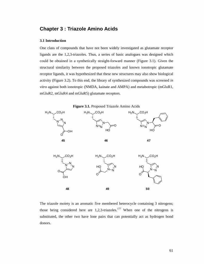
Chapter 3 : Triazole Amino Acids ...............................................................................61
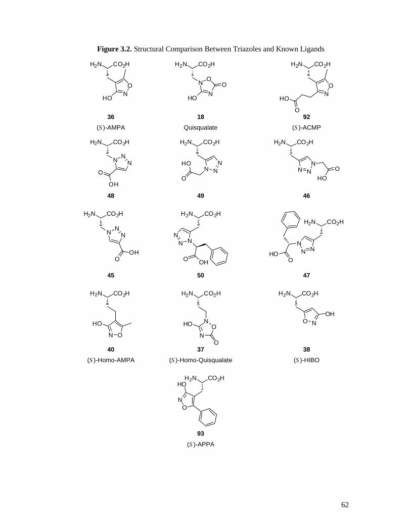
3.1 Introduction .......................................................................................................61
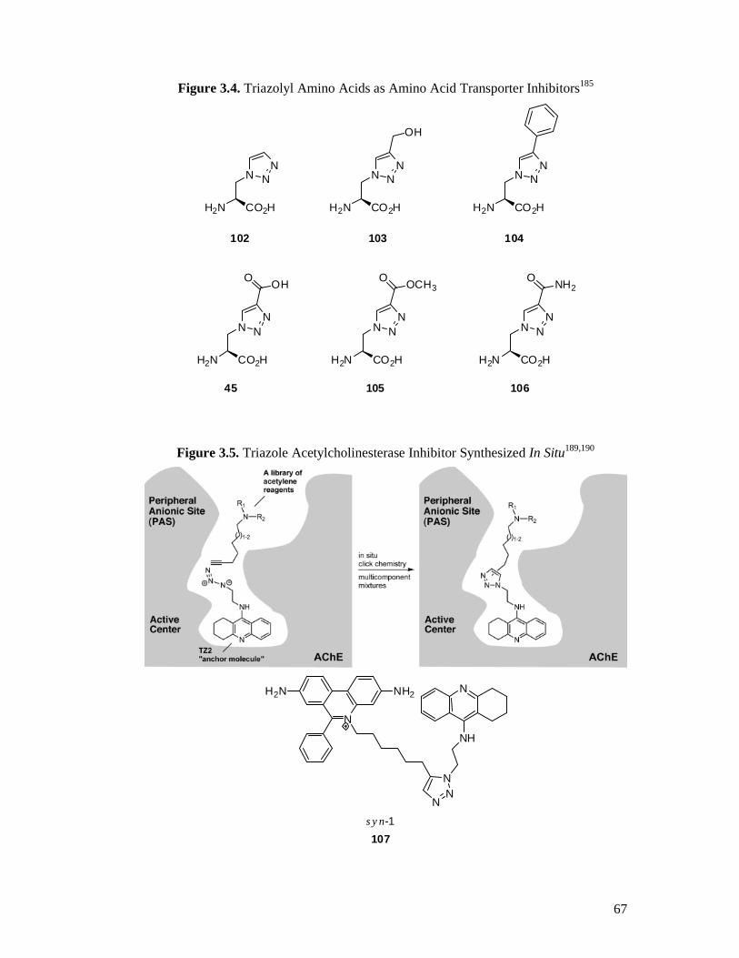
3.2 Triazoles in Drug Discovery ..............................................................................66
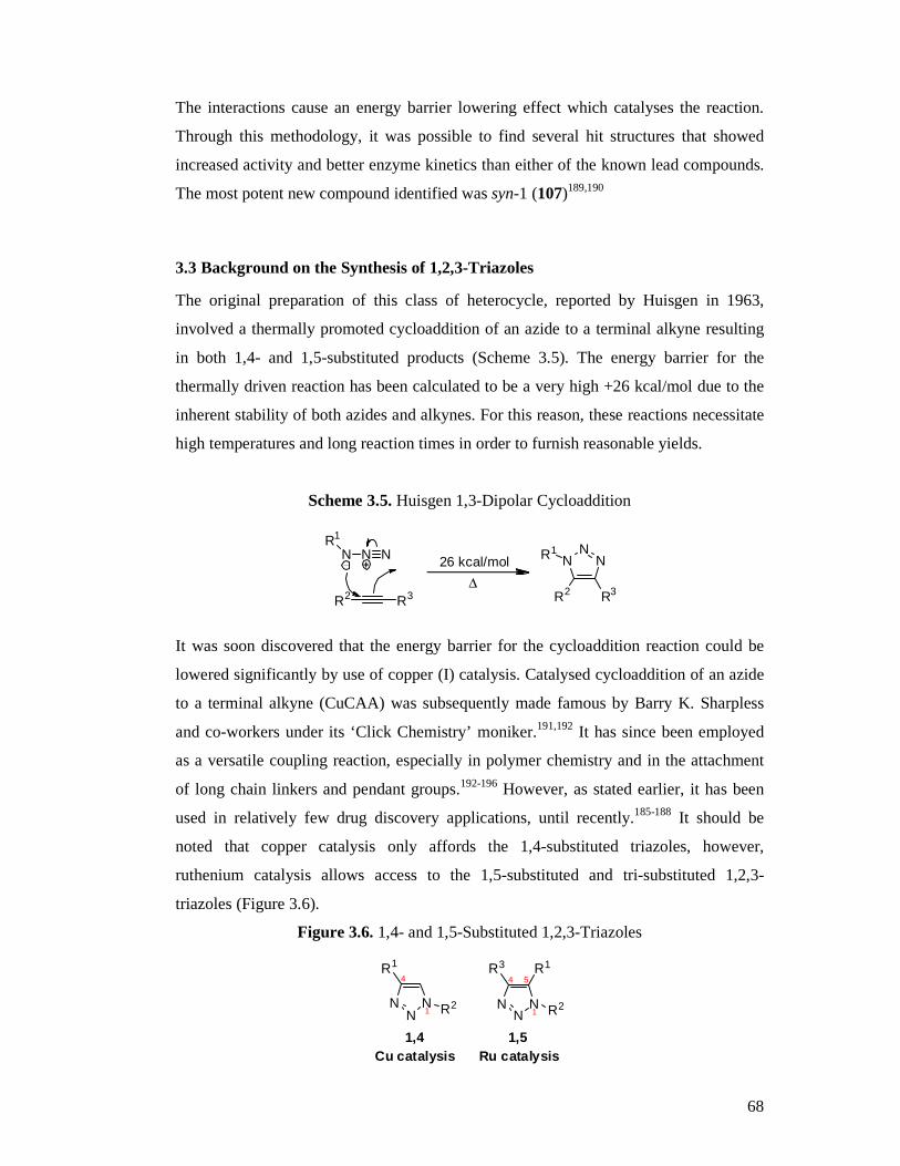
3.3 Background on the Synthesis of 1,2,3-Triazoles.................................................68
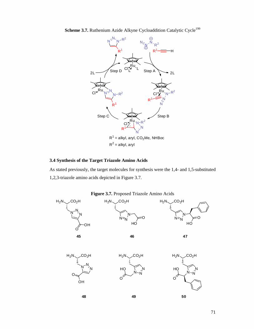
3.4 Synthesis of the Target Triazole Amino Acids ...................................................71
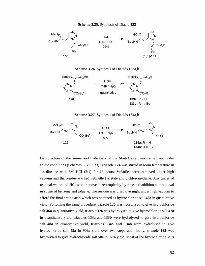
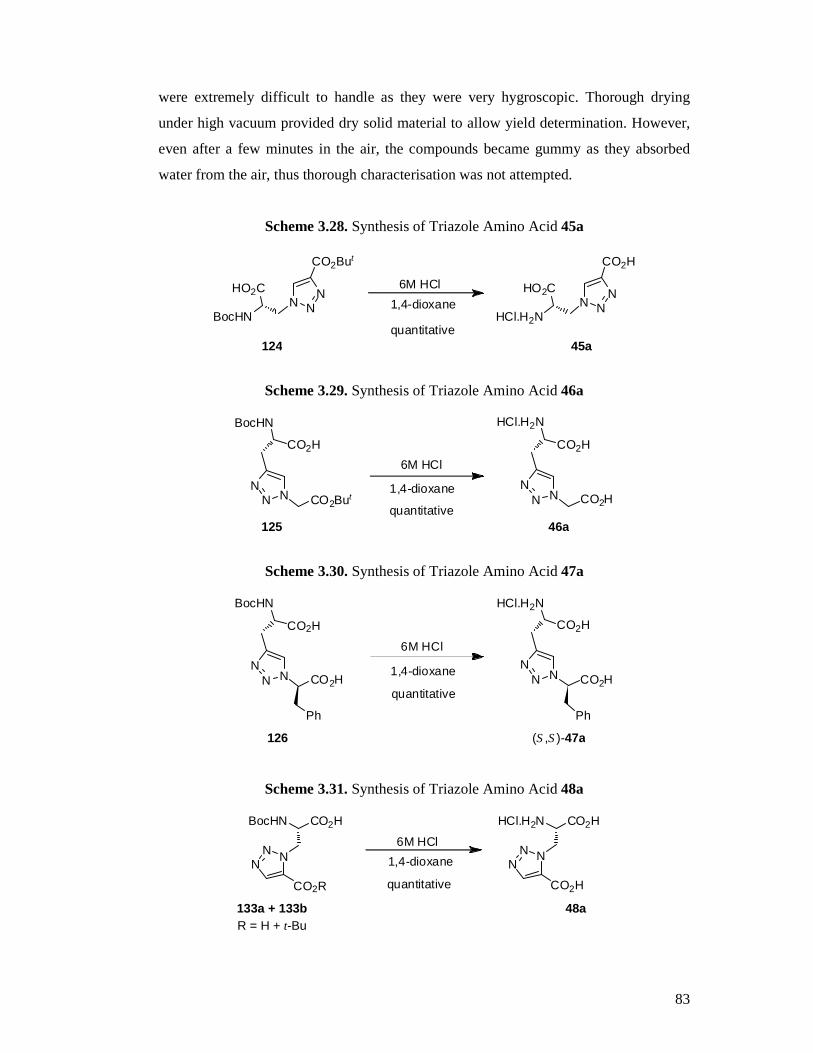
3.5 Summary ...........................................................................................................84
Chapter 4 : Pharmacology ...........................................................................................85
4.1 Pharmacological Testing....................................................................................85
4.2 In Vitro Studies..................................................................................................86
4.2.1 Binding assays at native iGlu receptors .......................................................86
4.2.2 Results ........................................................................................................87
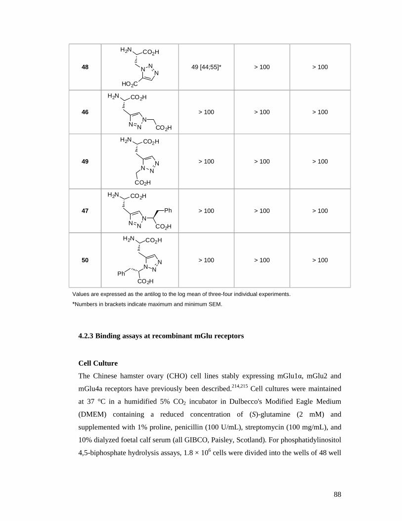
4.2.3 Binding assays at recombinant mGlu receptors ...........................................88
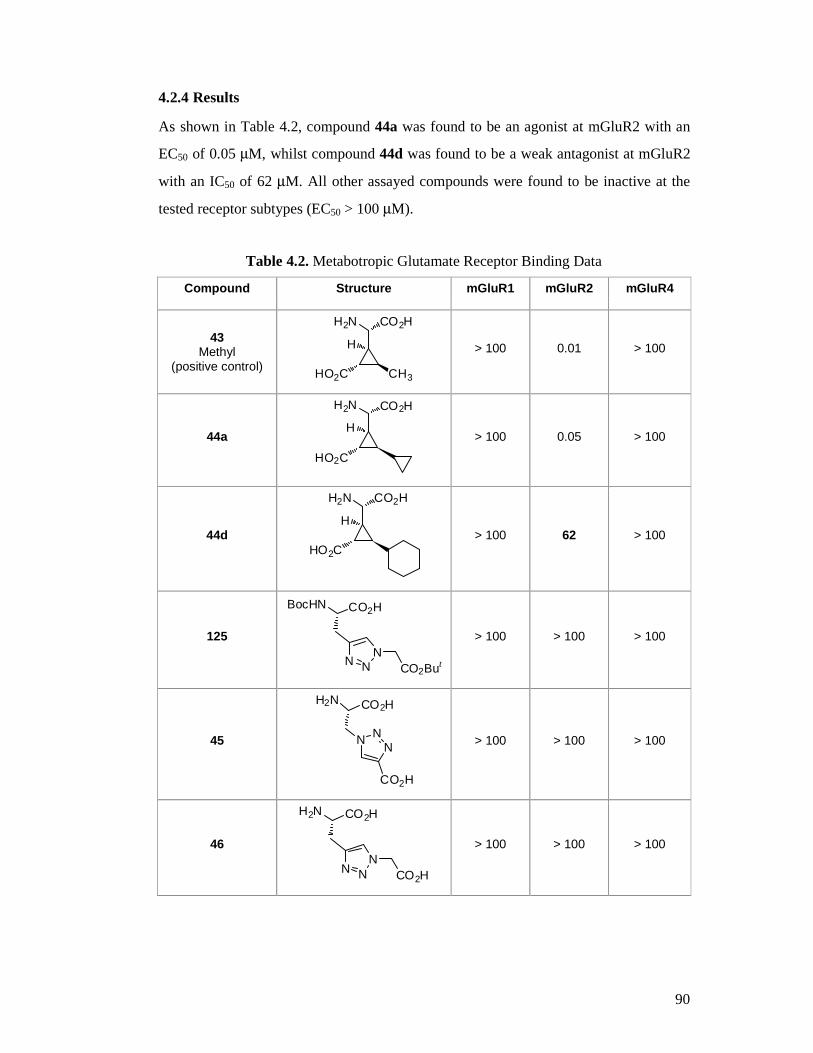
4.2.4 Results ........................................................................................................90
4.3 In Vivo Studies...................................................................................................91
4.3.1 Animals ......................................................................................................91
4.3.2 Ethics..........................................................................................................92
4.3.3 Drugs ..........................................................................................................92
4.3.4 Chronic Constriction Injury (CCI)...............................................................92
4.3.5 Von Frey Testing ........................................................................................93
4.3.6 Data Analysis..............................................................................................93
4.3.7 Results ........................................................................................................94
4.4 Discussion .........................................................................................................96
4.5 Summary .........................................................................................................102
Chapter 5 : In Silico Docking Simulations .................................................................103
5.1 Introduction .....................................................................................................103
5.2 Results and Discussion ....................................................................................104
5.2.1 Docking Validation...................................................................................104
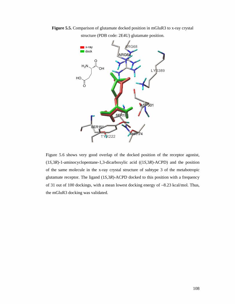
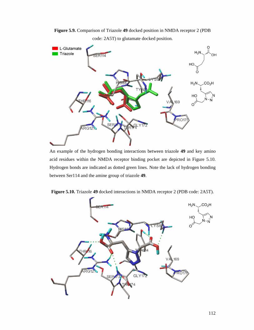
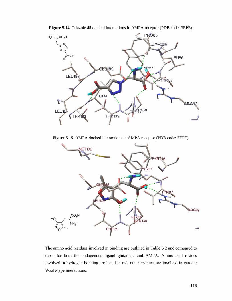
5.2.2 Docking Active Compounds .....................................................................111
5.3 Summary .........................................................................................................118
Chapter 6 : Experimental ...........................................................................................119
References .....................................................................................................157
Page 4
iv
Abstract
As part of the vital search towards improved therapeutic agents for the treatment of
neuropathic pain, the central nervous system ubiquitous glutamate receptors have
become a major focus of research. As such, the discovery of glutamate receptor ligands
with improved potency and selectivity has been an important area of study for many
decades, though there is still much knowledge to be gained.
Outlined herein are the syntheses towards a series of potentially biologically active 3’-
cycloalkyl-substituted carboxycyclopropylglycine analogues. These syntheses utilize
novel synthetic chemistry to construct the cyclopropane core with all required
stereochemistry. As a consequence of this work, two new
cycloalkylcarboxycycloproplyglycine analogues were successfully synthesized,
utilizing the reaction of 1,2-dioxines with protected phosphonates in a 20% overall yield
for one diastereoisomer.
Secondly, the syntheses of a series of 1,4- and 1,5-substituted 1,2,3-triazole amino acids
as a new class of potential glutamate receptor ligands. Briefly, a series of six 1,4- and
1,5-triazole amino acids were successfully synthesized utilizing both copper (I) and
ruthenium-catalysed cycloaddition of functionalized azides and alkynes.
Furthermore, contained within Chapter 4 are the details and results of in vitro binding
assays used in screening for possible active compounds. As an example, in vitro drug
screening at NMDA, kainate and AMPA ionotropic glutamate receptor subtypes
revealed activity of triazole amino acid 48 with an EC50 value of 49 µM at AMPA
receptors. Also, drug screening at metabotropic glutamate receptor subtypes 1, 2 and 4
revealed potent agonist activity of cyclopropane amino acid 44a at mGluR2 with an
EC50 value of 0.05 µM. Cyclopropane amino acid 44a was thus selected for further
testing in vivo in a rodent model of neuropathic pain. The results indicated that
cyclopropane amino acid 44a significantly and dose-dependently decreased mechanical
allodynia, one of the symptoms of neuropathic pain. It was suggested that this effect
was due to activation of mGlu2 and 3 receptors located on both neuronal and glial cells
within the dorsal horn of the spinal cord.
Page 5
v
Lastly, in an effort to rationalize the in vitro binding data, the newly synthesized
cyclopropane and triazole amino acids were docked in silico into the NMDA, AMPA,
mGluR1 and mGluR3 receptors available as x-ray crystal structures. Only limited data
was obtained regarding the mGluR1 and mGluR3 dockings. However, AMPA receptor
docking of the new in vitro active triazole amino acids 45 and 48 revealed positive
docking interactions in agreement with those seen for the endogenous ligand, glutamate
and the selective agonist AMPA. The docking of these new compounds was also
computed to be highly energetically favourable, thus suggesting plausible binding
modes.
Page 6
vi
Declaration
This work contains no material which has been accepted for the award of any other
degree or diploma in any university or other tertiary institution to Nathan Stanley and,
to the best of my knowledge and belief, contains no material previously published or
written by another person, except where due reference has been made in the text.
I give consent to this copy of my thesis, when deposited in the University Library, being
made available for loan and photocopying, subject to the provisions of the Copyright
Act 1968.
I also give permission for the digital version of my thesis to be made available on the
web, via the University’s digital research repository, the Library catalogue, the
Australasian Digital Theses Program (ADTP) and also through web search engines,
unless permission has been granted by the University to restrict access for a period of
time.
Page 7
vii
Acknowledgements
I would like to extend sincere thanks to my supervisors Professor Dennis Taylor,
Professor Andrew Abell and Associate Professor Rod Irvine who have encouraged me
to keep pushing forward even when things were turning pear-shaped.
Special thanks goes out to Dr Thomas Avery and Dr Daniel Pedersen for all their
invaluable help and practical tips in the lab. Without Dr Mark Hutchinson, the in vivo
experiments and in silico docking work would not have happened; thankyou. Thanks
also to Peter Grace for helping with the in vivo testing.
Many thanks to Birgitte Nielsen, Trine Kvist and especially Professor Hans Bräuner-
Osborne at the University of Copenhagen in Denmark for kindly doing the in vitro
receptor binding assays.
Gratitude is expressed to the Faculty of Sciences for providing the financial support
necessary for this research to be undertaken.
Finally, to my beautiful wife, Penelope; I could not have made it through this without
your support. Thankyou for putting up with me over the past four years throughout my
PhD.
Page 8
viii
Abbreviations
Ac acetyl
AcOH acetic acid
Anal. Calcd. analysis calculated
Bn benzyl
Boc tertiary-butoxycarbonyl
Cbz carboxybenzyl
CCG carboxy cyclopropyl glycine
CNS central nervous system
COSY correlated spectroscopy
Cp* pentamethylcyclopentadiene
∆ heat
DCM dichloromethane
DCVC dry column vacuum chromatography
DIAD diisopropyl azodicarboxylate
DMSO dimethyl sulfoxide
DPPA diphenyl phosphoryl azide
EC50 concentration which elicits a 50% maximal effect
EDC 1-ethyl-3-(3-dimethylaminopropyl) carbodiimide
ee enantiomeric excess
EI electron impact
ESI electrospray ionisation
Et ethyl
equiv. equivalent(s)
de diastereomeric excess
g gram(s)
HOBt N-Hydroxybenzotriazole
HRMS high resolution mass spectrometry
h hour(s)
hv light
Hz hertz
IC50 concentration which elicits 50% maximum inhibition
Page 9
ix
iGluR ionotropic glutamate receptor
IR infrared
i.t. intrathecal
J coupling constant
lit. literature
m meta
M moles per litre
m-CPBA meta-chloroperbenzoic acid
m/z mass to charge ratio
Me methyl
MeOH methanol
mGluR metabotropic glutamate receptor
MIRC Michael initiated ring closure
mol mole(s)
mp melting point
NIS N-iodosuccinimide
NMR nuclear magnetic resonance
PDC pyridinium dichromate
Pd/C palladium on carbon
Ph phenyl
ppm parts per million
Rf retention factor
ROESY Rotating Frame Overhauser Effect Spectroscopy
rt room temperature
t-Bu, But tertiary-butyl
TEA triethylamine
TFAA trifluoroacetic anhydride
THF tetrahydrofuran
TLC thin layer chromatography
TPP triphenylphosphine
TPPO triphenylphosphine oxide
UV ultraviolet
Page 10
10
Chapter 1 : Introduction
1.1 Introduction
The negative experience of pain embraces all of humankind, young and old, wealthy
and poor and those of every land, culture and language. Yet even after many years of
research, we are yet to alleviate ourselves completely of this universal burden. Not only
is this an individual burden, but pain is a great economic burden also. It has been
reported by the MBF Foundation that pain costs Australia an estimated total of $34
billion annually.1
1.2 What is Pain?
Pain can broadly be divided into two distinct types. Firstly, acute pain, such as common
headache or pain associated with a temporary injury such as a laceration. Secondly,
chronic pain, such as permanent spinal injury resulting in back pain or phantom limb
pain. Pain can be described as the negative aversive sensation caused by an actual or
perceived injury.2
1.3 Pain Pathways
Peripheral pain usually consists of a combination of either tactile or thermal along with
affective sensory information. Referring to Figure 1.1, the tactile and thermal sensory
pathways, consisting of Aβ and Aδ fibres, enter the central nervous system (CNS) via
the dorsal horn of the spinal cord and from there, pathways ascend to the thalamus,
where an involuntary reflex may be elicited in order to avoid injury and prevent
ongoing pain. Pathways also ascend from the thalamus to the somatosensory cortex
where the quality and location of the tactile or thermal stimulus is interpreted and
suitable action is consciously decided. However, this is only half the story, since these
pathways alone do not communicate anything about the noxious or painful nature of the
stimulus. There are other pathways, consisting of C fibres, which also enter the CNS via
the dorsal horn. Within the dorsal horn, the pain signals are integrated and a pathway
ascends to the parabrachial area and from there, directly to the amygdala. The amygdala
sends projections to the substantia innominata, which in turn projects to the thalamus
and cortex; thus the painful or affective aspects of the painful stimulus are conveyed. It
Page 11
11
is these pathways also that are involved in pairing context and aversive emotions with
the noxious stimuli.3-6
Figure 1.1. Pain Pathways3
1.4 Neuropathic Pain
Chronic neuropathic pain affects a significant portion of the population worldwide7 and
decreases quality of life.8,9 Many of the current medications used for neuropathic pain
do not give adequate and effective pain relief in all cases10-12 and so, much research has
been focussed on finding alternative pain treatments.
Neuropathic pain results from tissue damage, inflammation or injuries in which
affective pain pathways can become hypersensitive. Constant pain signalling causes
neuroplastic changes, resulting in persistent pain even after the initial insult has healed
or subsided.13 This sensory disorder is characterized by hyperalgesia, the sensitization to
painful stimuli, allodynia, the sensation of normal tactile stimuli as painful as well as
other sensory disorders including hyperesthesia, paresthesia, dysesthesia and
a1172507
Text Box
NOTE: This figure is included on page 11 of the print copy of the thesis held in the University of Adelaide Library.
Page 12
12
hypoesthesia. Spontaneous pain is also sometimes evident.13,14 Although the actual
mechanistic causes behind neuropathic pain are poorly understood, it is known to be
associated with direct nerve or spinal cord injury, herpes zoster infection, multiple
sclerosis, diabetes, stroke and cancer.12,14
1.5 The Pain Control Loop
Spinal pain signalling is modulated by both descending inhibitory and facilitatory
systems15,16 (Figure 1.2). Ascending pathways from the dorsal horn project to the
thalamus, however there are also descending pathways which project towards the
periaqueductal grey and then to the rostral ventromedial medulla (RVM) and back to the
dorsal horn via the dorsolateral funiculus. Animal behavioural experiments show
activation of these systems via electrical or chemical stimulation of the PAG or areas of
the RVM inhibits nociceptive reflexes such as the tail-flick and hotplate response.17
Following these experiments, it has been found by electrophysiological studies that
stimulation of either the RVM of PAG can cause inhibition of spinal nociceptive
transmission via pathways descending back to the dorsal horn.16,18 Of the projections
from the RVM to the dorsal horn, one group of neurons has been labelled ‘ON’ cells,
another group has been labelled ‘OFF’ cells and a final group labelled ‘neutral’ cells.
These groups of neurons terminate in laminae I, II and V. As the labels suggest,
‘neutral’ cells have no effect on pain modulation, ‘ON’ cells are thought to be
descending facilitatory neurons which potentiate dorsal horn pain signalling whereas the
‘OFF’ cells are descending inhibitory neurons which attenuate signalling.18-20 There are
also suggestions that descending pathways from the RVM and surrounding areas are not
involved in pain signalling alone, but modulate a range of homeostatic functions and
may play more of a role in stimulus-evoked arousal.21 However, dysfunction of ‘ON’
cells has been implicated as part of the cause of neuropathic pain, whereby there is
excess pain facilitation.16,22
Page 13
13
1.6 Current Treatments
Opioids, with morphine as gold-standard, have traditionally been the mainstay of pain
treatment. Be that as it may, these ancient therapeutics are generally considered to be
less effective in alleviating the symptoms of neuropathic pain.23 Though they do show
some efficacy, there is a lot of inter-individual variation along with side-effects,
particularly respiratory depression, sedation, tolerance and gastrointestinal upset, still
remaining a problem.24 Administered chronically, opioids can also cause adverse
endocrine effects25 and analgesic tolerance can also develop26 which results in the need
for dose escalation in the clinical setting and there is a well known risk of developing
opioid dependence.27 There is also evidence which implicates the endogenous opioid
system in the induction and maintenance of chronic pain28,29 and work which suggests
that chronic opioids can actually cause apoptosis of certain inhibitory neurons in the
dorsal horn, causing hyperalgesia which has the appearance of opioid tolerance in the
clinical pain setting.30 Further to this, individuals who are opioid dependent and who are
Figure 1.2. The Pain Control Loop
a1172507
Text Box
NOTE: This figure is included on page 13 of the print copy of the thesis held in the University of Adelaide Library.
Page 14
14
receiving opioids such as methadone and buprenorphine as substitution treatment often
have problems with pain sensitivity.26 This manifests itself in a similar way to
neuropathic pain, being characterised by hyperalgesia and allodynia.31 In this group of
people, pain management can be difficult due to analgesic tolerance and opioid
addiction, especially since there is strong evidence to suggest that chronic opioid use
can actually be the cause of such sensory disorders.30
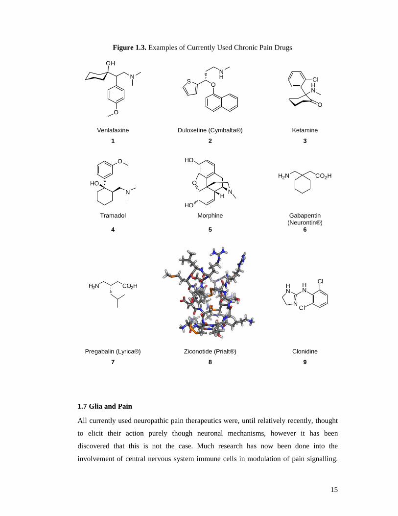
Owing to the fact that opioids have proved to lack efficacy in many types of chronic and
neuropathic pain, much research has centred on finding alternative drugs. At present
there are many drugs being used clinically to treat chronic pain (Figure 1.3) many of
these are being used ‘off-label’ including antidepressants such as venlafaxine (1) and
duloxetine (2) (Cymbalta®, Eli Lilly), NMDA antagonists like ketamine (3),
anticonvulsant drugs like gabapentin (6) (Neurontin®, Pfizer), pregabalin (7) (Lyrica®,
Pfizer) and lamotrigine (Lamictal®, GlaxoSmithKline), voltage-gated calcium channel
blockers, for example ziconotide (8) (Prialt®, Elan Pharmaceuticals), adrenergic drugs
such as clonidine (9) and more traditionally, opioids including tramadol (4) and
oxycodone as well as topical medications.12,32-35 There is often a lot of inter-individual
variation in efficacy of these drugs in neuropathic pain patients. As such combination
therapies are usually employed using several drugs with differing sites of action. Other
underlying diseases or conditions also need to be taken into account such that the use of
certain medications is prohibited due to drug interactions and the risk of serious side
effects.12,32
Page 15
15
Figure 1.3. Examples of Currently Used Chronic Pain Drugs
O
N
OH
OS
NH
O
HN
Cl
Venlafaxine Duloxetine (Cymbalta®) Ketamine
1 2 3
HON
O
HO
HOH
NO
H2N CO2H
Tramadol Morphine Gabapentin (Neurontin®)
4 5 6
H2N CO2H
HN
N
HN
Cl
Cl
Pregabalin (Lyrica®) Ziconotide (Prialt®) Clonidine
7 8 9
1.7 Glia and Pain
All currently used neuropathic pain therapeutics were, until relatively recently, thought
to elicit their action purely though neuronal mechanisms, however it has been
discovered that this is not the case. Much research has now been done into the
involvement of central nervous system immune cells in modulation of pain signalling.
Page 16
16
These cells, comprising mainly microglia and astrocytes (collectively known simply as
‘glia’), are equivalent in number to neurons in the CNS and are known to be involved in
the maintenance, support and immuno-protection of neurons.36,37 However, mounting
evidence indicates that glia can modulate the functional signalling and plasticity of
neurons.38-41 It has been shown that glia release a host of pro-inflammatory mediators
which act on neurons to increase and sustain excitability. Further to this, it has been
demonstrated that glia are intrinsically involved in opioid analgesic tolerance,
hyperalgesia, allodynia and withdrawal symptoms.42 Binding of opioids to toll-like
receptors (TLR) located in association with glia, is proposed to cause activation of these
cells which leads to increased levels of pro-inflammatory cytokines. It is becoming very
clear that opioids such as morphine are a double edged sword, not only mediating
analgesia in the short term, but actually increasing pain sensation in the longer term via
two distinct mechanisms.43 The excitatory neurotransmitter glutamate (10) is known to
be involved in signalling between neurons and glia.40 Crucially, metabotropic glutamate
receptors, the target of this research, are also located in association with glial cells and
as such activation or blockade of these receptors by external ligands may modulate how
glial cells are behaving in the neuropathic pain state.44
Figure 1.4. Structure of L-Glutamate
H2N
HO
O
OH
O
10
L-Glutamate
1.8 Glutamate Receptors
L-Glutamate (10) is the principal excitatory neurotransmitter in the central nervous
system (CNS). It plays an important role in neuronal synaptic plasticity and in particular,
changes to neuron signalling known as long-term potentiation and long-term
depression.45-47 A high density of glutamatergic projections are found in the
hippocampus and neocortex where glutamate plays a vital role in learning and
memory.46,48,49 There are two main classes of glutamate receptors, the ionotropic and
Page 17
17
the metabotropic. The ionotropic glutamate receptors (iGluRs) are ligand-gated sodium
and calcium ion channels and consist of various forms of the N-methyl-D-aspartate
(NMDA), α-amino-3-hydroxy-5-methyl-4-isoxazolepropionic acid (AMPA) and 2-
carboxy-3-carboxymethyl-4-isopropenylpyrrolidine (kainate) receptors. The
metabotropic glutamate receptors (mGluRs) are GTP-binding protein (G-protein)
coupled receptors (GPCRs), having eight known subtypes divided into three groups
depending on sequence homology, signal transduction mechanisms and
agonist/antagonist interactions. These receptors consist of a ‘venus fly trap’
extracellular binding domain coupled to a heptahelical transmembrane domain, coupled
to an intracellular signal transduction domain (Figure 1.5). Group I contains mGluR1
and mGluR5 and these receptors are excitatory, being coupled to Gq/11, leading to
activation of phospholipase C (PLC). Group II contains mGluR2 and mGluR3 and
group III contains mGluR4, R6, R7 and R8 and these are all inhibitory, being coupled to
Gi/0, leading to inhibition of adenylyl cyclase and decreased cyclic
adenosinemonophosphate (cAMP) production.50
Metabotropic glutamate receptors have been implicated as targets for a whole host of
neurological disorders including neuropathic pain,51-54 generalized anxiety disorder,55-57
Parkinson’s disease,58-60 psychosis,61,62 epilepsy,63 depression,64-66 dementia and
Alzheimer’s disease-related neuro-degeneration67 and drug dependence.68-70
Figure 1.5. A Representation of the Metabotropic Glutamate Receptor
a1172507
Text Box
NOTE: This figure is included on page 17 of the print copy of the thesis held in the University of Adelaide Library.
Page 18
18
1.9 Glutamatergic Origins of Neuropathic Pain
Glutamate plays a significant role in the modulation of pain signalling.71,72 There is
substantial evidence to support the involvement of neuroplastic changes such as central
sensitization and long term potentiation in the induction and maintenance of neuropathic
pain73,74 and previous studies have revealed that metabotropic glutamate receptors are
important as modulators of neuroplasticity.75 In the past, a large amount of research has
been focussed on the possibility of targeting NMDA receptors which have been shown
to be involved in the initiation and maintenance of neuropathic pain. The potential
mechanism of treatment is by the use of NMDA antagonists such as ketamine35 or
dextromethorphan,76 however, there are problems with side effects and lack of potency.
The NMDA receptors are fast excitatory receptors as they are ligand-gated ion channels
and as such, they elicit fast excitatory responses, whereas mGlu receptors are GTP-
binding protein (G-protein) coupled receptors (GPCRs) which elicit a slower, more
regulated response on nerve transmission. It has previously been shown that drugs
targeting mGlu receptors show efficacy in various pain models, including those where
allodynia and hyperalgesia are present.71,77,78
1.10 Distribution of Metabotropic Receptors
Metabotropic glutamate receptors have been identified in many key regions of the CNS
known to play a role in pain signalling and processing. The main receptor sub-types
present are those from Group I, mGluR1/R5 and those from Group II, mGluR2/R3 and
it appears from second-messenger assays, electrophysiological studies and
immunohistochemistry, that the Group I receptors act postsynaptically in an excitatory
manner and the Group II receptors act presynaptically as inhibitory autoreceptors, in the
case of mGluR3 or extrasynaptically, in the case of mGluR2 (Figure 1.6).79,80
Page 19
19
Figure 1.6. A Representation of the Typical Synaptic Locations of Metabotropic Glutamate Receptors and Their Signalling Pathways81
1.11 Pain Memory
There is recent research which points to the involvement of the amygdala in neuropathic
pain through the spino-parabrachio-amygdaloid pathway.82,83 The amygdala also sends
descending projections to the PAG which in turn projects to the RVM, then to the dorsal
horn (see Figure 1.2). The amygdala is involved in the emotional “colouring” of sensory
information, for example fear conditioning, and has been implicated as playing a role in
modulating the affective component of pain.82-85 Pathways within the amygdala can
undergo neuroplastic changes due to long-term potentiation of neuron signalling.86
Glutamate is a key player in this process and mGlu receptors have been identified
within the amygdala which are involved in synaptic plasticity and modulation of
signalling.87-89
One aspect of neuropathic pain is the presence of the affective component of pain in the
absence of any tactile or thermal insult or injury and the amygdala may well be involved
in this signalling. The specific involvement of mGluR2 in the amygdala has been
confirmed and early studies demonstrate that mGluR2 agonists can cause long term
depression of synaptic transmission in this area.90-92 Therefore, it is possible that
Page 20
20
modulation of amygdala function by targeting mGlu receptors may result in alleviation
of some aspects of neuropathic pain such as the negative feelings and depression
associated with it and possibly alleviation of the ‘pain’ itself.
1.12 The Benefit of Targeting Metabotropic Glutamate Receptors
Glutamate is important for signal transmission in pain signalling structures and mGlu
receptors are known to be involved in the RVM, the PAG93,94 and the dorsal horn (see
Figure 1.2).95 Research found that activation of Group II mGlu receptors by DCG-IV
(Figure 1.7, 11) within the RVM produces a powerful inhibition of the spinal
nociceptive tail-flick reflex.17 However, DCG-IV also activates NMDA receptors which
may have contributed to the antinociceptive effects.
Figure 1.7. Known mGluR Ligands
HO2C CO2H
CO2HH2N
H
CO2H
NH2
HO2C
H2N CO2H
CO2H CO2HNH2
CO2H
DCG-IV (1S,3R)-ACPD (2S)-α-Eglu (R,S)-AIDA
11 12 13 14
It has also been shown that microinjection of mGluR Group I and II agonist (1S,3R)-
ACPD (Figure 1.7, 12) into the PAG causes a dose-dependent increase in nociceptive
response latency in the mouse hotplate test. Pre-treatment with a Group II antagonist,
(2S)-α-Eglu (13), caused a brief but significant reversal of the antinociceptive effects,
whereas pre-treatment with a Group I antagonist, (RS)-AIDA (14), caused a partial, yet
significant potentiation of the antinociceptive effect produced by (1S,3R)-ACPD.93
These results suggest that both Group I and II mGlu receptors are involved in thermal
nociception and that blockade of Group I and activation of Group II receptors can elicit
antinociception. A further study directly injected the Group I agonist, (S)-3,5-DHPG
(Figure 1.8, 15) and Group II agonist, L-CCG-I (Figure 1.8, 16) into the PAG which
decreased the late phase formalin-induced nociceptive response.94 These results appear
to be contradictory since here a Group I agonist was antinociceptive whereas results
Page 21
21
found previously suggested that a Group I antagonist would be effective. However,
several research groups have found activation of Group II receptors to be
antinociceptive. This theory is also supported in experimental models of neuropathic
pain, where hyperalgesia, allodynia and spontaneous pain are evident. Sharpe et al.96
administered the Group II mGlu agonist, LY379268 (Figure 1.8, 17), to both rats and
mice and found a significant reduction in hyperalgesia both in models of thermal and
neurogenic inflammation. Simmons et al.52 found intraperitoneal injection of mGluR2
agonists resulted in antinociception in the late phase of the formalin test as well as
significantly reducing neuropathic allodynia in a rat model. Activation of Group II
mGluRs within the amygdala by agonist L-CCG-I, produced long term depression of
synaptic transmission which points to such receptor modulation being potentially useful
in targeting the affective component of pain.91
Figure 1.8 HO OH
H2N CO2H HO2C
CO2HH2N
H
OCO2HH2N
HO2CH
H
(S)-3,5-DHPG L-CCG-I LY379268
15 16 17
1.13 Structure Activity Relationship of Metabotropic Glutamate Ligands
Over the past twenty years, there has been an immense amount of research into the
structure activity relationship of metabotropic glutamate receptor ligands and the search
for more potent and selective compounds is far from over. Outlined herein is an
overview of the advances made in understanding the structure and function relationship
of the receptors themselves and how this relates to the design of new and improved
ligands. The metabotropic class of glutamate receptors was first recognized in 1987 by
Sugiyama and colleagues where it was shown that the potent AMPA receptor agonist,
quisqualate (18) and the potent NMDA agonist, ibotenate (19) as well as glutamate
could activate phosphoinositide hydrolysis in rat brain slices in vitro.97 It was shown
that this effect could not be replicated by using other ionotropic receptor agonists such
as NMDA or kainate, nor blocked by known antagonists of these receptors.98 This work
Page 22
22
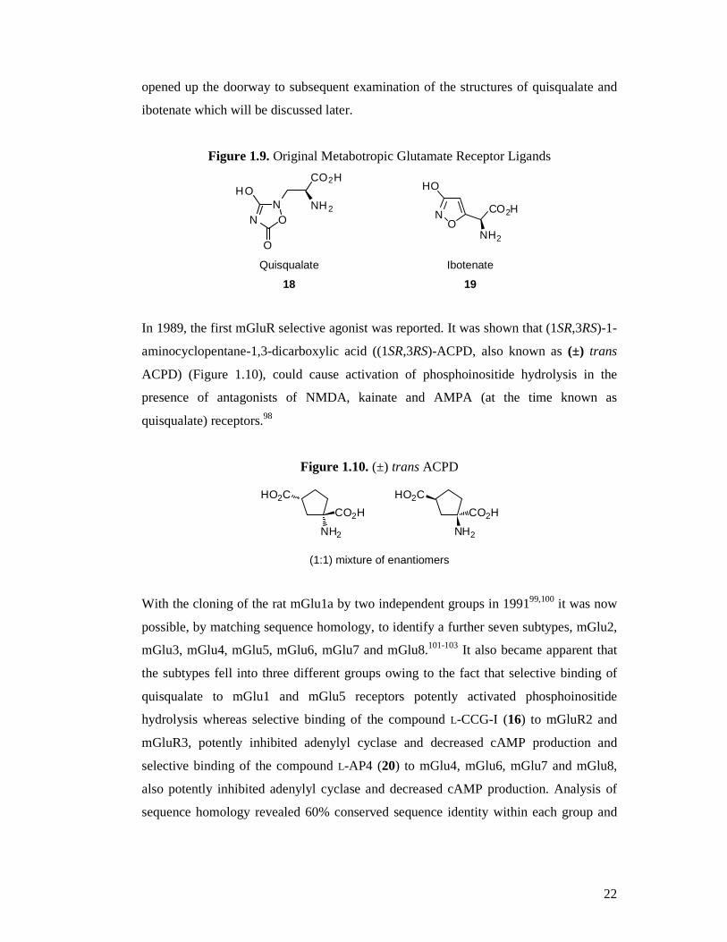
opened up the doorway to subsequent examination of the structures of quisqualate and
ibotenate which will be discussed later.
Figure 1.9. Original Metabotropic Glutamate Receptor Ligands
NN O
HO
O
CO2H
NH2
NO
HO
CO2H
NH2
Quisqualate Ibotenate
18 19
In 1989, the first mGluR selective agonist was reported. It was shown that (1SR,3RS)-1-
aminocyclopentane-1,3-dicarboxylic acid ((1SR,3RS)-ACPD, also known as (±) trans
ACPD) (Figure 1.10), could cause activation of phosphoinositide hydrolysis in the
presence of antagonists of NMDA, kainate and AMPA (at the time known as
quisqualate) receptors.98
Figure 1.10. (±) trans ACPD
CO2H
NH2
HO2C
CO2H
NH2
HO2C
(1:1) mixture of enantiomers
With the cloning of the rat mGlu1a by two independent groups in 199199,100 it was now
possible, by matching sequence homology, to identify a further seven subtypes, mGlu2,
mGlu3, mGlu4, mGlu5, mGlu6, mGlu7 and mGlu8.101-103 It also became apparent that
the subtypes fell into three different groups owing to the fact that selective binding of
quisqualate to mGlu1 and mGlu5 receptors potently activated phosphoinositide
hydrolysis whereas selective binding of the compound L-CCG-I (16) to mGluR2 and
mGluR3, potently inhibited adenylyl cyclase and decreased cAMP production and
selective binding of the compound L-AP4 (20) to mGlu4, mGlu6, mGlu7 and mGlu8,
also potently inhibited adenylyl cyclase and decreased cAMP production. Analysis of
sequence homology revealed 60% conserved sequence identity within each group and
Page 23
23
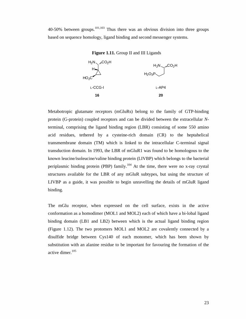
40-50% between groups.101,103 Thus there was an obvious division into three groups
based on sequence homology, ligand binding and second messenger systems.
Figure 1.11. Group II and III Ligands
HO2C
H2N CO2H
H
H2O3P
H2N CO2H
L-CCG-I L-AP4
16 20
Metabotropic glutamate receptors (mGluRs) belong to the family of GTP-binding
protein (G-protein) coupled receptors and can be divided between the extracellular N-
terminal, comprising the ligand binding region (LBR) consisting of some 550 amino
acid residues, tethered by a cysteine-rich domain (CR) to the heptahelical
transmembrane domain (TM) which is linked to the intracellular C-terminal signal
transduction domain. In 1993, the LBR of mGluR1 was found to be homologous to the
known leucine/isoleucine/valine binding protein (LIVBP) which belongs to the bacterial
periplasmic binding protein (PBP) family.104 At the time, there were no x-ray crystal
structures available for the LBR of any mGluR subtypes, but using the structure of
LIVBP as a guide, it was possible to begin unravelling the details of mGluR ligand
binding.
The mGlu receptor, when expressed on the cell surface, exists in the active
conformation as a homodimer (MOL1 and MOL2) each of which have a bi-lobal ligand
binding domain (LB1 and LB2) between which is the actual ligand binding region
(Figure 1.12). The two protomers MOL1 and MOL2 are covalently connected by a
disulfide bridge between Cys140 of each monomer, which has been shown by
substitution with an alanine residue to be important for favouring the formation of the
active dimer.105
Page 24
24
Figure 1.12 Metabotropic Glutamate Receptor Domains105
As has been briefly discussed above, there is an ever-growing body of research focussed
on the elucidation of the structure-function relationship and pharmacodynamics of the
metabotropic glutamate receptors. This has in turn necessitated the development of
improved ligands with greater subtype selectivity and potency as experimental tools.
Outlined below are the classes of compounds that have been investigated thus far.
1.13.1 The Cyclopentane Analogues
The cyclopentane class of compounds were the first investigated, due to their relation to
known iGluR ligands. Desai and Conn reported that (±) trans ACPD (1SR,3RS)-ACPD)
(see Figure 1.10) stimulates phosphoinositide hydrolysis, an effect similar to the iGluR
agonist ibotenate (19, Figure 1.9).106 However, this effect was not blocked by NMDA,
AMPA or kainate receptor antagonists indicating that the ligand was activating a
different type of receptor. This new class of receptor was consequently labelled the
metabotropic glutamate receptor after it was discovered to be a G-protein-coupled
receptor. Although these compounds all bear a similar core structure, there is great
diversity in their pharmacological profiles. Referring to Figure 1.13, (1S,3R)-ACPD
(12), the active enantiomer of (±) trans ACPD, is a broad spectrum, non-selective
agonist showing in vitro activity in the order: mGluR2 > mGluR5 > mGluR1 > mGluR8
> mGluR6 > mGluR4. Addition of a carboxylic acid group in the 4-position converts
amino acid 12 into (3S,4S)-ACPT-III (21), a selective mGluR4 agonist. Introduction of
a nitrogen atom into the cyclopentane ring of (1S,3R)-ACPD (12) at the 4-position
increases affinity for the Group II receptors, mGluR2 and R3 and the compound
a1172507
Text Box
NOTE: This figure is included on page 24 of the print copy of the thesis held in the University of Adelaide Library.
Page 25
25
becomes the potent and selective agonist, (2R,4R)-APDC (22). This compound is
approximately equipotent at both R2 and R3, however, if the nitrogen atom is
substituted with an amine group, as in (2R,4R)-amino-APDC (23) this results in a
compound which is about ten times more selective for mGluR2 over R3. If the nitrogen
atom is substituted with a benzyl group, as in (2R,4R)-benzyl-APDC (24) this results in
a compound which exhibits agonist activity at mGluR6, whilst being a weak antagonist
at mGluR2 (EC50: 200 µM) and mGluR5 (EC50: 600 µM). Finally, homologation of
(1S,3R)-ACPD (12) at the 3-position, produces (25) which increases the compound’s
affinity as an agonist for mGluR2 over all other subtypes.107
Figure 1.13. Cyclopentane Analogues
CO2H
NH2
HO2C
CO2H
NH2
CO2H
HO2C
(1S,3R)-ACPD (3S,4S)-ACPT-III
12 21
N
CO2H
NH2
R
HO2C
CO2H
NH2
HO2C
22 R = H: (2R,4R)-APDC (1S,3R)-Homo-ACPD 23 R = NH3: (2R,4R)-amino-APDC 25 24 R = CH2Ph: (2R,4R)-benzyl-APDC
1.13.2 The mGluR Binding Site
The ligand binding region of all known metabotropic glutamate receptors consists of six
conserved amino acid residues that are essential for binding L-glutamate and all known
competitive ligands. The sequence of the amino acids varies slightly between the three
receptor groups and the details of these are outlined in Table 1.1 below.
Page 26
26
Table 1.1. Amino Acid Residues Involved in Ligand Binding
Group I Group II Group III
distal carboxylate salt bridge Arg78 Arg68 Arg78
Lys409 Lys389 Lys407
proximal carboxylic hydrogen bonds Ser165 Ser151 Ser159
Thr188 Thr174 Thr182
amine salt bridge Asp318 Asp301 Asp314
van der Waals and lone pair interaction with
side group Tyr236 Tyr222 Tyr230
hydrogen bond acceptor (phenolic) Tyr236 Tyr222 Tyr230
However, simply knowing the residues involved in binding is not sufficient to predict
ligand binding. It is now becoming apparent that the actual size of the binding region
varies amongst the receptor subtypes. This is due to more or less bulky amino acid side
chains occupying the region at the edge of the binding cleft. These residues alone can
prevent entry of excessively bulky ligands into the respective receptor LBR.
Energetic and entropic effects also come into play. The LBR in the open form contains
a concentration of solvent molecules that is at equilibrium with the surrounding.
Binding of a ligand always requires displacement of some solvent molecules in order
for the receptor to convert to the closed form. From an entropic point of view, the
greater the number of solvent molecules that must be displaced in order for the ligand to
bind, the less favourable the binding. Furthermore, in x-ray crystallography studies it
has also been observed that some ligands actually require residual solvent molecules in
order to facilitate binding.
1.13.3 Phenylglycine Derivatives
In the early 1990s, a group of selective mGluR antagonists was reported that were based
on a phenylglycine core structure.108-110 There are three main types of compound in this
class, the hydroxyphenylglycines (Figure 1.14), the carboxyphenylglycines (Figure
Page 27
27
1.15) and the phosphonophenylglycines (Figure 1.16). The simplest compound in the
first class is (S)-3-HPG (26) which is primarily an mGluR5 agonist, but also exhibits
weak agonist activity at mGluR1. Addition of a second hydroxyl group at the 5-position
gives (S)-3,5-DHPG (15) which has increased potency over compound 26, however it
has less selectivity for mGluR5 over mGluR1. Further addition of a chlorine atom to the
6-position results in (R,S)-CHPG (27), which is 100 times less potent than compound 15,
but is 10-fold more selective for mGluR1 over mGluR5. The simplest
carboxyphenylglycine is (S)-4CPG (28) which is a selective mGluR1 antagonist, but
also shows some weak agonist activity at mGluR2. The addition of a hydroxyl group at
the 3-position of compound 28 gives (S)-4C3HPG (29), which is both an mGluR1
antagonist and an mGluR2 agonist with equal potency at both subtypes. Inclusion of a
methyl group at the 2-position of compound 29 provides (R,S)-4C3H2MPG (31) which
is solely a selective mGluR1 antagonist with no activity at mGluR2. The compound (+)-
4C2MPG (30) exhibits equivalent activity to compound 31 at mGluR1. (S)-M4CPG
(32) is compound 28 with methyl substitution on the alpha carbon and is also an
mGluR1 antagonist along with LY367366 (33) which also shows mGluR5 antagonist
activity. Finally, the phosphonophenylglycine analogues have a different
pharmacological profile entirely. (R,S)-PPG (34) is a very potent mGluR8 agonist and
also a moderately active mGluR4 and mGluR6 agonist, whereas (R,S)-MPPG (35) is an
mGluR2 antagonist.
Figure 1.14. Hydroxyphenylglycine Compounds
HO R1
R2
H2N CO2H
R1 R2
26 H H (R,S)-3-HPG
15 OH H (S)-3,5-DHPG
27 OH Cl (R,S)-CHPG
Page 28
28
Figure 1.15. Carboxyphenylglycine Compounds
R1
H2N CO2H
CO2H
R2
R3
R1 R2 R3
28 H H H (S)-4CPG
29 OH H H (S)-4C3HPG
30 H Me H (+)-4C2MPG
31 OH Me H (R,S)-4C3H2MPG
32 H H Me (S)-M4CPG
33 H H
LY367366
Figure 1.16. Phosphonophenylglycine Compounds
H2N CO2HR
PO3H2
R
34 H (R,S)-PPG
35 Me (R,S)-MPPG
1.13.4 The Isoxazoles and Oxadiazoles
These compounds were amongst the first to be discovered as having activity at mGluRs.
Most are also active at ionotropic glutamate receptors, such as the prototypes quisqualic
acid (quisqualic acid, 18) and ibotenate (ibotenic acid, 19) as well as (S)-AMPA (36).
S
Page 29
29
Figure 1.17. Isoxazoles and Oxadiazoles
NN O
HO
O
CO2H
NH2
NO
HO
CO2H
NH2 N
O
HONH2
CO2H
Quisqualate Ibotenate (S)-AMPA
18 19 36
NN O
HO
O
CO2HH2N
NO
HO R
CO2HH2N N
O
HOCO2H
H2N
(S)-Homo-quisqualate 38 R = H: (S)-HIBO
39 R = n-Bu: (S)-Bu-HIBO
(S)-Homo-AMPA
37 40
1.13.5 The Carboxycyclopropylglycines
Several isomers of 2-carboxycyclopropylglycine were first isolated as natural products
from the seeds of Blighia sapida and later from the stems of Ephedra altissima and
Ephedra foeminea where they were suggested to play a role as an anti-feeding
agents.111-114 Subsequent synthesis and testing revealed activity in L-glutamate pathways
in the central nervous system. In particular, these compounds were found to be
relatively selective for metabotropic glutamate receptors and were used as tools to gain
insight into the molecular conformation required for subtype selectivity. It was found
that NMDA receptor binding required the molecule to adopt a folded conformation,
whereas metabotropic binding required an extended conformation.115-117 In order to
investigate this further, a hybrid molecule (2,3-dicarboxycyclopropyl)glycine (DCG-IV)
(Figure 1.7, 11), was synthesized which incorporated both the extended and folded
conformations in the same molecule.118 Further to this, certain types of substitution have
been found to result in either agonist or antagonist activity (Table 1.2). It was found that
addition of a methyl, phenyl, xanthenylmethyl or xanthenylethyl groups to the 2-
position resulted in antagonist activity and phenyl, xanthenylmethyl or xanthenylethyl
substitution at the 3’-position also resulted in antagonist activity.119,120 The
cyclopropane core is also a key element as its rigid nature holds the functional groups in
Page 30
30
a conformation that very closely resembles L-glutamate’s folded conformation as
opposed to the fully extended conformation; this is vital for subtype selectivity.
Examination of the relationship between substitution at the 3’-position and ligand
potency as measured by the EC50 value suggests that a group comprising a one carbon
chain with a lone pair donor atom attached, such as oxygen, gives the greatest potency
of all tested CCGs thus far.121 Simple methyl substitution is also fairly potent.122
Table 1.2. Known Carboxycyclopropylglycine Glutamate Receptor Ligands
H2N
HO
O
OH
O
H
R3
R1R2
EC50 (µµµµM)*
Compound R1 R2 R3 mGluR2 mGluR3
L-Glutamate123 26 6.1
L-CCG-I121 H H H 0.3 0.6
L-F2CCG-I121 F F H 0.09
cis MCG-I121 H CH2OCH3 H 0.1
trans MCG-I121 CH2OCH3 H H 0.3
DCG-IV121 CO2H H H 0.3 0.2
PCCG-4124 phenyl H H 0.8
XM-CCG-I119 xanthenylmethyl H H 6.4 1.3
XE-CCG-I119 xanthenylethyl H H 0.2 0.75
LY341495119 H H xanthenylmethyl 0.2 0.16
HM-CCG-I122 CH2OH H H 0.005 0.012
Thiolmethyl122 CH2SH H H 0.047 0.059
Methyl122 CH3 H H 0.008 0.039
Ethyl122 CH2CH3 H H 0.17 3.6
CN-CCG122 CN H H 0.19 0.064 *Values shown in bold indicate antagonist activity
It has been found that certain minimal elements of the carboxycyclopropyl glycine
molecule are necessary for any binding to occur; this is known as the pharmacophore
Page 31
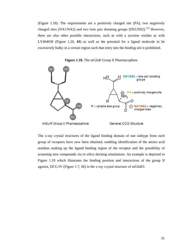
31
(Figure 1.18). The requirements are a positively charged site (PA), two negatively
charged sites (NA1/NA2) and two lone pair donating groups (DS1/DS2).116 However,
there are also other possible interactions, such as with a tyrosine residue as with
LY404039 (Figure 1.20, 43) as well as the potential for a ligand molecule to be
excessively bulky in a certain region such that entry into the binding site it prohibited.
Figure 1.18. The mGluR Group II Pharmacophore
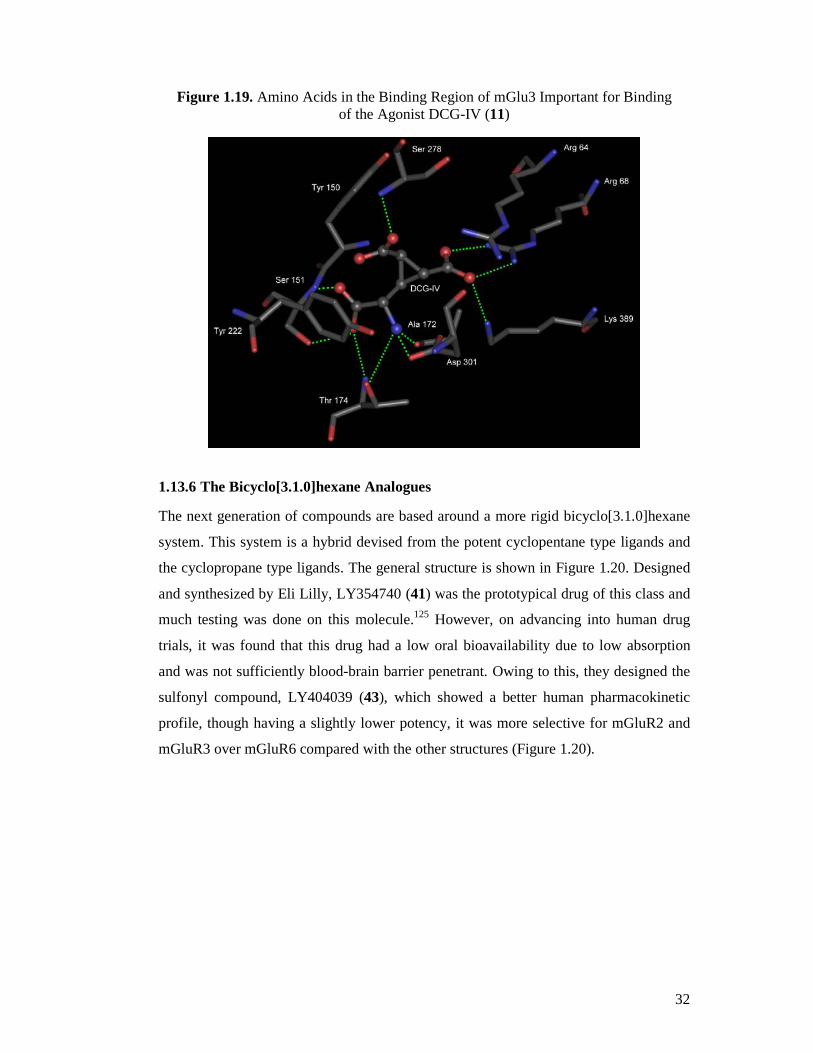
The x-ray crystal structures of the ligand binding domain of one subtype from each
group of receptors have now been obtained, enabling identification of the amino acid
residues making up the ligand binding region of the receptor and the possibility of
screening new compounds via in silico docking simulations. An example is depicted in
Figure 1.19 which illustrates the binding position and interactions of the group II
agonist, DCG-IV (Figure 1.7, 11) in the x-ray crystal structure of mGluR3.
Page 32
32
Figure 1.19. Amino Acids in the Binding Region of mGlu3 Important for Binding of the Agonist DCG-IV (11)
1.13.6 The Bicyclo[3.1.0]hexane Analogues
The next generation of compounds are based around a more rigid bicyclo[3.1.0]hexane
system. This system is a hybrid devised from the potent cyclopentane type ligands and
the cyclopropane type ligands. The general structure is shown in Figure 1.20. Designed
and synthesized by Eli Lilly, LY354740 (41) was the prototypical drug of this class and
much testing was done on this molecule.125 However, on advancing into human drug
trials, it was found that this drug had a low oral bioavailability due to low absorption
and was not sufficiently blood-brain barrier penetrant. Owing to this, they designed the
sulfonyl compound, LY404039 (43), which showed a better human pharmacokinetic
profile, though having a slightly lower potency, it was more selective for mGluR2 and
mGluR3 over mGluR6 compared with the other structures (Figure 1.20).
Page 33
33
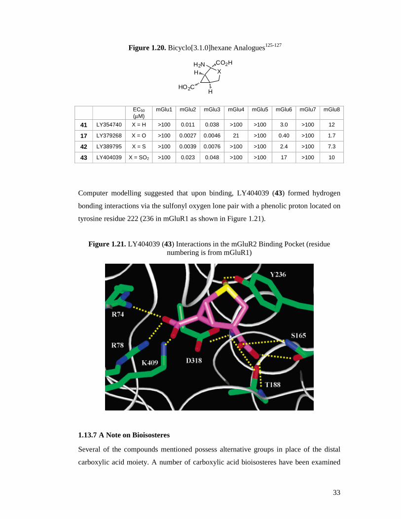
Figure 1.20. Bicyclo[3.1.0]hexane Analogues125-127
XCO2HH2N
HO2C H
H
EC50 (µM)
mGlu1 mGlu2 mGlu3 mGlu4 mGlu5 mGlu6 mGlu7 mGlu8
41 LY354740 X = H >100 0.011 0.038 >100 >100 3.0 >100 12
17 LY379268 X = O >100 0.0027 0.0046 21 >100 0.40 >100 1.7
42 LY389795 X = S >100 0.0039 0.0076 >100 >100 2.4 >100 7.3
43 LY404039 X = SO2 >100 0.023 0.048 >100 >100 17 >100 10
Computer modelling suggested that upon binding, LY404039 (43) formed hydrogen
bonding interactions via the sulfonyl oxygen lone pair with a phenolic proton located on
tyrosine residue 222 (236 in mGluR1 as shown in Figure 1.21).
Figure 1.21. LY404039 (43) Interactions in the mGluR2 Binding Pocket (residue numbering is from mGluR1)
1.13.7 A Note on Bioisosteres
Several of the compounds mentioned possess alternative groups in place of the distal
carboxylic acid moiety. A number of carboxylic acid bioisosteres have been examined
Page 34
34
including a phenolic moiety as in the phenylglycine analogues (Figures 1.14-1.16),
phosphonic acid as in L-AP4 (Figure 1.11, 20) and variations on isoxazoles and
oxadiazoles as in (S)-HIBO (Figure 1.17, 38). Analogues of this kind not only retain
potency, but also show increased subtype selectivity suggesting that the ligand binding
region of each subtype is sufficiently different such that some can accept certain
bioisosteric groups, whilst others cannot. This gives merit to the search for novel
bioisosteric groups which may convey further subtype selectivity whilst maintaining
ligand potency.
1.14 Current Research
In the search for new therapeutic agents for the treatment of neuropathic pain, outlined
herein are the syntheses towards a series of potentially biologically active 3’-cycloalkyl-
substituted carboxycyclopropylglycine analogues, utilizing novel synthetic chemistry to
construct the cyclopropane core with all required stereochemistry (Figure 1.22).
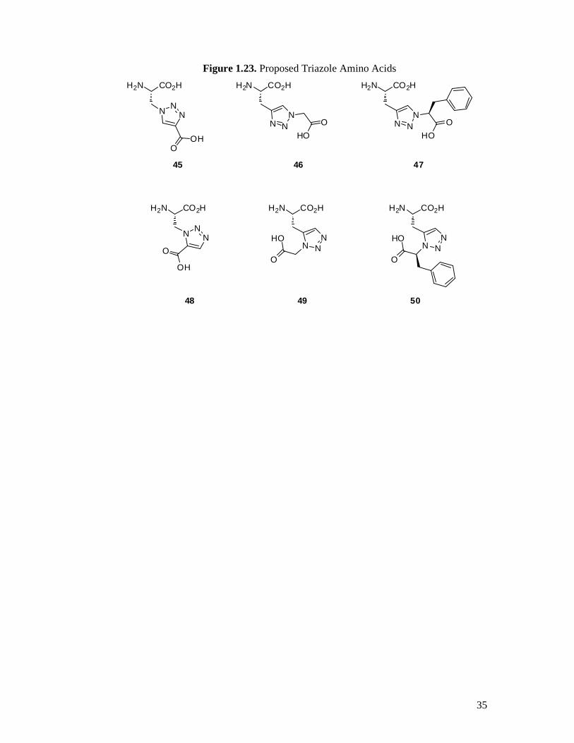
Secondly, the syntheses of a series of 1,4- and 1,5-substituted 1,2,3-triazole amino acids
as a new class of potential glutamate receptor ligands (Figure 1.23). Also included are
the details and results of in vitro binding assays used to screen for possible active
compounds, investigations into in silico glutamate receptor docking analysis of the
active and non-active test compounds in an effort to rationalize in vitro data and finally,
the details of in vivo anti-allodynic activity of one compound in an animal model of
neuropathic pain.
Figure 1.22. Proposed Cyclopropane Amino Acids
H2N
RHO
O
OH
O
H
H2N
RHO
O
OH
O
H
(R) 44b, d, f, h, j (S) 44a, c, e, g, i
R = c-C3H5, c-C4H7, c-C5H9, c-C6H11, c-C7H13
Page 35
35
Figure 1.23. Proposed Triazole Amino Acids
H2N CO2H
N NN
H2N CO2H
N NN
H2N CO2H
N NN
O
OH
OOH
OHO
H2N CO2H
N NN
O
HO
48 49
4645
50
47
H2N CO2H
N NN
OHO
H2N CO2H
N NN
O
HO
Page 36
36
Chapter 2 : Cyclopropane Amino Acids
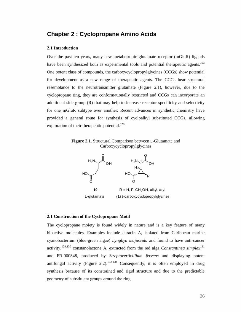
2.1 Introduction
Over the past ten years, many new metabotropic glutamate receptor (mGluR) ligands
have been synthesized both as experimental tools and potential therapeutic agents.103
One potent class of compounds, the carboxycyclopropylglycines (CCGs) show potential
for development as a new range of therapeutic agents. The CCGs bear structural
resemblance to the neurotransmitter glutamate (Figure 2.1), however, due to the
cyclopropane ring, they are conformationally restricted and CCGs can incorporate an
additional side group (R) that may help to increase receptor specificity and selectivity
for one mGluR subtype over another. Recent advances in synthetic chemistry have
provided a general route for synthesis of cycloalkyl substituted CCGs, allowing
exploration of their therapeutic potential.128
Figure 2.1. Structural Comparison between L-Glutamate and Carboxycyclopropylglycines
L-glutamate (1S)-carboxycyclopropylglycines
R
H2N
HO
O
OH
O
H
H2N
HO
O
OH
O
10 R = H, F, CH2OH, alkyl, aryl
1
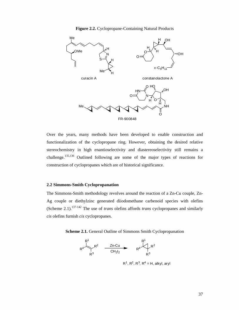
2.1 Construction of the Cyclopropane Motif
The cyclopropane moiety is found widely in nature and is a key feature of many
bioactive molecules. Examples include curacin A, isolated from Caribbean marine
cyanobacterium (blue-green algae) Lyngbya majuscula and found to have anti-cancer
activity,129,130 constanolactone A, extracted from the red alga Constantinea simplex131
and FR-900848, produced by Streptoverticillium fervens and displaying potent
antifungal activity (Figure 2.2).132-134 Consequently, it is often employed in drug
synthesis because of its constrained and rigid structure and due to the predictable
geometry of substituent groups around the ring.
Page 37
37
Figure 2.2. Cyclopropane-Containing Natural Products
H
H
Me
NS
Me
OMe
n-C5H11
H
H
NHMe
O
O
OHHO
NHN
O
OH
H
HO
O
H
OH
OH
curacin A constanolactone A
FR-900848
Over the years, many methods have been developed to enable construction and
functionalization of the cyclopropane ring. However, obtaining the desired relative
stereochemistry in high enantioselectivity and diastereoselectivity still remains a
challenge.135,136 Outlined following are some of the major types of reactions for
construction of cyclopropanes which are of historical significance.
2.2 Simmons-Smith Cyclopropanation
The Simmons-Smith methodology revolves around the reaction of a Zn-Cu couple, Zn-
Ag couple or diethylzinc generated diiodomethane carbenoid species with olefins
(Scheme 2.1).137-142 The use of trans olefins affords trans cyclopropanes and similarly
cis olefins furnish cis cyclopropanes.
Scheme 2.1. General Outline of Simmons Smith Cyclopropanation
R4
R1
R2
R3R4
R1
R2
R3
Zn-Cu
CH2I2
R1, R2, R3, R4 = H, alkyl, aryl
Page 38
38
Scheme 2.2 outlines an example of the Simmons Smith cyclopropanation utilizing
diethylzinc where a catalytic amount of the dipeptide N-Boc-L-Val-L-Pro-OMe (51),
used as a chiral directing species, and ethylmethoxyacetate, used to prevent side
reactions, provided a high yield of cyclopropane 53 with high enantioselectivity from
olefin 52 which lacks directing groups.143
Scheme 2.2. Example of a Simmons Smith Cyclopropanation
Ph BocHN NCO2MeO
CH2Cl2
ZnEt2, CH2I2
96% yield89% ee
ethylmethoxyacetate
51
5352
Ph
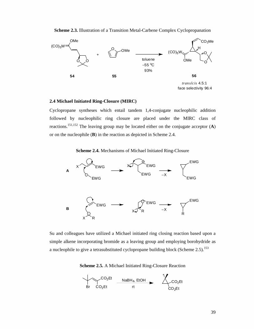
2.3 Transition Metal-Carbene Complexes
Carbene ligands can be transferred to olefins via a transition metal catalysed reaction to
provide enantioenriched cyclopropanes.139,142,144-148 In the literature it has been found
that carbenes can be added across double bonds using a range of chiral metal complexes
including iron, palladium, cobalt, ruthenium and rhodium. This manifold requires use of
diazo-compounds which pose a potential explosion risk, making large scale synthesis
difficult.149
Barluenga and colleges have outlined a procedure involving the use of a Fischer
tungsten metal carbene complex (54) reacting with 2-methoxyfuran (55) to furnish a
versatile tri-substituted cyclopropane building block in excellent yield and with high
diastereoselectivity (56, Scheme 2.3).150 Simple oxidation of the carbene product and
subsequent elaboration gives access to alcohols, diols and cyclopropanecarbaldehydes.
Page 39
39
Scheme 2.3. Illustration of a Transition Metal-Carbene Complex Cyclopropanation
OMe(CO)5W
O O
O OMe
O
O(CO)5W
OMe
CO2Me
+
54 55 56
H
toluene−55 oC93%
trans/cis 4.5:1face selectivity 96:4
2.4 Michael Initiated Ring-Closure (MIRC)
Cyclopropane syntheses which entail tandem 1,4-conjugate nucleophilic addition
followed by nucleophilic ring closure are placed under the MIRC class of
reactions.151,152 The leaving group may be located either on the conjugate acceptor (A)
or on the nucleophile (B) in the reaction as depicted in Scheme 2.4.
Scheme 2.4. Mechanisms of Michael Initiated Ring-Closure
X EWG X EWG
EWG
EWG
EWG−X
EWG
EWGEWG
RX R
R
EWG
X
A
B −X
Su and colleagues have utilized a Michael initiated ring closing reaction based upon a
simple alkene incorporating bromide as a leaving group and employing borohydride as
a nucleophile to give a tetrasubstituted cyclopropane building block (Scheme 2.5).153
Scheme 2.5. A Michael Initiated Ring-Closure Reaction
CO2Et
Br CO2EtCO2Et
CO2Et
NaBH4, EtOH
rt
Page 40
40
Work by Sun and Tang has found that cyclopropanes can be synthesized by use of
telluronium ylides. Starting from methyl cinnamate and under basic conditions, chiral
1,2,3-trisubstituted cyclopropanes may be obtained in excellent yields and with high
enantiomeric excess as outlined in Scheme 2.6.154
Scheme 2.6. Tri-substituted Cyclopropanes via Michael Initiated Ring-Closure
Te TMS
BPh4
PhCO2Me
CO2Me
Ph TMS
LiTMP / HMPA
2.
1.
96% ee95%
2.5 The Carboxycyclopropylglycines
As discussed earlier, several isomers of 2-carboxycyclopropylglycine have been
isolated as natural products.111-114 Up until now, few general routes for synthesis of di-
substituted cyclopropyl glycines have been developed. Most syntheses have previously
been aimed towards obtaining a single isomer, with no possibility of varying the
substitution pattern and involve as many as 21 synthetic steps. This has hindered the
pharmacological investigation of their therapeutic potential.
A previous enantioselective synthesis of 3’-alkyl substituted
carboxycyclopropylglycines (CCGs) (57, 58) was carried out by an addition and then
elimination of a chiral lithium bislactam ether anion to a racemic 4-alkyl-4-bromo-2-
butenoate (59) as shown in Scheme 2.7.155 However, all CCGs synthesized by this route
had the R group cis to the carboxyl group on the cyclopropane ring and it was found
that the more active isomers had the R group anti to the carboxyl group and these could
not be synthesized via this route.
Page 41
41
Scheme 2.7. Enantioselective Synthesis of 3’-Alkyl Substituted Carboxycyclopropylglycines
N
N OMe
MeO Br CO2Et
R
R = Me, Et, n-Pr
1.
2.
n-BuLi, −78 oC, THF
N
N OMe
MeOH
HN
N OMe
MeOH
R
H
0.1M HCl / THF 0.1M HCl / THF
6M HCl
R
H
CO2HH2N
R
H
CO2HH2N
6M HCl
59
57 58
CO2Et CO2Et
R
CO2H CO2H
+
A stereo-specific synthesis of the group II metabotropic glutamate receptor antagonist
(2S,3S,4S)-MCCG (60) was put forward by Pajouhesh et al., starting with a chiral
protected oxazolidinone (61).156 A transition metal-carbene complex formed by use of
diazomethane and palladium (II) acetate was employed to construct the cyclopropane
core from chiral olefin 62. This route afforded product with 99% ee (Scheme 2.8).
Scheme 2.8. Stereo-Specific Synthesis of (2S,3S,4S)-MCCG
CbzN
O OPh
CH3CbzN
O OPh
CH3CO2CH3
CbzN
O OPh
CH3CO2CH3
CO2HH3C
CO2H
CbzHN
CO2HH3C
CO2H
NH2
KHMDS (0.5 M)
BrCO2CH3
−78 oCCH2N2Pd(OAc)2
LiOH / H2O
HBr / AcOH
(2S,3S,4S)-MCCG
60
61 62
Page 42
42
Further syntheses have been investigated by researchers at Eli Lilly to produce the 3’-
alkyl substituted biologically active carboxycyclopropyl glycines (Scheme 2.9).55 This
time, starting from the readily available crotyl alcohol and building up the cyclopropane
core by use of di-rhodium catalysed intramolecular carbene chemistry (step c).
Scheme 2.9. Eli Lilly Synthesis of 3’-Alkyl Substituted Biologically Active Carboxycyclopropyl Glycines
OH
Me
O
Me
O
O
O
Me
N2
O
O
MeH
H
O
Me
CO2Me
OH
MeH(O)C
CO2Me
MeH(O)C
CO2Me Me
CO2Me
NHPh
OH
NC
Me
CO2H
NH2
HO2C
a
83%
h 89%
(42% isolated yield)
64% ee 67% ee from 2 steps
H
H
63
b
90%
c
77%
d e
98%
f
83%
g
88%
a: Diketene, AcONa, THF, reflux; b: (i) p-AcNHC6H4SO2N3, Et3N, CH3CN, rt, (ii) LiOH, H2O,
rt; c: Rh2(5R-MEPY)4, CH2Cl2, reflux; d: (i) 2.8 N LiOH, THF, rt, (ii) CH2N2, Et2O, 0 °C; e:
TPAP, NMO, mol sieves (4 Å), CH2Cl2, rt; f: (i) 10% NaOH, MeOH, rt, (ii) CH2N2, Et2O, 0 °C;
g: (i) (R)-Phenylglycinol, MeOH, rt, (ii) TMSCN, rt; h: (i) Pb(OAc)4, CH2Cl2, MeOH, 0 °C, (ii)
6 N HCl, (iii) Dowex 50 x 8-100.
Page 43
43
This enantioselective synthesis of the 3’-methyl substituted cyclopropane (63) was
accomplished over a total of 14 steps with a 12% overall yield. The same synthetic
strategy was used to synthesize the hydroxymethyl compound, using cis 4-benzyloxy-2-
buten-1-ol in place of crotyl alcohol.121
A Michael-Initiated Ring Closure reaction based on asymmetric sulfonium ylide (64)
chemistry was used in the synthesis of the related metabotropic glutamate receptor
ligand, LY354740.157 This molecule has a more rigid bicyclo[3.1.0]hexane core
(Scheme 2.10).
Scheme 2.10. Synthesis of LY354740 via Michael-Initiated Ring Closure
S CO2But
O
O H
HCO2But
OH
HCO2But
O
+ +
95% ee 82% ee49% yield 26% yield
H
HCO2H
CO2HH2N
LY354740
64
2.6 Construction of Cyclopropanes Using 1,2-Dioxines
The synthesis of 3,6-dihydro-1,2-dioxines or endoperoxides has undergone considerable
research, with a vast library of analogues having been synthesized.158-167 The most
common method for preparing these compounds is the [4π + 2π] cycloaddition of
singlet state oxygen onto a 1,3-butadiene. Singlet state oxygen is usually produced by
irradiation of a saturated solution of triplet state oxygen in the presence of a dye acting
as a photosensitizer. The most commonly used dyes are tetraphenylporphine or rose
bengal.
Page 44
44
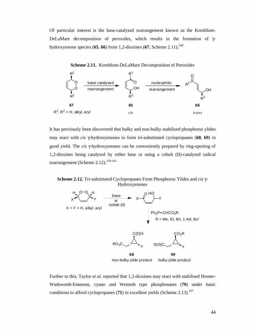
Of particular interest is the base-catalysed rearrangement known as the Kornblum-
DeLaMare decomposition of peroxides, which results in the formation of γ-
hydroxyenone species (65, 66) from 1,2-dioxines (67, Scheme 2.11).168
It has previously been discovered that bulky and non-bulky stabilised phosphorus ylides
may react with cis γ-hydroxyenones to form tri-substituted cyclopropanes (68, 69) in
good yield. The cis γ-hydroxyenones can be conveniently prepared by ring-opening of
1,2-dioxines being catalysed by either base or using a cobalt (II)-catalysed radical
rearrangement (Scheme 2.12).159-161
Scheme 2.12. Tri-substituted Cyclopropanes From Phosphorus Ylides and cis γ-Hydroxyenones
OO
YX
X = Y = H, alkyl, aryl
HOOX Y
Ph3P CHCO2R
R = Me, Et, Bn, 1-Ad, But
C(O)X
RO2C
CO2R
X(O)CY Y
non-bulky ylide product bulky ylide product
baseor
cobalt (II)
68 69
HH
Further to this, Taylor et al. reported that 1,2-dioxines may react with stabilised Horner-
Wadsworth-Emmons, cyano and Weinreb type phosphonates (70) under basic
conditions to afford cyclopropanes (71) in excellent yields (Scheme 2.13).167
Scheme 2.11. Kornblum-DeLaMare Decomposition of Peroxides
OO
OHO
R2
R1
OH
R2
R1
O
base catalysed
rearrangement
nucleophilic
67 65 66
cis trans
rearrangement
R1, R2 = H, alkyl, aryl
R2
R1
Page 45
45
Scheme 2.13
OO
YX
P R1O
EtOEtO
YX
O
R1
70
71
MeLi, THF
R2 R2
0 oC to rt
HH
Yield
X = Ph Y = Ph R1 = CO2Me R2 = H 81%
X = Ph Y = Ph R1 = CO2t-Bu R2 = H 75%
X = Ph Y = Ph R1 = CN R2 = H 53%
X = Ph Y = Ph R1 = C(O)N(Me)OMe R2 = H 91%
X = Ph Y = H R1 = CO2Me R2 = H 80%
X = Ph Y = H R1 = CN R2 = H 51%
X = Ph Y = Ph R1 = CO2Et R2 = Me 30%
This methodology has subsequently been employed in the synthesis of CCGs through
reaction with an aminophosphonate (72, Scheme 2.14). Base-catalysed ring opening of
the 1,2-dioxine is followed by Michael addition of the phosphonate nucleophile and
intramolecular ring closure to form the desired cyclopropanes as a 1:1 mixture of
diastereoisomers (73, 74), in good overall yield.128
Scheme 2.14. Synthesis of Substituted Carboxycyclopropyl Glycine Precursors128
73 74
OOR1R
(MeO)2P CO2Me
NHR2
O
72
LDA-THF in cyclohexane R1
R2HN CO2Me
H
R
OR1
R2HN CO2Me
H
R
OTHF
+
Total Yield
R = Ph R1 = Ph R2 = Cbz 47%
R = Ph R1 = H R2 = Cbz 54%
R = Ph R1 = Me R2 = Cbz 53%
R = Ph R1 = CH2OTBDMS R2 = Cbz 67%
R = 2-MeOPh R1 = CH2OTBDMS R2 = Boc 66%
R = Ph R1 = c-C6H11 R2 = Cbz 50%
Page 46
46
The cyclopropane diastereoisomers are easily separated by flash column
chromatography and can then easily be converted to the desired biologically active 3’-
cycloalkyl substituted carboxycyclopropylglycines. This synthetic route has several
advantages over those found in the literature in that a total of only seven steps are
required and the 3’-substitution can be altered simply by preparing the corresponding
1,2-dioxine.
2.7 3’-Cycloalkyl Carboxycyclopropylglycines
To extend the previous work and investigate novel carboxycyclopropyl glycines,
investigations into the synthesis of new cyclopropyl compounds having cycloalkyl
substitution were to be conducted. Based on previous structure-activity work found in
the literature, it was hypothesized that these compounds may be potent agonists or
antagonists at selected subtypes of metabotropic glutamate receptors. Scheme 2.15
below depicts the retro-synthetic strategy for making these compounds and follows
work previously carried out by the Taylor group, with the exception of the starting 1,2-
dioxines being cycloalkyl-substituted. The cyclopropane amino acids to be synthesized
were to have cycloalkyl ring sizes ranging from a 3-membered cyclopropyl right
through to a relatively bulky 7-membered cycloheptyl. Based on the structure-activity
relationships of previously tested CCGs, it was hypothesized that the activity of these
compounds may switch between agonist to antagonist as the ring size increases.
Page 47
47
Scheme 2.15. Retrosynthetic Strategy
R
H2N
H
HO
O
OH
O
R
CbzHN
H
HO
O
OH
O
R
CbzHN
H
MeO
O
OMe
O
R
CbzHN
H
PhO
O
OMe
O
R
CbzHN
H
Ph
O
OMe
O
OOPh R
P CO2Me
NHCbz
O
MeOMeO
+
deprotection
hydrolysis
trans esterification
Baeyer-Villigeroxidation
cyclopropanation
R = c-C3H5, c-C4H7,c-C5H9, c-C6H11, c-C7H13
Ph ROPh
R
PPh3Cl
Wittig
photo-oxidation
2.8 Synthesis of Target Cyclopropane Amino Acids
The requisite 1,3-butadienes (75a−−−−f) were prepared in good to excellent yields using
phosphorus ylide chemistry utilizing cycloalkanecarboxaldehydes and cinnamyl
triphenylphosphonium chloride except for diene 75f where (E)-2-
methoxycinnamaldehyde (76f) and cycloheptylmethyl triphenylphosphonium iodide
(77b) were employed (See Scheme 2.19). The cycloheptylmethyl
triphenylphosphonium iodide was prepared from cycloheptyliodomethane (78) which
Page 48
48
was conveniently synthesized from the corresponding alcohol via one of two routes
(A169 or B170) as illustrated in Scheme 2.16 following.
Scheme 2.16. Preparation of Cycloheptyliodomethane (75) Starting Material A. NIS, PPh3
B. I2, imidazole, PPh3
78a: 67% (A)
78b: 96% (B)
OH I
Where the required cycloalkanecarboxaldehyde was not available commercially, it was
prepared from the carboxylic acid via lithium aluminium hydride reduction followed by
oxidation with pyridinium dichromate. Some difficulty was experienced in obtaining the
aldehydes due to over-oxidation and so Swern conditions171 were investigated as well
as Parikh-Doering conditions,172,173 however, these proved to either produce mainly by-
products or to be too mild and the reaction too slow to be satisfactory. The Swern
oxidation of alcohol 79 produced only traces of aldehyde 76c, but mainly a by-product
which was determined by 1H NMR analysis to most likely be ester 80 (Scheme 2.17).
Scheme 2.17. Attempted Swern Oxidation
OH O
H
O
ODMSO, (COCl)2
Et3NCH2Cl2
+
79 76c 80trace
The Parikh-Doering oxidation of alcohol 79 also produced only a trace of aldehyde 76c
even after 24 hours reaction time and so this methodology was abandoned (Scheme
2.18).
Scheme 2.18. Attempted Parikh-Doering Oxidation
OH O
HDMSO, SO3.pyr
Et3NCH2Cl2
79 76c
Page 49
49
The crude 1,3-butadienes 75a−−−−f were obtained in high (58%) to excellent (97%) yields
with the desired isomer being the (E,E) as this can undergo addition to singlet oxygen to
form the 1,2-dioxine, whereas the (E,Z) isomer cannot. The reaction solvent used was
either anhydrous diethyl ether or anhydrous THF, however, it was found that THF gave
predominantly the (E,E) product whereas using ether afforded a mix of (E,E) and (E,Z).
This was not a great problem as subjecting the diene to photolysis conditions induces
isomerism from the (E,Z) to the desired (E,E) isomer.174
Scheme 2.19. Preparation of Butadienes via Wittig Reaction
R1
H
O R2 PPh3 R3 R1X
KOBut
(E,Z), (E ,E)
Yield
76a: R1 = c-C3H5 77a: R2 = (E)-cinnamyl 75a: R1 = c-C3H5 R3 = Ph 90%
76b: R1 = c-C4H7 77a: R2 = (E)-cinnamyl 75b: R1 = c-C4H7 R3 = Ph 74%
76c: R1 = c-C5H9 77a: R2 = (E)-cinnamyl 75c: R1 = c-C5H9 R3 = Ph 93%
76d: R1 = c-C6H11 77a: R2 = (E)-cinnamyl 75d: R1 = c-C6H11 R3 = Ph 97%
76e: R1 = c-C7H13 77a: R2 = (E)-cinnamyl 75e: R1 = c-C7H13 R3 = Ph 58%
76f: R1 = (E)-2-MeO-cinnamyl 77b: R2 = c-C7H13 75f: R1 = c-C7H13 R3 = 2-MeO-Ph 86%
X = Cl or I
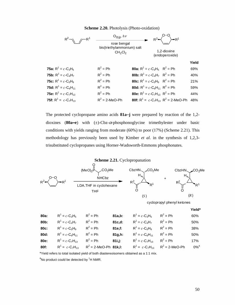
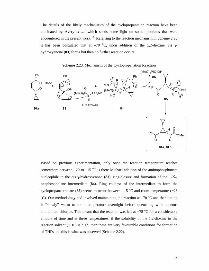
Photolysis (dye sensitised photo-oxidation) of the 1,3-butadienes 75a−−−−f employing rose
bengal bis(triethylammonium) salt as the sensitiser, in the presence of oxygen, afforded
the desired 3-cycloalkyl-6-phenyl-3,6-dihydro-1,2-dioxines 80a−−−−f in moderate (21%) to
high (69%) yields, including the known compound 80d128 (Scheme 2.20). This reaction
proceeds via a [4π + 2π] cycloaddition of singlet oxygen. Due to the inefficient and
non-selective nature of this reaction, there were many side products formed (ene,
cleavage) and this always meant that the maximum yield obtainable was reduced to a
greater or lesser extent, depending on the substrate 1,3-butadiene. It was often necessary
to carry out the photo-oxidation over several sessions in order to obtain a decent overall
yield with reaction times ranging from a total of 8 to 18 hours. Highlighting the
inefficiency of this photolysis process for these particular substrates, there was always a
large amount of un-reacted starting material remaining, even after subjecting the
reaction to photo-oxidation conditions for 8 hours or more.
Page 50
50
Scheme 2.20. Photolysis (Photo-oxidation)
R2 R1O O
R2 R1
rose bengalbis(triethylammonium) salt
O2(g), hν
1,2-dioxine(endoperoxide)
CH2Cl2
Yield
75a: R1 = c-C3H5 R2 = Ph 80a: R1 = c-C3H5 R2 = Ph 69%
75b: R1 = c-C4H7 R2 = Ph 80b: R1 = c-C4H7 R2 = Ph 40%
75c: R1 = c-C5H9 R2 = Ph 80c: R1 = c-C5H9 R2 = Ph 21%
75d: R1 = c-C6H11 R2 = Ph 80d: R1 = c-C6H11 R2 = Ph 59%
75e: R1 = c-C7H13 R2 = Ph 80e: R1 = c-C7H13 R2 = Ph 44%
75f: R1 = c-C7H13 R2 = 2-MeO-Ph 80f: R1 = c-C7H13 R2 = 2-MeO-Ph 48%
The protected cyclopropane amino acids 81a−−−−j were prepared by reaction of the 1,2-
dioxines (80a−−−−e) with (±)-Cbz-α-phosphonoglycine trimethylester under basic
conditions with yields ranging from moderate (60%) to poor (17%) (Scheme 2.21). This
methodology has previously been used by Kimber et al. in the synthesis of 1,2,3-
trisubstituted cyclopropanes using Horner-Wadsworth-Emmons phosphonates.
Scheme 2.21. Cyclopropanation
OOR2 R1
(MeO)2P CO2MeO
NHCbz
CO2MeH
CbzHN
R1R2
O
CO2MeH
CbzHN
R1R2
(S) (R)
LDA.THF in cyclohexane
+
cyclopropyl phenyl ketones
OTHF
Yield*
80a: R1 = c-C3H5 R2 = Ph 81a,b: R1 = c-C3H5 R2 = Ph 60%a
80b: R1 = c-C4H7 R2 = Ph 81c,d: R1 = c-C4H7 R2 = Ph 50%a
80c: R1 = c-C5H9 R2 = Ph 81e,f: R1 = c-C5H9 R2 = Ph 38%a
80d: R1 = c-C6H11 R2 = Ph 81g,h: R1 = c-C6H11 R2 = Ph 50%a
80e: R1 = c-C7H13 R2 = Ph 81i,j: R1 = c-C7H13 R2 = Ph 17%a
80f: R1 = c-C7H13 R2 = 2-MeO-Ph 81k,l: R1 = c-C7H13 R2 = 2-MeO-Ph 00%a
*Yield refers to total isolated yield of both diastereoisomers obtained as a 1:1 mix. aNo product could be detected by 1H NMR.
Page 51
51
However, it was shown that substitution even by a simple methyl group at the α-
position on the phosphonate caused a decrease in yield from 81% to 30%. The reason
for this was suggested to be the decreased nucleophilicity of the aminophosphonate.167
The attempted synthesis of cyclopropanes 81k and 81l was to investigate the effects of
2-methoxy substitution of the phenyl ketone on the rate of the subsequent reaction in the
synthetic sequence. Ortho-methoxy substitution had previously been used by our
research group to decrease reaction times of the Baeyer-Villiger oxidation, as this
increases electron density of the phenyl ring and speeds up migration.128 In this instance,
the change in electronic character of the phenyl ring seems to have caused the reaction
to fail. The cyclopropanation reaction was found to be generally unreliable, with
fluctuating yields and corresponding changes in the number of by-products formed.
Over the course of experimenting to optimize the reaction, different bases, reaction
temperatures and reaction concentrations were studied by other members of our
research group.128 However, these all resulted in even poorer yields or no isolated
product whatsoever. The reaction affords a 1:1 ratio of diastereoisomers which can be
separated by one or two rounds of careful flash chromatography. The difference
between the two Rf values for each isomer was approximately 0.05−0.10 and it was
noted that the first isomer off the column for all the cyclopropanes was the (S) isomer,
followed by the (R) isomer. Cyclopropanes 81g, h have previously been synthesized by
the Taylor group and our data was found identical to that reported.128
The main by-product that featured in these reactions was tetrahydrofurans (THFs)
(Scheme 2.22, 82), which were quite difficult to separate from the desired cyclopropane
products in any of the tested solvent systems, due to similar polarities of the THFs and
cyclopropanes.
Scheme 2.22. Formation of Tetrahydrofurans
Base, [83]
83 82
O
PhO
PhO
HOOHO
Ph
H
Page 52
52
The details of the likely mechanistics of the cyclopropanation reaction have been
elucidated by Avery et al. which sheds some light on some problems that were
encountered in the present work.128 Referring to the reaction mechanism in Scheme 2.23,
it has been postulated that at −78 oC, upon addition of the 1,2-dioxine, cis γ-
hydroxyenone (83) forms but then no further reaction occurs.
Scheme 2.23. Mechanism of the Cyclopropanation Reaction
HO
(MeO)2PO
H
H
Ph
O
RO
MeOPh
O
O
OMeR
H
OH
O
H
Ph
O
O
Ph
O(MeO)2P CO2Me
R
80a 83
85H
Ph
O
O
OMeR
H
84
81a, 81b
Base
R = HNCbz
(MeO)2P(O)OH
86
Based on previous experimentation, only once the reaction temperature reaches
somewhere between −20 to −15 oC is there Michael addition of the aminophosphonate
nucleophile to the cis γ-hydroxyenone (83), ring-closure and formation of the 1-2λ-
oxaphospholane intermediate (84). Ring collapse of the intermediate to form the
cyclopropane enolate (85) seems to occur between −15 oC and room temperature (~23 oC). Our methodology had involved maintaining the reaction at −78 oC and then letting
it “slowly” warm to room temperature overnight before quenching with aqueous
ammonium chloride. This meant that the reaction was left at −78 oC for a considerable
amount of time and at these temperatures, if the solubility of the 1,2-dioxine in the
reaction solvent (THF) is high, then these are very favourable conditions for formation
of THFs and this is what was observed (Scheme 2.22).
Page 53
53
The THFs form due to base-promoted self-condensation of two molecules of γ-
hydroxyenone via an oxa-Michael/Michael ring-closing reaction; the mechanism is
depicted in Scheme 2.24.
Scheme 2.24. Mechanism of Tetrahydrofuran Formation OO
R1 R2LiOH or alkoxide HOO
R1 R2
−OH
R2
R1
O
OHOO
R1
R2O
OR1
R2R2
HO
R1
O
O
OR1
R2R2
HO
R1
O
R1, R2 = H, alkyl, aryl
Eventually a procedure was found which requires addition of base and 1,2-dioxine at
−78 oC followed by warming to −15 to −10 oC for about 4 hours and then slow warming
to room temperature overnight. Using this protocol, it was possible to scale the reaction
up to 2.5 g for some 1,2-dioxines with total isolated yield for both diastereoisomers
ranging from 38 to 60%. Formation of the cyclopropane product was confirmed by the
appearance of a strong IR absorption due to the phenyl ketone at 1648−1670 cm-1. This
absorption peak would be expected to appear around 1685 cm-1, however, the frequency
is lowered since the ketone is also adjacent to the cyclopropane ring, which exhibits a
double bond character.175 The relative stereochemistry of the cyclopropanes could not
be determined using 1H NMR or 2D NMR due to overlap of the cyclopropyl and
cycloalkyl signals and identical ROESY interactions for both diastereoisomers. The
relative stereochemistry of cyclopropanes 81a, 81b and 81g were determined by single
crystal x-ray crystallography (Figures 2.3, 2.4, 2.5).
Page 54
54
Figure 2.3. X-ray Crystal Structure of Cyclopropane 81a
Molecular structure (50% probability ellipsoids) and crystallographic numbering scheme for
C24H25NO5
Figure 2.4. X-ray Crystal Structure of Cyclopropane 81b
Molecular structure (50% probability ellipsoids) and crystallographic numbering scheme for
C24H25NO5
Page 55
55
Figure 2.5. X-ray Crystal Structure of Cyclopropane 81e
Molecular structure (50% probability ellipsoids) and crystallographic numbering scheme for
C26H29NO5 (for reasons of clarity only one component of the disordered cyclopentane ring is
shown)
Page 56
56
Scheme 2.25. Baeyer-Villiger Oxidation
RPh
O
CbzHNOMe
O
H
RO
O
CbzHNOMe
O
H
Ph
ORtrifluoroperacetic acida
meta-chloroperbenzoic acidb
1 1
Yield
81a: (1S) R = c-C3H5 87a: (1S) R = c-C3H5 24%a* 77%b
81b: (1R) R = c-C3H5 87b: (1R) R = c-C3H5 11%a*
81c: (1S) R = c-C4H7 87c: (1S) R = c-C4H7 00%b*
81d: (1R) R = c-C4H7 87d: (1R) R = c-C4H7 47%b*
81e: (1S) R = c-C5H9 87e: (1S) R = c-C5H9 64%a*
81f: (1R) R = c-C5H9 87f: (1R) R = c-C5H9 19%a*
81g: (1S) R = c-C6H11 87g: (1S) R = c-C6H11 44%a*
81h: (1R) R = c-C6H11 87h: (1R) R = c-C6H11 47%a*
*Products readily decomposed under reaction conditions
The Baeyer-Villiger oxidation (Scheme 2.25) was performed using trifluoroperacetic
acid produced in situ from trifluoroacetic anhydride and hydrogen peroxide as per
Anastasia et al.176 These harsher conditions were used in lieu of the conventional m-
CPBA since the phenyl group is very slow to migrate when using m-CPBA, which
results in greatly increased reaction times and lower yields. This method worked fairly
well with the cyclohexyl-substituted cyclopropane (81g, h). However, yields decreased
significantly when it was attempted with the cyclopropyl (81a, b) and cyclobutyl (81c,
d) compounds where little or no product could be isolated. It was thought that the
strongly acidic conditions had caused the product to decompose and so it was opted to
try using m-CPBA, despite expecting a very long reaction time and potentially low
yields. After 5 weeks phenyl ester 87a was isolated in 77% yield. This methodology
was subsequently employed in the attempt to form the cyclobutyl compounds 87c and
87d, however after workup and purification, 1H NMR analysis revealed that the
products were prone to decomposition. The exact nature of the instability is unknown.
Owing to the long reaction times, an attempt was made to increase the speed of Baeyer-
Villiger phenyl group migration by 2-methoxy substitution and thus avoiding the use of
trifluoroperacetic acid as oxidant. The 2-methoxyphenyl cycloheptyl 1,2-dioxine was
synthesized successfully, however all attempts to form the cyclopropane failed.
Purification of the Baeyer-Villiger products was usually accomplished via flash column
Page 57
57
chromatography without much difficulty. However, due to the fact that when using m-
CPBA as oxidant, the reaction did not proceed to completion, it was sometimes not
possible to fully separate the starting ketone from the desired ester product. In these
cases, the impure product was simply carried through to the next step of the synthesis
and fully purified after the next synthetic transformation. Formation of the phenyl ester
could be confirmed by 1H NMR as an obvious disappearance of aromatic and
cyclopropane peaks associated with the protons adjacent to the ketone at approximately
8.0 ppm and 2.9 ppm respectively and the appearance of phenyl ester peaks at ca. 7.1
ppm. A corresponding shift was also observed in the 13C NMR with a shift in the
cyclopropyl carbonyl peak from approximately 198 ppm to ca. 172 ppm after
conversion to the ester. Also noted in the IR spectra, was the disappearance of the
absorption peak due to the phenyl ketone at 1648−1670 cm-1, which was replaced by an
ester carbonyl absorption that was largely obscured by the methyl ester absorption at
approximately 1740 cm-1.
Scheme 2.26. Trans-esterification
RMeO
O
CbzHNOMe
O
H
RO
O
CbzHNOMe
O
H
Ph
MeOH, H2SO4
1 1
ref lux
Yield
87a: (1S) R = c-C3H5 88a: (1S) R = c-C3H5 96%
87e: (1S) R = c-C5H9 88b: (1S) R = c-C5H9 80%
87f: (1R) R = c-C5H9 88c: (1R) R = c-C5H9 82%
87g: (1S) R = c-C6H11 88d: (1S) R = c-C6H11 76%
The trans-esterification reactions outlined in Scheme 2.26 went smoothly with yields
ranging from good (76%) to excellent (96%). This step is necessary in order to separate
the phenol by-product form the cyclopropanes, which would be difficult if the esters
were simply hydrolysed. The phenol that was formed could be clearly visualized via
TLC as a red spot, when developed with vanillin and was confirmation that the reaction
was proceeding. It was not difficult to purify the products by flash column
chromatography to remove all traces of the phenol by-product and residual starting
Page 58
58
material. All the dimethyl esters (88a−−−−d) were isolated as white solids and were easy to
handle, except the cyclopropyl substituted compound (88a) which was a colourless,
viscous, sticky gum. 1H NMR revealed the presence of a single phenyl ring and the
appearance of a second, sharp methyl ester peak at approximately 3.6 ppm. An IR
absorption peak due to the new methyl ester carbonyl group could be distinguished at
approximately 1720 cm-1.
Scheme 2.27. Ester Hydrolysis
LiOH(aq)
RHO
O
CbzHNOH
O
H
RMeO
O
CbzHNOMe
O
H
THF
1 1
Yield
88a: (1S) R = c-C3H5 89a: (1S) R = c-C3H5 92%
88b: (1S) R = c-C5H9 89b: (1S) R = c-C5H9 99%
88c: (1R) R = c-C5H9 89c: (1R) R = c-C5H9 96%
88d: (1S) R = c-C6H11 89d: (1S) R = c-C6H11 97%
Ester hydrolysis gave almost quantitative yields of 89a−−−−d (Scheme 2.27), however due
to the workup using ethyl acetate and concentrated hydrochloric acid, it was necessary
to add toluene and then remove it in vacuo to remove all traces of acetic acid that was
formed by hydrolysis of the solvent. The success of the hydrolysis was indicated by 1H
NMR by the disappearance of both methyl peaks. The IR spectra showed a shift in
frequencies corresponding to the carboxylic acid groups at ca. 1694 and 1680 cm-1.
Yields were in the range of 92 to 99%.
Page 59
59
Scheme 2.28. Hydrogenolysis (Amine Deprotection)
H2(g), 5% Pd/C
RHO
O
H2NOH
O
H
RHO
O
CbzHNOH
O
H
1 1
Yield
89a: (1S) R = c-C3H5 90a: (1S) R = c-C3H5 95%
89b: (1S) R = c-C5H9 90b: (1S) R = c-C5H9 ND
89c: (1R) R = c-C5H9 90c: (1R) R = c-C5H9 ND
89d: (1S) R = c-C6H11 90d: (1S) R = c-C6H11 ND
ND: not determined
The carboxybenzyl (Cbz) amine protecting group was utilized so that amine
deprotection could be done as the final step via a hydrogenolysis reaction, leaving the
amine in need of minimal purification (Scheme 2.28). The method previously used for
workup by our research group was to add water to the reaction followed by filtration
though a pad of kenite, washing with ethyl acetate and evaporation of the water to give
pure amino acid. This method depended on the product being fairly water soluble and it
was found to be successful in the isolation of 90a, however, it was found that the
cyclopentyl- (90b, c) and cyclohexyl-substituted (90d) compounds were methanol
soluble, which complicated their purification. Conversion of the requisite amino acid to
the hydrochloride salt was needed in order to afford pure product, however as the free
amino acid was desired, this necessitated removal of the HCl as the last step. The
method employed to do this was the same as used by Collado55 and required stirring a
concentrated methanol solution of the hydrochloride salt with dry propylene oxide
followed by filtration to obtain the solid product (Scheme 2.29). The HCl reacts with
the propylene oxide to form 1-chloro-2-propanol which can be removed by drying the
product under a hard vacuum.
Page 60
60
Scheme 2.29. Removal of HCl
RHO
O
H2NOH
O
H
RHO
O
HCl.H2NOH
O
H
1propylene oxide
1
MeOH
Yield
90b: (1S) R = c-C5H9 91b: (1S) R = c-C5H9 00%
90c: (1R) R = c-C5H9 91c: (1R) R = c-C5H9 00%
90d: (1S) R = c-C6H11 91d: (1S) R = c-C6H11 16%
This protocol was attempted on the cyclopentyl (90b, c) and the cyclohexyl (90d)
compounds. The cyclopropyl compound was sufficiently water soluble to negate its
conversion to the hydrochloride salt for purification. It was found upon filtration of the
cyclopentyl reactions that no product remained, presumably due to decomposition.
Decomposition also occurred in the cyclohexyl reaction, however, a small amount of
91d was able to be recovered upon filtration.
2.9 Summary
Two new cycloalkylcarboxycycloproplyglycine analogues were successfully
synthesized utilizing the reaction of 1,2-dioxines with protected phosphonates to rapidly
construct the cyclopropane core with all necessary stereochemistry of pendant groups,
in a 20% overall yield for one diastereoisomer.
Page 61
61
Chapter 3 : Triazole Amino Acids
3.1 Introduction
One class of compounds that have not been widely investigated as glutamate receptor
ligands are the 1,2,3-triazoles. Thus, a series of basic analogues was designed which
could be obtained in a synthetically straight-forward manner (Figure 3.1). Given the
structural similarity between the proposed triazoles and known ionotropic glutamate
receptor ligands, it was hypothesized that these new structures may also show biological
activity (Figure 3.2). To this end, the library of synthesized compounds was screened in
vitro against both ionotropic (NMDA, kainate and AMPA) and metabotropic (mGluR1,
mGluR2, mGluR4 and mGluR5) glutamate receptors.
Figure 3.1. Proposed Triazole Amino Acids
H2N CO2H
N NN
H2N CO2H
N NN
H2N CO2H
N NN
O
OH
OOH
OHO
H2N CO2H
N NN
O
HO
48 49
4645
50
47
H2N CO2H
N NN
OHO
H2N CO2H
N NN
O
HO
The triazole moiety is an aromatic five membered heterocycle containing 3 nitrogens;
those being considered here are 1,2,3-triazoles.177 When one of the nitrogens is
substituted, the other two have lone pairs that can potentially act as hydrogen bond
donors.
Page 62
62
Figure 3.2. Structural Comparison Between Triazoles and Known Ligands
H2N CO2H
NO
HO
H2N CO2H
N
N
O
HO
O
H2N CO2H
NO
HO
O 36 18 92
(S)-AMPA Quisqualate (S)-ACMP
H2N CO2H
N NN
O
OH
H2N CO2H
N NN
O
HO
H2N CO2H
N NN
OHO
48 49 46
H2N CO2H
N NN
OOH
H2N
NN N
CO2H
O OH
H2N
NN N
CO2H
HOO
45 50 47
H2N CO2H
N O
HO
H2N CO2H
N
NOHO
O
H2N CO2H
O NOH
40 37 38
(S)-Homo-AMPA (S)-Homo-Quisqualate (S)-HIBO
H2N
NO
HOCO2H
93 (S)-APPA
Page 63
63
As is depicted in Figure 3.2, this configuration mimics the arrangement found in some
competitive AMPA and NMDA receptor ligands such as (S)-AMPA (36), Quisqualate
(18) and (S)-ACMP (92) where either an isoxazole or oxadiazolone ring is
present.107,178,179 Many of these known ligands incorporate hydroxyl substitution on the
heterocycle, which bears an acidic proton and thus acts as a carboxylic acid bioisostere.
However, one compound, (S)-ACMP (92) is a known AMPA receptor antagonist which
bears a 2-carbon chain terminated with a carboxylic acid group.180
The current research was seeking novel metabotropic receptor ligands as opposed to
ionotropic ligands and so it was chosen to employ carboxylic acid substitution on the
heterocycle, instead of a hydroxyl group. This was done in an effort to increase affinity
towards the mGluRs over the iGluRs. Another structural feature of some iGluR ligands,
for example, the AMPA receptor agonist, (S)-APPA (93), is substitution on the
heterocycle by another aromatic ring.181 This is usually a simple phenyl or pyridinyl
group. Compounds 47 and 50 were designed in order to probe the effects of phenyl
substitution on the carboxylic acid chain adjacent to the heterocycle.
The 1,2,3-triazole core structure offers some advantages. Firstly, it is readily prepared
by a high yielding copper or ruthenium catalysed cycloaddition reaction. Secondly, it is
possible to establish a library of compounds for biological screening by cycloaddition of
a few simple azide and alkyne building blocks in different combinations (Figure 3.3).
Compounds 94, 98 and 101 have previously been reported and our analytical data was
found to be in agreement with the literature.182-184
Figure 3.3. Key Azide and Alkyne Building Blocks
BocHN CO2R
R = H, Me
BocHN CO2R
N3
R = H, Me
N3 CO2R
Ph
R = H, Me
N3 CO2But CO2But
96, 97 98, 99 100, 101
94 95
Page 64
64
Thus, the following retro-synthetic strategies were devised (Scheme 3.1-Scheme 3.4).
The desired triazolyl amino acid hydrochloride salts (45a, 46a, 47a, 48a, 49a, 50a)
could be obtained from the Boc-protected amino acids via treatment with HCl. These in
turn could be obtained from the methyl or t-butyl ester protected carboxylic acids by
alkaline hydrolysis. Formation of the protected triazoles is accomplished either by a
‘click’-style copper-catalysed cycloaddition (Schemes 3.1, Scheme 3.2) or a ruthenium-
catalysed cycloaddition (Schemes 3.3, Scheme 3.4) involving the reaction between a
functionalized azide (94, 98, 99, 100, 101) and alkyne (95, 96, 97).
Scheme 3.1. Retro-Synthetic Strategies
NNN
HCl.H2N CO2H
CO2H
NNN
BocHN CO2H
CO2But
N3
BocHN CO2H
CO2Bu t
Cu (I) Cycloaddition
Amine Deprotection
45a
96
101
Page 65
65
Scheme 3.2. Retro-Synthetic Strategies
NN
N
HCl.H2N CO2H
CO2H
R1N
NN
BocHN CO2H
CO2H
R1
NN
N
BocHN CO2H
CO2R2
R1
N3
BocHN CO2H
CO2R2
R1
R1 = H, R2 = t-BuR1 = CH2Ph, R2 = Me
Cu (I) Cycloaddition
Ester Hydrolysis
Amine Deprotection
46a, 47a
94
100:99:
Scheme 3.3. Retro-Synthetic Strategies
NNN
HCl.H2N CO2H
NNN
BocHN CO2H
HO2C HO2C
NNN
BocHN CO2Me
t -BuO2C
N3
BocHN CO2Me
t-BuO2C
[Ru] Cycloaddition
Amine Deprotection
Ester Hydrolysis
48a
97
101
Page 66
66
Scheme 3.4. Retro-Synthetic Strategies
NN
N
HCl.H2N CO2H
HO2C
R1
NN
N
BocHN CO2H
HO2C
R1
NN
N
BocHN CO2Me
R2O2C
R1
N3
BocHN CO2Me
R2O2C
R1
R1 = H, R2 = t-BuR1 = CH2Ph, R2 = Me
[Ru] Cycloaddition
Ester Hydrolysis
Amine Deprotection
49a, 50a
95
100:99:
3.2 Triazoles in Drug Discovery
Examination of the current literature reveals that there have been relatively few efforts
to investigate 1,2,3-trizoles as potential drug molecules.185-188 Gajewski and co-workers
have reported the design and synthesis of several triazolyl amino acids as potential
inhibitors of the neutral amino acid transporter SN1 (Figure 3.4).185 This transporter is
an L-glutamine shuttle responsible for transporting the endogenous amino acid in and
out of cells. Six potential ligands (45, 102−−−−106) were synthesized including compound
45 that was investigated in the present work, however, none showed significant
inhibitory activity in the amino acid transporter assay.185
An interesting advance put forward by Sharpless and co-workers is the synthesis of an
acetylcholinesterase inhibitor by in situ click chemistry. The process involved
incubation of the enzyme from Torpedo californica with azide-functionalized tacrine, a
known main binding site ligand, and allowing a variety of acetylene-functionalized
phenylphenanthridinium derivatives (with known activity) to interact with peripheral
binding sites (Figure 3.5). Triazoles are only formed when there is a favourable
interaction between the enzyme and both the azide and alkyne.
Page 67
67
Figure 3.4. Triazolyl Amino Acids as Amino Acid Transporter Inhibitors185
N NN
H2N CO2H
N NN
H2N CO2H
OH
N NN
H2N CO2H
N NN
H2N CO2H
OHO
N NN
H2N CO2H
OCH3O
N NN
H2N CO2H
NH2O
102 103 104
45 105 106
Figure 3.5. Triazole Acetylcholinesterase Inhibitor Synthesized In Situ189,190
N
H2N NH2
NNN
NH
N
syn-1
107
Page 68
68
The interactions cause an energy barrier lowering effect which catalyses the reaction.
Through this methodology, it was possible to find several hit structures that showed
increased activity and better enzyme kinetics than either of the known lead compounds.
The most potent new compound identified was syn-1 (107)189,190
3.3 Background on the Synthesis of 1,2,3-Triazoles
The original preparation of this class of heterocycle, reported by Huisgen in 1963,
involved a thermally promoted cycloaddition of an azide to a terminal alkyne resulting
in both 1,4- and 1,5-substituted products (Scheme 3.5). The energy barrier for the
thermally driven reaction has been calculated to be a very high +26 kcal/mol due to the
inherent stability of both azides and alkynes. For this reason, these reactions necessitate
high temperatures and long reaction times in order to furnish reasonable yields.
Scheme 3.5. Huisgen 1,3-Dipolar Cycloaddition
R1
N N N
R3R2
NN
NR1
R2 R3∆
26 kcal/mol
It was soon discovered that the energy barrier for the cycloaddition reaction could be
lowered significantly by use of copper (I) catalysis. Catalysed cycloaddition of an azide
to a terminal alkyne (CuCAA) was subsequently made famous by Barry K. Sharpless
and co-workers under its ‘Click Chemistry’ moniker.191,192 It has since been employed
as a versatile coupling reaction, especially in polymer chemistry and in the attachment
of long chain linkers and pendant groups.192-196 However, as stated earlier, it has been
used in relatively few drug discovery applications, until recently.185-188 It should be
noted that copper catalysis only affords the 1,4-substituted triazoles, however,
ruthenium catalysis allows access to the 1,5-substituted and tri-substituted 1,2,3-
triazoles (Figure 3.6).
Figure 3.6. 1,4- and 1,5-Substituted 1,2,3-Triazoles
NN
N
R1
R2 NN
N
R1
R2
R3
1 1
4 4 5
1,4Cu catalysis
1,5Ru catalysis
Page 69
69
The catalytic cycle for the cycloaddition has been elucidated and is outlined in Scheme
3.6. Briefly, the Cu(I) displaces a proton from the acetylene followed by coordinate
bonding of the azide to the copper. The binding of the acetylene to the copper increases
the electrophilicity of the alkyne, enabling attack by the azide to form a six-membered
cupro-cycle. Ring-collapse followed by proton displacement of the copper releases the
final 1,2,3-triazole product.
Scheme 3.6. Copper Azide Alkyne Cycloaddition Catalytic Cycle197,198
NN
N
HR1
R2
[CuLx]
NN
N
CuLxR1
R2
R1 H
R1 H
CuLx
R1 CuLx
N N NR2
R1 CuLx N N NR2
R1 CuLx
CuLx
N N NCuLx
R2
R1
N N NR2
RDS
23 kcal/mol
18 kcal/mol
H+ H+
R1 = alkyl, aryl, CO2H, NH2
R2 = alkyl, aryl
Page 70
70
It is possible to obtain 1,5-substituted triazoles as the sole product by use of ruthenium
catalysis. Several catalysts have been investigated including those outlined in Table 3.1.
As summarized in Table 3.1 above, in order to obtain 1,5-substituted triazoles in good
yields, a pentamethylcyclopentadiene (Cp*) ruthenium catalyst must be used.
[Cp*RuCl]4 (108) is the most potent catalyst thus far reported and can be used to
produce tri-substituted triazoles. Ruthenium complex 109 is also very potent and shows
complete selectivity for 1,5-cycloaddition. However, this catalyst has an advantage over
more potent catalyst 108 in that it can be stored for prolonged periods and does not have
to be prepared each time it is required.
The mechanism for ruthenium catalysis is shown in Scheme 3.7. Briefly, both the azide
and alkyne coordinate to the metal centre simultaneously, thus displacing two ligands.
This causes activation of both the azide and alkyne allowing them to form a cyclic
intermediate which collapses to form the tethered triazole which is subsequently
displaced from the metal centre by re-coordination to the starting ligands.
Table 3.1. Ruthenium (II) catalysis product distribution of benzyl azide and phenyl acetylene cycloaddition199
Ru catalyst 1,5-Substitution 1,4-Substitution
RuCl2(PPh3)3 - 1.7%
Ru(OAc)2(PPh3)3 - 46.5%
RuHCl(CO)(PPh3)3 - 2.5%
RuH2(CO)(PPh3)3 - 55.8%
CpRuCl(PPh3)2 12.9% 1.3%
Cp*RuCl(COD) 100% -
Cp*RuCl(NBD) 93.5% -
[Cp*RuCl]4 (108) 100% -
Cp*RuCl(PPh3)2 (109) 100% -
Page 71
71
Scheme 3.7. Ruthenium Azide Alkyne Cycloaddition Catalytic Cycle199
RuLCl L
NN
NR2
R1 H
RuNCl R2
NN
R1
Ru NCl R2
NR1
N
RuCl N N
NR2
R1
NN
N R2
R1
2L 2LStep A
Step BStep C
Step D
R1 = alkyl, aryl, CO2Me, NHBoc
R2 = alkyl, aryl
3.4 Synthesis of the Target Triazole Amino Acids
As stated previously, the target molecules for synthesis were the 1,4- and 1,5-substituted
1,2,3-triazole amino acids depicted in Figure 3.7.
Figure 3.7. Proposed Triazole Amino Acids
H2N CO2H
N NN
H2N CO2H
N NN
H2N CO2H
N NN
O
OH
OOH
OHO
H2N CO2H
N NN
O
HO
48 49
4645
50
47
H2N CO2H
N NN
OHO
H2N CO2H
N NN
O
HO
Page 72
72
Following is a discussion on the synthesis of the starting materials required in these
syntheses. The most basic azide building block was tert-butyl azidoacetate (94) which
was prepared from bromo ester 110 by reaction with sodium azide (Scheme 3.8). This
reaction employed chloroform as solvent, however, these conditions can give rise to
some diazidomethane formation, which is dangerously explosive.200 One literature
synthesis reports dimethylformamide as solvent, however, due to its high boiling point,
this would make purification difficult, since the product is volatile. A more suitable
method has been reported by Vollmar and Dunn where the reaction is carried out in
aqueous acetone, followed by removal of the acetone and ether extraction.201
Nevertheless, the azide was obtained, albeit in low yield (38%). During TLC analysis, it
was expected that it would be possible to visualize the product using UV light, due to
the p-orbital conjugation of the azide moiety. Contrary to this, it was discovered that the
bromo starting material was actually fluorescent under UV light. This caused confusion
during isolation and purification, until IR analysis was carried out on the suspected
desired product and no diagnostic azide peak was observed. The presence of the desired
product in the other major fraction with an IR spectrum revealing a strong absorption
peak at approximately 2100 cm-1.
Scheme 3.8. Preparation of tert-Butyl Azidoacetate (94)
BrOBut
ON3
OBut
O
NaN3
[C4H9]4NBrCHCl3
38% 110 94
Preparation of Boc-protected propargyl glycine (96) was accomplished by reaction of
amino acid 111 with di-tert-butyl dicarbonate with aqueous sodium hydroxide as base
(Scheme 3.9). Initially, the base used for this reaction was triethylamine, however, after
workup the desired product was not evident in the 1H NMR. It’s possible that the
triethylamine salt formed and was more resistant to re-protonation with acid during the
workup than would be expected.
Page 73
73
Scheme 3.9. Synthesis of Boc-Propargylglycine (96) H2N CO2H BocHN CO2H(Boc)2O, NaOH(aq)
98% 111 96
The methyl ester 97 was prepared in 83% yield from 111 by a literature procedure
consisting of a two step, one pot synthesis employing thionyl chloride and forming the
methyl ester hydrochloride salt as an intermediate (Scheme 3.10).202
Scheme 3.10. Synthesis of Boc-Propargylglycine Methyl Ester (97) H2N CO2H BocHN CO2Me
2. (Boc)2O, NaHCO3
1. SOCl2, MeOH
83%(over 2 steps)
111 97
Boc-protected azide (98) was prepared from Boc-L-2,3-diaminopropionic acid (112)
using freshly prepared triflyl azide as an azide transfer reagent (Scheme 3.11). This
method of diazotransfer, devised by Yan and colleagues, has significant advantages
over other methods.184 For example, it is usually carried out using water as co-solvent,
however, this requires the use of a large excess of the relatively expensive and highly
toxic triflic anhydride. By generating the triflyl azide in acetonitrile, it is possible to
decrease the amount of triflic anhydride required; the amount of sodium azide required
is also reduced compared to other methods. The main by-product of this reaction (trifyl
amine) is quite difficult to remove completely. However, its low solubility in
chloroform does allow separation from the target material. Due to the difficultly in
purification of this starting material, a quite low 32% yield was obtained. The IR
spectrum showed the expected sharp azide absorption at 2111 cm-1.
Scheme 3.11. Synthesis of Boc-Azidoalanine (98) BocHN CO2H
NH2
BocHN CO2H
N3MeCN, H2O
TfN3, Et3N, CuSO4
32% 112 98
Page 74
74
An alternative procedure to prepare 99 was sought, owing to the difficulty in
purification and low yield when using triflyl azide, as for synthesis of 98. Hydroxyl
groups are conveniently transformed into azides by means of a Mitsunobu reaction
(Scheme 3.12).203 Thus, Boc-serine methyl ester (113) was converted to azide 99 under
basic, anhydrous conditions. However, it was found that purification of this reaction
was more difficult that the previous method due to similar polarities of starting material
and desired product, even so the overall yield was significantly improved over that for
the synthesis of azide 98. The product was confirmed as an azide by appearance of a
diagnostic sharp IR absorption around 2108 cm-1.
Scheme 3.12. Synthesis of Boc-Azidoalanine Methyl Ester (99)
BocHN CO2Me
OH
BocHN CO2Me
N3THF
DPPA, DIAD, PPh3
69% 113 99
The use of triflyl azide as an azide transfer reagent smoothly converted L-phenylalanine
(114) to provide azide 100 as a pale yellow gum (Scheme 3.13). The presence of the
azide functionality was indicated by IR absorption at 2118 cm-1.
Scheme 3.13. Synthesis of 2-Azido-3-Phenyl-Propanoic Acid (100)
H2N CO2H
Ph
N3 CO2H
PhMeCN, H2O
TfN3, Et3N, CuSO4
44% 114 100
The corresponding known methyl ester 101 was prepared by dissolving compound 100
in methanol and adding minimal thionyl chloride (Scheme 3.14).184 Infrared
spectroscopy analysis revealed expected absorptions for the azide moiety at 2112 cm-1
and for the ester carbonyl at 1747 cm-1.
Page 75
75
Scheme 3.14. Synthesis of Methyl 2-Azido-3-Phenyl-Propanoate (101)
N3 CO2H
Ph
N3 CO2Me
Ph
SOCl2, MeOH
97% 100 101
It was brought to our attention that there was a possibility that in the preparation of
azides 99 and 101 and alkyne 97, some racemisation may have occurred. In order to
investigate this, it was necessary to deprotect the molecules and couple them to another
optically pure amino acid (Schemes 3.15, 3.16). By doing this, if the starting materials
were not optically pure, then diastereoisomers would be formed, which could be
detected by 1H NMR analysis. For simplicity of the resulting spectra, it was opted to
couple to L- and D-valine to provide both isomers for analysis. Thus, alkyne 97 was
stirred in anhydrous dichloromethane to which was added 0.6 mL of concentrated HCl.
After stirring at room temperature overnight, the solvent and excess HCl were removed
under reduced pressure to furnish hydrochloride salt 115. This was coupled to Boc-L-
valine (116) using HOBt and EDC under basic conditions to provide di-peptide 117 in
89% yield over two steps. Azide methyl esters 99 and 101 were dissolved in THF and
treated with 2M LiOH overnight to provide the free carboxylic acids. These were
coupled separately to both L-valine methyl ester (118) and D-valine methyl ester (119)
using HOBt and EDC under basic conditions to provide di-peptides 120, 121, 122 and
123 in 69 to 92% yield over two steps.
Scheme 3.15. Synthesis of Dipeptide 117
HCl.H2N CO2MeBocHN CO2H
MeMe
EDC.HCl, HOBt, TEA
DCM
BocHN
MeMe
NH
CO2Me
O
+
89% 116 115 117
Page 76
76
Scheme 3.16. Synthesis of Dipeptides 120−−−−123
R1 CO2H
R2
HCl.H2N CO2Me
MeMe
R1NH
CO2Me
OMe Me
R2
EDC.HCl, HOBt, TEA
DCM+
Yield 118 120:R1 = N3 R2 = Ph (S,S) 69% 119 121:R1 = N3 R2 = Ph (S,R) 86% 118 122:R1 = NHBoc R2 = N3 (S,S) 92% 119 123:R1 = NHBoc R2 = N3 (S,R) 77%
After coupling and workup, no crude 1H NMR of the di-peptides revealed more than 8%
diastereoisomer. Examples of excerpts from the 1H NMR spectra for compounds 121
and 122 are found in Figure 3.8. The ratio of diastereoisomers was determined by
comparison of the relative areas of the peaks corresponding to the methoxy groups
(highlighted in yellow) found between 3.60 and 3.80 ppm. Considering that the
conditions used for the amino acid coupling can also induce some epimerization, the
presence of this amount of optical impurity in the starting materials was not considered
to be a major problem.
Figure 3.8. Expansions from the 1H NMR Spectra Used for Determination of the Relative Diasteromeric Ratios
Page 77
77
With the basic building blocks (Figure 3.3, 94−−−−101) in hand, we set about synthesizing
a number of 1,2,3-triazole amino acid analogues. Referring to Scheme 3.17, alkyne 95
and azide 98 were dissolved in a 2:1 mix of t-butanol and water. Copper sulfate was
added at 5 mol% loading followed by sodium ascorbate to enable the necessary
reduction of Cu2+ to Cu1+ and enabling the cycloaddition to proceed. Stirring overnight
at room temperature was usually sufficient to obtain maximal yield, however, the
reaction time was occasionally extended to two days. After removal of solvents under
vacuum, pure triazole 124 was usually obtained after a single pass of flash column
chromatography. Likewise, referring to Scheme 3.18, azides 94 and 101 were allowed
to react with alkyne 96 and pure triazoles 125 and 126 respectively, obtained in good
yield.
Page 78
78
Scheme 3.17. Synthesis of Triazole 124
BocHN CO2H
N3
CO2But
N NN
CO2Bu t
BocHN
HO2CCuSO4 . 5H2ONa ascorbate
t -BuOH / H2O (2:1)
95
96% 98 124
The reaction time ranged from 16 to 60 hours with yields of triazoles ranging from good
(64%) to excellent (96%). Formation of the desired triazoles was indicated by the
appearance in the 1H NMR spectrum of a diagnostic aromatic triazole peak at ca. 7.50
ppm for compounds 125 and 126 and at ca. 8.04 ppm for compound 124.
Scheme 3.18. Synthesis of 1,4-Triazoles 125, 126
BocHN CO2H N3 CO2R1
R2
NN
N
BocHN
HO2C
R2
CO2R1
CuSO4 . 5H2ONa ascorbate
t-BuOH / H2O (2:1)96
Yield 94 R1 = t-Bu R2 = H (S,S)-125 85%* 101 R1 = Me R2 = CH2Ph (S,S)-126 64%*
In pursuit of the 1,5-substituted triazoles obtainable by ruthenium catalysis, preparation
of the most potent catalyst thus far reported, 108 from ruthenium compound 127 was
attempted.199,204 However, all attempts were unsuccessful (Scheme 3.19). This catalyst
has the disadvantage that it must be freshly prepared each time it is required and cannot
be stored.
Scheme 3.19. Attempted Preparation of Ruthenium Catalyst (108)
LiEt3BHCp*RuCl2 [Cp*RuCl]4
THF, N21 h
127 108 No product isolated
Page 79
79
The literature procedure describes a colour change from orange to blue-green, however
this was not observed.204 One explanation is that excess oxygen in the reaction caused
decomposition, though oxygen sensitivity has not been reported in the literature. Most
likely the lithium triethylborohydride purchased from Aldrich was contaminated or not
of sufficient concentration to enable the reaction to proceed. Owing to time constraints,
synthesis of this particular catalyst was abandoned and the synthesis of ruthenium
catalyst 109 pursued.
Ruthenium catalyst Cp*Ru(PPh3)2Cl (109) was prepared from Cp*RuCl2 (127) which
was synthesized by another member of our research group, Dr. Daniel Pedersen
(Scheme 3.20).205 This catalyst has the advantage that, unlike the more potent catalyst
108, it may be stored under an inert atmosphere for several weeks without significant
decomposition occurring. Following Morandini et al.205, the catalyst was obtained in
moderate yield (49%) as a brick red / orange solid which was used without further
analysis, as the catalyst is known to undergo decomposition in deuterated chloroform.
Scheme 3.20. Preparation of Ruthenium Catalyst (109)
PPh3Cp*RuCl2 Cp*Ru(PPh3)2Cl
EtOH, reflux, N26 h
49% 127 109
Unlike the copper-based chemistry, the ruthenium-catalysed reaction must be carried
out under an inert atmosphere and under anhydrous reflux conditions. Initially, the
reaction was attempted using azide 98 and alkyne 95, as used for the copper-catalysed
cycloaddition, but it was found that the ruthenium catalyst was sensitive to free
carboxylic acid groups on the reaction substrates (Scheme 3.21). The presence of a
carboxylic acid group (as in 98), resulted in a complex mixture of products and signs of
catalyst decomposition were evident (colour change from orange to brown). Although
traces of the desired triazole product could be detected in the 1H NMR, it was not
possible to cleanly isolate the compound.
Page 80
80
Scheme 3.21. Attempted Synthesis of 1,5-Triazole 111 (128)
BocHN CO2H
N3
CO2But
THF
Cp*Ru(PPh3)2Cl (2 mol%)
reflux, 3h
95
could not be isolated
NNN
CO2But
CO2HBocHN
98 128
In order to avoid this, all carboxylic acid groups were protected as methyl esters. Thus,
the reaction of azide 99 with alkyne 95 was carried out in refluxing anhydrous THF and
using the ruthenium catalyst 109 at 2 mol% loading (Scheme 3.22). Though it has not
been reported for this particular catalyst, organometallic ruthenium complexes are often
sensitive to oxygen, which can oxidize the metal centre. Because of this, the THF was
de-oxygenated by bubbling nitrogen through the solution just prior to use. The solution
was heated at reflux for 3 hours and over the course of the reaction the colour of the
solution changed from orange to brown indicating that the catalyst had undergone some
decomposition. Following this, volatiles were removed in vacuo and the residue
subjected to flash column chromatography. A second pass of column chromatography
was necessary in order to remove all traces of brown-coloured ruthenium species.
Protected 1,5-triazolyl amino acid 128 was obtained in low (33%) yield.
Scheme 3.22. Synthesis of 1,5-Triazole 128
BocHN CO2Me
N3
CO2Bu t
THF
Cp*Ru(PPh3)2Cl (2 mol%)
reflux, 3h
95
33%
NNN
CO2But
CO2MeBocHN
99 128
Employing the same methodology, azides 94 and 101 were allowed to react with alkyne
97 and the pure triazoles 129 and 130 were obtained in good yield (64% and 85%
respectively) (Scheme 3.23). Formation of the desired triazoles was indicated by the
appearance in the 1H NMR spectrum of a diagnostic aromatic triazole peak at ca. 8.02
ppm for compound 128 and at ca. 7.50 ppm for compounds 129 and 130.
Page 81
81
Scheme 3.23. Synthesis of 1,5-Triazoles 129−130
BocHN CO2Me N3 CO2R1
R2
THF
Cp*Ru(PPh3)2Cl (2 mol%)
reflux, 3 h
NN
N
CO2R1BocHN
MeO2C
R2
97 Yield
94 R1 = t-Bu R2 = H (S,S)-129 85%*
101 R1 = Me R2 = CH2Ph (S,S)-130 64%*
The methyl esters (126, 128, 129 and 130) were hydrolysed with LiOH. The reactions
were stirred overnight at room temperature and the solvent removed in vacuo before
being acidified with 1M HCl. The product was partitioned into ethyl acetate and
solvents removed under vacuum to give the crude carboxylic acid. Traces of acetic acid
formed during workup were removed azeotropically by repeated addition and removal
in vacuo of benzene or toluene. The hydrolysis afforded the dicarboxylic acids 132 and
131 in excellent (96%) and quantitative yields, respectively (Schemes 3.24−3.27). It
should be noted that the compounds 128 and 129 contained a t-butyl ester which
resulted in a mixture of hydrolysis products, since the t-butyl ester group was partially
hydrolysed during workup. These compounds were isolated as a mix of di-carboxylic
acid and t-butyl ester (133a,b and 134a,b, respectively) and were carried to the next
deprotection step without further purification.
Scheme 3.24. Synthesis of Diacid 131
NN
N
BocHN
HO2C
CO2Me
NN
N
BocHN
HO2C
CO2HLiOH
THF / H2O
quantitative
PhPh
126 (S,S)-131
Page 82
82
Scheme 3.25. Synthesis of Diacid 132
NN
N
BocHN
MeO2C LiOH
CO2Me
NN
N
BocHN
HO2C
CO2H
Ph Ph
THF / H2O96%
130 (S,S)-132
Scheme 3.26. Synthesis of Diacids 133a,b
THF / H2O
LiOH
quantitative
NNN
CO2R
CO2HBocHN
NNN
CO2But
CO2MeBocHN
128 133a: R = H
133b: R = t-Bu
Scheme 3.27. Synthesis of Diacids 134a,b
NN
N
BocHN
MeO2CLiOH
CO2But
NN
N
BocHN
HO2C
CO2RTHF / H2O
65% 129 134a: R = H
134b: R = t-Bu
Deprotection of the amine and hydrolysis of the t-butyl ester was carried out under
acidic conditions (Schemes 3.28−3.33). Triazole 124 was stirred at room temperature in
1,4-dioxane with 6M HCl (2:1) for 16 hours. Volatiles were removed under high
vacuum and the residue washed with ethyl acetate and dichloromethane. Any traces of
residual water and HCl were removed azeotropically by repeated addition and removal
in vacuo of benzene and toluene. The residue was dried overnight under high vacuum to
afford the final amino acid which was obtained as hydrochloride salt 45a in quantitative
yield. Following the same procedure, triazole 125 was hydrolysed to give hydrochloride
salt 46a in quantitative yield, triazole 126 was hydrolysed to give hydrochloride salt 47a
in quantitative yield, triazoles 133a and 133b were hydrolysed to give hydrochloride
salt 48a in quantitative yield, triazoles 134a and 134b were hydrolysed to give
hydrochloride salt 49a in 90% yield over two steps and finally, triazole 132 was
hydrolysed to give hydrochloride salt 50a in 92% yield. Most of the hydrochloride salts
Page 83
83
were extremely difficult to handle as they were very hygroscopic. Thorough drying
under high vacuum provided dry solid material to allow yield determination. However,
even after a few minutes in the air, the compounds became gummy as they absorbed
water from the air, thus thorough characterisation was not attempted.
Scheme 3.28. Synthesis of Triazole Amino Acid 45a
N NN
BocHN
HO2C
CO2But
N NN
HCl.H2N
HO2C
CO2H
6M HCl
1,4-dioxane
quantitative 124 45a
Scheme 3.29. Synthesis of Triazole Amino Acid 46a
6M HCl
1,4-dioxaneN N
N
HCl.H2N
CO2H
CO2HN NN
BocHN
CO2H
CO2But
quantitative 125 46a
Scheme 3.30. Synthesis of Triazole Amino Acid 47a
6M HCl
1,4-dioxaneN N
N
HCl.H2N
CO2H
CO2H
Ph
N NN
BocHN
CO2H
CO2H
Ph
quantitative
126 (S,S)-47a
Scheme 3.31. Synthesis of Triazole Amino Acid 48a
6M HCl
1,4-dioxane
quantitative
NNN
CO2H
CO2HHCl.H2N
NNN
CO2R
CO2HBocHN
133a + 133b 48a R = H + t-Bu
Page 84
84
Scheme 3.32. Synthesis of Triazole Amino Acid 49a
6M HCl
1,4-dioxaneN N
NRO2C
BocHN
CO2H
N NN
HO2C
HCl.H2N
CO2H
90%(over 2 steps)
134a + 134b 49a R = H + t-Bu
Scheme 3.33. Synthesis of Triazole Amino Acid 50a
6M HCl
1,4-dioxaneN N
NHO2C
Ph
BocHN
CO2H
N NN
HO2C
Ph
HCl.H2N
CO2H
92% 132 (S,S)-50a
3.5 Summary
A series of six 1,4- and 1,5-triazole amino acids were synthesized utilizing both copper
(I) and ruthenium catalysis. These compounds were obtained as the hydrochloride salts
in good yields from the starting azides and alkynes. Subsequent in vitro receptor
binding assays and in silico docking simulations are discussed in Chapters 4 and 5
respectively.
Page 85
85
Chapter 4 : Pharmacology
4.1 Pharmacological Testing
With the cyclopropane and triazole amino acids in hand, it was necessary to determine
their biological activity. This was accomplished by our collaborators at the University
of Copenhagen, where in vitro glutamate receptor binding assays were carried out. It
was desirable that the compounds be active only at the metabotropic receptor class, as
opposed to the ionotropic glutamate receptors. The reason for this is that competitive
metabotropic ligands show promise as potential neuropathic pain therapeutics,52,54,78,206
whereas ionotropic ligands have been associated with lack of efficacy and unwanted
side effects.32,35
Based on comparison with previously tested compounds, the cyclopropane amino acids
were predicted to be active at metabotropic glutamate receptor 2 (mGluR2).121,122
Research has made it clear that it is desirable for mGluR2 ligands to show agonist
activity in order to effectively alleviate neuropathic pain symptoms.17,52,54,71,207 As such,
antagonists of this receptor would be excluded from in vivo testing. Antagonists at
mGluR1 were also desirable, as studies have also shown their potential therapeutic
benefit.53,54,208 Any compounds showing activity at ionotropic receptors were also
excluded from in vivo testing. Only compounds having EC50 values less than 10 µM
would be considered for further study in vivo and compounds with EC50 greater than
100 µM were considered inactive.
The cyclopropane amino acid 44a showed potent and selective activity in the in vitro
binding assays. Based on this, 44a was then tested in vivo in a neuropathic pain model,
the chronic constriction injury of the sciatic nerve in Sprague-Dawley rats. Von Frey
filaments were employed in order to test for neuropathic pain, as observed through the
presence of mechanical allodynia. During this testing procedure, fine filaments of
increasing diameter and bending force are touched to the underside of the hind paw
until the animal shows a withdrawal response.
Page 86
86
4.2 In Vitro Studies
The in vitro binding assays were performed by our collaborators at the University of
Copenhagen according to the following methodology. The ionotropic assays measured
the displacement of selective radio-labelled ligands from each of the sub-types, NMDA,
AMPA and kainate, expressed natively in rat brain homogenates. This assay does not
provide any insight into whether the compound is blocking or activating the receptors,
but simply indicates the binding affinity. The metabotropic receptor assays, however,
were functional assays, enabling the assessment of agonist or antagonist activity by
measurement of concentrations of the second messengers cyclic AMP (cAMP) and
inositol phosphates. Because of this, it is necessary to use recombinant receptors where
a single receptor sub-type is expressed in a cell expressing no other similar receptors.
Here, Chinese hamster ovary (CHO) cells were employed so as to provide a clear
measurement of changes to inter-cellular cAMP and inositol second messenger
concentrations and thus determine ligand activity.
4.2.1 Binding assays at native iGlu receptors
All binding assays were performed using rat brain synaptic membranes of cortex and
the central hemispheres from adult male Sprague-Dawley rats with tissue preparation as
earlier described.209 Affinities for native AMPA, KAIN and NMDA receptors were
determined using 5 nM [3H]AMPA,210 5 nM [3H]KAIN,211 and 2 nM [3H]CGP39653212
with some modifications. On the day of experiments, frozen membranes were quickly
thawed and homogenised in 40 volumes of ice-cold buffer (pH 7.4) (30 mM Tris-HCl
containing 2.5 mM CaCl2, 50 mM Tris-HCl, or 50 mM Tris-HCl containing 2.5 mM
CaCl2, for [3H]AMPA, [3H]KAIN, or [3H]CGP39653 binding, respectively), and
centrifuged (48,000 × g for 10 min). This step was repeated four times. In [3H]AMPA
binding experiments, 100 mM KSCN was added to the buffer during the final wash and
during incubation. The final pellet was re-suspended in ice-cold buffer, corresponding
to approx. 0.4−0.5 mg protein/mL. [3H]AMPA, [3H]KAIN, and [3H]CGP39653 binding
were carried out in aliquots consisting of 25 µL [3H]ligand, 25 µL test solution, and 200
µL membrane suspension and incubated for 30 min, 60 min, and 60 min, respectively.
Binding was terminated by filtration through GF/B filters using a 96-well Packard
Filter-Mate Cell Harvester and washing with 3 × 250 µL buffer. After drying, 25 µL
Page 87
87
Microscint 0 (Perkin-Elmer) per well was added and the plate was counted on a
Topcounter (Perkin-Elmer). Non-specific binding was determined using 1 mM (S)-Glu.
The Bradford213 protein assay was used for protein determination using bovine serum
albumin as a standard, according to the protocol of the supplier (Bio-Rad, Milan, Italy).
4.2.2 Results
As shown in Table 4.1, in vitro drug screening at NMDA, kainate and AMPA
ionotropic glutamate receptor subtypes revealed activity of compound 45 with an EC50
value of 63 µM and compound 48 with an EC50 value of 49 µM at AMPA receptors. All
other compounds were inactive at all of the tested ionotropic receptor subtypes as
indicated by IC50 / Ki > 100 µM.
Table 4.1. Ionotropic Glutamate Receptor Binding Data
[[[[3H]]]]AMPA [[[[3H]]]]Kainate [[[[3H]]]]CGP39653
(NMDA) Structure
IC50 (µµµµM) IC50 (µµµµM) Ki (µµµµM)
Methyl (negative control)
CH3HO2C
H2N CO2H
H
> 100 > 100 > 100
44a
HO2C
H2N CO2H
H
> 100 > 100 > 100
44d HO2C
H2N CO2H
H
> 100 > 100 > 100
45 NNN
H2N CO2H
CO2H
63 [54;74]* > 100 > 100
Page 88
88
48 N
NN
H2N CO2H
HO2C
49 [44;55]* > 100 > 100
46
N NN
H2N CO2H
CO2H
> 100 > 100 > 100
49 N N
N
H2N CO2H
CO2H
> 100 > 100 > 100
47
N NN
H2N CO2H
CO2H
Ph
> 100 > 100 > 100
50 N N
N
H2N CO2H
CO2HPh
> 100 > 100 > 100
Values are expressed as the antilog to the log mean of three-four individual experiments.
*Numbers in brackets indicate maximum and minimum SEM.
4.2.3 Binding assays at recombinant mGlu receptors
Cell Culture
The Chinese hamster ovary (CHO) cell lines stably expressing mGlu1�, mGlu2 and
mGlu4a receptors have previously been described.214,215 Cell cultures were maintained
at 37 °C in a humidified 5% CO2 incubator in Dulbecco's Modified Eagle Medium
(DMEM) containing a reduced concentration of (S)-glutamine (2 mM) and
supplemented with 1% proline, penicillin (100 U/mL), streptomycin (100 mg/mL), and
10% dialyzed foetal calf serum (all GIBCO, Paisley, Scotland). For phosphatidylinositol
4,5-biphosphate hydrolysis assays, 1.8 × 10⁶ cells were divided into the wells of 48 well
Page 89
89
plates 2 days before assay. For cyclic AMP assays, 1 × 10⁶ cells were divided into the
wells of 96 well plates 2 days before assay.
Second Messenger Assays
Phosphatidylinositol 4,5-biphosphate hydrolysis was measured as described
previously.109,216 Briefly, the cells were labelled with [3H]-inositol (2 �Ci/mL) 24 h
prior to the assay. For agonist assays, the cells were incubated with ligand dissolved in
phosphate buffered saline (PBS)–LiCl for 20 min, and agonist activity was determined
by measurement of the level of [3H]-labelled mono-, bis- and tris-inositol phosphates by
ion-exchange chromatography. For antagonist assays, the cells were pre-incubated with
the ligand dissolved in PBS–LiCl for 20 min prior to incubation with ligand and 20 �M
(S)-Glu for 20 min. The antagonist activity was then determined as the inhibitory effect
of the (S)-Glu-mediated response. The assay of cyclic AMP formation was performed as
described previously.109,216 Briefly, the cells were incubated for 10 min in PBS
containing the ligand and 10 �M forskolin and 1 mM 3-isobutyl-1-methylxanthine
(IBMX) (both Sigma, St. Louis, MO). The agonist activity was then determined as the
inhibitory effect of the forskolin-induced cyclic AMP formation. For antagonist assay,
the cells were pre-incubated with ligand dissolved in PBS containing 1 mM IBMX for
20 min prior to a 10 min incubation in PBS containing the ligand, 30 �M (S)-Glu, 10
�M forskolin, and 1 mM IBMX.
Data Analysis
All experiments were performed in triplicate and the results are given as mean ± S.E.M.
of at least two independent experiments. Antagonist potencies were calculated from
inhibition curves using the ‘functional equivalent' of the Cheng–Prusoff equation; Kb =
IC50 / (1 + ([A]/EC50)), where IC50 is the concentration of antagonist required to reduce
the response to 50% of the maximal response, EC50 is the concentration of agonist
which elicits 50% of the maximal response, and [A] is the fixed concentration of the
agonist.217
Page 90
90
4.2.4 Results
As shown in Table 4.2, compound 44a was found to be an agonist at mGluR2 with an
EC50 of 0.05 µM, whilst compound 44d was found to be a weak antagonist at mGluR2
with an IC50 of 62 µM. All other assayed compounds were found to be inactive at the
tested receptor subtypes (EC50 > 100 µM).
Table 4.2. Metabotropic Glutamate Receptor Binding Data
Compound Structure mGluR1 mGluR2 mGluR4
43 Methyl
(positive control) CH3HO2C
H2N CO2H
H
> 100 0.01 > 100
44a
HO2C
H2N CO2H
H
> 100 0.05 > 100
44d HO2C
H2N CO2H
H
> 100 62 > 100
125
N NN
BocHN CO2H
CO2But
> 100 > 100 > 100
45 NNN
H2N CO2H
CO2H
> 100 > 100 > 100
46
N NN
H2N CO2H
CO2H
> 100 > 100 > 100
Page 91
91
126
N NN
BocHN CO2H
CO2Me
Ph
> 100 > 100 > 100
132 N N
N
BocHN CO2H
CO2HPh
> 100 > 100 > 100
49 N N
N
H2N CO2H
CO2H
> 100 > 100 > 100
47
N NN
H2N CO2H
CO2H
Ph
> 100 > 100 > 100
aPlain and bold text refer to agonist and antagonist potencies in µM, respectively.
4.3 In Vivo Studies
The in vivo drug testing in an animal model of neuropathic pain was performed in the
Discipline of Pharmacology, University of Adelaide with the help of Dr. Mark
Hutchinson.
4.3.1 Animals
Experiments were carried out on 12 male, pathogen-free Sprague-Dawley rats
(325−500g) housed in groups of three per cage. Rats were given free access to food and
water and maintained on a controlled 12/12 hour light/dark cycle with lights on at 0700
h.
Page 92
92
4.3.2 Ethics
Ethical approval was obtained from the University of Adelaide Animal Ethics
Committee for all animal tests and manipulations and care was taken to minimize the
extent of suffering and duration of pain, where doing so would not interfere with the
project. All experimental work involving animals abided by the guidelines found in the
Australian Code of Practise for the Care and Use of Animals for Scientific Purposes.
4.3.3 Drugs
The test compound (±) (1R,2S,3S)-3-[(S)-amino(carboxy)methyl]-1,1'-bi(cyclopropyl)-
2-carboxylic acid (44a) was synthesized in the Discipline of Chemistry, University of
Adelaide and the positive control (2R,4R)-aminopyrrolidine-2,4-dicarboxylate (APDC)
was purchased from Tocris Bioscience (Bristol, UK). Both drugs were administered
intrathecally, compound 44a as a suspension (500 nmols) or solution (50 and 250
nmols) and APDC as a solution in Milli-Q water containing 12 mM HCl. The method of
acute intrathecal (i.t.) drug administration was based on that described previously.218
Briefly, intrathecal operations were conducted under isoflurane anaesthesia (Phoenix
Pharmaceuticals, St Joseph, MO, USA) by threading sterile polyethylene-10 tubing (PE-
10 Intramedic Tubing; Becton Dickinson Primary Care Diagnostics, Sparks, MD, USA)
guided by an 18-gauge needle between the L5 and L6 vertebrae. The catheter was
inserted such that the proximal catheter tip lay over the lumbosacral enlargement. The
catheters were pre-loaded with drugs at the distal end in a total volume of no greater
than 25 µL and delivered over 20−30 s once the catheter was in position. The catheters
were 17 cm in length, and were attached to a pre-loaded Hamilton syringe.
4.3.4 Chronic Constriction Injury (CCI)
Neuropathic pain was induced using the chronic constriction injury (CCI) model of
partial sciatic nerve injury.219 CCI was performed at the mid-thigh level of the left hind
leg as previously described.220 Animals were anesthetized with isoflurane. The shaved
skin was treated with ethanol to cleanse and the surgery was aseptically performed.
Animals were monitored post-operatively until fully ambulatory prior to return to their
home cage, and checked daily for any sign of infection. No such cases occurred in this
study. In brief, four sterile chromic gut sutures (cuticular 4-0 chromic gut, FS-2;
Page 93
93
Ethicon, Somerville, NJ, USA) were loosely tied around the gently isolated sciatic nerve.
Drug testing was delayed until 16 days after surgery to ensure that neuropathic pain was
well established prior to the initiation of drug delivery.
4.3.5 Von Frey Testing
All testing was conducted blind with respect to group assignment. Rats received at least
three 60-min habituations to the test environment prior to behavioural testing. The von
Frey test221 was performed within the sciatic innervation region of the hind paws as
previously described in detail.222,223 Assessments were made prior to (baseline) and at 1
and 3 hours after intrathecal (i.t.) drug dosing. A logarithmic series of 10 calibrated
Semmes-Weinstein monofilaments (von Frey hairs; Stoelting, Wood Dale, IL, USA)
was applied randomly to the left and right hind paws to define the threshold stimulus
intensity required to elicit a paw withdrawal response. Log stiffness of the hairs was
determined by log10 (milligrams × 10) and ranged from 3.61 (4.07 g) to 5.18 (15.136 g).
The behavioural responses were used to calculate absolute threshold (the 50% paw
withdrawal threshold) by fitting a Gaussian integral psychometric function using a
maximum likelihood fitting method,224 as described in detail previously.223,225 This
fitting method allows parametric analyses that otherwise would not be appropriate.223,225
4.3.6 Data Analysis
All data is reported as mean ± SEM. Von Frey data was analysed as the interpolated
50% thresholds (absolute threshold) in log base 10 of stimulus intensity (monofilament
stiffness in milligrams × 10). Pre-drug baseline measures were analysed by one-way
ANOVA. Post drug time course measures were analysed by repeated measures two-way
ANOVAs followed by Bonferroni post hoc tests. The three hour time point data was
compared to vehicle by one-way ANOVA followed by Dunnett’s t test post hoc. Where
appropriate, P < 0.05 was considered statistically significant.
Page 94
94
4.3.7 Results
Figure 4.1. Effects of an acute dose of compound 44a (500 nmols i.t.) and a positive
control, APDC (500 nmols i.t.) on mechanical allodynia following chronic constriction
injury of the sciatic nerve in Sprague-Dawley rats. Data are expressed as mean ± SEM,
n = 6. BL = baseline, PDBl = pre-drug baseline. **P < 0.01, ***P < 0.001 compared to
vehicle.
Illustrated in Figure 4.1, prior to surgery, all animals responded only at a bending force
of about 9.0 g, both ipsilaterally and contralaterally as indicated at baseline (BL).
Following surgery, subsequent von Frey testing on days 3, 10 and 14 revealed a
decrease in response threshold to about 0.4 g bending force for both sides, indicating
that all animals were allodynic. On the day of drug dosing, a pre-drug baseline (PDBl)
measurement was also made to ensure the reliability of inter-day test results. One hour
after an intrathecal dose of (2R,4R)-APDC, von Frey response thresholds were
significantly increased up to about 1.6 g bending force on both sides, compared to
animals receiving vehicle. Three hours after drug dosing, response thresholds were
similar to the one hour post-drug time point at 1.5 g bending force. One hour after an
intrathecal dose of compound 44a, von Frey response thresholds of drug-treated animals
were significantly increased up to about 3.0 g bending force on both sides, compared to
animals receiving vehicle. Three hours after drug dosing, von Frey response thresholds
of drug-treated animals were significantly increased up to about 5.0 g bending force on
both sides, compared to animals receiving vehicle. After 24 hours, all animals had
returned to responding to a bending force of 0.6 g, with positive control and drug-
Page 95
95
treated animals no longer significantly different from vehicle-treated controls. No other
behavioural effects of either drug were noted except very slight sedation.
Figure 4.2. Comparison of the changes in mechanical threshold three hours after
intrathecal administration of (2R,4R)-APDC (500 nmols), compound 44a (50, 250 and
500 nmols) or vehicle. Data expressed in grams ± SEM, n = 6. **P < 0.01, ***P <
0.001 compared to vehicle.
In order to investigate any possible dose dependency of the anti-allodynic effect of 44a,
two lower doses, 50 nmols and 250 nmols, were also administered. For clarity, only the
data relating to the three hour time point is shown in Figure 4.2. The data strongly
indicated a dose dependency of compound 44a with 50 nmols causing an increase in
response threshold to 5.0 g, 250 nmols causing a significant increase to 7.5 g and 500
nmols causing a significant increase in response threshold to 10.0 g compared to vehicle.
Page 96
96
4.4 Discussion
In Vitro Binding Assays
Referring to Table 4.1, the data obtained from the competitive binding assays at the
ionotropic glutamate receptors indicates that only two compounds exhibited any
bioactivity. Triazole amino acid 45 bound to the AMPA receptor with an IC50 of 63 µM
and triazole 48 also bound to the AMPA receptor, but with an IC50 of 49 µM; these
values indicate only weak activity. This is an interesting outcome considering the
structural similarities between these compounds and some known ligands of these
receptors. However, in order for the compounds to be potential candidates as
neuropathic pain drugs, they must not be agonists at any of the iGluRs, as activation of
these receptors is pro-nociceptive.206 It also must be noted that the assays used cannot
indicate agonist or antagonist activity of the active compounds 45 and 48.
Referring to Table 4.2, the data obtained from functional assays at the metabotropic
glutamate receptors indicates that only two of the new compounds exhibited any
bioactivity. The second messenger assays to measure mGluR2 activity involved the
radioimmunoassay detection of forskolin-stimulated cAMP concentrations. Agonists
caused a decrease in cAMP, whereas antagonist activity was measured in the presence
of the endogenous agonist, glutamate and indicated as an increase in cAMP levels. The
methyl-substituted cyclopropyl amino acid was used as a positive control and had a
measured EC50 value of 0.01 µM at mGluR2, which is comparable to, though slightly
higher than, that reported previously by Collado et al.55 Test compound 44a showed
agonist activity at mGluR2 which was approximately five-fold lower than the positive
control, with a measured EC50 of 0.05 µM. Overall, the in vitro data indicates that
compound 44a is quite potent and is also selective for mGluR2 over mGluR1 and
mGluR4 as well as having no activity at the ionotropic receptors. This made
cyclopropyl amino acid 44a a good candidate for further investigation in vivo. Finally,
test compound 44d showed weak antagonist activity at mGluR2 with an EC50 of 62 µM.
The in vitro assays confirm the initial hypothesis where it was expected that the small
ring size, as in compound 44a would provide an agonist, whereas the large ring size, as
in compound 44d, would provide an antagonist. Compound 44a is structurally quite
similar to the positive control, however the cyclopropyl-substitution is slightly more
bulky than a simple methyl group, which may account for the loss of potency. On the
Page 97
97
other hand, compound 44d is substituted with a comparatively bulky cyclohexyl group
which seems to convey antagonist activity. Substitution by other bulky, hydrophobic
groups, such as phenyl and xanthenyl has been reported previously to consistently
produce receptor antagonists, thus this result is hardly surprising.119,124 It was only
possible to test the compounds at three of the eight sub-types of the metabotropic
glutamate receptors; mGluR1, mGluR2 and mGluR4, representing the three groups of
receptors. It’s possible that these compounds may have also shown some activity at the
five receptor sub-types which were not investigated; mGluR3, mGluR5, mGluR6,
mGluR7 and mGluR8.
In Vivo Testing
Finally, work was carried out to ascertain the in vivo effects of the novel, selective
Group II metabotropic glutamate receptor (mGluR) agonist 44a in a rodent model of
neuropathic pain. The model employed in this study was chronic constriction of the
sciatic nerve, where the nerve bundle undergoes loose ligation with chromic gut at the
mid-thigh level. The outcome is a neuropathic pain state that develops over a period of
two weeks and persists for months thereafter.219 This model results in reliably
measurable allodynia which resembles that reported and measured in human
neuropathic pain states.219 Although the surgery manipulates the sciatic nerve on only
one side of the animal, allodynia develops on both the operated side (ipsilateral) and the
non-operated side (contralateral) as can be seen in Figure 4.1.226 This phenomena is
known as mirror-image allodynia and there is mounting evidence to suggest that it is
caused by glial activation and the associated action of pro-inflammatory cytokines.227-229
Due to a lack of test compound, it was opted to administer the drugs via the intrathecal
route, by lumbar puncture between the L5 and L6 vertebrae. Drugs that act at the spinal
level have the advantage of avoiding CNS side effects and offer the possibility of
chronic administration via an indwelling catheter in a chronic pain setting. However,
Jones and colleagues have identified the potential for development of therapeutic
tolerance to mGluR agonists upon repeated dosing, which could potentially limit their
effectiveness as therapeutics.230 Intrathecal administration also confines drug action to
the spinal dorsal horn and thus limits the possible mechanisms responsible for drug
effects, thus simplifying interpretation of the data obtained.
Page 98
98
Although only in vitro receptor binding data relating to mGlu2 receptors was obtained,
it is reasonable to assert that compound 44a would also activate mGlu3 receptors, given
that all known ligands in this compound class are approximately equipotent at mGluR2
and R3.121,122 It is widely reported that agonists at these receptors can significantly
decrease the behavioural signs associated with neuropathic pain, including thermal and
mechanical hyperalgesia and mechanical allodynia.52,54,206 The intrathecal route of drug
administration employed in this study distributes the drug in the region of the dorsal
horn of the spinal cord and so it is in this region that the test compounds are eliciting
their pharmacological action. Several in situ hybridization studies have identified
mRNA encoding for the mGlu3 receptor in the spinal dorsal horn, however, levels of
mRNA encoding for the mGlu2 receptor were found to be very low.207,231 Jia and co-
workers used light and electron microscopic immunocytochemistry to show the
presence of mGluR2/R3 in the inner part of lamina II and also the presence of these
receptors pre-synaptically on GABAergic neurons within the dorsal horn.79 These
results were confirmed by Azkue and colleagues using an immunogold technique.80
Finally, both Gerber et al. and Carlton et al. have provided evidence that mGlu2 and 3
receptors were also located pre-synaptically on glutamatergic primary afferent neurons
within the dorsal horn.207,232 The involvement of mGlu2 and 3 receptors on sensory
transmission within the dorsal horn has been verified by a number of studies, including
work by Dolan and Nolan, who showed an increase in mechanical nociception threshold
in sheep after intrathecal administration of the selective Group II agonist, L-CCG-I.233
The known selective Group II mGluR agonist, (2R,4R)-APDC, here used as a positive
control, has previously been shown by Fisher and co-workers to prevent the
development of mechanical allodynia, following loose ligation of the sciatic nerve
chronic constriction injury, and using a repeated intrathecal dosing regime.54 Similarly,
we observed that this compound is capable of producing a significant reversal of
mechanical allodynia with the greatest effect apparent at the 1 hour time point. There
was no possibility to use receptor antagonism to verify that the anti-allodynic effects
observed were indeed due to Group II mGluR activation, as receptor blockade would be
pro-nociceptive, which was undesirable.71
Referring to Figure 4.1, the test compound 44a was found to have greater in vivo anti-
allodynic activity than the positive control, (2R,4R)-APDC. This result was expected
given the EC50 value of amino acid 44a at mGluR2 is 8 times higher, indicating the
Page 99
99
higher in vitro potency of 44a. Interestingly, the effects on withdrawal threshold were
maximal for test drug 44a when measured at the 3 hour time point, compared to 1 hour
post drug dosing for the positive control, (2R,4R)-APDC. This observation is difficult to
explain, apart from potential differences in the polarizability and lipophilicity of the two
compounds, which may have affected their distribution and absorption. Slight sedation
was noted after both test drug and positive control treatment, an effect which has not
been reported previously. Generally, metabotropic glutamate receptor ligands are
reported to not produce sedation, thus further investigation is needed to verify that this
effect was, in actual fact, drug related.52,54
Endogenous glutamate within the dorsal horn is known to act pro-nociceptively on
glutamatergic neurons which project to the brain stem and higher brain centres.234
Hence, pre-synaptic modulation of these projecting neurons is capable of attenuating
pain signalling. Since mGluR2 and R3 have been identified on glutamatergic primary
afferent neurons, it is reasonable to assume that mGluR2/R3 ligands could modulate
signalling in these projecting neurons, thus resulting in anti-nociception (See Figure
4.3).207,232
It is well known that there exists descending inhibitory pathways which serve to
dampen and attenuate pain signalling at the spinal dorsal horn level.6 Research by Zhou
and colleagues has highlighted the action of endogenous glutamate, acting by
stimulation of Group II mGluRs in the spinal dorsal horn, in causing a decrease in
GABAergic neuron activity and a subsequent decrease in GABA inhibition due to
nociceptive input.235 Their data suggests that GABA inhibition in the dorsal horn is anti-
nociceptive and hence is evidence for disinhibition of descending anti-nociceptive
neurons which would result in analgesia (See Figure 4.3).
Referring to Figure 4.3, metabotropic glutamate receptors 2 and 3 have been identified
both on glutamatergic primary afferent neurons and GABAergic axon terminals.
Binding to excitatory glutamatergic neurons would result in decreased ascending nerve
transmission via ascending projecting neurons, whereas binding to inhibitory
GABAergic neurons likely causes disinhibition of descending inhibitory neurons. Thus,
these tandem mechanisms are partly responsible for the observed reversal of mechanical
allodynia.
Page 100
100
Involvement of Glial Cells
Glial cells are also known to be present in the spinal dorsal horn and much
accumulating evidence points to the presence of mGluR3 receptors on glia.79,80,236-238
Recent research has demonstrated the important contribution of glial cell activation and
consequent changes to glial and neuronal signalling to the development and
maintenance of neuropathic pain.42,226-229,239 Considering this, it is essential that the
mechanism of action of any new pharmacological agent be discussed, not only in terms
of possible neuronal actions, but also in light of its potential effects on glial cells.
Illustrated as a simplified schematic in Figure 4.4, glial cell activation leads to increased
release of pro-inflammatory cytokines such as tumour necrosis factor alpha (TNFα),
interleukin 1 beta (IL-1β) and interleukin 6 (IL-6).239 It has been shown that levels of
pro-inflammatory cytokines are increased both centrally and spinally due to chronic
constriction injury of the sciatic nerve.240,241 These cytokines cause inflammation
responses that result in changes to nerve signalling function which contributes to the
Figure 4.3. Neuronal and Glial Pathways in the Spinal Dorsal Horn
Page 101
101
allodynia and hyperalgesia which is evident in the neuropathic pain state.41,242 Not only
this, but under these circumstances, neurons can release fractalkine, which creates a
positive feedback loop by binding to fractalkine receptors on glia and further increasing
release of pro-inflammatory cytokines.226 The activation of Group II metabotropic
glutamate receptors has been demonstrated to modulate glial cell activation and play a
role in alterations to levels of pro-inflammatory cytokines.44,243
Figure 4.4. Changes to Glial Cell Function and Neuronal Excitability Due to Nerve Damage
Further evidence to support this hypothesis regards the activation of glia and release of
cytokines induced by morphine.244 It is by this mechanism that chronic opioid
administration is now believed to result in opioid tolerance.244-246 Work by Popik et al.
has revealed that the potent, selective Group II metabotropic glutamate agonist,
LY354740, is able to reverse opioid tolerance in vivo; an effect which is quite feasibly
due to the drug’s action at glial mGlu receptors.247 In the present study, it is likely that
the selective mGlu2/3 receptor agonist, 44a, is acting to reverse mechanical allodynia
by interactions with both neurons and glia. Compound 44a may have caused a decrease
Page 102
102
in cytokine levels through activation of glial metabotropic glutamate receptors, which in
turn decreased neuronal cell activation and led to an attenuation of pain signalling.
The pharmacological action of compound 44a on metabotropic glutamate receptors 2
and 3 expressed both on glutamatergic and GABAergic neurons and on glial cells
within the dorsal horn of the spinal cord, most likely mediated the reversal of
mechanical allodynia observed in this study.
4.5 Summary
In vitro drug screening at NMDA, kainate and AMPA ionotropic glutamate receptor
subtypes revealed activity of compound 45 with an EC50 value of 63 µM and compound
48 with an EC50 value of 49 µM at AMPA receptors. All other compounds showed no
activity at any of these receptor subtypes as indicated by EC50 > 100 µM. In vitro drug
screening at metabotropic glutamate receptor subtypes 1, 2 and 4 revealed agonist
activity for compound 44a at mGluR2 with an EC50 value of 0.05 µM and weak
antagonist activity of compound 44d at mGluR2 with an EC50 value of 62 µM. All other
compounds tested, apart from the positive control, were found to be inactive at all
subtypes of the metabotropic receptors which were tested as indicated by EC50 > 100
µM. Compound 44a was then tested in vivo in a rodent model of neuropathic pain and
was found to significantly and dose-dependently decrease mechanical allodynia,
measured at one hour and three hours post intrathecal dosing. It is highly likely that this
effect is due to activation of mGlu2 and 3 receptors located on both neuronal and glial
cells within the dorsal horn of the spinal cord.
Page 103
103
Chapter 5 : In Silico Docking Simulations
5.1 Introduction
In an effort to rationalize the results obtained from the in vitro binding assays, in silico
docking simulations were carried out using Autodock 4. A library was constructed
consisting of known active compounds, newly synthesized compounds and theoretical
compounds and were docked into mGluR1, mGluR3, NMDA receptor (NMDAR) and
AMPA receptor (AMPAR).
There were 100 simulations carried out for each ligand with 5 million iterations each
over a cubic grid with a 0.375 Å spacing resolution. The grid box was sufficiently large
so as to encompass the entire extracellular domain. Two different versions of the
NMDA x-ray crystal structure were used as found in the Protein Data Bank (PDB). The
first was the NR2A type (NMDA: PDB code 2A5S) and the second consisted of an
NR1/NR2A ligand-binding cores complex (NMDA2: PDB code 2A5T).248 The
AMPAR ligand binding domain used was the GluR4flip subunit (PDB code 3EPE).249
Two versions of the mGluR1 receptor were also used, the 1ISR (mGluR1) and the
1EWK (mGluR1a) x-ray structures.105,250 Finally, the mGluR3 x-ray crystal structures
used were PDB entries 2E4X and 2E4U.251 All receptors were prepared beforehand by
removal of any ligands and co-factors present, followed by re-docking of the glutamate
ligand to ensure reliable results could be obtained. In order to further confirm the
reliability of the docking, a comparison was made between the docking of known
ligands and the available x-ray crystal structures of the same ligand bound to the
receptor.
The results were analysed using Autodock Tools by comparison of the docked position
of glutamate to that of the particular compound. The mean lowest docked energy
structures were considered with docking frequency and total number of docked
positions also being taken into account.
The amino acid (AA) residues involved in receptor / ligand interactions for each
receptor, NMDA AMPA, mGluR3 and mGluR1 are outlined in Table 5.1. The data was
Page 104
104
obtained from the literature and comparison to the in silico data revealed identical
amino acid residues within the ligand binding region.248,249,252
Table 5.1. Amino Acid Residues Involved in Receptor Binding
Receptor AA residue Receptor AA residue
NMDA248 Ser114 AMPA249 Tyr451 (57)*
Thr116 Pro479 (85)*
Arg121 Thr656 (87)*
Ser173 Arg486 (92)*
Thr174 Gly452 (137)*
Thr174 Ser655 (138)*
Tyr214 Glu706 (189)*
mGluR1252 Arg78 mGluR3252 Arg68
Ser165 Arg68
Thr188 Ser151
Asp318 Thr174
Lys409 Thr174
Asp301
Lys389
*Numbering in parentheses is taken from the PDB x-ray crystal structure (3EPE) and will be used throughout this chapter.
5.2 Results and Discussion
5.2.1 Docking Validation
Before screening of new structures, it was necessary to ensure the docking simulation
could correctly predict the receptor binding position and orientation in a reliable and
repeatable manner. As such, some known ligands including the endogenous ligand,
glutamate, were first docked in silico. As is depicted in Figure 5.1, when docked into
NMDAR, there was excellent overlap between the docked position of the ligand
glutamate and the position of the same molecule in the x-ray crystal structure.
Glutamate docked to this position with a frequency of 7 out of 100 dockings, with a
Page 105
105
mean lowest docking energy of −5.90 kcal/mol. For these dockings, the frequency was
low, since glutamate is a highly flexible molecule. However, in this case the docking
energy holds more weight than the docking frequency and so the NMDA receptor
docking was validated.
Figure 5.1. Comparison of glutamate docked position in NMDA receptor to x-ray
crystal structure (PDB code: 2A5S) glutamate position.
Depicted in Figure 5.2, when docked into AMPAR, there was excellent overlap between
the docked position of the ligand glutamate and the position of the same molecule in the
x-ray crystal structure. Glutamate docked to this position with a frequency of 64 out of
100 dockings, with a mean lowest docking energy of −6.14 kcal/mol. Figure 5.3
illustrates the docked position of the ligand AMPA compared to the position of
glutamate from the x-ray crystal structure; there was good overlap between the two
molecules. AMPA docked to this position with a frequency of 95 out of 100 dockings,
with a mean lowest docking energy of −9.60 kcal/mol. Thus, the AMPA receptor
docking was validated.
Page 106
106
Figure 5.2. Comparison of glutamate docked position in AMPA receptor to x-ray
crystal structure (PDB code: 3EPE) glutamate position.
Figure 5.3. Comparison of AMPA docked position in AMPA receptor to x-ray crystal
structure (PDB code: 3EPE) glutamate position.
Page 107
107
Figure 5.4 shows that the docked position of the ligand glutamate and the position of the
same molecule in the x-ray crystal structure of subtype 1 of the metabotropic glutamate
receptor were significantly different. This immediately rendered the mGluR1 in silico
docking invalid. Attempts were made, by decreasing the size of the docking grid box, to
improve the docking data, however, this also failed. Due to this, the mGluR1 docking
was abandoned.
Figure 5.4. Comparison of glutamate docked position in mGluR1 receptor to x-ray
crystal structure (PDB code: 1ISR) glutamate position.
Figure 5.5 depicts the overlap of the docked position of the ligand glutamate and the
position of the same molecule in the x-ray crystal structure of subtype 3 of the
metabotropic glutamate receptor. The docked position agrees very well with the x-ray
crystal structure; thus, the mGluR3 docking was validated. Glutamate docked to this
position with a frequency of 30 out of 100 dockings, with a mean lowest docking
energy of −6.20 kcal/mol.
Page 108
108
Figure 5.5. Comparison of glutamate docked position in mGluR3 to x-ray crystal
structure (PDB code: 2E4U) glutamate position.
Figure 5.6 shows very good overlap of the docked position of the receptor agonist,
(1S,3R)-1-aminocyclopentane-1,3-dicarboxylic acid ((1S,3R)-ACPD) and the position
of the same molecule in the x-ray crystal structure of subtype 3 of the metabotropic
glutamate receptor. The ligand (1S,3R)-ACPD docked to this position with a frequency
of 31 out of 100 dockings, with a mean lowest docking energy of −8.23 kcal/mol. Thus,
the mGluR3 docking was validated.
Page 109
109
Figure 5.6. Comparison of (1S,3R)-ACPD docked position in mGluR3 to x-ray
crystal structure (PDB code: 2E4X) (1S,3R)-ACPD position.
Figure 5.7 illustrates the docked positions of glutamate and several
carboxycyclopropylglycine analogues. For comparison, the position of glutamate from
the x-ray crystal structure is shown in red. As would be expected, substitution in the 3’-
position of the cyclopropane ring causes the ligand to occupy a new region of space
within the binding pocket of the receptor. The substitution patterns on the ligands
shown are either small and non-polar (methyl), or slightly larger and polar
(hydroxymethyl and carboxy (DCG-IV)). These molecules all docked in the correct
location and with the correct orientation. However, when docking was attempted on
compounds having larger or bulkier groups in the 3’-position, such as cycloalkyl,
phenyl and xanthenyl groups, the structures docked at a location distant from the
competitive binding site (Figure 5.8). The reason for this was the docking was initially
carried out using a rigid receptor protein structure, whereas in an in vitro or in vivo
system, the receptor would be flexible and able to accommodate different structures. It
was hypothesized, based on work by Yao and co-workers, that the position of tyrosine
150 (Tyr150) in the rigid receptor binding site may have been at least partly responsible
for the inaccurate docking.253 In an effort to provide a more reliable system in which
new compounds could be docked, amino acid residues in the binding site of the receptor
and known to be involved in binding were made flexible. Thus, the docking was
Page 110
110
repeated with glutamate and known ligands to verify that the model was reliable. Even
so, when the docking simulation was run with flexible residues, the mean docking
energy increased dramatically, providing essentially meaningless data. Finally, the
docking was attempted with a smaller grid box so as to restrict possible docking
locations and increase the likelihood of the ligand docking in the correct position;
however, this also failed to give accurate results. As a consequence of this, the mGluR3
docking work was also abandoned.
Figure 5.7. Overlap of glutamate with several carboxycyclopropylglycine ligands in
mGluR3 x-ray crystal structure (PDB code: 2E4U).
CH3
H2N
HOH
HO
O
O
H2N
HOH
HO
O
O
O
OH
CH2
H2N
HOH
HO
O
O
OH methyl DCG-IV hydroxymethyl
Page 111
111
Figure 5.8. Structures of Incorrectly Docked Cyclopropane Amino Acids
H2N
HOH
HO
O
O
H2N
HOH
HO
O
O
H2N
HOH
HO
O
O
CH2
H2N
HOH
HO
O
O
cyclopropyl phenyl cyclohexyl xanthenylmethyl
Overall, only the NMDA and AMPA receptor docking provided reliable results,
whereas limited data was obtained for the metabotropic glutamate receptor 3 and the
metabotropic glutamate receptor 1 docking was not further investigated.
5.2.2 Docking Active Compounds
There were few novel compounds screened in silico for which the docking suggested
favourable interactions. There was indication that triazoles 48 and 49 (Figure 5.9) could
potentially bind to NMDA receptors; however, the in vitro data indicated that this was
not significant. It’s possible that the receptor adopts a different conformation in vitro,
which prevents the molecules from either accessing the binding site or binding as
predicted by the in silico docking. The docking indicated that glutamate made eight
hydrogen bonds, whereas triazole 48 made seven and triazole 49 made five. Although
the mean lowest docking energies for these triazoles were lower than that for glutamate
(−5.48 kcal/mol) at −7.91 and −9.04 kcal/mol respectively, there were particular
interactions not present, such as hydrogen bonds to the amine group of the ligand.
Triazole 49 is shown docked to the NMDA receptor in Figure 5.9 along with glutamate
for comparison; there was good overlap between the two structures.
Page 112
112
Figure 5.9. Comparison of Triazole 49 docked position in NMDA receptor 2 (PDB
code: 2A5T) to glutamate docked position.
An example of the hydrogen bonding interactions between triazole 49 and key amino
acid residues within the NMDA receptor binding pocket are depicted in Figure 5.10.
Hydrogen bonds are indicated as dotted green lines. Note the lack of hydrogen bonding
between Ser114 and the amine group of triazole 49.
Figure 5.10. Triazole 49 docked interactions in NMDA receptor 2 (PDB code: 2A5T).
Page 113
113
The in vitro binding assays showed weak activity for triazoles 45 and 48 at AMPA
receptors. Triazole 48 was the more potent compound with an IC50 of 49 µM at AMPA
receptors. It can be observed from Figure 5.11 that there was good overlap of the
glutamate (red) docked position to the docked position of triazole 48 (green).
Figure 5.11. Comparison of Triazole 48 docked position in AMPA receptor (PDB code:
3EPE) to glutamate docked position.
Triazole 48 docked to this location with a frequency of 75 out of 100 dockings.
Examination of the receptor-ligand interactions reveals strong agreement with the
interactions for glutamate (See Figure 5.12 and Table 5.2). On comparison of the mean
lowest docking energies, it was discovered that the binding of triazole 48 (−8.71
kcal/mol) was more favourable energetically than glutamate (−6.14 kcal/mol). Triazole
48 makes six hydrogen bonds with the receptor as does glutamate. The amino acid
residues involved in binding are outlined in Table 5.2 and compared to those for both
the endogenous ligand glutamate and AMPA.
Page 114
114
Figure 5.12. Triazole 48 docked interactions in AMPA receptor (PDB code: 3EPE).
Triazole 45 was slightly less potent than triazole 48, with an IC50 of 63 µM at AMPA
receptors. It can be observed from Figure 5.13 that there was good overlap of the
glutamate (red) docked position to the docked position of triazole 45 (green). Triazole
45 docked to this location with a frequency of 55 out of 100 dockings. Examination of
the receptor-ligand interactions reveals strong agreement with the interactions for
glutamate (See Figure 5.14). A comparison of the mean lowest docking energies
suggests that the binding of triazole 45 (−8.48 kcal/mol) was more favourable
energetically than glutamate (−6.14 kcal/mol). Triazole 45 makes seven hydrogen bonds
with the receptor, which is one more than glutamate.
Page 115
115
Figure 5.13. Comparison of Triazole 45 docked position in AMPA receptor (PDB code:
3EPE) to glutamate docked position.
The low docking energy of glutamate is no doubt mainly attributable to its very flexible
structure. Upon examination of the amino acid residues involved in binding, it seems as
if hydrogen bonding with Pro85 also has a negative effect in terms of docking energy.
Triazole 45 also interacts with Pro85 and is the less potent of the two active compounds.
AMPA is the potent, selective agonist after which the receptor is named and upon
binding does not appear to interact with Pro85 and neither does the more active of the
tested compounds, triazole 48 (Compare Figures 5.15 and 5.12). However, the docking
was carried out employing a rigid receptor x-ray crystal structure form which glutamate
was removed. The literature sheds some light on this by clearly stating that AMPA does
in fact interact with Pro85 upon binding and thus the lack of Pro85 hydrogen bonding
observed here is due to the rigid receptor that was used.249,254 Future work making use
of flexible amino acid residues in the ligand binding domain may serve to improve the
docking data obtained.
Page 116
116
Figure 5.14. Triazole 45 docked interactions in AMPA receptor (PDB code: 3EPE).
Figure 5.15. AMPA docked interactions in AMPA receptor (PDB code: 3EPE).
The amino acid residues involved in binding are outlined in Table 5.2 and compared to
those for both the endogenous ligand glutamate and AMPA. Amino acid resides
involved in hydrogen bonding are listed in red; other residues are involved in van der
Waals-type interactions.
Page 117
117
Table 5.2. Interactions of Glutamate, AMPA, Triazole 45 and Triazole 48 in the
AMPA Receptor
Compound
Amino Acid
Residue
Receptor
Interactions
Ligand
Interactions
Hydrogen
Bonds
Mean Lowest
Docking Energy
Glutamate Tyr57 phenyl ring hydrophobic 6 −6.14 kcal/mol
(Agonist) Pro85 C=O NH
Thr87 NH OH / C=O (prox)
Arg92 NH (dist) OH / C=O (prox)
Leu134 alkyl chain hydrophobic
Ser138 NH OH / C=O (prox)
Thr139 NH C=O (dist)
Glu189 C=O (dist) NH
Triazole 45 Tyr57 phenyl ring hydrophobic 7 −8.48 kcal/mol
(Active) Pro85 C=O NH
Thr87 NH OH (prox)
Arg92 NH (dist) OH / C=O (prox)
Thr133 NH OH (dist)
Ser138 NH OH / C=O (prox)
Thr139 NH N (triazole)
Glu189 C=O (dist) NH
Triazole 48 Tyr57 phenyl ring hydrophobic 6 −8.71 kcal/mol
(Active) Thr87 NH OH (prox)
Arg92 NH (dist) OH / C=O (prox)
Ser138 NH C=O (prox)
Thr139 NH OH (dist)
Glu189 C=O (dist) NH
Glu189 NH N (triazole)
AMPA Tyr57 phenyl ring hydrophobic 5 −9.60 kcal/mol
(Agonist) Thr87 NH OH (prox)
Arg92 NH (dist) OH (prox)
Ser138 NH C=O (prox)
Thr139 NH OH (phenolic)
Glu189 C=O (dist) NH
dist: distal; prox: proximal
Page 118
118
5.3 Summary
In an effort to rationalize the in vitro binding data, the newly synthesized cyclopropane
and triazole amino acids were docked in silico into the NMDA, AMPA, mGluR1 and
mGluR3 receptors available as x-ray crystal structures. The mGluR1 in silico docking
could not be successfully validated. The endogenous ligand, glutamate, failed to dock in
the correct location, even when a small docking grid box was employed. Due to this, no
further useful data could be gained regarding docking to the mGlu1 receptor. The
mGluR3 in silico docking was successfully validated. However, certain known receptor
ligands, particularly those with bulky side groups and including the new biologically
active cyclopropane amino acid, failed to dock correctly. This is suggested to be due to
the receptor structure being rigid and unable to accommodate these structures. As a
consequence of this, limited data was obtained from the mGluR3 docking. Both the
NMDA and AMPA in silico docking models were validated successfully. AMPA
receptor docking of the new in vitro active triazole amino acids 45 and 48 revealed
positive docking interactions in agreement with those seen for the endogenous ligand,
glutamate and the selective agonist AMPA. The docking of these new compounds was
also computed to be highly energetically favourable, thus suggesting plausible binding
modes.
Page 119
119
Chapter 6 : Experimental
General Experimental
Diethyl ether and dichloromethane were dried over 4Å sieves. Methanol was dried over
3Å sieves. THF was dried over sodium wire with benzophenone as indicator and
distilled just prior to use. All organic extracts were dried over anhydrous magnesium
sulfate. Thin layer chromatography was carried out using aluminium sheets coated with
silica gel 60 F254 (40 × 80 mm) from Merck and visualized under 254 nm light, or
developed in either vanillin or ninhydrin dip. Flash chromatography was accomplished
using Merck silica gel 60 (230−400 mesh). All yields reported refer to isolated material
judged to be >95% homogenous by TLC and NMR spectroscopy.
1H and 13C NMR spectra were obtained using either a Bruker ACP-300 (300 MHz) or
Varian INOVA (600 MHz) instrument. NMR spectra were recorded in CDCl3 solution
using TMS (0 ppm) and CDCl3 (77.0 ppm) as internal standards and D2O (4.87 ppm)
using t-butanol as external zero for 13C NMR. NMR spectra collected in CD3OD were
calibrated to CD3OD (3.31 and 49.0 ppm). NMR spectra collected in d6-DMSO were
calibrated to d6-DMSO (2.50 and 39.5 ppm). All resonances are given in parts per
million (ppm). 1H multiplicities are given the following abbreviations: singlet (s),
doublet (d), triplet (t), multiplet (m) and broad (br) referring to broadened signals. All
coupling constants are reported in Hertz (Hz).
Infrared spectra were recorded on an ATI Mattson Genesis Series FTIR
spectrophotometer between solid plates (NaCl) as a nujol mull or thin film. Melting
points were taken on a Reichert Thermovar Kofler apparatus and are uncorrected.
Electron impact (EI) mass spectra were recorded using a VG ZAB 2HF mass
spectrometer operating at 70 eV. Accurate mass measurements were performed at the
School of Chemistry, Monash University, Victoria, Australia using electrospray
ionisation (ESI). Elemental analysis was conducted in the Department of Chemistry,
University of Otago, Dunedin, New Zealand. X-ray crystallography was performed by
Edward R. T. Tiekink at The University of Texas, San Antonio, USA, using a Bruker
AXS SMART CCD.
Page 120
120
The following compounds were purchased from Sigma Aldrich Fine Chemicals and
used without further purification: rose bengal bis(triethylammonium)salt, trans
cinnamyl chloride, trans 2-methoxycinnamaldehyde, cyclopropanecarboxaldehyde,
cyclobutanecarboxylic acid, cyclopentanecarboxaldehyde, cyclohexanecarboxaldehyde,
cycloheptanecarboxylic acid, (±)-Cbz-α-phosphonoglycine trimethyl ester, vanillin,
tert-butyl bromoacetate, tert-butyl propiolate and HOBt. (S)-Propargyl glycine, Boc-L-
2,3-diaminopropionic acid, (S)-Boc-serine methyl ester and EDC hydrochloride were
purchased from Chem Impex International and used without further purification.
General Procedure for the Preparation of Cycloalkanemethanols. Anhydrous
diethyl ether (100 mL) was cooled to 0 oC under an N2 atmosphere. LiAlH4 (0.1 mol)
was added in one portion with stirring followed by dropwise addition of the
cycloalkanecarboxylic acid (0.09 mol) in diethyl ether (50 mL). Once addition was
complete, the mixture was heated under reflux overnight. The reaction was allowed to
cool to rt before being diluted with ether and cooled to 0 oC. Water was slowly added
dropwise followed by 15% NaOH(aq) and further addition of water. The solution was
allowed to warm to rt and stirred for 15 minutes at which point formation of a white
precipitate was observed and anhydrous MgSO4(s) was added. After stirring for an
additional 15 minutes, the solution was filtered through a pad of kenite and the ether
removed in vacuo to afford the desired pure cycloalkanemethanol.
General Procedure for the Preparation of Cycloalkanecarboxaldehydes. The
cycloalkanemethanol (0.03 mol) was dissolved in anhydrous dichloromethane (150 mL)
to which was added freshly prepared pyridinium dichromate (0.04 mol) in one portion
and the solution was stirred under an N2 atmosphere for 8−24 h. Upon consumption of
the starting material and formation of the aldehyde, as monitored by TLC (30% ethyl
acetate in hexanes, v/v), the solvent was carefully removed in vacuo. The remaining
brown residue was taken up in 50:50 diethyl ether:n-pentane and filtered through a pad
of silica (4 cm × 5 cm ID) to remove all insoluble chromium salts. The filtrate was
collected and the volatiles removed in vacuo to afford the desired pure
cycloalkanecarboxaldehyde.
Cinnamyltriphenylphosphonium chloride. The phosphonium salt was prepared by
reaction of neat cinnamyl chloride (36.8 g, 0.2 mol) with triphenylphosphine (76 g, 0.2
Page 121
121
mol) at ~90 oC. Heating was continued until formation of the salt as a glass. The glass
was broken up and a small amount of toluene added followed by further heating to
ensure the reaction was complete. The salt was then ground to a fine powder, vacuum
filtered and washed with ether to afford pure cinnamyl triphenylphosphonium chloride
as an off-white solid (90 g, 90%); 1H NMR (300 MHz, CDCl3) δ 5.10−5.17 (m, 2H),
5.71−6.04 (m, 1H), 6.72−6.79 (m, 1H), 7.18−7.25 (m, 5H), 7.64−7.94 (m, 15H).
Cycloheptyliodomethane255
Method A (78a).169 Cycloheptanemethanol (6.5 g, 50 mmol) and N-iodosuccinimide
(17 g, 75 mmol) were dissolved in anhydrous dichloromethane (60 mL) under a N2
atmosphere. The solution was cooled to ca. 5 oC. Triphenylphosphine (19.8 g, 75 mmol)
dissolved in anhydrous dichloromethane (60 mL) was added dropwise over
approximately 20 minutes. The reaction was then stirred for 1 h at 5 oC followed by
stirring for 1 h at room temperature. Solvents were stripped under vacuum and the
residue taken up in 10% ether / n-pentane to precipitate all TPPO by-product, followed
by vacuum filtration through a silica pad. The filter cake was rinsed with further 10%
ether / n-pentane and the solvents removed from the filtrate in vacuo to afford the pure
iodo compound as a pale yellow oil (8 g, 67%). The crude product was used without
further purification.
Method B (78b).170 Cycloheptanemethanol (7.0 g, 55 mmol) was dissolved in
anhydrous THF (130 mL). To the stirred solution was added triphenylphosphine (17.2 g,
66 mmol) and imidazole (7.4 g, 110 mmol) followed by cooling to 0 oC. Elemental
iodine (16.6 g, 66 mmol) was added and the stirred reaction allowed to warm to rt
overnight. The reaction was quenched with saturated sodium metabisulfite solution and
washed with saturated sodium bicarbonate solution followed by extraction with diethyl
ether. Ether extracts were combined, dried over MgSO4(s), filtered and volatiles removed
in vacuo providing cycloheptyliodomethane as a pale yellow oil (12.5 g, 96%) which
was used without further purification.
Attempted Preparation of Cyclopentanecarboxaldehyde by Swern Oxidation171
(76c). To a 0.5 M solution of oxalyl chloride (2.5 g, 20 mmol) in DCM (40 mL) held at
−78 oC was added anhydrous DMSO (3.1 g, 40 mmol) with stirring for 10 min.
Page 122
122
Cyclopentanemethanol (1.0 g, 10 mmol) in DCM (10 mL) was added dropwise
followed by stirring for 1 h. TEA (4.6 g, 45 mmol) was added and the reaction stirred at
−78 oC for a further 5 min before being allowed to warm to rt at which point stirring
was continued for another 15 min. The reaction was diluted with H2O (80 mL) and
extracted with DCM (2 × 50 mL) and the combined organic extracts washed with brine
(80 mL). Drying of the DCM solution, filtration and removal of the solvent in vacuo
provided the crude aldehyde. 1H NMR, however, revealed only traces of desired product,
approximately 50 % remaining starting material and significant amounts of ester.
Attempted Preparation of Cyclopentanecarboxaldehyde by Parikh-Doering
Oxidation172 (76c). To a stirred slurry of sulfur trioxide pyridine complex (3.2 g, 20
mmol) and DMSO (3.1 g, 40 mmol) in DCM (16 mL) was added dropwise
cyclopentanemethanol (1.0 g, 10 mmol) and TEA (6.0 g, 60 mmol) in DCM (16 mL) at
0 oC under an N2 blanket. After 1 h, the reaction was allowed to warm to rt and stirring
was continued for a further 3 h. The solution was washed with H2O (50 + 50 mL) and
extracted with DCM (50 + 50 mL). The organic extracts were combined, washed with
brine (50 mL), dried over MgSO4(s), filtered and the solvent carefully removed under
reduced pressure to give the crude aldehyde. 1H NMR, however, revealed only traces of
desired product and mostly starting material.
General Procedure for the Preparation of 1-cycloalkyl-4-phenyl-1,3-butadienes
(75a−−−−f). Cinnamyl triphenylphosphonium chloride (36 mmol) was added to anhydrous
diethyl ether or anhydrous THF (150 mL). Whilst the slurry was stirred under N2,
potassium tert-butoxide (39 mmol) was added in one portion. After stirring for 15
minutes, the cycloalkanecarboxaldehyde (30 mmol), dissolved in anhydrous ether (50
mL), was added dropwise over approximately 20 minutes. The reaction was stirred
overnight at which point the excess base was quenched with half-saturated NH4Cl(aq),
the volatiles removed in vacuo, the residue diluted with water (100 mL) and extracted
with dichloromethane (3 × 100 mL). After drying over anhydrous MgSO4(s), filtering
and removal of the solvent in vacuo, the residue was taken up in hexanes and filtered
through a pad of silica (4 cm × 5 cm ID) to remove all triphenylphosphine oxide.
Removal of the hexanes in vacuo afforded the desired crude 1,3-butadienes.
Page 123
123
Ph
[(1E,3E)-4-Cyclopropylbuta-1,3-dien-1-yl]benzene (75a). Colourless oil (17 g, 90%); 1H NMR (300 MHz, CDCl3) δ 0.43−0.48 (m, 2H), 0.77−0.90 (m, 2H), 1.42−1.54 (m,
1H), 1.83−1.93 (m, 1H), 4.85−4.92 (m, 1H), 5.27−5.39 (m, 2H), 6.07−6.15 (m, 1H),
6.25−6.55 (m, 3H), 6.68−6.76 (m, 1H), 7.14−7.44 (m, 10H). The crude product was
used without further purification.
Ph
[(1E,3E)-4-Cyclobutylbuta-1,3-dien-1-yl]benzene (75b). Colourless oil (4.73 g, 74%); 1H NMR (300 MHz, CDCl3) δ 1.73−2.31 (m, 14H), 2.96−3.07 (m, 1H), 3.39 (d, J =
13.8 Hz, 2H), 3.40−3.55 (m, 1H), 5.04−5.11 (m, 2H), 5.63 (t, J = 9.6 Hz, 1H),
5.88−6.05 (m, 2H), 6.10−6.29 (m, 2H), 6.37−6.51 (m, 3H), 6.75 (dd, J = 10.2, 5.4 Hz,
1H), 7.01 (ddd, J = 9.9, 3.6, 1.2 Hz, 1H), 7.14−7.41 (m, 20H); 13C NMR (75 MHz,
CDCl3) δ 18.7, 19.2, 29.1, 30.3, 34.7, 38.8, 40.5, 116.0, 124.9, 125.9, 126.0, 126.3,
126.3, 126.5, 126.9, 127.2, 127.3, 127.5, 128.6, 128.8, 129.5, 130.6, 131.2, 132.2, 137.7,
137.8, 138.6, 140.3. The crude product was used without further purification.
Ph
[(1E,3E)-4-Cyclopentylbuta-1,3-dien-1-yl]benzene (75c). Colourless oil (12 g, 93%);
IR (film): 3080, 3060, 3024, 3003, 2953, 2867, 1641, 1595, 1494, 1448, 1295, 1265 cm-
1; 1H NMR (300 MHz, CDCl3) δ 1.25−1.40 (m, 4H), 1.56−1.92 (m, 12H), 2.53 (sextet, J
= 8.4, 8.1, 7.8 Hz, 1H), 3.00 (sextet, J = 8.4 Hz, 1H), 5.46 (dd, J = 10.2 Hz, 1H), 5.82
(dd, J = 8.1, 7.2 Hz, 1H), 6.02−6.29 (m, 2H), 6.47 (dd, J = 15.3, 7.2 Hz, 2H), 6.75 (dd, J
= 10.5, 5.1 Hz, 1H), 7.18−7.43 (m, 10H); 13C NMR (75 MHz, CDCl3) δ 25.2, 25.4, 33.2,
34.0, 38.9, 43.6, 124.8, 125.8, 126.1, 126.3, 127.0, 127.2, 127.3, 128.5, 128.6, 129.5,
129.9, 131.8, 137.4, 138.7, 140.6. The crude product was used without further
purification.
Page 124
124
Ph
[(1E,3E)-4-Cyclohexylbuta-1,3-dien-1-yl]benzene (75d). Colourless oil (22 g, 97%);
Rf 0.81 (35% dichloromethane in hexanes, v/v). All other physical and chemical
properties were identical with those previously reported.256 The crude product was used
without further purification.
Ph
[(1E,3E)-4-Cycloheptylbuta-1,3-dien-1-yl]benzene (75e). Colourless oil (3.75 g,
58%); Rf 0.81 (35% dichloromethane in hexanes, v/v) IR (film): 3080 (weak), 3060
(weak), 3025, 3003 (weak), 2923, 2853, 1639 (weak), 1596 (weak), 1494, 1447 cm-1.
The crude product was used without further purification.
OMe
2-[(1E,3E)-4-Cycloheptylbuta-1,3-dien-1-yl]phenyl methyl ether (75f). Colourless
oil (5.2 g, 86%); 1H NMR (300 MHz, CDCl3) δ 1.26−1.82 (m, 12H), 2.68−2.79 (m, 1H),
3.84 (s, 3H), 5.41−5.48 (m, 1H), 6.00−6.07 (m, 1H), 6.75−6.95 (m, 3H), 7.01−7.24 (m,
2H), 7.49−7.51 (m, 1H). The crude product was used without further purification.
General Procedure for the Preparation of 1,2-dioxines (80a−−−−f). All 1,2-dioxines
were prepared by the rose bengal bis(triethylammonium)salt sensitized [4π + 2π]
cycloaddition of singlet oxygen to the corresponding 1,3-butadiene. The 1,3-butadiene
(3 g, 15 mmol) and rose bengal bis(triethylammonium)salt (100 mg, 0.09 mmol) were
dissolved in dichloromethane (100 mL) and the reaction vessel semi-immersed in an ice
bath so that the reaction mixture was maintained at a temperature of ca. 5−10 oC. A
stream of oxygen was then passed through the solution, whilst irradiating with two or
three tungsten halogen lamps (500 W) at a distance of 10 cm from the reaction vessel
for 6−12 h. Upon consumption of the diene, as monitored by TLC (35%
dichloromethane in hexanes, v/v), the volatiles were then removed in vacuo and the
residue subjected to flash column chromatography (10% ethyl acetate in hexanes, v/v)
which afforded the pure 1,2-dioxine.
Page 125
125
OOPh
(±) (3S,6S)-3-Cyclopropyl-6-phenyl-3,6-dihydro-1,2-dioxine (80a). Recrystallization
from hot hexanes gave the title compound as colourless needles (15.4 g, 69%); mp:
54−55 oC; Rf 0.49 (10% ethyl acetate in hexanes, v/v); IR (nujol): 2922, 2852, 2725,
2672, 1715, 1455, 1378, 1305, 1256, 1190, 1160 cm-1; 1H NMR (300 MHz, CDCl3) δ
0.31−0.67 (m, 4H), 1.02−1.13 (m, 1H), 3.87−3.92 (m, 1H), 5.48 (dd, J = 1.8, 2.1 Hz,
1H), 6.09−6.20 (m, 2H), 7.30−7.43 (m, 5H); 13C NMR (75 MHz, CDCl3) δ 1.7, 3.4,
13.2, 79.9, 82.7, 137.8; MS (EI) (C13H14O2) m/z (%): (M+ 202, < 1%), 173 (10), 160
(20), 133 (10), 118 (20), 105 (100), 97 (5), 77 (40), 69 (35), 51 (10); HRMS (ESI, [M +
Na]+) calcd for C13H14O2Na1: 225.0894, found 225.0884; Anal. Calcd. for C13H14O2: C,
77.20; H, 6.98. Found: C, 77.48; H, 6.88.
OOPh
(±) (3S,6S)-3-Cyclobutyl-6-phenyl-3,6-dihydro-1,2-dioxine (80b). Colourless oil
(2.11 g, 40%); Rf 0.54 (10% ethyl acetate in hexanes, v/v); IR (film): 3032, 2976, 2863,
1603 (weak), 1493, 1454, 1385, 1291, 1257 cm-1; 1H NMR (300 MHz, CDCl3) δ
1.83−2.14 (m, 6H), 2.56−2.65 (m, 1H), 4.55 (dd, J = 6.3, 2.1 Hz, 1H), 5.46−5.47 (m,
1H), 6.07−6.15 (m, 2H), 7.31−7.40 (m, 5H); MS (EI) (C14H16O2) m/z (%): (M+ 216, <
1%), 172 (8), 161 (100), 145 (8), 133 (16), 115 (14), 105 (100), 91 (8), 83 (22), 77 (60),
55 (60).
OO
Ph
(±) (3S,6S)-3-Cyclopentyl-6-phenyl-3,6-dihydro-1,2-dioxine (80c). Colourless oil
(1.43 g, 21%); Rf 0.57 (10% ethyl acetate in hexanes, v/v); IR (film): 3032, 2953, 2868,
2360 (weak), 1603 (weak), 1493, 1453, 1387, 1258 cm-1; 1H NMR (300 MHz, CDCl3) δ
1.24−1.90 (m, 8H), 2.18 (sextet, J = 8.4, 8.4, 8.1 Hz, 1H), 4.37 (ddd, J = 2.4, 2.1, 1.8 Hz,
1H), 5.48 (dd, J = 2.4, 2.1 Hz, 1H), 6.08−6.21 (m, 2H), 7.30−7.42 (m, 5H); 13C NMR
(75 MHz, CDCl3) δ 25.5, 25.7, 28.7, 29.6, 43.0, 80.4, 82.5, 126.8, 128.3, 128.7, 128.8,
128.9, 138.1; �MS (EI) (C15H18O2) m/z (%): (M+ 230, 5%), 189 (10), 161 (100), 145 (5),
Page 126
126
133 (15), 115 (5), 105 (45), 97 (12), 91 (5), 77 (25), 69 (30), 51 (5); HRMS (ESI, [M +
Na)+) calcd for C15H18O2Na1: 253.1204, found 253.1198.
OOPh
(±) (3S,6S)-3-Cyclohexyl-6-phenyl-3,6-dihydro-1,2-dioxine (80d). Colourless solid
(2.05 g, 59%); Rf 0.84 (30% ethyl acetate in hexanes, v/v); IR (nujol): 3082, 3054, 3032,
2932, 2862, 1491, 1453, 1337, 1316, 1292, 1272, 1254 cm-1; 1H NMR (300 MHz,
CDCl3) δ 1.30−1.90 (m, 10H), 2.18 (sextet, J = 8.7, 8.4, 8.1 Hz, 1H), 4.37 (ddd, J = 2.1,
2.1 Hz, 1H), 5.48 (dd, J = 2.4, 2.1 Hz, 1H), 6.08−6.18 (m, 2H), 7.35−7.47 (m, 5H); 13C
NMR (75 MHz, CDCl3) δ 25.3, 25.4, 28.4, 29.4, 42.7, 80.1, 82.3, 126.6, 128.0, 128.4,
128.6, 128.6, 137.8; MS (EI) (C16H20O2) m/z (%): (M+ 244, < 1%), 198 (100), 169 (15),
156 (30), 142 (30), 129 (73), 115 (25), 105 (12), 79 (10); HRMS (ESI, [M + H]+) calcd
for C16H21O2: 248.1776, found 248.1647. All data was identical to that reported
previously.128
OOPh
(±) (3S,6S)-3-Cycloheptyl-6-phenyl-3,6-dihydro-1,2-dioxine (80e). Colourless oil
(1.90 g, 44%); Rf 0.60 (10% ethyl acetate in hexanes, v/v); IR (film): 3086, 3063, 3032,
2920, 2854, 2685, 1950, 1883, 1806, 1602, 1492, 1454, 1258 cm-1; 1H NMR (300 MHz,
CDCl3) δ 1.33−1.94 (m, 13H), 4.40−4.44 (m, 1H), 5.46 (dd, 1H), 6.10−6.21 (m, 2H),
7.33−7.42 (m, 5H);13C NMR (75 MHz, CDCl3) δ 26.8, 26.9, 28.5, 28.8, 29.7, 30.5, 43.0,
127.1, 128.0, 128.7, 128.8, 138.3; MS (EI) (C17H22O2) m/z (%): (M+ 258, < 1%), 226
(100), 170 (12), 130 (55), 129 (55), 128 (55), 115 (35), 105 (30), 93 (8), 91 (10), 79 (8),
77 (8); HRMS (ESI, [M + NH4]+) calcd for C17H26O2N1: 276.1964, found 276.1962.
OOOMe
(±) (3S,6S)-3-Cycloheptyl-6-(2-methoxyphenyl)-3,6-dihydro-1,2-dioxine (80f).
Colourless oil (2.8 g, 48%); Rf 0.57 (10% ethyl acetate in hexanes, v/v); IR (film): 3047,
3002, 2918, 2857, 2687, 2045, 1904, 1724, 1601, 1589, 1493, 1463, 1439, 1387, 1331,
1287, 1241, 1191 cm-1; 1H NMR (300 MHz, CDCl3) δ 1.35−1.93 (m, 13H), 3.85 (s, 3H),
Page 127
127
4.49 (dd, J = 1.5, 2.0 Hz, 1H), 5.90 (m, 1H), 6.07−6.16 (m, 2H), 6.88−6.98 (m, 2H),
7.26−7.32 (m, 2H), 7.40−7.43 (m, 1H); 13C NMR (75 MHz, CDCl3) δ 26.7, 26.7, 28.3,
28.5, 29.3, 30.2, 42.6, 55.4, 74.2, 82.5, 110.5, 120.3, 126.5, 127.0, 127.5, 129.2, 129.6,
157.3; MS (EI) (C18H24O3) m/z (%): (M+ 288, < 1%), 256 (100), 213 (8), 200 (20), 185
(8), 159 (25), 148 (12), 134 (30), 121 (22), 107 (8), 93 (10), 91 (10), 79 (5); HRMS
(ESI, [M + NH4]+) calcd for C18H28O3N1: 306.2069, found 306.2070.
General Procedure for Construction of the Cyclopropane Core (81a−−−−j). The
phosphonate, (±)-Cbz-α-phosphonoglycine trimethyl ester (15 mmol) was dissolved in
anhydrous, freshly distilled THF, under N2. The solution was cooled to ca. −78 oC and
freshly prepared, titrated LDA (mono-tetrahydrofuran complex in cyclohexane) (13
mmol) was added dropwise. After ca. 30 minutes, the 1,2-dioxine (14 mmol) was added
and allowed to react for ca. 15 minutes before the reaction was warmed to ca. −15 to
−10 oC. The reaction was maintained at this temperature for at least 4 hours and then
allowed to warm to rt overnight. The reaction was quenched with half-saturated
NH4Cl(aq), extracted with diethyl ether, dried over MgSO4(s), filtered and the solvents
removed in vacuo to yield the crude reaction extract containing a 1:1 mixture of
diastereoisomers. Flash column chromatography (20% ethyl acetate in hexanes, v/v)
afforded the desired pure diastereoisomers.
Ph
CbzHN CO2Me
O
H
Methyl (2S)-[(1R,2S,3S)-3-benzoyl-1,1'-bi(cyclopropyl)-2-
yl]{[(benzyloxy)carbonyl]amino}ethanoate (81a). Colourless needles (500 mg, 27%);
mp: 84−85 oC; Rf 0.25 (20% ethyl acetate in hexanes, v/v); IR (nujol): 3384, 3073, 2924,
2727, 1740, 1704, 1662, 1597, 1580, 1520, 1456, 1377, 1294, 1284, 1230 cm-1; 1H
NMR (600 MHz, CDCl3) δ 0.28−0.31 (m, 1H), 0.39−0.42 (m, 1H), 0.61−0.68 (m, 2H),
1.01−1.06 (m, 1H), 1.67 (ddd, J = 6.0, 3.6, 1.8 Hz, 1H), 2.02 (ddd, J = 6.0, 4.8, 3.0 Hz,
1H), 2.88 (dd, J = 4.8, 4.8 Hz, 1H), 3.82 (s, 3H), 4.51 (dd, J = 9.0, 1.8 Hz, 1H), 5.10 (dd,
J = 12.6, 12.6 Hz, 1H), 5.57 (d, J = 8.4 Hz, 1H), 7.26−7.30 (m, 4H), 7.34−7.37 (m, 1H),
7.42−7.45 (m, 2H), 7.54−7.57 (m, 1H), 7.96−7.97 (m, 2H); 13C NMR (150 MHz,
Page 128
128
CDCl3) δ 4.8, 8.8, 28.1, 34.2, 34.3, 52.7, 53.0, 67.1, 127.9, 128.2, 128.5, 128.6, 133.0,
136.1, 137.4, 156.1, 171.6, 198.3; MS (EI) (C24H25O5N) m/z (%): (M+ 407, < 1%), 240
(6), 185 (100), 167 (6), 157 (6), 144 (12), 129 (8), 115 (9), 105 (100), 91 (9), 77 (70), 59
(4), 51 (8); HRMS (ESI, [M + H]+) calcd for C24H26O5N: 408.1811, found 408.1813;
Anal. Calcd. for C24H25O5N: C, 70.74; H, 6.18; N, 3.44. Found: C, 70.74; H, 6.13; N,
3.48.
Ph
CbzHN CO2Me
O
H
Methyl (2R)-[(1R,2S,3S)-3-benzoyl-1,1'-bi(cyclopropyl)-2-
yl]{[(benzyloxy)carbonyl]amino}ethanoate (81b). Colourless needles (500 mg, 27%);
mp: 104−105 oC; Rf 0.18 (20% ethyl acetate in hexanes, v/v); IR (nujol): 3342, 3181,
2924, 2727, 2671, 1749, 1725, 1716, 1648, 1596, 1577, 1531, 1462, 1377, 1340, 1270,
1202 cm-1; 1H NMR (600 MHz, CDCl3) δ 0.22−0.26 (m, 1H), 0.29−0.33 (m, 1H),
0.53−0.57 (m, 1H), 0.59−0.63 (m, 1H), 0.81−0.82 (m, 1H), 1.54 (ddd, J = 8.4, 7.2, 4.8
Hz, 1H), 1.95 (ddd, J = 4.8, 4.2, 2.4 Hz, 1H), 2.80 (dd, J = 4.8, 4.2 Hz, 1H), 3.69 (s, 3H),
4.33−4.36 (m, 1H), 5.14 (dd, J = 21, 12 Hz, 2H), 5.45 (d, J = 7.8 Hz, 1H), 7.31−7.59 (m,
8H), 7.98−8.00 (m, 2H); 13C NMR (150 MHz, CDCl3) δ 4.9, 5.1, 8.3, 29.5, 31.9, 33.1,
52.5, 53.3, 67.1, 128.0, 128.1, 128.2, 128.5, 128.6, 133.1, 136.2, 137.4, 155.6, 172.1,
197.9; MS (EI) (C24H25O5N) m/z (%): (M+ 407, < 1%), 240 (3), 185 (100), 167 (6), 157
(6), 144 (15), 129 (8), 115 (8), 105 (100), 91 (9), 77 (65), 59 (4), 51 (6); HRMS (ESI,
[M + H]+) calcd for C24H26O5N: 408.1811, found 408.1816; Anal. Calcd. for
C24H25O5N: C, 70.74; H, 6.18; N, 3.44. Found: C, 70.66; H, 6.20; N, 3.46.
CbzHN CO2Me
O
Ph
H
Methyl (2S)-[(1S,2S,3R)-2-benzoyl-3-
cyclobutylcyclopropyl]{[(benzyloxy)carbonyl]amino}ethanoate (81c). Colourless
needles (230 mg, 14%); mp: 115−117 oC; Rf 0.28 (20% ethyl acetate in hexanes, v/v);
IR (nujol): 3383, 2926, 2727, 2671, 1737, 1703, 1662, 1596, 1579, 1520, 1463, 1377,
Page 129
129
1288 cm-1; 1H NMR (300 MHz, CDCl3) δ 1.81−2.24 (m, 8H), 2.46−2.51 (m, 1H),
2.78−2.81 (m, 1H), 3.77 (s, 3H), 4.21−4.28 (m, 1H), 5.04−5.13 (m, 2H), 5.50 (d, J = 8.7
Hz, 1H), 7.27−7.59 (m, 8H), 7.96−7.99 (m, 2H); MS (EI) (C25H27O5N) m/z (%): (M+
421, < 1%), 254 (9), 199 (50), 171 (40), 105 (100), 91 (55), 77 (50), 65 (5), 51 (6);
HRMS (ESI, [M + Na]+) calcd for C25H27O5N1Na1: 444.1787, found 444.1779; Anal.
Calcd. for C25H27O5N: C, 71.24; H, 6.46; N, 3.32. Found: C, 71.52; H, 6.38; N, 3.29.
CbzHN CO2Me
O
Ph
H
Methyl (2R)-[(1S,2S,3R)-2-benzoyl-3-
cyclobutylcyclopropyl]{[(benzyloxy)carbonyl]amino}ethanoate (81d). Colourless
needles (436 mg, 27%); mp: 127−128 oC; Rf 0.19 (20% ethyl acetate in hexanes, v/v);
IR (nujol): 3352, 2921, 2727, 2671, 2363, 1741, 1696, 1657, 1597, 1578, 1518, 1460,
1377, 1269, 1240, 1213 cm-1; 1H NMR (600 MHz, CDCl3) δ 1.77−1.96 (m, 6H),
1.98−2.03 (m, 1H), 2.10−2.16 (m, 1H), 2.26−2.32 (m, 1H), 2.69 (dd, J = 4.8, 4.8 Hz,
1H), 3.66 (s, 3H), 4.12 (dd, J = 10.2, 9.0 Hz, 1H), 5.13 (dd, J = 17.4, 12.6 Hz, 2H), 5.33
(br d, J = 8.4 Hz, 1H), 7.30−7.38 (m, 5H), 7.47−7.49 (m, 2H), 7.56−7.59 (m, 1H),
7.98−7.99 (m, 2H); 13C NMR (150 MHz, CDCl3) δ 18.5, 28.1, 28.6, 29.7, 31.6, 34.5,
35.5, 52.5, 53.2, 67.2, 128.1, 128.2, 128.5, 128.6, 133.0, 136.1, 137.5, 155.5, 172.0,
198.0; MS (EI) (C25H27O5N) m/z (%): (M+ 421, < 1%), 362 (2), 318 (2), 270 (2), 254 (5),
242 (2), 199 (57), 171 (43), 157 (3), 143 (2), 128 (3), 115 (3), 105 (100), 91 (50), 77
(45), 65 (3), 51 (6); HRMS (ESI, [M + Na]+) calcd for C25H27O5N1Na1: 444.1787, found
444.1783; Anal. Calcd. for C25H27O5N1: C, 71.24; H, 6.46; N, 3.32. Found: C, 71.32; H,
6.45; N, 3.33.
Ph
CO2MeCbzHN
O
H
Methyl (2S)-[(1S,2S,3R)-2-benzoyl-3-
cyclopentylcyclopropyl]{[(benzyloxy)carbonyl]amino}ethanoate (81e). Fine,
colourless needles (294 mg, 17%); mp: 153−154 oC; Rf 0.65 (30% ethyl acetate in
Page 130
130
petroleum spirit, v/v); IR (nujol): 3462, 3381, 2923, 2854, 2728, 1956, 1900, 1813,
1738, 1704, 1662, 1596, 1580, 1520, 1456, 1377, 1290 cm-1; 1H NMR (600 MHz,
CDCl3) δ 1.34−1.38 (m, 2H), 1.39−1.54 (m, 1H), 1.58−1.75 (m, 4H), 1.88−1.95 (m, 3H),
2.08−2.12 (m, 1H), 2.80−2.81 (m, 1H), 3.80 (s, 3H), 4.33−4.36 (m, 1H), 5.09 (dd, J =
12.6 6.6 Hz, 2H), 5.48 (d, J = 9.0 Hz, 1H), 7.26−7.28 (m, 5H), 7.43−7.45 (m, 2H),
7.55−7.58 (m, 1H), 7.97−7.99 (m, 2H); 13C NMR (150 MHz, CDCl3) δ 25.1, 25.5, 29.2,
32.2, 33.6, 34.4, 38.3, 39.3, 52.6, 52.9, 67.1, 128.1, 128.2, 128.5, 128.6, 132.9, 136.0,
137.4, 156.0, 171.4, 198.6; MS (EI) (C26H29O5N) m/z (%): (M+ 435, < 1%), 213 (100),
145 (10), 105 (80), 91 (20), 77 (30), 67 (5), 51 (5); HRMS (ESI, [M + Na]+) calcd for
C26H29O5N1Na1: 458.1943, found 458.1941.
Ph
CO2MeCbzHN
O
H
Methyl (2R)-[(1S,2S,3R)-2-benzoyl-3-
cyclopentylcyclopropyl]{[(benzyloxy)carbonyl]amino}ethanoate (81f). Fine,
colourless needles (255 mg, 15%); mp: 115−117 oC; Rf 0.56 (30% ethyl acetate in
petroleum spirit, v/v); IR (nujol): 3373, 3063, 2922, 2728, 1737, 1698, 1658, 1598,
1581, 1528, 1456, 1377, 1283 cm-1; 1H NMR (600 MHz, CDCl3) δ 1.28−1.37 (m, 2H),
1.47−1.86 (m, 8H), 1.94−1.98 (m, 1H), 2.74−2.76 (m, 1H), 3.65 (s, 3H), 4.20 (dd, J =
9.0, 1.8 Hz, 1H), 5.13 (dd, J = 31.8, 12 Hz, 2H), 5.39 (br d, J = 8.4 Hz, 1H), 7.31−7.37
(m, 5H), 7.47−7.49 (m, 2H), 7.57−7.59 (m, 1H), 7.99−8.01 (m, 2H); 13C NMR (150
MHz, CDCl3) δ 25.1, 25.5, 30.0, 32.0, 32.5, 33.3, 36.2, 38.7, 52.5, 53.2, 67.2, 128.1,
128.2, 128.5, 128.6, 133.0, 136.2, 137.5, 155.6, 172.1, 198.2; MS (EI) (C26H29O5N) m/z
(%): (M+ 435, < 1%), 344 (8), 300 (8), 283 (8), 268 (8), 213 (100), 195 (20), 145 (20),
105 (75), 91 (40); HRMS (ESI, [M + NH4]+) calcd for C26H33O5N2: 453.2389, found
453.2391.
Ph
CO2MeCbzHN
O
H
Page 131
131
Methyl (2S)-[(1S,2S,3R)-2-benzoyl-3-
cyclohexylcyclopropyl]{[(benzyloxy)carbonyl]amino}ethanoate (81g). White solid
(642 mg, 19%); mp: 150−151 oC; Rf 0.62 (30% ethyl acetate in hexanes, v/v); IR
(nujol): 3387, 2926, 2727, 2669, 1737, 1708, 1659, 1595, 1580, 1519, 1455, 1377, 1292
cm-1; 1H NMR (600 MHz, CDCl3) δ 1.08−2.13 (m, 13H), 2.74−2.76 (m, 1H), 3.79 (s,
3H), 4.29−4.32 (m, 1H), 5.07 (d, J = 12.0 Hz, 1H), 5.12 (d, J = 12.0 Hz, 1H), 5.48 (d, J
= 8.4 Hz, 1H), 7.26−7.57 (m, 8H), 7.98−7.99 (m, 2H); 13C NMR (150 MHz, CDCl3)
δ 25.8, 26.1, 26.2, 28.0, 32.9, 33.6, 34.3, 37.0, 39.0, 52.6, 52.9, 67.1, 128.0, 128.1,
128.2, 128.5, 128.6, 133.0, 136.0, 137.3, 156.0, 171.4, 198.6; MS (EI) (C27H29O5N) m/z
(%): (M+ 450, < 1%), 227 (100), 209 (20), 145 (30), 105 (90), 91 (30); HRMS (ESI, [M
+ NH4]+) calcd for C27H35O5N2: 467.2546, found 467.2548. Data were identical to that
previously reported.128
Ph
CO2MeCbzHN
O
H
Methyl (2R)-[(1S,2S,3R)-2-benzoyl-3-
cyclohexylcyclopropyl]{[(benzyloxy)carbonyl]amino}ethanoate (81h). White solid
(638 mg, 19%); mp: 156−158 oC; Rf 0.44 (30% ethyl acetate in hexanes, v/v); IR
(nujol): 3349, 2924, 2854, 2727, 2668, 1774, 1742, 1696, 1670, 1598, 1581, 1518, 1456,
1378, 1286 cm-1; 1H NMR (600 MHz, CDCl3) δ 1.07−1.28 (m, 6H), 1.06−1.76 (m, 6H),
1.93−1.97 (m, 1H), 2.70−2.72 (m, 1H), 3.65 (s, 3H), 4.17−4.21 (m, 1H), 5.08 (d, J =
12.6 Hz, 1H), 5.18 (d, J = 12.6 Hz, 1H), 5.36 (br d, J = 8.4 Hz, 1H), 7.31−7.37 (m, 5H),
7.47−7.50 (m, 2H), 7.56−7.59 (m, 1H), 8.00−8.01 (m, 2H); 13C NMR (150 MHz,
CDCl3) δ 25.8, 26.0, 26.1, 28.8, 32.3, 33.3, 33.6, 36.7, 37.1, 52.5, 53.2, 67.2, 128.0,
128.1, 128.2, 128.5, 128.6, 133.0, 136.2, 137.4, 155.5, 172.2, 198.3; MS (EI)
(C27H29O5N) m/z (%): (M+ 450, < 1%), 390 (7), 358 (5), 346 (7), 329 (8), 297 (7), 265
(5), 239 (15), 227 (75), 209 (25), 145 (30), 131 (15), 105 (100), 91 (60); HRMS (ESI,
[M + NH4]+) calcd for C27H35O5N2: 467.2546, found 467.2547. Data were identical to
that previously reported.128
Page 132
132
Ph
CO2MeCbzHN
O
H
Methyl (2S)-[(1S,2S,3R)-2-benzoyl-3-
cycloheptylcyclopropyl]{[(benzyloxy)carbonyl]amino}ethanoate (81i). Off-white
solid (45 mg, 10%); Rf 0.36 (3% diethyl ether in dichloromethane, v/v); 1H NMR (300
MHz, CDCl3) δ 1.33−1.90 (m, 14H), 2.11 (dddd, J = 4.5, 4.2, 3.0, 1.5 Hz, 1H), 2.76 (dd,
J = 4.8, 4.8 Hz, 1H), 3.78 (s, 3H), 4.31 (dd, J = 9.9, 9.6 Hz, 1H), 5.09 (dd, J = 12.3, 3.9
Hz, 2H), 5.47 (br d, J = 8.4 Hz, 1H), 7.28−7.56 (m, 8H), 7.97−8.00 (m, 2H).
Ph
CO2MeCbzHN
O
H
Methyl (2R)-[(1S,2S,3R)-2-benzoyl-3-
cycloheptylcyclopropyl]{[(benzyloxy)carbonyl]amino}ethanoate (81j). Off-white
solid (33 mg, 7%); Rf 0.25 (3% diethyl ether in dichloromethane, v/v); 1H NMR (300
MHz, CDCl3) δ 1.36−1.96 (m, 15H), 2.71−2.74 (m, 1H), 3.65 (s, 3H), 4.20 (dd, J = 9.3,
1.2 Hz, 1H), 5.13 (dd, J = 15.6, 12.3 Hz, 2H), 5.40 (br d, J = 8.7 Hz, 1H), 7.31−7.61 (m,
8H), 7.99−8.02 (m, 2H).
General Procedures for the Preparation of Baeyer-Villiger Oxidation Products
(87a−−−−h).
Method A.176 To a stirred mixture of 30% H2O2 (29 mmol) and dichloromethane (10
mL), cooled to 0 oC was added trifluoroacetic anhydride (92 mmol) dropwise. On
completion of addition, temperature was maintained at 0 oC with the dropwise addition
of the phenyl ketone (0.44 mol) dissolved in dichloromethane (10 mL). The reaction
was stirred under nitrogen and allowed to warm to rt overnight. Following this, the
reaction was quenched by pouring into 2% K2CO3(aq) solution and extracting with
dichloromethane. The organic extracts were pooled, dried over MgSO4, filtered and the
volatiles removed in vacuo. Flash column chromatography (30% ethyl acetate in
hexanes, v/v) afforded the pure phenyl ester.
Page 133
133
Method B. The phenyl ketone (3 mmol) along with meta-chloroperbenzoic acid (20
mmol), were dissolved in chloroform and the solution left in the dark at rt for 1 month.
After quenching the reaction with saturated sodium thiosulfate solution, the resulting
solution was extracted with dichloromethane. The organic extracts were washed with
saturated NaHCO3(aq) solution followed by saturated NaCl(aq) solution. The organic
extracts were pooled, dried over MgSO4, filtered and the volatiles removed in vacuo.
Flash column chromatography (30% ethyl acetate in hexanes, v/v) afforded the pure
phenyl ester.
CbzHN CO2Me
O
PhO
H
Phenyl (1R,2S,3S)-3-[(1S)-1-{[(benzyloxy)carbonyl]amino}-2-methoxy-2-oxoethyl]-
1,1'-bi(cyclopropyl)-2-carboxylate (87a). Prepared via method A: Pale yellow oil (75
mg, 24%); Prepared via method B: Colourless, sticky, viscous oil (954 mg, 77%); Rf
0.45 (5% diethyl ether in dichloromethane, v/v); IR (nujol): 3351, 3066, 3033, 3004,
2954, 2850, 1732, 1593, 1494, 1456, 1376 cm-1; 1H NMR (300 MHz, CDCl3)
δ 0.35−0.43 (m, 2H), 0.65−0.68 (m, 2H), 0.96−0.98 (m, 1H), 1.64 (ddd, J = 5.4, 3.6, 1.5
Hz, 1H), 1.87−1.96 (m, 2H), 3.82 (s, 3H), 4.41 (t, J = 9.6 Hz, 1H), 5.15 (dd, J = 12.0,
9.9 Hz, 2H), 5.53 (d, J = 8.1 Hz, 1H), 7.04−7.07 (m, 2H), 7.19−7.39 (m, 3H); 13C NMR
(75 MHz, CDCl3) δ 4.6, 4.8, 8.3, 23.3, 31.1, 31.7, 52.6, 52.7, 67.2, 121.4, 125.8, 128.1,
128.2, 128.6, 129.3, 136.1, 150.6, 155.9, 171.5, 171.6; ΜS (EI) (C24H25O6N) m/z (%):
(M+ 423, < 1%), 201 (50), 162 (6), 134 (5), 119 (6), 107 (20), 91 (100), 79 (25), 65 (10),
51 (5); HRMS (ESI, [M + NH4]+) calcd for C24H29O6N2: 441.2026, found 441.2027.
CbzHN CO2Me
O
PhO
H
Phenyl (1R,2S,3S)-3-[(1R)-1-{[(benzyloxy)carbonyl]amino}-2-methoxy-2-oxoethyl]-
1,1'-bi(cyclopropyl)-2-carboxylate (87b). Prepared via method A: Pale yellow oil (57
mg, 11%); 1H NMR (300 MHz, CDCl3) δ 0.23−0.79 (m, 5H), 1.45−1.52 (m, 1H),
1.82−1.97 (m, 2H), 3.82 (s, 3H), 4.25−4.31 (m, 1H), 5.08−5.18 (m, 2H), 5.40 (d, J = 8.1
Page 134
134
Hz, 1H), 6.99−7.04 (m, 2H), 7.07−7.43 (m, 8H). Compound decomposition prevented
further analysis.
CbzHN CO2Me
O
PhO
H
Phenyl (1S,2S,3R)-2-[(1S)-1-{[(benzyloxy)carbonyl]amino}-2-methoxy-2-oxoethyl]-
3-cyclobutylcyclopropanecarboxylate (87d). Prepared via method B: Yellow-brown
solid (145 mg, 47%); 1H NMR (300 MHz, CDCl3) δ 0.23−0.79 (m, 5H), 1.45−1.52 (m,
1H), 1.82−1.97 (m, 2H), 3.82 (s, 3H), 4.25−4.31 (m, 1H), 5.08−5.18 (m, 2H), 5.40 (d, J
= 8.1 Hz, 1H), 6.99−7.04 (m, 2H), 7.07−7.43 (m, 8H). Compound decomposition
prevented further analysis.
PhO
CO2MeCbzHN
O
H
Phenyl (1S,2S,3R)-2-[(1S)-1-{[(benzyloxy)carbonyl]amino}-2-methoxy-2-oxoethyl]-
3-cyclopentylcyclopropanecarboxylate (87e). Prepared via method A: White solid
(334 mg, 64% + 84 mg starting material, inseparable); Rf 0.64 (30% ethyl acetate in
petroleum spirit, v/v); IR (film): 3337, 3033, 2952, 2866, 1747, 1722, 1704, 1667, 1597,
1524, 1496, 1453, 1437, 1340, 1277, 1233, 1198, 1150, 1051 cm-1; 1H NMR (300 MHz,
CDCl3) δ 1.26−2.15 (m, 12H), 3.80 (s, 3H), 4.20−4.26 (m, 1H), 5.08−5.20 (m, 2H), 5.53
(d, J = 8.1 Hz, 1H), 7.05−7.07 (m, 2H), 7.11−7.59 (m, 8H); 13C NMR (75 MHz, CDCl3)
(partial) δ 24.4, 25.0, 25.5, 31.4, 32.1, 33.5, 35.3, 38.9, 52.5, 52.6, 67.3, 121.5, 125.7,
128.1, 128.3, 128.5, 129.3, 132.6, 136.0, 150.6, 155.7, 171.5.
PhO
CO2MeCbzHN
O
H
Phenyl (1S,2S,3R)-2-[(1R)-1-{[(benzyloxy)carbonyl]amino}-2-methoxy-2-oxoethyl]-
3-cyclopentylcyclopropanecarboxylate (87f). Prepared via method A: White solid
Page 135
135
(135 mg, 19%); mp: 117−118 oC; Rf 0.59 (30% ethyl acetate in petroleum spirit, v/v);
IR (nujol): 3372, 3065, 2920, 2727, 1732, 1698, 1592, 1520, 1456, 1377, 1285 cm-1; 1H
NMR (300 MHz, CDCl3) δ 1.28−1.30 (m, 1H), 1.41−1.70 (m, 8H), 1.80−1.92 (m, 3H),
3.81 (s, 3H), 4.12 (dd, J = 3, 8.1 Hz, 1H), 5.13 (dd, J = 12.3 Hz, 2H), 5.33 (d, J = 9 Hz,
1H), 7.04−7.07 (m, 2H), 7.19−7.24 (m, 1H), 7.30−7.40 (m, 7H); 13C NMR (75 MHz,
CDCl3) δ 25.0, 25.3, 25.5, 29.6, 32.3, 33.2, 33.6, 38.3, 52.6, 52.8, 67.2, 121.4, 125.8,
128.0, 128.2, 128.5, 129.3, 136.1, 150.6, 155.6, 171.5, 172.1; MS (EI) (C26H29O6N) m/z
(%): (M+ 452, < 1%), 358 (15), 250 (15), 222 (33), 190 (8), 162 (8), 135 (10), 119 (8),
108 (25), 91 (100), 79 (30), 65 (10), 51 (8); HRMS (ESI, [M + Na]+) calcd for
C26H29O6N1Na1: 474.1893, found 474.1890.
PhO
CO2MeCbzHN
O
H
Phenyl (1S,2S,3R)-2-[(1S)-1-{[(benzyloxy)carbonyl]amino}-2-methoxy-2-oxoethyl]-
3-cyclohexylcyclopropanecarboxylate (87g). Prepared via method A: Colourless
needles (415 mg, 73%); mp: 143−145 oC; Rf 0.67 (30% ethyl acetate in petroleum spirit,
v/v); IR (nujol): 3387, 3069, 2922, 2727, 2671, 1740, 1702, 1656, 1590, 1520, 1499,
1460, 1377, 1346, 1293 cm-1; 1H NMR (300 MHz, CDCl3) δ 0.88−2.03 (m, 14H), 3.79
(s, 3H), 4.16−4.23 (m, 1H), 5.15 (dd, J = 10.2, 12.3 Hz, 2H), 5.51 (d, J = 8.4 Hz, 1H),
7.03−7.07 (m, 2H), 7.18−7.38 (m, 8H); 13C NMR (75 MHz, CDCl3) δ 23.4, 25.7, 26.0,
26.1, 30.9, 32.7, 33.3, 36.1, 36.7, 52.4, 52.7, 67.2, 121.4, 125.7, 128.1, 128.3, 128.6,
129.3, 136.0, 150.6, 155.9, 171.5, 171.8; MS (EI) (C27H31O6N) m/z (%): (M+ 466, <
1%), 372 (30), 344 (8), 328 (25), 310 (23), 268 (15), 251 (15), 223 (20), 91 (100);
HRMS (ESI, [M + H]+) calcd for C27H32O6N1: 466.2230, found 466.2233; Anal. calcd
for C27H31O6N1: C, 69.66; H, 6.71; N, 3.01. Found: C, 69.40; H, 6.98; N, 2.97.
PhO
CO2MeCbzHN
O
H
Phenyl (1S,2S,3R)-2-[(1R)-1-{[(benzyloxy)carbonyl]amino}-2-methoxy-2-oxoethyl]-
3-cyclohexylcyclopropanecarboxylate (87h). Prepared via method A: White solid (97
Page 136
136
mg, 47%); mp: 134−135oC; Rf 0.63 (30% ethyl acetate in petroleum spirit, v/v); IR
(nujol): 3350, 3065, 2922, 2728, 2668, 1746, 1732, 1699, 1592, 1518, 1496, 1459, 1377,
1345, 1286, 1286, 1202 cm-1; 1H NMR (300 MHz, CDCl3) δ 1.12−1.89 (m, 14H), 3.81
(s, 3H), 4.07−4.14 (m, 1H), 5.05−5.20 (m, 2H), 5.37 (d, J = 9 Hz, 1H), 7.04−7.06 (m,
2H), 7.19−7.39 (m, 8H); 13C NMR (75 MHz, CDCl3) δ 24.4, 25.7, 25.9, 26.1, 29.6, 33.2,
33.5, 34.5, 36.4, 52.7, 52.8, 67.2, 121.4, 125.8, 128.0, 128.2, 128.5, 129.4, 136.1, 150.6,
155.5, 155.5, 171.6, 172.2; MS (EI) (C27H31O6N) m/z (%): (M+ 466, < 1%), 372 (9), 344
(5), 328 (30), 310 (45), 268 (9), 250 (13), 223 (10), 142 (9), 91 (100); HRMS (ESI, [M
+ NH4]+) calcd for C27H35O6N2: 483.2495, found 483.2497.
General Procedure for Bis Methyl Ester Formation (88a−−−−d). The phenyl ester was
dissolved in anhydrous methanol and concentrated H2SO4 (2 drops) was added. The
solution was heated under reflux for 16 h at which point solid NaHCO3 (120 mg) was
added and the methanol removed in vacuo until a volume of 5 mL remained.
Dichloromethane was added and the solution extracted with sat. NaHCO3 solution. The
aqueous extract was extracted with further dichloromethane, the organic extracts pooled,
dried over MgSO4, filtered and the volatiles removed in vacuo. Flash column
chromatography (5% diethyl ether in dichloromethane, v/v) of the residue afforded the
pure methyl ester.
MeO2C
CbzHN CO2Me
H
Methyl (1R,2S,3S)-3-[(1S)-1-{[(benzyloxy)carbonyl]amino}-2-methoxy-2-oxoethyl]-
1,1'-bi(cyclopropyl)-2-carboxylate (88a). Colourless, viscous oil (385 mg, 96%); Rf
0.43 (5% diethyl ether in dichloromethane, v/v); IR (film): 3345, 3066, 3034, 3006,
2954, 2903, 2846, 1724, 1709, 1587, 1526, 1455, 1395 cm-1; 1H NMR (600 MHz,
CDCl3) δ 0.26−0.28 (m, 1H), 0.32−0.35 (m, 1H), 0.57−0.63 (m, 2H), 0.90−0.91 (m, 1H),
1.49 (ddd, J = 6.0, 3.0, 1.8 Hz, 2H), 1.70 (t, J = 4.2 Hz, 1H), 1.76 (dddd, J = 4.8, 4.8,
3.0, 1.2 Hz, 1H), 3.64 (s, 3H), 3.79 (s, 3H), 4.31 (t, J = 9.6 Hz, 1H), 5.12 (dd, J = 14.4,
12.0 Hz, 2H), 5.44 (d, J = 8.4 Hz, 1H), 7.34 (m, 5H); 13C NMR (150 MHz, CDCl3)
δ 0.2, 4.7, 4.8, 8.4, 23.2, 30.5, 31.0, 52.1, 52.8, 67.3, 128.3, 128.4, 128.7, 136.3, 156.1,
171.9, 173.5; MS (EI) (C19H23O6N) m/z (%): (M+ 361, < 1%), 258 (5), 226 (3), 194 (3),
Page 137
137
166 (4), 139 (70), 107 (24), 91 (100), 79 (45), 59 (10); HRMS (ESI, [M + Na]+) calcd
for C19H23O6N: 384.1423, found 384.1421.
MeO
CO2MeCbzHN
O
H
Methyl (1S,2S,3R)-2-[(1S)-1-{[(benzyloxy)carbonyl]amino}-2-methoxy-2-oxoethyl]-
3-cyclopentylcyclopropanecarboxylate (88b). White solid (316 mg, 80%); mp:
96−97.5 oC; Rf 0.57 (30% ethyl acetate in petroleum spirit, v/v); IR (nujol): 3348, 2919,
1746, 1723, 1705, 1525, 1463, 1377, 1351, 1285 cm-1; 1H NMR (600 MHz, CDCl3) δ
1.30−1.91 (m, 12H), 3.66 (s, 3H), 3.78 (s, 3H), 5.08−5.15 (m, 2H), 5.39 (d, J = 7.8 Hz,
1H), 7.31−7.38 (m, 5H); 13C NMR (150 MHz, CDCl3) δ 24.2, 25.0, 25.5, 30.6, 32.1,
33.3, 34.4, 38.9, 51.9, 52.5, 52.6, 67.2, 128.1, 128.2, 128.5, 136.1, 155.9, 171.6, 173.7;
MS (EI) (C21H27O6N) m/z (%): (M+ 389, < 1%), 330 (4), 286 (3), 250 (3), 222 (12), 194
(10), 167 (100), 154 (6), 135 (75), 126 (9), 119 (19), 108 (55), 91 (100), 79 (74), 67 (35),
59 (19), 51 (13); HRMS (ESI, [M + Na]+) calcd for C21H27O6N1Na1: 412.1736, found
412.1734; Anal. calcd for C21H27O6N1: C, 64.77; H, 6.99; N, 3.60. Found: C, 64.86; H,
7.19; N, 3.62.
MeO
CO2MeCbzHN
O
H
Methyl (1S,2S,3R)-2-[(1R)-1-{[(benzyloxy)carbonyl]amino}-2-methoxy-2-oxoethyl]-
3-cyclopentylcyclopropanecarboxylate (88c). White solid (96 mg, 82%); mp: 95−97 oC; Rf 0.53 (30% ethyl acetate in petroleum spirit, v/v); IR (nujol): 3435, 3385, 2958,
2926, 2857, 1747, 1719, 1702, 1516, 1456, 1377, 1338, 1204, 1048 cm-1; 1H NMR (600
MHz, CDCl3) δ 1.22−1.84 (m, 12H), 3.66 (s, 3H), 3.76 (s, 3H), 4.01−4.04 (m, 1H),
5.07−5.16 (m, 2H), 5.27 (d, J = 8.4 Hz, 1H), 7.31−7.36 (m, 5H); 13C NMR (150 MHz,
CDCl3) δ 0.0, 25.0, 25.5, 28.8, 32.4, 32.9, 33.1, 38.3, 52.0, 52.6, 52.9, 67.2, 128.1,
128.2, 128.5, 136.1, 155.6, 172.2, 173.3; MS (EI) (C21H27O6N) m/z (%): (M+ 389, <
1%), 330 (10), 286 (4), 254 (5), 222 (3), 204 (3), 194 (4), 167 (26), 135 (19), 119 (6),
Page 138
138
108 (22), 91 (100), 79 (30), 67 (10), 59 (6), 51 (6); HRMS (ESI, [M + H]+) calcd for
C21H28O6N1: 390.1917, found 390.1916.
MeO
CO2MeCbzHN
O
H
Methyl (1S,2S,3R)-2-[(1S)-1-{[(benzyloxy)carbonyl]amino}-2-methoxy-2-oxoethyl]-
3-cyclohexylcyclopropanecarboxylate (88d). Colourless solid (258 mg, 76%); mp:
129−130 oC; Rf 0.64 (30% ethyl acetate in petroleum spirit, v/v); IR (nujol): 3345, 2916,
2728, 2671, 2360, 1751, 1718, 1706, 1523, 1461, 1377, 1364, 1344, 1282, 1231 cm-1; 1H NMR (600 MHz, CDCl3) δ 1.07−1.25 (m, 6H), 1.39−1.41 (m, 1H), 1.55−1.91 (m,
7H), 3.65 (s, 3H), 3.77 (s, 3H), 4.08−4.11 (m, 1H), 5.09−5.15 (m, 2H), 5.37 (d, J = 7.8
Hz, 1H), 7.32−7.38 (m, 5H); 13C NMR (150 MHz, CDCl3) δ 0.0, 23.2, 25.8, 26.0, 26.2,
30.2, 32.8, 33.3, 35.2, 36.6, 51.9, 52.6, 67.2, 128.1, 128.3, 128.6, 136.1, 155.8, 171.7,
173.7; MS (EI) (C21H27O6N) m/z (%): (M+ 403, < 1%), 344 (6), 300 (5), 268 (5), 236 (4),
204 (5), 181 (33), 149 (16), 133 (6), 121 (6), 108 (26), 91 (100), 79 (32), 67 (10), 55
(7); HRMS (ESI, [M + H]+) calcd for C22H30O6N1: 404.2073, found 404.2076.
General Procedure for Ester Hydrolysis (89a−−−−d). The methyl ester (1.0 mol) was
dissolved in THF (5 mL) and stirred with 2.5M LiOH (10 mL, 25 mmol) solution at rt
overnight. Brine (10 mL) was added followed by acidification with concentrated HCl (2
drops). The solution was then extracted with ether (2 × 15 mL), dried over MgSO4,
filtered and the solvents removed in vacuo.
HO2C
CbzHN CO2H
H
(1R,2S,3S)-3-[(S)-{[(Benzyloxy)carbonyl]amino}(carboxy)methyl]-1,1'-
bi(cyclopropyl)-2-carboxylic acid (89a). White solid (298 mg, 92%); mp: 130−132 oC;
IR (nujol): 3303, 2922, 2727, 2672, 1702, 1456, 1377, 1301, 1245 cm-1; 1H NMR (600
MHz, CD3OD) δ 0.25−0.30 (m, 2H), 0.54−0.62 (m, 2H), 0.95−0.96 (m, 1H), 1.50−1.53
(m, 2H), 1.83−1.87 (m, 1H), 4.14 (d, J = 10.2 Hz, 1H), 5.10 (dd, J = 12.6, 10.2 Hz, 2H),
Page 139
139
7.28−7.36 (m, 5H); 13C NMR (150 MHz, CD3OD) δ 5.5, 5.6, 9.6, 24.7, 31.7, 32.7, 68.1,
129.2, 129.4, 130.0, 138.8, 159.2, 177.4, 210.3; MS (EI) (C17H19O6N) m/z (%): (M+ 333,
7%), 328 (14), 310 (10), 239 (14), 212 (27), 166 (45), 152 (14), 107 (100), 91 (55), 79
(36); HRMS (ESI, [M + H]+) calcd for C17H20O6N1: 334.1291, found 334.1289.
HO2C
CO2HCbzHN
H
(1S,2S,3R)-2-[(S)-{[(Benzyloxy)carbonyl]amino}(carboxy)methyl]-3-
cyclopentylcyclopropanecarboxylic acid (89b). Off-white solid (238 mg, 99%); mp:
165−167 oC; IR (nujol): 3335, 2924, 2858, 2727, 2677, 1710, 1694, 1678, 1535, 1464,
1377, 1342, 1298, 1254, 1224 cm-1; 1H NMR (600 MHz, CD3OD) δ 1.28−1.98 (m,
12H), 3.98 (d, J = 10.2 Hz, 1H), 5.10 (dd, J = 15.6, 12.6 Hz, 2H), 7.26−7.36 (m, 5H);
13C NMR (150 MHz, CD3OD) δ 26.0, 26.4, 26.9, 31.8, 33.8, 34.8, 36.1, 40.9, 54.6, 68.2,
129.1, 129.4, 130.0, 138.7, 159.1, 174.9, 177.8; MS (EI) (C19H22O6N) m/z (%): (M+ 361,
< 1%), 343 (4), 236 (4), 226 (8), 210 (7), 208 (7), 180 (10), 164 (8), 135 (7), 117 (7), 91
(100); HRMS (ESI, [M + 2H]+) calcd for C19H24O6N1: 362.1604, found 362.1603.
HO2C
CO2HCbzHN
H
(1S,2S,3R)-2-[(R)-{[(Benzyloxy)carbonyl]amino}(carboxy)methyl]-3-
cyclopentylcyclopropanecarboxylic acid (89c). Off-white solid (68 mg, 96%); dec:
210 oC; IR (nujol): 3307, 2924, 2862, 2727, 2671, 1714, 1694, 1652, 1538, 1464, 1377,
1342, 1297, 1226 cm-1; 1H NMR (300 MHz, CD3OD) δ 1.26−1.85 (m, 12H), 3.75−3.82
(m, 1H), 4.99−5.14 (m, 2H), 7.26−7.35 (m, 5H); 13C NMR (75 MHz, CD3OD) δ 26.1,
26.6, 26.8, 29.8, 30.9, 33.6, 34.3, 40.1, 54.6, 67.8, 129.0, 129.1, 129.6, 138.4, 158.4,
175.5, 177.2; MS (EI) (C19H22O6N) m/z (%): (M+ 361, < 1%), 313 (1), 285 (2), 239 (8),
212 (23), 166 (45), 152 (15), 107 (100); HRMS (ESI, [M + 2H]+) calcd for C19H24O6N1:
362.1604, found 362.1600.
Page 140
140
HO2C
CO2HCbzHN
H
(1S,2S,3R)-2-[(S)-{[(Benzyloxy)carbonyl]amino}(carboxy)methyl]-3-
cyclohexylcyclopropanecarboxylic acid (89d). White solid (185 mg, 97%); mp:
160−167 oC; IR (nujol): 3306, 2923, 2854, 2729, 1728, 1694, 1682, 1538, 1463, 1409,
1377, 1326, 1285, 1246 cm-1; 1H NMR (600 MHz, CD3OD) δ 1.06−1.41 (m, 8H),
1.63−2.02 (m, 6H), 3.93−3.95 (m, 1H), 5.05−5.12 (m, 2H), 7.25−7.35 (m, 5H); 13C
NMR (150 MHz, CD3OD) δ 25.1, 27.4, 27.7, 27.9, 31.7, 34.4, 34.9, 37.0, 38.5, 54.6,
68.2, 129.2, 129.4, 130.0, 138.7, 159.1, 175.4, 178.1; MS (EI) (C20H25O6N) m/z (%):
(M+ 375, < 1%), 357 (12), 250 (15), 240 (15), 224 (16), 194 (14), 178 (32), 131 (35),
108 (47), 91 (100), 79 (70); HRMS (ESI, [M + H]+) calcd for C20H26O6N1: 376.1760,
found 376.1760.
General Procedure for Amine Deprotection. The protected amine (0.8 mmol) was
dissolved in methanol (10 mL) to which 5% Pd/C (20 wt%) was added. The reaction
was purged of air by placing under vacuum and subsequently filled with H2(g) held in a
hydrogen balloon. The reaction was stirred at rt for 48 h and then filtered through a pad
of kenite, using methanol and water to flush the product off of the catalyst. All solvents
were removed in vacuo and the residue completely dried under a hard vacuum.
General Procedure for Purification of Free Amine
To the crude residue obtained after hydrogenation was added water (5 mL) and conc.
HCl (2 drops). The solvent was then stripped under hard vacuum and the residue (0.5
mmol) dissolved in methanol (0.6 mmol). Anhydrous propylene oxide (25 mL/mmol)
was then added and the solution stirred under an N2 atmosphere at rt overnight. The
precipitated solid was collected and washed with ethyl acetate to give the pure free
amine.
HO2C
H2N CO2H
H
Page 141
141
(1R,2S,3S)-3-[(S)-Amino(carboxy)methyl]-1,1'-bi(cyclopropyl)-2-carboxylic acid
(90a). Compound was prepared according to the general procedure for amine
deprotection. White solid (158 mg, 95%); dec: 234 oC; IR (solid): 3013, 1693, 1656,
1612, 1562, 1508, 1470, 1457, 1393, 1343, 1321, 1300, 1239 cm-1; 1H NMR (600 MHz,
D2O) δ 0.05−0.13 (m, 2H), 0.30−0.35 (m, 1H), 0.40−0.44 (m, 1H), 0.72−0.77 (m, 1H),
1.51−1.55 (m, 2H), 1.76 (ddd, J = 4.8, 4.2, 2.4 Hz, 1H), 3.82 (d, J = 11.4 Hz, 1H); 13C
NMR (150 MHz, D2O) δ 5.5, 9.1, 25.1, 28.7, 32.6, 54.0, 172.4, 178.0; HRMS (ESI, [M
+ H]+) calcd for C9H14O4N1: 200.0923, found 200.0917.
HO2C
CO2HH2N
H
(1S,2S,3R)-2-[(S)-Amino(carboxy)methyl]-3-cyclohexylcyclopropanecarboxylic
acid (91d). Compound was prepared according to the general procedure for amine
deprotection and purification of free amine. Off-white solid (16 mg, 16%); IR (solid):
2924, 2851, 1680, 1586, 1554, 1451, 1391, 1320, 1231 cm-1; 1H NMR (600 MHz, D2O)
δ 0.80−0.93 (m, 6H), 1.27−1.43 (m, 6H), 1.64−1.73 (m, 2H), 3.60 (d, J = 10.8, 1H); 13C
NMR (150 MHz, D2O) δ 25.2, 27.3, 29.1, 33.8, 34.3, 37.1, 37.2, 37.8, 53.6, 53.8, 172.2,
178.5; HRMS (ESI, [M + H]+) calcd for C12H20O4N1: 242.1392, found 242.1387.
N3OBut
O Preparation of tert-Butyl azidoacetate (94). tert-Butyl bromoacetate (2.5 g, 13 mmol)
was dissolved in chloroform (100 mL) and sodium azide (2.5 g, 39 mmol) was added at
0 oC with stirring, followed by tetrabutylammonium bromide (40 mg, 0.13 mmol). The
resulting slurry was stirred at 0 oC for 20 min, followed by heating to 40 oC under an
inert atmosphere overnight. The reaction was then diluted with CH2Cl2 (100 mL),
washed with 2M HCl(aq) (2 × 200 mL) and brine (200 mL) and the organic extract dried
over MgSO4(s). Filtration and removal of solvents in vacuo yielded the crude product.
Flash column chromatography (10% diethyl ether in petroleum spirit, v/v) afforded the
pure azide (94) as a colourless, volatile oil (4.00 g, 38%); Rf 0.49 (10% diethyl ether in
petroleum spirit, v/v); IR (film): 3004, 2982, 2935, 2108, 1740, 1479, 1457, 1425, 1394,
Page 142
142
1370, 1356, 1299, 1258, 1227, 1157, 1111, 953, 842 cm-1; 1H NMR (300 MHz, CDCl3)
δ 1.49 (s, 9H), 3.76 (s, 2H); 13C NMR (75 MHz, CDCl3) δ 27.8, 50.9, 82.8, 166.2; MS
(EI) (C6H11O2N3) m/z (%): (M+ 157, < 1%), 59.2 (35), 57.2 (100). Spectral data
matched that reported previously.183
BocHN CO2H
(2S)-2-[(tert-Butoxycarbonyl)amino]pent-4-ynoic acid (96). (S)-Propargyl glycines
(111) (500 mg, 4.4 mmol) was dissolved in a 2:1 mix of 1,4-dioxane/H2O (9 mL) and
cooled to 0 oC. 2M NaOH solution (2.2 mL, 4.4 mmol) was added dropwise followed
by di-tert-butyl dicarbonate (1.1 g, 4.9 mmol) as a solid in one portion. The reaction
was stirred for 15 min at 0 oC, then allowed to warm to rt and stirred for an additional 2
h. The 1,4-dioxane was removed in vacuo and the resulting aqueous residue cooled to 0 oC and washed with ethyl acetate (20 mL). The aqueous solution was acidified to pH
2−3 with NaHSO4 solution and extracted with ethyl acetate (3 × 40 mL). The combined
organics were washed with water, dried over MgSO4(s), filtered and the solvent stripped
to afford the target material (96) as a colourless gum (923 mg, 98%); Rf 0.25 (20% ethyl
acetate in hexanes, v/v); IR (film): 3414, 3303, 3114, 3055, 2981, 2934, 1715, 1513,
1456, 1429, 1396, 1370, 1344, 1253, 1224, 1162, 1064, 1027 cm-1; 1H NMR (300 MHz,
CDCl3) δ 1.47 (s, 9H), 2.06−2.12 (m, 1H), 2.77−2.86 (m, 2H), 4.50−4.56 (m, 1H), 5.36
(d, J = 8.1 Hz, 1H); 13C NMR (75 MHz, CDCl3) δ 22.5, 28.2, 51.8, 71.2, 78.3, 80.7,
155.4, 175.2; MS (EI) m/z (%): (M+ 213, < 1%), 157(60), 129(20), 115(2), 101(5),
87(22), 57(100); HRMS (ESI, [M − H]+) calcd for C10H14O4N: 212.0923, found
212.0931.
BocHN CO2Me
Methyl (2S)-2-[(tert-butoxycarbonyl)amino]pent-4-ynoate (97). (S)-Propargyl
glycine (111) (500 mg, 4.4 mmol) was dissolved in anhydrous methanol (15 mL) and
cooled to 0 oC. Thionyl chloride (1.05 g, 8.8 mmol) was added dropwise. After 15 min,
cooling was removed and the solution was heated under reflux conditions for 2 h.
Removal of volatiles in vacuo gave a syrupy residue which was dissolved in 1:1 1,4-
Page 143
143
dioxine/H2O (30 mL) and cooled to 0 oC. Solid NaHCO3 (1.1 g, 13 mmol) was added
with stirring, followed by di-tert-butyl dicarbonate (1.9 g, 8.8 mmol) as a solid in one
portion. Cooling was removed and the reaction was stirred at rt for a further 2 h at
which time the 1,4-dioxane was stripped under vacuum. The aqueous residue was
extracted with ethyl acetate (3 × 25 mL), organic extracts combined, dried over MgSO4
and filtered. Crude product was stripped of solvent under vacuum and subjected to flash
column chromatography (15% ethyl acetate in petroleum spirit, v/v) to afford the pure
target material as a colourless oil (838 mg, 83%); Rf 0.22 (15% ethyl acetate in
petroleum spirit, v/v); IR (film): 3378, 3295, 2979, 2934, 1749, 1726, 1506, 1454, 1439,
1393, 1367, 1357, 1284, 1251, 1220, 1166, 1063, 1026, 994, 869, 780, 646 cm-1; 1H
NMR (300 MHz, CDCl3) δ 1.46 (s, 9H), 2.05 (t, J = 5.4 Hz, 1H), 2.67−2.82 (m, 2H),
3.79 (s, 3H), 4.46−4.52 (m, 1H), 5.36 (d, J = 7.5 Hz, 1H); 13C NMR (75 MHz, CDCl3) δ
22.7, 28.2, 51.8, 52.5, 71.5, 78.4, 80.0, 155.0, 171.0. All other chemical and physical
properties were identical to those previously reported.202
BocHN CO2H
N3 3-Azido-N-(tert-butoxycarbonyl)-L-alanine (98). As per Yan et al.184 Sodium azide
(3.8 g, 58 mmol) was suspended in anhydrous acetonitrile (60 mL) and cooled to 0 oC
with stirring under a nitrogen atmosphere. Triflic anhydride (13.7 g, 49 mmol) was
added dropwise over 10 min and the cooled solution stirred for 2 h. The mixture was
filtered through cotton wool to give crude triflic azide in solution. A suspension of Boc-
L-2,3-diaminopropionic acid (112) (8.3 g, 41 mmol) was vigorously stirred in
acetonitrile (100 mL) and water (40 mL) to which was added triethylamine (12.3 g, 122
mmol) and CuSO4(s) (0.1 g, 0.4 mmol). The resulting solution was cooled to 0 oC and
the triflic azide solution added dropwise. The reaction was allowed to stir overnight and
warm to rt. The acetonitrile was removed in vacuo and the remaining aqueous solution
washed with ethyl acetate (3 × 200 mL). The aqueous phase was then acidified with 2M
HCl and extracted with ethyl acetate, dried over MgSO4(s), filtered and the solvent
stripped under vacuum and subjected to dry column vacuum chromatography (5−10%
methanol in chloroform containing 0.5% acetic acid, v/v) which afforded crude product.
Addition of cold chloroform to precipitate the triflyl amide by-product, followed by
filtration through cotton wool and removal of volatiles under hard vacuum gave pure
Page 144
144
azide as a yellow, viscous oil (2.96 g, 32%); Rf 0.56 (20% methanol in chloroform, v/v);
IR (film): 3386, 3279, 3041, 2983, 2938, 2109, 1724, 1694, 1612, 1520, 1450, 1351,
1236, 1187, 1070, 1030, 955, 868 cm-1; 1H NMR (300 MHz, CDCl3) δ 1.47 (s, 9H),
3.74−3.87 (m, 2H), 4.51−4.54 (m, 1H), 5.38 (d, J = 7.8 Hz, 1H); 13C NMR (75 MHz,
CDCl3) δ 28.2, 52.3, 53.3, 81.1, 155.5, 174.0; MS (EI) m/z (%): (M+ 230, 1%), 202(5),
185(6), 174(2), 157(3), 147(8), 130(10), 111(2), 102(35), 85(10), 74(5), 57(100). All
other chemical and physical properties were identical to those previously reported.257
BocHN CO2Me
N3 Methyl 3-azido-N-(tert-butoxycarbonyl)-L-alaninate (99). As per Gomez-Vidal and
Silverman:203 (S)-Boc-serine methyl ester (113) (3.0 g, 14 mmol) and
triphenylphosphine (4.3 g, 16 mmol) were dissolved in anhydrous THF (100 mL) under
an inert atmosphere and cooled to 0 oC. Diisopropyl azodicarboxylate (3.6 g, 18 mmol)
was added dropwise followed by diphenyl phosphorylazide (4.5 g, 16 mmol). After 1h,
the reaction was allowed to warm to rt and left stirring overnight. Volatiles were
stripped in vacuo and the bright yellow syrupy residue was subjected to flash column
chromatography (20% ethyl acetate in petroleum spirit, v/v) to afford pure target
material as a colourless oil (2.29g, 69%); Rf 0.38 (20% ethyl acetate in petroleum spirit,
v/v); IR (film): 3362, 2980, 2936, 2108, 1749, 1716, 1506, 1449, 1393, 1368, 1252,
1211, 1164, 1066, 1048 cm-1; 1H NMR (300 MHz, CDCl3) δ 1.46 (s, 9H), 3.73 (d, J =
3.6 Hz, 2H), 4.46−4.49 (m, 1H), 5.37 (d, J = 6.3 Hz, 1H); 13C NMR (75 MHz, CDCl3) δ
28.2, 52.6, 52.8, 53.5, 80.4, 155.0, 170.2; HRMS (ESI, [M + Na]+) calcd for
C9H16O4N4Na: 267.1069, found 267.1066.
N3 CO2H
(2S)-2-Azido-3-phenylpropanoic acid (100). As per Yan et al.184 Sodium azide (2.3 g,
35 mmol) was suspended in anhydrous acetonitrile (35 mL) and cooled to 0 oC with
stirring. Triflic anhydride (8.2 g, 29 mmol) was added dropwise over 10 min and the
cooled solution stirred for 1.5 h followed by 30 min at rt. The mixture was filtered
through cotton wool to give crude triflic azide in solution. A suspension of L-
Page 145
145
phenylalanine (114) (4.0 g, 24 mmol) was vigorously stirred in acetonitrile (50 mL) and
water (20 mL) to which was added triethylamine (7.4g, 73 mmol) and CuSO4(s) (60 mg,
0.24 mmol). The resulting solution was cooled to 0 oC and the triflic azide solution
added dropwise. After 30 min, cooling was removed and the reaction was allowed to
stir at rt overnight. The acetonitrile was removed in vacuo and the remaining aqueous
solution washed with ethyl acetate (2 × 100 mL). The aqueous phase was then acidified
with 2M HCl followed by conc. HCl and extracted with ethyl acetate (3 × 100 mL),
dried over MgSO4(s), filtered and the solvent stripped to afford crude target material.
Chloroform was added and the solution refrigerated and filtered to remove some triflyl
amine by-product. Removal of the solvent in vacuo, followed by flash column
chromatography (5% methanol in chloroform, v/v) afforded the pure azide as a pale
yellow oil (2.05 g, 44%); IR (film): 3089, 3068, 3032, 2928, 2118, 1720, 1497, 1456,
1441, 1420, 1264, 1228, 1081, 1030, 751, 700 cm-1; 1H NMR (300 MHz, CDCl3) δ 3.04
(dd, J = 5.1, 8.7, 9.0 Hz, 1H), 3.24 (dd, J = 5.1, 5.1, 9.0 Hz, 1H), 4.16 (dd, J = 3.9, 5.1
Hz, 1H), 7.24−7.38 (m, 5H), 10.68 (s, 1H); 13C NMR (75 MHz, CDCl3) δ 37.4, 63.0,
127.4, 128.8, 129.2, 135.5, 176.0.
N3 CO2Me
Methyl (2S)-2-azido-3-phenylpropanoate (101). To a well stirred solution of (2S)-2-
azido-3-phenylpropanoic acid (100) (223mg, 1.2 mmol) in anhydrous methanol (5 mL)
was added thionyl chloride (2 drops) at 0 oC. The solution was stirred at rt under an
inert atmosphere overnight. Volatiles were carefully removed under vacuum to yield
pure target material as a colourless oil (233 mg, 97%); Rf 0.74 (40% ethyl acetate in
petroleum spirit, v/v); IR (film): 3088, 3065, 3031, 2955, 2112, 1747, 1605, 1498, 1456,
1437, 1357, 1278, 1262, 1210, 1176, 1081, 1030, 1017, 750, 700 cm-1; 1H NMR (300
MHz, CDCl3) δ 3.01 (dd, J = 5.4, 8.7, 8.7 Hz, 1H), 3.18 (dd, J = 5.7, 5.7, 8.4 Hz, 1H),
3.77 (s, 3H), 4.07 (dd, J = 3.3, 5.4, 5.4 Hz, 1H), 7.20−7.36 (m, 5H); 13C NMR (75 MHz,
CDCl3) δ 37.5, 52.6, 63.1, 127.2, 128.6, 129.1, 135.8, 170.3; MS (EI) m/z (%): (M+ 205,
<1%), 177(7), 162(13), 118(20), 91(100), 65(10); HRMS (ESI, [M + Na]+) calcd for
C10H11O2N3Na: 228.0749, found 228.0746. Data were identical to that previously
reported.184
Page 146
146
BocHNNH
CO2Me
O
Me Me Methyl (2S)-2-{[N-(tert-butoxycarbonyl)-L-valyl]amino}pent-4-ynoate (117). To a
stirred solution of (S)-2-tert-Butoxycarbonylamino-pent-4-ynoic acid methyl ester (97)
(15 mg, 0.07 mmol) in anhydrous CH2Cl2, (2 mL) was added conc. HCl (0.6 mL) and
H2O (4 drops). The resulting solution was stirred at rt overnight followed by removal of
solvent and excess HCl in vacuo to provide the crude hydrochloride salt (9 mg, 82%).
To this was added Boc-L-valine (116) (12 mg, 0.06 mmol), EDC hydrochloride (14 mg,
0.07 mmol) and HOBt (11 mg, 0.08 mmol) and the solids dissolved in anhydrous
CH2Cl2 (1.5 mL). Diisopropylethylamine (7.8 mg, 0.06 mmol) was added dropwise
with stirring and the solution allowed to stir overnight at rt. After dilution with CH2Cl2,
the solution was washed with sat. NaHCO3 solution (2 × 10 mL), 1M HCl (2 × 10 mL)
and brine (2 × 10 mL). Drying over MgSO4(s), filtering and removal of volatiles in
vacuo gave crude di-peptide 117 (16 mg, 89%).
General Procedure for Amino Acid Coupling (120−−−−123).
The carboxylic acid, amine hydrochloride salt, EDC hydrochloride and HOBt were
combined and dissolved in anhydrous CH2Cl2 with stirring at rt. Upon dissolution, TEA
was added dropwise. The reaction was allowed to stir at rt overnight at which point the
solution was diluted with CH2Cl2 and washed alternately with 1M HCl (10 mL) and
saturated sodium bicarbonate solution (10 mL). This process was repeated followed by
washing with brine (15 mL), drying over MgSO4(s), filtering and removal of all volatiles
in vacuo. The product amino acid was analysed without further purification.
BocHNNH
CO2Me
Me MeO
N3 Methyl 3-azido-N-(tert-butoxycarbonyl)-L-alanyl- L-valinate (120). Compound was
prepared according to the general procedures for ester hydrolysis and amino acid
coupling. Isolated yield: 70 mg, 92% (pale yellow gum); 1H NMR (600 MHz, CDCl3) δ
Page 147
147
0.92 (d, J = 6.6 Hz, 3H), 0.95 (d, J = 7.2 Hz, 3H), 1.47 (s, 9H), 2.18−2.21 (m, 1H),
3.54−3.57 (m, 1H), 3.75 (s, 3H), 3.86−3.89 (m, 1H), 4.28 (m, 1H), 4.53−4.55 (m, 1H),
5.29 (d, J = 8.4 Hz, 1H), 6.81 (d, J = 1.2 Hz, 1H); 13C NMR (150 MHz, CDCl3) δ 17.6,
18.9, 28.4, 32.2, 51.8, 52.2, 53.6, 57.4, 80.9, 155.4, 169.4, 171.8.
BocHNNH
CO2Me
Me MeO
N3 Methyl 3-azido-N-(tert-butoxycarbonyl)-L-alanyl-D-valinate (121). Compound was
prepared according to the general procedures for ester hydrolysis and amino acid
coupling. Isolated yield: 54 mg, 77% (pale yellow gum); 1H NMR (600 MHz, CDCl3) δ
0.91 (d, J = 6.6 Hz, 3H), 0.96 (d, J = 7.2 Hz, 3H), 1.47 (s, 9H), 2.18−2.21 (m, 1H),
3.55−3.58 (m, 1H), 3.75 (s, 3H), 3.84−3.87 (m, 1H), 4.33 (m, 1H), 4.55 (dd, J = 4.8, 4.2
Hz, 1H), 5.32 (m, 1H), 6.78 (d, J = 6.6 Hz, 1H); 13C NMR (150 MHz, CDCl3) δ 17.5,
18.9, 28.2, 31.2, 51.9, 52.2, 57.2, 155.8, 169.3, 172.0.
N3 NH
CO2Me
Me MeO
Methyl N-[(2S)-2-azido-3-phenylpropanoyl]-L-valinate (122). Compound was
prepared according to the general procedures for ester hydrolysis and amino acid
coupling. Isolated yield: 70 mg, 92% (pale yellow gum); 1H NMR (600 MHz, CDCl3) δ
0.81 (d, J = 7.2 Hz, 3H), 0.82 (d, J = 7.2 Hz, 3H), 2.07−2.12 (m, 1H), 3.02−3.36 (m,
2H), 3.74 (s, 3H), 4.25 (dd, J = 4.2, 3.6 Hz, 1H), 4.47 (dd, J = 5.4, 3.0 Hz, 1H),
7.25−7.33 (m, 5H); 13C NMR (150 MHz, CDCl3) δ 17.7, 18.7, 31.1, 38.6, 52.2, 57.1,
65.5, 127.2, 128.6, 129.6, 135.9, 168.3, 171.8.
N3 NH
CO2Me
Me MeO
Page 148
148
Methyl N-[(2S)-2-azido-3-phenylpropanoyl]-D-valinate (123). Compound was
prepared according to the general procedures for ester hydrolysis and amino acid
coupling. Isolated yield: 74 mg, 86% (pale yellow gum); 1H NMR (600 MHz, CDCl3) δ
0.88 (d, J = 6.6 Hz, 3H), 0.90 (d, J = 7.2 Hz, 3H), 2.13−2.16 (m, 1H), 2.98−3.36 (m,
2H), 3.72 (s, 3H), 4.20 (dd, J = 4.8, 3.6 Hz, 1H), 4.50 (dd, J = 5.4, 3.6 Hz, 1H),
7.26−7.33 (m, 5H); 13C NMR (150 MHz, CDCl3) δ 17.8, 18.9, 31.2, 38.6, 52.2, 57.1,
65.5, 127.2, 128.7, 129.4, 136.1, 168.6, 171.6.
General Procedure for Copper-Catalysed Cycloaddition (124−−−−126).
Alkyne (0.7 mmol) and azide (0.9 mmol) were dissolved in t-butanol (2 mL) and
CuSO4(s) (0.04) was added in one portion followed by water (1 mL) and sodium
ascorbate (0.15 mmol). The mixture was stirred at rt overnight at which point solvents
were stripped under vacuum. The residue was taken up in ethyl acetate (15 mL) and
washed with 0.5 M HCl (3 × 15 mL). The ethyl acetate extracts were washed with water
and dried over MgSO4(s), filtered and the solvent removed in vacuo to afford crude
triazole. Flash column chromatography (10% methanol in chloroform, v/v) provided the
pure target material.
N NN
CO2But
BocHN
HO2C
N-(tert-Butoxycarbonyl)-3-[4-(tert-butoxycarbonyl)-1H-1,2,3-triazol-1-yl]-L-alanine
(124). Compound was prepared according to the general procedure for copper-catalysed
cycloaddition from alkyne 95 and azide 98. Isolated yield: 135 mg, 96% (white solid);
mp: 85−87 oC (water); IR (nujol): 3396, 3147, 2927, 2854, 1714, 1615, 1538, 1461,
1377, 1247, 1167, 1058, 1028, 908, 842, 779, 735 cm-1; 1H NMR (300 MHz, CDCl3) δ
1.45 (s, 9H), 1.59 (s, 9H), 4.71−4.73 (m, 1H), 4.91−5.06 (m, 2H), 5.49 (d, J = 5.7 Hz,
1H), 8.04 (s, 1H); 13C NMR (75 MHz, CDCl3) δ 28.1, 28.2, 51.6, 55.4, 79.5, 82.3, 128.7,
140.4, 155.6, 160.3, 174.2; HRMS (ESI, [M + H]+) calcd for C15H25O6N4: 357.1774,
found 357.1770.
Page 149
149
NN
N
CO2But
BocHN
CO2H
N-(tert-Butoxycarbonyl)-3-[1-(2-tert-butoxy-2-oxoethyl)-1H-1,2,3-triazol-4-yl]-L-
alanine (125). Compound was prepared according to the general procedure for copper-
catalysed cycloaddition from alkyne 96 and azide 94. Isolated yield: 82 mg, 85% (white
solid); Rf 0.38 (10% methanol in chloroform, v/v); mp: 59−61 oC (water); IR (nujol):
3354, 3148, 2954, 2922, 2852, 2726, 1743, 1714, 1464, 1455, 1377, 1302, 1169, 1151,
1052, 858, 722 cm-1; 1H NMR (300 MHz, CDCl3) δ 1.45 (s, 9H), 1.48 (s, 9H),
3.27−3.44 (m, 2H), 4.56−4.57 (m, 1H), 5.04 (s, 2H), 5.62 (d, J = 6.6 Hz, 1H), 7.58 (s,
1H); 13C NMR (75 MHz, CDCl3) δ 27.5, 27.9, 28.3, 51.7, 52.9, 80.3, 84.0, 124.4, 142.7,
155.8, 165.0, 172.7; HRMS (ESI, [M + Na]+) calcd for C16H26O6N4Na: 393.1750, found
393.1745.
NN
N
CO2Me
BocHN
CO2H
N-(tert-Butoxycarbonyl)-3-{1-[(2S)-1-methoxy-1-oxo-3-phenylpropan-2-yl]-1H-
1,2,3-triazol-4-yl}-L-alanine (126). Compound was prepared according to the general
procedure for copper-catalysed cycloaddition from alkyne 96 and azide 101. Isolated
yield: 101 mg, 64% (white solid); [ ]25
Dα −39.0 (c 0.6, MeOH); mp: 52−55 oC (water); IR
(neat): 3417, 3329, 3145, 3062, 3032, 1979, 2932, 1749, 1713, 1499, 1456, 1438, 1393,
1368, 1266, 1168, 1054, 1029, 802, 738, 702 cm-1; 1H NMR (300 MHz, CDCl3) δ 1.43
(s, 9H), 3.24−3.53 (m, 4H), 3.74 (s, 3H), 4.53−4.55 (m, 1H), 5.44−5.52 (m, 2H),
6.96−6.99 (m, 2H), 7.18−7.28 (m, 3H), 7.49 (s, 1H); 13C NMR (75 MHz, CDCl3) δ 27.4,
28.3, 38.5, 53.1, 64.4, 80.1, 123.4, 127.6, 128.8, 128.9, 134.5, 142.2, 155.7, 168.3,
173.0; HRMS (ESI, [M + H]+) calcd for C20H27O6N4: 419.1931, found 419.1924.
Attempted Preparation of [Cp*RuCl]4 Catalyst (108).
According to Fagan et al.204 To a stirred solution of Cp*RuCl2 (60 mg, 0.20 mmol) in
anhydrous THF (2 mL) under a nitrogen blanket was added 1M LiEt3BH in THF (0.2
Page 150
150
mL, 0.20 mmol). The brown solution was stirred for 1 hour and then filtered, however
no product could be isolated.
Preparation of Cp*Ru(PPh3)2Cl Catalyst (109).
According to Morandini et al.205 Triphenylphosphine (170 mg, 0.65 mmol) and
Cp*RuCl2 (77 mg, 0.25 mmol) were dried under a hard vacuum overnight. Anhydrous
ethanol (3 mL) was added with stirring and the solution heated under reflux for 6 h
under a nitrogen atmosphere. The solids were collected by vacuum filtration and
washed with minimal petroleum spirit. Drying under hard vacuum overnight afforded
the product as a brick red / orange solid (98 mg, 49%).
General Procedure for Ruthenium-Catalysed Cycloaddition (128−−−−130).
According to Boren et al.199 Alkyne (0.5−0.6 mmol) and azide (0.5−0.7 mmol) were
dissolved in anhydrous THF (4 mL) and the solvent deoxygenated by bubbling nitrogen
through the solution. The catalyst, Cp*Ru(PPh3)2Cl (2 mol%) was added in one portion
with stirring and the reaction heated under reflux conditions for 3 h. Volatiles were
stripped under vacuum and the residue subjected to two passes of flash column
chromatography (50% ethyl acetate in petroleum spirit, v/v) to provide pure target
material.
NNN
CO2But
CO2MeBocHN
tert-Butyl 1-{(2S)-2-[(tert-butoxycarbonyl)amino]-3-methoxy-3-oxopropyl}-1H-
1,2,3-triazole-5-carboxylate (128). Compound was prepared according to the general
procedure for ruthenium-catalysed cycloaddition from alkyne 95 and azide 99. Isolated
yield: 58 mg, 33% (pale yellow gum); Rf 0.62 (50% ethyl acetate in petroleum spirit,
v/v); IR (film): 3378, 2980, 2935, 1745, 1720, 1529, 1508, 1454, 1438, 1394, 1369,
1325, 1255, 1211, 1163, 1124, 1096, 1057 cm-1; 1H NMR (300 MHz, CDCl3) δ 1.38 (s,
9H), 1.60 (s, 9H), 3.78 (s, 3H), 4.86−4.93 (m, 1H), 5.00−5.18 (m, 2H), 5.37 (d, J = 8.4
Hz, 1H), 8.02 (s, 1H); 13C NMR (75 MHz, CDCl3) δ 28.1, 28.1, 50.5, 52.8, 53.1, 80.3,
83.9, 129.8, 137.6, 154.9, 157.6, 169.8; HRMS (ESI, [M + H]+) calcd for C16H27O6N4:
371.1931, found 371.1928.
Page 151
151
NN
N
CO2ButBocHN
MeO2C
Methyl N-(tert-butoxycarbonyl)-3-[1-(2-tert-butoxy-2-oxoethyl)-1H-1,2,3-triazol-5-
yl]-L-alaninate (129). Compound was prepared according to the general procedure for
ruthenium-catalysed cycloaddition from alkyne 97 and azide 94. Isolated yield: 158 mg,
85% (colourless gum); [ ]25
Dα −12.1 (c 1.6, MeOH); Rf 0.36 (50% ethyl acetate in
petroleum spirit, v/v); IR (film): 3370, 2981, 2937, 2252, 1747, 1712, 1518, 1458, 1438,
1394, 1369, 1289, 1246, 1160 cm-1; 1H NMR (300 MHz, CDCl3) δ 1.44 (s, 9H), 1.48 (s,
9H), 3.13−3.27 (m, 2H), 4.56−4.58 (m, 1H), 5.06 (dd, J = 16.5, 17.7 Hz, 2H), 5.34 (d, J
= 6.6 Hz, 1H), 7.49 (s, 1H); 13C NMR (75 MHz, CDCl3) δ 25.9, 27.9, 28.2, 49.6, 52.5,
52.8, 80.6, 83.8, 133.2, 133.3, 155.0, 165.3, 171.0; HRMS (ESI, [M + H]+) calcd for
C17H29O6N4: 385.2087, found 385.2087.
NN
N
CO2MeBocHN
MeO2C
Methyl N-(tert-butoxycarbonyl)-3-{1-[(2S)-1-methoxy-1-oxo-3-phenylpropan-2-yl]-
1H-1,2,3-triazol-5-yl}-L-alaninate (130). Compound was prepared according to the
general procedure for ruthenium-catalysed cycloaddition from alkyne 97 and azide 101.
Isolated yield: 194 mg, 92% (pale yellow gum); [ ]25
Dα −9.7 (c 0.6, MeOH); Rf 0.25
(50% ethyl acetate in petroleum spirit, v/v); IR (film): 3422, 3376, 3031, 3005, 2980,
2956, 1748, 1711, 1515, 1498, 1456, 1438, 1393, 1368, 1352, 1286, 1253, 1166, 1098,
1056, 1015, 986, 913, 733, 702, 648 cm-1; 1H NMR (300 MHz, CDCl3) δ 1.45 (s, 9H),
2.65−2.78 (m, 2H), 3.60−3.73 (m, 2H), 3.67 (s, 3H), 3.77 (s, 3H), 5.07 (d, J = 6.9 Hz,
1H), 5.28−5.33 (m, 1H), 6.97−7.00 (m, 2H), 7.18−7.20 (m, 3H), 7.37 (s, 1H); 13C NMR
(75 MHz, CDCl3) δ 25.3, 28.1, 37.6, 52.2, 52.5, 53.0, 61.6, 80.4, 127.1, 128.5, 128.9,
132.8, 133.6, 135.8, 154.8, 168.0, 170.8; HRMS (ESI, [M + H]+) calcd for C21H29O6N4:
433.2087, found 433.2086.
Page 152
152
General Procedure for Ester Hydrolysis (131−−−−134).
The triazole ester was dissolved in THF (5 mL) and cooled to 0 oC. Pre-cooled 1M
LiOH(aq) (8.0 equiv.) was added dropwise with stirring and the reaction allowed to stir at
rt overnight. The suspension was diluted with water and washed with ethyl acetate
before being acidified with 1M HCl and extracted with ethyl acetate. The ethyl acetate
extracts were combined and washed with brine, dried over MgSO4(s), filtered and the
solvent removed in vacuo. The residue was re-dissolved in toluene and the solvent
stripped under hard vacuum to provide the pure desired triazolyl carboxylic acid.
NN
N
CO2H
BocHN
HO2C
N-(tert-Butoxycarbonyl)-3-{1-[(1S)-1-carboxy-2-phenylethyl]-1H-1,2,3-triazol-4-
yl}-L-alanine (131). Compound was prepared according to the general procedure for
ester hydrolysis from compound 126. Isolated yield: 50 mg, quantitative (white solid);
IR (nujol): 3352, 3148, 2925, 2854, 1714, 1513, 1457, 1394, 1369, 1255, 1165, 1058,
1026 cm-1; 1H NMR (300 MHz, CD3OD) δ 1.30 (s, 9H), 2.89−3.19 (m, 2H), 3.27−3.48
(m, 2H), 4.14−4.23 (m, 1H), 5.38−5.39 (m, 1H), 6.90−6.98 (m, 2H), 7.01−7.15 (m, 3H),
7.68 (s, 1H); 13C NMR (75 MHz, CD3OD) δ 23.89, 28.94, 39.43, 53.88, 66.29, 80.85,
124.78, 128.25, 129.77, 130.16, 130.51, 137.48, 157.99, 171.56, 174.92; HRMS (ESI,
[M + H]+) calcd for C19H25O6N4: 405.1774, found 405.1768. The crude product was
used without further purification.
NN
N
CO2HBocHN
CO2H
N-(tert-Butoxycarbonyl)-3-{1-[(1S)-1-carboxy-2-phenylethyl]-1H-1,2,3-triazol-5-
yl}-L-alanine (132). Compound was prepared according to the general procedure for
Page 153
153
ester hydrolysis from compound 130. Isolated yield: 134 mg, 96% (white solid); mp:
130−134 oC; IR (nujol): 3334, 2927, 2600, 1916, 1714, 1652, 1496, 1457, 1376, 1278,
1250, 1155, 1113, 1055, 1028 cm-1; 1H NMR (300 MHz, CD3OD) δ 1.43 (s, 9H),
2.68−2.94 (m, 2H), 3.48−3.74 (m, 2H), 4.10−4.23 (m, 1H), 5.43−5.49 (m, 1H),
7.01−7.05 (m, 2H), 7.17−7.20 (m, 3H), 7.45 (s, 1H); 13C NMR (75 MHz, CD3OD) δ
26.25, 28.79, 38.83, 53.60, 63.37, 80.96, 128.25, 128.29, 129.77, 130.21, 137.85,
137.88, 157.86, 170.90, 173.95; HRMS (ESI, [M + H]+) calcd for C19H25O6N4:
405.1774, found 405.1772. The crude product was used without further purification.
NNN
CO2H
CO2HBocHN
1-{(2S)-2-[(tert-Butoxycarbonyl)amino]-2-carboxyethyl}-1H-1,2,3-triazole-5-
carboxylic acid (133a,b). Compound was prepared according to the general procedure
for ester hydrolysis from compound 128 and isolated as a mix of free carboxylic acid
and t-butyl ester. The crude mix of products was used without further purification.
NN
N
CO2HBocHN
HO2C
N-(tert-Butoxycarbonyl)-3-[1-(carboxymethyl)-1H-1,2,3-triazol-5-yl]-L-alanine
(134a,b). Compound was prepared according to the general procedure for ester
hydrolysis from compound 129 and isolated as a mix of free carboxylic acid and t-butyl
ester; white solid (66 mg, 65%). The crude mix of products was used without further
purification.
General Procedure for Boc Deprotection (45a−−−−50a).
The Boc-protected triazolyl amino acid was dissolved in 1,4-dioxane (4 mL) and cooled
to 0 oC with stirring. 6M HCl (2 mL) was added dropwise and the reaction allowed to
stir at rt overnight. All volatiles were removed under vacuum and the residue re-
dissolved in dry benzene or toluene followed by removal of solvent in vacuo. Washing
with ether and thorough drying under hard vacuum provided the pure target triazole
amino acids as hydrochloride salts.
Page 154
154
N NN
CO2H
HCl.H2N
HO2C
1-[(2S)-2-Amino-2-carboxyethyl]-1H-1,2,3-triazole-4-carboxylic acid
hydrochloride (45a). Compound was prepared according to the general procedure for
Boc deprotection. Isolated yield: 42 mg, quantitative (pale brown solid); [ ]25
Dα −7.1 (c
1.0, 5% NaOH(aq)); IR (nujol): 3121, 3030, 2920, 2854, 2688, 2628, 2562, 2463, 1743,
1688, 1600, 1556, 1543, 1492, 1459, 1421, 1377, 1350, 1259, 1231, 1051 cm-1; 1H
NMR (300 MHz, d6-DMSO) δ 4.56−4.59 (m, 1H), 4.92−5.05 (m, 2H), 8.72 (s, 1H); 13C
NMR (75 MHz, d6-DMSO) δ 48.65, 51.75, 130.51, 139.74, 161.63, 168.17. Due to the
extremely hygroscopic nature of this material, no further analysis could be undertaken.
NN
N
CO2H
HCl.H2N
HO2C
3-[1-(Carboxymethyl)-1H-1,2,3-triazol-4-yl]-L-alanine hydrochloride (46a).
Compound was prepared according to the general procedure for Boc deprotection.
Isolated yield: 49 mg, quantitative (off white solid); [ ]25
Dα +4.6 (c 0.9, MeOH); 1H NMR
(300 MHz, D2O) δ 3.38−3.51 (m, 2H), 4.22−4.44 (m, 1H), 5.34 (s, 2H), 7.98 (s, 1H);
13C NMR (75 MHz, D2O) δ 26.24, 51.65, 53.22, 126.94, 141.67, 171.41, 171.52. Due to
the extremely hygroscopic nature of this material, no further analysis could be
undertaken.
NN
N
HCl.H2N
HO2C
CO2H
3-{1-[(1S)-1-Carboxy-2-phenylethyl]-1H-1,2,3-triazol-4-yl}-L-alanine
hydrochloride (47a). Compound was prepared according to the general procedure for
Boc deprotection. Isolated yield: 27 mg, quantitative (white solid); [ ]25
Dα −5.1 (c 0.5,
Page 155
155
MeOH); IR (solid crystal): 3392, 2936, 2527, 1744, 1631, 1497, 1456, 1439, 1296,
1249, 1143, 1083, 1057 cm-1; 1H NMR (600 MHz, CD3OD) δ 3.27−3.37 (m, 2H),
3.60−3.65 (m, 2H), 4.31−4.33 (m, 1H), 5.72−5.74 (m, 1H), 7.08−7.11 (m, 2H),
7.16−7.22 (m, 3H), 7.98 (s, 1H); 13C NMR (150 MHz, CD3OD) δ 27.52, 39.36, 54.00,
66.07, 125.98, 128.78, 130.17, 130.18, 130.53, 137.23, 170.68, 171.02; HRMS (ESI, [M
+ H]+) calcd for C14H16O4N4Cl: 339.0860, found 339.1936.
HCl.H2N CO2H
NNN
CO2H 1-[(2S)-2-Amino-2-carboxyethyl]-1H-1,2,3-triazole-5-carboxylic acid
hydrochloride (48a). Compound was prepared according to the general procedure for
Boc deprotection. Isolated yield: 28 mg, quantitative (off white solid); 1H NMR (300
MHz, D2O) δ 4.66−4.69 (m, 1H), 5.21−5.40 (m, 2H), 8.24 (s, 1H); 13C NMR (75 MHz,
D2O) δ 49.17, 53.25, 131.66, 138.55, 161.31, 169.47. Due to the extremely hygroscopic
nature of this material, no further analysis could be undertaken. Spectral data were
identical to that previously reported.185
NN
N
CO2HHCl.H2N
HO2C
3-[1-(Carboxymethyl)-1H-1,2,3-triazol-5-yl]-L-alanine hydrochloride (49a).
Compound was prepared according to the general procedure for Boc deprotection.
Isolated yield: 62 mg, 90% (over two steps) (off white solid); [ ]25
Dα +8.4 (c 1.0, MeOH);
1H NMR (300 MHz, CDCl3) δ 3.46−3.70 (m, 2H), 4.51−4.52 (m, 1H), 5.60 (s, 2H), 8.36
(s, 1H); ); 13C NMR (75 MHz, CD3OD) δ (partial) 25.18, 56.42, 102.81; HRMS (ESI,
[M + H]+) calcd for C7H12O4N4Cl: 251.0547, found 251.0953. Due to the extremely
hygroscopic nature of this material, no further analysis could be undertaken.
Page 156
156
N
NN
CO2HHCl.H2N
HO2C
3-{1-[(1S)-1-Carboxy-2-phenylethyl]-1H-1,2,3-triazol-5-yl}-L-alanine
hydrochloride (50a). Compound was prepared according to the general procedure for
Boc deprotection. Isolated yield: 70 mg, 92% (pale yellow solid); [ ]25
Dα −1.4 (c 1.2,
MeOH); IR (solid crystal): 3584, 3402, 2919, 2362, 1738, 1604, 1497, 1456, 1236,
1143, 1081, 963 cm-1; 1H NMR (300 MHz, D2O) δ 2.77−3.06 (m, 2H), 3.38−3.56 (m,
1H), 3.59−4.17 (m, 2H), 5.50−5.62 (m, 1H), 6.94−6.97 (m, 2H), 7.18−7.19 (m, 3H),
7.66 (s, 1H); 13C NMR (75 MHz, D2O) δ 4.79, 8.79, 28.06, 34.20, 34.34, 52.68, 53.01,
65.36, 67.07, 126.95, 127.62, 127.93, 128.16, 128.50, 128.54, 128.58, 132.97, 136.07,
137.37, 140.89; HRMS (ESI, [M – Cl + NH3]+) calcd for C14H20O4N5: 322.1515, found
322.1591.
Page 157
157
References 1. MBF Foundation Ltd First study of Australia’s high cost of pain reveals $34
billion price tag 2007, Media Release, Accessed: 03/07/09
2. Bonica J.J. Pain 1979, 6, 247-252.
3. Anaesthesia UK Pain Pathways (edited) 2009, Web Resource, Accessed:
7/12/09
4. DeLeo J.A. J. Bone Joint Surg. Am. 2006, 88 Suppl 2, 58-62.
5. Gauriau C. and Bernard J.F. Exp. Physiol. 2002, 87, 251-258.
6. Millan M.J. Prog. Neurobiol. 2002, 66, 355-474.
7. Torrance N., Smith B.H., Bennett M.I., and Lee A.J. J. Pain 2006, 7, 281-289.
8. McCarberg B.H. and Billington R. Am. J. Manag. Care 2006, 12, S263-268.
9. Tang N.K. and Crane C. Psychol. Med. 2006, 36, 575-586.
10. McQuay H.J. Eur. J. Pain 2002, 6 Suppl A, 11-18.
11. Sen D. and Christie D. Best Prac. Res. Clin. Rheum. 2006, 20, 369-386.
12. Colombo B., Annovazzi P.O., and Comi G. Neurol. Sci. 2006, 27 Suppl 2, S183-
189.
13. Ossipov M.H. and Porreca F. J. Am. Soc. Exp. NeuroTher. 2005, 2, 650-661.
14. Jensen T.S., Gottrup H., Sindrup S.H., and Bach F.W. Eur. J. Pharmacol. 2001,
429, 1-11.
15. Suzuki R., Rygh L.J., and Dickenson A.H. Trends Pharmacol. Sci. 2004, 25,
613-617.
16. Porreca F., Ossipov M.H., and Gebhart G.F. Trends Neurosci. 2002, 25, 319-325.
17. Kim S.J., Calejesan A.A., and Zhuo M. Pharmacol. Biochem. Behav. 2002, 73,
429-437.
18. Gebhart G.F. Neurosci. Biobehav. Rev. 2004, 27, 729-737.
19. Neubert M.J., Kincaid W., and Heinricher M.M. Pain 2004, 110, 158-165.
20. Urban M.O. and Gebhart G.F. PNAS 1999, 96, 7687-7692.
21. Mason P. Annu. Rev. Neurosci. 2001, 24, 737-777.
22. Vera-Portocarrero L.P., Zhang E.T., Ossipov M.H., Xie J.Y., King T., Lai J.,
and Porreca F. Neurosci. 2006, 140, 1311-1320.
23. Polomano R.C. and Farrar J.T. Am. J. Nurs. 2006, 106, 39-47.
24. Kalso E., Edwards J.E., Moore R.A., and McQuay H.J. Pain 2004, 112, 372-380.
Page 158
158
25. Abs R., Verhelst J., Maeyaert J., Van Buyten J.P., Opsomer F., Adriaensen H.,
Verlooy J., Van Havenbergh T., Smet M., and Van Acker K. J. Clin. Endocr.
Metab. 2000, 85, 2215-2222.
26. Compton P., Charuvastra V.C., and Ling W. Drug Alcohol Depend. 2001, 63,
139-146.
27. Kouyanou K., Pither C.E., and Wessely S. J. Psychosom. Res. 1997, 43, 497-504.
28. Porreca F., Burgess S.E., Gardell L.R., Vanderah T.W., Malan T.P., Jr., Ossipov
M.H., Lappi D.A., and Lai J. J. Neurosci. 2001, 21, 5281-5288.
29. Lai J., Ossipov M.H., Vanderah T.W., Malan T.P., Jr., and Porreca F. Mol.
Interventions 2001, 1, 160-167.
30. Mao J., Sung B., Ji R.R., and Lim G. J. Neurosci. 2002, 22, 7650-7661.
31. Doverty M., White J.M., Somogyi A.A., Bochner F., Ali R., and Ling W. Pain
2001, 90, 91-96.
32. Chong M.S. and Brandner B. Biomed. Pharmacother. 2006, 60, 318-322.
33. Martin T.J. and Eisenach J.C. J. Pharmacol. Exp. Ther. 2001, 299, 811-817.
34. Mico J.A., Ardid D., Berrocoso E., and Eschalier A. Trends Pharmacol. Sci.
2006, 27, 348-354.
35. Visser E. and Schug S.A. Biomed. Pharmacother. 2006, 60, 341-348.
36. Azevedo F.A., Carvalho L.R., Grinberg L.T., Farfel J.M., Ferretti R.E., Leite
R.E., Jacob Filho W., Lent R., and Herculano-Houzel S. J. Comp. Neurol. 2009,
513, 532-541.
37. Bruno V., Battaglia G., Casabona G., Copani A., Caciagli F., and Nicoletti F. J.
Neurosci. 1998, 18, 9594-9600.
38. Allen N.J. and Barres B.A. Curr. Opin. Neurobiol. 2005, 15, 542-548.
39. Araque A., Carmignoto G., and Haydon P.G. Annu. Rev. Physiol. 2001, 63, 795-
813.
40. Verkhratsky A. and Kirchhoff F. J. Anat. 2007, 210, 651-660.
41. Watkins L.R., Hutchinson M.R., Milligan E.D., and Maier S.F. Brain Res. Rev.
2007, 56, 148-169.
42. Watkins L.R., Milligan E.D., and Maier S.F. Trends Neurosci. 2001, 24, 450-
455.
43. Hutchinson M.R., Bland S.T., Johnson K.W., Rice K.C., Maier S.F., and
Watkins L.R. ScientificWorldJournal 2007, 7, 98-111.
Page 159
159
44. Aronica E., Gorter J.A., Rozemuller A.J., Yankaya B., and Troost D. Neurosci.
2005, 130, 927-933.
45. Corlew R., Brasier D.J., Feldman D.E., and Philpot B.D. Neuroscientist 2008,
14, 609-625.
46. Anwyl R. Neuropharmacol. 2009, 56, 735-740.
47. Bliss T.V. and Collingridge G.L. Nature 1993, 361, 31-39.
48. Kim S.J. and Linden D.J. Neuron 2007, 56, 582-592.
49. Kullmann D.M. and Lamsa K.P. Nat. Rev. Neurosci. 2007, 8, 687-699.
50. Kew J.N. and Kemp J.A. Psychopharmacol. 2005, 179, 4-29.
51. Chen S.R. and Pan H.L. J. Pharmacol. Exp. Ther. 2005, 312, 120-126.
52. Simmons R.M., Webster A.A., Kalra A.B., and Iyengar S. Pharmacol. Biochem.
Behav. 2002, 73, 419-427.
53. Zheng G.Z., Bhatia P., Kolasa T., Patel M., El Kouhen O.F., Chang R., Uchic
M.E., Miller L., Baker S., Lehto S.G., Honore P., Wetter J.M., Marsh K.C.,
Moreland R.B., Brioni J.D., and Stewart A.O. Bioorg. Med. Chem. Lett. 2006,
16, 4936-4940.
54. Fisher K., Lefebvre C., and Coderre T.J. Pharmacol. Biochem. Behav. 2002, 73,
411-418.
55. Collado I., Pedregal C., Mazon A., Espinosa J.F., Blanco-Urgoiti J., Schoepp
D.D., Wright R.A., Johnson B.G., and Kingston A.E. J. Med. Chem. 2002, 45,
3619-3629.
56. Helton D.R., Tizzano J.P., Monn J.A., Schoepp D.D., and Kallman M.J. J.
Pharmacol. Exp. Ther. 1998, 284, 651-660.
57. Johnson M.P., Barda D., Britton T.C., Emkey R., Hornback W.J., Jagdmann
G.E., McKinzie D.L., Nisenbaum E.S., Tizzano J.P., and Schoepp D.D.
Psychopharmacol. 2005, 179, 271-283.
58. Feeley Kearney J.A. and Albin R.L. Exp. Neurol. 2003, 184 Suppl 1, S30-36.
59. Ossowska K., Konieczny J., Wardas J., Golembiowska K., Wolfarth S., and Pilc
A. Amino Acids 2002, 23, 193-198.
60. Senkowska A. and Ossowska K. Pol. J. Pharmacol. 2003, 55, 935-950.
61. Gonzalez-Maeso J., Ang R.L., Yuen T., Chan P., Weisstaub N.V., Lopez-
Gimenez J.F., Zhou M., Okawa Y., Callado L.F., Milligan G., Gingrich J.A.,
Filizola M., Meana J.J., and Sealfon S.C. Nature 2008, 452, 93-97.
62. Moghaddam B. Psychopharmacol. 2004, 174, 39-44.
Page 160
160
63. Alexander G.M. and Godwin D.W. Epilepsy Res. 2006, 71, 1-22.
64. Chaki S., Yoshikawa R., Hirota S., Shimazaki T., Maeda M., Kawashima N.,
Yoshimizu T., Yasuhara A., Sakagami K., Okuyama S., Nakanishi S., and
Nakazato A. Neuropharmacol. 2004, 46, 457-467.
65. Markou A. Biol. Psych. 2007, 61, 17-22.
66. Pilc A., Chaki S., Nowak G., and Witkin J.M. Biochem. Pharmacol. 2008, 75,
997-1006.
67. Spillson A.B. and Russell J.W. Exp. Neurol. 2003, 184 Suppl 1, S97-105.
68. Kenny P.J., Boutrel B., Gasparini F., Koob G.F., and Markou A.
Psychopharmacol. 2005, 179, 247-254.
69. Kenny P.J. and Markou A. Trends Pharmacol. Sci. 2004, 25, 265-272.
70. Peters J. and Kalivas P.W. Psychopharmacol. 2006, 186, 143-149.
71. Fisher K. and Coderre T.J. Pain 1996, 68, 255-263.
72. Chizh B.A. Amino Acids 2002, 23, 169-176.
73. Willis W.D. Brain Res. Rev. 2002, 40, 202-214.
74. Sandkuhler J. Pain 2000, 88, 113-118.
75. Anwyl R. Brain Res. Rev. 1999, 29, 83-120.
76. Carlsson K.C., Hoem N.O., Moberg E.R., and Mathisen L.C. Acta Anaesthesiol.
Scand. 2004, 48, 328-336.
77. Bhave G., Karim F., Carlton S.M., and Gereau R.W.t. Nat. Neurosci. 2001, 4,
417-423.
78. Jang J.H., Kim D.W., Sang Nam T., Se Paik K., and Leem J.W. Neurosci. 2004,
128, 169-176.
79. Jia H., Rustioni A., and Valtschanoff J.G. J. Comp. Neurol. 1999, 410, 627-642.
80. Azkue J.J., Mateos J.M., Elezgarai I., Benitez R., Osorio A., Diez J., Bilbao A.,
Bidaurrazaga A., and Grandes P. Neurosci. Lett. 2000, 287, 236-238.
81. De Blasi A., Conn P.J., Pin J., and Nicoletti F. Trends Pharmacol. Sci. 2001, 22,
114-120.
82. Neugebauer V., Li W., Bird G.C., and Han J.S. Neuroscientist 2004, 10, 221-
234.
83. Neugebauer V. and Li W. J. Neurophysiol. 2002, 87, 103-112.
84. Nandigama P. and Borszcz G.S. Brain Res. 2003, 959, 343-354.
85. LeDoux J.E. Annu. Rev. Neurosci. 2000, 23, 155-184.
86. Han J.S., Li W., and Neugebauer V. J. Neurosci. 2005, 25, 10717-10728.
Page 161
161
87. Li W. and Neugebauer V. J. Neurophysiol. 2004, 91, 13-24.
88. Han J.S., Fu Y., Bird G.C., and Neugebauer V. Mol. Pain 2006, 2, 18.
89. Han J.S., Bird G.C., and Neugebauer V. Neuropharmacol. 2004, 46, 918-926.
90. Linden A.M., Baez M., Bergeron M., and Schoepp D.D. Neuropharmacol. 2006,
91. Lin H.C., Wang S.J., Luo M.Z., and Gean P.W. J. Neurosci. 2000, 20, 9017-
9024.
92. Lin C.H., Lee C.C., Huang Y.C., Wang S.J., and Gean P.W. Learn. Memory
2005, 12, 130-137.
93. Maione S., Marabese I., Leyva J., Palazzo E., de Novellis V., and Rossi F.
Neuropharmacol. 1998, 37, 1475-1483.
94. Maione S., Oliva P., Marabese I., Palazzo E., Rossi F., Berrino L., and Filippelli
A. Pain 2000, 85, 183-189.
95. Budai D. and Larson A.A. Neurosci. 1998, 83, 571-580.
96. Sharpe E.F., Kingston A.E., Lodge D., Monn J.A., and Headley P.M. Br. J.
Pharmacol. 2002, 135, 1255-1262.
97. Sugiyama H., Ito I., and Hirono C. Nature 1987, 325, 531-533.
98. Palmer E., Monaghan D.T., and Cotman C.W. Eur. J. Pharmacol. 1989, 166,
585-587.
99. Masu M., Tanabe Y., Tsuchida K., Shigemoto R., and Nakanishi S. Nature 1991,
349, 760-765.
100. Houamed K.M., Kuijper J.L., Gilbert T.L., Haldeman B.A., O'Hara P.J.,
Mulvihill E.R., Almers W., and Hagen F.S. Science 1991, 252, 1318-1321.
101. Pin J.P. and Duvoisin R. Neuropharmacol. 1995, 34, 1-26.
102. Schoepp D.D. Neurochem. Int. 1994, 24, 439-449.
103. Schoepp D.D., Jane D.E., and Monn J.A. Neuropharmacol. 1999, 38, 1431-1476.
104. O'Hara P.J., Sheppard P.O., Thogersen H., Venezia D., Haldeman B.A.,
McGrane V., Houamed K.M., Thomsen C., Gilbert T.L., and Mulvihill E.R.
Neuron 1993, 11, 41-52.
105. Kunishima N., Shimada Y., Tsuji Y., Sato T., Yamamoto M., Kumasaka T.,
Nakanishi S., Jingami H., and Morikawa K. Nature 2000, 407, 971-977.
106. Desai M.A. and Conn P.J. Neurosci. Lett. 1990, 109, 157-162.
107. Bräuner-Osborne H., Egebjerg J., Nielsen E.O., Madsen U., and Krogsgaard-
Larsen P. J. Med. Chem. 2000, 43, 2609-2045.
108. Watkins J. and Collingridge G. Trends Pharmacol. Sci. 1994, 15, 333-342.
Page 162
162
109. Hayashi Y., Sekiyama N., Nakanishi S., Jane D.E., Sunter D.C., Birse E.F.,
Udvarhelyi P.M., and Watkins J.C. J. Neurosci. 1994, 14, 3370-3377.
110. Eaton S.A., Jane D.E., Jones P.L., Porter R.H., Pook P.C., Sunter D.C.,
Udvarhelyi P.M., Roberts P.J., Salt T.E., and Watkins J.C. Eur. J. Pharmacol.
1993, 244, 195-197.
111. Caveney S. and Starratt A. Nature 1994, 372, 509.
112. Fowden L. and Smith A. Phytochem. 1969, 8, 1043-1045.
113. Millington D.S. and Sheppard R.C. Phytochem. 1969, 8, 437-443.
114. Starratt A.N. and Caveney S. Phytochem. 1995, 40, 479-481.
115. Bessis A.S., Jullian N., Coudert E., Pin J.P., and Acher F. Neuropharmacol.
1999, 38, 1543-1551.
116. Costantino G., Macchiarulo A., and Pellicciari R. J. Med. Chem. 1999, 42, 2816-
2827.
117. Jullian N., Brabet I., Pin J.P., and Acher F.C. J. Med. Chem. 1999, 42, 1546-
1555.
118. Kawai M., Horikawa Y., Ishihara T., Shimamoto K., and Ohfune Y. Eur. J.
Pharmacol. 1992, 211, 195-202.
119. Pellicciari R., Costantino G., Marinozzi M., Macchiarulo A., Amori L., Josef
Flor P., Gasparini F., Kuhn R., and Urwyler S. Bioorg. Med. Chem. Lett. 2001,
11, 3179-3182.
120. Thomsen C., Bruno V., Nicoletti F., Marinozzi M., and Pellicciari R. Mol.
Pharmacol. 1996, 50, 6-9.
121. Collado I., Pedregal C., Bueno A.B., Marcos A., Gonzalez R., Blanco-Urgoiti J.,
Perez-Castells J., Schoepp D.D., Wright R.A., Johnson B.G., Kingston A.E.,
Moher E.D., Hoard D.W., Griffey K.I., and Tizzano J.P. J. Med. Chem. 2004, 47,
456-466.
122. Gonzalez R., Collado I., de Uralde B.L., Marcos A., Martin-Cabrejas L.M.,
Pedregal C., Blanco-Urgoiti J., Perez-Castells J., Fernandez M.A., Andis S.L.,
Johnson B.G., Wright R.A., Schoepp D.D., and Monn J.A. Bioorg. Med. Chem.
2005, 13, 6556-6570.
123. Amori L., Serpi M., Marinozzi M., Costantino G., Diaz M.G., Hermit M.B.,
Thomsen C., and Pellicciari R. Bioorg. Med. Chem. Lett. 2006, 16, 196-199.
124. Pellicciari R., Marinozzi M., Natalini B., Costantino G., Luneia R., Giorgi G.,
Moroni F., and Thomsen C. J. Med. Chem. 1996, 39, 2259-2269.
Page 163
163
125. Monn J.A., Valli M.J., Massey S.M., Wright R.A., Salhoff C.R., Johnson B.G.,
Howe T., Alt C.A., Rhodes G.A., Robey R.L., Griffey K.R., Tizzano J.P.,
Kallman M.J., Helton D.R., and Schoepp D.D. J. Med. Chem. 1997, 40, 528-537.
126. Monn J.A., Valli M.J., Massey S.M., Hansen M.M., Kress T.J., Wepsiec J.P.,
Harkness A.R., Grutsch J.L., Jr., Wright R.A., Johnson B.G., Andis S.L.,
Kingston A., Tomlinson R., Lewis R., Griffey K.R., Tizzano J.P., and Schoepp
D.D. J. Med. Chem. 1999, 42, 1027-1040.
127. Monn J.A., Massey S.M., Valli M.J., Henry S.S., Stephenson G.A., Bures M.,
Herin M., Catlow J., Giera D., Wright R.A., Johnson B.G., Andis S.L., Kingston
A., and Schoepp D.D. J. Med. Chem. 2007, 50, 233-240.
128. Avery T.D., Greatrex B.W., Pedersen D.S., Taylor D.K., and Tiekink E.R. J.
Org. Chem. 2008, 73, 2633-2640.
129. Gerwick W.H., Proteau P.J., Nagle D.G., Hamel E., Blokhin A., and Slate D.L. J.
Org. Chem. 1994, 59, 1243-1245.
130. Gu L., Jia J., Liu H., Hakansson K., Gerwick W.H., and Sherman D.H. J. Am.
Chem. Soc. 2006, 128, 9014-9015.
131. Nagle D.G. and Gerwick W.H. J. Org. Chem. 1994, 59, 7227-7237.
132. Wallock N.J. and Donaldson W.A. J. Org. Chem. 2004, 69, 2997-3007.
133. Tokiwano T., Watanabe H., Seo T., and Oikawa H. Chem. Comm. 2008, 6016-
6018.
134. Yoshida M., Ezaki M., Hashimoto M., Yamashita M., Shigematsu N., Okuhara
M., Kohsaka M., and Horikoshi K. J. Antibiot. 1990, 43, 748-754.
135. Deng X.-M., Cai P., Ye S., Sun X.-L., Liao W.-W., Li K., Tang Y., Wu Y.-D.,
and Dai L.-X. J. Am. Chem. Soc. 2006, 128, 9730-9740.
136. Hartikka A. and Arvidsson P.I. J. Org. Chem. 2007, 72, 5874-5877.
137. Denmark S.E., Christenson B.L., O'Connor S.P., and Murase N. Pure Appl.
Chem. 1996, 68, 23-27.
138. Denmark S.E., Edwards J.P., and Wilson S.R. J. Am. Chem. Soc. 1992, 114,
2592-2602.
139. Doyle M.P. and Protopopova M.N. Tetrahedron 1998, 54, 7919-7946.
140. Furukawa J., Kawabata N., and Nishimura J. Tetrahedron Lett. 1966, 3353-3354.
141. Nishimura J., Kawabata N., and Furukawa J. Tetrahedron 1969, 25, 2647-2659.
142. Salaun J. Chem. Rev. 1989, 89, 1247-1270.
143. Long J., Du H., Li K., and Shi Y. Tetrahedron Lett. 2005, 46, 2737-2740.
Page 164
164
144. Doyle M.P., Austin R.E., Bailey A.S., Dwyer M.P., Dyatkin A.B., Kalinin A.V.,
Kwan M.M.Y., Liras S., and Oalmann C.J. J. Am. Chem. Soc. 1995, 117, 5763-
5775.
145. Nishiyama H., Itoh Y., Matsumoto H., Park S.-B., and Itoh K. J. Am Chem. Soc.
1994, 116, 2223-2224.
146. Ichiyanagi T., Shimizu M., and Fujisawa T. Tetrahedron 1997, 53, 9599-9610.
147. Park S.-W., Son J.-H., Kim S.-G., and Ahn K.H. Tetrahedron: Asymmetry 1999,
10, 1903-1911.
148. Reissig H.-U. Angew. Chem. Int. Ed. 1996, 35, 971-973.
149. Struempel M., Ondruschka B., Daute R., and Stark A. Green Chem. 2008, 10,
41-43.
150. Barluenga J., de Prado A., Santamaria J., and Tomas M. Chem. 2007, 13, 1326-
1331.
151. Li A.-H., Dai L.-X., and Aggarwal V.K. Chem. Rev. 1997, 97, 2341-2372.
152. Little R.D. and Dawson J.R. Tetrahedron Lett. 1980, 21, 2609-2612.
153. Su J., Qiu G., Liang S., and Hu X. Syn. Comm. 2005, 35, 1427-1433.
154. Sun X.-L. and Tang Y. Acc. Chem. Res. 2008, 41, 937-948.
155. Mazon A., Pedregal C., and Prowse W. Tetrahedron 1999, 55, 7057-7064.
156. Pajouhesh H., Curry K., Pajouhesh H., Meresht M.H., and Patrick B.
Tetrahedron: Asymmetry 2003, 14, 593-596.
157. Aggarwal V.K. and Grange E. Chem. 2006, 12, 568-575.
158. Avery T.D., Culbert J.A., and Taylor D.K. Org. Biomol. Chem. 2006, 4, 323-330.
159. Avery T.D., Fallon G., Greatrex B.W., Pyke S.M., Taylor D.K., and Tiekink
E.R.T. J. Org. Chem. 2001, 66, 7955-7966.
160. Avery T.D., Haselgrove T.D., Rathbone T.J., Taylor D.K., and Tiekink E.R.T.
Chem. Comm. 1998, 333-334.
161. Avery T.D., Taylor D.K., and Tiekink E.R.T. J. Org. Chem. 2000, 65, 5531-
5546.
162. Brown Rachel C., Taylor Dennis K., and Elsey Gordon M. Org. Lett. 2006, 8,
463-466.
163. Greatrex B.W. and Taylor D.K. J. Org. Chem. 2005, 70, 470-476.
164. Greatrex B.W., Kimber M.C., Taylor D.K., Fallon G., and Tiekink E.R.T. J. Org.
Chem. 2002, 67, 5307-5314.
Page 165
165
165. Greatrex B.W., Kimber M.C., Taylor D.K., and Tiekink E.R.T. J. Org. Chem.
2003, 68, 4239-4246.
166. Greatrex B.W., Taylor D.K., and Tiekink E.R.T. J. Org. Chem. 2004, 69, 2580-
2583.
167. Kimber M.C. and Taylor D.K. J. Org. Chem. 2002, 67, 3142-3144.
168. Kornblum N. and DeLaMare H.E. J. Am. Chem. Soc. 1951, 73, 880-881.
169. Guan H.-P., Qiu Y.-L., Ksebati M.B., Kern E.R., and Zemlicka J. Tetrahedron
2002, 58, 6047-6059.
170. Fujiwara N., Kinoshita M., and Akita H. Tetrahedron: Asymmetry 2006, 17,
3037-3045.
171. Omura K. and Swern D. Tetrahedron 1978, 34, 1651-1660.
172. Banwell M.G., McLeod M.D., Premraj R., and Simpson G.W. Aust. J. Chem.
2000, 53, 659-664.
173. Parikh J.R. and Doering W.v.E. J. Am. Chem. Soc. 1967, 89, 5505-5507.
174. Wakamatsu K., Takahashi Y., Kikuchi K., and Miyashi T. J. Chem. Soc. 1996,
2105-2109.
175. Meijere A.D. Angew. Chem. 1979, 91, 867-884.
176. Anastasia M., Allevi P., Ciuffreda P., Fiecchi A., and Scala A. J. Org. Chem.
1985, 50, 321-325.
177. Huisgen R. 1,3-Dipolar Cycloadd. Chem. 1984, 1, 1-176.
178. Krogsgaard-Larsen P., Honore T., Hansen J.J., Curtis D.R., and Lodge D.
Nature 1980, 284, 64-66.
179. Hansen J.J., Lauridsen J., Nielsen E., and Krogsgaard-Larsen P. J. Med. Chem.
1983, 26, 901-903.
180. Madsen U., Slok F.A., Stensbol T.B., Brauner-Osborne H., Lutzhoft H.H.,
Poulsen M.V., Eriksen L., and Krogsgaard-Larsen P. Eur. J. Med. Chem. 2000,
35, 69-76.
181. Ebert B., Lenz S., Brehm L., Bregnedal P., Hansen J.J., Frederiksen K., Bogeso
K.P., and Krogsgaard-Larsen P. J. Med. Chem. 1994, 37, 878-884.
182. Rosenberg S.H., Spina K.P., Woods K.W., Polakowski J., Martin D.L., Yao Z.,
Stein H.H., Cohen J., Barlow J.L., Egan D.A., and et al. J. Med. Chem. 1993, 36,
449-59.
183. Afonso C.A.M. Syn. Comm. 1998, 28, 261 - 276.
Page 166
166
184. Yan R.-B., Yang F., Wu Y., Zhang L.-H., and Ye X.-S. Tetrahedron Lett. 2005,
46, 8993-8995.
185. Gajewski M., Seaver B., and Esslinger C.S. Bioorg. Med. Chem. Lett. 2007, 17,
4163-4166.
186. Hou D.-R., Alam S., Kuan T.-C., Ramanathan M., Lin T.-P., and Hung M.-S.
Bioorg. Med. Chem. Lett. 2009, 19, 1022-1025.
187. Ito S., Satoh A., Nagatomi Y., Hirata Y., Suzuki G., Kimura T., Satow A.,
Maehara S., Hikichi H., Hata M., Kawamoto H., and Ohta H. Bioorg. Med.
Chem. Lett. 2008, 16, 9817-9829.
188. Srinivasan R., Tan L.P., Wu H., Yang P.-Y., Kalesh K.A., and Yao S.Q. Org.
Biomol. Chem. 2009, 7, 1821-1828.
189. Lewis W.G., Green L.G., Grynszpan F., Radic Z., Carlier P.R., Taylor P., Finn
M.G., and Sharpless K.B. Angew. Chem. Int. Ed. 2002, 41, 1053-1057.
190. Krasinski A., Radic Z., Manetsch R., Raushel J., Taylor P., Sharpless K.B., and
Kolb H.C. J. Am. Chem. Soc. 2005, 127, 6686-6692.
191. Kolb H.C., Finn M.G., and Sharpless K.B. Angew. Chem. Int. Ed. 2001, 40,
2004-2021.
192. Kolb H.C. and Sharpless K.B. Drug Discovery Today 2003, 8, 1128-1137.
193. Breinbauer R. and Koehn M. ChemBioChem 2003, 4, 1147-1149.
194. Hotha S. and Kashyap S. J. Org. Chem. 2006, 71, 364-367.
195. Lutz J.-F. Angew. Chem. Int. Ed. 2007, 46, 1018-1025.
196. Vora A., Singh K., and Webster D.C. Polymer 2009, 50, 2768-2774.
197. Ahlquist M. and Fokin V.V. Organometallics 2007, 26, 4389-4391.
198. Himo F., Lovell T., Hilgraf R., Rostovtsev V.V., Noodleman L., Sharpless K.B.,
and Fokin V.V. J. Am. Chem. Soc. 2005, 127, 210-216.
199. Boren B.C., Narayan S., Rasmussen L.K., Zhang L., Zhao H., Lin Z., Jia G., and
Fokin V.V. J. Am. Chem. Soc. 2008, 130, 8923-8930.
200. Conrow R.E. and Dean W.D. Org. Process Res. Dev. 2008, 12, 1285-1286.
201. Vollmar A. and Dunn M.S. J. Org. Chem. 1960, 25, 387-390.
202. Van Esseveldt B.C.J., Van Delft F.L., Smits J.M.M., De Gelder R., Schoemaker
H.E., and Rutjes F.P.J.T. Adv. Syn. Catal. 2004, 346, 823-834.
203. Gomez-Vidal J.A. and Silverman R.B. Org. Lett. 2001, 3, 2481-2484.
204. Fagan P.J., Ward M.D., and Calabrese J.C. J. Am. Chem. Soc. 1989, 111, 1698-
1719.
Page 167
167
205. Morandini F., Dondana A., Munari I., Pilloni G., Consiglio G., Sironi A., and
Moret M. Inorg. Chim. Acta 1998, 282, 163-172.
206. Bleakman D., Alt A., and Nisenbaum E.S. Semin. Cell Dev. Biol. 2006, 17, 592-
604.
207. Gerber G., Zhong J., Youn D., and Randic M. Neurosci. 2000, 100, 393-406.
208. Ahn D.K., Kim K.H., Jung C.Y., Choi H.S., Lim E.J., Youn D.H., and Bae Y.C.
Pain 2005, 118, 53-60.
209. Ransom R.W. and Stec N.L. J. Neurochem. 1988, 51, 830-836.
210. Honoré T. and Nielsen M. Neurosci. Lett. 1985, 54, 27-32.
211. Braitman D.J. and Coyle J.T. Neuropharmacol. 1987, 26, 1247-1251.
212. Sills M.A., Fagg G., Pozza M., Angst C., Brundish D.E., Hurt S.D., Wilusz E.J.,
and Williams M. Eur. J. Pharmacol. 1991, 192, 19-24.
213. Bradford M.M. Anal. Biochem. 1976, 72, 248-254.
214. Aramori I. and Nakanishi S. Neuron 1992, 8, 757-765.
215. Tanabe Y., Masu M., Ishii T., Shigemoto R., and Nakanishi S. Neuron 1992, 8,
169-179.
216. Hayashi Y., Tanabe Y., Aramori I., Masu M., Shimamoto K., Ohfune Y., and
Nakanishi S. Br. J. Pharmacol. 1992, 107, 539-543.
217. Craig D.A. Trends Pharmacol. Sci. 1993, 14, 89-91.
218. Milligan E.D., Hinde J.L., Mehmert K.K., Maier S.F., and Watkins L.R. J.
Neurosci. Method. 1999, 90, 81-86.
219. Bennett G.J. and Xie Y.K. Pain 1988, 33, 87-107.
220. Milligan E.D., Maier S.F., and Watkins L.R. Method. Mol. Med. 2004, 99, 67-89.
221. Chaplan S.R., Bach F.W., Pogrel J.W., Chung J.M., and Yaksh T.L. J. Neurosci.
Method. 1994, 53, 55-63.
222. Chacur M., Milligan E.D., Gazda L.S., Armstrong C., Wang H., Tracey K.J.,
Maier S.F., and Watkins L.R. Pain 2001, 94, 231-244.
223. Milligan E.D., O'Connor K.A., Nguyen K.T., Armstrong C.B., Twining C.,
Gaykema R.P., Holguin A., Martin D., Maier S.F., and Watkins L.R. J. Neurosci.
2001, 21, 2808-2819.
224. Treutwein B. and Strasburger H. Percep. Psychophys. 1999, 61, 87-106.
225. Milligan E.D., Mehmert K.K., Hinde J.L., Harvey L.O., Martin D., Tracey K.J.,
Maier S.F., and Watkins L.R. Brain Res. 2000, 861, 105-116.
Page 168
168
226. Milligan E.D., Zapata V., Chacur M., Schoeniger D., Biedenkapp J., O'Connor
K.A., Verge G.M., Chapman G., Green P., Foster A.C., Naeve G.S., Maier S.F.,
and Watkins L.R. Eur. J. Neurosci. 2004, 20, 2294-2302.
227. Milligan E.D., Twining C., Chacur M., Biedenkapp J., O'Connor K., Poole S.,
Tracey K., Martin D., Maier S.F., and Watkins L.R. J. Neurosci. 2003, 23, 1026-
1040.
228. Wieseler-Frank J., Maier S.F., and Watkins L.R. Neurochem. Int. 2004, 45, 389-
395.
229. Hansson E. Acta Physiol. 2006, 187, 321-327.
230. Jones C.K., Lutz Eberle E., Peters S.C., Monn J.A., and Shannon H.E.
Neuropharmacol. 2005, 49, 206-218.
231. Ohishi H., Shigemoto R., Nakanishi S., and Mizuno N. J. Comp. Neurol. 1993,
335, 252-266.
232. Carlton S.M., Hargett G.L., and Coggeshall R.E. Neurosci. 2001, 105, 957-969.
233. Dolan S. and Nolan A.M. Neuropharmacol. 2000, 39, 1132-1138.
234. Fundytus M.E. CNS Drugs 2001, 15, 29-58.
235. Zhou H.Y., Zhang H.M., Chen S.R., and Pan H.L. J. Neurophysiol. 2007, 97,
871-882.
236. Aronica E., van Vliet E.A., Mayboroda O.A., Troost D., da Silva F.H., and
Gorter J.A. Eur. J. Neurosci. 2000, 12, 2333-2344.
237. Petralia R.S., Wang Y.X., Niedzielski A.S., and Wenthold R.J. Neurosci. 1996,
71, 949-976.
238. Tamaru Y., Nomura S., Mizuno N., and Shigemoto R. Neurosci. 2001, 106, 481-
503.
239. Watkins L.R. and Maier S.F. Physiol. Rev. 2002, 82, 981-1011.
240. Garrison C.J., Dougherty P.M., Kajander K.C., and Carlton S.M. Brain Res.
1991, 565, 1-7.
241. Üçeyler N., Tscharke A., and Sommer C. Brain Behav. Immun. 2007, 21, 553-
560.
242. Cao H. and Zhang Y.Q. Neurosci. Biobehav. Rev. 2008, 32, 972-983.
243. Jung C.Y., Lee S.Y., Choi H.S., Lim E.J., Lee M.K., Yang G.Y., Han S.R.,
Youn D.H., and Ahn D.K. Neurosci. Lett. 2006, 409, 173-178.
244. Hutchinson M.R., Coats B.D., Lewis S.S., Zhang Y., Sprunger D.B., Rezvani N.,
Baker E.M., Jekich B.M., Wieseler J.L., Somogyi A.A., Martin D., Poole S.,
Page 169
169
Judd C.M., Maier S.F., and Watkins L.R. Brain Behav. Immun. 2008, 22, 1178-
1189.
245. Song P. and Zhao Z.Q. Neurosci. Res. 2001, 39, 281-286.
246. Watkins L.R., Hutchinson M.R., Johnston I.N., and Maier S.F. Trends Neurosci.
2005, 28, 661-669.
247. Popik P., Kozela E., and Pilc A. Br. J. Pharmacol. 2000, 130, 1425-1431.
248. Furukawa H., Singh S.K., Mancusso R., and Gouaux E. Nature 2005, 438, 185-
192.
249. Gill A., Birdsey-Benson A., Jones B.L., Henderson L.P., and Madden D.R.
Biochem. 2008, 47, 13831-13841.
250. Tsuchiya D., Kunishima N., Kamiya N., Jingami H., and Morikawa K. PNAS
2002, 99, 2660-2665.
251. Muto T., Tsuchiya D., Morikawa K., and Jingami H. PNAS 2007, 104, 3759-
3764.
252. Bertrand H.O., Bessis A.S., Pin J.P., and Acher F.C. J. Med. Chem. 2002, 45,
3171-3183.
253. Yao Y., Pattabiraman N., Michne W.F., Huang X.P., and Hampson D.R. J.
Neurochem. 2003, 86, 947-957.
254. Kasper C., Frydenvang K., Naur P., Gajhede M., Pickering D.S., and Kastrup
J.S. FEBS Lett. 2008, 582, 4089-4094.
255. Webb H.K., Wu Z., Sirisoma N., Ha H.C., Casero R.A., Jr., and Woster P.M. J.
Med. Chem. 1999, 42, 1415-1421.
256. Murakami M., Ubukata M., and Ito Y. Tetrahedron Lett. 1998, 39, 7361-7364.
257. Kim H., Cho Jin K., Aimoto S., and Lee Y.-S. Org. Lett. 2006, 8, 1149-1151.