Page 1
RESEARCH PAPER
Synthesis of ZnO nanoparticles with tunable sizeand surface hydroxylation
Giang Van Ngo • Andre Margaillan •
Sylvie Villain • Christine Leroux •
Christine Bressy
Received: 10 July 2012 / Accepted: 20 November 2012 / Published online: 13 December 2012
� Springer Science+Business Media Dordrecht 2012
Abstract Zinc oxide (ZnO) is an important metal
oxide for hybrid inorganic–organic devices in which
the surface properties can dictate the overall charac-
teristics of the system. The particle size and the
amount of hydroxyl groups’ density at the surface are
key parameters to promote further bonding of organic
phase on metal oxide. The precipitation method was
used to successfully prepare ZnO nanoparticles at
room temperature with a wurtzite structure and a
controlled surface hydroxylation. Spherical nanopar-
ticles with diameters around 6–8 nm were synthesized
in ethanolic solutions whereas the addition of water in
the reaction mixture led to bigger particles within the
range of 20–50 nm together with a change in mor-
phology. The X-ray diffraction data revealed that a
high crystal quality of ZnO with hexagonal (wurtzite-
type) crystal structure could be obtained with increas-
ing the amount of water and the annealing tempera-
ture. Transmission electronic microscopy images
demonstrated the presence of two populations of
particles size synthesized in an ethanol/water reaction
mixture together with the presence of a zinc
dihydroxide amorphous layer surrounding the well-
crystallized grains in water solution. The amount of
physically and chemically adsorbed water on to ZnO
particles was determined through thermogravimetric
analysis. The surface hydroxylation of ZnO particles
and the hydrophilic character of the particles surface
were shown to be modulated by the solvent, the time,
and the annealing temperature of the precipitated
particles. In ethanol/water solutions, the use of a
reactive silane capping agent such a 3-(trimethoxysi-
lyl)propylmethacrylate was shown to limit the growth
of ZnO particles with diameters around 5 nm to switch
their wetting characteristics from a hydrophilic to a
hydrophobic surface.
Keywords Zinc oxide � ZnO � Precipitation method �Surface hydroxylation � TEM � Silane � XRD
Introduction
Zinc oxide (ZnO) is an important semiconductor with
associated properties such as good transparency, high
electron mobility, wide bandgap, and room tempera-
ture luminescence (Wang 2004). These properties
have already been used in emerging applications,
e.g., solar cells (Gao and Nagai 2006; Mawyin et al.
2011), gas sensors (Kim and Yong 2011; Li et al.
2010), varistors (Wu et al. 2002; Singhai et al. 1997),
catalysts (Curri et al. 2003; Kamat et al. 2002; Seung
and Yun 1997), electrical and optical devices
G. Van Ngo � A. Margaillan � C. Bressy (&)
Universite de Toulon, MAPIEM, EA 4323, 83957
La Garde, France
e-mail: [email protected]
S. Villain � C. Leroux
IM2NP, UMR 6242 CNRS–Universite de Toulon,
Campus de La Garde, Bat R, BP 20132,
83957 La Garde, France
123
J Nanopart Res (2013) 15:1332
DOI 10.1007/s11051-012-1332-4
Page 2
(Feldmann 2003; Zheng et al. 2002; Wu and Xie
2004), transistors (Nomura et al. 2003), light-emitting
diodes (Bakin et al. 2007), and electrostatic dissipative
coatings (Kitano and Shiojiri 1997). In addition, ZnO
nanoparticles have received much attention due to a
variety of other applications such as antibacterial
treatment (Applerot et al. 2009) and biocidal pigments
in antifouling paints (Yebra et al. 2004, 2006; Singh
and Turner 2009). Particular technological interest is
the insertion of nanosized ZnO powders in organic
materials such as plastics or rubbers (Begum et al.
2008). Various synthetic ways were reported to
synthesize ZnO nanoparticles such as laser-ablation
(Scarisoreanu et al. 2005), spray pyrolysis (Tani et al.
2002), hydrothermal method (Søndergaard et al. 2011;
Ni et al. 2005), sol–gel method (Ivanova et al. 2010;
Znaidi et al. 2003a; Znaidi et al. 2003b), vapor
condensation method (Haldar et al. 2010), common
thermal evaporation method (Takahashi et al. 2000;
Zhou and Li 2005), and precipitation method (Zhou
and Li 2005; Wu et al. 2007; Briseno et al. 2010; Zhou
et al. 2007; Liewhiran et al. 2006). In recent years, the
precipitation method have been used for the synthesis
of ZnO with a wurtzite structure, and a wide range of
particle sizes and morphologies such as nanowires
(Zhou and Li 2005), nanorods (Zhou and Li 2005;
Briseno et al. 2010), and spherical nanoparticles (Zhou
and Li 2005; Wu et al. 2007; Briseno et al. 2010; Zhou
et al. 2007; Liewhiran et al. 2006). The particle
formation including nucleation and growth steps and
the particle size and morphology depend on several
parameters such as: (1) the nature of the precursor and
its concentration (Zhang and Li 2003), (2) the type of
solvent (Hu et al. 2003, 2005) and the acidity/basicity
of the mixture (Demir et al. 2006, Viswanatha et al.
2007a), (3) the type of stabilizers or capping agent and
their concentrations (Guo et al. 2000), (4) the aging
time and temperature of the mixture (Viswanatha et al.
2007b), and (5) the annealing temperature of the
precipitated particles (Noack and Eychmuller 2002).
Among all the studies focused on the synthesis of ZnO
nano-objects, few of them investigated the impact of
the above parameters on the surface hydroxylation
(Asakuma et al. 2003; Traeger and Kauer 2011). The
surface of all metal and metalloid oxides is known to
be covered by varying degrees of hydroxyl groups or
ions, which play an important role in the adsorption
processes occurring at the oxide surface (Armistead
et al. 1969). In this context, nanometer inorganic
particles easily agglomerate because of their high
surface energy. On the other hand, it is difficult to
achieve homogeneous dispersion of nanoparticles in
polymer matrix. Surface modification of an inorganic
particle with an organic substance is a useful way to
reduce its surface energy, to increase its compatibility
with polymer matrixes and its dispersion, and thus to
improve the properties of the polymer/inorganic
particles nanocomposites (Posthumus et al. 2004;
Bourgeat-Lami 2004). The formation of an organic
layer on to metal oxide surfaces involves adsorption or
covalent bonding from hydroxyl groups present on the
particles surface. Organosilanes bearing a vinyl group
could be also used to modify the surface properties and
further initiate the grafting of polymer chains from the
surface (Bourgeat-Lami 2004). To enhance the graft-
ing efficiency of such organic molecules or macro-
molecules on metal oxide particles, high amounts of
hydroxyl groups on to the surface are needed (Allen
et al. 2008). A large number of papers reported the
methods used for the determination of the amount of
hydroxyl groups on to the surface of oxides such as
infrared spectroscopy (Fripiat and Uytterhoeven
1962), chemical methods (Mueller et al. (2003);
Gilpin et al. 1997) and TGA (Kellum and Smith
1967; Nagao 1971; Costa et al. 1997; Mueller et al.
2003; Jal et al. 2004). Most of the commercially
available metal oxide particles were usually annealed
at temperatures higher than 500� C to remove any
volatile compounds and stabilizers coming from the
synthetic process. Thus, low concentrations of hydro-
xyl groups were found on the particle surface (Noack
and Eychmuller 2002).
In this context, we report a simple method to
produce ZnO particles in the nanometer scale with a
large range of hydroxyl groups concentration on to the
particle surface. The synthetic pathway was based on
the transformation of precipitated zinc hydroxide to
ZnO particles under mild conditions, i.e., at room or
low temperature and under atmospheric pressure. The
effects of several parameters on the particles size and
morphology and on the concentration of hydroxyl
groups on to the surface of particles were investigated
in detail. The solvent of reaction, the aging time, the
annealing temperature were considered. A new route
to synthesize ZnO nanoparticles in using MPS as
reactive capping agent was also reported. XRD and
TEM were used to study the crystal structure and
morphology together with the distribution of particles
Page 2 of 15 J Nanopart Res (2013) 15:1332
123
Page 3
size. The amount of physically and chemically
adsorbed water was determined through TGA. Both
the related concentration of hydroxyl groups and the
wetting properties of the ZnO surface was intended to
be modulated.
Experimental
Materials
Zinc acetate dihydrate (98 %) and powered NaOH
(85 % min) were received from Acros company.
Commercially, zinc oxide nanopowder (ZnO-ref) with
a density of 5.67 g/cm3, a median particles size from 50
to 70 nm and a surface area from 15 to 25 m2/g) and
MPS (98 % purity, 1.045 g/mL) were received from
Sigma-Aldrich. Absolute ethanol was analytical grade
(99 % purity) and purchased from Sigma-Aldrich.
Synthesis of ZnO nanoparticles
ZnO nanoparticles were synthesized via the basic
hydrolysis of zinc acetate dihydrate by sodium
hydroxide. The reaction can be written as depicted in
Scheme 1.
In a typical procedure, Zn(CH3COO)2.2H2O
(74 mmoL, 16.24 g) was dissolved in a beaker with
1 L absolute ethanol (E) or deionized water (W) under
magnetic stirring at room temperature for 1 h. At the
same time, powdered NaOH 85 % min (148 mmoL,
5.92 g) was dissolved in another beaker with 1.48 L of
absolute ethanol or deionized water and stirred for
30 min. Then, this basic solution was dropped into the
zinc acetate solution for 20 min. The mixture was
stirred slowly for 4 h at room temperature. Two flasks
were prepared in the same way but with different aging
times at room temperature, 1 day (24 h, 1d) and
3 days (72 h, 3d), respectively. During the aging step,
the solution was not stirred. Separated ZnO nanopar-
ticles can be obtained by centrifugation with a rotation
of 6,000 rpm. The solid product was washed three
times with ethanol and deionized water, and treated
separately under vacuum at different drying temper-
atures (25 and 120 �C) for 6 h to remove the volatile
compounds. The reaction yield of the synthesis of ZnO
from zinc acetate dihydrate was higher than 70 % for
all samples. In addition, small amounts of residual
organic compounds were found on dried powders. The
elemental analysis revealed the presence of impurities
with a percentage of carbon atoms less than 0.15 %.
Considering zinc acetate as the main residual com-
pound, a maximum value of 0.65 wt% of impurities
was estimated. Detailed conditions are presented in
Table 1. The ZnO samples were named indicating the
solvent used for the Zn(CH3COO)2.2H2O solution
(E for ethanol and W for water), the solvent used for
the NaOH solution (E for ethanol and W for water), the
aging time at 25 �C (1 or 3d) and the drying
temperature (25 or 120 �C), respectively. ZnO-E/E-
3d-120/T200, ZnO-E/E-3d-120/T400, ZnO-E/E-3d-
120/T600, and ZnO-E/E-3d-120/T800 were prepared
from the ZnO-E/E-3d-120 sample annealed at 200,
400, 600, and 800 �C under atmospheric pressure for
3 h, respectively. Two ZnO samples were prepared
using MPS as capping agent. This liquid compound
MPS (10.2 mmoL, 2.52 g) was added after 4 h of slow
stirring at room temperature and the reaction mixture
was aged for 1 and 3 days. The samples are named
ZnO-E/W-1d-MPS and ZnO-E/W-3d-MPS, respec-
tively. In addition, the sample ZnO-E/W-1d-MPS was
further pyrolyzed under air at 600 �C for 3 h to
remove the organic part of the capping agent.
Characterization of ZnO nanoparticles
X-ray diffraction patterns were recorded on a Sie-
mens-Bruker D5000 equipment working in a classical
h–2h angles coupled mode, with copper X-ray source
(k = 0.15406 nm), soller slits, a secondary mono-
chromator and a rotating sample holder. Particle sizes
and intensity values of peaks were estimated from a
Lorentzian fit of X-ray diffraction intensity profiles
using Origin� as software. ZnO particles diameter D
was assessed using the Debye–Scherrer formula as
follows (Eq. 1):
D ¼ K � kb � cosh
ð1Þ
Where K is the Scherrer constant, K = 0.94 for
Lorentzian peaks, k is the X-rays’ wavelength
+ 2NaOH Zn(OH)2Zn(CH3COO)2 2CH3COONa+
Zn(OH)2 ZnO + H2O
Scheme 1 Synthesis of ZnO nanoparticles through the precip-
itation method
J Nanopart Res (2013) 15:1332 Page 3 of 15
123
Page 4
(k = 0.154056 nm), b is integrated breadth of peaks
corrected from the instrumental broadening, and h is the
Bragg diffraction angle. TEM images were taken using
a Tecnai G2, operated at 200 kV (k = 0.251 pm), with
a point-to-point resolution of 0.25 nm. The powders
were dispersed in ethanol, and a few drops were put on a
holey carbon grid and dried by evaporation in air.
Images were taken from particles suspended over holes,
to avoid any misinterpretation with the amorphous
carbon film of the grid. Energy dispersive spectroscopy
(EDS) was performed, using commercial ZnO as a
standard.
Fourier transform infra-red spectroscopic (FTIR)
measurements were realized on a Thermo-Nicolet-
Nexus spectrometer. The samples were prepared in
KBr pellets with a weight content of 1 %. The
characterizations of physically and chemically
adsorbed water were performed on a TGA–DSC
instruments-Q600 from TA Instrument. The samples
were heated under nitrogen (N2) at a rate of 10 �C/min,
from 30 to 800 �C. The ZnO particles exhibit two
distinct decomposition zones during the heating
procedure. The first step removes the physically
adsorbed water (100–180 �C) (Nagao 1971). The
second step represents the weight loss associated
to the removal of chemically adsorbed water
(180–550 �C). The hydroxyl group concentration
[OH] (mmoL/g ZnO) was assessed according to
Eq. 2 assuming a complete dehydroxylation in the
second step (Mueller et al. 2003):
OH½ � ¼ 2:103
MH2O
� Weight loss ð%Þ180�550�C
ð100� Weight loss ð%Þ100�180�C
� �ð2Þ
Where MH2O is the molar mass of water (g/moL). The
hydroxyl group concentration [OH] (mmoL/gZnO) of
ZnO powders was calculated using the TGA weight
loss in step 2 of a physically water-desorbed sample.
The weight percentages of carbon atoms were
assessed by elemental analysis performed at Vernai-
son, CNRS laboratory, France. The wetting behavior
of the nanoparticle surface was evaluated by measur-
ing the water contact angle using a contact angle meter
DIGIDROP from GBX Instrument. ZnO powders
were firstly pressed into compact pellets with a
pressure of about 300 bars. The volume of the
deionized water droplet was fixed as 1.0 lL. Five
measurements were done per sample.
Results and discussion
Structural and morphological characterizations
In order to compare the morphology and structure of the
various synthezised ZnO powders, we characterized
first a commercial ZnO powder. The grains were
crystallized in the wurtzite structure and the relative
intensities of the various peaks in the X-ray diffraction
Table 1 Experimental
conditions for the synthesis of
ZnO nanoparticles: solvent of
reaction, aging time and drying
temperature values
Sample Solvent of reaction Aging
time at
25 �C (day)
Drying
temperature
for 6 h (�C)
NaOH solution Zn(CH3COO)2�2H2O solution
ZnO-E/E-1d-120 Ethanol Ethanol 1 120
ZnO-E/E-3d-25 Ethanol Ethanol 3 25
ZnO-E/E-3d-120 Ethanol Ethanol 3 120
ZnO-E/W-1d-25 Water Ethanol 1 25
ZnO-E/W-1d-120 Water Ethanol 1 120
ZnO-E/W-3d-25 Water Ethanol 3 25
ZnO-E/W-3d-120 Water Ethanol 3 120
ZnO-W/W-3d-25 Water Water 3 25
ZnO-W/W-3d-120 Water Water 3 120
ZnO-E/W-1d-MPS Water Ethanol 1 25
ZnO-E/W-3d-MPS Water Ethanol 3 25
Page 4 of 15 J Nanopart Res (2013) 15:1332
123
Page 5
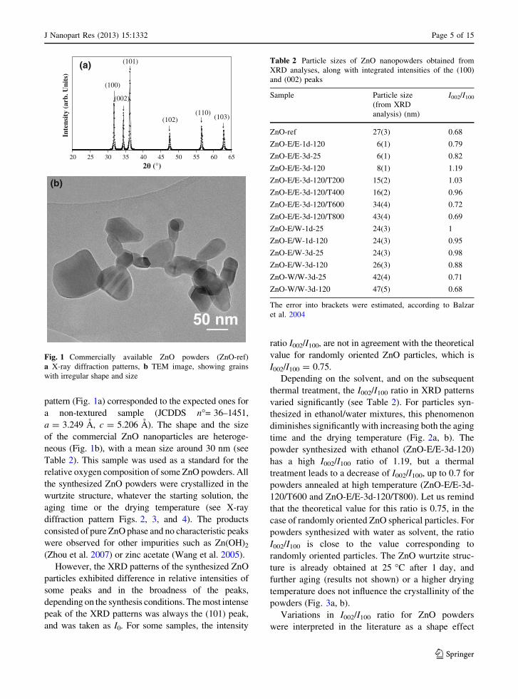
pattern (Fig. 1a) corresponded to the expected ones for
a non-textured sample (JCDDS n�= 36–1451,
a = 3.249 A, c = 5.206 A). The shape and the size
of the commercial ZnO nanoparticles are heteroge-
neous (Fig. 1b), with a mean size around 30 nm (see
Table 2). This sample was used as a standard for the
relative oxygen composition of some ZnO powders. All
the synthesized ZnO powders were crystallized in the
wurtzite structure, whatever the starting solution, the
aging time or the drying temperature (see X-ray
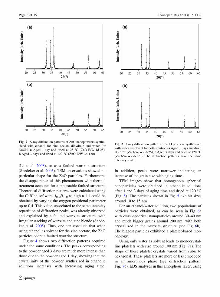
diffraction pattern Figs. 2, 3, and 4). The products
consisted of pure ZnO phase and no characteristic peaks
were observed for other impurities such as Zn(OH)2
(Zhou et al. 2007) or zinc acetate (Wang et al. 2005).
However, the XRD patterns of the synthesized ZnO
particles exhibited difference in relative intensities of
some peaks and in the broadness of the peaks,
depending on the synthesis conditions. The most intense
peak of the XRD patterns was always the (101) peak,
and was taken as I0. For some samples, the intensity
ratio I002/I100, are not in agreement with the theoretical
value for randomly oriented ZnO particles, which is
I002/I100 = 0.75.
Depending on the solvent, and on the subsequent
thermal treatment, the I002/I100 ratio in XRD patterns
varied significantly (see Table 2). For particles syn-
thesized in ethanol/water mixtures, this phenomenon
diminishes significantly with increasing both the aging
time and the drying temperature (Fig. 2a, b). The
powder synthesized with ethanol (ZnO-E/E-3d-120)
has a high I002/I100 ratio of 1.19, but a thermal
treatment leads to a decrease of I002/I100, up to 0.7 for
powders annealed at high temperature (ZnO-E/E-3d-
120/T600 and ZnO-E/E-3d-120/T800). Let us remind
that the theoretical value for this ratio is 0.75, in the
case of randomly oriented ZnO spherical particles. For
powders synthesized with water as solvent, the ratio
I002/I100 is close to the value corresponding to
randomly oriented particles. The ZnO wurtzite struc-
ture is already obtained at 25 �C after 1 day, and
further aging (results not shown) or a higher drying
temperature does not influence the crystallinity of the
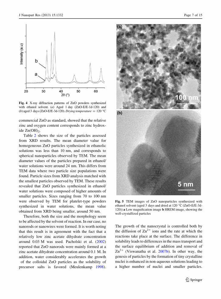
powders (Fig. 3a, b).
Variations in I002/I100 ratio for ZnO powders
were interpreted in the literature as a shape effect
20 25 30 35 40 45 50 55 60 65
Inte
nsit
y (a
rb. U
nits
)
(002)
(101)
(102)(110)
(a)
(100)
(103)
(b)
Fig. 1 Commercially available ZnO powders (ZnO-ref)
a X-ray diffraction patterns, b TEM image, showing grains
with irregular shape and size
Table 2 Particle sizes of ZnO nanopowders obtained from
XRD analyses, along with integrated intensities of the (100)
and (002) peaks
Sample Particle size
(from XRD
analysis) (nm)
I002/I100
ZnO-ref 27(3) 0.68
ZnO-E/E-1d-120 6(1) 0.79
ZnO-E/E-3d-25 6(1) 0.82
ZnO-E/E-3d-120 8(1) 1.19
ZnO-E/E-3d-120/T200 15(2) 1.03
ZnO-E/E-3d-120/T400 16(2) 0.96
ZnO-E/E-3d-120/T600 34(4) 0.72
ZnO-E/E-3d-120/T800 43(4) 0.69
ZnO-E/W-1d-25 24(3) 1
ZnO-E/W-1d-120 24(3) 0.95
ZnO-E/W-3d-25 24(3) 0.98
ZnO-E/W-3d-120 26(3) 0.88
ZnO-W/W-3d-25 42(4) 0.71
ZnO-W/W-3d-120 47(5) 0.68
The error into brackets were estimated, according to Balzar
et al. 2004
J Nanopart Res (2013) 15:1332 Page 5 of 15
123
Page 6
(Li et al. 2008), or as a faulted wurtzite structure
(Snedeker et al. 2005). TEM observations showed no
particular shape for the ZnO particles. Furthermore,
the disappearance of this phenomenon with thermal
treatment accounts for a metastable faulted structure.
Theoretical diffraction patterns were calculated using
the CaRIne software. I002/I100 as high a 1.1 could be
obtained by varying the oxygen positional parameter
up to 0.4. This value, associated to the same intensity
repartition of diffraction peaks, was already observed
and explained by a faulted wurtzite structure, with
irregular stacking of wurtzite and zinc blende (Snede-
ker et al. 2005). Thus, one can conclude that when
using ethanol as solvent for the zinc acetate, the ZnO
particles adopt a faulted wurtzite structure.
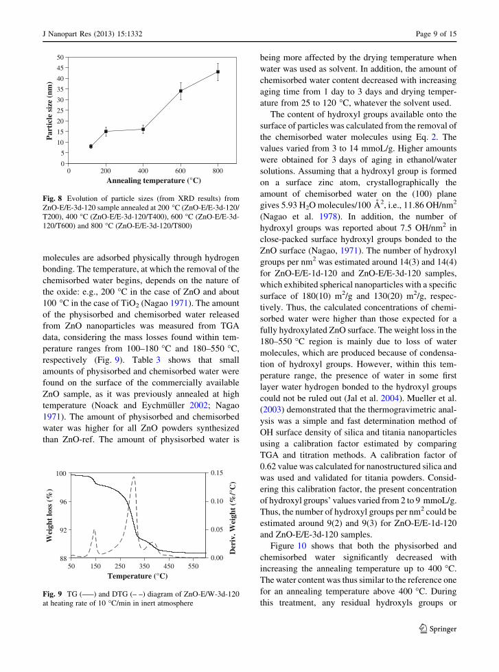
Figure 4 shows two diffraction patterns acquired
under the same conditions. The peaks corresponding
to the powder aged 3 days are much more intense than
those due to the powder aged 1 day, showing that the
crystallinity of the powder synthesized in ethanolic
solutions increases with increasing aging time.
In addition, peaks were narrower indicating an
increase of the grain size with aging time.
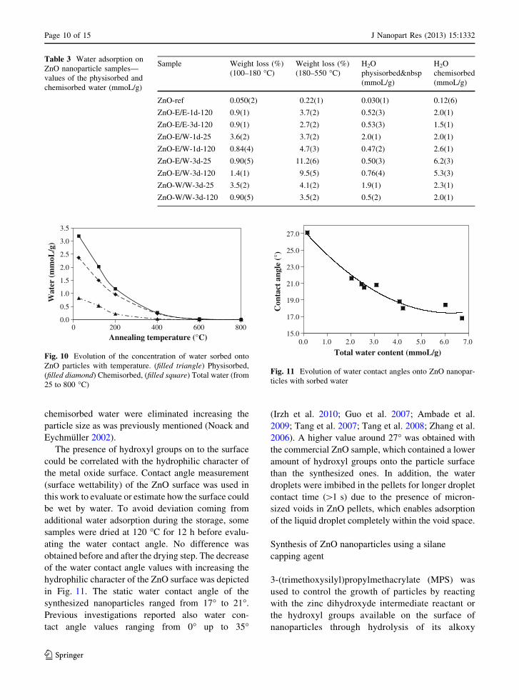
TEM images show that homogenous spherical
nanoparticles were obtained in ethanolic solutions
after 1 and 3 days of aging time and dried at 120 �C
(Fig. 5). The particles shown in Fig. 5 exhibit sizes
around 10 to 15 nm.
For an ethanol/water solution, two populations of
particles were obtained, as can be seen in Fig. 6a
with quasi-spherical nanoparticles around 30–40 nm
and much bigger grains around 200 nm, with both
crystallized in the wurtzite structure (see Fig. 6b).
The biggest particles exhibited a platelet-based mor-
phology.
Using only water as solvent leads to monocrystal-
line platelets with size around 100 nm (Fig. 7a). The
shape of these platelet crystals varied from cubic to
hexagonal. These platelets are more or less embedded
in an amorphous phase (see diffraction pattern,
Fig. 7b). EDS analyses in this amorphous layer, using
20 25 30 35 40 45 50 55 60 65
Inte
nsit
y (a
rb. U
nits
)
(a)
20 25 30 35 40 45 50 55 60 65
Inte
nsit
y (a
rb. U
nits
)
(b)
Fig. 2 X-ray diffraction patterns of ZnO nanopowders synthe-
sized with ethanol for zinc acetate dihydrate and water for
NaOH. a Aged 1 day and dried at 25 �C (ZnO-E/W-1d-25),
b Aged 3 days and dried at 120 �C (ZnO-E/W-3d-120)
20 25 30 35 40 45 50 55 60 65
Inte
nsit
y (a
rb. U
nits
)
(a)
20 25 30 35 40 45 50 55 60 65
Inte
nsit
y (a
rb. U
nits
)
(b)
Fig. 3 X-ray diffraction patterns of ZnO powders synthesized
with water as solvent for both solutions a Aged 3 days and dried
at 25 �C (ZnO-W/W-3d-25), b Aged 3 days and dried at 120 �C
(ZnO-W/W-3d-120). The diffraction patterns have the same
intensity scale
Page 6 of 15 J Nanopart Res (2013) 15:1332
123
Page 7
commercial ZnO as standard, showed that the relative
zinc and oxygen content corresponds to zinc hydrox-
ide Zn(OH)2.
Table 2 shows the size of the particles assessed
from XRD results. The mean diameter value for
homogeneous ZnO particles synthesized in ethanolic
solutions was less than 10 nm, and corresponds to
spherical nanoparticles observed by TEM. The mean
diameter values of the particles prepared in ethanol/
water solutions were around 24 nm. This differs from
TEM data where two particle size populations were
found. Particle sizes from XRD analysis matched with
the smallest particles observed by TEM. These results
revealed that ZnO particles synthesized in ethanol/
water solutions were composed of higher amounts of
smaller particles. Sizes ranging from 70 to 100 nm
were observed by TEM for platelet-type powders
synthesized in water solutions, the mean value
obtained from XRD being smaller, around 50 nm.
Therefore, both the size and the morphology seem
to be affected by the solvent of reaction. In our case, no
nanorods or nanowires were formed. It is worth noting
that this result is in agreement with the fact that a
relatively low zinc acetate dihydrate concentration
around 0.03 M was used. Pacholski et al. (2002)
reported that ZnO nanorods were mainly formed at a
zinc acetate dihydrate concentration around 0.1 M. In
addition, water considerably accelerates the growth
of the colloidal ZnO particles as the solubility of
precursor salts is favored (Meulenkamp 1998).
The growth of the nanocrystal is controlled both by
the diffusion of Zn2? ions and the rate at which the
reactions take place at the surface. The difference in
solubility leads to differences in the mass transport and
the surface equilibrium of addition and removal of
Zn2? (Viswanatha et al. 2007b). In other way, the
genesis of particles by the formation of tiny crystalline
nuclei is enhanced in non-aqueous solutions leading to
a higher number of nuclei and smaller particles.
Fig. 4 X-ray diffraction patterns of ZnO powders synthesized
with ethanol solvent. (a) Aged 1 day (ZnO-E/E-1d-120) and
(b) aged 3 days (ZnO-E/E-3d-120). Drying temperature = 120 �C
Fig. 5 TEM images of ZnO nanoparticles synthesized with
ethanol solvent (aged 3 days and dried at 120 �C (ZnO-E/E-3d-
120)) a Low magnification image b HREM image, showing the
well-crystallized particles
J Nanopart Res (2013) 15:1332 Page 7 of 15
123
Page 8
The size of the particles was shown to increase with
increasing annealing temperature (Noack and Eychmul-
ler 2002). The size of ZnO particles synthesized in
ethanol/water solutions doubled above 400 �C (Fig. 8).
According to Ostwald ripening, the increase in the
particle size is due to the transfer of materials from the
smaller particles to the larger once as a result of potential
energy difference between small and large particles. In
addition, the crystal structure evolved from a faulted
wurtzite structure to a perfect one with the awaited
intensity values of (100) and (002) peaks (Table 2).
Concentration of hydroxyl groups onto the surface
of ZnO nanoparticles
Most metal oxides chemisorb water molecules to form
surface hydroxyl groups on which further water
Fig. 6 TEM images of ZnO nanoparticles synthesized with
ethanol/water solvent (aged 3 days and dried at 120 �C, ZnO-E/
W-3d-120) a Low magnification image b HREM image,
showing the well-crystallized particles
Fig. 7 TEM images and electron diffraction of ZnO synthe-
sized with water as solvent and aged 3 days, drying temperature
25 �C (ZnO-W/W-3d-25). a TEM image showing a platelet-like
grain, covered by an amorphous phase. b Electron diffraction
pattern of a corresponding to a [010] zone axis of the wurtzite
structure
Page 8 of 15 J Nanopart Res (2013) 15:1332
123
Page 9
molecules are adsorbed physically through hydrogen
bonding. The temperature, at which the removal of the
chemisorbed water begins, depends on the nature of
the oxide: e.g., 200 �C in the case of ZnO and about
100 �C in the case of TiO2 (Nagao 1971). The amount
of the physisorbed and chemisorbed water released
from ZnO nanoparticles was measured from TGA
data, considering the mass losses found within tem-
perature ranges from 100–180 �C and 180–550 �C,
respectively (Fig. 9). Table 3 shows that small
amounts of physisorbed and chemisorbed water were
found on the surface of the commercially available
ZnO sample, as it was previously annealed at high
temperature (Noack and Eychmuller 2002; Nagao
1971). The amount of physisorbed and chemisorbed
water was higher for all ZnO powders synthesized
than ZnO-ref. The amount of physisorbed water is
being more affected by the drying temperature when
water was used as solvent. In addition, the amount of
chemisorbed water content decreased with increasing
aging time from 1 day to 3 days and drying temper-
ature from 25 to 120 �C, whatever the solvent used.
The content of hydroxyl groups available onto the
surface of particles was calculated from the removal of
the chemisorbed water molecules using Eq. 2. The
values varied from 3 to 14 mmoL/g. Higher amounts
were obtained for 3 days of aging in ethanol/water
solutions. Assuming that a hydroxyl group is formed
on a surface zinc atom, crystallographically the
amount of chemisorbed water on the (100) plane
gives 5.93 H2O molecules/100 A2, i.e., 11.86 OH/nm2
(Nagao et al. 1978). In addition, the number of
hydroxyl groups was reported about 7.5 OH/nm2 in
close-packed surface hydroxyl groups bonded to the
ZnO surface (Nagao, 1971). The number of hydroxyl
groups per nm2 was estimated around 14(3) and 14(4)
for ZnO-E/E-1d-120 and ZnO-E/E-3d-120 samples,
which exhibited spherical nanoparticles with a specific
surface of 180(10) m2/g and 130(20) m2/g, respec-
tively. Thus, the calculated concentrations of chemi-
sorbed water were higher than those expected for a
fully hydroxylated ZnO surface. The weight loss in the
180–550 �C region is mainly due to loss of water
molecules, which are produced because of condensa-
tion of hydroxyl groups. However, within this tem-
perature range, the presence of water in some first
layer water hydrogen bonded to the hydroxyl groups
could not be ruled out (Jal et al. 2004). Mueller et al.
(2003) demonstrated that the thermogravimetric anal-
ysis was a simple and fast determination method of
OH surface density of silica and titania nanoparticles
using a calibration factor estimated by comparing
TGA and titration methods. A calibration factor of
0.62 value was calculated for nanostructured silica and
was used and validated for titania powders. Consid-
ering this calibration factor, the present concentration
of hydroxyl groups’ values varied from 2 to 9 mmoL/g.
Thus, the number of hydroxyl groups per nm2 could be
estimated around 9(2) and 9(3) for ZnO-E/E-1d-120
and ZnO-E/E-3d-120 samples.
Figure 10 shows that both the physisorbed and
chemisorbed water significantly decreased with
increasing the annealing temperature up to 400 �C.
The water content was thus similar to the reference one
for an annealing temperature above 400 �C. During
this treatment, any residual hydroxyls groups or
0
5
10
15
20
25
30
35
40
45
50
0 200 400 600 800
Annealing temperature (°C)
Par
ticl
e si
ze (
nm)
Fig. 8 Evolution of particle sizes (from XRD results) from
ZnO-E/E-3d-120 sample annealed at 200 �C (ZnO-E/E-3d-120/
T200), 400 �C (ZnO-E/E-3d-120/T400), 600 �C (ZnO-E/E-3d-
120/T600) and 800 �C (ZnO-E/E-3d-120/T800)
88
92
96
100
50 150 250 350 450 550
Temperature (°C)
Wei
ght
loss
(%
)
0.00
0.05
0.10
0.15
Der
iv. W
eigh
t (%
/°C
)
Fig. 9 TG (—–) and DTG (– –) diagram of ZnO-E/W-3d-120
at heating rate of 10 �C/min in inert atmosphere
J Nanopart Res (2013) 15:1332 Page 9 of 15
123
Page 10
chemisorbed water were eliminated increasing the
particle size as was previously mentioned (Noack and
Eychmuller 2002).
The presence of hydroxyl groups on to the surface
could be correlated with the hydrophilic character of
the metal oxide surface. Contact angle measurement
(surface wettability) of the ZnO surface was used in
this work to evaluate or estimate how the surface could
be wet by water. To avoid deviation coming from
additional water adsorption during the storage, some
samples were dried at 120 �C for 12 h before evalu-
ating the water contact angle. No difference was
obtained before and after the drying step. The decrease
of the water contact angle values with increasing the
hydrophilic character of the ZnO surface was depicted
in Fig. 11. The static water contact angle of the
synthesized nanoparticles ranged from 17� to 21�.
Previous investigations reported also water con-
tact angle values ranging from 0� up to 35�
(Irzh et al. 2010; Guo et al. 2007; Ambade et al.
2009; Tang et al. 2007; Tang et al. 2008; Zhang et al.
2006). A higher value around 27� was obtained with
the commercial ZnO sample, which contained a lower
amount of hydroxyl groups onto the particle surface
than the synthesized ones. In addition, the water
droplets were imbibed in the pellets for longer droplet
contact time ([1 s) due to the presence of micron-
sized voids in ZnO pellets, which enables adsorption
of the liquid droplet completely within the void space.
Synthesis of ZnO nanoparticles using a silane
capping agent
3-(trimethoxysilyl)propylmethacrylate (MPS) was
used to control the growth of particles by reacting
with the zinc dihydroxyde intermediate reactant or
the hydroxyl groups available on the surface of
nanoparticles through hydrolysis of its alkoxy
0.0
0.5
1.0
1.5
2.0
2.5
3.0
3.5
0 200 400 600 800
Annealing temperature (°C)
Wat
er (
mm
oL/g
)
Fig. 10 Evolution of the concentration of water sorbed onto
ZnO particles with temperature. (filled triangle) Physisorbed,
(filled diamond) Chemisorbed, (filled square) Total water (from
25 to 800 �C)
Table 3 Water adsorption on
ZnO nanoparticle samples—
values of the physisorbed and
chemisorbed water (mmoL/g)
Sample Weight loss (%)
(100–180 �C)
Weight loss (%)
(180–550 �C)
H2O
physisorbed 
(mmoL/g)
H2O
chemisorbed
(mmoL/g)
ZnO-ref 0.050(2) 0.22(1) 0.030(1) 0.12(6)
ZnO-E/E-1d-120 0.9(1) 3.7(2) 0.52(3) 2.0(1)
ZnO-E/E-3d-120 0.9(1) 2.7(2) 0.53(3) 1.5(1)
ZnO-E/W-1d-25 3.6(2) 3.7(2) 2.0(1) 2.0(1)
ZnO-E/W-1d-120 0.84(4) 4.7(3) 0.47(2) 2.6(1)
ZnO-E/W-3d-25 0.90(5) 11.2(6) 0.50(3) 6.2(3)
ZnO-E/W-3d-120 1.4(1) 9.5(5) 0.76(4) 5.3(3)
ZnO-W/W-3d-25 3.5(2) 4.1(2) 1.9(1) 2.3(1)
ZnO-W/W-3d-120 0.90(5) 3.5(2) 0.5(2) 2.0(1)
15.0
17.0
19.0
21.0
23.0
25.0
27.0
0.0 1.0 2.0 3.0 4.0 5.0 6.0 7.0
Total water content (mmoL/g)
Con
tact
ang
le (
°)
Fig. 11 Evolution of water contact angles onto ZnO nanopar-
ticles with sorbed water
Page 10 of 15 J Nanopart Res (2013) 15:1332
123
Page 11
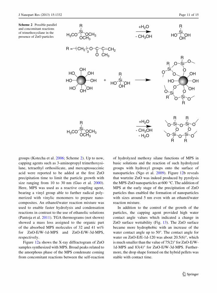
groups (Kotecha et al. 2006; Scheme 2). Up to now,
capping agents such as 3-aminopropyl trimethoxysi-
lane, tetraethyl orthosilicate, and mercaptosuccinic
acid were reported to be added at the first ZnO
precipitation time to limit the particle growth with
size ranging from 10 to 30 nm (Guo et al. 2000).
Here, MPS was used as a reactive coupling agent,
bearing a vinyl group able to further radical poly-
merized with vinylic monomers to prepare nano-
composites. An ethanol/water reaction mixture was
used to enable faster hydrolysis and condensation
reactions in contrast to the use of ethanolic solutions
(Pantoja et al. 2011). TGA thermograms (not shown)
showed a mass loss assigned to the organic part
of the absorbed MPS molecules of 32 and 41 wt%
for ZnO-E/W-1d-MPS and ZnO-E/W-3d-MPS,
respectively.
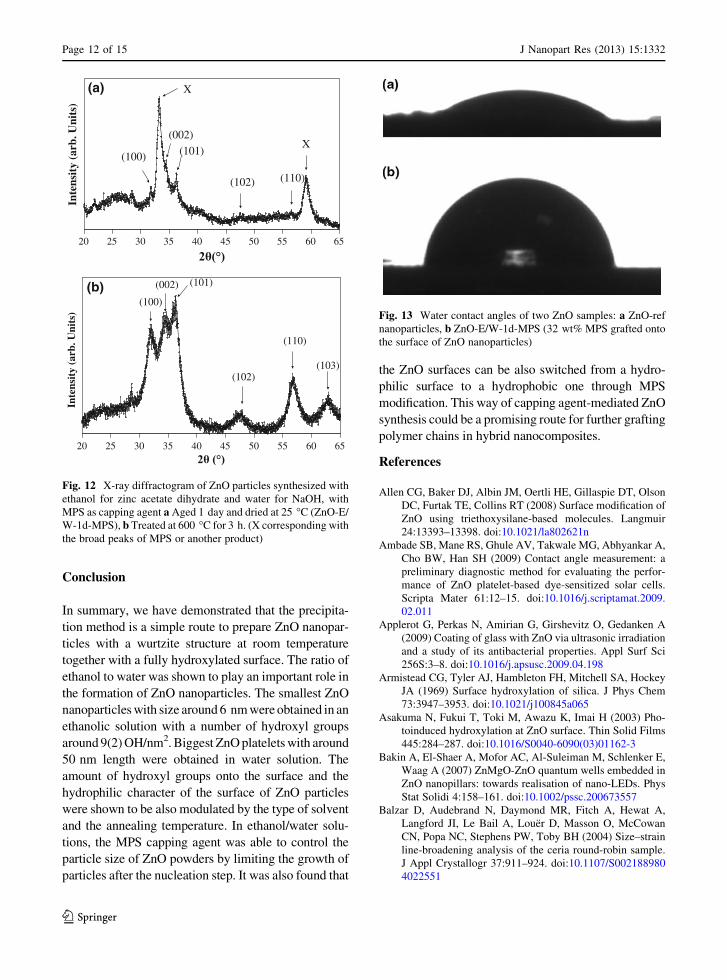
Figure 12a shows the X-ray diffractogram of ZnO
samples synthesized with MPS. Broad peaks related to
the amorphous phase of the MPS condensate coming
from concomitant reactions between the self-reaction
of hydrolyzed methoxy silane functions of MPS in
basic solutions and the reaction of such hydrolyzed
groups with hydroxyl groups onto the surface of
nanoparticles (Ngo et al. 2009). Figure 12b reveals
that wurtzite ZnO was indeed produced by pyrolysis
the MPS-ZnO nanoparticles at 600 �C. The addition of
MPS at the early stage of the precipitation of ZnO
particles thus enabled the formation of nanoparticles
with sizes around 5 nm even with an ethanol/water
reaction mixture.
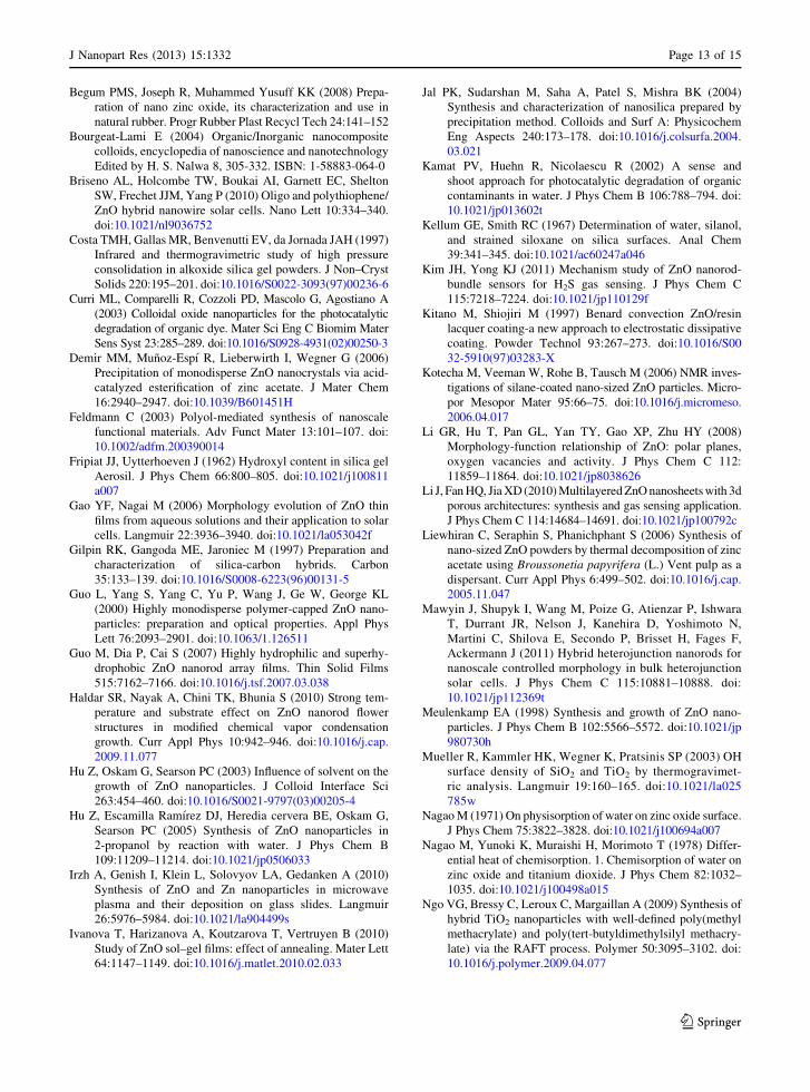
In addition to the control of the growth of the
particles, the capping agent provided high water
contact angle values which indicated a change in
ZnO surface wettability (Fig. 13). The ZnO surface
became more hydrophobic with an increase of the
water contact angle up to 50�. The contact angle for
water on ZnO-E/E-1d-120 was about 20.5(6)�, which
is much smaller than the value of 75(2)� for ZnO-E/W-
1d-MPS and 83(4)� for ZnO-E/W-3d-MPS. Further-
more, the drop shape formed on the hybrid pellets was
stable with contact time.
+H2O
- CH3OH
R = CH2 O CO
C CH23CH3
SiR
OCH3H3COOCH3
SiR
OHHOOH
OH
OH
HO OH
HO
HO OHOH
+
O
OH
O OH
HOO OH
OSiR
O
O Si OR
SiO
O
R Si OR
SiR
OHHOOH
+H2O SiR
- CH3OH
Si
Si Si- H2O
SiR
OCH3H3COOCH3
R
R
OO
O
O
O
OO
RO
Scheme 2 Possible parallel
and concomitant reactions
of trimethoxysilane in the
presence of ZnO particles
J Nanopart Res (2013) 15:1332 Page 11 of 15
123
Page 12
Conclusion
In summary, we have demonstrated that the precipita-
tion method is a simple route to prepare ZnO nanopar-
ticles with a wurtzite structure at room temperature
together with a fully hydroxylated surface. The ratio of
ethanol to water was shown to play an important role in
the formation of ZnO nanoparticles. The smallest ZnO
nanoparticles with size around 6 nm were obtained in an
ethanolic solution with a number of hydroxyl groups
around 9(2) OH/nm2. Biggest ZnO platelets with around
50 nm length were obtained in water solution. The
amount of hydroxyl groups onto the surface and the
hydrophilic character of the surface of ZnO particles
were shown to be also modulated by the type of solvent
and the annealing temperature. In ethanol/water solu-
tions, the MPS capping agent was able to control the
particle size of ZnO powders by limiting the growth of
particles after the nucleation step. It was also found that
the ZnO surfaces can be also switched from a hydro-
philic surface to a hydrophobic one through MPS
modification. This way of capping agent-mediated ZnO
synthesis could be a promising route for further grafting
polymer chains in hybrid nanocomposites.
References
Allen CG, Baker DJ, Albin JM, Oertli HE, Gillaspie DT, Olson
DC, Furtak TE, Collins RT (2008) Surface modification of
ZnO using triethoxysilane-based molecules. Langmuir
24:13393–13398. doi:10.1021/la802621n
Ambade SB, Mane RS, Ghule AV, Takwale MG, Abhyankar A,
Cho BW, Han SH (2009) Contact angle measurement: a
preliminary diagnostic method for evaluating the perfor-
mance of ZnO platelet-based dye-sensitized solar cells.
Scripta Mater 61:12–15. doi:10.1016/j.scriptamat.2009.
02.011
Applerot G, Perkas N, Amirian G, Girshevitz O, Gedanken A
(2009) Coating of glass with ZnO via ultrasonic irradiation
and a study of its antibacterial properties. Appl Surf Sci
256S:3–8. doi:10.1016/j.apsusc.2009.04.198
Armistead CG, Tyler AJ, Hambleton FH, Mitchell SA, Hockey
JA (1969) Surface hydroxylation of silica. J Phys Chem
73:3947–3953. doi:10.1021/j100845a065
Asakuma N, Fukui T, Toki M, Awazu K, Imai H (2003) Pho-
toinduced hydroxylation at ZnO surface. Thin Solid Films
445:284–287. doi:10.1016/S0040-6090(03)01162-3
Bakin A, El-Shaer A, Mofor AC, Al-Suleiman M, Schlenker E,
Waag A (2007) ZnMgO-ZnO quantum wells embedded in
ZnO nanopillars: towards realisation of nano-LEDs. Phys
Stat Solidi 4:158–161. doi:10.1002/pssc.200673557
Balzar D, Audebrand N, Daymond MR, Fitch A, Hewat A,
Langford JI, Le Bail A, Louer D, Masson O, McCowan
CN, Popa NC, Stephens PW, Toby BH (2004) Size–strain
line-broadening analysis of the ceria round-robin sample.
J Appl Crystallogr 37:911–924. doi:10.1107/S002188980
4022551
20 25 30 35 40 45 50 55 60 65
Inte
nsit
y (a
rb. U
nits
)
(100)
(002)
(101)
(110)(102)
X
X
(a)
20 25 30 35 40 45 50 55 60 65
Inte
nsit
y (a
rb. U
nits
)
(b) (101)
(100)
(103)(102)
(110)
(002)
Fig. 12 X-ray diffractogram of ZnO particles synthesized with
ethanol for zinc acetate dihydrate and water for NaOH, with
MPS as capping agent a Aged 1 day and dried at 25 �C (ZnO-E/
W-1d-MPS), b Treated at 600 �C for 3 h. (X corresponding with
the broad peaks of MPS or another product)
Fig. 13 Water contact angles of two ZnO samples: a ZnO-ref
nanoparticles, b ZnO-E/W-1d-MPS (32 wt% MPS grafted onto
the surface of ZnO nanoparticles)
Page 12 of 15 J Nanopart Res (2013) 15:1332
123
Page 13
Begum PMS, Joseph R, Muhammed Yusuff KK (2008) Prepa-
ration of nano zinc oxide, its characterization and use in
natural rubber. Progr Rubber Plast Recycl Tech 24:141–152
Bourgeat-Lami E (2004) Organic/Inorganic nanocomposite
colloids, encyclopedia of nanoscience and nanotechnology
Edited by H. S. Nalwa 8, 305-332. ISBN: 1-58883-064-0
Briseno AL, Holcombe TW, Boukai AI, Garnett EC, Shelton
SW, Frechet JJM, Yang P (2010) Oligo and polythiophene/
ZnO hybrid nanowire solar cells. Nano Lett 10:334–340.
doi:10.1021/nl9036752
Costa TMH, Gallas MR, Benvenutti EV, da Jornada JAH (1997)
Infrared and thermogravimetric study of high pressure
consolidation in alkoxide silica gel powders. J Non–Cryst
Solids 220:195–201. doi:10.1016/S0022-3093(97)00236-6
Curri ML, Comparelli R, Cozzoli PD, Mascolo G, Agostiano A
(2003) Colloidal oxide nanoparticles for the photocatalytic
degradation of organic dye. Mater Sci Eng C Biomim Mater
Sens Syst 23:285–289. doi:10.1016/S0928-4931(02)00250-3
Demir MM, Munoz-Espı R, Lieberwirth I, Wegner G (2006)
Precipitation of monodisperse ZnO nanocrystals via acid-
catalyzed esterification of zinc acetate. J Mater Chem
16:2940–2947. doi:10.1039/B601451H
Feldmann C (2003) Polyol-mediated synthesis of nanoscale
functional materials. Adv Funct Mater 13:101–107. doi:
10.1002/adfm.200390014
Fripiat JJ, Uytterhoeven J (1962) Hydroxyl content in silica gel
Aerosil. J Phys Chem 66:800–805. doi:10.1021/j100811
a007
Gao YF, Nagai M (2006) Morphology evolution of ZnO thin
films from aqueous solutions and their application to solar
cells. Langmuir 22:3936–3940. doi:10.1021/la053042f
Gilpin RK, Gangoda ME, Jaroniec M (1997) Preparation and
characterization of silica-carbon hybrids. Carbon
35:133–139. doi:10.1016/S0008-6223(96)00131-5
Guo L, Yang S, Yang C, Yu P, Wang J, Ge W, George KL
(2000) Highly monodisperse polymer-capped ZnO nano-
particles: preparation and optical properties. Appl Phys
Lett 76:2093–2901. doi:10.1063/1.126511
Guo M, Dia P, Cai S (2007) Highly hydrophilic and superhy-
drophobic ZnO nanorod array films. Thin Solid Films
515:7162–7166. doi:10.1016/j.tsf.2007.03.038
Haldar SR, Nayak A, Chini TK, Bhunia S (2010) Strong tem-
perature and substrate effect on ZnO nanorod flower
structures in modified chemical vapor condensation
growth. Curr Appl Phys 10:942–946. doi:10.1016/j.cap.
2009.11.077
Hu Z, Oskam G, Searson PC (2003) Influence of solvent on the
growth of ZnO nanoparticles. J Colloid Interface Sci
263:454–460. doi:10.1016/S0021-9797(03)00205-4
Hu Z, Escamilla Ramırez DJ, Heredia cervera BE, Oskam G,
Searson PC (2005) Synthesis of ZnO nanoparticles in
2-propanol by reaction with water. J Phys Chem B
109:11209–11214. doi:10.1021/jp0506033
Irzh A, Genish I, Klein L, Solovyov LA, Gedanken A (2010)
Synthesis of ZnO and Zn nanoparticles in microwave
plasma and their deposition on glass slides. Langmuir
26:5976–5984. doi:10.1021/la904499s
Ivanova T, Harizanova A, Koutzarova T, Vertruyen B (2010)
Study of ZnO sol–gel films: effect of annealing. Mater Lett
64:1147–1149. doi:10.1016/j.matlet.2010.02.033
Jal PK, Sudarshan M, Saha A, Patel S, Mishra BK (2004)
Synthesis and characterization of nanosilica prepared by
precipitation method. Colloids and Surf A: Physicochem
Eng Aspects 240:173–178. doi:10.1016/j.colsurfa.2004.
03.021
Kamat PV, Huehn R, Nicolaescu R (2002) A sense and
shoot approach for photocatalytic degradation of organic
contaminants in water. J Phys Chem B 106:788–794. doi:
10.1021/jp013602t
Kellum GE, Smith RC (1967) Determination of water, silanol,
and strained siloxane on silica surfaces. Anal Chem
39:341–345. doi:10.1021/ac60247a046
Kim JH, Yong KJ (2011) Mechanism study of ZnO nanorod-
bundle sensors for H2S gas sensing. J Phys Chem C
115:7218–7224. doi:10.1021/jp110129f
Kitano M, Shiojiri M (1997) Benard convection ZnO/resin
lacquer coating-a new approach to electrostatic dissipative
coating. Powder Technol 93:267–273. doi:10.1016/S00
32-5910(97)03283-X
Kotecha M, Veeman W, Rohe B, Tausch M (2006) NMR inves-
tigations of silane-coated nano-sized ZnO particles. Micro-
por Mesopor Mater 95:66–75. doi:10.1016/j.micromeso.
2006.04.017
Li GR, Hu T, Pan GL, Yan TY, Gao XP, Zhu HY (2008)
Morphology-function relationship of ZnO: polar planes,
oxygen vacancies and activity. J Phys Chem C 112:
11859–11864. doi:10.1021/jp8038626
Li J, Fan HQ, Jia XD (2010) Multilayered ZnO nanosheets with 3d
porous architectures: synthesis and gas sensing application.
J Phys Chem C 114:14684–14691. doi:10.1021/jp100792c
Liewhiran C, Seraphin S, Phanichphant S (2006) Synthesis of
nano-sized ZnO powders by thermal decomposition of zinc
acetate using Broussonetia papyrifera (L.) Vent pulp as a
dispersant. Curr Appl Phys 6:499–502. doi:10.1016/j.cap.
2005.11.047
Mawyin J, Shupyk I, Wang M, Poize G, Atienzar P, Ishwara
T, Durrant JR, Nelson J, Kanehira D, Yoshimoto N,
Martini C, Shilova E, Secondo P, Brisset H, Fages F,
Ackermann J (2011) Hybrid heterojunction nanorods for
nanoscale controlled morphology in bulk heterojunction
solar cells. J Phys Chem C 115:10881–10888. doi:
10.1021/jp112369t
Meulenkamp EA (1998) Synthesis and growth of ZnO nano-
particles. J Phys Chem B 102:5566–5572. doi:10.1021/jp
980730h
Mueller R, Kammler HK, Wegner K, Pratsinis SP (2003) OH
surface density of SiO2 and TiO2 by thermogravimet-
ric analysis. Langmuir 19:160–165. doi:10.1021/la025
785w
Nagao M (1971) On physisorption of water on zinc oxide surface.
J Phys Chem 75:3822–3828. doi:10.1021/j100694a007
Nagao M, Yunoki K, Muraishi H, Morimoto T (1978) Differ-
ential heat of chemisorption. 1. Chemisorption of water on
zinc oxide and titanium dioxide. J Phys Chem 82:1032–
1035. doi:10.1021/j100498a015
Ngo VG, Bressy C, Leroux C, Margaillan A (2009) Synthesis of
hybrid TiO2 nanoparticles with well-defined poly(methyl
methacrylate) and poly(tert-butyldimethylsilyl methacry-
late) via the RAFT process. Polymer 50:3095–3102. doi:
10.1016/j.polymer.2009.04.077
J Nanopart Res (2013) 15:1332 Page 13 of 15
123
Page 14
Ni YH, Wei XW, Hong JM, Ye Y (2005) Hydrothermal and
optical properties of ZnO nanorods. Mater Sci Eng B
121:42–47. doi:10.1016/j.mseb.2005.02.065
Noack V, Eychmuller A (2002) Annealing of nanometer-sized
zinc oxide particles. Chem Mater 14:1411–1417. doi:
10.1021/cm011262i
Nomura K, Ohta H, Ueda K, Kamiya T, Hirano M, Hosono H
(2003) Thin-film transistor fabricated in single-crystalline
transparent oxide semiconductor. Science 300:1269–1272.
doi:10.1126/science.1083212
Pacholski C, Kornowski A, Weller H (2002) Self-assembly of
ZnO: from nanodots to nanorods. Angew Chem Int Ed
41:1188–1191. doi:10.1002/1521-3757(20020402)114:7
Pantoja M, Martınez MA, Abenojar J, Encinas N, Ballesteros Y
(2011) Effect of EtOH/H2O ratio and pH on bis-sulfur silane
solutions for electrogalvanized steel joints based on anaer-
obic adhesives. J Adhes 87:688–708. doi:10(1080/
00218464),2011,596771
Posthumus W, Magusin PCMM, Brokken-Zijp JCM, Tinne-
mans AHA, van der Linde R (2004) Surface modification
of oxidic nanoparticles using 3-methacryloxypropyl-
trimethoxysilane. J Colloid Interface Sci 269:109–116. doi:
10.1016/j.jcis.2003.07.008
Scarisoreanu N, Matei DG, Dinescu G, Epurescu G, Ghica C,
Nistor LC, Dinescu M (2005) Properties of ZnO thin films
prepared by radio-frequency plasma beam assisted laser
ablation. Appl Surf Sci 247:518–525. doi:10.1016/j.
apsusc.2005.01.140
Seung BP, Yun CK (1997) Photocatalytic activity of nanometer
size ZnO particles prepared by spray pyrolysis. J Aerosol
Sci 28:473–474. doi:10.1016/S0021-8502(97)00283-8
Singh N, Turner A (2009) Leaching of copper and zinc from
spent antifouling paint particles. Environ Pollut 157:371–
376. doi:10.1016/j.envpol.2008.10.003
Singhai M, Chhabra V, Kang P, Shah DO (1997) Synthesis of
ZnO nanoparticles for varistor application using Zn-
substituted aerosol OT microemulsion. Mater Res Bull
32:239–247. doi:10.1016/S0025-5408(96)00175-4
Snedeker LP, Risbud AS, Masala O, Zhang JP, Seshadri R
(2005) Organic phase conversion of bulk (wurtzite) ZnO
to nanophase (wurtzite and zinc blende) ZnO. Solid State
Sci 7:1500–1505. doi:10.1016/j.solidstatesciences.2005.
08.020
Søndergaard M, Bøjesen ED, Christensen M, Iversen BoB
(2011) Size and morphology dependence of ZnO nano-
particles synthesized by a fast continuous flow hydrother-
mal method. Cryst Growth Des 11:4027–4033. doi:
10.1021/cg200596c
Takahashi N, Kaiya K, Omichi K, Nakamura T, Okamoto S,
Yamamoto H (2000) Atmospheric pressure vapor-phase
growth of ZnO using a chloride source. J Cryst Growth
209:822–827. doi:10.1016/S0022-0248(99)00611-9
Tang L, Zhou B, Tian Y, Bala H, Pan Y, Ren S, Wang Y, Lv XT,
Li M, Wang Z (2007) Preparation and surface modification
of uniform ZnO nanorods via a one-step process. Colloids
and Surf A: Physicochem Eng Aspects 296:92–96. doi:
10.1016/j.colsurfa.2006.09.035
Tang L, Zhou B, Tian Y, Sun F, Li YL, Wang Z (2008) Syn-
thesis and surface hydrophobic functionalization of ZnO
nanocrystals via a facile one-step solution method. Chem
Eng J 139:642–648. doi:10.1016/j.cej.2008.01.027
Tani T, Madler L, Pratsinis SE (2002) Homogeneous ZnO
nanoparticles by flame spray pyrolysis. J Nanopart Res
4:337–343. doi:10.1023/A:1021153419671
Traeger F, Kauer M (2011) Analysis of surface, subsurface, and
bulk hydrogen in ZnO using nuclear reaction analysis. Phys
Rev B 84:075462. doi:10.1103/PhysRevB.84.075462
Viswanatha R, Amenitsch H, Sarma DD (2007a) Growth
kinetics of ZnO nanocrystals: a few surprises. J Am Chem
Soc 129(4470):4475. doi:10.1021/ja068161b
Viswanatha R, Santra PK, Dasgupta C, Sarma DD (2007b)
Growth mechanism of nanocrystals in solution: ZnO, a
case study. Phys Rev Lett 98:255501-1–255501-4. doi:
10.1103/PhysRevLett.98.255501
Wang ZL (2004) Zinc oxide nanostructures: growth, properties
and applications. J Phys Condens Matter 16:829–858. doi:
10.1088/0953-8984/16/25/R01
Wang CL, Shen EH, Wang EH, Gao L, Kang ZK, Tian CG, Lan
Y, Zhang Ch (2005) Controllable synthesis of ZnO nano-
crystals via a surfactant-assisted alcohol thermal process at
a low temperature. Mater Lett 59:2867–2871. doi:10.1016/
j.matlet.2005.04.031
Wu R, Xie CS (2004) Formation of tetrapod ZnO nanowhiskers
and its optical properties. Mater Res Bull 39:637–645. doi:
10.1016/j.materresbull.2003.12.009
Wu J, Xie CS, Bai ZK, Zhu BL, Huang KJ, Wu R (2002) Prep-
aration of ZnO–glass varistor from tetrapod ZnO nano-
powders. Mater Sci Eng B Solid-State Mater Adv Technol
95:157–161. doi:10.1016/S0921-5107(02)00227-1
Wu YL, Tok AIY, Boey FYC, Zeng XT, Zhang ZH (2007)
Surface modification of ZnO nanocrystals. Appl Surf Sci
253:5473–5479. doi:10.1016/j.apsusc.2006.12.091
Yebra DM, Kiil S, Dam-Johansen K (2004) Antifouling tech-
nology—past, present and future steps towards efficient
and environmentally friendly antifouling coatings. Prog
Org Coat 50:75–104. doi:10.1016/j.porgcoat.2003.06.001
Yebra DM, Kiil S, Weinell CE, Dam-Johansen K (2006) Dis-
solution rate measurements of sea water soluble pigments
for antifouling paints: ZnO. Prog Org Coat 56:327–337.
doi:10.1016/j.porgcoat.2006.06.007
Zhang SC, Li XG (2003) Preparation of ZnO particles by pre-
cipitation transformation method and its inherent forma-
tion mechanisms. Colloids and Surf A Physicochem Eng
Aspects 226:35–44. doi:10.1016/S0927-7757(03)00383-2
Zhang JJ, Gao G, Zhang M, Zhang D, Wang CL, Zhao DC, Liu
FQ (2006) ZnO/PS core–shell hybrid microspheres pre-
pared with miniemulsion polymerization. J Colloid Inter-
face Sci 301:78–84. doi:10.1016/j.jcis.2006.05.005
Zheng MJ, Zhang LD, Li GH, Shen WZ (2002) Fabrication and
optical properties of large-scale uniform zinc oxide nano-
wire arrays by one-step electrochemical deposition tech-
nique. Chem Phys Lett 363:123–128. doi:10.1016/S0009-
2614(02)01106-5
Zhou H, Li Z (2005) Synthesis of nanowires, nanorods andnanoparticles of ZnO through modulating the ratio of water
to methanol by using a mild and simple solution method.
Mater Chem Phys 89:326–331. doi:10.1016/j.matche
mphys.2004.09.006
Zhou J, Zhao F, Wang Y, Zhang Y, Yang L (2007) Size-con-
trolled synthesis of ZnO nanoparticles and their photolu-
minescence properties. J Lumin 122–123:195–197. doi:
10.1016/j.jlumin.2006.01.089
Page 14 of 15 J Nanopart Res (2013) 15:1332
123
Page 15
Znaidi L, Soler Illia GJAA, Benyahia S, Sanchez C, Kanaev AV
(2003a) Oriented ZnO thin films synthesis by sol–gel
process for laser application. Thin Solid Films 428:
257–262. doi:10.1016/S0040-6090(02)01219-1
Znaidi L, Soler Illia GJAA, Le Guennic R, Sanchez C, Kanaev A
(2003b) Elaboration of ZnO thin films with preferential
orientation by a soft chemistry route. J Sol–Gel Sci Tech-
nol 26:817–821. doi:10.1023/A:1020795515478
J Nanopart Res (2013) 15:1332 Page 15 of 15
123