http://crcp.sciedupress.com Case Reports in Clinical Pathology 2016, Vol. 3, No. 2 CASE REPORT Systemic eosinophilia with skin and pulmonary infiltrates in a patient with chronic lymphocytic leukemia: A case report Veronika Nagy *1 , Corina Dommann-Scherrer 2 , Alexandar Tzankov 3 , Miklos Pless 1 1 Department of Medical Oncology, Kantonsspital Winterthur, Winterthur, Switzerland 2 Institute of Pathology, Kantonsspital Winterthur, Winterthur, Switzerland 3 Institute of Pathology, University Hospital Basel, Basel, Switzerland Received: January 8, 2016 Accepted: February 6, 2016 Online Published: February 24, 2016 DOI: 10.5430/crcp.v3n2p15 URL: http://dx.doi.org/10.5430/crcp.v3n2p15 ABSTRACT We report on a patient with chronic lymphocytic leukemia (CLL) under treatment with fludarabine who developed skin ulcers, systemic eosinophilia and pulmonary infiltrates. Despite extensive diagnostic investigations, no cause of the clinical findings could be detected. After unsuccessful treatment with prednisone and hydroxyurea, the patient died. Autopsy revealed a surprising diagnosis, explaining all symptoms and findings. Key Words: Chronic lymphocytic leukemia, Eosinophilia, Eosinophilic granuloma, Richter, Hodgkin lymphoma, Skin ulcer, Lung infiltrate 1. I NTRODUCTION Chronic lymphocytic leukemia (CLL) is the most common leukemia in adults in the Western world and is frequently accompanied by eosinophilia. The differential diagnosis of eosinophilia is very wide, including infection, allergic diseases, myeloproliferative disorders, [1] lymphoid malig- nancies, [2, 3] side effects of medical treatment of CLL with fludarabine, [4] and other causes. [5] We present and discuss the case of a patient who developed dramatic clinical fea- tures: rapid deterioration of performance status, high fever, extremely high eosinophilic counts in peripheral blood, as well as eosinophilic infiltrates in skin and lungs. 2. CASE PRESENTATION In 2006, a 75-year-old man who received fludarabine for B-CLL presented with several small, crusted excoriations on his forehead. In addition, he demonstrated a 4-centimeter axillary ulcer (see Figure 1) which he had first noticed two weeks after the most recent fludarabine infusion. Except for a minor generalized pruritus, he reported no further symptoms. Ten years ago, the patient initially presented with supra- clavicular, axillary and inguinal lymphadenopathy. Biopsy of a supraclavicular lymph node revealed small lympho- cytic lymphoma SLL/CLL, CD20, CD5, CD23 positive with lambda light chain restriction, RAI stage I. In the subsequent years, symptomatic lymphadenopathy with involvement of the submandibular gland and left tonsil, lymphocytosis and thrombocytopenia developed. Over the years, six cycles of chlorambucil and prednisone and four cycles of cyclophos- phamide, adriamycine, vincristine, etoposide and prednisone (CHOEP) respectively, were administered, each leading to * Correspondence: Veronika Nagy; Email: [email protected]; Address: Department of Medical Oncology, Kantonsspital Winterthur, Brauer- strasse 15 8401 Winterthur, Winterthur, Switzerland. Published by Sciedu Press 15
Transcript
http://crcp.sciedupress.com Case Reports in Clinical Pathology 2016, Vol. 3, No. 2
CASE REPORT
Systemic eosinophilia with skin and pulmonaryinfiltrates in a patient with chronic lymphocyticleukemia: A case report
Veronika Nagy ∗1, Corina Dommann-Scherrer2, Alexandar Tzankov3, Miklos Pless1
1Department of Medical Oncology, Kantonsspital Winterthur, Winterthur, Switzerland2Institute of Pathology, Kantonsspital Winterthur, Winterthur, Switzerland3Institute of Pathology, University Hospital Basel, Basel, Switzerland
Received: January 8, 2016 Accepted: February 6, 2016 Online Published: February 24, 2016DOI: 10.5430/crcp.v3n2p15 URL: http://dx.doi.org/10.5430/crcp.v3n2p15
ABSTRACT
We report on a patient with chronic lymphocytic leukemia (CLL) under treatment with fludarabine who developed skin ulcers,systemic eosinophilia and pulmonary infiltrates. Despite extensive diagnostic investigations, no cause of the clinical findingscould be detected. After unsuccessful treatment with prednisone and hydroxyurea, the patient died. Autopsy revealed a surprisingdiagnosis, explaining all symptoms and findings.
1. INTRODUCTIONChronic lymphocytic leukemia (CLL) is the most commonleukemia in adults in the Western world and is frequentlyaccompanied by eosinophilia. The differential diagnosisof eosinophilia is very wide, including infection, allergicdiseases, myeloproliferative disorders,[1] lymphoid malig-nancies,[2, 3] side effects of medical treatment of CLL withfludarabine,[4] and other causes.[5] We present and discussthe case of a patient who developed dramatic clinical fea-tures: rapid deterioration of performance status, high fever,extremely high eosinophilic counts in peripheral blood, aswell as eosinophilic infiltrates in skin and lungs.
2. CASE PRESENTATIONIn 2006, a 75-year-old man who received fludarabine forB-CLL presented with several small, crusted excoriations on
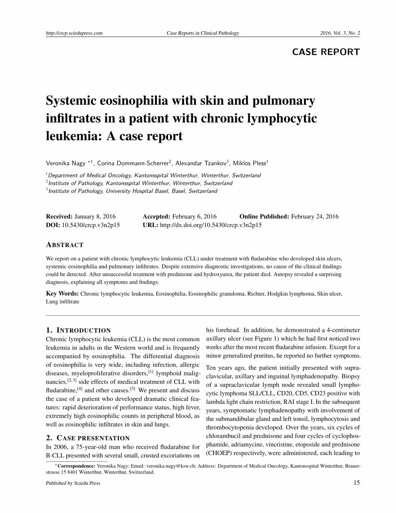
his forehead. In addition, he demonstrated a 4-centimeteraxillary ulcer (see Figure 1) which he had first noticed twoweeks after the most recent fludarabine infusion. Except for aminor generalized pruritus, he reported no further symptoms.
Ten years ago, the patient initially presented with supra-clavicular, axillary and inguinal lymphadenopathy. Biopsyof a supraclavicular lymph node revealed small lympho-cytic lymphoma SLL/CLL, CD20, CD5, CD23 positive withlambda light chain restriction, RAI stage I. In the subsequentyears, symptomatic lymphadenopathy with involvement ofthe submandibular gland and left tonsil, lymphocytosis andthrombocytopenia developed. Over the years, six cycles ofchlorambucil and prednisone and four cycles of cyclophos-phamide, adriamycine, vincristine, etoposide and prednisone(CHOEP) respectively, were administered, each leading to
∗Correspondence: Veronika Nagy; Email: [email protected]; Address: Department of Medical Oncology, Kantonsspital Winterthur, Brauer-strasse 15 8401 Winterthur, Winterthur, Switzerland.
Published by Sciedu Press 15
http://crcp.sciedupress.com Case Reports in Clinical Pathology 2016, Vol. 3, No. 2
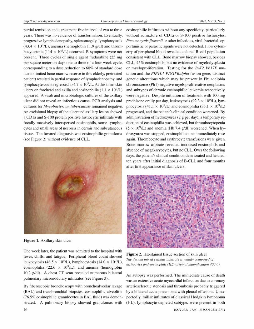
partial remission and a treatment-free interval of two to threeyears. There was no evidence of transformation. Eventually,progressive lymphadenopathy, splenomegaly, lymphocytosis(43.4 × 10⁹/L), anemia (hemoglobin 11.9 g/dl) and throm-bocytopenia (114 × 109/L) occurred. B-symptoms were notpresent. Three cycles of single agent fludarabine (25 mgper square meter on days one to three of a four-week cycle,corresponding to a dose reduction to 60% of standard dosedue to limited bone marrow reserve in this elderly, pretreatedpatient) resulted in partial response of lymphadenopathy, andlymphocyte count regressed to 4.7 × 10⁹/L. At this time, skinulcers on forehead and axilla and eosinophilia (1.1 × 10⁹/L)appeared. A swab and microbiologic cultures of the axillaryulcer did not reveal an infectious cause. PCR analysis andcultures for Mycobacterium tuberculosis remained negative.An excisional biopsy of the ulcerated axillary lesion showeda CD1a and S-100 protein positive histiocytic infiltrate withfocally massively interspersed eosinophils, some lympho-cytes and small areas of necrosis in dermis and subcutaneoustissue. The favored diagnosis was eosinophilic granuloma(see Figure 2) without evidence of CLL.
Figure 1. Axillary skin ulcer
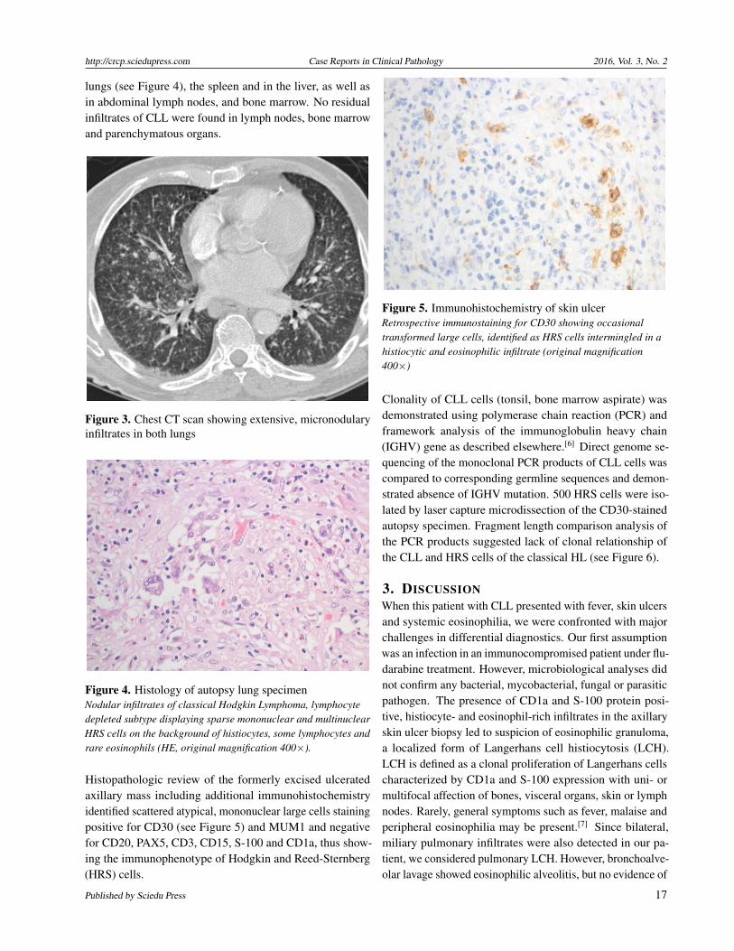
One week later, the patient was admitted to the hospital withfever, chills, and fatigue. Peripheral blood count showedleukocytosis (46.5 × 10⁹/L), lymphocytosis (14.0 × 10⁹/L),eosinophilia (22.6 × 10⁹/L), and anemia (hemoglobin10.2 g/dl). A chest CT scan revealed numerous bilateralpulmonary micronodulary infiltrates (see Figure 3).
By fiberoscopic bronchoscopy with bronchoalveolar lavage(BAL) and transbronchial biopsies, eosinophilic alveolitis(76.5% eosinophilic granulocytes in BAL fluid) was demon-strated. A pulmonary biopsy showed granulomas with
eosinophilic infiltrates without any specificity, particularlywithout admixture of CD1a or S-100 positive histiocytes.Pneumocystis jirovecii or other infectious, viral, bacterial, op-portunistic or parasitic agents were not detected. Flow cytom-etry of peripheral blood revealed a clonal B-cell-populationconsistent with CLL. Bone marrow biopsy showed, besidesCLL, 45% eosinophils, but no evidence of myelodysplasiaor myeloproliferation. Testing for the JAK2 V617F mu-tation and the FIP1L1-PDGFRalpha fusion gene, distinctgenetic alterations which may be present in Philadelphiachromosome (Ph1) negative myeloproliferative neoplasmsand subtypes of chronic eosinophilic leukemia respectively,were negative. Despite initiation of treatment with 100 mgprednisone orally per day, leukocytosis (92.3 × 10⁹/L), lym-phocytosis (41.1 × 10⁹/L) and eosinophilia (35.1 × 10⁹/L)progressed, and the patient’s clinical condition worsened. Byadministration of hydroxyurea (2 g per day), a temporary re-duction of eosinophilia was achieved, but thrombocytopenia(5 × 10⁹/L) and anemia (Hb 7.4 g/dl) worsened. When hy-droxyurea was stopped, eosinophil counts immediately roseagain. Thrombocyte and erythrocyte transfusions were given.Bone marrow aspirate revealed increased eosinophils andabsence of megakaryocytes, but no CLL. Over the followingdays, the patient’s clinical condition deteriorated and he died,ten years after initial diagnosis of B-CLL and four monthsafter first appearance of skin ulcers.
Figure 2. HE-stained tissue section of skin ulcerThe dermal mixed cellular infiltrate is mainly composed ofhistiocytes and eosinophils (HE, original magnification 400×).
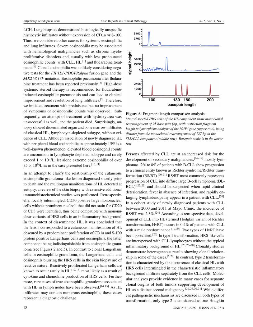
An autopsy was performed. The immediate cause of deathwas an extensive acute myocardial infarction due to coronaryarteriosclerotic stenosis and thrombosis probably triggeredby a bilateral acute pneumonia with pleural effusions. Unex-pectedly, miliar infiltrates of classical Hodgkin lymphoma(HL), lymphocyte-depleted subtype, were present in both
16 ISSN 2331-2726 E-ISSN 2331-2734
http://crcp.sciedupress.com Case Reports in Clinical Pathology 2016, Vol. 3, No. 2
lungs (see Figure 4), the spleen and in the liver, as well asin abdominal lymph nodes, and bone marrow. No residualinfiltrates of CLL were found in lymph nodes, bone marrowand parenchymatous organs.
Figure 3. Chest CT scan showing extensive, micronodularyinfiltrates in both lungs
Figure 4. Histology of autopsy lung specimenNodular infiltrates of classical Hodgkin Lymphoma, lymphocytedepleted subtype displaying sparse mononuclear and multinuclearHRS cells on the background of histiocytes, some lymphocytes andrare eosinophils (HE, original magnification 400×).
Histopathologic review of the formerly excised ulceratedaxillary mass including additional immunohistochemistryidentified scattered atypical, mononuclear large cells stainingpositive for CD30 (see Figure 5) and MUM1 and negativefor CD20, PAX5, CD3, CD15, S-100 and CD1a, thus show-ing the immunophenotype of Hodgkin and Reed-Sternberg(HRS) cells.
Figure 5. Immunohistochemistry of skin ulcerRetrospective immunostaining for CD30 showing occasionaltransformed large cells, identified as HRS cells intermingled in ahistiocytic and eosinophilic infiltrate (original magnification400×)
Clonality of CLL cells (tonsil, bone marrow aspirate) wasdemonstrated using polymerase chain reaction (PCR) andframework analysis of the immunoglobulin heavy chain(IGHV) gene as described elsewhere.[6] Direct genome se-quencing of the monoclonal PCR products of CLL cells wascompared to corresponding germline sequences and demon-strated absence of IGHV mutation. 500 HRS cells were iso-lated by laser capture microdissection of the CD30-stainedautopsy specimen. Fragment length comparison analysis ofthe PCR products suggested lack of clonal relationship ofthe CLL and HRS cells of the classical HL (see Figure 6).
3. DISCUSSIONWhen this patient with CLL presented with fever, skin ulcersand systemic eosinophilia, we were confronted with majorchallenges in differential diagnostics. Our first assumptionwas an infection in an immunocompromised patient under flu-darabine treatment. However, microbiological analyses didnot confirm any bacterial, mycobacterial, fungal or parasiticpathogen. The presence of CD1a and S-100 protein posi-tive, histiocyte- and eosinophil-rich infiltrates in the axillaryskin ulcer biopsy led to suspicion of eosinophilic granuloma,a localized form of Langerhans cell histiocytosis (LCH).LCH is defined as a clonal proliferation of Langerhans cellscharacterized by CD1a and S-100 expression with uni- ormultifocal affection of bones, visceral organs, skin or lymphnodes. Rarely, general symptoms such as fever, malaise andperipheral eosinophilia may be present.[7] Since bilateral,miliary pulmonary infiltrates were also detected in our pa-tient, we considered pulmonary LCH. However, bronchoalve-olar lavage showed eosinophilic alveolitis, but no evidence of
Published by Sciedu Press 17
http://crcp.sciedupress.com Case Reports in Clinical Pathology 2016, Vol. 3, No. 2
LCH. Lung biopsies demonstrated histologically unspecifichistiocytic infiltrates without expression of CD1a or S-100.Thus, we considered other causes for systemic eosinophiliaand lung infiltrates. Severe eosinophilia may be associatedwith hematological malignancies such as chronic myelo-proliferative disorders and, usually with less pronouncedeosinophilic counts, with CLL, HL,[1] and fludarabine treat-ment.[4] Clonal eosinophilia was unlikely considering nega-tive tests for the FIP1L1-PDGFRalpha fusion gene and theJAK2 V617F mutation. Eosinophilic pneumonia after fludara-bine treatment has been reported previously.[8] High-dosesystemic steroid therapy is recommended for fludarabine-induced eosinophilic pneumonitis and can lead to clinicalimprovement and resolution of lung infiltrates.[9] Therefore,we initiated treatment with prednisone, but no improvementof symptoms or eosinophilic counts was observed. Sub-sequently, an attempt of treatment with hydroxyurea wasunsuccessful as well, and the patient died. Surprisingly, au-topsy showed disseminated organ and bone marrow infiltratesof classical HL, lymphocyte-depleted subtype, without evi-dence of CLL. Although association of newly diagnosed HLwith peripheral blood eosinophilia in approximately 15% is awell-known phenomenon, elevated blood eosinophil countsare uncommon in lymphocyte-depleted subtype and rarelyexceed 1 × 10⁹/L, let alone extreme eosinophilia of over35 × 109/L as in the case presented here.[10, 11]
In an attempt to clarify the relationship of the cutaneouseosinophilic granuloma-like lesion diagnosed shortly priorto death and the multiorgan manifestations of HL detected atautopsy, a review of the skin biopsy with extensive additionalimmunohistochemical studies was performed. Retrospectiv-elly, focally intermingled, CD30 positive large mononuclearcells without prominent nucleoli that did not stain for CD20or CD3 were identified, thus being compatible with mononu-clear variants of HRS cells in an inflammatory background.In the context of disseminated HL, it was concluded thatthe lesion corresponded to a cutaneous manifestation of HLobscured by a predominant proliferation of CD1a and S-100protein positive Langerhans cells and eosinophils, the lattercomponent being indistinguishable from eosinophilic granu-loma (see Figures 2 and 5). In contrast to clonal Langerhanscells in eosinophilic granuloma, the Langerhans cells andeosinophils blurring the HRS cells in the skin biopsy are ofreactive nature. Reactively proliferated Langerhans cells areknown to occur rarely in HL,[12, 13] most likely as a result ofcytokine and chemokine production of HRS cells. Further-more, rare cases of true eosinophilic granuloma associatedwith HL in lymph nodes have been observed.[14, 15] As HLinfiltrates may contain numerous eosinophils, these casesrepresent a diagnostic challenge.
Figure 6. Fragment length comparison analysisMicrodissected HRS cells of the HL component show monoclonalrearrangement of 95 base pair (bp) with restriction fragmentlength polymorphism analysis of the IGHV gene (upper row), beingdistinct from the monoclonal rearrangement of 127 bp in theSLL/CLL component (middle row). Basepair scale is in the lowerrow
Persons affected by CLL are at an increased risk for thedevelopment of secondary malignancies,[16–19] mostly lym-phomas. 2% to 8% of patients with B-CLL show progressionto a clinical entity known as Richter syndrome/Richter trans-formation (RS/RT).[20, 21] RS/RT most commonly representsprogression of CLL into diffuse large B-cell lymphoma (DL-BCL),[22, 23] and should be suspected when rapid clinicaldeterioration, fever in absence of infection, and rapidly en-larging lymphadenopathy appear in a patient with CLL.[20]
In a cohort study of newly diagnosed patients with CLLbetween 2000 and 2011 at Mayo Clinic, the incidence ofRS/RT was 2.3%.[24] According to retrospective data, devel-opment of CLL into HL (termed Hodgkin variant of Richtertransformation, HvRT) occurs in 0.4% of patients with CLLwith a male predominance.[19, 25] Two types of HvRT havebeen postulated:[26] In type 1 transformation, HRS-like cellsare interspersed with CLL lymphocytes without the typicalinflammatory background of HL.[20, 25–28] Clonality studiesdemonstrate heterogeneous results showing clonal relation-ship in some of the cases.[6, 29] In contrast, type 2 transforma-tion is characterized by the occurrence of classical HL withHRS cells intermingled in the characteristic inflammatorybackground infiltrate separately from the CLL cells. Molec-ular analyses provide evidence in many cases for separateclonal origins of both tumors supporting development ofHL as a distinct second malignancy.[20, 26, 30, 31] While differ-ent pathogenetic mechanisms are discussed in both types oftransformation, only type 2 is considered as true Hodgkin
18 ISSN 2331-2726 E-ISSN 2331-2734
http://crcp.sciedupress.com Case Reports in Clinical Pathology 2016, Vol. 3, No. 2
transformation irrespective of clonal relationship with theunderlying CLL cells.[6, 20, 29–32] In our patient, morpholog-ical and immunohistochemical findings at autopsy showedclassical HL, namely type 2 transformation. Approximatelyhalf of CLL cases carry a mutation in the variable regionof the IGHV gene.[33] In CLL, unmutated IGHV status (asin our patient) is one of several adverse prognostic markersthat are independently associated with more aggressive, ad-vanced stage CLL with significantly lower overall survivaland higher risk for development of RS/RT.[24, 34] In a recentseries of classic RS/RT, 78% of CLL cases showed clonalprogression into DLBCL with identical IGHV sequences inboth lymphoma components. Among those clonally relatedRS/RT cases, more than 70% carried unmutated IGHV genes,whereas most clonally unrelated cases displayed IGHV genemutations in the CLL component.[6] In contrast, CLL withHvRT (type 1 and type 2) predominantly carry mutated IGHVgenes, independent of a clonal relationship.[6, 30, 35] Thus, ourcase represents an unusual HvRT developing in the back-ground of IGHV-unmutated CLL, which fits to the lack ofclonal relationship of both components suggested by the re-sults of fragment length comparison analysis of IGHV PCRproducts.
Whereas primary HL is associated with EBV infection in30% in developed countries,[36–39] the role of EBV in thepathogenesis of HvRT is ambiguous. Negativity for EBVhas been described mainly in clonally related cases of CLLand HL. In contrast, in many published cases of clonallyunrelated HL and CLL, EBV-infection was present.[6, 31, 40]
Immunosuppression induced by fludarabine treatment hasbeen suggested to play a role in the pathogenesis of EBVpositive HvRT,[31] and cases of clonally related CLL andHL associated with EBV after fludarabine therapy have beendescribed.[40] Interestingly, in the case reported here, theHRS cells of HvRT were EBV-negative in the absence ofclonal relationship between CLL and HL.
Within the last decade, major advances in treatment ofCLL have been accomplished. In the mid to late 2000s,standard first line treatment of CLL changed from singleagent chemotherapy to rituximab-based chemoimmunother-apy.[41] As our patient was treated prior to approval ofrituximab for CLL in Switzerland, he was unable to ben-efit from this development. Currently, anti-CD20 mono-
clonal antibodies (rituximab, ofatumumab or obinutuzumab)in combination with chemotherapeutic agents are standardof care in treatment of CLL.[41, 42] Considering the currentapproach for elderly patients with CLL, this patient wouldmost likely have received a chemoimmunotherapeutic treat-ment regimen such as obinutuzumab/chlorambucil[42, 43] orrituximab/bendamustine.[44, 45] Recently, another paradigmshift in treatment of relapsed or refractory CLL has beenheralded by the introduction of promising new treatment con-cepts including inhibition of Bruton’s tyrosine kinase (BTK),phospoinositide 3-kinase (PI3K) delta or B-cell lymphoma2 (Bcl2) and many other novel agents.[41, 42] Given the ex-tended treatment armamentarium available nowadays and inthe near future, this elderly patient would probably not betreated with fludarabine today.
HvRT commonly shows an aggressive clinical course andtypically leads to death in a majority of patients within oneyear from diagnosis.[46] When multiagent chemotherapy isadministered, overall response rates of around 50% and shortprogression-free intervals have been reported.[20, 25] Gener-ally, clinical outcomes are worse than in de novo HL.[20, 25, 32]
A retrospective analysis demonstrated that patients previ-ously treated for CLL with fludarabine displayed a moreaggressive course of HL and a shorter mean survival of 0.7years compared to 2.1 years in the non-fludarabine group.[20]
4. CONCLUSIONWe conclude that unexpected clinical findings, especiallyeosinophilia, in a patient with CLL, as described in thisreport, should lead to inclusion of HvRT in differential diag-nosis. Repeated invasive diagnostic procedures with biopsyof suspicious lesions are necessary to come to a clear diagno-sis in such complex cases, and close collaboration betweenclinicians and pathologists within the diagnostic process ismandatory.
ACKNOWLEDGEMENTSThe authors gratefully acknowledge Sergio Cogliatti for per-formance of molecular analyses and valuable scientific in-put on molecular genetics and help with the section of themanuscript concerning molecular pathology.
CONFLICTS OF INTEREST DISCLOSUREThe authors declare no conflicts of interest.
REFERENCES[1] Andersen CL, Siersma VD, Hasselbalch HC, et al. Eosinophilia in
routine blood samples and the subsequent risk of hematological ma-lignancies and death. American journal of hematology. 2013; 88(10):
http://crcp.sciedupress.com Case Reports in Clinical Pathology 2016, Vol. 3, No. 2
duction. The Journal of allergy and clinical immunology. 1993; 92(1Pt 1): 123-31. PMid: 8335848. http://dx.doi.org/10.1016/0091-6749(93)90046-I
[3] Gruss HJ, Brach MA, Drexler HG, et al. Expression of cytokinegenes, cytokine receptor genes, and transcription factors in culturedHodgkin and Reed-Sternberg cells. Cancer research. 1992; 52(12):3353-60. PMid: 1596893.
[4] Sezer O, Schmid P, Hallek M, et al. Eosinophilia during fludarabinetreatment of chronic lymphocytic leukemia. Annals of hematology.1999; 78(10): 475-7. PMID: 10550560. http://dx.doi.org/10.1007/s002770050602
[5] Mejia R, Nutman TB. Evaluation and differential diagnosis ofmarked, persistent eosinophilia. Seminars in hematology. 2012; 49(2):149-59. PMid: 22449625. http://dx.doi.org/10.1053/j.seminhematol.2012.01.006
[6] Mao Z, Quintanilla-Martinez L, Raffeld M, et al. IgVH muta-tional status and clonality analysis of Richter’s transformation: dif-fuse large B-cell lymphoma and Hodgkin lymphoma in associa-tion with B-cell chronic lymphocytic leukemia (B-CLL) represent2 different pathways of disease evolution. The American journalof surgical pathology. 2007; 31(10): 1605-14. PMid: 17895764.http://dx.doi.org/10.1097/PAS.0b013e31804bdaf8
[8] Trojan A, Meier R, Licht A, et al. Eosinophilic pneumonia afteradministration of fludarabine for the treatment of non-Hodgkin’s lym-phoma. Annals of hematology. 2002; 81(9): 535-7. PMid: 12373357.http://dx.doi.org/10.1007/s00277-002-0497-9
[9] Kane GC, McMichael AJ, Patrick H, et al. Pulmonary toxicityand acute respiratory failure associated with fludarabine monophos-phate. Respiratory medicine. 1992; 86(3): 261-3. PMid: 1377831.http://dx.doi.org/10.1016/S0954-6111(06)80066-1
[10] Samoszuk M. IgE in Reed-Sternberg cells of Hodgkin’s diseasewith eosinophilia. Blood. 1992; 79(6): 1518-22. PMid: 1532136.http://dx.doi.org/10.1093/heapol/czh031
[11] Vaughan HB, Linch DC, Macintyre EA, et al. Selective peripheralblood eosinophilia associated with survival advantage in Hodgkin’sdisease (BNLI Report No 31). British National Lymphoma Inves-tigation. Journal of clinical pathology. 1987; 40(3): 247-50. PMid:3558857. http://dx.doi.org/10.1136/jcp.40.3.247
[12] Benharroch D, Guterman G, Levy I, et al. High content of Langerhanscells in malignant lymphoma-incidence and significance. VirchowsArchiv: an international journal of pathology. 2010; 457(1): 63-7.PMid: 20473767. http://dx.doi.org/10.1007/s00428-010-0931-7
[13] Greaves WO, Bueso-Ramos C, Fayad L. Classical Hodgkin’s lym-phoma associated with Langerhans cell histiocytosis: multiagentchemotherapy resulted in histologic resolution of both the classicalHodgkin’s lymphoma and Langerhans cell proliferation components.Journal of clinical oncology: official journal of the American So-ciety of Clinical Oncology. 2011; 29(4): e76-8. PMid: 21041711.http://dx.doi.org/10.1200/JCO.2010.31.2413
[14] Kjeldsberg CR, Kim H. Eosinophilic granuloma as an incidentalfinding in malignant lymphoma. Archives of pathology & laboratorymedicine. 1980; 104(3): 137-40. PMid: 6892595.
[15] L’Hoste RJ Jr, Arrowsmith WR, Leonard GL, et al. Eosinophilic gran-uloma occurring in a patient with Hodgkin disease. Human pathology.1982; 13(6): 592-5. PMid: 7076242. http://dx.doi.org/10.1016/S0046-8177(82)80278-5
[17] Maddocks-Christianson K, Slager SL, Zent CS, et al. Risk fac-tors for development of a second lymphoid malignancy in patientswith chronic lymphocytic leukaemia. British journal of haematol-ogy. 2007; 139(3): 398-404. http://dx.doi.org/10.1111/j.1365-2141.2007.06801.x
[18] Royle JA, Baade PD, Joske D, et al. Second cancer incidence andcancer mortality among chronic lymphocytic leukaemia patients: apopulation-based study. British journal of cancer. 2011; 105(7): 1076-81. PMid: 21847118. http://dx.doi.org/10.1038/bjc.2011.313
[19] Travis LB, Curtis RE, Hankey BF, et al. Second cancers in pa-tients with chronic lymphocytic leukemia. Journal of the NationalCancer Institute. 1992; 84(18): 1422-7. PMid: 1512794. http://dx.doi.org/10.1093/jnci/84.18.1422
[20] Bockorny B, Codreanu I, Dasanu CA. Hodgkin lymphoma as Richtertransformation in chronic lymphocytic leukaemia: a retrospectiveanalysis of world literature. British journal of haematology. 2012;156(1): 50-66. PMid: 22017478. http://dx.doi.org/10.1111/j.1365-2141.2011.08907.x
[21] Tadmor T, Shvidel L, Goldschmidt N, et al. Hodgkin’s variant ofRichter transformation in chronic lymphocytic leukemia; a retrospec-tive study from the Israeli CLL study group. Anticancer research.2014; 34(2): 785-90. PMid: 24511013.
[22] Richter MN. Generalized Reticular Cell Sarcoma of Lymph NodesAssociated with Lymphatic Leukemia. The American journal ofpathology. 1928; 4(4): 285-92. PMid: 19969796.
[23] Swerdlow SCE, Harris NL, et al. in: Vardiman JW, ed. WHO Classi-fication of Tumours of the Haematopoietic and Lymphoid Tissues,4th edition. Geneva, Switzerland: World Health Organization. 2008.
[24] Parikh SA, Rabe KG, Call TG, et al. Diffuse large B-cell lym-phoma (Richter syndrome) in patients with chronic lymphocyticleukaemia (CLL): a cohort study of newly diagnosed patients. Britishjournal of haematology. 2013; 162(6): 774-82. PMid: 23841899.http://dx.doi.org/10.1111/bjh.12458
[25] Tsimberidou AM, O’Brien S, Kantarjian HM, et al. Hodgkin trans-formation of chronic lymphocytic leukemia: the M. D. AndersonCancer Center experience. Cancer. 2006; 107(6): 1294-302. PMid:16902984. http://dx.doi.org/10.1002/cncr.22121
[26] Ohno T, Smir BN, Weisenburger DD, et al. Origin of theHodgkin/Reed-Sternberg cells in chronic lymphocytic leukemia with"Hodgkin’s transformation". Blood. 1998; 91(5): 1757-61. PMid:9473243.
[27] Shin SS, Ben-Ezra J, Burke JS, et al. Reed-Sternberg-like cells inlow-grade lymphomas are transformed neoplastic cells of B-cell lin-eage. American journal of clinical pathology. 1993; 99(6): 658-62.PMid: 8322699.
[28] van den Berg A, Maggio E, Rust R, et al. Clonal relation in a caseof CLL, ALCL, and Hodgkin composite lymphoma. Blood. 2002;100(4): 1425-9. PMid: 12149227.
[29] Kanzler H, Kuppers R, Helmes S, et al. Hodgkin and Reed-Sternberg-like cells in B-cell chronic lymphocytic leukemia represent the out-growth of single germinal-center B-cell-derived clones: potentialprecursors of Hodgkin and Reed-Sternberg cells in Hodgkin’s dis-ease. Blood. 2000; 95(3): 1023-31. PMid: 10648418.
[30] Pescarmona E, Pignoloni P, Mauro FR, et al. Hodgkin/Reed-Sternberg cells and Hodgkin’s disease in patients with B-cell chroniclymphocytic leukaemia: an immunohistological, molecular and clin-ical study of four cases suggesting a heterogeneous pathogenetic
http://crcp.sciedupress.com Case Reports in Clinical Pathology 2016, Vol. 3, No. 2
background. Virchows Archiv: an international journal of pathology.2000; 437(2): 129-32. PMid: 10993272.
[31] de Leval L, Vivario M, De Prijck B, et al. Distinct clonal ori-gin in two cases of Hodgkin’s lymphoma variant of Richter’s syn-drome associated With EBV infection. The American journal ofsurgical pathology. 2004; 28(5): 679-86. PMid: 15105659. http://dx.doi.org/10.1097/00000478-200405000-00018
[32] Kazmierczak M, Kroll-Balcerzak R, Balcerzak A, et al. Hodgkinlymphoma transformation of chronic lymphocytic leukemia: casesreport and discussion. Medical oncology. 2014; 31(1): 800.PMid: 24338339. http://dx.doi.org/10.1007/s12032-013-0800-8
[33] Fais F, Ghiotto F, Hashimoto S, et al. Chronic lymphocytic leukemiaB cells express restricted sets of mutated and unmutated antigen re-ceptors. The Journal of clinical investigation. 1998; 102(8): 1515-25.PMid: 9788964. http://dx.doi.org/10.1172/JCI3009
[34] Hamblin TJ, Davis Z, Gardiner A, et al. Unmutated Ig V(H) genesare associated with a more aggressive form of chronic lymphocyticleukemia. Blood. 1999; 94(6): 1848-54. PMid: 10477713.
[35] Tzankov A, Fong D. Hodgkin’s disease variant of Richter’s syndromeclonally related to chronic lymphocytic leukemia arises in ZAP-70negative mutated CLL. Medical hypotheses. 2006; 66(3): 577-9.PMid: 16223567. http://dx.doi.org/10.1016/j.mehy.2005.09.007
[36] Brauninger A, Schmitz R, Bechtel D, et al. Molecular biology ofHodgkin’s and Reed/Sternberg cells in Hodgkin’s lymphoma. In-ternational journal of cancer Journal international du cancer. 2006;118(8): 1853-61. PMid: 16385563. http://dx.doi.org/10.1002/ijc.21716
[37] Glaser SL, Lin RJ, Stewart SL, et al. Epstein-Barr virus-associatedHodgkin’s disease: epidemiologic characteristics in internationaldata. International journal of cancer Journal international du cancer.1997; 70(4): 375-82. PMid: 9033642.
[38] Weiss LM. Epstein-Barr virus and Hodgkin’s disease. Current on-cology reports. 2000; 2(2): 199-204. PMid: 11122844. http://dx.doi.org/10.1007/s11912-000-0094-9
[39] Kuppers R, Duhrsen U, Hansmann ML. Pathogenesis, diagnosis, andtreatment of composite lymphomas. The Lancet Oncology. 2014;
[40] Fong D, Kaiser A, Spizzo G, et al. Hodgkin’s disease variant ofRichter’s syndrome in chronic lymphocytic leukaemia patients previ-ously treated with fludarabine. British journal of haematology. 2005;129(2): 199-205. PMid: 15813847. http://dx.doi.org/10.1111/j.1365-2141.2005.05426.x
[41] Rai KR, Jain P. Chronic lymphocytic leukemia (CLL) - Then andNow. American journal of hematology. 2015. PMid: 26690614.http://dx.doi.org/10.1002/ajh.24282
[42] Cramer P, Langerbeins P, Eichhorst B, et al. Advances in first-linetreatment of chronic lymphocytic leukemia: current recommenda-tions on management and first-line treatment by the German CLLStudy Group (GCLLSG). European journal of haematology. 2016;96(1): 9-18. PMid: 26332019. http://dx.doi.org/10.1111/ejh.12678
[43] Goede V, Fischer K, Busch R, et al. Obinutuzumab plus chlorambucilin patients with CLL and coexisting conditions. The New Englandjournal of medicine. 2014; 370(12): 1101-10. PMid: 24401022.http://dx.doi.org/10.1056/NEJMoa1313984
[44] Eichhorst B, Fink AM, Busch R, et al. Frontline Chemoimmunother-apy with Fludarabine (F), Cyclophosphamide (C), and Rituximab(R) (FCR) Shows Superior Efficacy in Comparison to Bendamustine(B) and Rituximab (BR) in Previously Untreated and Physically FitPatients (pts) with Advanced Chronic Lymphocytic Leukemia (CLL):Final Analysis of an International, Randomized Study of the GermanCLL Study Group (GCLLSG) (CLL10 Study). ASH Annual MeetingAbstracts. 2014.
[45] Fischer K, Cramer P, Busch R, et al. Bendamustine in combina-tion with rituximab for previously untreated patients with chroniclymphocytic leukemia: a multicenter phase II trial of the GermanChronic Lymphocytic Leukemia Study Group. Journal of clini-cal oncology: official journal of the American Society of Clin-ical Oncology. 2012; 30(26): 3209-16. PMid: 22869884. http://dx.doi.org/10.1200/JCO.2011.39.2688
[46] Fayad L, Robertson LE, O’Brien S, et al. Hodgkin’s disease variantof Richter’s syndrome: experience at a single institution. Leukemia& lymphoma. 1996; 23(3-4): 333-7. PMid: 9031114. http://dx.doi.org/10.3109/10428199609054836