Modern Pathology (2020) 33:2046–2057https://doi.org/10.1038/s41379-020-0568-2
ARTICLE
T-cell clones of uncertain significance are highly prevalent and showclose resemblance to T-cell large granular lymphocytic leukemia.Implications for laboratory diagnostics
Min Shi1 ● Horatiu Olteanu 1● Dragan Jevremovic1 ● Rong He 1
AbstractBenign clonal T-cell expansions in reactive immune responses often complicate the laboratory diagnosis T-cell neoplasia.We recently introduced a novel flow cytometry assay to detect T-cell clones in blood and bone marrow, based on theidentification of a monophasic T-cell receptor (TCR) β chain constant region-1 (TRBC1) expression pattern within aphenotypically distinct TCRαβ T-cell subset. In routine laboratory practice, T-cell clones of uncertain significance (T-CUS)were detected in 42 of 159 (26%) patients without T-cell malignancy, and in 3 of 24 (13%) healthy donors. Their phenotype(CD8+/CD4−: 78%, CD4−/CD8−: 12%, CD4+/CD8+: 9%, or CD4+/CD8−: 2%) closely resembled that of 26 cases of T-celllarge granular lymphocytic leukemia (T-LGLL) studied similarly, except for a much smaller clone size (p < 0.0001), slightlybrighter CD2 and CD7, and slightly dimmer CD3 expression (p < 0.05). T-CUS was not associated with age, gender,comorbidities, or peripheral blood counts. TCR-Vβ repertoire analysis confirmed the clonality of T-CUS, and identifiedadditional clonotypic CD8-positive subsets when combined with TRBC1 analysis. We hereby report the phenotypic featuresand incidence of clonal T-cell subsets in patients with no demonstrable T-cell neoplasia, providing a framework for thedifferential interpretation of T-cell clones based on their size and phenotypic properties.
Introduction
Clonal T-cell proliferations can be generated as part of anormal immune response, following recognition of a cog-nate antigen presented within an appropriate major histo-compatibility complex, and aided by costimulatory signals[1]. Detection of a T-cell clone in clinical laboratory prac-tice, however, is usually interpreted in the context of thepossibility of T-cell malignancy. By far, the most com-monly utilized clinical test to detect T-cell clonality is theelectrophoretic analysis of polymerase chain reaction (PCR)products using standard BIOMED-2 primers targeting theT-cell receptor (TCR) variable region [2]. While most
physiologic T-cell clones produced by normal immuneresponses are believed to be sufficiently small to remainundetectable by this assay, positive clonality test results inthe absence of a T-cell malignancy do occur fairly fre-quently. Indeed, false-positive results (equivocal or positiveclonality testing in the absence of T-cell neoplasia) havebeen consistently reported in up to 20% of clinical samplesof various sources [3–6]. Thus, interpretation of laboratorytest results indicative of a T-cell clone remains difficult andoccasionally leads to unnecessary laboratory work up oreven a misdiagnosis of T-cell neoplasia.
Although the presence of T-cell clones in the absence ofT-cell malignancy has been extensively reported [1, 7–10],this knowledge has not yet been consistently translated intoa conceptual diagnostic interpretation, mainly due to thelimited capacity of most routine T-cell clonality assays toprovide immunophenotypic information on small T-cellclones. In contrast, other clonal proliferations of hemato-poietic cells not amounting to a malignant process havebeen clearly defined and extensively studied based on theroutine availability of highly sensitive and informativelaboratory assays, such as in the case of monoclonal B
1 Division of Hematopathology, Mayo Clinic, Rochester, MN, USA
Supplementary information The online version of this article (https://doi.org/10.1038/s41379-020-0568-2) contains supplementarymaterial, which is available to authorized users.
lymphocytosis, monoclonal gammopathy or undeterminedsignificance, and clonal hematopoiesis of indeterminatepotential.
We recently reported a novel and highly sensitive flowcytometry assay to routinely detect and immunophenotypeclonal T-cell populations [11]. This approach relies on therandom utilization of one of two mutually exclusive TCR βchain constant region (TRBC) genes during TCR generearrangement, and the capacity of a monoclonal antibody(clone JOVI.1) to detect only one of these constant regions(TRBC1) by flow cytometry [12] (Fig. 1a). A high dis-criminatory capacity between positive and negative eventsallows for a clear-cut identification of TRBC1-restricted andTRBC1-negative (thus presumably TRBC2-restricted) T-cell subsets, in a fashion similar to the routine assessment ofkappa and lambda immunoglobulin light chain restrictionfor the detection of B-cell clones. Thus, adding this singleantibody to a comprehensive flow cytometry T-cell panelprovides a rapid and reliable approach to routinely assessfor clonality within gated T-cell subsets, without resourcingto a separate laboratory assay or setting up additional ana-lysis tubes.
While implementing this laboratory test in clinicalpractice, we noticed a high frequency of small clonal T-cellpopulations in patients lacking features diagnostic for a T-cell malignancy. We hereby report that these highly pre-valent T-cell clones of uncertain significance (T-CUS)exhibit immunophenotypic features closely resemblingthose of T-cell large granular lymphocytic leukemia (T-LGLL). Moreover, we show that similar minute T-cellclones can be also detected in healthy individuals,expanding the spectrum of clonal T-cell large granularpopulations to include physiologically normal T-cellsubsets.
Materials and methods
Patient and sample selection
Fresh peripheral blood or bone marrow aspirate specimenswere received for diagnostic flow cytometric analysis atMayo Clinic, Rochester, Minnesota, between June 2018 andNovember 2019. Electronic medical records, includingpathology reports, laboratory test results, clinical notes andimaging reports were retrospectively reviewed to identifypatients with either (1) no diagnostic clinical or laboratoryevidence of a current, prior, or subsequently diagnosed T-cell malignancy, or (2) a definitive diagnosis of T-LGLL,with confirmed disease involvement on the specimen stu-died. At least three of the four following criteria needed tobe met to diagnose T-LGLL: (1) a distinct T-cell populationwith co-expression of one or more natural killer cell-associated antigens (CD16, CD56, or CD57) and decreasedCD2 or CD5 expression; (2) a clonal T-cell population bymolecular T-cell gene rearrangement studies or killer cellimmunoglobulin-like receptor flow cytometric study; (3)intrasinusoidal cytotoxic T-cell infiltrates in the bone mar-row; and (4) persistence of the abnormal T-cell populationfor more than 6 months associated with unexplained cyto-penia. In addition, fresh peripheral blood specimens from29 adult healthy donors were made available by the Bios-pecimen Program at Mayo Clinic. This study was approvedby the Mayo Clinic Institutional Review Board.
Flow cytometry
A single-tube T-cell panel with fluorescent labeled anti-bodies recognizing CD2, CD3, CD4, CD5, CD7, CD8,CD45, TCRγδ, and TRBC1, was routinely utilized, as
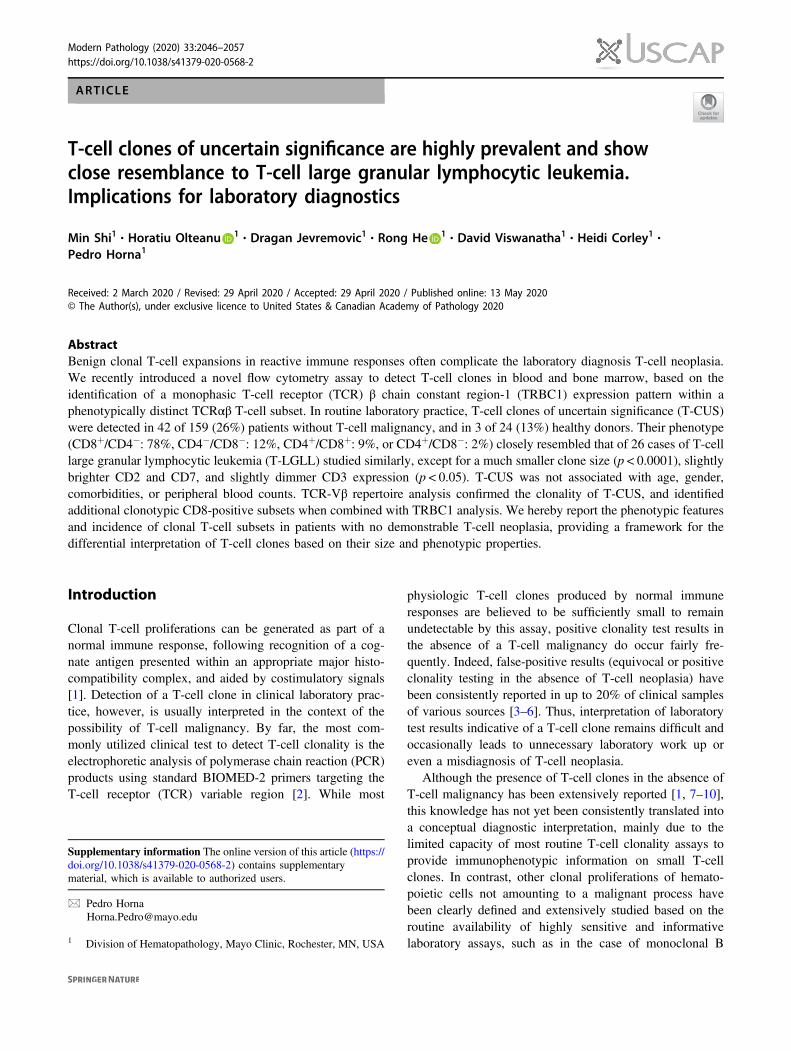
Fig. 1 T-cell receptor β chain constant (TRBC)-1 or TRBC2 geneselection during T-cell receptor (TCR) gene rearrangement isindependent of selection of TCR variable β (TCR-Vβ) genes. aGene rearrangement of the TCR β chain locus involves the selection ofone of two mutually exclusive TRBC genes, and one of 52 TCR-Vβgenes. A specific anti-TRBC1 antibody can be used by flow cytometry
to distinguish TRBC1 and TRBC2-expressing T-cells. b Comparisonof the TCR-Vβ repertoire between TRBC1-positive and TRBC1-negative peripheral blood T-cells from five healthy donors, showingremarkably similar distributions. Lines and ranges represent the meanand standard deviation of the mean.
T-cell clones of uncertain significance are highly prevalent and show close resemblance to T-cell large. . . 2047
previously described [11]. In short, CD3-positive/TCRγδ-negative T-cells (TCRαβ T-cells) were displayed on a CD4versus CD8 dot-plot to identify CD4-positive, CD8-posi-tive, CD4/CD8 double-positive, and CD4/CD8 double-negative subsets. CD4-posiitve and CD8-positive T-cellswere separately studied for the presence of immunopheno-typically distinct subsets based on CD2, CD5, and CD7expression using several dot plots and a radar (three-dimensional) plot. Expression of CD3, CD45, CD4, andCD8 was carefully studied on the gating plots to identifyadditional discrete subsets. The immunophenotype of CD3-negative events (including CD7-positive NK cells) was alsoevaluated to identify abnormal CD3-negative T-cell subsets.Each identified T-cell subset was evaluated on a histogramof TRBC1 expression. Clonal T-cell populations wereidentified as immunophenotypically distinct TCRαβ T-cellsubsets exhibiting discretely homogenous T-cell antigenexpression properties, and a monophasic TRBC1 stainingpattern defined as either (1) >85% of TRBC1-positiveevents, (2) <15% TRBC1-positive events, or (3) homo-genous TRBC1-dim expression. These thresholds werearbitrarily defined to capture all T-cell neoplasms so farevaluated in our practice (over 200 T-cell malignancies)using our T-cell panel; while excluding total benign CD4-positive and CD8-positive T-cell subsets from healthyindividuals and patients without T-cell neoplasia (with theexception of very rare large CD8-positive T-CUS).
The median fluorescence of CD2, CD3, CD4, CD5,CD7, CD8, CD45, and TRBC1 expression was calculatedfor each clonal T-cell subset, and expressed as a percentagerelative to the median fluorescence of non-clonal back-ground CD4-positive T-cells (for CD2, CD3, CD4, CD5,and CD45), non-clonal background CD8-positive/TCRγδ-negative T-cells (for CD8), NK cells (for CD7), or CD4-positive/TRBC1-positive T-cells (for TRBC1). In all casesof T-LGLL and selected cases of T-CUS, additional tubeswere set up to study the expression of the NK-cell-associated antigens CD16, CD56, CD57, CD94, andNKG2A (using antibodies conjugated to PerCP-Cy5.5,Horizon V450, FITC, APC, and PE, respectively), onrelevant T-cell subsets gated based CD3 (PE-Cy7) and CD8(APC-H7) expression.
A combined TRBC1/TCR variable β (TCR-Vβ) flowcytometry assay was developed based on the previouslydescribed IOTest Beta Mark TCR-Vβ repertoire kit assay(Beckman Coulter, Brea, CA, USA), combined with anti-bodies recognizing CD2, CD3, CD4, CD5, CD7, CD8, andTRBC1 (conjugated to PerCP-Cy5.5, PE-Cy7, APC-R700,APC, Horizon V450, APC-H7, and BV605, respectively).In brief, each detectable TCR-Vβ specificity was separatelyanalyzed for clonality based on the pattern of TRBC1expression within phenotypically distinct subsets, asdescribed above. Tube 6 of the Beta Mark test (Vβ23, Vβ1,
and Vβ21.3) was excluded from the analysis due to sub-optimal staining when utilized within this custom panel.
For all flow cytometry assays, at least 100,000 totalevents were acquired per analysis tube on a FacsCanto IIflow cytometer (BD Biosciences, San Jose, CA, USA). Allantibodies were obtained from BD Biosciences, except for aFITC-conjugated anti-TRBC1 antibody (clone JOVI.1,Ancell Corporation, Bayport, MN, USA), and the anti-TCR-Vβ antibodies from the Beta Mark kit (BeckmanCoulter). All analyses were performed on Kaluza version2.1 (Beckman Coulter), considering only populationscomprised by 100 or more events. All cases were analyzedretrospectively by two of the authors (PH and MS) in ablinded fashion, to ensure consistency.
TCR gene rearrangement studies
Total cellular DNA was extracted and PCR amplificationperformed in five multiplex PCR tubes with ASR Biomed-2primers (Invivoscribe Technologies, San Diego, CA, USA)targeting TCR-Vβ, Dβ, Jβ, Vγ, and Jγ regions. The productswere separated and detected by capillary gel electrophoresison the ABI Prism 3130xl genetic analyzer (Applied Bio-systems, Warrington, UK).
Statistical analysis
All statistic calculations were performed using GraphPadPrism, version 8.2.1 for Windows (GraphPad Software, SanDiego, CA, USA). Comparisons of measurement valuesbetween two groups were performed using theMann–Whitney test (clone size) or an unpaired two-tailed ttest (age, median fluorescence percentage, TCR-Vβ classpercentages, and peripheral blood cell counts). Comparisonsof nominal variables were performed using the chi-squaredtest for expected values equal or greater than 5, and theFisher’s exact test for expected values less than 5. Corre-lation between two variables was evaluated using theSpearman nonparametric test. A statistical significant Pvalue was considered as less than 0.05.
Results
A previously reported single-tube flow cytometry assay forthe detection T-cell clones [11] was developed and vali-dated for implementation into laboratory diagnostics. Thisassay relies on the restricted expression of one of twomutually exclusive TRBCs within immunophenotypicallydistinct clonal TCRαβ T-cell subsets (Fig. 1a), assumingthat selection of TRBC1 or TRBC2 is a random process thatoccurs independently of TCR-Vβ gene selection. To con-firm this hypothesis, we tested five healthy donors with a
2048 M. Shi et al.
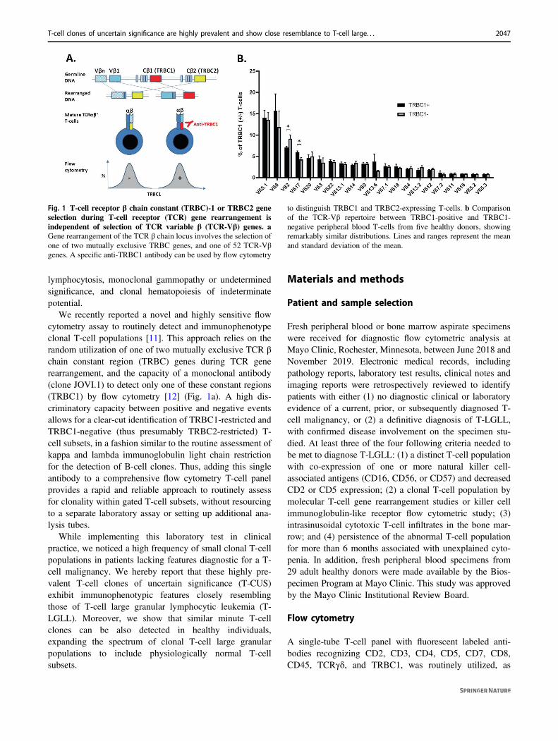
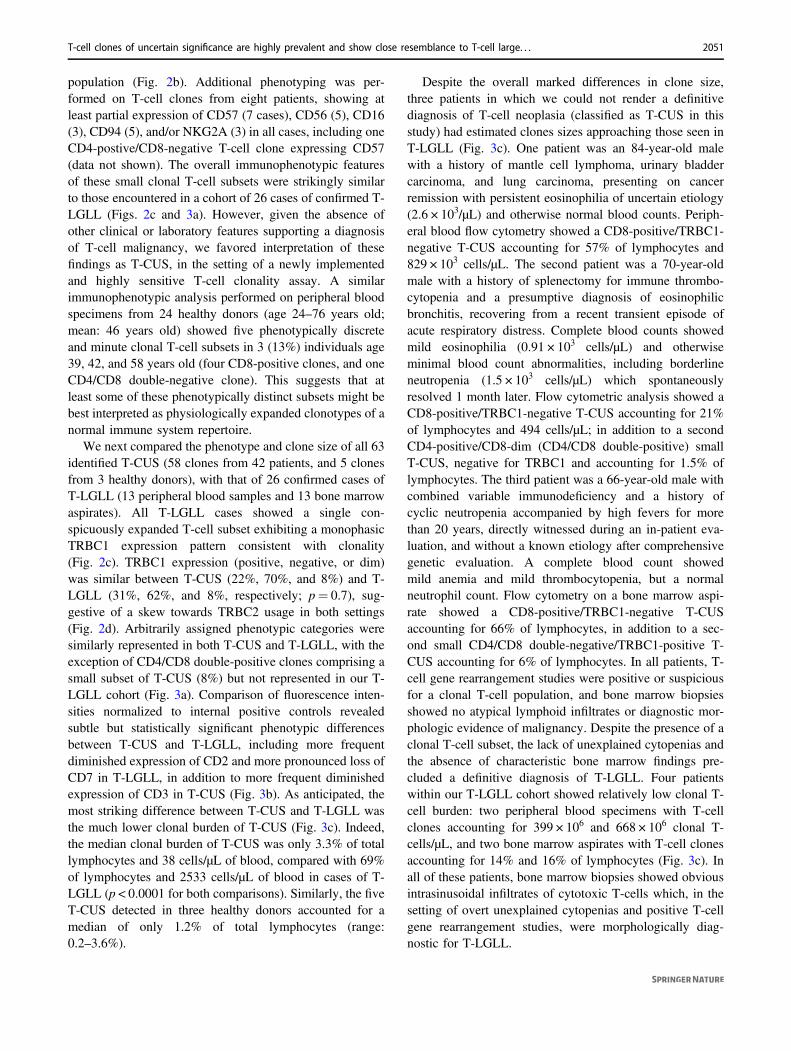
Fig. 2 T-cell clones of uncertain significance (T-CUS) are fre-quently detected by flow cytometry on patients with no evidence ofT-cell neoplasia. a Peripheral blood flow cytometry plots from threepatients with T-CUS: A 58-year-old man with a history of hyper-trophic cardiomyopathy presenting with diarrhea, dysuria, and pruritus(top); a 57-year-old female with acute hypersensitivity reaction toLamotrigine (middle); and a 73-year-old male with newly diagnosedinclusion body myositis and no cytopenias or lymphocytosis (bottom).The red events correspond to CD8-positive T-CUS that are homo-genously negative (top), dim (middle), or positive (bottom) forTRBC1, accounting for 0.5%, 17%, and 16% of lymphocytes,respectively. The last patient (bottom) also features two additionalTRBC1-negative T-CUS, which are CD8-positive (violet) or CD4/CD8 double-negative (blue), and account for 6% and 1.3% of lym-phocytes, respectively. Also displayed are background non-clonal
CD4-positive (cyan) and CD8-positive (orange) T-cells, and percen-tages of TRBC1-positive events for each population adjacent to eachhistogram. N/A not applicable. b Incidence and basic phenotypicfeatures of T-CUS detected in 159 patients with no demonstrable T-cell neoplasm. c Three typical cases of T-cell large granular lympho-cytic leukemia (T-LGLL) are shown for comparison, featuring similarclonal CD8-positive T-cell populations (red events) on bone marrow(top and bottom) or peripheral blood (middle) specimens, which arehomogenously negative (top), dim (middle), or positive (bottom) forTRBC1. d Percentages of TRBC1-positive events on CD4-positiveand CD8-positive T-cells from 24 healthy donors; and on gated clonalT-cell populations from patients with T-CUS and patients with T-LGLL. Dotted lines depict previously established thresholds for T-cellclonality. TRBC1-dim clonal T-cell populations are not shown.
T-cell clones of uncertain significance are highly prevalent and show close resemblance to T-cell large. . . 2049
modified flow cytometry assay where our diagnostic panelwas combined with antibodies to detect different Vβfamilies. Overall, the TCR-Vβ repertoire of TRBC1-positive and TRBC1-negative (TRBC2-positive) T-cellsclosely mirrored each other, with no detectable selectionbias between TCR-Vβ and TRBC expression, except forminor percentage differences for Vβ2 and Vβ17 (Fig. 1b).
A total of 159 patients (104 peripheral blood and 55 bonemarrow specimens) with no diagnostic clinical or laboratoryevidence of T-cell neoplasia were then studied with our
diagnostic T-cell flow cytometry panel including core T-cellantigens and TRBC1. Phenotypically discrete clonal T-cellsubsets were detected in 42 patients (26%), with a singleclonal T-cell subset observed in most subjects. Elevenpatients had two clonal subsets, while three and four clonalsubsets were detected in one patient, each (Fig. 2a). Of the58 total T-cell clones detected in these patients, 45 (78%)were CD8-positive, 7 (12%) were CD4/CD8 double-nega-tive, 5 (9%) were CD4/CD8 double-positive, and only 1(2%) corresponded to a CD4-positive/CD8-negative T-cell
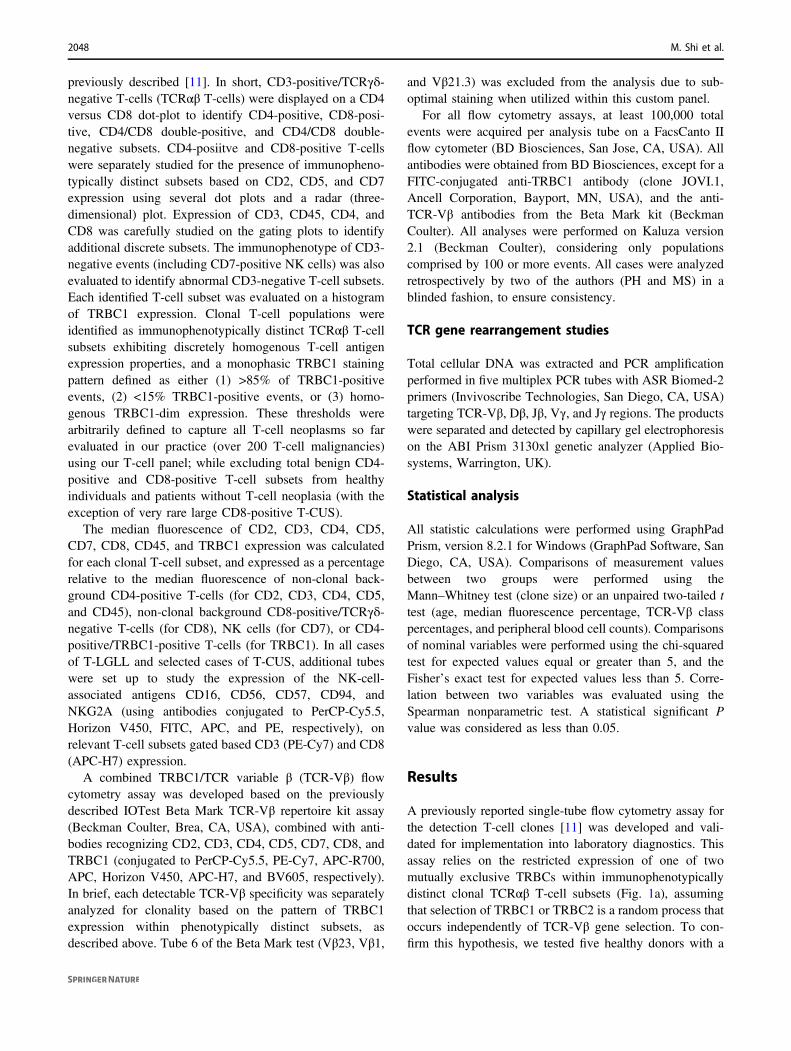
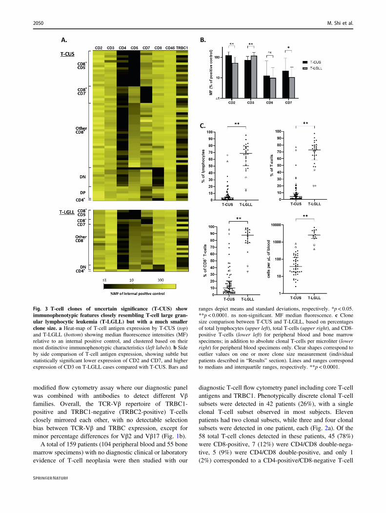
Fig. 3 T-cell clones of uncertain significance (T-CUS) showimmunophenotypic features closely resembling T-cell large gran-ular lymphocytic leukemia (T-LGLL) but with a much smallerclone size. a Heat-map of T-cell antigen expression by T-CUS (top)and T-LGLL (bottom) showing median fluorescence intensities (MF)relative to an internal positive control, and clustered based on theirmost distinctive immunophenotypic characteristics (left labels). b Sideby side comparison of T-cell antigen expression, showing subtle butstatistically significant lower expression of CD2 and CD7, and higherexpression of CD3 on T-LGLL cases compared with T-CUS. Bars and
ranges depict means and standard deviations, respectively. *p < 0.05.**p < 0.0001. ns non-significant. MF median fluorescence. c Clonesize comparison between T-CUS and T-LGLL, based on percentagesof total lymphocytes (upper left), total T-cells (upper right), and CD8-positive T-cells (lower left) for peripheral blood and bone marrowspecimens; in addition to absolute clonal T-cells per microliter (lowerright) for peripheral blood specimens only. Clear shapes correspond tooutlier values on one or more clone size measurement (individualpatients described in “Results” section). Lines and ranges correspondto medians and interquartile ranges, respectively. **p < 0.0001.
2050 M. Shi et al.
population (Fig. 2b). Additional phenotyping was per-formed on T-cell clones from eight patients, showing atleast partial expression of CD57 (7 cases), CD56 (5), CD16(3), CD94 (5), and/or NKG2A (3) in all cases, including oneCD4-postive/CD8-negative T-cell clone expressing CD57(data not shown). The overall immunophenotypic featuresof these small clonal T-cell subsets were strikingly similarto those encountered in a cohort of 26 cases of confirmed T-LGLL (Figs. 2c and 3a). However, given the absence ofother clinical or laboratory features supporting a diagnosisof T-cell malignancy, we favored interpretation of thesefindings as T-CUS, in the setting of a newly implementedand highly sensitive T-cell clonality assay. A similarimmunophenotypic analysis performed on peripheral bloodspecimens from 24 healthy donors (age 24–76 years old;mean: 46 years old) showed five phenotypically discreteand minute clonal T-cell subsets in 3 (13%) individuals age39, 42, and 58 years old (four CD8-positive clones, and oneCD4/CD8 double-negative clone). This suggests that atleast some of these phenotypically distinct subsets might bebest interpreted as physiologically expanded clonotypes of anormal immune system repertoire.
We next compared the phenotype and clone size of all 63identified T-CUS (58 clones from 42 patients, and 5 clonesfrom 3 healthy donors), with that of 26 confirmed cases ofT-LGLL (13 peripheral blood samples and 13 bone marrowaspirates). All T-LGLL cases showed a single con-spicuously expanded T-cell subset exhibiting a monophasicTRBC1 expression pattern consistent with clonality(Fig. 2c). TRBC1 expression (positive, negative, or dim)was similar between T-CUS (22%, 70%, and 8%) and T-LGLL (31%, 62%, and 8%, respectively; p= 0.7), sug-gestive of a skew towards TRBC2 usage in both settings(Fig. 2d). Arbitrarily assigned phenotypic categories weresimilarly represented in both T-CUS and T-LGLL, with theexception of CD4/CD8 double-positive clones comprising asmall subset of T-CUS (8%) but not represented in our T-LGLL cohort (Fig. 3a). Comparison of fluorescence inten-sities normalized to internal positive controls revealedsubtle but statistically significant phenotypic differencesbetween T-CUS and T-LGLL, including more frequentdiminished expression of CD2 and more pronounced loss ofCD7 in T-LGLL, in addition to more frequent diminishedexpression of CD3 in T-CUS (Fig. 3b). As anticipated, themost striking difference between T-CUS and T-LGLL wasthe much lower clonal burden of T-CUS (Fig. 3c). Indeed,the median clonal burden of T-CUS was only 3.3% of totallymphocytes and 38 cells/µL of blood, compared with 69%of lymphocytes and 2533 cells/µL of blood in cases of T-LGLL (p < 0.0001 for both comparisons). Similarly, the fiveT-CUS detected in three healthy donors accounted for amedian of only 1.2% of total lymphocytes (range:0.2–3.6%).
Despite the overall marked differences in clone size,three patients in which we could not render a definitivediagnosis of T-cell neoplasia (classified as T-CUS in thisstudy) had estimated clones sizes approaching those seen inT-LGLL (Fig. 3c). One patient was an 84-year-old malewith a history of mantle cell lymphoma, urinary bladdercarcinoma, and lung carcinoma, presenting on cancerremission with persistent eosinophilia of uncertain etiology(2.6 × 10³/μL) and otherwise normal blood counts. Periph-eral blood flow cytometry showed a CD8-positive/TRBC1-negative T-CUS accounting for 57% of lymphocytes and829 × 103 cells/µL. The second patient was a 70-year-oldmale with a history of splenectomy for immune thrombo-cytopenia and a presumptive diagnosis of eosinophilicbronchitis, recovering from a recent transient episode ofacute respiratory distress. Complete blood counts showedmild eosinophilia (0.91 × 10³ cells/µL) and otherwiseminimal blood count abnormalities, including borderlineneutropenia (1.5 × 10³ cells/µL) which spontaneouslyresolved 1 month later. Flow cytometric analysis showed aCD8-positive/TRBC1-negative T-CUS accounting for 21%of lymphocytes and 494 cells/μL; in addition to a secondCD4-positive/CD8-dim (CD4/CD8 double-positive) smallT-CUS, negative for TRBC1 and accounting for 1.5% oflymphocytes. The third patient was a 66-year-old male withcombined variable immunodeficiency and a history ofcyclic neutropenia accompanied by high fevers for morethan 20 years, directly witnessed during an in-patient eva-luation, and without a known etiology after comprehensivegenetic evaluation. A complete blood count showedmild anemia and mild thrombocytopenia, but a normalneutrophil count. Flow cytometry on a bone marrow aspi-rate showed a CD8-positive/TRBC1-negative T-CUSaccounting for 66% of lymphocytes, in addition to a sec-ond small CD4/CD8 double-negative/TRBC1-positive T-CUS accounting for 6% of lymphocytes. In all patients, T-cell gene rearrangement studies were positive or suspiciousfor a clonal T-cell population, and bone marrow biopsiesshowed no atypical lymphoid infiltrates or diagnostic mor-phologic evidence of malignancy. Despite the presence of aclonal T-cell subset, the lack of unexplained cytopenias andthe absence of characteristic bone marrow findings pre-cluded a definitive diagnosis of T-LGLL. Four patientswithin our T-LGLL cohort showed relatively low clonal T-cell burden: two peripheral blood specimens with T-cellclones accounting for 399 × 106 and 668 × 106 clonal T-cells/µL, and two bone marrow aspirates with T-cell clonesaccounting for 14% and 16% of lymphocytes (Fig. 3c). Inall of these patients, bone marrow biopsies showed obviousintrasinusoidal infiltrates of cytotoxic T-cells which, in thesetting of overt unexplained cytopenias and positive T-cellgene rearrangement studies, were morphologically diag-nostic for T-LGLL.
T-cell clones of uncertain significance are highly prevalent and show close resemblance to T-cell large. . . 2051
Given prior reports describing clonal T-cell large gran-ular proliferations in a variety of specific clinical settings,we queried whether an association between T-CUS andcertain clinical or laboratory features could be encounteredin our cohort. We found no positive association between T-CUS and any particular disease or condition, although anunexplained inverse association between T-CUS and auto-immune disease was noted (Table 1). Also, there was nocorrelation with age, sex, or peripheral blood counts. Asexpected, T-cell gene rearrangement studies were positive
or suspicious for clonality in most patients with T-CUS onwhom this test was performed (13 of 15, 87%), with theexception of two cases where a low clonal burden (<3% oftotal T-cells) might have precluded the detection of a clonalpeak. In our patient population, a positive or suspicious T-cell gene rearrangement study was also fairly common inpatients without detectable T-CUS (9/43, 21%), raising thepossibility of TCRγδ clones or immunophenotypicallyindistinct clones that could not be detected by our flowcytometry assay.
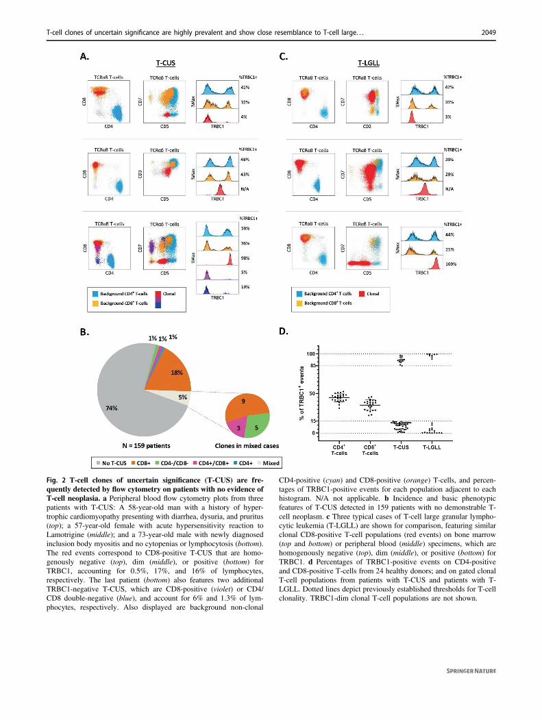
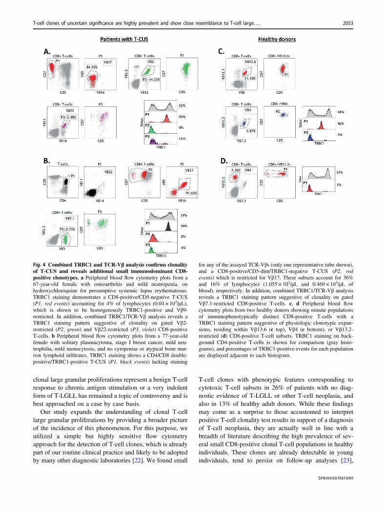
To further confirm the validity of TRBC1 staining as anindicator of a clonotypic T-cell population, we studied theTCR-Vβ repertoire in four additional peripheral bloodsamples from patients with no demonstrable T-cell neo-plasia on which a putative clonal T-cell subset was detectedby TRBC1 staining. In all samples, these subsets showedeither a restricted TCR-Vβ class expression or a lack ofexpression of all assayed TCR-Vβ classes, confirmatory ofT-cell clonality. In addition, a comprehensive study of theTCR-Vβ repertoire in these samples showed one or twoadditional CD8-positive TCR-Vβ classes exhibiting aTRBC1 staining pattern suggestive of an immunodominantclonotype (Fig. 4a). A similar study on five peripheral bloodsamples from healthy donors showed two individuals wherea minute phenotypically distinct T-cell population sugges-tive of an immunodominant clonotype could be demon-strated by TRBC1 analysis within a single CD8-positive/TCR-Vβ class-positive subset (Fig. 4b). Remarkably, asimilar comprehensive analysis of the CD4-positive T-cellcompartment did not yield any clonotypic subsets.
Discussion
T-LGLL is a lymphoproliferative disorder characterized bya persistent clonal expansion of cytotoxic T-cells involvingperipheral blood and bone marrow, which frequently resultsin neutropenia or other cytopenias that may require ther-apeutic intervention [13]. Chronic antigen stimulation issuspected to be responsible for the initial emergence of acytotoxic T-cell clone, which is then believed to undergomalignant transformation resulting in clonal overgrowth andsuppression of hematopoiesis. The laboratory diagnosis ofT-LGLL and its distinction from reactive T-cell largegranular lymphocytes can be extremely challenging. Indeed,persistent clonal expansion of large granular lymphocyteshave been reported in a variety of clinical settings includingmyelodysplastic syndromes [14], B-cell lymphoprolifera-tive disorders, plasma cell proliferative disorders, auto-immunity [15], unexplained neutropenia [16], paroxysmalnocturnal hemoglobinuria [17], following allogeneic orautologous hematopoietic stem cell transplantation [18–20],and after solid organ transplantation [21]. Whether these
Table 1 Clinical and laboratory characteristics of 159 patients with noevidence of T-cell malignancy, with or without T-cell clones ofuncertain significance (T-CUS).
No T-CUS T-CUS P value
N 117 42 –
Age in years (±SD) 56 (±18) 62 (±18) 0.07
Male:female 0.8:1 1:1 0.7
Diagnoses [n (%)]
Autoimmune 32 (27%) 4 (10%) 0.01
Neurologic 4 (3%) 2 (5%) 0.1
Infectious 11 (9%) 1 (2%) 0.1
B-cell/plasma cellneoplasm
23 (20%) 8 (19%) 0.8
Carcinoma/sarcoma 3 (3%) 4 (10%) 0.07
MDS/MPN 7 (6%) 6 (14%) 0.1
Hypereosinophilia 2 (2%) 1 (2%) 0.8
Inflammatory dermatosis 10 (9%) 7 (17%) 0.2
Immunodeficiency 8 (7%) 4 (10%) 0.6
Post allo-HSCT 1 (1%) 0 (0%) 1
Post solid organ transplant 3 (3%) 3 (7%) 0.2
Blood counts [cells × 103/µL (±SD)]
White blood cells 8.4 (±5.9) 7.3 (±4.5) 0.3
Neutrophils 4.7 (±8.2) 3.5 (±2.6) 0.3
Lymphocytes 3.1 (±3.8) 2.4 (±2.0) 0.3
Eosinophils 0.6 (±1.3) 0.4 (±0.8) 0.3
Platelets 228 (±134) 224 (±144) 0.9
Hemoglobin (g/dL) 12.19 (±2.-6)
12.53 (±2.3) 0.5
Peripheral blood subsets analysis
Blood specimens studied 74 30 –
CD4:CD8 2.7:1(±3.3:1)
2.0:1 (±1.5:1) 0.3
% NK cells (oflymphocytes)
11.6%(±11.2%)
12.3% (±10.6%) 0.8
% γδ T-cells (oflymphocytes)
4.3%(±8.4%)
3.1% (4.0%) 0.4
T-cell gene rearrangementpositive or suspicious
9/43 (21%) 13/15 (87%) <0.0001
SD standard deviation, MDS/MPN myelodysplastic and/or myelopro-liferative neoplasm, Allo-HSCT allogeneic hematopoietic stem celltransplant. Statistically significant differences highlighted in bold.
2052 M. Shi et al.
clonal large granular proliferations represent a benign T-cellresponse to chronic antigen stimulation or a very indolentform of T-LGLL has remained a topic of controversy and isbest approached on a case by case basis.
Our study expands the understanding of clonal T-celllarge granular proliferations by providing a broader pictureof the incidence of this phenomenon. For this purpose, weutilized a simple but highly sensitive flow cytometryapproach for the detection of T-cell clones, which is alreadypart of our routine clinical practice and likely to be adoptedby many other diagnostic laboratories [22]. We found small
T-cell clones with phenotypic features corresponding tocytotoxic T-cell subsets in 26% of patients with no diag-nostic evidence of T-LGLL or other T-cell neoplasia, andalso in 13% of healthy adult donors. While these findingsmay come as a surprise to those accustomed to interpretpositive T-cell clonality test results in support of a diagnosisof T-cell neoplasia, they are actually well in line with abreadth of literature describing the high prevalence of sev-eral small CD8-positive clonal T-cell populations in healthyindividuals. These clones are already detectable in youngindividuals, tend to persist on follow-up analyses [23],
Fig. 4 Combined TRBC1 and TCR-Vβ analysis confirms clonalityof T-CUS and reveals additional small immunodominant CD8-positive clonotypes. a Peripheral blood flow cytometry plots from a67-year-old female with osteoarthritis and mild neutropenia, onhydroxychloroquine for presumptive systemic lupus erythematosus.TRBC1 staining demonstrates a CD8-positive/CD5-negative T-CUS(P1, red events) accounting for 4% of lymphocytes (0.04 × 10³/µL),which is shown to be homogenously TRBC1-positive and Vβ9-restricted. In addition, combined TRBC1/TCR-Vβ analysis reveals aTRBC1 staining pattern suggestive of clonality on gated Vβ2-restricted (P2, green) and Vβ22-restricted (P3, violet) CD8-positiveT-cells. b Peripheral blood flow cytometry plots from a 77-year-oldfemale with solitary plasmacytoma, stage I breast cancer, mild neu-trophilia, mild monocytosis, and no cytopenias or atypical bone mar-row lymphoid infiltrates. TRBC1 staining shows a CD4/CD8 double-positive/TRBC1-positive T-CUS (P1, black events) lacking staining
for any of the assayed TCR-Vβs (only one representative tube shown),and a CD8-positive/CD5-dim/TRBC1-negative T-CUS (P2, redevents) which is restricted for Vβ17. These subsets account for 36%and 16% of lymphocytes (1.055 × 10³/µL and 0.469 × 10³/µL ofblood), respectively. In addition, combined TRBC1/TCR-Vβ analysisreveals a TRBC1 staining pattern suggestive of clonality on gatedVβ7.1-restricted CD8-positive T-cells. c, d Peripheral blood flowcytometry plots from two healthy donors showing minute populationsof immunophenotypically distinct CD8-positive T-cells with aTRBC1 staining pattern suggestive of physiologic clonotypic expan-sions, residing within Vβ13.6 (c top), Vβ4 (c bottom), or Vβ13.2-restricted (d) CD8-positive T-cell subsets. TRBC1 staining on back-ground CD4-positive T-cells is shown for comparison (gray histo-grams), and percentages of TRBC1-positive events for each populationare displayed adjacent to each histogram.
T-cell clones of uncertain significance are highly prevalent and show close resemblance to T-cell large. . . 2053
become more prominent with aging [24, 25], and arebelieved to represent physiologic expansions of mostlyeffector/memory cytotoxic T-cell subsets in response toeither highly prevalent chronic infections such as cytome-galovirus and Epstein–Bar virus [8, 26–30], resolved acuteinfections [31], neoplastic processes, or other sources ofantigen exposure. Also in line with our results is the factthat physiologic clonal expansions of CD4-positive/CD8-negative T-cells are much less frequently represented or notdetected at all, depending on the sensitivity of the assayutilized, consistent with the concept that clonal expansionsof CD4-positive and CD8-positive T-cells are controlled bysubstantially different mechanisms [1, 32]. Although we didnot find a statistically significant correlation between ageand the presence (Table 1) or clone size (SupplementaryFig. 1A, B) of T-CUS, the age spectrum of our cohort wastoo narrow to formally study the dynamics of T-CUSwith aging.
We propose utilizing the term T-CUS, modeled afterprior reports describing T-cell clones/clonopathies ofunknown/uncertain/undetermined significance [33–36], inorder to facilitate our understanding of frequently encoun-tered clonal expansions of cytotoxic T-cells that are largerthan expected for physiologic T-cell clones but do not meetcriteria for T-LGLL at the time of evaluation. Based on ourdata on healthy individuals, a clonal size of 5% lympho-cytes or 50 cells/µL of blood might be an appropriatearbitrary threshold to “screen out” normal clonotypic T-cellsubsets which should not be reported in routine laboratorypractice as they are detectable in healthy individuals. Higherpractical thresholds, such as 20% of lymphocytes or 500cells/µL of blood, might also be appropriate to preventunnecessary work up of small T-CUS commonly encoun-tered in patients with unrelated conditions. However, asmall but significant overlap in clone size between T-CUSand T-LGLL should be acknowledged, often requiring bonemarrow evaluation and clinical follow-up for a final dis-tinction. Although testing for STAT3 mutations was notperformed in this study, assessment of these genetic lesionscould be contributory to the evaluation of patients where adistinction between T-CUS and T-LGLL cannot be easilydetermined, as these mutations have been reported in ~40%of patients with T-LGLL [37–39]. However, STAT3mutations have also been identified in T large granularproliferations from patients with aplastic anemia, myelo-dysplastic syndrome [40], and Felty syndrome [41], where adiagnosis of T-LGLL could not be unequivocallyestablished.
The definition of T-CUS proposed in this paperencompasses a spectrum of phenotypic variants, mirroringthose of T-LGLL. These include previously describedindolent CD4-positive largec granular proliferations withvariable expression of CD8 [30, 42, 43], classified in our
study as CD4/CD8 double-positive T-CUS or a singlecase of CD4-positive/CD57-positive T-CUS. Recognitionof this unique phenotypic variant is important, as it isunlikely to be representative of bona fide T-LGLL. Wealso describe a significant proportion of CD4/CD8double-negative T-CUS which has not been as widelyrecognized but has been encountered in a prior study [35].While many our patients without T-cell neoplasia hadeosinophilia (main indication for flow cytometric analysisin 16% of patients) (Supplementary Table 2), the lackcorrelation between the degree of eosinophilia and T-CUS, and the rarity of clinical hypereosinophilia in ourcohort (Table 1) precludes interpretation of these clonesas an abnormality with the spectrum of lymphocyticvariant of hypereosinophilic syndrome (LV-HES). Indeed,most (but not all) abnormal T-cells identified in LV-HEScorrespond to CD4-positive/CD3-negative subsets [44],which markedly differs from the phenotypic spectrum ofT-CUS.
Upon comprehensive phenotypic analysis, we found thatthe degree of CD5 loss by T-CUS is largely indistinguish-able from that encountered in T-LGLL, despite theemphasis commonly made on this phenotypic characteristicfor the detection of T-LGLL clones [45, 46]. We didhowever detect some phenotypic differences in theexpression of CD2, CD3, and CD7, which were not initiallyrecognized on visual inspection of the flow cytometry plotsand currently remain largely unexplained (Fig. 3b). Ofinterest, a gene expression profiling study also demonstrateda lower level of CD2 transcripts in T-LGLL compared withreactive cytotoxic T-cells [47]. Limited numeric analysis ofoverall T-cell and NK-cell subsets did not yield significantdifferences between patients with and without T-CUS(Table 1), but did show decreased numbers of NK cells inpatients with T-LGLL (Supplementary Table 1), as pre-viously reported [48].
The high reliance of our assay on immunophenotypicmarkers and gating strategies to define clonality warrantscaution and extensive validation when implementing thisanalysis in clinical practice. For example, while our initialanalysis of a limited number of T-cell malignancies sug-gested a narrow TRBC1 expression pattern for neoplasia(>97% or <3%) [11], our accumulated experience withmany more cases favors using a wider threshold (>85% or<15%) to accommodate for neoplasms on which the phe-notypic distinction from background benign T-cells is not asperfect, and the percentage of tumor cells among total T-cells not as high. Given this interdependence betweenimmunophenotypic gating and clonality assessment byTRBC1 staining, selection of accompanying T-cell markersthat would best separate neoplastic cells from backgroundbenign T-cells is of upmost importance for an optimal testperformance.
2054 M. Shi et al.
In addition to estimating percentages of TRBC1-positiveevents, analysis strategies should also include the identifi-cation of monophasic TRBC1-dim expression as a surrogatefor clonality, as this often produces falsely normal percen-tages overlapping the threshold between positive andnegative events. We have encountered this pattern in varietyof T-cell neoplasms [11] and in T-CUS, always in a min-ority of cases, and to the best of our knowledge not asso-ciated with any particular phenotypic feature, patientdemographic, specimen type, or pre-analytical variable. Asexpected, CD3-negative neoplasms are always TRBC1-negative (based on authors’ experience, data not shown) dueto lack of expression of the TCR complex. However, wehave not found a consistent correlation between dimTRBC1 and dim CD3 expression that would support thehypothesis that this pattern is due to decreased expression ofthe TCR (Supplementary Fig. 1C). As such, the molecularbasis of dim TRBC1 expression remains uncertain.
While most routine T-cell clonality assays rely on therelative abundance of a T-cell clone as detected by adominant TCR-Vβ class [49, 50] or prominent TCRamplification product [2], our flow cytometry T-cell panel iscapable of detecting very small T-cell clones that are notparticularly abundant relative to total T-cells, but havedistinct immunophenotypic features that allows them to begated and analyzed separately for clonality. Moreover, weshow that a combined TRBC1/TCR-Vβ analysis provides anovel method to detect even smaller clonal T-cell popula-tions that have replaced most of the repertoire of a specificVβ class, but have not expanded beyond the size of otherVβ classes. Thus, we were able to detect small T-cell clonesthat would have otherwise been missed by other commonlaboratory tests, many of which correspond to physiologicclonotypic T-cell expansions of normal immunity. Despitethese advantages, our routine flow cytometry panel does nothave the capacity to detect TCRγδ T-cell clones or clonal T-cell subsets that lack distinguishable phenotypic featureswith the antigens studied.
By systematically studying the phenotypic properties ofclonal T-cell subsets in patients with no demonstrable T-cellneoplasms and healthy individuals, we provide a frameworkfor the interpretation of laboratory test results where small T-cell clones and/or immunophenotypically distinct T-cell sub-sets might be detected. The first corollary of our study relatesto the rarity of physiologic or reactive CD4-positive/CD8-negative clonotypic T-cell expansions detected by our assay,which should prompt careful evaluation for the possibility of aT-cell lymphoproliferative disorder irrespective of the clonesize. Second, a CD4-negative or CD4/CD8 double-positiveclonal T-cell subset should be interpreted in the context ofclone size, as clones smaller than 20% of total lymphocytesare highly prevalent in patients with no T-cell malignancy (T-CUS) and show no particular disease association in our series,
while clones smaller than 5% of lymphocytes are within theexpected size for physiologic clonotypic expansions in heal-thy individuals. Third, it should be noted that the phenotypicspectrum of T-CUS closely resembles that of T-LGLL, whichdirectly relates to the common difficulties in distinguishing areactive clonal proliferation of T-cell large granular lympho-cytes from a bone fide T-cell neoplasm. Finally, the highprevalence of reactive T-cell clones in our series warrantscaution in the interpretation of highly sensitive assays of T-cell clonality, as low-positive results might not be con-tributory to the overall clinical evaluation or may result inmisinterpretation of suspicious morphologic, phenotypic, orclinical findings.
Compliance with ethical standards
Conflict of interest The authors declare that they have no conflict ofinterest.
Publisher’s note Springer Nature remains neutral with regard tojurisdictional claims in published maps and institutional affiliations.
References
1. Maini MK, Casorati G, Dellabona P, Wack A, Beverley PC. T-cellclonality in immune responses. Immunol Today.1999;20:262–266.
2. Langerak AW, Groenen PJ, Bruggemann M, Beldjord K, BellanC, Bonello L, et al. EuroClonality/BIOMED-2 guidelines forinterpretation and reporting of Ig/TCR clonality testing in sus-pected lymphoproliferations. Leukemia. 2012;26:2159–2171.
3. Shin S, Kim AH, Park J, Kim M, Lim J, Kim Y, et al. Analysis ofimmunoglobulin and T cell receptor gene rearrangement in thebone marrow of lymphoid neoplasia using BIOMED-2 multiplexpolymerase chain reaction. Int J Med Sci. 2013;10:1510–1517.
4. Zhang B, Beck AH, Taube JM, Kohler S, Seo K, Zwerner J, et al.Combined use of PCR-based TCRG and TCRB clonality tests onparaffin-embedded skin tissue in the differential diagnosis ofmycosis fungoides and inflammatory dermatoses. J Mol Diagn.2010;12:320–327.
5. Langerak AW, Molina TJ, Lavender FL, Pearson D, Flohr T,Sambade C, et al. Polymerase chain reaction-based clonalitytesting in tissue samples with reactive lymphoproliferations: use-fulness and pitfalls. A report of the BIOMED-2 Concerted ActionBMH4-CT98-3936. Leukemia. 2007;21:222–229.
6. Bonzheim I, Fröhlich F, Adam P, Colak S, Metzler G, Quintanilla-Martinez L, et al. A comparative analysis of protocols for detec-tion of T cell clonality in formalin-fixed, paraffin-embedded tissue—implications for practical use. J Hematop. 2012;5:7–16.
7. Monteiro J, Batliwalla F, Ostrer H, Gregersen PK. Shortenedtelomeres in clonally expanded CD28(-)CD8(+) T cells imply areplicative history that is distinct from their CD28(+)CD8(+)counterparts. J Immunol. 1996;156:3587–3590.
8. Maini MK, Gudgeon N, Wedderburn LR, Rickinson AB, Bev-erley PC. Clonal expansions in acute EBV infection are detectablein the CD8 and not the CD4 subset and persist with a variableCD45 phenotype. J Immunol. 2000;165:5729–5737.
9. Ricalton NS, Roberton C, Norris JM, Rewers M, Hamman RF,Kotzin BL. Prevalence of CD8+ T-cell expansions in relation toage in healthy individuals. J Gerontol A Biol Sci Med Sci.1998;53:B196–203.
T-cell clones of uncertain significance are highly prevalent and show close resemblance to T-cell large. . . 2055
10. Hingorani R, Choi IH, Akolkar P, Gulwaniakolkar B, PergolizziR, Silver J, et al. Clonal predominance of T-cell receptors withinthe Cd8+ Cd45ro+ subset in normal human-subjects. J Immunol.1993;151:5762–5769.
11. Shi M, Jevremovic D, Otteson GE, Timm MM, Olteanu H, HornaP. Single antibody detection of T-cell receptor alphabeta clonalityby flow cytometry rapidly identifies mature T-cell neoplasms andmonotypic small CD8-positive subsets of uncertain significance.Cytom B Clin Cytom. 2020;98:99–107.
12. Maciocia PM, Wawrzyniecka PA, Philip B, Ricciardelli I, AkarcaAU, Onuoha SC, et al. Targeting the T cell receptor beta-chainconstant region for immunotherapy of T cell malignancies. NatMed. 2017;23:1416–1423.
13. Chan WC, Foucar K, Morice WG, Matutes E. T-cell large gran-ular lymphocytic leukaemia. In: Swerdlow SH, Campo E, LeeHarris N, Jaffe ES, Pileri SA, Stein H, et al., editors. WHOClassification of Tumors of Haematopoietic and Lymphoid Tis-sues. Lyon: IARC; 2017.
14. Zhang X, Sokol L, Bennett JM, Moscinski LC, List A, Zhang L.T-cell large granular lymphocyte proliferation in myelodysplasticsyndromes: Clinicopathological features and prognostic sig-nificance. Leuk Res. 2016;43:18–23.
15. Zhang R, Shah MV, Loughran TP Jr. The root of many evils:indolent large granular lymphocyte leukaemia and associateddisorders. Hematol Oncol. 2010;28:105–117.
16. Wlodarski MW, Nearman Z, Jiang Y, Lichtin A, Maciejewski JP.Clonal predominance of CD8(+) T cells in patients with unex-plained neutropenia. Exp Hematol. 2008;36:293–300.
17. Risitano AM, Maciejewski JP, Muranski P, Wlodarski M,O’Keefe C, Sloand EM, et al. Large granular lymphocyte (LGL)-like clonal expansions in paroxysmal nocturnal hemoglobinuria(PNH) patients. Leukemia. 2005;19:217–222.
18. Munoz-Ballester J, Chen-Liang TH, Hurtado AM, Heras I, deArriba F, Garcia-Malo MD, et al. Persistent cytotoxic T lym-phocyte expansions after allogeneic haematopoietic stem celltransplantation: kinetics, clinical impact and absence of STAT3mutations. Br J Haematol. 2016;172:937–946.
19. Wolniak KL, Goolsby CL, Chen YH, Chenn A, Singhal S, MehtaJayesh, et al. Expansion of a clonal CD8+CD57+ large granularlymphocyte population after autologous stem cell transplant inmultiple myeloma. Am J Clin Pathol. 2013;139:231–241.
20. Mohty M, Faucher C, Vey N, Chabannon C, Sainty D, ArnouletC, et al. Features of large granular lymphocytes (LGL) expansionfollowing allogeneic stem cell transplantation: a long-term ana-lysis. Leukemia. 2002;16:2129–2133.
21. Sabnani I, Zucker MJ, Tsang P, Palekar S. Clonal T-large granularlymphocyte proliferation in solid organ transplant recipients.Transpl Proc. 2006;38:3437–3440.
22. Novikov ND, Griffin GK, Dudley G, Drew M, Rojas-Rudilla V,Lindeman NI, et al. Utility of a simple and robust flow cytometryassay for rapid clonality testing in mature peripheral T-cell lym-phomas. Am J Clin Pathol. 2019;151:494–503.
23. Bernardin F, Doukhan L, Longone-Miller A, Champagne P,Sekaly R, Delwart E. Estimate of the total number of CD8+clonalexpansions in healthy adults using a new DNA heteroduplex-tracking assay for CDR3 repertoire analysis. J Immunol Methods.2003;274:159–175.
24. Messaoudi I, Lemaoult J, Guevara-Patino JA, Metzner BM,Nikolich-Zugich J. Age-related CD8 T cell clonal expansionsconstrict CD8 T cell repertoire and have the potential to impairimmune defense. J Exp Med. 2004;200:1347–1358.
25. Wedderburn LR, Patel A, Varsani H, Woo P. The developinghuman immune system: T-cell receptor repertoire of children andyoung adults shows a wide discrepancy in the frequency of per-sistent oligoclonal T-cell expansions. Immunology.2001;102:301–309.
26. Blackman MA, Woodland DL. The narrowing of the CD8 T cellrepertoire in old age. Curr Opin Immunol. 2011;23:537–542.
27. Hadrup SR, Strindhall J, Kollgaard T, Seremet T, Johansson B,Pawelec G, et al. Longitudinal studies of clonally expanded CD8T cells reveal a repertoire shrinkage predicting mortality and anincreased number of dysfunctional cytomegalovirus-specificT cells in the very elderly. J Immunol. 2006;176:2645–2653.
28. Khan N, Shariff N, Cobbold M, Bruton R, Ainsworth JA, SinclairAJ, et al. Cytomegalovirus seropositivity drives the CD8 T cellrepertoire toward greater clonality in healthy elderly individuals. JImmunol. 2002;169:1984–1992.
29. Beverley PC, Maini MK. Differences in the regulation of CD4 andCD8 T-cell clones during immune responses. Philos Trans R SocLond B Biol Sci. 2000;355:401–406.
31. Ely KH, Ahmed M, Kohlmeier JE, Roberts AD, Wittmer ST,Blackman MA, et al. Antigen-specific CD8(+) T cell clonalexpansions develop from memory T cell pools established byacute respiratory virus infections. J Immunol.2007;179:3535–3542.
32. Wack A. Age-related modifications of the human alphabeta T cellrepertoire due to different clonal expansions in the CD4+ andCD8+ subsets. Int Immunol. 1998;10:1281–1288.
33. Dippel E, Klemke D, Hummel M, Stein H, Goerdt S. T-cellclonality of undetermined significance. Blood. 2001;98:247–248.
34. Dhodapkar MV, Li CY, Lust JA, Tefferi A, Phyliky RL. Clinicalspectrum of clonal proliferations of T-large granular lymphocytes:a T-cell clonopathy of undetermined significance? Blood.1994;84:1620–1627.
35. Singleton TP, Yin B, Teferra A, Mao JZ. Spectrum of clonal largegranular lymphocytes (LGLs) of alphabeta T cells: T-cell clonesof undetermined significance, T-cell LGL leukemias, and T-cellimmunoclones. Am J Clin Pathol. 2015;144:137–144.
36. Sabnani I, Tsang P. Are clonal T-cell large granular lymphocytesto blame for unexplained haematological abnormalities? Br JHaematol. 2007;136:30–37.
37. Koskela HL, Eldfors S, Ellonen P, van Adrichem AJ, KuusanmakiH, Andersson EI, et al. Somatic STAT3 mutations in large gran-ular lymphocytic leukemia. N Engl J Med. 2012;366:1905–1913.
38. Fasan A, Kern W, Grossmann V, Haferlach C, Haferlach T,Schnittger S. STAT3 mutations are highly specific for largegranular lymphocytic leukemia. Leukemia. 2013;27:1598–1600.
39. Shi M, He R, Feldman AL, Viswanatha DS, Jevremovic D, ChenD, et al. STAT3 mutation and its clinical and histopathologiccorrelation in T-cell large granular lymphocytic leukemia. HumPathol. 2018;73:74–81.
40. Jerez A, Clemente MJ, Makishima H, Rajala H, Gomez-Segui I,Olson T, et al. STAT3 mutations indicate the presence ofsubclinical T-cell clones in a subset of aplastic anemia andmyelodysplastic syndrome patients. Blood. 2013;122:2453–2459.
41. Savola P, Bruck O, Olson T, Kelkka T, Kauppi MJ, Kovanen PE,et al. Somatic STAT3 mutations in Felty syndrome: an implicationfor a common pathogenesis with large granular lymphocyte leu-kemia. Haematologica. 2018;103:304–312.
42. Lima M, Almeida J, Dos Anjos Teixeira M, Alguero Md Mdel C,Santos AH, Balanzategui A, et al. TCRalphabeta+/CD4+ largegranular lymphocytosis: a new clonal T-cell lymphoproliferativedisorder. Am J Pathol. 2003;163:763–771.
43. Olteanu H, Karandikar NJ, Eshoa C, Kroft SH. Laboratory find-ings in CD4(+) large granular lymphocytoses. Int J Lab Hematol.2010;32:e9–16.
2056 M. Shi et al.
44. Roufosse F, Cogan E, Goldman M. Lymphocytic variant hyper-eosinophilic syndromes. Immunol Allergy Clin North Am.2007;27:389–413.
45. Lundell R, Hartung L, Hill S, Perkins SL, Bahler DW. T-cell largegranular lymphocyte leukemias have multiple phenotypicabnormalities involving pan-T-cell antigens and receptors forMHC molecules. Am J Clin Pathol. 2005;124:937–946.
46. Ohgami RS, Ohgami JK, Pereira IT, Gitana G, Zehnder JL, ArberDA. Refining the diagnosis of T-cell large granular lymphocyticleukemia by combining distinct patterns of antigen expressionwith T-cell clonality studies. Leukemia. 2011;25:1439–1443.
47. Wlodarski MW, Nearman Z, Jankowska A, Babel N, Powers J,Leahy P, et al. Phenotypic differences between healthy effectorCTL and leukemic LGL cells support the notion of antigen-triggered clonal transformation in T-LGL leukemia. J LeukocBiol. 2008;83:589–601.
48. Shi M, Neff JL, Jevremovic D, Morice WG. Decreased normalNK-cells is a characteristic of T-cell large granular lymphocyticleukemia and is strongly associated with cytopenia. Leuk Lym-phoma. 2016;57:1230–1233.
49. Feng B, Jorgensen JL, Hu Y, Medeiros LJ, Wang SA. TCR-Vbeta flow cytometric analysis of peripheral blood for assessingclonality and disease burden in patients with T cell largegranular lymphocyte leukaemia. J Clin Pathol. 2010;63:141–146.
50. Morice WG, Kimlinger T, Katzmann JA, Lust JA, HeimgartnerPJ, Halling KC, et al. Flow cytometric assessment of TCR-Vbeta expression in the evaluation of peripheral bloodinvolvement by T-cell lymphoproliferative disorders: a com-parison with conventional T-cell immunophenotyping andmolecular genetic techniques. Am J Clin Pathol. 2004;121:373–383.
T-cell clones of uncertain significance are highly prevalent and show close resemblance to T-cell large. . . 2057