+ All Categories
Home > Documents > T-regulatory Cells in Renal Ischemic injury Alvaro Pacheco-Silva Laboratory of Clinical and...

T-regulatory Cells in Renal Ischemic injury Alvaro Pacheco-Silva Laboratory of Clinical and...

Date post: 18-Dec-2015
Category:
View: 217 times
Download: 4 times
Share this document with a friend
Popular Tags:
35
T-regulatory Cells in Renal Ischemic injury Alvaro Pacheco-Silva Laboratory of Clinical and Experimental Immunology Division of Nephrology Universidade Federal de São Paulo Hospital do Rim e Hipertensão Hospital Israelita Albert Einstein São Paulo, Brasil
Transcript

T-regulatory Cells in Renal Ischemic injury

Alvaro Pacheco-Silva

Laboratory of Clinical and Experimental Immunology Division of Nephrology

Universidade Federal de São Paulo Hospital do Rim e Hipertensão

Hospital Israelita Albert EinsteinSão Paulo, Brasil

Ischemia and Reperfusion InjuryIschemia and Reperfusion Injury

Bonventre & Weinberg, JASN 14:2199-2210, 2003; Schrier et al, J Clin Invest 14(1):5-14, 2004

Acute Renal Failure (ARF) Delayed Graft Funciton (DGF)

Graft Rejection

Very complex

Incompletely

understood

Ischemic phase - blockade of blood influx, oxygen and

nutrients

Reperfusion phase – enhancement of tissue damage

Interaction between vasculature (endothelium), tubular

cells and incoming cells.

Ischemia/Reperfusion Injury

Oxygen deprivation Reactive oxygen species (ROS)

Cellular events Inflammatory response

Haemodynamic events

Cytoskeletal breakdown

Loss of polarityApoptosis and

necrosisDesquamation of

viable and necrotic cells

Tubular obstruction

Endothelial activationLeukocyte activation

and migration – neutrophils,

lymphocytes, macrophages

Cytokines, chemokines

Adhesion molecules

Vasoconstriction VasodilatationEndothelial and

smooth muscle cells structural damage

Endothelial-leukocyte adhesion, vascular obstruction

Immune response in IRIImmune response in IRI

Boros and Bromberg, Am J. Transplantation 2005

Impact of ischemia/Reperfusion

injuryAcute Rejection Renal Fibrosis

P 0,01

P 0,01

Discharge 6 months > 6 months0

5

10

15

20

25

30

35

40

45NO DGF

With DGF

Re

jec

tio

n

(%)

Burne-Tarne al. Kidney Int 2005.Ojo et al. Transplantation 1997.

Prediction of Clinical outcomes (DGF)Variables p valueDonor type <0,001CIT 0,005WIT 0,013TNFα <0,001CD25 <0,001TGF-β <0,001A20 <0,001IL-10 <0,001ICAM 0,006

CD4+T lymphocytes and IRI

A phase I trial of immunossupression with anti-ICAM-1 (CD54) mAb in renal allograft recipients. Haug C. et al. Transplantation 55(4):766-772, 1993.

Graft TreatmentPrimary Non

FunctionGraft Survival

Kidney BIRR1 (anti-ICAM) 0 78 %

Contralateral KidneyCurrent

immunosuppression3 56 %

N= 18 hight risk for delayed graft function patientsFollow up 16 to 30 months

CD4+T lymphocytes and IRI

A prospective, randomized, clinical trial of intraoperative versus postoperative Thymoglobulin in adult cadaveric renal transplant recipients.

Goggins WC. et al. Transplantation. 76(5):798-802, 2003

Post Op Days

Fig. 1 Fig. 2

CD4+T lymphocytes participates on IR injury

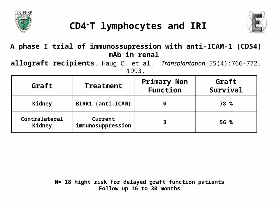
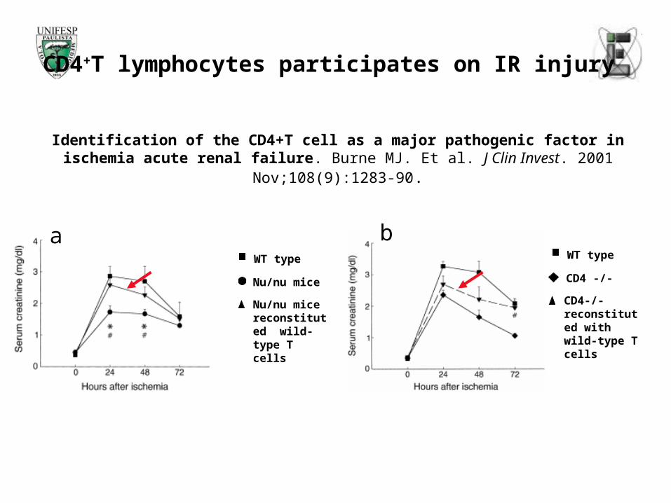
Identification of the CD4+T cell as a major pathogenic factor in ischemia acute renal failure. Burne MJ. Et al. J Clin Invest. 2001 Nov;108(9):1283-90.

a

Nu/nu mice

WT type

Nu/nu mice reconstituted wild-type T cells

WT type

CD4-/- reconstituted with wild-type T cells

CD4 -/-

b

Strategies of treatment IRI

INJURYPROTECTIVE

MECHANISMS

Tregs and Innate immune response

Enhanced Regulatory T Cell Activity is an Element od the Host response to Injury Choileain NN. Et al. J immunol 2006 Jan 1;176(1):225-36

Regulatory T cells

Developmental classification for T Reg

Naturally arising T reg cells:

Constitutive expression at higher levels:Interleukin (IL)-2 receptor alpha chain (CD25) (Sakaguchi et al., 2004)

CTLA-4 (CD152) (Sakaguchi et al., 2004)

Glucocorticoid induced TNF receptor (GITR)(McHugh et al., 2002, Shimizu et al., 2002)

FOXP3 (Schubert and Ziegler et al, 2001, Fontenot et al, 2003; Hori et al, 2003)

Adaptative T regs:

Induced from naive T cells by antigenic stimulation and cytokine millieu

IL-10 (Roncarolo et al, 2006)

TGF-beta (Nakamura et al, 2004)

CD4+T lymphocytes in Renal Ischemia reperfusion model

Strategies

CD4+ T cells Post ischemic injury

CD4+CD25+Foxp3+

T CellsCD4+ effector T Cells

Post ischemic injury

?Decrease number

(Anti-CD25 depleting antibody)

Decrease suppressor activity(Anti-GITR)

Increase CD4+ effector T cells activity

PC61: depleting rat IgG1 anti-CD25 (alpha chain of IL-2R)

Choileain NN. Et al J. Immunol, 2006

DTA-1: agonist rat IgG2a anti-GITR (Glucocorticoid-induced TNF receptor)

DTA-1 mAb abrogates suppression mediated by CD25+CD4+ T cells breaking immunological self- tolerance

DTA-1 mAb abrogates suppression mediated by CD25+CD4+ T cells leading to development of autoimmune gastritis in mice

IR antibody treatmentProtocol

Day 0 Day 1 Day 2 (24 hs) Day 4 (72hs)

Sacrifice: Blood, kidneys, spleen, lymph nodes harvested

Treatment PC61 200 gDTA-1 400 gIgG 400 g Isquemia 45 min

Microclamps for renal artery and vein (renal pedicle)

cm

Background: C57 BL/6 male

IRI model adapted from KELLY et al., 1996

Reperfusion times : 24 and 72hs

Analyses: blood: creatinine and urea

LN, spleen: flow cytometry

Kidneys: morphometry,

Real Time PCR

Renal Ischemia Reperfusion Model

Effect of Antibody treatment in TCD4+CTLA4+Foxp3+ cells

IR antibody treatment

32,240,2

69,6

90,2

0

20

40

60

80

100

% d

ep

leti

on

SPl LN

Organ

Depletion of TCD4+CTLA4+FOXP3+ cells by PC61 treatment

IR24

IR72

14,0

7,6

13,2

50,1

0

10

20

30

40

50

60

% d

ep

leti

on

SPl LN

Organ

Effect of by by DTA-1 treatment in the TCD4+CTLA4+Foxp3+ population

IR24

IR72

DTA-1 Treatment PC61 Treatment

IR antibody treatment

Depletion of TCD4+CTLA4+Foxp3+ cells

12.6210.52 8.08

18.22 3.11 1.57

Spleen

pRenal LN

IgG PC61 IR 24 PC61 IR 72

IR antibody treatmentRenal Function Outcome

Blood Urea

0

20

40

60

80

100

120

140

160

180

200

24 72

Hours

Blo

od U

rea (

mg/dL)

I gGPC61DTA-1

weight loss after IR24 + antibody treatment

00,5

11,5

22,5

33,5

4

IgG PC61 DTA-1

Treatment

Wei

ght L

oss

(g)

P=0,0018

P=0,0002

weight loss after IR 72 + antibody treatment

0

1

2

3

4

5

6

7

IgG PC61 DTA-1

TreatmentW

eigh

t Lo

ss (

g)

P=0,0001

P=0,0033

* p< 0.001

Morphometric analysesAcute tubular necrosis and tubular regeneration

IR 24 hours

Morphmetric analyses IR 24 groups

0

10

20

30

40

50

60

70

80

IgG IR 24 PC61 IR 24 DTA-1 IR 24

Groups

Sco

re necrosis

regeneration

*

*

*

* p< 0.001

Morphometric analysesAcute tubular necrosis and tubular regeneration

IR 72 hoursMorhmetric analyses IR72 groups

0

10

20

30

40

50

60

70

IgG IR 72 PC61 IR 72 DTA-1 IR 72

Groups

Score necrosis

regeneration

*

*

*

Kidney Tissue HE. A, B, C, D: IR 24 hs. A: Sham, B: IgG treated, 400 micrograms C: PC61 treated, 200 micrograms. D: DTA-1 treated, 400 micrograms, E, F, G, H: IR 72 hs. E: Sham, F: IgG treated, 400 micrograms, G: PC61 treated 200 micrograms mg, H: DTA-1 treated, 400 microgams.

IR antibody treatment Histopathological analyses HE

I

Sham PC61 200 gIgG

72 H

24 H

DTA-1 400 g

A B C

GFE

D

H

IR antibody treatment Anti-Inflammatory Genes/TH2 response

GATA-3 Relative Expression PC61

00,5

11,5

22,5

33,5

4

NR IgG IR24

PC61IR 24

IgG IR72

PC61IR 72

Groups

Gata

-3

GATA-3 Relative ExpressionDTA-1

012345678

NR IgG IR24

DTA-1IR24

IgG IR72

DTA-1IR72

Groups

Gata

-3

HO-1 Relative Expression PC61

0

1

2

3

4

5

6

NR IgG IR24

PC61 IR24

IgG IR72

PC61 IR72

Groups

HO

-1

HO-1 Relative Expression DTA-1

05

10152025303540

NR IgG IR24

DTA-1IR24

IgG IR72

DTA-1IR72

Groups

HO

-1

IR antibody treatment Pro-Inflammatory Genes

I L6 Relative ExpressionPC61

0

1

2

3

4

5

NR IgG IR24

PC61 IR24

IgG IR72

PC61 IR72

Groups

IL6

I L6 Relative ExpressionDTA-1

00,20,40,60,8

11,21,4

NR IgG IR24

DTA-1IR24

IgG IR72

DTA-1IR72

Groups

IL6

I L-1b Relative ExpressionPC61

012345

NR IgGIR 24

PC61IR 24

IgGIR 72

PC61IR 72

Groups

IL-1

b

I L-1b Relative Expression DTA-1

0

5

10

15

20

NR I gG IR24

DTA-1I R24

I gG IR72

DTA-1I R72

Groups

IL-1

b

IR antibody treatment

Foxp3 Relative Expression PC61

0

1

2

3

4

5

NR I gGI R 24

PC61I R 24

I gGI R 72

PC61I R 72

Groups

Foxp3

TGFb Relative ExpressionDTA-1

01530456075

NR I gGI R 24

DTA-1

I R24

I gGI R 72

DTA-1

I R72

Groups

TG

Fb

TGFb Relative Expression PC61

01530456075

NR IgGIR24

PC61IR24

IgGIR72

PC61IR72

Groups

TG

Fb

Foxp3 Relative Expression DTA-1

0

2

4

6

8

10

NR IgGIR 24

DTA-1

IR24

IgGIR 72

DTA-1

IR72

Groups

Fo

xp

3

IR antibody treatment Conclusions and Perspectives

At 24 hours of reperfusion, depletion of TCD4+CTLA-4+Foxp3+ cells was 30,3% (spleen) and 67,8% (para renal lymphnodes).After 72 hours of reperfusion, depletion of TCD4+CTLA-4+Foxp3+ was 43,1% (spleen) and 90,22% (para renal lymphnodes). This depletion was efficient in generate significant responses in both 24 hours and 72 hours if reperfusion Depleted mice presented similar renal function to control animals at 24 hours, but 72 hours after IRI, PC61 treated mice presented significant worst renal function compared to the group that received IgG. DTA-1 treated animals presented significant protection at the same timepoint, indicating that different subsets of cells can be acting at these timepoints.

Furthermore, histopathological analyses showed that there was a pronounced incidence of necrosis for both PC61 treated and IgG in IR 24 hours experiments. On the other hand, in IR 72 hours experiments we observed a regeneration pattern in both PC61 and IgG treated animals, but in the PC61 treated group there was a significant necrosis index (p<0.001), comparing with IgG treated group, suggesting that TCD4+CTLA4+FOXP3+ cell population could be important in a late phase of injury recover.

It is known that the stress and tissue damage associated with IRI influence the development of a immune response to protect the tissue damage. Thus, our results suggests a role for TCD4+CTLA4+FOXP3+ cells (naturally arising T teg cells) in renal IRI experimental model.

IR antibody treatment Hypothesis

CD4+CD25+Foxp3+

T CellsCD4+ effector T Cells

Post ischemic injury

24 hs:TCD4+ effectorStimulated by DTA-1escape from T Reg suppression and make injury worst

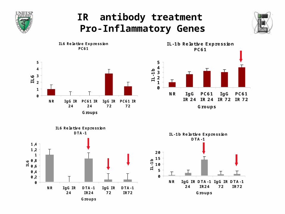
Proinflamatory cytokinesProinflamatory cytokinesIL-1b and IL-6IL-1b and IL-6

72 hs:TRegs GITRhigh stimulated by DTA-1 start to suppressTCD4+ response

Anti inflamatory Anti inflamatory genesgenesSuch as HO-1 and Such as HO-1 and polarizationpolarizationtoward Th2 (GATA-3) toward Th2 (GATA-3) transcription factortranscription factor

72 hs:If there is no T Reg at this point (PC61 treatment), there is no recovery from injury

Persistence of necrosisPersistence of necrosis

IR antibody treatment Perspectives

Acknowledgements

Rebecca M. M. MonteiroMarcio J. DamiãoGiselle GonçalvesCarla Q. FeitozaMarcos CenedezeNephrology Division - Universidade Federal de São PauloBrazil

Prof. Dr.Mauricio M. RodriguesFanny TzelepisInterdisciplinary Center for Gene Therapy CINTERGENUniversidade Federal de São Paulo, Brazil

Prof. Dr.Niels Olsen S. CamaraImmunology Division Universidade de São Paulo USP, Brazil

Vicente de Paula A. TeixeiraMarlene A. dos ReisDepartment of Pathology, Universidade Federal de Uberaba, Minas Gerais, Brazil

Prof Dr. S. SakaguchiT. Yamaguchi (DTA-1)H. Uryu (PC61)K. Nagahama (IRI)M. Ono (Real Time PCR)Institute for Frontier Medical SciencesKyoto University Japan


Recommended