Page 1
Research Signpost
37/661 (2), Fort P.O.
Trivandrum-695 023
Kerala, India
Original Article
Recent Res. Devel. Mat. Sci., 10 (2013): 45-58 ISBN: 978-81-308-0518-4
3. Synthesis and characterization of anatase
TiO2 nanofibers using Stevia rebaudiana leaf
aqueous extract Mirla Rodríguez1, Ángela B. Sifontes1, Franklin J. Méndez 1, Edgar Cañizales2
Andrea Mónaco1, María Tosta1 and Joaquín L. Brito1 1Centro de Química, Instituto Venezolano de Investigaciones Científicas, Apartado 20632, Caracas 1020-A, Venezuela; 2Área de Análisis Químico Inorgánico. PDVSA. INTEVEP. Los Teques 1070-A,
Venezuela
Abstract. Anatase TiO2 nanofibers with high surface area
(252 m2/g), diameter in the range of 25-60 nm and length of
100-150 nm, have been synthesized at room temperature by
template method using titanium isopropoxide and Stevia rebaudiana
leaf extract. Characterization was carried out using scanning
electron microscopy (SEM), transmission electron microscopy
(TEM), N2adsorption porosimetry, X-ray diffraction (XRD), Fourier
transform infrared (FT-IR), ultraviolet–visible diffuse reflectance
spectroscopy (UV-vis DRS) and thermogravimetric analysis (TGA).
The template effect of the diterpene glycosides molecules present in
S. rebaudiana leaf aqueous extract was confirmed by TEM and
SEM. The band gap energy was determined using Kubelka-Munk
function. The TiO2 nanofibers synthesized offer potential
applications in catalysis and photocatalysis.
Correspondence/Reprint request: Dr. Ángela B. Sifontes, Centro de Química, Instituto Venezolano de
Investigaciones Científicas, Apartado 20632, Caracas 1020-A Venezuela. E-mail: [email protected]
Page 2
Mirla Rodríguez et al.
46
Introduction
Titanium dioxide (TiO2, Eg = 3.2 eV ) has been widely studied due to its
promising applications in photocatalyst, solar cells, environmental pollution
degradation, biomaterials, optical devices, sensors and catalyst processes
[1- 4]. Titanium dioxide has three different structure types: rutile, anatase
and brookite. All of these crystalline forms of TiO2 occur in nature as
mineral, but only rutile and anatase have been able to be synthesized in pure
form at low temperature until recent days [5]. Anatase TiO2 is
thermodynamically metastable and can be easily transformed into the stable
rutile phase when it is heated to 500–600 °C. Such transformation of the
TiO2 crystalline phase is usually accompanied with a severe sintering or
growth of TiO2 crystallites, resulting in a severe decrease in photocatalytic
activity [6]. Anatase TiO2 with higher crystallinity is preferred for
photocatalysis, since higher crystallinity would mean fewer defects for the
recombination of photogenerated electrons and holes [6-7].
Nanosized TiO2 powders are prepared by several methods such as
hydrothermal, sol gel, microemulsion, and thermal decomposition of alkoxides
[7]. On the other hand, nanometric TiO2 compounds with different morphologies
have been reported: nanotubes of 10-20 nm diameter and 50-80 nm length [8],
nanofibers [9], nanoparticles of 30-50 nm in diameter [10].
Functional properties of TiO2 are influenced by many factors such as
crystallinity, particle size, and surface area [11]. From the point of view of
surface area nanosheets and nanotubes of TiO2 offer high surface areas
(about 100-400 m2/g) [12].
Nowadays, many researches are giving great importance to the
development of environmentally friendly synthesis methods using products
less toxic and of low cost. In recent years, S. rebaudiana leaf extract which
contains a complex mixture of eight sweet diterpene glycosides including
stevioside, rebaudioside A, rebaudioside B, rebaudioside C, rebaudioside D,
rebaudioside E, dulcoside and stelviol bioside [13] has been successfully
used for the synthesis of gold and silver nanoparticles [14-16] and most
recently, the synthesis of mesoporous hollow Al2O3 nanorods [13].
Therefore, in this study, we describe for synthesizing anatase TiO2
nanofibers using S. rebaudiana leaf extract as template in aqueous medium.
Nanofibers offer highest surface areas (per unit volume and mass) that the
bulk materials and approaching those of nanoparticles [17]. The materials
prepared by this method exhibit properties potentially useful for many
applications in nanotechnology, electronics, optics, catalysis and other fields.
Page 3
TiO2 nanofibers
47
Materials and methods
Preparation of the S. rebaudiana leaf extract
Portions of 1.2 g of leaves of S. rebaudiana were extracted with 50 mL
of hot water (65 ºC) for 3 h, as described previously by Nishiyama et al.
[18]. The crude extract was filtered through a Whatman Qualitative filter
paper No. 1.
Synthesis of TiO2 nanofibers
Synthesis of porous titania was carried out from aqueous solutions
employing titanium isopropoxide (Sigma-Aldrich) as metal precursor and
S. rebaudiana leaf extract as template. In a typical preparation, 5.84 g of
titanium isopropoxide was dissolved in 54 mL of distilled water. The
resultant solution was magnetically stirred at room temperature for 2 h.
Subsequently, the extract was added dropwise. The pH value was adjusted to
5 using a diluted acid nitric aqueous solution. The obtained solution was
evaporated and dried at 80 ºC for 48 h. Finally, the resulting solid was
calcined to remove the template. This was carried out in a tubular furnace
under air atmosphere, with a heating rate of 5 ºC/min up to 500 ºC, 600 ºC
and 750 ºC and kept at the maximum temperature for 6 h.
Characterization and instrumentation
Characterization was carried out by X-ray diffraction, using a Bruker
D-8 Focus diffractometer and CuKα radiation in the 2θ range between 5°
and 90°, operating at 40 kV and 30 mA. Thermogravimetric analysis was
performed from room temperature to 750 °C in a Mettler Toledo
thermogravimetric analyzer under air flow (100 mL/min) at a heating rate of
10 °C/min. Fourier transform infrared (FT-IR) spectra, of samples prepared
before and after calcinations, were recorded with a Perkin Elmer 100
spectrometer in the range of 2000-400 cm-1
. The textural properties of the
calcined oxides were characterized by N2 adsorption porosimetry
(Micromeritics, ASAP 2010). The samples were degassed at 300 °C under
vacuum. Nitrogen adsorption isotherms were measured at liquid N2
temperature (77 K) and N2 relative pressures ranging from 10-6
to 1.0 P/Po.
Specific surface areas were calculated according to Brunauer-Emmett-Teller
(BET) method and the pore size distribution were obtained according to the
Barret-Joyner-Halenda (BJH) method [19]. The morphologies were observed
Page 4
Mirla Rodríguez et al.
48
by field emission scanning electron microscopy (FE-SEM), using a Quanta
250 FEG scanning electron microscope (accelerating voltage of 30 kV). The
evaluation by transmission electron microscopy was performed in a
HITACHI 7100 microscope. The samples were prepared by suspending the
powders in an ethanol-based liquid and pipetting the suspension onto a
carbon/collodion-coated 200 mesh copper grid.
The optical absorption edge of the titania nanofibers was measured using
a Lambda 35 UV−vis spectrophotometer (Perkin Elmer) equipped with an
integrating sphere, employing BaSO4 as the reflectance reference sample.
Reflectance spectra was recorded at 190 – 800 nm wavelength. The band gap
energy of the titania nanofibers was determined from the reflectance using
Kubelka-Munk function, F(R), and the extrapolation of Tauc plot, plot of
(F(R).hν)1/2
against hν [20].
Results and discussion
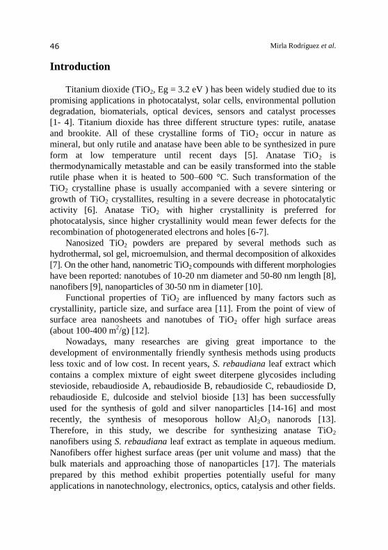
Figure 1 shows the wide angle X-ray diffraction pattern of the
synthesized samples calcined at different temperatures (500, 600 and
750 ºC). XRD analyses were conducted to investigate the relationship
between the thermal stability and anatase–rutile phase transformation. The
peaks of samples calcined were identified by comparison with JCPDS-84-
1286 according 2θ which confirmed that an anatase structure at 2θ=25.40. The XRD diffractograms corresponding to the treated samples at
500 and 600 °C, indicated that the obtained TiO2 had relatively high
crystallinity, attributable to the anatase phase (101), (004), (105), (200) and
(211) Miller indexes [5, 21-22]. It is noteworthy that these diffractograms do
not present any peaks assigned to the rutile phase (2θ=27.36 o
). On the other
hand, the sample calcined at 750 ºC gave diffraction peaks of anatase
nanocrystal along with minor peaks corresponding to rutile phase.
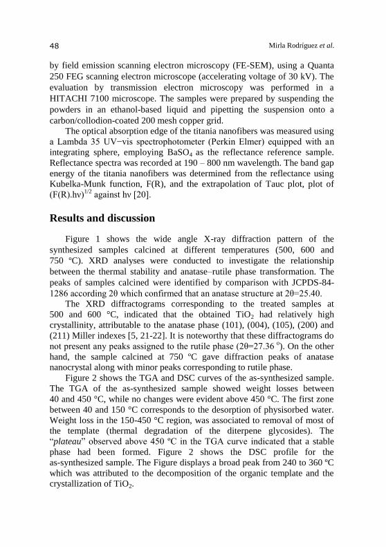
Figure 2 shows the TGA and DSC curves of the as-synthesized sample.
The TGA of the as-synthesized sample showed weight losses between
40 and 450 °C, while no changes were evident above 450 °C. The first zone
between 40 and 150 °C corresponds to the desorption of physisorbed water.
Weight loss in the 150-450 °C region, was associated to removal of most of
the template (thermal degradation of the diterpene glycosides). The
“plateau” observed above 450 ºC in the TGA curve indicated that a stable
phase had been formed. Figure 2 shows the DSC profile for the
as-synthesized sample. The Figure displays a broad peak from 240 to 360 ºC
which was attributed to the decomposition of the organic template and the
crystallization of TiO2.
Page 5
TiO2 nanofibers
49
20 30 40 50 60
2θ/Degree
Rel
ativ
e in
ten
sity
(a.
u.)
500°C
600°C
750°C
TiO2 (anatase)
TiO2 (rutile)
30
00
a.u.
(101)
(004) (105)
(200)(211)
Figure 1. XRD patterns of the synthesized samples with different calcination
temperature.
-100
-80
-60
-40
-20
0
60
70
80
90
100
30 230 430 630 830
Wei
gh
tlo
ss(%
) HF
(mW
)
Temperature (°C)
Figure 2. TGA and DSC curves of the as-synthesized sample.
Page 6
Mirla Rodríguez et al.
50
In the synthesis medium, titanium alcoxides hydrolyze and subsequently
polymerize to form a three-dimensional oxide network. These reactions can
be schematically represented as follows:
Ti(OR)4 + 4H2O→ Ti(OH)4 + 4ROH (hydrolysis), (Ι)
Ti(OH)4→ TiO2 xH2O + (2− x)H2O (condensation), (ΙΙ)
where R is ethyl, i-propyl, n-butyl, etc.[23]. Even so, hydrolysis in the
presence of excess water is rapid and complete within seconds. The size,
stability and morphology of the sol produced from alcoxides is strongly
affected by the water-to-titanium molar ratio (x = [H2O]/[Ti]). High x ratios
would favors the formation of colloidal TiO2. Frequently, particles of small
size are formed under these conditions [23].
The condensation and hydrolysis products possess proton–donors
centers (OH-groups, as well as water molecules) adsorbed on the surface.
OH-groups are well-known to have ability for H-bond creation and can
appear as centers for the physical adsorption of molecules (especially of
polar ones), which are acceptors of protons [24-26]. On the other hand, the
hydrolysis products of the diterpene glycosides molecules present in
S. rebaudiana leaf aqueous extract (stevioside, rebaudioside A, rebaudioside
B, rebaudioside C, rebaudioside D, rebaudioside E, dulcoside and stelviol
bioside) can act as proton acceptors. These molecules would be bound to the
three-dimensional titanium oxide forming a supramolecular structure. The
literature provide similar examples of supramolecular structures obtained on
the basis of diterpene glycosides derivatives [24-26].
Figure 3 shows TEM and SEM images of the titania powders
synthesized at 500 ºC, indicating the fibers-like morphology. The nanofibers
have diameter in the range of 25-60 nm and length of 100-150 nm.
The observed morphology was similar to the morphology of alumina
nanorods reported in our previous publication [13], which was synthesized
employing aluminum isopropoxide. The formation of structures based on the
template effect of the diterpene glycosides molecules present in
S. rebaudiana leaf aqueous extract was confirmed by these results. An
explanation of the template effect in the course of the synthesis was
proposed as the result of a complex system of intermolecular interactions
between the diterpene glycosides molecules and the metal alcoxide [13]. The
TEM image (Figure 3A) of the S. rebaudiana dried extract is evidence of the
template effect. Figure 3 clearly demonstrates the morphology of the template,
which was replicated by the synthesized metal oxides.
Page 7
TiO2 nanofibers
51
Figure 3. TEM image of the S. rebaudiana dried extract (A); TEM (B) and SEM
(C, D) images of the titania powders calcined at 500 ºC.
It is interesting to reference the formation of molecular complex of
isosteviol with the substituted organic molecules proposed by Andreeva et al.
[26]. In this study it was demonstrated that these compounds can form
crystalline molecular complexes, whose supramolecular structures look like a
double chiral helix cross-linked [25-26]. This would be consistent with the
morphology of the titania obtained after calcination at 500 ºC.
Figure 4 and 5, show the TEM image of samples calcined at 600 and
750 ºC. It is seen that the morphology of the synthesized titania powders at
higher calcination temperature has a change. The nanofibers are completely
destroyed and only crystallites with size of 25-100 nm and morphology of
sphere-like form are observed. It would be attributed to the high temperatures
which cause the sintering of the nanocrystallites.
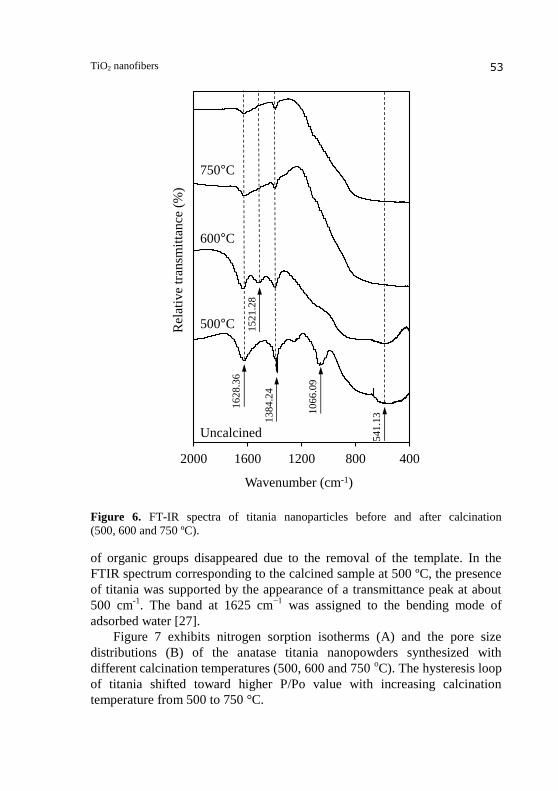
In order to identify the chemical nature of the synthesized nanofibers,
FTIR measurements were carried out yielding spectra as shown in Figure 6.
The characteristic vibrational bands in the as-synthesized sample appeared in
the range of 1650-1519 cm-1
for (COO-) stretching bands, 1470-1300 cm-1
for
Page 8
Mirla Rodríguez et al.
52
50 nm
Figure 4. TEM image of the titania powders calcined at 600 ºC.
154 nm
Figure 5. TEM image of the titania powders calcined at 750 ºC.
C-H stretching, scissoring and bending. These carboxilate groups were attributed
to diterpenic molecules like steviol and isosteviol generated in the synthesis
medium [13, 27]. A broad band between 400 cm−1
and 700 cm−1
should be
due to the bands of Ti –O- Ti bond. After calcination, the transmittance peaks
Page 9
TiO2 nanofibers
53
400800120016002000
Rel
ativ
e tr
ansm
itta
nce
(%
)
Wavenumber (cm-1)
500°C
600°C
750°C
Uncalcined
16
28
.36
13
84
.24
10
66
.09
54
1.1
3
15
21
.28
Figure 6. FT-IR spectra of titania nanoparticles before and after calcination
(500, 600 and 750 ºC).
of organic groups disappeared due to the removal of the template. In the
FTIR spectrum corresponding to the calcined sample at 500 ºC, the presence
of titania was supported by the appearance of a transmittance peak at about
500 cm-1
. The band at 1625 cm−1
was assigned to the bending mode of
adsorbed water [27].
Figure 7 exhibits nitrogen sorption isotherms (A) and the pore size
distributions (B) of the anatase titania nanopowders synthesized with
different calcination temperatures (500, 600 and 750 oC). The hysteresis loop
of titania shifted toward higher P/Po value with increasing calcination
temperature from 500 to 750 °C.
Page 10
Mirla Rodríguez et al.
54
Figure 7. Nitrogen sorption isotherms (A) and the pore size distributions (B) of the
anatase titania nanopowders synthesized with different calcination temperatures
(500, 600 and 750 ºC).
Table 1. Surface area, pore volume and pore diameter of TiO2 samples
calcined at 500, 600 and 750 ºC.
The titania synthesized showed the maximum of adsorbed nitrogen
volume at 500 °C, and the adsorbed nitrogen volume rapidly decreased from
500 to 750 °C. The titania calcined at 750 °C almost lost its hysteresis loop.
Table 1 presents pore properties calculated from the nitrogen sorption
isotherms shown in Figure 7.
Page 11
TiO2 nanofibers
55
The pore size of titania nanofibers increased from ~5 to ~24 nm with an
increase in the calcination temperature from 500 to 750 °C. The maximum
pore volume was 0.33 cm3/g at 500 °C, and the surface area decreased from
253 to 18 m2/g with an increase in the temperature from 500 to 750 °C. The
isotherm for titania calcined at 500 ºC exhibits typical type IV pattern
with inflection point of nitrogen adsorbed volume at P/P0 about 0.42 (type
H2 hysteresis loop), indicating the existence of mesopores [19]. Adsorption
isotherm of TiO2 sample calcined at 600 °C also exhibit hysteresis loop.
However, an increase in adsorption volume of N2 was observed and located
in the P/P0 range of 0.70- 0.95, indicating collapse and loss of mesoporosity.
The pore size distribution obtained by BJH approach (Figure 7B) is
noticeably broad, in the range of 4-45 nm. The mesostructure of the material
totally collapses in the calcination process at 600-750 ºC, indicating a
substantial loss of porosity.
The UV-vis DRS spectra for titania powders calcined at 500 ºC is shown
in Figure 8. Diffuse reflectance spectra was used to study the possible
transitions between the valence band and the conduction band, as well as
other possible transitions due to impurities. For semiconductor materials, the
direct band gap can be derived by establishing the Tauc Plot of transformed
Kubelka-Munk function versus the absorbed light energy [20]. As can be
seen in Figure 8C, the so-called Tauc optical band gap is obtained at the
intercept between the extension line of slop and the base line. This indicates
that titania nanofibers have a band gap of 3.2 eV, which corresponds to
approximately 385 nm. This calculated band gap energy is consistent with
data reported in the literature for other forms of titania materials [28].
0
20
40
60
80
100
200 300 400 500 600 700
0,0
0,4
0,8
1,2
1,6
0 1 2 3 4 5 6 7 80,0
0,4
0,8
1,2
1,6
200 300 400 500 600 700
Ref
lect
ance
(%
)
Wavelength (nm) Wavelength (nm) Eg (eV)
1.6
1.2
0.8
0.4
0.0
Ku
bel
ka-
Mu
nk
1.6
1.2
0.8
0.4
0.0
Ku
bel
ka-
Mu
nk
(A) (B) (C)
Figure 8. Diffuse reflectance spectra (A), the absorption spectra obtained by
applying a Kubelka-Munk function to the diffuse reflectance spectra (B), and plot of
transformed Kubelka Munk function vs the energy of the light absorbed (C) for
titania powders calcined at 500 ºC.
Page 12
Mirla Rodríguez et al.
56
Conclusions
Titania nanofibers with high surface area (252 m2/g), diameter of
25-60 nm and length of 100-150 nm could be prepared at room temperature in
an aqueous medium by using titanium isopropoxide as precursor and the Stevia
rebaudiana leaf extract. The prepared titania nanofibers exhibit a mesoporous
structure and are composed of nanocrystalline anatase titania. Due to their
porous structure and band gap of 3.2 eV, these materials may find numerous
applications in photocatalysis, solar energy conversion and catalysis.
Acknowledgements
The authors would like to thank the Microbiology Center (IVIC) and
M.Sc. Freddy Sanchez for the TEM micrographs; Diffraction and
Fluorescence Laboratory, Dr. Reinaldo Atencio and Miguel Ángel Ramos
García (INZIT) for DRX analysis; Dra. Tamara Zoltan and Ing. Maibelin
Rosales (IVIC) for the UV-vis DRS analysis.
References
1. Fujishima A, Honda K (1972) Electrochemical photolysis of water at a
semiconductor electrode. Nature 238: 37-38.
2. Liu C, Wang X (2012) Mesoporous titanium dioxide nanobelts: synthesis,
morphology evolution, and photocatalytic properties. Mater. Res. Soc. 27:
2265-2270.
3. Calleja G, Serrano DP, Sanz R, Pizarro P, García (2004) Study on the synthesis
of high-surface-area mesoporous in the presence of nonionic surfactant. Ind.
Eng. Chem. Res. 43: 2485-2492.
4. Madhugiri S, Sun B, Smirniotis P G, Ferraris J P, Balkus KJ Jr (2004)
Electrospun mesoporous titanium dioxide fibers. Micropor. Mesopor. Mater. 69:
77-83.
5. Park J-Y, Lee C, Jung K W, Jung D (2009) Structure related photocatalytic
properties of TiO2. Bull. Korean Chem. Soc. 30:402-404.
6. He D, Lin F (2007) Preparation and photocatalytic activity of anatase TiO2
nanocrystallites with high thermal stability. Mater. Lett. 61: 3385-3387.
7. Rashidzadeh M, Synthesis of High-Thermal stable titanium dioxide
nanoparticles. Int. J. Photoenergy doi:10.1155/2008/245981.
8. Chen X, Schriver M, Suen T, Mao S S (2007) Fabrication of 10 nm diameter
TiO2 nanotube arrays by titanium anodization. Thin Solid Films 515: 8511-8514.
9. Pavasupree S, Suzuki Y,Yoshikawa S, Kawahata R (2005) Synthesis of titanate,
TiO2 (B) and anatase TiO2 nanofibers from natural rutile sand. J Solid State
Chemistry 178: 3110-3116.
Page 13
TiO2 nanofibers
57
10. Pavasupree S, Ngamsinlapasathian S, Nakajima M, Suzuki Y, Yushikawa S
(2006) Synthesis, characterization,photocatalytic activity and die-sensitized solar
cell performance of nanorods/nanoparticles TiO2 with mesoporous structure. J
Photochem Photobiol A: Chem 184: 163-169.
11. Carp O, Huisman C L, Reller A. (2004) Photoinduced reactivity of titanium
dioxide. Progr. state solid Chem. 32: 33-177.
12. Tsai Ch-Ch, Teng H (2004) Regulation of the Physical Characteristics of Titania
Nanotube Aggregates Synthesized from Hydrothermal Treatment. Chem.
Mater.16:4352-4358.
13. Rodríguez M, Sifontes A B, Méndez F, Díaz I, Cañizales E, Brito J L (2013)
Template synthesis and characterization of mesoporous γ-Al2O3 hollow
nanorods using Stevia rebaudiana leaf aqueous extract. Ceram Int. 39:
4499-4506.
14. Mishra A N, Bhadauria S, Gaur M S, Pasricha R, Kushwa B S (2010) Synthesis
of gold nanoparticles leaves of zero-calorie sweetener herb (Stevia rebaudiana)
and their nanoscopic characterization by spectroscopy and microscopy. Int J.
Green Nanotechnology Phys. Chem 1: 118-124.
15. Varshney R, Bhadauria S, Gaur M S (2010) Biogenic synthesis of silver
nanocubes and nanorods using sundried Stevia rebaudiana leaves. Adv. Mater.
Lett. 1: 232-237.
16. Yilmaz M, Turkdemir H, Akif Kilic M, Bayram E, Cicek A, Mete A, Ulug B
(2011) Biosynthesis of silver particles using leaves of Stevia rebaudiana. Mater
Chem Phys 130: 1195-1202.
17. Park S-J, Kang YC, Park J Y, Ed A. Evans EA, Ramsier R D, Chase G G (2010)
Physical Characteristics of titania nanofibers synthesized by sol-gel and
electrospinning techniques. J Eng Fiber 5: 50-56.
18. Nishiyama P, Alvarez M, Vieira LG (1992) Quantitative analysis of stevioside
in the leaves of Stevia rebaudiana by near infrared reflectance spectroscopy. J.
Sci. Food Agric 59: 277-281.
19. Gregg SJ, Sing KSW (1982) Adsorption, Surface Area and Porosity. Academic
Press, 2nd edition, London.
20. Tauc J, Grigorovici R, Vancu A, (1966) Optical properties and electronic
structure of amorphous germanium, Phys, Status Solidi B.15 :627-637.
21. Lee D-W, Lee K-H (2011) Novel eco-friendly synthesis of sucrose-templated
mesoporous titania with high thermal stability. Micropor. Mesopor. Mat. 142:
98-103.
22. Yu J, Wang B (2010) Effect of calcination temperature on morphology and
photoelectrochemical properties of anodized titanium dioxide nanotube arrays.
Applied Catalysis B: Environmental 94:295-302.
23. Mahshid S, Sasani Ghamsari M, Askari M, Afshar N, Lahuti S (2006) Synthesis
of TiO2 nanoparticles by hydrolysis and peptization of titanium isopropoxide
solution. Semicond. Phys. Quantum Electron. Optoelectron. 9: 65-68.
24. Gubaidullin A, Beskrovnyj D V, Litvinov I A (2005) Crystal structure model
based on the analysis of hydrophilic-hydrophobic ratio in molecules. isosteviol
derivatives. J. Struct. Chem. 46: 195-201
Page 14
Mirla Rodríguez et al.
58
25. Alfonsov V A, Bakaleynik G A, Gubaidullin A T, Kataev V E,
Kovyljaeva G I, Konovalov A I, Litvinov I A, Strobykina I Y , Andreeva O V,
Korochkina M G, (1999) Molecular complex of isosteviol with aniline.
Mendeleev Commun. 9:227-228.
26. Andreeva O V, Garifullin B F, Gubaidullin A T, Al’fonsov V A, Kataev V E, Ryzhikov D V (2007) Crystalline inclusion complexes of diterpenoid isosteviol
with aromatic compounds. J. Struct. Chem. 48: 540-546.
27. Tao J, Shen Y, Gu F, Zhu J, Zhang J (2007) Synthesis and Characterization of
mesoporous titania particles and thin films. J. Mater. Sci. Technol, 23: 513-516.
28. Valencia S, Marin J M, Restrepo G (2010) Study of the bandgap of synthesized
titanium dioxide nanoparticles using the sol-gel method and a hydrothermal
treatment. The Open Mater. Sci. J. 4: 9-14.


![[MS-PDF]: Microsoft Edge ISO 32000-1 Portable Document ...interoperability.blob.core.windows.net/web/MS-PDF/[MS-PDF].pdf · Microsoft Edge ISO 32000-1 Portable Document Format (PDF)](https://static.documents.pub/doc/80x56/5ad426737f8b9a5d058b85b6/ms-pdf-microsoft-edge-iso-32000-1-portable-document-ms-pdfpdfmicrosoft.jpg)


