A new alpha helix motif required for protein glycosylation A new helical binding domain mediates a glycosyltransferase activity of a bifunctional protein Hua Zhang a , Meixian Zhou a , Tiandi Yang b , Stuart M. Haslam b , Anne Dell b and Hui Wu a a Departments of Pediatric Dentistry and Microbiology, Schools of Dentistry and Medicine, University of Alabama at Birmingham, Birmingham, Alabama 35294, USA b Department of Life Sciences, Imperial College London, London, SW7, 2AZ, UK Running title: A new alpha helix motif required for protein glycosylation *Address for correspondence: Dr. Hui Wu, Department of Pediatric Dentistry, University of Alabama at Birmingham, School of Dentistry, 1919 7 th Avenue South, Birmingham, AL 35294; E-mail: [email protected]Key words: Streptococcus, glycoproteins, alpha helix domain, glycosyltransferase, protein-protein interaction, crystal structure Abstract Serine-rich repeat glycoproteins (SRRPs) conserved in streptococci and staphylococci are important for bacterial colonization and pathogenesis. Fap1, a well- studied SRRP is a major surface constituent of Streptococcus parasanguinis and is required for bacterial adhesion and biofilm formation. Biogenesis of Fap1 is a multi-step process that involves both glycosylation and secretion. A series of glycosyltransferases catalyze sequential glycosylation of Fap1. We have identified a unique hybrid protein dGT1 (dual glycosyltransferase 1) that contains two distinct domains. N-terminal DUF1792 is a novel GT-D type glycosyltransferase, transferring Glc residues to Glc-GlcNAc-modified Fap1. C- terminal dGT1 (CgT) is predicted to possess a typical GT-A type glycosyltransferase, however the activity remains unknown. In this study, we determine that CgT is a distinct glycosyltransferase, transferring GlcNAc residues to Glc-Glc-GlcNAc-modified Fap1. 2.0 Å X-ray crystal structure reveals that CgT has a unique binding domain consisting of three alpha helices in addition to a typical GT-A type glycosyltransferase domain. The helical domain is crucial for the oligomerization of CgT. Structural and biochemical 1
Transcript
A new alpha helix motif required for protein glycosylation
A new helical binding domain mediates a glycosyltransferase activity of a bifunctional protein
Hua Zhanga, Meixian Zhoua, Tiandi Yangb, Stuart M. Haslamb, Anne Dellb and Hui Wua
aDepartments of Pediatric Dentistry and Microbiology, Schools of Dentistry and Medicine, University of Alabama at Birmingham, Birmingham, Alabama 35294, USA
bDepartment of Life Sciences, Imperial College London, London, SW7, 2AZ, UK
Running title: A new alpha helix motif required for protein glycosylation*Address for correspondence: Dr. Hui Wu, Department of Pediatric Dentistry, University of Alabama at Birmingham, School of Dentistry, 1919 7th Avenue South, Birmingham, AL 35294; E-mail: [email protected]
Serine-rich repeat glycoproteins (SRRPs) conserved in streptococci and staphylococci are important for bacterial colonization and pathogenesis. Fap1, a well-studied SRRP is a major surface constituent of Streptococcus parasanguinis and is required for bacterial adhesion and biofilm formation. Biogenesis of Fap1 is a multi-step process that involves both glycosylation and secretion. A series of glycosyltransferases catalyze sequential glycosylation of Fap1. We have identified a unique hybrid protein dGT1 (dual glycosyltransferase 1) that contains two distinct domains. N-terminal DUF1792 is a novel GT-D type glycosyltransferase, transferring Glc residues to Glc-GlcNAc-modified Fap1. C-terminal dGT1 (CgT) is predicted to possess a typical GT-A type glycosyltransferase, however the activity remains unknown. In this study, we determine that CgT is a distinct glycosyltransferase, transferring GlcNAc residues to Glc-Glc-GlcNAc-modified Fap1. 2.0 Å X-ray crystal structure reveals that CgT has a unique binding domain consisting of three alpha helices in addition to a typical GT-A type glycosyltransferase domain. The helical domain is crucial for the oligomerization of CgT. Structural and biochemical studies revealed that the helix domain is required for the protein-protein interaction and crucial for the glycosyltransferase activity of CgT in vitro and in vivo. As the helix domain presents a novel structural fold, we conclude that CgT represents
a new member of GT-A type glycosyltransferases.
Introduction
Protein glycosylation is one of the most common post-translational modifications. There are two major types of modifications resulting in N-linked and O-linked glycosylation, respectively. Both are involved in the regulation of various biological processes (1), and have been implicated in human health and diseases (2).
Serine-rich repeat proteins (SRRPs) are O-linked glycoproteins. They belong to a growing family of bacterial adhesins (3-7). Fimbriae associate protein (Fap1) of S. parasanguinis is the first SRRP identified, and is required for biofilm formation (8-10). Fap1-like proteins are highly conserved in other oral streptococci (11-13) and widespread in pathogenic streptococci and staphylococci (4,5). For instance, Srr-2 of Streptococcus agalactiae is associated with bacterial virulence (5), Hsa of Streptococcus gordonii and SraP of Staphylococcus aureus contribute to the pathogenesis of infective endocarditis (4,14). PsrP of S. pneumoniae mediates frequency of invasive pneumococcal disease (15). Thus Fap1-like SRRPs represent a new family of adhesins that are important in bacterial virulence, but little is known about their biogenesis.
1
A new alpha helix motif required for protein glycosylation
Glycosylation of Fap1 is crucial for bacterial adhesion (16), and biofilm formation(17) and is required for the assembly of fimbriae-like surface structure in S. parasanguinis (18). An eleven gene cluster flanking the fap1 gene locus is essential for Fap1 glycosylation and biogenesis (17). Several glycosyltransferase genes including gly, gtf3, dgT1, galT2 are found located upstream of fap1, and genes coding for accessory secretion proteins such as SecY2, Gap1, Gap2, Gap3, SecA2, and glycosyltransferase Gtf1, and its chaperone Gtf2 are located downstream of fap1. SecY2 and SecA2 encode accessory Sec proteins. SecY2 is essential for the biogenesis of mature Fap1 (19), and SecA2 is important for export mature Fap1 to the cell wall (20,21). The glycosylation-associated proteins (Gap1, Gap2, and Gap3) modulate Fap1 secretion and biogenesis (18,22,23). A glycosyltransferase enzyme complex, consisting of Gtf1 and its chaperone Gtf2, catalyzes the first step of Fap1 glycosylation, transferring GlcNAc residues to the Fap1 polypeptide (17,24). Gtf3 mediates the second step by transferring Glc to Gtf1-2-modified Fap1 (25,26). Our previous studies have determined that N-terminal dGT1 (DUF1792) transfers glucose residues to Gtf1-2-3-modified Fap1 (27,28). The subsequent step of Fap1 glycosylation is still unknown.
dGT1 of S. parasanguinis contains a conserved new glycosyltransferase (DUF1792) at the N-terminus and a putative glycosyltransferase at the C-terminus (27). In this study, we determined the crystal structure of C-terminal dGT1, and found that the C-terminal dGT1 is a GlcNAc transferase, which possesses a glycosyltransferase activity that is distinct from the N-terminal activity, transferring GlcNAc to the Fap1 substrate modified first by the N-terminal DUF1792 glycosyltransferase. The 2.0 Å X-ray crystal structure reveals a new helix domain that is crucial for both oligomerization and enzymatic activity of CgT. Together, we determine that dGT1 is a bifunctional protein that catalyzes successive steps of glycosylation of Fap1 in vitro and in vivo in S. parasanguinis.
Experimental procedures
Strains and primers
Bacterial strains and plasmids used in this study are listed in Table 1. E. coli and S. parasanguinis strains were cultured as previously described (29).
Primers used in this study are listed in Table 2. Plasmid isolation, genomic DNA purification, PCR amplification, and purification of PCR products were carried out as described (29). DNA digestion, ligation, and transformation were performed using standard methods.
Reductive Elimination and Permethylation
The glycans were reductively eliminated from rFap1R1 and purified in a Dowex® column as previously described (30), and the purified glycans were permethylated and further purified according to published methods (30). Briefly, freeze-dried rFap1 samples and 22 mg/mL potassium borohydride were dissolved in 400 µL 0.1 M potassium hydroxide solution. The mixture was incubated at 45°C for 18 hrs and added 5-6 drops of acetic acid for quenching the reaction. The solution was loaded onto the 50E-8C Dowex® column and eluted with 5% acetic acid. The collected solution was concentrated and lyophilized. Excessive borates were co-evaporated with 10% methanolic acetic acid.
For the permethylation, sodium hydroxide (3-5 pellets per sample) was crushed in 3 mL dry dimethyl sulfoxide solvent. The resulting slurry (0.75 mL) and methyl iodine (750 μL) were added to the sample. The mixture was agitated for 30 min and added 2 mL ultra-pure water with shaking in order to quench the reaction. The permethylated glycans were extracted with chloroform (2mL) and washed with ultra-pure water for two more times. Chloroform was removed under a stream of nitrogen. The permethylated glycans were loaded onto a conditioned C18 Sep-pak® column, washed with 5 mL ultra-pure water, and successively eluted with 3 mL each of 15%, 35%, 50% and 75% aq. acetonitrile. The solution was collected
2
A new alpha helix motif required for protein glycosylation
and lyophilized.
Mass Spectrometry
MS data were acquired by using either a Voyager DE-STRTM MALDI-TOF or a 4800 MALDI-TOF/TOF mass spectrometer (Applied Biosystems, Darmstadt, Germany). The latter instrument was also used for acquiring MS/MS data. The MS mode was calibrated with 4700 Calibration standard kit (Applied Biosystems), and the MS/MS mode was calibrated with fibrinopeptide B human (Sigma). The collision energy for CID MS/MS was set to 1 kV, and the collision gas was argon. 2, 5-dihydroxybenzoic acid was used as matrix. Permethylated glycans were dissolved in 10 μL methanol, and 1 μL of this solution was premixed with 1 μL matrix and spotted onto the plate.
E. coli glycosylation system
A small fragment of Fap1 (rFap1R1) that contains the first repeat region (100 to 200 amino acid residues of Fap1) was used as a model to reconstitute Fap1 glycosylation in E. coli (27). Recombinant BL21 strains were constructed by co-expression of pET28a-rFap1RI(27) with pVPT-Gtf123(27), pVPT-Gtf123-DUF1792(27), pVPT-Gtf123-CgT, or pVPT-Gtf123-dGT1 individually, in which rFap1R1 was differentially modified by different combinations of glycosyltransferases. All plasmids were confirmed by PCR and DNA sequencing. Glycosylated rFap1R1 proteins from different recombinant strains were induced, and purified using the same method as described for the recombinant CgT, and were then examined by SDS-PAGE analysis.
Protein expression, purification and crystallization
The DNA fragment coding for the CgT protein (residue 287-582) was cloned into pET-sumo vector with a primer set of CgT-sumo-BamH1-F and CgT-sumo-Xho1-R (Table 2), and expressed in Escherichia coli BL21(DE3), purified as previously described (25,31) and
stored at 4°C in buffer contained 20mM Tris-HCl, pH 8, 100mM NaCl, 0.2 mM TCEP. Crystallization screens were conducted using the sitting drop vapor diffusion method with a Phoenix crystallization robot on a 96-well Intelli-plate (Art Robbins Instrument). Designed crystallizations were set up manually using the vapor diffusion, hanging drop method with 24-well plates. The drops were set up at a 1:1 ratio of protein to mother liquor and incubated at 20°C. Crystals grown in a well solution containing 0.1M Sodium citrate tribasic dehydrate pH 5.5, 22% PEG 1000 of IndexHT (Hampton Research) were used to determine the structure of CgT.
X-ray data collection and processing
Diffraction datasets for CgT (2.0 Å) were collected at the Argonne National Laboratory on beam stations ID22 at 100 K. Paratone-N was used as a cryoprotectant. The datasets were processed by HKL2000 (32).
Molecular replacement and refinement
Phases for CgT were calculated using the structure of a glycosyltransferase from Bacteroides fragilis (PDB code: 3BCV) as a starting model by the molecular replacement method using PHENIX software suite(33). Further refinement and model building were performed using the PHENIX software suite and COOT(33,34). The final CgT structure was obtained through several refinement cycles and lastly using TLS for the final refinement.
UDP-Glo assay
UDP-GloTM assay (Promega) was used to determine in vitro glycosyltransferase activity of CgT. His-tagged recombinant Fap1 (amino acid, 100-200) proteins differentially modified by different glycosyltransferases were purified and used as acceptors. Purified dGT1, DUF1792 and CgT were used as enzymes, and either UDP-Glc or UDP-GlcNAc as a sugar donor. A corresponding acceptor, an enzyme, and a sugar donor were reconstituted in the same reaction
3
A new alpha helix motif required for protein glycosylation
buffer (20mM Tris pH 8.0, 100mM NaCl, 0.5mM MnCl), in a total volume of 5 µL and incubated for 2 hrs. 5 µL of UDP-Glo reagent were added into each mixture to react for 1 hr. Luminescence was determined to monitor glycosyltransferase activity.
Isothermal Titration Calorimetry (ITC)
Thermodynamics of binding of enzyme (either CgT or CgTΔH1-3) and ligand UDP-GlcNAc were characterized using MicroCal Auto-iTC200 at 25 °C. Enzymes were dialyzed extensively against 20 mM Tris-HCl buffer, pH 8.0, containing 100 mM NaCl and 20mM MgCl. Ligand UDP-GlcNAc was dissolved in the same buffer as the enzymes. 400 µL of enzyme solutions (CgT or CgTΔH1-3 at 1mM) was titrated with 20 injections of the ligand (2 µL of UDP-GlcNAc at 15 mM). Each injection of the ligand lasted for 4s with 300s intervals between successive injections. Binding isotherms were generated by plotting the corrected heats of binding against the ratio of the ligand to enzymes. Software supplied by the manufacturer (Origin version 7.0 from Microcal Inc.) was used to calculate dissociation constants (Kd), enthalpies of binding (∆H), stoichiometry (N), and entropy of binding (∆S).
GST Pull-down assays
To examine in vitro self-interaction of dGT1, relevant GST and His tagged fusion proteins were prepared. Strains carry plasmids pGEX-6P-1 and pGEX-dGT1/DUF1792/CgT were used for the preparation of GST, GST-dGT1, GST-DUF1792 and GST-CgT fusion proteins, and the strain carrying the plasmid pET-28b-CgT was used for the preparation of the His tagged CgT protein. A primer set of CgT-28b-BamH1-F and CgT-28b-Xho1-R was used to amplify the CgT fragment to construct pET-28b-CgT. A primer set of CgT-sumo-BamH1-F and CgT-sumo-Xho1-R was used to amplify the CgT gene and construct pGEX-6p-1-CgT. Confirmed constructs were used to transform into appropriate E. coli strains to produce recombinant proteins.
GST pull-down assay was conducted as we described (23,35). In brief, GST fusion proteins were purified and bound to glutathione Sepharose 4B beads (Amersham) according to manufacturer’s instructions. His-CgT was purified by His trap column (GE) according to manufacturer’s instruction. 50 µg of the Glutathione-Sepharose bound GST fusion proteins were mixed with 40 µg of His-tagged CgT in the NETN buffer (20mM Tris-HCl, pH7.2, 100mM NaCl, 1mM EDTA, 0.2% NP40) and incubated overnight on a rotary shaker at 4°C. The beads were washed 3 times with 600μl NETN buffer and the proteins were eluted with SDS-PAGE sample buffer, boiled for 10min, and subjected to Western blotting analysis with anti-His antibody at 1:2000 dilution.
Complementation of dGT1 mutant with dGT1 variants
Shuttle vector pVPT-HSV-His was used as a vector to complement dGT1 mutant. DNA fragment corresponding to CgT was PCR amplified using a primer pair of CgT-pvpt-sal1-F and CgT-pvpt-Kpn1-R from the genomic DNA of S. parasanguinis FW213. The amplified fragment was purified and then cloned into the pVPT-Hsv-His vector to obtain pVPT-CgT. The CgT gene fragment with a functional promoter from pVPT-CgT was amplified using a primer set of pvpt-promoter-kpn1-F and CgT-pvpt-Kpn1-R and then cloned into pVPT-DUF1792 to yield pVPT-CgT/DUF172. Plasmid constructs were confirmed by DNA sequencing, and then transformed into the dGT1 mutant by electroporation as described (27). Transformants were selected on TH agar containing 125µg/mL kanamycin and 10 µg/mL Erythromycin, and further verified by PCR analysis. The confirmed strains were used in this study.
Site-directed mutagenesis and truncated helix variants
Site-direct mutagenesis was carried out using QuickChange mutagenesis kit as described (Stratagene) (25). The plasmid pVPT-dGT1 was used as a template, and primers sets of dGT-D378A-F and dGT-D378A-R, and dGT-D380A-F and dGT-D380A-R (Table 2) were used to
4
A new alpha helix motif required for protein glycosylation
construct designed mutant alleles. Mutant constructs were identified and confirmed by DNA sequencing.
To construct truncated helix motif variants, the plasmid pET-sumo-CgT was used as a template, and the following primer sets Deletion-helix1-F and Deletion-helix1-R (helix1), Deletion-helix2-F and Deletion-helix2-R (helix2), Deletion-helix3-F and Deletion-helix3-R (helix3), CgTΔH1-3 and CgT- ΔH1-3-R (the entire helix domain)(Table 2) were used to delete helix1, helix2, helix2 or the entire helix domain respectively. Mutant alleles were identified and confirmed by PCR and DNA sequencing.
Results
CgT plays an important role in the Fap1 glycosylation
dGT1 contains an N-terminal unknown function domain (DUF1792) and C-terminal putative glycosyltransferase domain (CgT). Our studies have showed that DUF1792 is a new type of glycosyltransferases, transferring glucose residues to Glc-GlcNAc-Fap1, and that the C-terminus did not have the same activity in vitro (27,28). However DUF1792 alone failed to complement the dGT1 mutant in S. parasanguinis (27), suggesting there is an unknown activity of CgT. In order to determine the CgT activity, we established an in vivo glycosylation system in E. coli, using a small recombinant Fap1 (rFap1R1) as a substrate (27). In the system, rFap1R1 was co-expressed with N-terminal DUF1792, C-terminal dGT1 (CgT) or the full-length dGT1 in addition to three glycosyltransferases, Gtf1, Gtf2, and Gtf3. The presence of DUF1792 super-shifted the recombinant Fap1 modified by Gtf123 (Fig. 1. lane 2 versus 1). Introduction of the C-terminal domain alone did not modify the recombinant Fap1 glycosylated by Gtf123 (Fig. 1. lane 3). Only when the full –length dGT1 (Fig. 1. lane 4) was used, was the recombinant Fap1 further super-shifted to two higher molecular weight positions, indicating that the glycosylation of
rFap1RI by DUF1792 is a prerequisite for the subsequent modification by CgT. These data suggest that CgT catalyzes the fourth step of the Fap1 glycosylation.
To investigate glycan residue(s) transferred by CgT, we performed MS-based glycomics. The recombinant Fap1R1 proteins glycosylated by DUF1792 and dGT1 were purified and subjected to reductive elimination to release O-linked glycans. The isolated glycans were derivatized and subsequently analyzed by mass spectrometry. Matrix-assisted laser desorption/ionization-time of flight (MALDI-TOF) mass fingerprinting (Fig. 2) of the permethylated glycans released from dGT1 and Gtf123 modified rFap1R1, showed a peak corresponding to a tetrasaccharide comprising two HexNAcs and two hexoses. The sequence of the monosaccharides was further characterized by MALDI-TOF/TOF, which generates fragmentation patterns consistent with annotated glycan structures (Fig. 2). Previous studies indicate that dGT1 modified rFap1R1 carries a tri-saccharide with a sequence of Glc-Glc-GlcNAc and that rFap1R1 can only carry Glc as the hexose and GlcNAc as the HexNAc. Therefore, the presence of an additional HexNAc in the dGT1 modified glycan suggests that the C-terminal dGT1 catalyzes the transfer of GlcNAc to the Gtf123 and DUF1792 modified Fap1, so we hypothesized that CgT has the activity to transfer GlcNAc.
To test this, we performed an in vitro glycosyltransferase assay using the UDP-Glo method (Promega) (36). Gtf123-modified-Fap1 was used as a substrate, and either UDP-Glc or UDP-GlcNAc was evaluated as a sugar donor. Consistent with our previous data, DUF1792 transfers Glc not GlcNAc residues to Gtf123-modified Fap1 (Fig. 3A, B), while CgT did not have the activity to transfer either GlcNAc or Glc residues to Gtf123-modified Fap1 (Fig. 3B). However, when Gtf123-DUF1792 modified Fap1 was used as a substrate, CgT was able to transfer GlcNAc but not Glc to the substrate (Fig. 3 C and D). These data are consistent with the findings from the E. coli glycosylation system (Fig. 1), demonstrating that CgT is a glycosyltransferase, transferring GlcNAc
5
A new alpha helix motif required for protein glycosylation
residues to the Fap1 that is first modified by DUF1792. Taken together, we conclude that CgT mediates the fourth step of Fap1 glycosylation.
CT1 interacts with itself
Gel filtration profiling of purified CgT shows two peaks (Fig. 4A). The first peak is close to the void volume of this column, SDS-PAGE analysis reveal the presence of an unknown high molecular weight protein (Supplementary Fig. 1B). The second peak is calculated to accommodate a dimeric CgT, suggesting CgT can bind to itself. An in vitro pull-down assay was used to validate this. GST-dGT1 and GST-CgT were able to pull down His-CgT (Fig. 4B, lanes1 & 3), while GST-DUF1792 failed to pull down His-CgT (Fig. 4B, lane2), demonstrating that CgT interacts with itself.
Structural insights into CgT
In order to further characterize CgT, we crystallized CgT. Crystals of CgT were obtained at a condition of 0.1M Sodium citrate tribasic dehydrate pH 5.5, 22% PEG 1000. X-ray crystal structure of CgT (286aa – 582aa) was determined at a resolution of 2.0 Å (Table 3). The space group of the CgT structure is R3, shown in dimer in symmetry unit (Fig. 5).
Each CgT monomer has a triangle shape and contains three regions (Fig. 6A): an N-terminal domain, a core region and a helix domain. The N-terminal region consists of β1-β2-β3-β4 and α1-α2-α3. β1, β2 and β3 make a parallel β-sheet surrounded by α1, α2 and α3 to form a Rossmann fold. Strand β4 connects the N-terminal region with the core of the protein. The core region consists of six strands (β5 to β10) and six helixes (α4 to α9). β7, β8, β9 and β10 lined in the center clamped by α5, α6, α7 to form a sandwich shape (Fig. 6B). Two loops (aa154-159 and aa219-228) were missing in the core region. At the very C-terminus there is a unique helix domain consisting of three helices.
In the CgT structure, two monomers bind tail to tail together through the helix domain
(Fig. 5). Since all key binding residues of this CgT dimer were located in the helix domain, we designate it as a helical binding domain.
CgT possesses a unique helix structure
Structural alignment of CgT was performed using the Secondary Structure Matching server (SSM) from the European Bioinformatics Institute (37). The result shows that only the N-terminal region and a part of the core region are matched to SpsA (Protein Data Bank entry, 1H7L), the first defined GT-A type glycosyltransferase (38). SpsA contains a Rossmann fold and a metal binding site. The match is limited to this region (Fig. 6C). We also searched the CgT structure using Dali server (39). The best match identified is also SpsA. The helix domain does not share any similarity with known glycosyltransferases. The domain was then extracted from the CgT structure, and evaluated using the SSM. The best match from the search is TPR2 of FimV (PDB code: 4MBQ) from Pseudomonas aeruginosa with a low Z score of 3.9. In addition, a Dali search alone revealed it only partially matches bacterial ferritin (FtnA) (PDB code: 3R2K) of Pseudomonas aeruginosa with a weak Z score of 5.9. These results suggest the helix domain represents a novel structural fold.
The helical binding domain is important for the glycosyltransferase activity
We found all amino acid residues key to the binding distributed along the entire binding interface. Deletion of the entire binding domain from CgT, rendered the CgTΔH1-3 mutant fail to form dimer but only exist as a monomer (Fig. 4A, the red profile graph). And the CgTΔH1-3 mutant significantly reduced the glycosyltransferase activity in vitro (Fig. 7). We also attempted to delete each of three helixes (H1-3) in the binding domain. Interestingly deletion of the first and second helix made CgT unstable and drastically reduced production of CgT (data not shown), which precluded us from studying these deletion mutants. Deletion of the third helix (CgTΔH3) gave rise to a stable
6
A new alpha helix motif required for protein glycosylation
protein, the mutant behaved normally but dramatically reduced the glycosyltransferase activity (Fig. 7). These data suggest the helical binding domain is important for the CgT activity.
The helical binding domain does not mediate UDP-GlcNAc binding
Based on the Dali search, the best matched structure identified is SpsA, a GT-A type glycosyltransferase. SpsA is a metal dependent enzyme, containing a DXD metal binding motif (40). Structure superimpose between SpsA and CgT revealed the conserved UDP and metal binding residues (Fig. 8A). The DXD motif is essential for the CgT function, as DXD mutants in dGT1 failed to complement dGT1 knockout in vivo (Fig. 8B, lanes 5 & 6).
As the deletion of the helical binding domain reduced the glycosyltransferase activity, and the UDP binding is crucial and close to the helical domain, we also examined whether the helical domain affects the binding of CgT to its substrate UDP-GlcNAc using Isothermal Titration Calorimetry (ITC) (Fig. 9). The Kd
value was determined to be at 10.67 mM and 12.15 mM for CgT and CgT∆H1-3 binding respectively (Tab. 4), which is much weaker than reported interactions between glycosyltransferases and their sugar donors. Human α1,4-N-acteylhexosaminyltransferase binds to UDP-GlcNAc at Kd of 61 uM (41). Both CgT and CgT∆H1-3 have same binding ratio to its substrate being 1:1. The ITC data reveal that both CgT and CgTΔH1-3 bind to UDP-GlcNAc, and the binding affinity is very similar, suggesting the deletion of the helical domain did not affect the binding to UDP-GlcNAc.
CgT is a distinct glycosyltranferase and important for the Fap1 glycosylation in vivo.
As CgT plays an important role in the Fap1 glycosylation in vitro, transferring GlcNAc to Glc-Glc-GlcNAc-modified Fap1, we also evaluated its function in vivo. N-terminal DUF1792 and C-terminal CgT alone fail to
complement dGT1 mutant in S. parasanguinis (Fig. 10, lanes5&6), while the intact full-length dGT1 complements (Fig. 10, lane 4). Intriguingly the split N-terminal DUF1792 and CgT placed on a same plasmid driven by their own promoter was able to complement the dGT1 mutant (Fig. 10, lane 8). These data suggest that CgT is an independent glycosyltransferase and important for the Fap1 glycosylation. Consistent with the in vitro data, the modification of Fap1 by Gtf1, 2, 3 and DUF1792 is prerequisite for the activity of CgT in vivo. DUF1792 partially rescued Fap1 modification, and CgT failed to complement. Only when both DUF1792 and CgT in place, was the production of mature Fap1 rescued (Fig. 10, lane8). In addition, dGT1 with the deletion of a helical motif also failed to produce mature Fap1 in vivo (Fig. 10, lane 7), suggesting the importance of the helical domain in vivo.
Discussion
Glycosylation catalyzed by a series of glycosyltransferases is crucial for biogenesis of SRRP adhesins in gram-positive bacteria (7). dGT1 is a unique bifunctional protein involved in the biosynthesis of an SRRP adhesin Fap1 of S. parasanguinis. It is essential for glycosylation of Fap1 by two distinct glycosyltransferase activities derived from the N-terminal and C-terminal region respectively. The N-terminus of dGT1 was originally annotated as a domain of unknown function (DUF1792) but our recent studies have determined that it represents a new family of glycosyltransferases transferring Glc to Glc-GlcNAc-modified Fap1(27). In this study we demonstrate that the C-terminus of dGT1 (CgT) is an independent glycosyltransferase, transferring GlcNAc to the Glc-Glc-GlcNAc-modified Fap1. The N-terminus and C-terminus of dGT1 can be separated completely into two enzymes and are still active as long as the split domains are co-expressed. The co-expression fully rescued the dGT1 mutant. On the other hand, the CgT activity depends on the completion of the glycosylation by N-terminal DUF1792. CgT can only glycosylate Fap1 after the Fap1 intermediate is first modified by DUF1792. This claim was substantiated by
7
A new alpha helix motif required for protein glycosylation
several pieces of evidence in vitro and in vivo, 1) In vivo E. coli glycosylation system reconstituted by rFap1 and corresponding glycosyltransferases; 2) Mass Spectrometric analysis of glycans released from recombinant Fap1 differentially modified by dGT1 or DUF1792; 3) In vitro glycosyltransferase reactions using UDP-GlcNAc sugar donor and the DUF1792 modified Fap1 substrate; and 4) In vivo glycosylation of Fap1 in the native host S. parasanguinis. Thus we conclude that CgT catalyzes the fourth step of Fap1 following the action of Gtf1/Gtf2, Gtf3 and DUF1792.
The organization of one protein consisting of two glycosyltransferase activities in two separate domains is unique in the SRRP glycosylation pathway. A search of protein databases using dGT1 reveals a number of proteins that share a similar domain organization. This two-domain organization would maximize the enzymatic efficiency since the reaction from one domain should provide the substrate for the subsequent modification by the other domain. Such protein homologs are only found in streptococcal species such as Streptococcus iniae, Streptococcus agalactiae, Streptococcus suis, Streptococcus plurextorum, Streptococcus lutetiensis, Streptococcus hyovaginalis, and Streptococcus gallolyticus with the exception of Enterococcus cecorum. Most of those bacterial genomes contain genes coding for putative SRRP biosynthetic pathways, however their function and engagement in the biogenesis of SRRPs have not been investigated. Bifunctional glycosyltransferases have been documented in one-domain proteins in eukaryotic cells. Saccharomyces cerevisiae β1,4-mannosyl-transferase Alg1 catalyzes the transfer of the first mannose to the glycan during the biosynthesis of asparagine-linked glycoproteins, and the bifunctional mannosyltransferase Alg2, which carries out the dual function of the transfer of two different linkages of mannoses, an α1,3-mannosylation followed by an addition of an α1,6-mannose to complete the first branched pentasaccharide of the glycosylation pathway (42). However Alg2 only possesses one GT-B type of glycosyltransferase domain, how the dual activity is executed and regulated is
unknown. It is also interesting to note the two-domain dGT1 protein we reported here catalyzes the branching of the glycan sequence (27) as Alg2, how these two domains are coordinated to complete the branched reaction is not clear and awaits further investigation.
2.0 Å X-ray crystal structure of CgT revealed another new feature of this glycosyltransferase. CgT contains a unique helix binding domain in addition to possessing a typical GT-A type glycosyltransferase domain. The helix binding domain is crucial for the oligomerization of CgT and dGT1. The helix binding domain does not appear to mediate the binding of CgT to its sugar donor, UDP-GlcNAc. Noticeably the predicated UDP-GlcNAc binding site in CgT is about 19Aº away from the helical motif (Fig. 8A). Further, the deletion of this helical domain significantly reduced glycosyltransferase activity in vitro and failed to complement the dGT1 mutant in vivo. In addition to the studies of the dual functionality from single domain glycosyltransferases, few distinct two-domain bifunctional glycosyltransferases have been characterized in bacteria (43-47). The hyaluronan synthase of Pasteurella multocida (48) and the chondroitin polymerase of E. coli K4 (49) are two close examples as they resemble dGT1 and belong to known structural superfamily, GT-A. Capsule polymerases, SiaDW-135 and SiaDY, responsible for the biosynthesis of Neisseria meningitides capsule polysaccharides, are another unique family of bifunctional glycosyltransferases. The N-terminal GT-B type glycosyltransferase domain is separated from the C-terminal GT-B type domain by a structurally unknown center region. They function as galactosyltransferase and sialyltransferase respectively, however it is not clear whether the central domain regulates their activities (50). N-acetylglucosamine 1-phosphate uridyltransferase (GlmU) is a cytoplasmic bifunctional enzyme involved in the biosynthesis of the nucleotide-activated UDP-GlcNAc (51). GlmU structure contains two domains that are linked by a helix arm. The N-terminal domain contains a Rossmann fold that is involved in the uridyltransferase activity, and C-terminal domain has a left-handed parallel β-
8
A new alpha helix motif required for protein glycosylation
helix structure that is responsible for in the acetyltransferases activity (51). GlmU forms a trimer through the helix arm. Noticeably, this helix arm is not crucial for GlmU activities. This is in the sharp contrast to the helix domain of CgT, which is important for the enzymatic activity of CgT in addition to its role in the oligomerization of dGT1. Moreover, the helix
domain of CgT doesn’t share any sequence and structure homology to any known proteins, and is mostly found in Streptococcus, thus representing a unique structure. Since CgT is essential for the Fap1 glycosylation, and the helix domain is a unique binding structure in streptococci, it can be targeted to develop anti-bacteria drugs.
Acknowledgements: This work was supported by NIH/NIDCR grant R01DE17954 (HW) and F33DE022215 (HW), by a Biotechnology and Biological Sciences Research Council grant (BB/K016164/1, Core Support for Collaborative Research (AD and SMH), and by a Welcome Trust Senior Investigator Award (AD). We thank Zhengrong Yang for helping to access to the Auto-iTC200 in the Biocalorimetry Laboratory supported by the NIH Shared Instrumentation Grant # 1S10RR026478 and Shared Facility Program of the UAB Comprehensive Cancer Center, Grant # 316851. X-ray data were collected at Southeast Regional Collaborative Access Team (SER-CAT) 22-ID beamline at the Advanced Photon Source, Argonne National Laboratory.
Conflict of Interest: The authors declare that they have no conflicts of interest with the contents of this article.
Author contributions: HZ, MZ, SMH, AD and HW designed the study. HZ and HW drafted the paper. HZ and TY performed the experiments. HZ, TY, SMH, AD and HW analyzed the results and wrote the final version of the manuscript.
References
1. Drickamer, K., and Taylor, M. E. (1998) Evolving views of protein glycosylation. Trends in Biochemical Sciences 23, 321-324
2. Yang, Y. R., and Suh, P.-G. (2014) O-GlcNAcylation in cellular functions and human diseases. Advances in biological regulation 54, 68-73
3. Zhang, Y. Q., Ren, S. X., Li, H. L., Wang, Y. X., Fu, G., Yang, J., Qin, Z. Q., Miao, Y. G., Wang, W. Y., and Chen, R. S. (2003) Genome based analysis of virulence genes in a non biofilm forming Staphylococcus‐ ‐ ‐ epidermidis strain (ATCC 12228). Molecular microbiology 49, 1577-1593
4. Siboo, I. R., Chambers, H. F., and Sullam, P. M. (2005) Role of SraP, a serine-rich surface protein of Staphylococcus aureus, in binding to human platelets. Infection and immunity 73, 2273-2280
5. Seifert, K. N., Adderson, E. E., Whiting, A. A., Bohnsack, J. F., Crowley, P. J., and Brady, L. J. (2006) A unique serine-rich repeat protein (Srr-2) and novel surface antigen (ε) associated with a virulent lineage of serotype III Streptococcus agalactiae. Microbiology 152, 1029-1040
6. Sanchez, C. J., Shivshankar, P., Stol, K., Trakhtenbroit, S., Sullam, P. M., Sauer, K., Hermans, P. W., and Orihuela, C. J. (2010) The pneumococcal serine-rich repeat protein is an intra-species bacterial adhesin that promotes bacterial aggregation in vivo and in biofilms. PLoS pathogens 6, e1001044
7. Zhu, F., Zhang, H., and Wu, H. (2015) Glycosyltransferase-mediated Sweet Modification in Oral Streptococci. Journal of dental research, 0022034515574865
8. Wu, H., Mintz, K. P., Ladha, M., and Fives Taylor, P. M. (1998) Isolation and characterization of Fap1, a‐ fimbriae associated adhesin of Streptococcus parasanguis FW213. ‐ Molecular microbiology 28, 487-500
9. Wu, H., and Fives Taylor, P. M. (1999) Identification of dipeptide repeats and a cell wall sorting signal in the‐ fimbriae associated adhesin, Fap1, of Streptococcus parasanguis. ‐ Molecular microbiology 34, 1070-1081
9
A new alpha helix motif required for protein glycosylation
10. Froeliger, E. H., and Fives-Taylor, P. (2001) Streptococcus parasanguis fimbria-associated adhesin Fap1 is required for biofilm formation. Infection and immunity 69, 2512
11. Bensing, B. A., and Sullam, P. M. (2002) An accessory sec locus of Streptococcus gordonii is required for export of the surface protein GspB and for normal levels of binding to human platelets. Molecular microbiology 44, 1081-1094
12. Takahashi, Y., Yajima, A., Cisar, J. O., and Konishi, K. (2004) Functional analysis of the Streptococcus gordonii DL1 sialic acid-binding adhesin and its essential role in bacterial binding to platelets. Infection and immunity 72, 3876-3882
13. Plummer, C., Wu, H., Kerrigan, S. W., Meade, G., Cox, D., and Ian Douglas, C. (2005) A serine rich‐ glycoprotein of Streptococcus sanguis mediates adhesion to platelets via GPIb. British journal of haematology 129, 101-109
14. Takahashi, Y., Takashima, E., Shimazu, K., Yagishita, H., Aoba, T., and Konishi, K. (2006) Contribution of sialic acid-binding adhesin to pathogenesis of experimental endocarditis caused by Streptococcus gordonii DL1. Infection and immunity 74, 740-743
15. Shivshankar, P., Sanchez, C., Rose, L. F., and Orihuela, C. J. (2009) The Streptococcus pneumoniae adhesin PsrP binds to Keratin 10 on lung cells. Molecular microbiology 73, 663-679
16. Stephenson, A. E., Wu, H., Novak, J., Tomana, M., Mintz, K., and Fives Taylor, P. (2002) The Fap1 fimbrial‐ adhesin is a glycoprotein: antibodies specific for the glycan moiety block the adhesion of Streptococcus parasanguis in an in vitro tooth model. Molecular microbiology 43, 147-157
17. Wu, H., Zeng, M., and Fives-Taylor, P. (2007) The glycan moieties and the N-terminal polypeptide backbone of a fimbria-associated adhesin, Fap1, play distinct roles in the biofilm development of Streptococcus parasanguinis. Infection and immunity 75, 2181-2188
18. Peng, Z., Fives-Taylor, P., Ruiz, T., Zhou, M., Sun, B., Chen, Q., and Wu, H. (2008) Identification of critical residues in Gap3 of Streptococcus parasanguinis involved in Fap1 glycosylation, fimbrial formation and in vitro adhesion. BMC microbiology 8, 52
19. Wu, H., Bu, S., Newell, P., Chen, Q., and Fives-Taylor, P. (2007) Two gene determinants are differentially involved in the biogenesis of Fap1 precursors in Streptococcus parasanguis. Journal of bacteriology 189, 1390
20. Chen, Q., Wu, H., and Fives Taylor, P. M. (2004) Investigating the role of secA2 in secretion and‐ glycosylation of a fimbrial adhesin in Streptococcus parasanguis FW213. Molecular microbiology 53, 843-856
21. Zhou, M., Zhang, H., Zhu, F., and Wu, H. (2011) Canonical SecA associates with an accessory secretory protein complex involved in biogenesis of a streptococcal serine-rich repeat glycoprotein. Journal of bacteriology 193, 6560-6566
22. Peng, Z., Wu, H., Ruiz, T., Chen, Q., Zhou, M., Sun, B., and Fives Taylor, P. (2008) Role of gap3 in Fap1‐ glycosylation, stability, in vitro adhesion, and fimbrial and biofilm formation of Streptococcus parasanguinis. Oral microbiology and immunology 23, 70-78
23. Zhou, M., Zhu, F., Li, Y., Zhang, H., and Wu, H. (2012) Gap1 functions as a molecular chaperone to stabilize its interactive partner Gap3 during biogenesis of serine rich repeat bacterial adhesin. ‐ Molecular microbiology 83, 866-878
24. Wu, R., and Wu, H. (2011) A Molecular Chaperone Mediates a Two-protein Enzyme Complex and Glycosylation of Serine-rich Streptococcal Adhesins. Journal of Biological Chemistry 286, 34923-34931
25. Zhu, F., Erlandsen, H., Ding, L., Li, J., Huang, Y., Zhou, M., Liang, X., Ma, J., and Wu, H. (2011) Structural and functional analysis of a new subfamily of glycosyltransferases required for glycosylation of serine-rich streptococcal adhesins. Journal of Biological Chemistry 286, 27048-27057
26. Zhou, M., Zhu, F., Dong, S., Pritchard, D. G., and Wu, H. (2010) A novel glucosyltransferase is required for glycosylation of a serine-rich adhesin and biofilm formation by Streptococcus parasanguinis. Journal of Biological Chemistry 285, 12140
27. Zhang, H., Zhu, F., Yang, T., Ding, L., Zhou, M., Li, J., Haslam, S. M., Dell, A., Erlandsen, H., and Wu, H. (2014) The highly conserved domain of unknown function 1792 has a distinct glycosyltransferase fold. Nature communications 5
28. Zhang, H., Zhu, F., Ding, L., Zhou, M., Wu, R., and Wu, H. (2013) Preliminary X-ray crystallographic studies
10
A new alpha helix motif required for protein glycosylation
of an N-terminal domain of unknown function from a putative glycosyltransferase from Streptococcus parasanguinis. Acta Crystallographica Section F: Structural Biology and Crystallization Communications 69, 520-523
29. Zhou, M., Fives-Taylor, P., and Wu, H. (2008) The utility of affinity-tags for detection of a streptococcal protein from a variety of streptococcal species. Journal of microbiological methods 72, 249-256
30. North, S. J., Jang-Lee, J., Harrison, R., Canis, K., Ismail, M. N., Trollope, A., Antonopoulos, A., Pang, P.-C., Grassi, P., and Al-Chalabi, S. (2010) Chapter Two-Mass Spectrometric Analysis of Mutant Mice. Methods in Enzymology 478, 27-77
31. Zhu, F., Wu, R., Zhang, H., and Wu, H. (2013) Structural and biochemical analysis of a bacterial glycosyltransferase. in Glycosyltransferases, Springer. pp 29-39
32. Otwinowski, Z., and Minor, W. (2001) Denzo and Scalepack. in International Tables for Crystallography Volume F: Crystallography of biological macromolecules, Springer. pp 226-235
33. Adams, P. D., Afonine, P. V., Bunkóczi, G., Chen, V. B., Davis, I. W., Echols, N., Headd, J. J., Hung, L.-W., Kapral, G. J., and Grosse-Kunstleve, R. W. (2010) PHENIX: a comprehensive Python-based system for macromolecular structure solution. Acta Crystallographica Section D: Biological Crystallography 66, 213-221
34. Emsley, P., and Cowtan, K. (2004) Coot: model-building tools for molecular graphics. Acta Crystallographica Section D: Biological Crystallography 60, 2126-2132
35. Echlin, H., Zhu, F., Li, Y., Peng, Z., Ruiz, T., Bedwell, G. J., Prevelige, P. E., and Wu, H. (2013) Gap2 promotes the formation of a stable protein complex required for mature Fap1 biogenesis. Journal of bacteriology 195, 2166-2176
36. Zegzouti, H., Engel, L., Hennek, J., Alves, J., Vidugiris, G., and Goueli, S. (2013) Detection of Glycosyltransferase Activities with Homogeneous Bioluminescent UDP Detection Assay. in GLYCOBIOLOGY, OXFORD UNIV PRESS INC JOURNALS DEPT, 2001 EVANS RD, CARY, NC 27513 USA
37. Krissinel, E., and Henrick, K. (2004) Secondary-structure matching (SSM), a new tool for fast protein structure alignment in three dimensions. Acta Crystallographica Section D: Biological Crystallography 60, 2256-2268
38. Tarbouriech, N., Charnock, S. J., and Davies, G. J. (2001) Three-dimensional structures of the Mn and Mg dTDP complexes of the family GT-2 glycosyltransferase SpsA: a comparison with related NDP-sugar glycosyltransferases. Journal of molecular biology 314, 655-661
39. Holm, L., and Rosenström, P. (2010) Dali server: conservation mapping in 3D. Nucleic acids research 38, W545-W549
40. Charnock, S. J., and Davies, G. J. (1999) Structure of the nucleotide-diphospho-sugar transferase, SpsA from Bacillus subtilis, in native and nucleotide-complexed forms. Biochemistry 38, 6380-6385
41. Sobhany, M., Dong, J., and Negishi, M. (2005) Two-step mechanism that determines the donor binding specificity of human UDP-N-acetylhexosaminyltransferase. Journal of Biological Chemistry 280, 23441-23445
42. O'Reilly, M. K., Zhang, G., and Imperiali, B. (2006) In vitro evidence for the dual function of Alg2 and Alg11: essential mannosyltransferases in N-linked glycoprotein biosynthesis. Biochemistry 45, 9593-9603
43. Hölzl, G., Leipelt, M., Ott, C., Zähringer, U., Lindner, B., Warnecke, D., and Heinz, E. (2005) Processive lipid galactosyl/glucosyltransferases from Agrobacterium tumefaciens and Mesorhizobium loti display multiple specificities. Glycobiology 15, 874-886
44. van der Wel, H., Fisher, S. Z., and West, C. M. (2002) A Bifunctional Diglycosyltransferase Forms the Fucα1, 2Galβ1, 3-Disaccharide on Skp1 in the Cytoplasm ofDictyostelium. Journal of Biological Chemistry 277, 46527-46534
45. Wang, Z. A., van der Wel, H., Vohra, Y., Buskas, T., Boons, G.-J., and West, C. M. (2009) Role of a cytoplasmic dual-function glycosyltransferase in O2 regulation of development in Dictyostelium. Journal of Biological Chemistry 284, 28896-28904
46. Nakanishi, M., Karasudani, M., Shiraishi, T., Hashida, K., Hino, M., Ferguson, M. A., and Nomoto, H. (2014) TbGT8 is a bifunctional glycosyltransferase that elaborates N-linked glycans on a protein phosphatase AcP115 and a GPI-anchor modifying glycan in Trypanosoma brucei. Parasitology international 63, 513-518
47. Izquierdo, L., Acosta-Serrano, A., Mehlert, A., and Ferguson, M. A. (2014) Identification of a
11
A new alpha helix motif required for protein glycosylation
glycosylphosphatidylinositol anchor-modifying β1-3 galactosyltransferase in Trypanosoma brucei. Glycobiology, cwu131
48. Williams, K. J., Halkes, K. M., Kamerling, J. P., and DeAngelis, P. L. (2006) Critical elements of oligosaccharide acceptor substrates for the Pasteurella multocida hyaluronan synthase. Journal of Biological Chemistry 281, 5391-5397
49. Sobhany, M., Kakuta, Y., Sugiura, N., Kimata, K., and Negishi, M. (2008) The chondroitin polymerase K4CP and the molecular mechanism of selective bindings of donor substrates to two active sites. Journal of Biological Chemistry 283, 32328-32333
50. Romanow, A., Haselhorst, T., Stummeyer, K., Claus, H., Bethe, A., Mühlenhoff, M., Vogel, U., von Itzstein, M., and Gerardy-Schahn, R. (2013) Biochemical and biophysical characterization of the sialyl-/hexosyltransferase synthesizing the meningococcal serogroup W135 heteropolysaccharide capsule. Journal of Biological Chemistry 288, 11718-11730
51. Brown, K., Pompeo, F., Dixon, S., Mengin Lecreulx, D., Cambillau, C., and Bourne, Y. (1999) Crystal‐ structure of the bifunctional N acetylglucosamine 1 phosphate uridyltransferase from Escherichia coli: a‐ ‐ paradigm for the related pyrophosphorylase superfamily. The EMBO Journal 18, 4096-4107
Figure Legends
Fig. 1. Glycosylation of rFap1R1 in E. coli. A recombinant small Fap1 fragment (rFap1R1) was modified by Gtf1/2/3 (lane 1), by Gtf1/2/3 and DUF1792 (lane 2), by Gtf123 and CgT (lane 3), or by Gtf1/2/3 and dGT1 (lane 4).
Fig. 2. Mass spectra of dGT1 and DUF1792 modified rFap1. Glycans from recombinant Fap1 proteins were reductively released, and permethylated. Derivatized glycans were purified and analyzed by MS. Representative spectra were shown in (a) and (b). Peaks corresponding to sodiated glycans are colored red and annotated with m/z and glycan structures. Black signals are due to under-permethylation (minus 14 in m/z) or the matrix contaminations. (c) MS/MS of the MS peak at m/z 738 from (a); (d) MS/MS of the MS peak at m/z 983 from (b).
Fig. 3. Glycosyltransferase activities of N-terminus (DUF1792), C-terminus (CgT) and the full length dGT1. In vitro glycosyltransferase activity assays were performed using either Glc-GlcNAc-Fap1 (A and B) or Glc-Glc-GlcNAc-Fap1 as an acceptor substrate (C and D). Either UDP-Glc (A and C) or UDP-GlcNAc (B and D) was used as a sugar donor in all reactions.
Fig. 4. Gel filtration profile of CgT and CgTΔH1-3 (A) and pull-down assay (B). Elution curve of CgT (blue) and CgTΔH1-3(red) from GE Hiload 16/60 Superdex G200 column at 4 °C. GST pull-down of His tagged CgT (B). GST fused dGT1 (lane 1), DUF1792 (lane 2), and CgT (lane 3) were used to pull down His tagged CgT. Top Panel, His-CgT pulled down by GST fusion proteins, probed by anti-His antibody; Bottom Panel, Input control of His tagged CgT, probed by anti-His antibody.
Fig. 5. CgT Structure. The CgT structure exists in dimer in a space group of R3; each monomer is colored in green and cyan respectively. The interface in the CgT dimer is the helix domain that faced each other.
Fig. 6. Structure of the CgT monomer. N-terminal region is colored in red, core region is colored in yellow, and the helix domain is colored in blue. Rossmann fold and DxD motif are labeled (A). Topology diagram of the CgT monomer was color coded as the structure. Arrows stand for strands, and columns represent helices (B). Superimposing of CgT with the best matched structure model of SpsA (C). CgT is shown in green, whereas SpsA is shown in orange.
Fig. 7. The helix domain is important for CgT glycosyltransferase activity. UDP-Glo assay was employed to assess glycosyltransferase activity of CgT. Gtf123-DUF1792 modified recombinant Fap1
12
A new alpha helix motif required for protein glycosylation
was used as a substrate with UDP-GlcNAc as a sugar donor.
Fig. 8. Conserved UDP binding and metal binding sites identified from CgT (A). CgT structure (green) superimposed onto SpsA (Gray), the key residues in SpsA colored in blue, and in CgT colored in red. UDP, Mg and the DxD motif are labeled. The distance between key residues and UDP is shown in dotted lines. The DXD motif is important for the CgT function in vivo (B). Cell lysates of wild type S. parasanguinis FW213 (Lane 1); Fap1 mutant (Lane 2); dGT1 mutant (Lane 3); and dGT1 mutant complemented with dGT1 (Lane 4); with dGT1/AXD (Lane 5) or dGT1/DXA (Lane 6) were subjected to western blotting analysis using mature Fap1-specific monoclonal antibody F51 (top panel) and Fap1 peptide-specific monoclonal antibody E42 (bottom panel) to monitor Fap1 production.
Fig. 9. Isothermal titration calorimetric graphs of CgT and CgTΔH1-3 titrated with UDP-GlcNAc. Binding of UDP-GlcNAc to CgT or CgTΔH1-3 was assessed as follows. The reaction cells contained either CgT (A) or CgTΔH1-3 at 1mM (B) and the syringe was supplied with UDP-GlcNAc at 15 mM. Data obtained from 20 injections of 2 µL aliquots of UDP-GlcNAc at 3-min intervals are shown in the top graphs. The lower plots show the integrated binding isotherm with the experimental points (■) and best fit.
Fig. 10. CgT and the helix domain are required for dGT1 function in vivo. Wild type S. parasanguinis FW213(Lane 1) , Fap1 mutant (Lane 2 ), dGT1 mutant (Lane 3) and its complement variants, dGT1 complemented with the full-length dGT1 (Lane 4), with DUF1792 (Lane 5), with CgT (Lane 6), with dGT1without the helix domain (Lane 7), and with both DUF1792 and CgT (Lane 8), were examined for the production of Fap1 by western blotting analysis using Fap1 peptide-specific antibody E42 and mature Fap1 specific antibody F51.
Table 1. Strains and plasmids used in this studyStrains or plasmids Relevant properties SourceStrainsE. coli Top10 Host for propagation of the recombinant
plasmidsInvitrogen
E. coli BL21-Gold (DE3)
pET system hos strain Invitrogen
S. parasanguinis FW213 Wild typeS. parasanguinis FW213 Fap1 deletion mutant
Wild type; Fap1 knockout; Fap1::aphA3;Kanr
(8)
S. parasanguinis FW213 dGT1 deletion mutant
Wild type; dGT1 knockout; dGT1::aphA3;Kanr
(27)
Strains used to produce recombinant proteins:CgT pET-sumo: CgT transformed into BL21 In this
studyHis-CgT pET-28b: CgT transformed into BL21 In this
study
13
A new alpha helix motif required for protein glycosylation
CgT∆H1-3 pET-sumo: CgT∆H1-3 transformed into BL21
In this study
CgT-DH1 pET-sumo: CgT-DH1 transformed into BL21 In this study
CgT-DH2 pET-sumo: CgT-DH2 transformed into BL21 In this study
CgT-DH3 pET-sumo: CgT-DH3 transformed into BL21 In this study
His-sFap1-GlcNAc-Glc pET-28b: sFap1 and pvpt-Gtf123 co-transformed into BL21
(27)
His-sFap1-GlcNAc-Glc-Glc
pET-28b: sFap1 and pvpt-Gtf123-DUF1792 co-transformed into BL21
In this study
His-sFap1-GlcNAc-Glc(-Glc)-GlcNAc
pET-28b: sFap1 and pvpt-Gtf123-T1 co-transformed into BL21
In this study
GST-DUF1792 pGEx-5x-1: DUF1792 transformed into Top10
(27)
GST-dGT1 pGEx-5x-1: dGT1 transformed into Top10 (27)GST-CgT pGEx-5x-1: CgT transformed into Top10 In this
study
plasmidspvpt-hsv-his E. coli-streptococci shuttle vector; Ermr (29)pET-sumo His-SUMO fusion protein expression vector;
Kanr(25)
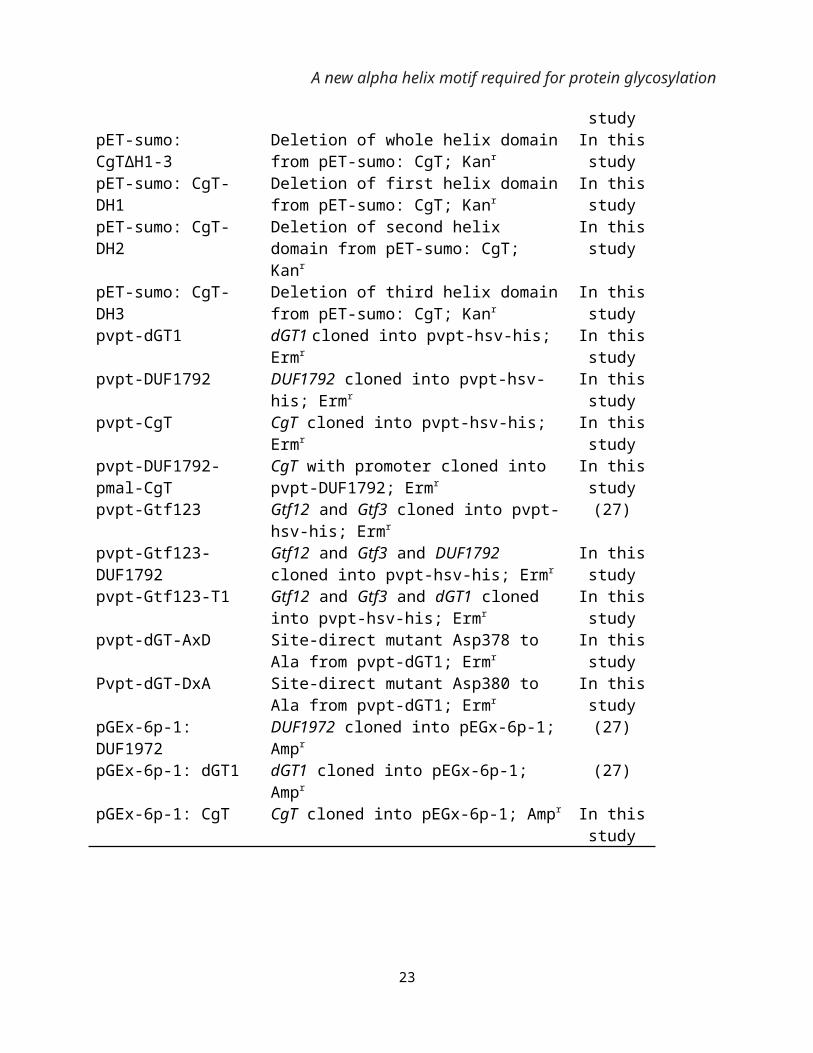
pET-28b His fusion protein expression vector; Kanr AmershampGEx-6p-1 GST fusion protein expression vector; Ampr AmershampET-sumo: CgT CgT cloned in pET-sumo; Kanr In this
studypET-28b: CgT CgT cloned in pET-28b; Kanr In this
studypET-sumo: CgT∆H1-3 Deletion of whole helix domain from pET-
sumo: CgT; KanrIn this study
pET-sumo: CgT-DH1 Deletion of first helix domain from pET-sumo: CgT; Kanr
In this study
pET-sumo: CgT-DH2 Deletion of second helix domain from pET-sumo: CgT; Kanr
In this study
pET-sumo: CgT-DH3 Deletion of third helix domain from pET-sumo: CgT; Kanr
In this study
pvpt-dGT1 dGT1 cloned into pvpt-hsv-his; Ermr In this study
pvpt-DUF1792 DUF1792 cloned into pvpt-hsv-his; Ermr In this study
pvpt-CgT CgT cloned into pvpt-hsv-his; Ermr In this study
pvpt-DUF1792-pmal-CgT
CgT with promoter cloned into pvpt-DUF1792; Ermr
In this study
14
A new alpha helix motif required for protein glycosylation
pvpt-Gtf123 Gtf12 and Gtf3 cloned into pvpt-hsv-his; Ermr
(27)
pvpt-Gtf123-DUF1792 Gtf12 and Gtf3 and DUF1792 cloned into pvpt-hsv-his; Ermr
In this study
pvpt-Gtf123-T1 Gtf12 and Gtf3 and dGT1 cloned into pvpt-hsv-his; Ermr
In this study
pvpt-dGT-AxD Site-direct mutant Asp378 to Ala from pvpt-dGT1; Ermr
In this study
Pvpt-dGT-DxA Site-direct mutant Asp380 to Ala from pvpt-dGT1; Ermr
In this study
pGEx-6p-1: DUF1972 DUF1972 cloned into pEGx-6p-1; Ampr (27)pGEx-6p-1: dGT1 dGT1 cloned into pEGx-6p-1; Ampr (27)pGEx-6p-1: CgT CgT cloned into pEGx-6p-1; Ampr In this
study
15
A new alpha helix motif required for protein glycosylation
Table 2. Primers used in this studyprimers SequenceCgT-sumo-BamH1-F 5’ – GATCAGGATCCATGGATAATGGTGAATTGATT – 3’
(B) CgTΔH1-3 UDP-GlcNAc 0.88±0.01 12.15±1.35 -2148±63 11.45±0.45∆H, ∆S, number of binding sites (n), and the binding constant (Kd) for donor binding with proteins in solution at 20 °C.
Figure 1.
1 2 3 4
17
75kD
50kD
A new alpha helix motif required for protein glycosylation
Figure 2.
18
A new alpha helix motif required for protein glycosylation
Figure 3.
Figure 3
19
UDP-GlcNAcUDP-Glc
UDP-GlcNAcUDP-Glc
(D)(C)
(B)(A)
A new alpha helix motif required for protein glycosylation
Figure 4. (A)
(B) 1 2 3
Figure 5.
20
0 20 120 mL 100 80 60 40
CgTΔH1-3
CgT
37 kD
37 kD
A new alpha helix motif required for protein glycosylation
Figure 6.
(C)
21
(B)(A)
Rossmann fold
DxD motif
Helix domain
Core region
N-terminal region
SpsA
CgT
A new alpha helix motif required for protein glycosylation
Figure 7.
dGT1
DUF1792 CgT
CgTΔH1-3
CgTΔH30
4000
8000
12000
Lum
iesc
ence
(RLU
)
22
A new alpha helix motif required for protein glycosylation