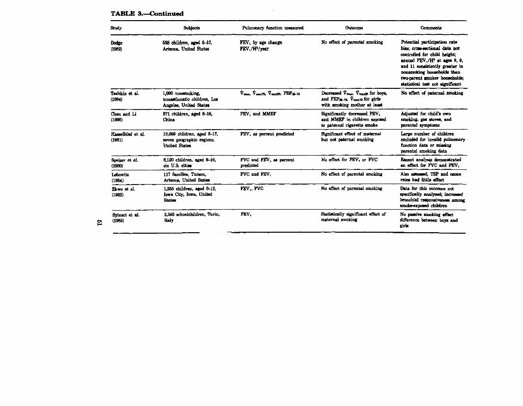
TABLE t.-Continud subjocte Pulmonery function meanued Outcome 668 chilhll, aged 8-10, Arir.om united St.&n No effect of parental smoking PotenW pnrticipation rate bias;cram+e&onaldatanot corkroIled for child hei& allmud FEx,/lP at e 8, 9, and 11 oonsietently gxater in nonomoking houoehobio tblm hreperent smoker hou&o@ 0tatica.I tast not lligdbnt lil&khetal. ww Chen and Li wm Haaeelblad et al. (1981). 1,080 noMmoking, nonasthmatic children, Lctl Al&e& united stateu 671 children, aged 6-16, chine 16,689 children, aged 5-17, eeven geqinphic regione, u0it.d state3 L, Vmu7~ Vd FE&e-7s FEV, snd MMEF FEV, 88 percent pwdkted Delmamd0~,0~forboys, Noeffe&ofp&malemoking and FJZFrm, k,m,a for &la with mnoking mother at led Slltly decremd FEV, Adjuoted for child’s own fC4dMMEFiIlChUdWSl@XpOOd smotiae~gas~~ to peteal cigarette Emoke w-d v-mm Significant effect of mE4ternal Largenmnherofchihiren but not paternal smoking exchdedforinvdidpuImonq tiuxtion data or mieoing porental omoking data Speizer et al. ww Lehowitz ww Ekwo et al. (1983) 8,120 children, eged 6-10, oix U.S. cltlm 117 femiliea, Tucaon, Arlmw united stat&i 1,266 chuhn, eged 6-12, Iowe City, low4 unitad SW40 FVC end FEV, en percent predicted FVC end FFW, FEV,, Fvc No effect for FEV, or PVC No effect of parental smoking No e.ffect of parental emoking Recent anelyeie demon&eted en effezt for FVC end FEV, Almemeaxd,lWendcaone rat&3hedIittIeeffect Data for thin outcome not epeciflallly enal- innasal blmchid reepfmlllvene4o emong imokm children Spineci et al. 2,386 echoolchildren, Turin, usw Italr statistialuy aigniiamt effect of metemel omoking No pemlve emoking effect diffemna betmen boyo and l&b
Transcript
TABLE t.-Continud
subjocte Pulmonery function meanued Outcome
668 chilhll, aged 8-10, Arir.om united St.&n
No effect of parental smoking PotenW pnrticipation rate bias;cram+e&onaldatanot corkroIled for child hei& allmud FEx,/lP at e 8, 9, and 11 oonsietently gxater in nonomoking houoehobio tblm hreperent smoker hou&o@ 0tatica.I tast not lligdbnt
16,689 children, aged 5-17, eeven geqinphic regione, u0it.d state3
L, Vmu7~ Vd FE&e-7s
FEV, snd MMEF
FEV, 88 percent pwdkted
Delmamd0~,0~forboys, Noeffe&ofp&malemoking and FJZFrm, k,m,a for &la with mnoking mother at led
Slltly decremd FEV, Adjuoted for child’s own fC4dMMEFiIlChUdWSl@XpOOd smotiae~gas~~ to peteal cigarette Emoke w-d v-mm Significant effect of mE4ternal Largenmnherofchihiren but not paternal smoking exchdedforinvdidpuImonq
tiuxtion data or mieoing porental omoking data
Speizer et al. ww
Lehowitz ww Ekwo et al. (1983)
8,120 children, eged 6-10, oix U.S. cltlm
117 femiliea, Tucaon, Arlmw united stat&i
1,266 chuhn, eged 6-12, Iowe City, low4 unitad SW40
FVC end FEV, en percent predicted
FVC end FFW,
FEV,, Fvc
No effect for FEV, or PVC
No effect of parental smoking
No e.ffect of parental emoking
Recent anelyeie demon&eted en effezt for FVC end FEV,
Almemeaxd,lWendcaone rat&3hedIittIeeffect
Data for thin outcome not epeciflallly enal- innasal blmchid reepfmlllvene4o emong imokm children
Spineci et al. 2,386 echoolchildren, Turin, usw Italr
statistialuy aigniiamt effect of metemel omoking
No pemlve emoking effect diffemna betmen boyo and l&b
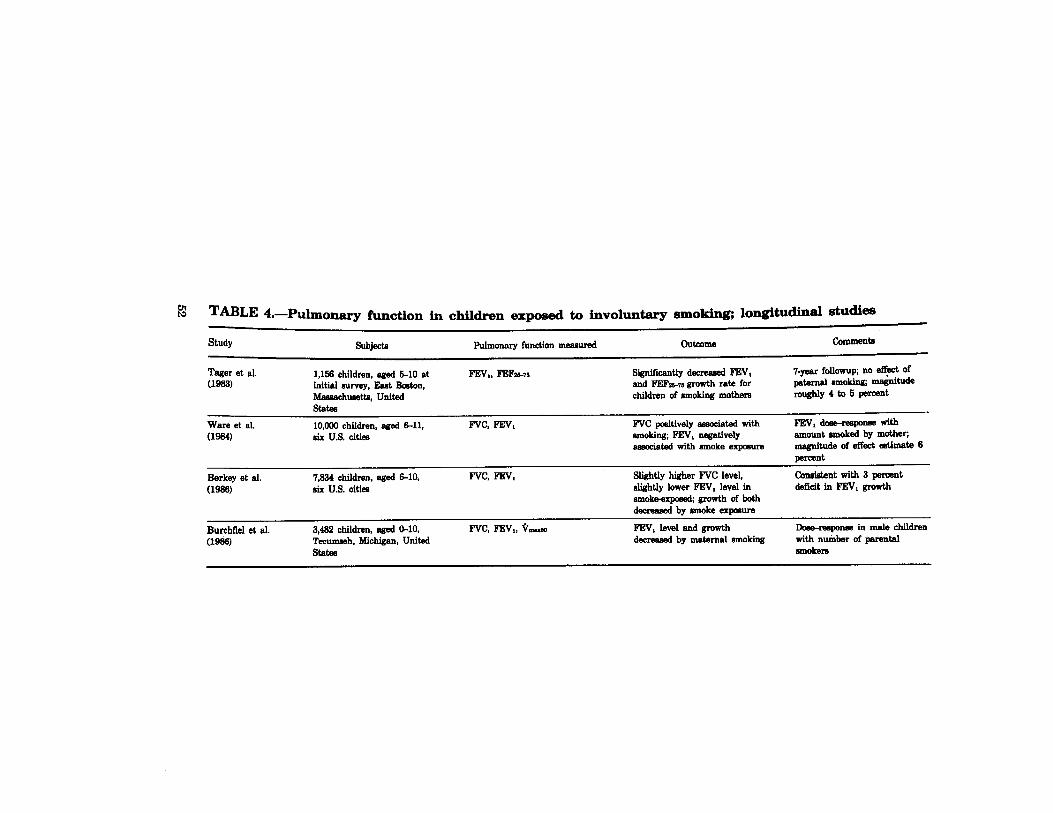
c TABLE 4.-Pulmonary function in cm&n exposed to involuntary smoking; longitudina\ ddies
Study Subjecta Pulmonary funOti0n mOamld Outcame c!ommenta
‘l&r et al. u9w
1,166 children, aged 6-10 at lnltial mvey, Enat Boston, Mamachuaetta, United St&M
FEW,, FEFow Sicantly decreasai FEW, and FJ!Tws growth rate for chUdren of emoking mothem
‘I-year followup; no eff$=-t of paternal amok&; magnitude roughIy 4 to 6 Pemnt
ware et al. 10,000 children, aged 8-11, ww six U.S. cities
WC, ml WC poeitively aooociaw with smoking; FEV, ne&ively amociated with omoke expooure
FEV, dose-response with amount omoked hy mother; magnitude of e&ct estimate 6 prant
Berkey et al. mw
7,834 children, agad 610, six U.S. cities
WC, FEV, Slightly higher FVC level, slightly lower FXV, level in omcbexposed; gmwth of both decreased by smoke expomue
coneistent with 3 penxlt de&it in FEV, growth
Burchfiel et al. (l=w
3,432 children, aged MO, Tecumeeh, Michigan, United Statee
WC, FEV,, vrrdn FEV, level and growth decread by maternal smoking
Dwe-reuponoe in maIe chlhimn with nuder of parental omokem
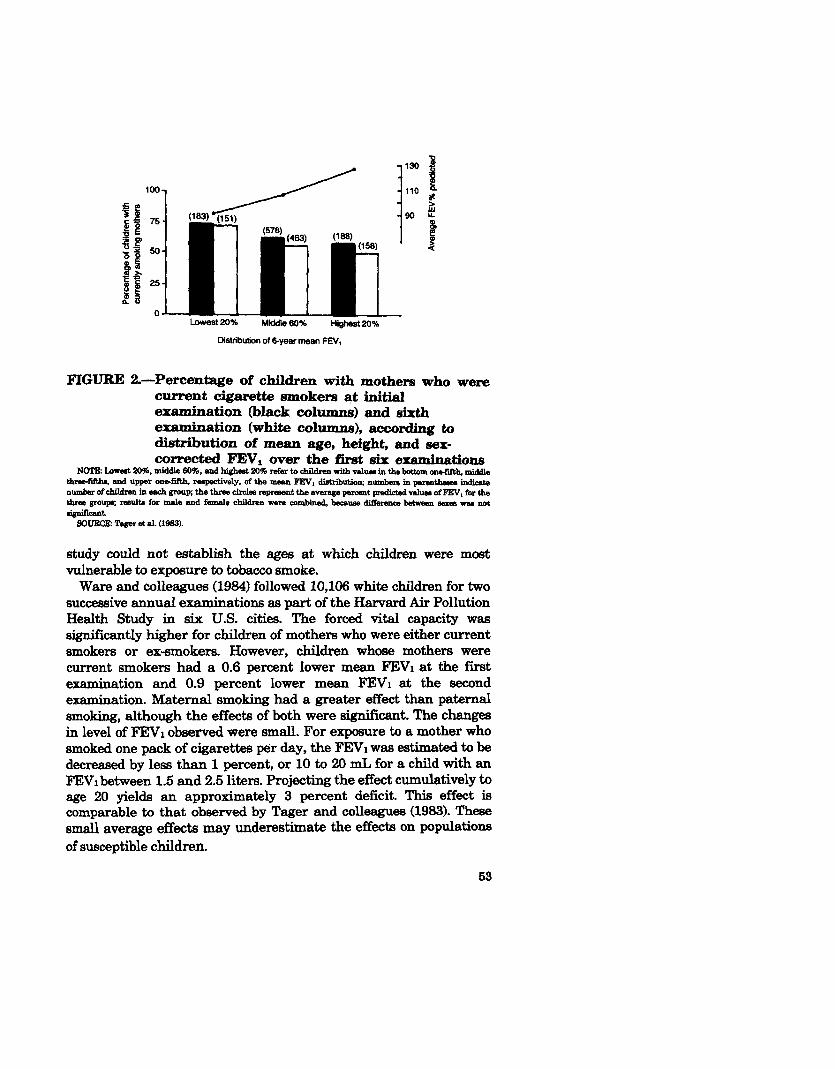
LOwmt20% Mile go% Highest20%
Dialibution Of syear mean FN,
FIGURE 2.-Percenwe of children with mothers who were current cigarette smokers at initial examination (black columns) and sixth examination (white columns), according to distribution of mean age, height, and sex- corrected FEV, over the firat six examinations
N(rPE:~20W,middle60%,andhiehat20%nefertoEhildraaarithvllluainthe~ollati(th,middla tliltxrfiftho, and upper onbflfth, rssp&vely. of the - FEY, diotributio~ nlmlbBnl in parentheua todioate number of children in each gmup; the three circlen rap&went the BVeraga pbresnt pImdh?d value4 of FEW, for the the group; remlta for male and famals children were combined, because difference t&mm - vu not signifmt.
SOURCE: !hget et al. w33).
study could not establish the ages at which children were most vulnerable to exposure to tobacco smoke.
Ware and colleagues (1964) followed 10,106 white children for two successive annual examin ations as part of the Harvard Air Pollution Health Study in six U.S. cities. The forced vital capacity was significantly higher for children of mothers who were either current smokers or ex-smokers. However, children whose mothers were current smokers had a 0.6 percent lower mean FEVl at the first examination and 0.9 percent lower mean F’EVl at the second examination. Maternal smoking had a greater effect than paternal smoking, although the effects of both were sign&ant. The changes in level of FEYI observed were small. For exposure to a mother who smoked one pack of cigarettes per day, the FEVl was estimated to be decreased by less than 1 percent, or 10 to 20 mL for a child with an F’EVl between 1.5 and 2.5 liters. Projecting the effect cumulatively to age 20 yields an approximately 3 percent deficit. This effect is comparable to that observed by Tager and colleagues (1963). These small average effects may underestimate the effects on populations of susceptible children.
53
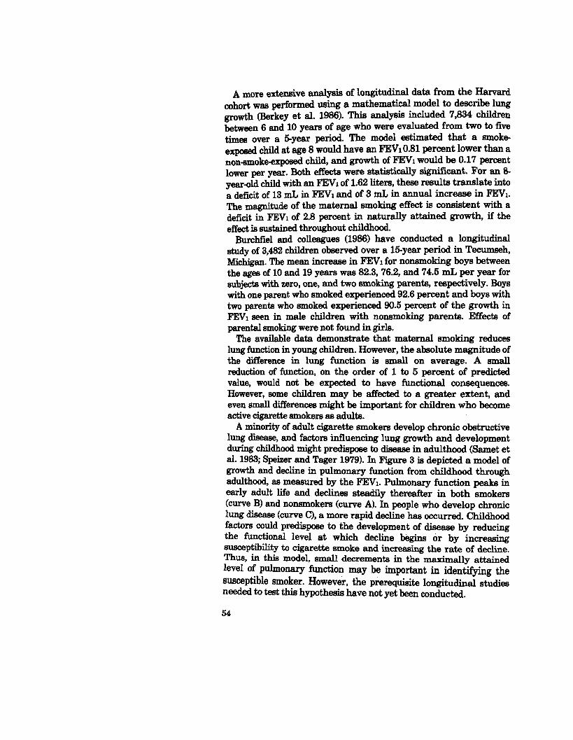
A mom e&hve analysis of longitudinal data from the Harvard cohort wm performed using a mathematical model to describe lung growth (Be&y et al. 1966). This ~IM&B~S included 7,834 Children beben 6 & 10 year6 of age who were evaluated from two to five hm over a ~-year period. The model estimated that a smoke expOeed child at age 8 would have an FE571 0.81 percent lower than a non-emokeexpoa3ed child, and growth of FEVI would be 0.17 percent lower per year. ~0th effects were statistidy significant. For ~I.I 8 yw& child tith an F’EVl of 1.62 liters, these result-8 translate into a deficit of 13 J.UL in FEVI and of 3 mL in annual increase in MI. ‘&e magnitude of the maternal smoking effect is consistent with a de&it in J?EVl of 2.8 percent in naturally attained growth, if the effect, ia sustained throughout ~hildhd.
Burchfiel and colleagues (1966) have conducted a longitudinal study of 3,462 children observed over a E-year period in Tecumseh, &.&h&an. The mean increase in FXVI for nonsmoking boys between the ages of 10 and 19 years was 82.3, 76.2, and 74.5 mL per year for subjects with zero, one, and two smoking parents, respectively. Boys with one parent who smoked experienced 92.6 percent and boys with two parents who smoked experienced 90.5 percent of the growth in FEVl seen in male children with nonsmoking parents. EXfecta of parental smoking were not found in girls.
The available data demonstrate that maternal smoking reduces lung function in young children. However, the absolute magnitude of the difference in lung function is tnnall on average. A Emall reduction of function, on the order of 1 to 5 percent of predicted value, would not be expected to have functional consequences. However, some children may be affected to a greater extent, and even small differences might be important for children who become active cigarette smokers as adults.
A minority of adult cigarette smokers develop chronic obstructive lung disease, and factors influencing lung growth and development during childhood might predispose to disease in adulthood (Samet et al. 1983; Speizer and Tager 1979). In Figure 3 is depicted a model of growth and decline in pulmonary function from childhood through adulthood, as measured by the F’EXl. Pulmonary function peaks in early adult life and declines steadily thereafter in both smokers (curve B) and nonsmokers (curve A). In people who develop chronic lung disease (curve CL a more rapid decline has occurred. Childhood factors could predispose to the development of disease by reducing the functional level at which decline begins tjr by increasing SusCePtibfitV to cigarette smoke and increasing the rate of d-be. Thus, in this model, small decrements in the ’ Ily atwed led Of PuhnOnary function may be important in identifying &e susceptible smoker. However, the prerequisite longitudinal stu&es needed to test this hypothesis have not yet been conducted,
FEV, as percent of value at age 2h25
I I I L 5 10 15 20 30 40 50 90 70 50
Age (uem)
FIGURE 3.-Theoretical curves representing varying rates of change in FJSV, by age
SOURCE: SpeLer and Tager (1979).
Bronchoconstriction Nonspecific bronchial responsiveness has been considered a poten-
tial risk factor for the development of chronic obstructive lung disease in both adults and children (US DHHS 1984). This physiolog- ic trait may be influenced by environmental exposures such as involuntary smoking by children and active smoking by adults, and by respiratory infections at all ages.
Asthma is a chronic disease characterized by bronchial hyperre sponsiveness. Epidemiologic studies of children have shown no consistent relationship between the report of a doctor’s diagnosis of asthma and exposure to involuntary smoking. Although one study showed an association between involuntary smoking and asthma (Gortmaker et al. 1982), others have not (Schenker et al. 1983; Horwood et al. 1985). This variability may reflect differing ages of the children studied, differing exposures, or uncontrolled bias. In several recent studies (Murray and Morrison 1986; O’Connor et al.
55
1986, web et al. 1985; Martinez et al. 198% Ekwo et al. 19831, noMpecific broncm responsiveness Was examined in relationship b ~voluntary smoking. The results of these ytusm .sugge”t that exposure b matid cigarette smoking b assocmted mth increased noMpecific wap req&ven~. Some ,repo* suggest that the hm respoevena is present only m chi&+% kWWn to be w-tic (Murray and Morrison 1986; o’~nnOr et al. 1986), whereas othm sw~ that the increased respo~iveness is seen in 4 cmw (~kw~ et al. 1983; Martinez et al. 1985). The pathophysi- ological magi underlying the increased responsiveness and the lowm~m consequences of the increased responsiveness remain horn. m s&ion reviews the studies on asthma and on bronchial hyperresponsiven~.
&rtln&er amj coworkers (1982) studied the relationship between paren~ ~&ing and the prevalence of asthma in children up to 17 ;years of age. Random community-based populations in Michigan (3,072 &U.ren) and Massachusetts (894 children) were surveyed. parents reported on their own smoking habits a&l on the asthma histories of t&r children. Biased reporting by parents who smoked ~88 d by e=mimng the relationship between parena smoking and other conditions, and considered not to be present. A&IM prevakmce declines with age, and asthmatic children are unlikely to tolerate active smoking; therefore, misclassification of activelp smoking asthmatic children ss nonsmokers seems unlikely. In comparison with children of nonsmokers, children whose parents smoked were more likely to have asthma (relative risks of 1.5 and 1.8 for Michigan and Massachusetts children, respectively) and sevely! asthma (relative risks of 2.0 and 2.4, respectively). The investigators estimated that between 18 and 23 percent of all childhood asthma and 28 and 34 percent of severe childhood asthma is attributable to exposure to maternal cigarette smoke.
Schenker and coworkers (1983) studied 4,071 children between 5 and 15 years of age in western Pennsylvania. These investigators found no relationship of parental smoking to the occurrence of asthma, after adjustment for potential confounding factors.
Horwood and coworkers (1985) conducted a cohort study of 1,058 children in New Zealand who were followed from birth to age 6 ye- A fdy history of allergy and male sex were the ody significant predictors of incident cases of asthma. Neither parental smoking nor respiratory illnesses were predictive of the occurrence Of asthma in this investigation.
A recently reported cross-sectional study by Murray and Morrison (1986) suggests a mechanism by which maternal cigarette smoking
might influence the severity of childhood asthma. These mvestiga- h-s StUdkd 94 children, aged 7 to 17 years, with a history of asthma. The children of mothers who smoked had 47 percent more symp-
56
41
2-
1 -
0.5 -
0.25 -
0.125 -
0.06 -
0.03 -
049
Nonsmoker smoker
00
0 0 0 %I?
0
p=o.o02
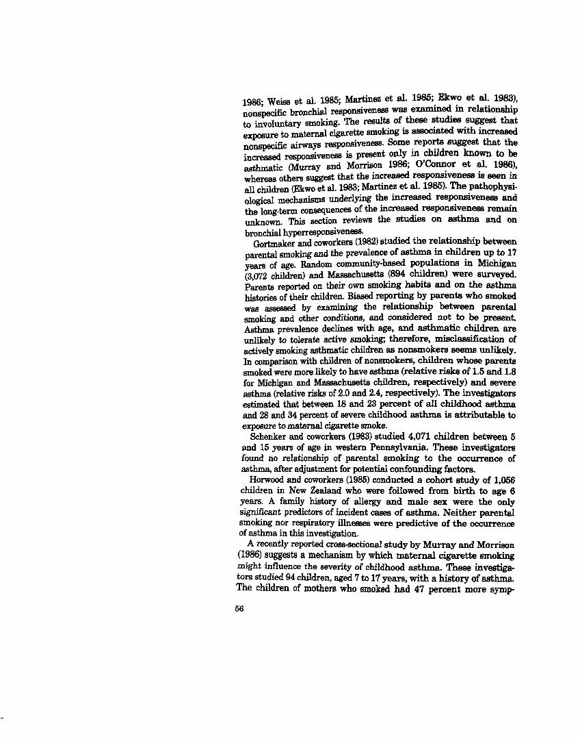
FIGURE 4.-P% in two groupa of children with a history
toms, a 13 percent lower FEYI, and a 23 percent lower FEFws than the children of nonsmoking mothers. Forty-one children, who had been able to discontinue med ication and had no recent respiratory illness, underwent a histamine chal lenge test. There was a fourfold greater responsiveness to hi&amine among the asthmatic children of mothers who smoked (Figure 4) compared with asthmatic children of nonsmoking mothers. Dose-response relationships were present for all outcome variables in this study: symptoms, pulmonary function, and airways responsiveness. The differences between children of smoking mothers and children of nonsmoking mothers were greatest in the older children. The father’s smoking behavior did not influence the child’s asthma severity. The sample of asthmatic children with mothers who smoked was small (N = lo), and only 41 of 96 children had histamine chal lenge tests. G iven the heterogeneity of asthma, the variable nature of bronchial hyperreactivity in asthma, and the potential for biased selection, these results must be interpreted with caution.
O’Connor and coworkers (1986) studied 286 children and young adults, 6 to 21 years of age, drawn from a community-based sample,
57
and confirmed the findings of Murray and Morrison (1986). Bronchi- al responsiveness was measured with eucapneic hyperpnea to subfreezing air. Among the 265 subjects without asthma there was no significant relationship between maternal cigarette smoking and nonspecific bronchial responsiveness. However, in the 21 subjects 6th adive asthma, maternal smoking was significantly associated with increased levels of bronchial responsiveness.
In a study of 1,355 children 6 to 12 years of age, significant increases in FEW and FF,F25-7s were observed following isoproterenol administration in children whose parents smoked (E~wo et al. 1983). Increases after isoproterenol were not observed in children of nonsmoking parents.
Weiss and coworkers (1985) evaluated 194 subjects between the ages of 12 and 16 drawn from the same population as those reported by O’Connor and coworkers (1986), with eucapneic hyperpnea to subfreezing air as a test for bronchial responsiveness and allergy skin tests as a test for atopy. Subjects defmed as atopic (any skin test wheal greater than or equal to 5 mm) had twice the frequency of lower respiratory illnesses in early childhood and were twice as likely to have a mother who smoked. However, there was no relationship between maternal smoking and increased bronchial responsiveness.
Martinez and associates (1985) studied 170 9-yearold children in Italy. Nonspecific bronchial responsiveness to methacholine and allergy prick test positivity in these subjects was significantly associated with maternal cigarette smoking.
These data suggest that maternal cigarette smoking may influence the severity of asthma; a mechanism for this effect may be through alteration of nonspecific bronchial responsiveness. Further investi- gation is needed to determine whether exposure to environmental cigarette smoke can induce asthma in children and whether ETS exposure increases the frequency or severity of attacks of broncho- constriction in asthmatics. The effect of involuntary smoking on increased bronchial responsiveness in asthmatics and in norm&h- matics has only recently been addressed. These initial data are provocative, but the magnitude of the effect, the target population at risk, the underlying mechanisms, and the long-term consequences have not been described. Furthermore, the complex interrelation- sops ==‘u3 respiratory illness, atopy, parental smoking, and tin-w responsiveness have not been clarified and require further study.
Ear, Nose, and Thmat Five studies (Said et al. 1978; Iverson et al. 1985; Kraemer et al.
1983; Black 1985; Pukander et al. 1985) show an excess of chronic
58
middle ear effusions and d&eases in children exposed to parental smoke.
Said and colleagues (1978) questioned 3,920 children between IO and 20 years of age about prior tonsillectomy or adenoidectomy, considered an index of frequent upper respiratory or ear infections. The investigators reported that, in general, this surgery was performed before the children were 5 years old. The prevalence of prior surgery ~INXMS~ with the number of currently smoking parents in the home.
Iverson and coworkers (1985) prospectively studied 337 children enrolled in all day-care institutions in a municipality over a 3month petid to evaluate the importance of involuntary smoking for middle ear effusion in children. Middle ear effusion was assessed with tympanometry, and the overall prevalence was found to be approxi- matsly 23 percent. Although various indoor environmental factors were assessed in this investigation, only parental smoking was significantly associated with middle ear effusion. The effect of parental smoking persisted with control for the number of siblings. The overall age-adjusted odds ratio was 1.6’ (95 percent confidence interval 1.0-2.6). In 5- to 7-year-old children, 10 to 36 percent of all chronic middle ear effusions could thus be attributed to smoking on the basis of these results.
Kraemer and coworkers (1983) performed a cas+control study of 76 children to examin e the relationship of environmental tobacco smoke exposure to the occurrence of persistent middle ear effusions. Frequent ear infections, nasal congestion, environmental tobacco smoke exposure, and atopy were all more frequent in children with ear effusions. The effect of involuntary smoking was observed only if nasal congestion was present, and was greatest in children who were atopic.
Black (1985) performed a case-control study of glue ear with 150 cases and 300 controls. Parental smoking was associated with a relative risk of 1.64 (95 percent C.I. 1.03-2.61) for glue ear. In Finland, Pukander and coworkers (1985) conducted a mntrol study of 264 2 to 3-year-old children with acute otitis media and 207 control children and found an association between parental smoking and this acute illness.
These studies are consistent in their demonstration of excess chronic middle ear effusions, a sign of chronic ear disease, in children exposed to parental cigarette smoke. Potential confounding factors for middle ear effusions should be examined carefully in future studies. The long-term implications of the excess middle ear problems deserve further study.
59
Acute Reqimtory Illness There are no studies of acute respiratory illness experience in
adulta exposed to environmental cigarette smoke.
cbugh, Phlegm, and wheezing Few studies have addressed the relationship of chronic respiratory
symptoms in nonsmoking adults with environmental tobacco smoke exposure. Schilling and colleagues (1977) found that symptoms in adult men and women were related to personal smoking habits and that the occurrence of cough, phlegm, or wheeze in nonsmokers was not related to the smoking habits of their spouses. Schenker and colleagues (1982) confirmed these results in a telephone survey of 5,000 adult women in western Pennsylvania.
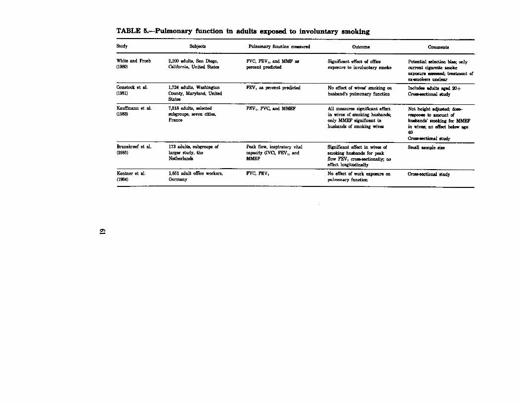
White and Froeb (1980) reported on 2,100 asymptomatic adults drawn from a population enrolled in a physical fitness program (Table 5). They reported statistically significant decreases in FEVl and maximum midexpiratory flow rate 0 as a percent of predicted in nonsmokers exposed to tobacco smoke in the work environment for at least 20 years compared with nonsmoking workers not exposed. The magnitude of effect was comparable to that of actively smoking 1 to 10 cigarettes per day. However, the absolute magnitude of the difference in mean levels of function between the smokeexpoeed group and the unexposed group was smalh 160 mL (5.5 percent) for FEW1 and 465 I& per second (13.5 percent) for MMEF. Carbon monoxide levels were measured in selected work- places and ranged from 3.1 to 25.8 ppm. The study population was se&selected, and the exposure classification was crude and did not account for people who changed jobs. It is unclear how the ex- smokers in the population were handled in the analysis. Kentner and coworkers (1984) performed a cross+ectionai investigation on 1,351 workers and found no influence of involuntary smoking on pulmonary function. In this study, involuntary smoking at home and at work was considered.
Comstock and colleagues (1981) examined 1,724 subjects drawn from two separate studies in Washington County, Maryland. Male and female nonsmokers married to smokers did not have a sign& CaMJy increased risk of having an FEWI less than 80 percent of Predicted or an F’EJVAWC ratio less than 70 percent. Schilling and colleagues (1977) also did not find an effect of involuntary smoking in adults. Effects were not examined within strata defined by age in either of these studies.
60
TABLE S.-Pulmonary function in adults exposed to involuntary smoking
Study Subjecta Pulmonary function measured Outcome Comments
White end Froeb 2,100 adults, Sen Diego, (1980) California, United States
Fvc, FBV,, end MhfF m percent predictcd
significant effect of office exposure to involuntary smoke
Potential e&&ion biaq only current c@rette emoke expceure & treatment of exsmokere unclear
Comtwk et al. a9811
1,724 adulte, Washington
County, Maryland United St&S
FEV, 88 percent predicted No effect of wives’ smoking on hueband’s pulmonary function
FFW,, FVC, end MMEF All meeauw signiEcant effect in wives of smoking husbands; only MMEF signGent in husbands of smoking wives
Not height edjw dear- reeponmtoamolmtof husbands’ smoking for MMEF in wivce; no effect below age 40
Brunekreef et al. w-w
Kentner et al. (1984)
173 adult.% subaroupe of larger study, the - Netherlands
1,851 adult of&e workers, Germany
Peak flow. in8Diratorv vital Significant effect in wives of smoking huebenda for peak flow FEW, cmmsctionally; no effect longitudinelly
NO effect of work exposure on pulmonery function
Chmectionel study
Smell sample .3&e
thmectional .&udy
Kauffmann and colleagues (1983) suggested that the effects of exposure from a spouse who smoked may be manifest only after many years of exposure. These investigators asses& the effects of marriage to a smoker in 7,818 adults drawn from several cities in France. Among 1,985 nonsmoking women aged 25 to 59,58 percent of whom had husbands who smoked, the level of MMEF was significantly reduced in women married to smokers compared with women married to nonsmokers; this effect did not become apparent until age 40. The reduction was small, on average.
Recently, studying another population, KaufXnann and colleagues (1986) suggd,ed that the FEWI/FVC ratio may be a more sensitive test for detecting differences between exposed and nonexpoeed subjects, particularly in those with symptoms of wheexing; however, this suggestion has not been evaluated in other populations.
Rrunekreef and coworkers (1985), from the Netherlands, reported on 173 nonsmoking women who were participants in a larger longitudinal study of pulmonary function. The women were classi- fied by whether they were or were not exposed to tobacco smoke at study onset or at followup. Cross-sectionally, significant differences in pulmonary function were observed between smoke-expoeed and nonexposed women. However, the rate of decline of lung function king the followup period was not affected by tobacco smoke exposure in the home. This study had a small number of subjects and inadequate statistical power to detect effects of exposure on rate of decline that were not extremely large.
Jones and colleagues (1983) selected women with either high or low FEVs from a population-based longitudinal study in Tecumseh, Michigan. Exposure to cigarette smoke at home from husbands who smoked was not significantly different in the two groups of women.
Nonsmoking men who participated in the Multiple Risk Factor Intervention Trial had significantly lower levels of pulmonary function if their wives smoked in comparison with similar men whose wives did not smoke (Svendsen et al. 1985).
The physiologic and clinical significance of the small changes in pulmonary function found in some studies of adults remains to be determined. The small magnitude of effect implies that a previously healthy individual would not develop chronic lung disease solely on the basis of involuntary tobacco smoke exposure in adult life. Whether particular characteristics increase susceptibility, such as Childhood exposures or illnesses, atopy, reduced pulmonary function from whatever cause, and increased airways responsiveness, rema,fns unknown. These sndl changes may also be markers of an irritant response, possibly transient, to the irritants known to be present in environmental tobacco smoke.
62
Bronchoconstriction
Normal Subjects Only limited data have been published on the acute effects of
inhalation of environmental tobacco smoke on pulmonary function in normal subjects (Table 6) and none on bronchial responsiveness. The available data have been obtained in exposure chambers under carefully monitored and controlled circumstances (Pimm et al. 1978; Shephard et al. 1979; Dahms et al. 1981).
Pimm and colleagues (1978) exposed nonsmoking adults to smoke in an exposure chamber. Relatively constant levels of carbon monoxide (approximately 24 ppm) were achieved in the chamber during involuntary smoking. Peak blood carboxyhemoglobin levels were always less than 1 percent in these subjects before smoke exposure, but were significantly greater after the study exposure, Lung volumes, flow volume curves, and heart rates were measured for all subjects. Measurements were made at rest and following exercise under control and smoke-exposure conditions. Flow at 25 percent of the vital capacity was reduced at rest in men and with exercise in women. Although statistically significant, the magnitude of the change was small: a 7 percent decrease in flow in men and 14 percent in women.
Shephard and coworkers (1979) utilized a similar cross-over design in a chamber of exactly the same size as that used by Pimm and associates. Their results were similar, with a small (3 to 4 percent) decrease in FVC, FEVI, Vti, and Vma~26. They concluded that these changes were of the magnitude anticipated from exposure to the smoke of less than one-half of a cigarette in 2 hours (the exposure anticipated for an involuntary smoker).
Dahms and colleagues (1981) used a slightly larger chamber and an exposure with an estimated peak carbon monoxide level of approximately 20 parts per million. They found no change in FVC, FEVl, or FEFs76 in normal subjects after 1 hour of exposure.
The active smoker manifests acute responses to the inhalation of cigarette smoke; thus, highdose involuntary exposure to tobacco smoke may plausibly induce similar responses in nonsmokers. The magnitude of these changes is quite small, even at moderate to high exposure levels, and it is unlikely that this change in airflow, per se, results in symptoms.
Asthmatics Dahms and colleagues (1981) exposed 10 patients with bronchial
asthma and 10 normal subjects to cigarette smoke in an environmen- tal chamber. Pulmonary function was measured at 15-minute intervals for 1 hour after smoke exposure. Blood carboxyhemoglobin levels were measured before and after the l-hour exposure. The
63
2 TABLE 6.-Acute effects on pulmonary function of passive exposure to cigarette smoke; normal subjects
Study Type of expceure Magnitude of expmure Effecta Comments
Pimm et al. (1978)
Chamber 14.6 In, furniture sparse, smoking machine in mm
Room levels not meanured; e&mated at peak [CO] - 20 wm
Low exposure: 3% decmaee FEW,, 4% decrease vm 6% decrease V- with exerch no increaeed effect with high =poeu~
0.9% increaee in Fvc, 6.2% increaw in m,, 2.2% llmenee in F+EF at 1 hour
Nonmokem: average age, men 23, women 25; sham expoeure en control; iubjwt eatiited inhalation - l/2 cigar&e!2 how
10 nonsmokera; a2e range 24- 63 yenm; not blinded; no aham expmure
carboxyhemoglobin levels in subjects with asthma increased from 0.82 to 1.20 percent. In normal subjects the increase was from 0.62 to 1.05 percent. The increases in carboxyhemoglobin in the two study groups were not significantly different. Asthmatic subjects had a decrease in forced vital capacity @‘VC), FEVI, and MMEF to a level significantly different from their preexposure values. The decreases in asthmatic subjects were present at 15 minutes, but worsened over the course of the hour to approximately 75 percent of the preexpo sure values. Normal subjects had no change in pulmonary fundion with this level of exposure. In this study, subjects were not blinded as to the exposure and were selected because of complaints about smoke sensitivity.
Shephard and colleagues (1979), in a very similar experiment, subjected 14 asthmatics to a Zhour cigarette smoke exposure in a closed room (14.6 ms). The carbon monoxide levels (24 ppm) were similar to those predicted in the study of Dahms and coworkers (1981). Blood carboxyhemoglobin levels were not measured. Subjects were randomized and blinded to sham (no smoke) and smoke exposure and tested on two separate occasions. Data were expressed as the percentage change from the sham exposure. Signi&ant changes in FVC and FEYI were not observed between the sham and the smoke exposure periods, although 5 of 12 subjects did report wheezing or tightness in the chest on the day of smoke exposure.
Wiedemann and associates (1986) examined nonspecific bronchial responsiveness to methacholine in 9 asthmatic subjects and 14 controls and the effect of acute involuntary smoking on nonspecific bronchial responsiveness. At the time of the study, all asthmatics were stable with normal or near normal pulmonary function. The subjects underwent baseline pulmonary function and methacholine challenge testing. On a separate day they were exposed to cigarette smoke for 1 hour at 40 to 50 ppm of carbon monoxide and underwent pulmonary function and methacholine challenge testing. J?uhnonary function was not influenced by exposure. Nonspecific bronchial responsiveness decreased significantly, rather than increasing, as would be anticipated following an irritant exposure.
Acute exposure in a chamber may not adequately represent exposure in the general environment. Biases in observation and the in selection of subjects and the subjects’ own expectations may account for the widely divergent results. Studies of large numbers of individuals with measurement of the relevant physiologic and exposure parameters will be necessary to adequately address the effects of environmental tobacco smoke exposure on asthmatics.
Ear, Nose, and Throat There are no studies of chronic ear, nose, and throat symptoms in
adults with involuntary smoking exposure.
65
Lung Cancer This se&ion reviews the epidemiological evidence on invohmtary
smoking and lung cancer in nonsmokers, which has been derived from retrospective and prospective epidemiological studies. First, common methodological issues that apply to all these Studies are considered. Second, for each type of study design, individual studies are reviewed for their methodological approach (Tables 7 and 81, findings associated with tobacco smoke exposure (Table 9, Figure 5), and strengths and limitations. Third, the lung cancer risk associated with involuntary smoking is e xamined as a low-dose exposure to cigarette smoke by combining the d-response relationships for active smoking with the exposure data for involuntary smoking to predict the expected lung cancer risk due to involuntary smoking. This expxted risk is then compared with the actual risks observed in studies of involuntary smoking. Finally, the existing epidemiologicsl evidence is summarized and the plausibility of the association between lung cancer and involuntary smoking is evaluated on the basis of our current knowledge.
ObfBNd Risk Geneml iUethodological Issues
For both retrospective and prospective studies, the common methodoltic concerns are disease misclassi&ation and miscla&fi- cation of the subject’s personal smoking status or exposure to ET& Disease misclassification, for example, refers to the incorrect classifi- cation of the lung as the primary site of a cancer that originated elsewhere. Disesss misclassification is of greatest concern in studies in which the diagn~is of lung cancer was not histologically confirmed. Such misclsssification tends to be random and to bias relative risk estimates toward unity (Copeland et al. 1977). Patients with lung cancer, or any disease associated with cigarette smoke exposure, may report exposure to ETS more frequently than controls becauseofbiasinrecall.
Misclassification of the subject’s personal smoking status may occur in both retrospective and prospective studies; this misclassifi- cation refers to incorrectly classifying a subject as a nonsmoker when the subject is actually an ex-smoker or a current smoker, or to incorrectly chdfying the subject as a smoker when the subject is a nonsmoker. Biochemical markers such as cotinine and nicotine, which can be used to detect unadmitted active smokers, are sensitive only to a recent exposure to tobacco smoke; thus, they are not Ptiicuh+Q useful for identifying ex-smokers who deny their past SIIlOking hisb?k~. Mis&ss&ation of smokers or ex-smokers as nonsmokers may produce the appearance of an involuntary smoking effect when, in fact, the true relationship is with aeve smoking.
&urcz of subjects
Age-
Yeara of earollment
l&St year of followup
Method of followup
VerScation of diagncei.9
Metbodandtypeof information obtained
hdex of pamive smokiug
Number of lung cancer deaths in nonsmokers
ceusus population, 29 he&h districts, Japan
1966
1961,1962
Recordlinkagebstweaa ri&factarrecvrdaand death certitica~
lnterview 0): smoking auddrinkinghabita, dietary history, oocupation, other health-related variables
Husband’s smoking at entry ooasmoker, ex- smoker, curteat smoker (oiglday)
mom
176769 (F)
85-84
196%1960
1972
llIonitored by ACS volunteers, death eartificatee from locallstate hsmlth departmenta
Verified method of dlagnosi9aud tilo6y for 6rat 6 yeam’ followup
self -red qw3tiomlaisz education, real- ocoupational exposure, smokiw and medical history
H&and’s smoking atentrj? -km current smoker, and OigldaJy exsmokers exoluded
163 0
mea 1,917 (F)
45-64
19724976
1962
Recordlioh@witb
Lomlcauoerre&try
Spouse’s smoklug at eatrJTcurrentm uever smoker, ex- smokers edlded (quit 26 year8 before atrg)
Misclassification of involuntary smoking exposure refers to the incorrect categorization of exposed subjects as nonexposed and of nonexposed subjects as exposed. Most studies of lung cancer to date have used the number of cigarettes smoked by spouses as a measure of exposure to involuntary smoking, and thus have disregarded duration of exposure, exposure from other sources, and factors that influence exposure, such as proximity to the smokers or size and ventilation of the room where the exposure occurred. Moreover, all
6'7
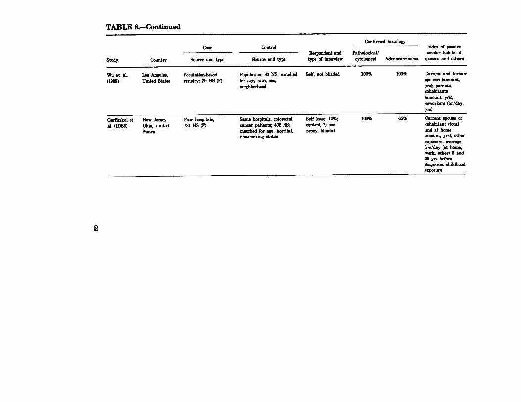
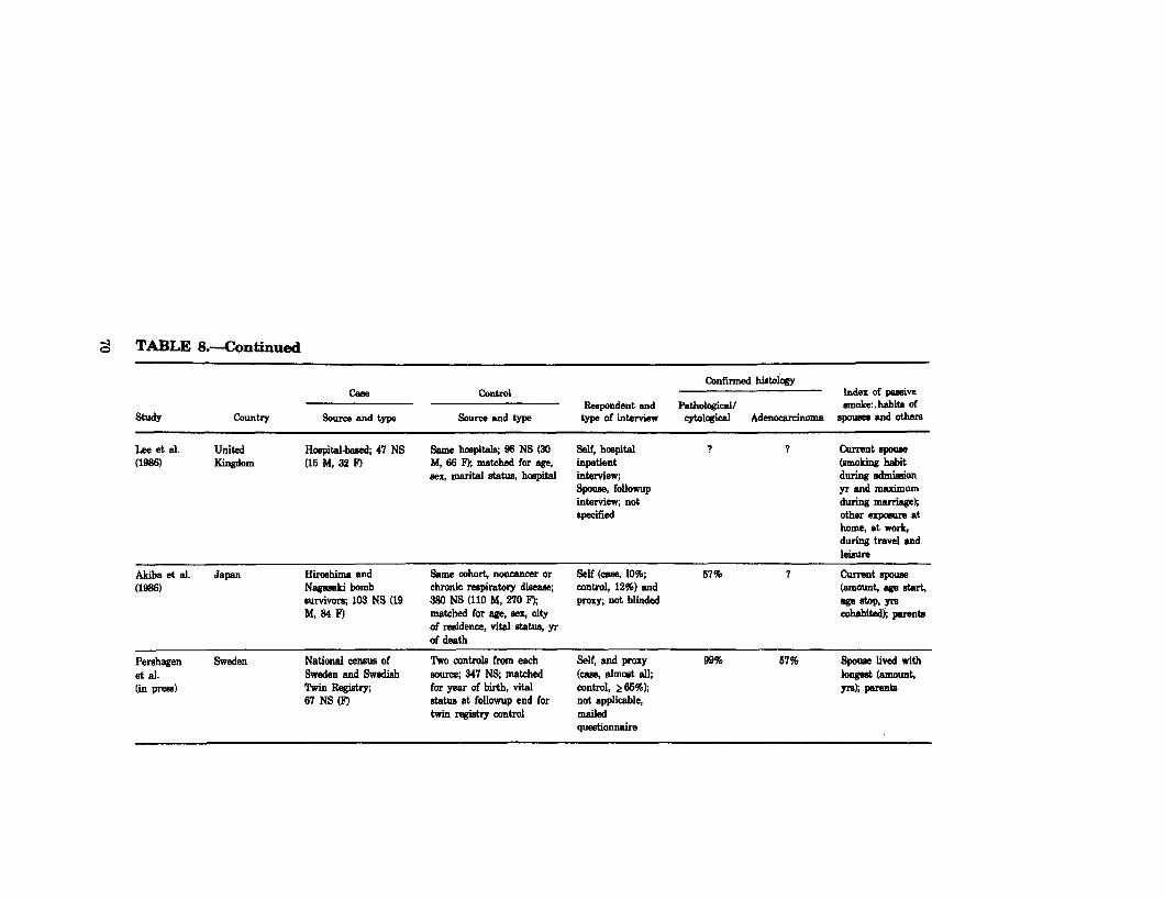
TABLE &-Description of case-control studies
Study cmntry
case
Source end type
control
Source end type
confirmed histology Iudexofpemd~
Respondent and Pathologicel/ mole: habita of type of interview cytological Adermarchoma spouam end others
Trichopouka Greece et al. (1981, 1983)
C&set and can& hospitals, 77 NS (FJ
Orthopedic hcepital; 225 Ns; not matched
Selfi not blinded 65%
Correa et al. (1989)
New Orleans, United States
Hoepit+ 30 NS (8 M, Same hospitals, non-~ Self, and proxy 97% 54% among current spoues 22 F) related diaeaaes; 313 NS bm, 23% women RYPe, amount Fk
(180 M, 133 Q matched for control, 11%); parents age, sex, raw hmitd blinded
Chsn and Hong Kong Four hoepit& Orthopedic, same hoepit& Self; not blinded 82% 45% Ndapouee Fun8 ww 64 NS 0 189 NS; not matched w=if-bi-
quiwtiom at home andatwork
Koo et al. Hong Kong Eight lmpitale; Population; 137 N& Self; not blinded 97% 69% Currentandr- wa 1981) 88 NS 0 matched for egm, race, sex, m b--k
emioewllomlc status, YmwiP@-B lwldence dlstrlct other cohablti&
am&em (amount PIW
K&at and united statea Most from one NY Same hospital 0; non- Self; not blinded 10096 M%td cul7ent spouss Wynder (19S4) hoepi~ 134 NS; tabaamrelated dieeasq 78 14% F of @nrentww
passive smoking data NS (25 M, 63 p); matched 134 NS fmoking habite on only 78 NS (26 M, for age, sex, raa+ hoepital, l?urrentwat Mm date of interview, hnmeandwork
nonsmoking status
TABLE 8.-Continued
St&- country
caes
Sourceandtype
Contml
Soweandtype
confll histology hiexofpamive
Respondent and Patholo6icaV srook habit8 of type of inte.rvlew ~hw Adenocarcinonm qouam and othara
wu et al. w=)
Population; 62 N9; matched self; not blinded 100% 100% current and former for age, ram sex, spa- (amount, nelghborImcd IT& puent*
cohabitant4 (=mt, Yd, coworkers &r/day, P-4
Garfinkel et al. (1985)
New Jersey, Ohio, united St&S
Four hoopit& 134 NS 0
Same haepitale, o~lorectal cancer patients; 402 NS; matched for age, hospital, IlonsmoLiDg status
self (cab& 12%; control, ?) and prory; blinded
100% 66% Current spouse or cohabitant (total and at home: amu& ~3); other ew-re, a-w hrdday (at home, worl, other) 5 and 2syrsbefore diagm!&chiMhad em--
d TABLE &-Continued
Study Country
case
Source and type
Control
Source end type
confirmed hb3bi~ Index of paudve
Respondent and Patholcgical/ smoke:, habite of type of interview cytological Adenocarcinoma spousee and others
Lee et al. wm
United Ha&al-beeed; 47 NS KlllgdOlll (15 M, 32 l9
Same hmpitals, 96 NS (30 Self, hospital 1 1 current spoum M, 66 F); matched for age, inpetient (smoking habit sex, marital status, hoepital interview; duriag admission
spouse, followup yr ad maximum intarvie-w; not during marrio& fqecified other expceure at
home, at work, dur@ travel end leisure
Akiba et al. ww
Japan Hllhlma and Nagasekl bomb survivors; 103 NS (19 M, 84 F)
Same cohort, noncancer or self (c-am, 10%; 67% 7 curlent spoum chronic respiratory disease; control, 12%) end bow llee de 380 NS (110 M, 270 IQ prow, not blinded aBgestop,yra matched for age, eeq city cohabitiS; perenta of residence, vital statue, yr of death
Perehagen et al. (in press)
Sweden National census of Sweden end Swedish TwlnFhgi&y; 67 NS 0
Two controls from each soume; 347 Ns; matched for year of bii, vital status at followup end for twin regktry control
Self, and proxy (case, almost all; mntrol, 265%); not applicable, mailed questionnaire
99% 57% spouse lived with longeat kImour& Yd; parenb
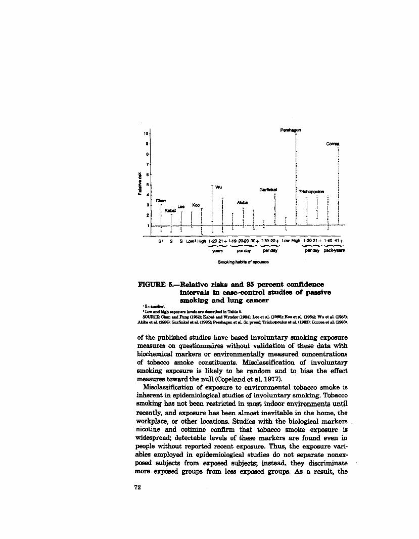
TABLE 9.-Results from selected prospective and cast+ control studies; lung cancer risk associated with spowes’ smoking
' Numbers in parentheses am the 95 percent confidence Ii&e. =Totd erpmre from spooma, cobabitante, coworkem * Husband smoked 5 16 cigarettes/day or 1 pack (50 g) of pipe tohncmhmek or any amount duriDg < 30 years of
manillge. ‘Husband smoking > 15 cigarettes/day or 1 pack of pipe tobacco/week during 2 30 years ofmarriaga.
71
IO.
8.
a,
7.
x 6. g5 = 4
3 ,'
of the published studies have baaed involuntary smoking exposure measures on questionnaires without validation of these data with biochemical markers or environmentally measured concentrations of tobacco smoke constituents. M isclassification of involuntary emoking exposure is likely to be random and to bias the effect measures toward the null (Copeland et al. 1977).
M isclassification of exposure to environmental tobacco smoke is inherent in epidemiological studies of involuntary smoking. Tobacco smoking has not been re&icted in most indoor environments until recently, and exposure has been almost inevitable in the home, the workplace, or other locations. Studies with the biological markem nicotine and cotinine confirm that tobacco smoke exposure is widespread; detectable levels of these markers are found even in people without reported recent exposure. Thus, the exposure vari- ables emp loyed in epidemiological studies do not separate nonex- posed subjects from exposed subjeds; instead, they discriminate more exposed groups from less exposed groups. As a result, the
72
epidemiological approach is conservative in estimating the effects of involuntary smoking. A truly nonexposed but otherwise equivalent comparison population has not been identified. The extent of the resulting bias cannot be readily estimated and probably varies with the exposure under consideration, which may be one reason for the variability in risk estimates obtained by different studies.
Information bias is an added concern in cas+control studies, since neither interviewer nor respondent bias can be ruled out. It is not feasible to blind interviewers to the case or control status of respondents because of the usually obvious manifestations of lung cancer and because of the setting in which some of the interviews are conducted. Moreover, blinding of interviewers and respondents to the study hypothesis is difficult because the majority of questions are concerned with exposure to tobacco smoke. The direction of the information bias may be dependent on the type of respondent. Self- respondents may be more apt to interpret their d&ease as related to exposure to tobacco smoke and thus overreport the exposure. However, the direction of the information bias is less clear when interviews are conducted with surrogate respondents. The ability of a surrogate to provide accurate information may depend on the relationship of the surrogate respondent to the subject, whether the surrogate lived with the subject during the time frame of the questions asked, the degree of detail requested, and the amount of time elapsed since the event in question @or&s 1982; Pickle et al. 1983; Lerchen and Samet 1986). Surrogate respondents may mini- mize the reporting of their own smoking because of guilt, or may overreport about involuntary smoking exposure in an attempt to explain their relative’s illness. Thus, depending on the direction of the information bias, it may dilute or strengthen the effect being. measured (Sackett 1979). In general, however, the information on smoking status and on amount smoked provided by surrogatea has been found to be fairly comparable to that provided by the individuals themselves (Blot and McLaughlin 1985).
F’inally, participants and nonparticipants in case-control studies may be inherently different with respect to their exposure to involuntary smoking because their awareness of the hypothesis under study may motivate the decision to participate. However, participants in cas+control studies are generally not informed of the hypothesis under study.
Spousal Exposure: Prospective Studies
The Japanese Cohort Study Hirayama (1981a, 1983, 1984a) has presented data from a large
cohort study that included 91,540 nonsmoking married women who were residents of 29 health districts in Japan. Subjects were 49 years
73
of age or older at enrollment in 1965; infOrm&iO~ W88 collected on smoking and drinking habits, diet (e.g., green-yellow vegetables, meat), occupation, and other health-related variables.
me initial report on invohmtary smoking ~88 baaed on 14 gears of f&owup (lg6&1979). The husbands’ smoking histories were avail- able for 174 of 240 lung cancer cases identified among the non- smoking &ed women (Hirayama 1981a); this number increased to 2~ with 2 additional years of followup -yama 1983, 1984a). &db p&thing to the association of spouses’ lung cancer risk with the husbands’ smoking were essentially identical in the first and second reports.
On the basis of the smoking habits of the husbands at entry, the 206 nonsmoking women were classified as married to a nonsmoker, an ex-smoker, or a current smoker. The lung cancer mortality ratios &d&jzed by husband’s age were 1.90,1.36,1.42,1.58, and 1.91 for women whose husbands were nonsmokers, ex-smokers, and daily smokers of 1 to 14,15 to 19, and 20 or more cigarettes, respectively (one-sided p for trend, 0.002). Similarly sign&ant dose-response trends were observed when the mortality ratios were standardized by age of the wives, by occupation of the husbands (agricultural, industrial, other), by age and occupation of the husbands, and by the time period of observation (19661977 versus 1978-1981). The risk of lung cancer &ong nonsmoking wives of smokers was reduced to 0.7 (two-sided p=O.O5) if they ate green-yellow vegetables daily com- pared with 1.0 if they ate such vegetables less often than daily (Hirayama 1984b). No other characteristic of the wives (e.g., drinking habits, parity, occupation, nonvegetahle dietary items) or of the husbands (e.g., drinking habits) was significantly predictive of lung cancer risk.
Nonsmoking men whose wives were smokers also showed an elevated lung cancer risk. On the basis of 67 lung cancers in nonsmoking married men, the lung cancer mortality ratios were 1.00,2.14, and 2.31 if their wives had never smoked or had smoked 1 to 19 cigarettes or 20 or more cigarettes per day, respectively (one- sided p for trend, 0.023) (Hirayama 198413).
This study has been critically discussed in correspondence since its initial publication. Because a detailed breakdown of the at-risk population was not presented in the initial report, the lung cancer mortality rate was thought by some to be higher in the unmarried nonsmoking women than in the nonsmoking women marriedto smokers CRutsch 1981; Grundmann et al. 1981). This impression was clarified by the researcher (Hirayama 1981b,c,d) and shown to be the result of incorrect interpretation of data in the original paper. Other potential problems cited were sampling bias in the study cohort, misclassification in the diagnosis of lung cancer, misclassification of the nonsmoking status of wives, misclassification of involuntary
74
smoking exposure, failure to control for potential confounders, and inadequate statistical treatment of data. Each of these points of criticism is discussed below.
MacDonald (1981a,b) questioned the representativeness of the 29 health districts selected in the study cohort and suggested that,
industrial pollution, such as asbestos exposure from shipbuilding industries specific to the selected health districts, may have biased the results. However, the levels of exposure to this factor would have to coincide with the husbands’ smoking level to explain the effect observed. Such an association seems unlikely. If the cohort were not representative, the generalizibility but not the validity of the findings would be challenged (Criqui 1979).
The accuracy of the diagnosis of primary lung cancer on the basis of death certificates and the adequacy of the data without informa- tion on the histology of the tumor were questioned (Grundmann et al. 1981; MacDonald 1981a). From a sample of 23 cases, Hirayama (1981b) reported that the distribution by histology of lung cancer in nonsmoking women whose husbands smoked was similar to that in women who smoked. Failure to discriminate in some cases between primary and metatastic lesions to the lung may be a potential problem with disease diagnosis. Although Hirayama was unable to assess the accuracy of the diagnosis listed on the death certificate, there is no reason to believe that error in recording the causes of death of wives was influenced by the smoking habits of their husbands, and any m isclassification is likely to be random. Inclusion of nonlung cancer cases would tend to bias the risk ratio toward unity or no effect (Barron 1977; Greenland 1980).
The relatively high risks observed for nonsmokers whose husbands smoked led to speculation that Japanese women may report them- selves as nonsmokers when they actually smoke (Lehnert 1934). However, some assurance of the reliability of the smoking data provided by the Japanese women comes from an investigation in Hiroshima and Nagasaki (Akiba et al. 1986) that found strong concordance between smoking status reported by the women them- selves and that reported by their next of km.
Classifying nonsmoking women solely on the basis of the smoking habits of their current husbands probably does not quantify their exposure with precision because it accounts for only one of the many possible sources of tobacco smoke exposure. Moreover, using the number of cigarettes smoked per day by the husbands as a measure of exposure dose assumes that the husbands’ increasing daily cigarette consumption is directly related to an increasing ETS exposure of the wives (Kornegay and Kastenbaum 1981; Lee 1982b).
The analyses were further criticized for not accounting for potential confounding factors such as socioeconomic status (SES) and exposure to indoor air pollutants (e.g., from heating and cooking