Department of Dermatology, Allergology and Venereology, Institute of Clinical Medicine, Helsinki University Central Hospital, University of Helsinki TACROLIMUS OINTMENT FOR LONG-TERM TREATMENT OF ATOPIC DERMATITIS Johanna Mandelin ACADEMIC DISSERTATION To be publicly discussed with the permission of the Faculty of Medicine of the University of Helsinki in the auditorium of the Skin and Allergy Hospital, Meilahdentie 2, Helsinki, on March 26, 2010, at 12 noon.
Transcript
Department of Dermatology, Allergology and Venereology, Institute of Clinical Medicine,
Helsinki University Central Hospital,University of Helsinki
Tacrolimus oinTmenT for long-Term TreaTmenT
of aTopic dermaTiTis
Johanna mandelin
academic disserTaTion
To be publicly discussed with the permission of the Faculty of Medicine of the University of Helsinki in the auditorium of the Skin and Allergy Hospital,
Meilahdentie 2, Helsinki, on March 26, 2010, at 12 noon.
supervised by
Docent Sakari Reitamo, M.D., Ph.D.Department of Dermatology, Allergology and and Venereology,Institute of Clinical Medicine,University of HelsinkiHelsinki, Finland
reviewed by
Docent Antti Lauerma, M.D., Ph.D.Control of Hypersensitivity DiseasesFinnish Institute of Occupational HealthHelsinki, Finland
Docent Erkka Valovirta, M.D., Ph.D. Department of Clinical AllergologyUniversity of TurkuTurku, Finland
official opponent
Professor Thomas Bieber, M.D., PhDDepartment of Dermatology and AllergyUniversity of BonnBonn, Germany
The cover shows the tacrolimus molecule.
ISBN 978-952-92-6977-8 (paperback) ISBN 978-952-10-6119-6 (pdf)http://ethesis.helsinki.fi Helsinki University PrintHelsinki 2010
To Erik, Isabella, and Bianca
4
TaBle of conTenTs
LIST OF ORIGINAL PUBLICATIONS ...................................................................... 6ABBREVIATIONS AND DEFINITIONS ................................................................... 7ABSTRACT ..................................................................................................................... 8REVIEW OF THE LITERATURE ............................................................................. 101. Atopic dermatitis ...................................................................................................... 10 1.1 Diagnosis and clinical features ............................................................ 10 1.2 Epidemiology ......................................................................................... 12 1.3 Genetics .................................................................................................. 13 1.4 Skin barrier function ............................................................................ 14 1.5 Pathogenesis ........................................................................................... 16 1.5.1 Cell-mediated immunity ............................................................. 19 1.5.2 Immunoglobulin E ...................................................................... 20 1.6 Environmental factors ......................................................................... 202. Relationship of atopic dermatitis, atopic sensitisation, and atopic airway disease ........................................................................... 23 2.1 Atopic sensitisation ............................................................................... 23 2.2 Allergic rhinitis ..................................................................................... 23 2.3 Asthma ................................................................................................... 243. Topical treatment modalities in atopic dermatitis .............................................. 26 3.1 Topical corticosteroids ......................................................................... 26 3.1.1 General properties and mechanism of action .......................... 26 3.1.2 Efficacy .......................................................................................... 27 3.1.3 Safety ............................................................................................. 27 3.1.4 Pharmacokinetics ........................................................................ 28 3.2 Tacrolimus ointment ............................................................................ 28 3.2.1 General properties and mechanism of action ......................... 28 3.2.2 Efficacy in short-term studies .................................................... 29 3.2.3 Efficacy in long-term studies ...................................................... 30 3.2.4 Clinical studies comparing topical corticosteroids and tacrolimus ointment ............................................................ 31 3.2.5 Safety ............................................................................................. 32 3.2.6 Pharmacokinetics ........................................................................ 33 3.3 Pimecrolimus cream ............................................................................ 34AIMS OF THE STUDY .............................................................................................. 35
5
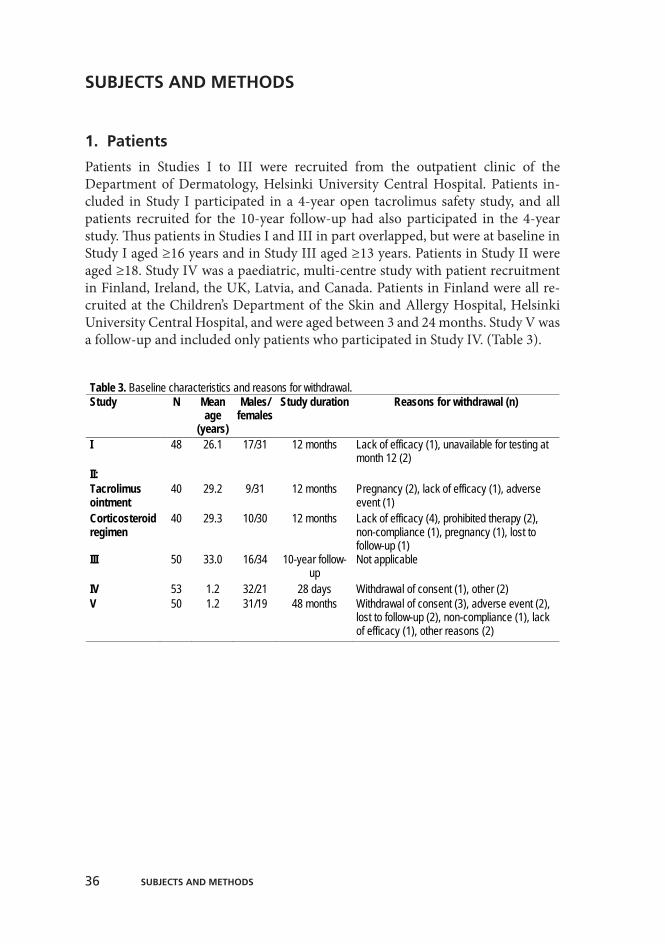
SUBJECTS AND METHODS ................................................................................... 361. Patients ..................................................................................................................... 36 1.1 Inclusion and exclusion critera .......................................................... 37 1.2 Concomitant medication .................................................................... 392. Study designs and protocols .................................................................................. 40 2.1 Medication ............................................................................................ 41 2.2 Study schedules .................................................................................... 41 2.3 Efficacy and safety assessments .......................................................... 42 2.4 Transepidermal water loss .................................................................. 44 2.5 Recall antigen testing .......................................................................... 44 2.6 Serum IgE and skin prick testing ....................................................... 44 2.7 Respiratory symptoms and findings .................................................. 45 2.7.1 Questionnaire .............................................................................. 45 2.7.2 Histamine challenge test ............................................................ 45 2.8 Tacrolimus pharmacokinetics ............................................................ 453. Statistical methods .................................................................................................. 46RESULTS ...................................................................................................................... 471. Clinical efficacy ....................................................................................................... 472. Safety ........................................................................................................................ 483. Transepidermal water loss ..................................................................................... 494. Recall antigen testing .............................................................................................. 495. Serum IgE and skin prick testing ......................................................................... 526. Respiratory symptoms and findings ..................................................................... 53 6.1 Respiratory symptoms ......................................................................... 53 6.2 Histamine challenge test ..................................................................... 537. Tacrolimus pharmacokinetics ............................................................................... 53DISCUSSION .............................................................................................................. 551. Long-term efficacy .................................................................................................. 552. Long-term safety ..................................................................................................... 563. Structural and functional effects on the skin barrier ......................................... 574. Effects on atopic airway disease ............................................................................ 585. Ideal long-term treatment of atopic dermatitis ................................................... 59SUMMARY AND CONCLUSIONS ........................................................................ 61ACKNOWLEDGEMENTS ....................................................................................... 62REFERENCES ............................................................................................................. 64 Appendix: ORIGINAL PUBLICATIONS
6 lisT of original puBlicaTions
lisT of original puBlicaTions
This thesis is based on the following original publications referred to in the text by their Roman numerals.
Mandelin J, Remitz A, Virtanen H, Reitamo S. Recall antigen reactions in patients with atopic dermatitis treated with tacrolimus ointment for 1 year. J Allergy Clin Immunol 121:777-9, 2008
Mandelin J, Remitz A, Virtanen H, Reitamo S. One-year treatment with 0.1% tacrolimus ointment versus a corticosteroid regimen in adults with moderate to severe atopic dermatitis: a randomized, double-blind, compar-ative trial. Acta Derm Venereol 90:170-4, 2010
Mandelin JM, Remitz A, Virtanen HM, Malmberg LP, Haahtela T, Reitamo S. A 10-year open follow-up of eczema and respiratory symptoms in pa-tients with atopic dermatitis treated with topical tacrolimus for the first 4 years. J Dermatol Treat, in press
Reitamo S, Mandelin J, Rubins A, Remitz A, Mäkelä M, Cirule K, Rubins S, Zigure S, Ho V, Dickinson J, Undre N. The pharmacokinetics of tacrolimus after first and repeated dosing with 0.03% ointment in infants with atopic dermatitis. Int J Dermatol 48:348-55, 2009
Mandelin JM, Rubins A, Remitz A, Cirule K, Dickinson J, Ho V, Mäkelä MJ, Rubins S, Reitamo S, Undre N. Long-term efficacy and tolerability of tacrolimus 0.03% ointment in infants: a 2-year open-label study. Submitted
Reprinted here with permission of the publishers.
I.
II.
III.
IV.
V.
7aBBreViaTions and definiTions
aBBreViaTions and definiTions
AD Atopic dermatitisAPT Atopy patch testAUC Area under the curveBHR Bronchial hyper-responsiveness BSA Body surface areaCLA Cutaneous lymphocyte antigenDTH Delayed-type hypersensitivityEASI Eczema Area and Severity IndexFcεRI High-affinity receptor for IgE type IFEV1 Forced expiratory volume in 1 secondFKBP FK-binding proteinGM-CSF Granulocyte-macrophage colony-stimulating factorHPLC High-pressure liquid chromatographyIDEC Inflammatory dendritic epidermal cellIFN InterferonIGA Investigator’s global assessmentIgE Immunoglobulin EIL InterleukinLC Langerhans cellLEKTI Lymphoepithelial kazal type-5 serine protease inhibitor mEASI modified Eczema Area and Severity IndexOR Odds ratioPD15FEV1 Provocative dose of inhaled histamine producing a 15% FEV1 decrease PINP/ PIIINP Aminoterminal propeptide of type I/ III procollagenS. aureus Staphylococcus aureusSPT Skin prick testTEWL Transepidermal water lossTGF Transforming growth factor Th T helperTLR Toll-like receptorTNF Tumour necrosis factorTSLP Thymic stromal lymphopoietinUV UltravioletVAS Visual analogue scale
8 aBsTracT
aBsTracT
Atopic dermatitis (= atopic eczema) is a common skin disease characterised by eczema, a superficial inflammation of the skin, and impaired function of the epidermal barrier. Patients also are at an increased risk for asthma and allergic rhinitis. Development of these atopic diseases in childhood is often referred to as the “atopic march”. As atopic dermatitis is a chronic disease, treatment modalities must be planned long-term. Treatment with tacrolimus ointment has shown good efficacy and safety in long-term studies in adults and in children over 2 years old. Since topical tacrolimus treatment targets the T cells in the skin, long-term safety in terms of skin infections and skin cancer has been of special interest.
The aim of this thesis was to further study the long-term efficacy and safety of treatment with tacrolimus ointment in atopic dermatitis, also in comparison to topical corticosteroid treatment. Two studies focused on cell-mediated immunity of the skin and one of them in addition on epidermal barrier function. Respiratory symptoms in patients with atopic dermatitis initially treated long-term with tac-rolimus ointment were evaluated in a 10-year follow-up study. Effective treatment of atopic dermatitis is especially important in infants to minimise sensitisation through the skin and thus possibly stop the atopic march, so two studies in chil-dren under 2 years of age investigated the pharmacokinetics, safety, and efficacy of their treatment with 0.03% tacrolimus ointment.
Adults or adolescents with moderate-to-severe atopic dermatitis were evaluated in three clinical studies and infants with atopic dermatitis in two clinical studies. In Study I, 48 patients, participating in a 4-year open safety study of 0.1% tacrolimus ointment, were tested for recall antigen reactivity at baseline and after 12 months of treatment. In addition, 28 healthy controls were tested. Study II was a long-term, double-blind study comparing the safety and efficacy of treatment with 0.1% tac-rolimus ointment to that of a corticosteroid regimen in 80 patients. Recall antigen reactivity, transepidermal water loss and serum IgE concentrations were tested at baseline and after 6 and 12 months of treatment. Study III was a 10-year follow-up of respiratory symptoms in 50 patients participating in the 4-year, open tacrolimus ointment safety study. Data on bronchial hyper-responsiveness, respiratory symp-toms, total serum IgE, and skin prick tests were collected at baseline and at the follow-up visit. Study IV was a 2-week pharmacokinetic study in 53 infants, 3 to 24 months of age, with atopic dermatitis treated with 0.03% tacrolimus ointment once or twice daily. Full pharmacokinetic profiles for tacrolimus blood concentra-tions were obtained at days 1 and 14. Study V investigated the efficacy and safety of 0.03% tacrolimus ointment in 50 infants treated for 2 years.
9aBsTracT
Long-term treatment with tacrolimus ointment showed good efficacy and did not result in any safety problems in adults, nor in infants with atopic dermati-tis. Treatment with 0.1% tacrolimus ointment for one year enhanced recall an-tigen reactivity in patients with atopic dermatitis almost to that of healthy con-trols. Treatment with either 0.1% tacrolimus ointment or a corticosteroid regi-men enhanced recall antigen reactivity after 12 months of treatment in a similar way. Transepidermal water loss, an indicator of skin barrier function, decreased at month 12 to approximately half its baseline value (P≤0.001) in both treatment groups. In patients with more than 60% improvement of the entire affected body surface area at month 12, serum IgE levels decreased from 666 at baseline to 584 at month 12 (P=0.02). Patients in the 10-year, open follow-up study showed a decrease in affected body surface area from a baseline 19.0% to a 10-year 1.6% (P<0.0001), and those with bronchial hyper-responsiveness at baseline showed an increase (P=0.02) in the provocative dose of inhaled histamine producing a 15% decrease in FEV1, indicating less hyper-responsiveness. A decrease in respiratory symptoms occurred in patients with active symptoms at baseline. A good treat-ment response (≥60% improvement in eczema) after one year of treatment with tacrolimus ointment predicted a good treatment response throughout the 10-year follow-up and a decrease in total serum IgE levels at the 10-year follow-up visit. The 2-week pharmacokinetic and the long-term safety study with 0.03% tacroli-mus ointment showed good and continuous improvement of the eczema in the infants. Tacrolimus blood levels were throughout the study low and over time de-creasing. Treatment was well tolerated.
This thesis underlines the importance of effective long-term topical treatment of atopic dermatitis. When the active skin inflammation, with a Th2 cell dominance, decreases, cell-mediated immunity of the skin improves, and a secondary marker for Th2 cell reactivity, total serum IgE, decreases. Respiratory symptoms seem to improve when the eczema area decreases, but the lack of any control group in our study makes it difficult to evaluate the role of the initial 4-year intervention with tacrolimus ointment. All these effects can be attributed to improvement of skin barrier function, indicated by a decrease in transepidermal water loss. One potential method to prevent a progression from atopic dermatitis to asthma and allergic rhinitis may be avoidance of early sensitisation through the skin, so early treatment of atopic dermatitis in infants is crucial. Long-term treatment of atopic dermatitis with 0.03% tacrolimus ointment was effective and safe in infants over age 3 months.
10
reVieW of THe liTeraTure
1. atopic dermatitis
1.1 diagnosis and clinical features
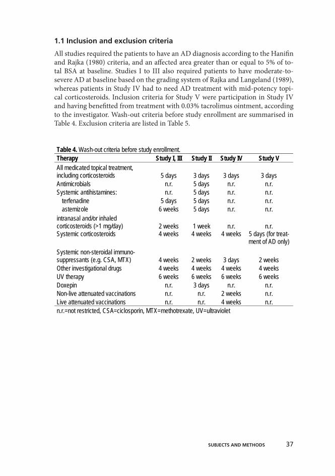
Atopic dermatitis (AD) is a chronic, relapsing, inflammatory skin disease which of-ten appears in conjunction with other atopic diseases like atopic allergies, asthma, and allergic rhinitis. The diagnosis is clinical with no specific diagnostic clinical sign or laboratory test. In 1980, Hanifin and Rajka listed the main clinical features of AD, and these criteria are those most often referred to and most widely applied in clinical studies when setting the diagnosis of AD (Table 1). In epidemiological studies, the more simplified criteria of the UK Working Party are the most widely applied. Due do these criteria, a diagnosis of AD requires a pruritic skin disease plus three or more of the following: history of involvement of the flexural regions, history of asthma or hay fever, history of generally dry skin, onset at less than 2 years of age, and visible flexural dermatitis (Williams et al 1994). Typical and mandatory for AD is the intense itch, an important cause of reduced quality of life in patients and their families. The majority of AD patients have increased levels of peripheral blood eosinophils and serum immunoglobulin E (IgE), and serum IgE levels show a high correlation with disease severity (Laske & Niggemann 2004, Clendenning et al 1973). Because some AD patients have normal serum IgE levels and no signs of sensitisation to allergens, AD can be divided into two forms: the extrinsic and the intrinsic. The extrinsic form affects 70 to 80% of patients, who show elevated serum IgE levels and sensitisation towards common allergens, while the intrinsic form affects 20 to 30%, who have normal serum IgE levels, no specific IgE to common allergens, and negative skin prick tests (Novak & Bieber 2003). The need for and validity of this type of classification has been debated (Williams & Flohr 2006), since IgE-mediated sensitisation may be only a transient factor. This is considered in a recently presented model which suggests that the natural history of AD has three consecutive phases: nonatopic, atopic, and autoallergic (Bieber 2008).
Skin lesions in the acute and sub-acute phases of AD are characterised by itch-ing, erythematous papules, excoriations, and serous exudates. Chronic lesions are characterised by dryness, scaling, lichenification, papules, and excoriations. Histopathology in AD shows epidermal intercellular oedema, and infiltration of lymphocytes, macrophages, and dendritic cells around blood vessels. In licheni-fied eczema, the epidermis is thickened and the upper layer hypertrophied. The normal-looking skin of AD patients is not normal but shows a subclinical inflam-matory infiltrate (Mihm et al 1976) and impaired barrier function (Proksch et al 2006).
Localisation of the eczema varies with age. In infancy (under 1 year) lesions are typically on the cheeks, scalp, trunk, and extensor sides of the extremities. In child-
reVieW of THe liTeraTure
11
hood (1-4 years) lesions can still be on the extensor sides of the extremities, but also in the flexures. In addition, face, neck, and hands can be affected. In ado-lescents (4-16 years) the eczema is usually symmetrically distributed on the face, flexural areas, hands, feet, and sometimes back of the thighs. Beginning from pu-berty and continuing into adulthood, lesions are typically on the face, upper body, flexures, and hands. Chronic hand eczema can be the main manifestation in many adults with AD.
The common clinical term “atopic skin diathesis” includes atopic skin features such as dry skin, hyperlinear palms and soles, orbital darkening, winter feet, and nipple eczema (Wütrich & Schmid-Grendelmeier 2002). Persons with atopic skin diathe-sis are at risk of developing occupational skin disease, and evidence supports the importance of recognizing this as part of an occupational skin disease-prevention strategy (Dickel et al 2003).
11
and continuing into adulthood, lesions are typically on the face, upper body, flexures, and hands. Chronic hand eczema can be the main manifestation in many adults with AD.
The common clinical term “atopic skin diathesis” includes atopic skin features such as dry skin, hyperlinear palms and soles, orbital darkening, winter feet, and nipple eczema (Wütrich & Schmid-Grendelmeier 2002). Persons with atopic skin diathesis are at risk of developing occupational skin disease, and evidence supports the importance of recognizing this as part of an occupational skin disease-prevention strategy (Dickel et al 2003).
Table 1. The Hanifin and Rajka criteria for diagnosis of AD (1980).
Must have three or more basic features: 1. Pruritus 2. Typical morphology and distribution:
Flexural lichenification or linearity in adults Facial or extensor involvement in infants and children
3. Chronic or chronically relapsing dermatitis 4. Personal or family history of atopy (asthma, allergic rhinitis, atopic dermatitis)
Plus three or more minor features: 1. Xerosis 2. Ichtyosis/ palmar hyperlinearity/ keratosis pilaris 3. Immediate (type I) skin test reactivity 4. Elevated serum IgE 5. Early age of onset 6. Tendancy toward repeated cutaneous infections (especially Staphylococcus aureus
and Herpes simplex)/ impaired cell-mediated immunity 7. Tendancy towards nonspecific hand or foot dermatitis 8. Nipple eczema 9. Cheilitis 10. Recurrent conjunctivitis 11. Dennie-Morgan infraorbital fold 12. Keratoconus 13. Anterior subcapsular cataracts 14. Orbital darkening 15. Facial pallor/ facial erythema 16. Pityriasis alba 17. Anterior neck folds 18. Itch when sweating 19. Intolerance to wool and lipid solvents 20. Perifollicular accentuation 21. Food intolerance 22. Course influenced by environmental or emotional factors 23. White dermographism/ delayed blanch
reVieW of THe liTeraTure
12
1.2 epidemiology
AD is a common disease with a lifetime prevalence of 10 to 20% in children and a point prevalence of 1 to 3% in adults (Schultz Larsen & Hanifin 2002). In Finnish young men, the prevalence of AD in 2003 was 1.2%, which, compared to its prevalence in 1966, is an over 6-fold increase (Latvala et al 2005). In a worldwide cross-sectional study known as the International Study of Asthma and Allergies in Childhood (ISAAC) the 12-month prevalence in 13- to 14-year-old children ranged from 0.9% in China to 15.6% in Finland, and to 21.1% in Bolivia (Odhiambo et al 2009). The ISAAC study also showed that the prevalence of AD during the last 5 to 10 years is levelling off or decreasing in some countries with previously high prevalences, while many previously low-prevalence developing countries have ex-perienced clear increases. No single environmental factor can explain the increase, and various risk factors, such as family size, hygiene, allergens, and changes in mi-crobial environment are likely to be important in different countries (Williams et al 2008). In general, a two-to-three fold increase in AD occurred in industrialised countries during the last 30 years, with higher prevalences in urban than in ru-ral regions. Agricultural regions, such as China, eastern Europe, and rural Africa show clearly lower prevalences (Taylor et al 1984). AD also seems to increase with higher socioeconomic status (Wolkewitz et al 2007).
In 60% of the patients, eczema starts during the first year of life, and in 85% before age 5 (Kay et al 1994). In children with early (under age 2) manifestation of AD, 43% were in complete remission after age 2. However, at age 7 nearly 20% had persistent symptoms and 38% intermittent symptoms (Illi et al 2004). In early ado-lescence, about 60% of the children with AD will be symptom-free (Rystedt 1985), although up to half of them may have a relapse in adulthood (Lammintausta et al 1991). Early-onset disease, severe early disease, concomitant asthma and allergic rhinitis, and a family history of AD may predict a more persistent course (Williams & Wüthrich 2000).
Parental and especially maternal AD is a major risk factor for AD. Another ma-jor risk factor, which also could partly explain the increase in prevalence, is the western life-style, which generally leads to smaller families, better vaccination programs, and more frequent use of antibiotics (von Mutius 2000). The “hygiene hypothesis” suggests that infections are required during the first months of life in an infant to switch the intrauterine T helper (Th) type 2 cell dominance towards a non-atopic Th1 cell dominance; in atopic infants, this sequence is thought to fail (Strachan 1989). At present, some data do not support a clear inverse relationship between infections and risk for AD (Williams & Flohr 2006). Other risk factors may include maternal smoking and early respiratory syncytial virus infections (Lau et al 2000).
reVieW of THe liTeraTure
13
Studies of prevention of AD lack sufficient evidence for maternal dietary restric-tions during pregnancy or lactation. For infants at high risk for atopic disease, exclusive breastfeeding for at least 4 months compared with feeding intact cow milk protein formula seems to reduce the cumulative incidence of AD (Greer et al 2008). Prevention studies with probiotics show variable results, which at least in part can be explained by the use of differing probiotic bacteria and study sched-ules. A study by Taylor and co-workers (2007) showed no reduction in AD risk in infants of 1 year of age who received probiotics for their first 6 months. In contrast, a large study in infants at high risk for allergy showed a reduced risk for AD by the age of 2 (Kukkonen et al 2007), but at 5 years of age a reduced risk appeared in only a very small subgroup, i.e., for IgE-associated eczema in cesarean-delivered children (Kuitunen et al 2009).
Scientific rationale implies that simple means, such as consuming farm milk and spending time in nature, can endorse tolerance to environmental antigens and reduce risk of atopy. This has recently been summarised in the Finnish Allergy Programme 2008-2018 (von Hertzen et al 2009). 1.3 genetics
AD results from a complex interplay between genetic and environmental factors. The genetic factors are strong, as can be seen in twin studies where the concord-ance rate in monozygotic twins has been 72% compared with a rate of 23% in dizygotic twins (Larsen et al 1986, Schultz Larsen 1993). The presence of eczema-specific genes is also supported by epidemiological studies which show that pa-rental eczema constitutes a higher risk for eczema in the infant than does parental asthma or allergic rhinitis (Dold et al 1992). The disease risk of an infant seems more often to be related to maternal than paternal disease status (Dold et al 1992, Ruiz et al 1992, Diepgen & Blettner 1996). This could be due to genomic imprint-ing, mitochondrial transmission, and gene-environment interactions involving the environment in the uterus or exposure to immunologic and nutritional factors of breast milk or both (Morar et al 2006).
Genes of the epidermal differentiation complex are located on chromosome 1q21. The filaggrin gene on chromosome 1q21.3 encodes filaggrin, a key protein in epi-dermal differentiation. Among European patients with AD, a nonfunctional mu-tation of the filaggrin gene occurs in about 30% (Palmer et al 2006, Weidinger et al 2006, Marenholz et al 2006), whereas around 40% of all carriers of the filaggrin gene-null alleles will have no eczema (Henderson et al 2008). It therefore seems likely that filaggrin mutations cause a defective skin barrier, which in conjunc-tion with additional genetic and environmental factors in many of the carriers, results in eczema. In European AD patients, six recurrent filaggrin gene mutations appear, plus several other family-specific mutations. The two most common are R501X and 2282del4 (Palmer et al 2006); AD patients with one of these are more
reVieW of THe liTeraTure
14
likely to have asthma, a persistent course of the disease, and atopic sensitisations (Marenholz et al 2006, Henderson et al 2008). Loss-of-function mutations of the gene from both parents is the cause of ichtyosis vulgaris, the most common inher-ited disorder of keratinisation and one of the most common single-gene disorders in humans (Smith et al 2006).
Genome-wide linkage studies have provided evidence of AD-related loci on chro-mosome 3q21, containing the COL29A1 gene, which encodes a new epidermal collagen XXIX (Söderhäll et al 2007). Other AD-related loci occur on chromo-some 17q25, which contains the keratin type I gene cluster genes, and on 20p and 16q, which have been mapped by combining phenotypes, such as AD and asthma or AD and total serum IgE-level (Cookson et al 2001). The gene which encodes the β subunit of the high-affinity receptor for IgE (FcεRI) is located on chromosome 11q12-13, found in a region with confirmed linkage to atopy.
On chromosome 5q31-33, a cytokine gene cluster has been identified encoding mediators involved in the immune response, such as interleukin (IL)-4, IL-5, IL-12, IL-13, and granulocyte-macrophage colony-stimulating factor (GM-CSF) (Morar et al 2006). This region also includes the SPINK5 gene, which encodes the lymphoepithelial kazal type-5 serine protease inhibitor (LEKTI), which inhibits two serine proteases: stratum corneum tryptic enzyme (kallikrein 5), and stra-tum corneum chymotryptic enzyme (kallikrein 7) (Egelrud et al 2005). Patients with Netherton syndrome, a rare disease with severe atopic manifestations, carry mutations in the SPINK5 gene on both alleles (Sprecher et al 2001). They show marked barrier dysfunction, with altered desquamation and impaired keratinisa-tion (Comel 1949). Several studies have linked mutations in the SPINK5 gene with AD, when maternally inherited (Walley et al 2001, Kato et al 2003, Nishio et al 2003, Weidinger et al 2008).
1.4 skin barrier function
The epidermal barrier, found in the lower layers of the stratum corneum, serves as the first-line defense against invading pathogens and allergens. It is composed of differentiated keratinocytes, corneocytes, which are held together with cor-neodesmosomes. Lipid lamellae, composed of ceramides, cholesterol, fatty acids, and cholesterol esters (Rawlings 2003), surround the corneocytes, providing a water-resistant layer which protects them from water loss (Lavker 1976). The cor-neocytes contain natural moisturising factor, a breakdown product of filaggrin, which helps them to store water and subsequently swell. This in turn prevents gaps between them and makes the stratum corneum resistant to the penetration of allergens (Figure 1) (Cork et al 2009). Filaggrin is, together with other structural proteins, part of the cornified envelope which strengthens the corneocytes and helps lipids attach to them (Elias & Menon 1991). Filaggrin is of special impor-tance, because it makes the corneocytes collapse into flattened discs with a large surface area (Steinert et al 1981).
reVieW of THe liTeraTure
15
Figure 1. Structural of the epidermal barrier. Corneocytes (green) are surrounded by lipid lamellae (yellow) and held together by corneodesmosomes (blue). Adapted from Cork et al 2008.
Transepidermal water loss (TEWL) can be measured by a special device (Pinnagoda et al 1990) and is widely used as a marker of skin barrier function. TEWL varies at different body sites, due to differences in skin thickness and in numbers of sweat glands. Patients with AD show dysfunction of the epidermal barrier, which can be seen as a two- to fivefold increase in TEWL, even in non-lesional skin. The degree of barrier impairment correlates with the phase of the disease – acute, subacute, and chronic – and with disease severity (Seidenari & Giusti 1995, Shahidullah et al 1969).
The skin of AD patients, both those with and without the filaggrin mutation, shows decreased levels of filaggrin (Seguchi et al 1996). Patients with a filaggrin mutation show lower levels of filaggrin in acute lesional than in non-lesional skin. Overexpression of Th2 cytokines downregulates filaggrin expression during the differentiation process of the keratinocytes (Howell et al 2007), which explains the filaggrin deficiency seen in AD patients without the filaggrin mutation. Reductions in filaggrin levels lead to reduced levels of natural moisturising factor, which is important for skin hydration, has strong antimicrobial effects, and decreases skin colonisation of pathogenic bacteria (Cork et al 2009).
AD patients have significantly elevated pH of both lesional and non-lesional skin compared with that in normal controls (Eberlein-König et al 2000, Seidenari & Giusti 1995). A sustained increase in pH elevates the activity of degradatory pro-teases and reduces the activity of the lipid synthesis enzymes. The activity of pro-teases such as the stratum corneum tryptic (kallikrein 5) and chymotryptic (kal-likrein 7) enzymes is strictly regulated by protease inhibitors (Suzuki et al 1994). LEKTI is a particularly important protease inhibitor and regulator of desquama-tion, the inhibitory potential of which is reduced as the skin pH becomes more
reVieW of THe liTeraTure
16
acidic (Deraison et al 2007). Lack of kallikrein 5 inhibition by LEKTI in Netherton syndrome initiates thymic stromal lymphopoietin (TSLP) expression and triggers AD-like lesions, without any contribution from the environment or adaptive im-mune system (Briot et al 2009).
The outside-inside hypothesis suggests that barrier dysfunction can raise the activ-ity of AD, by allowing antigens such as pollen and enterotoxins to penetrate the disrupted skin barrier. In contrast, the inside-outside hypothesis suggests that bar-rier breakdown in AD is secondary to the inflammatory response to irritants and allergens (Cork et al 2009).
1.5 pathogenesis
The initial cascade resulting in skin inflammation in AD is unknown. It may start with scratching, due to itching, irritation, or neuropeptides, leading to a release of pro-inflammatory cytokines from the keratinocytes, or may start with T cell reac-tions towards allergens in the skin which have been able to penetrate the disrupted skin barrier (Bieber 2008).
Both innate and adaptive immunity play roles in the pathogenesis of AD. The in-nate (non-adaptive) immune responses are non-specific, whereas the adaptive im-mune responses are highly specific for a particular pathogen. Innate responses do not alter during repeated exposure, whereas adaptive responses strengthen with each successive encounter with the target. T and B cells are central to all adaptive immune responses, since they exhibit the key features, specificity and memory, of adaptive immunity (Male & Roitt 1998).
Lesional skin in AD shows a dermal infiltrate of CD4+ and CD8+ T cells, in a ratio similar to that of peripheral blood. Some of the T cells in the immune system har-bour the cutaneous lymphocyte antigen (CLA), which enables them to be quickly recruited to the skin if foreign antigens penetrate the skin barrier (Trautmann et al 2001). In the skin, naïve T cells, after antigen presentation by the dendritic cells, differentiate into Th1 and Th2 cells. It is generally accepted that AD reveals a disturbance of the Th1/ Th2-cell bal-ance in the skin, with a shift towards overall Th2 dominance. The inflammation is biphasic, starting with an initial, acute Th2-predominant phase associated with increased secretion of IgE and cytokines such as IL-4, IL-5, and IL-13. This is fol-lowed by a chronic phase in which Th0 and Th1 cells are dominant, with secretion of interferon (IFN)-gamma, IL-12, IL-5, and GM-CSF (Grewe et al 1995, Taha et al 1998). The switch from the acute to the chronic phase is initiated by increased levels of IL-12 and IL-18 produced by eosinophils or inflammatory dendritic epi-dermal cells (IDECs), or both (Leung & Bieber 2003). Patients with AD commonly have elevated levels of eosinophils in serum and the skin. This is due to their in-creased production in the bone marrow, and their recruitment and chemotaxis to
reVieW of THe liTeraTure
17
the site of inflammation by Th2 cytokines together with some chemokines such as eotaxin and monocyte chemoattractant protein 4. In addition, delayed apoptosis may also contribute to tissue eosinophilia (Simon et al 1998). TSLP is an IL-7-like cytokine which in humans is expressed by keratinocytes in AD and by bronchial epithelial cells in asthmatic airways (Soumelis et al 2002, Ying et al 2005). TSLP is capable of activating CD11c+ myeloid dendritic cells to up-reg-ulate co-stimulatory molecules, leading to the differentiation of CD4+ T cells into Th2 cells. It therefore plays a key role in the development of allergic diseases such as AD or asthma (Ziegler & Liu 2006, Leonard 2002, Liu 2006). TSLP can also act directly on T cells to enhance Th2 cytokine production (He et al 2008).
A third Th cell subset, the Th17 cells, also seems to play an important role in AD by aggravation of the disease. The number of Th17 cells is increased in the pe-ripheral blood and in acute lesional AD skin. These cells produce inflammatory cytokines such as IL-17A, IL-17F, IL-22, and IL-26. IL-17 stimulates keratinocytes to produce GM-CSF, tumour necrosis factor (TNF)-α, IL-8, CXCL10, and vascular endothelial growth factor (Koga et al 2008). Subtypes of Th cells and their possible roles in AD are presented in Figure 2.
Figure 2. Cytokine profiles of various types of T helper cells. Symbols: ↑=increase or ↓=decrease in activity shown in studies cited in the text, ?=only a few studies have suggested this, necessitating further verification.
reVieW of THe liTeraTure
18
Regulatory T cells may also play a role in AD in which the circulating amount is increased (Ou et al 2004). One study of lesional atopic skin showed that the in-flammatory reaction of atopic skin was associated with an inadequate induction of tolerance due to the absence or defective function of human regulatory T cells (Verhagen et al 2006).
Langerhans cells (LCs) and IDECs are different types of antigen-presenting den-dritic cells (Wollenberg et al 1996), which in AD show increased expression of FcεRI on their surfaces, which is relevant for IgE binding (Bieber et al 1992, Klubal et al 1997, Wang et al 1992). The binding of specific IgE to the receptor leads to a 100- to 1000-fold increased antigen presentation to the Th1 and Th2 cells, and possibly also to regulatory T cells. LCs induce the production of some chemokines, but probably play a more important role in the induction and control of tolerance (Bieber 2007). In contrast, IDECs with activated FcεRIs may intensify the inflam-matory immune reaction in patients with AD by producing high amounts of pro-inflammatory cytokines and chemokines, and by contributing to the increased survival of monocytes and some antigen-presenting cells (Katoh et al 2000). The number of IDECs in the skin can be reduced by treatment with topical calcineurin inhibitors, but LCs are unaffected (Wollenberg et al 2001, Schuller et al 2004, Hoetzenecker et al 2005, Novak et al 2005). In contrast, topical corticosteroids induce apoptosis in LCs and impair their antigen-presenting capacity to T cells (Hoetzenecker et al 2004).
Patients with AD also show a variety of defects in their innate immune system that affect AD development and severity. Reduced function or migration into the skin of neutrophils, plasmacytoid dendritic, and natural killer cells occurs (Michaëlsson 1973, Novak et al 2004, Wollenberg et al 2002, Katsuta et al 2006), as well as dys-function of pattern-recognition receptors, such as Toll-like receptors (TLRs) 2 and 9 (Hasannejad et al 2007, Novak et al 2007). TLRs are key molecules involved in microbial recognition by the immune system. Recent studies suggest that a muta-tion in the TLR-2 modifies cytokine production and TLR expression in AD; as an end effect, both innate and adaptive responses in AD are modulated, which may be associated with enhanced susceptibility to skin infections with Staphylococcus aureus (Mrabet-Dahbi et al 2008, Niebuhr et al 2009).
AD patients also show decreased levels of antimicrobial peptides such as human beta-defensin-2 and -3, cathelicidin, and dermcidin. This reduced antimicrobial peptide expression is in part due to the inhibitory effects of the IL-4, -13, and -10 cytokines on the keratinocytes (Ong et al 2002, Nomura et al 2003, Howell et al 2006a). Antimicrobial peptides have antimicrobial properties and act as a link between innate and adaptive immune responses; for example, cathelicidins and some defensins have been chemoattractants for neutrophils, monocytes, and T cells (Yang et al 2001, Niyonsaba et al 2004). The defects in the innate immu-nity of patients with AD amplify their susceptibility to skin infections and make these more difficult to manage (De Benedetto et al 2009). Modulins derived from
reVieW of THe liTeraTure
19
Staphylococcus epidermidis, a normal resident of the skin, seem to selectively in-hibit the survival of pathogens, while maintaining the normal skin microbiome (Cogen et al 2010). Studies on skin microbiota will probably improve our under-standing of the cutaneous microbiota and also shift paradigms in the interpreta-tion of the roles microbes play in skin health and disease (Cogen et al 2008).
Itching is a dominant symptom of AD, and many studies have tried to identify the major pruritogen. Histamine was thought to be a major mediator of itch in AD, but the lack of effect of antihistamines contradicts this (Diepgen et al 2002). Mediators such as IL-2, GM-CSF, substance-P, acetylcholine, and prostaglandins induce itching (Ständer & Steinhoff 2002). IL-31, a T-cell-derived cytokine overex-pressed in pruritic atopic compared with non-pruritic psoriatic skin inflammation (Sonkoly et al 2006), seems to be produced mainly by Th2 cells (Neis et al 2006). In a murine model, IL-31 antibody reduced scratching, but had no effect on skin lesions (Grimstad et al 2009).
1.5.1 cell-mediated immunity
Cell-mediated immunity is a T cell-mediated defense mechanism against microbes that survive within phagocytes or that infect non-phagocytic cells. Activation of the phagocytes is dependent on Th1-produced interferon IFN-gamma, but Th17-derived IL-17 also seems to play an important role in cell-mediated immunity (Nakae et al 2002). A delayed-type hypersensitivity (DTH) skin reaction is the result of phagocytic cell activation and inflammation and can serve to assess cell-mediated immunity in vivo. DTH is commonly assessed by injecting an antigen such as tuberculin or tetanus intradermally; erythema and induration after 48 to 72 hours indicate a positive reaction. The reaction is initiated when antigens are presented by antigen-presenting cells in the skin to sensitised T memory cells. The T cell activation leads to an influx of macrophages, monocytes, and lymphocytes at the site, which produce inflammatory cytokines such as TNF-alfa, IL-17A, and IFN-gamma.
The lack of DTH to any recall antigens can indicate T cell immunodeficiency, and is termed anergy. Anergy is common in patients with sarcoidosis, rheuma-tological diseases, and severe viral infections such as influenza or mononucleosis (Jyonouchi 2005). Many studies have shown an inverse association between tuber-culin responses and atopy (Shirakawa et al 1997, Miyake et al 2008), while others have failed to do so (Grüber et al 2002). Impaired DTH reactions are evident in pa-tients with active AD compared with healthy controls (Stenger et al 1983), as well as in apparently normal patients who have had multiple skin cancers (Czarnecki et al 1995). Systemic immunosuppressive treatment with ciclosporin or with pred-nisolone and ciclosporin also inhibits DTH reactions (Ellis et al 1991, Rentenaar et al 2002).
reVieW of THe liTeraTure
20
1.5.2 immunoglobulin e
IgE synthesis is initiated by allergens which penetrate the epidermal barrier and are taken up via the FcεRIs by local antigen-presenting cells, which process and present them to Th cells. Th2 cells secrete cytokines that induce B cell proliferation and induce an allergen-specific IgE response. IgE production by B cells requires physical interaction with T cells, an interaction involving a number of surface ad-hesion molecules, as well as with IL-4 and IL-13 produced by T cells, basophils, and mast cells. Mast cells and basophils play a key role by producing inflammatory mediators, but they can also directly regulate IgE production independently of T cells (Gauchat et al 1993).
The elevated IgE levels in the majority of the AD patients are probably due to many factors, such as the disrupted epidermal barrier which allows allergens to enter the skin and the Th2 dominance which provides a cytokine milieu suitable for IgE pro-duction by B cells. Staphylococcal superantigens are able to stimulate IgE synthesis by peripheral blood mononuclear cells and this may contribute to elevated IgE levels in AD patients (Hofer et al 1995). In addition, the skin of AD patients shows higher expression of FcεRI than does the skin of healthy controls (Wollenberg et al 1995), and a correlation exists between FcεRI expression on skin dendritic cells and serum IgE levels (Kerschenlohr et al 2003).
The relevance of elevated IgE in patients with AD is not fully clarified, but many authors believe in a role for IgE in the pathogenesis of AD (Williams & Flohr 2006); this is supported by a clear correlation between disease severity and IgE levels (Laske & Niggemann 2004). Results from low-dose anti-IgE therapy (oma-lizumab) have been reported from an open study with 11 adult patients with gen-eralised AD and high levels of total IgE. Only two patients responded with more than a 50% reduction in their eczema score. Total IgE (bound and free IgE) slightly increased during therapy, whereas free IgE remained nearly stable over the treat-ment period (Belloni et al 2007). To date, no placebo-controlled studies have been published on anti-IgE therapy in the treatment of AD.
1.6 environmental factors
AD arises from an interaction between genetic and environmental factors. Factors such as bacteria and viruses, soap and detergents, stress, foods, and aeroallergens can trigger the inflammatory cascade and further break down the epidermal bar-rier (Akdis et al 2006).
In most patients with active AD, S. aureus colonises the skin (Leyden et al 1974), in contrast to colonisation in less than 5% of healthy controls. The colonisation in AD is facilitated by impaired barrier function, reduced skin lipid content, high skin surface pH, increased adherence due to increased fibronectin and fibrinogen, and decreased production of antimicrobial peptides by the keratinocytes (Leung 2008). In the majority of patients with AD, S. aureus secrets superantigens such
reVieW of THe liTeraTure
21
as enterotoxin A and B, and toxic shock syndrome toxin-1 (Nomura et al 1999). These play an important role in exacerbating AD and have induced T cell expres-sion of the CLA skin homing receptor via stimulation of IL-12 production (Leung et al 1995). They also aggravate allergen-induced skin inflammation by activating infiltrating mononuclear cells and by inducing mast cell degranulation. AD se-verity correlates with the presence of IgE against superantigens (Bunikowski et al 1999, Nomura et al 1999). S. aureus also secretes proteinases able to break down corneodesmosomes (Miedzobrodzki et al 2002, Cork et al 2009).
Patients with AD also suffer from viral skin infections such as Herpes simplex, Molluscum contagiosum, and Verruca vulgaris more often than do non-atopics. One reason for this can be the lower levels of interferon-gamma in the skin in AD patients than in healthy subjects, which can enable the overgrowth of viruses (Engler et al 2002). AD patients also show impaired skin recruitment of plasmacy-toid dendritic cells, which respond to viral infections by producing large amounts of antiviral type I IFN-alfa and -beta (Wollenberg et al 2002), and decreased levels of the antiviral antimicrobial peptide cathelicidin in lesional skin and skin affected by eczema herpeticum (Ong et al 2002, Howell et al 2006b). These changes in the AD skin probably explain its predisposition to viral skin infections.
Yeasts, especially Malassezia species, may play a role in AD pathogenesis, although study results are conflicting. Several studies show that patients with AD exclusive-ly—particularly those with head and neck dermatitis—are sensitised to Malassezia (Faergemann 2002, Allam & Bieber 2003, Roll et al 2004, Bayrou et al 2005). In two double-blind, controlled studies, topical antifungal treatment added to 1% hydro-cortisone cream has showed no benefit compared to that of hydrocortisone cream alone (Wong et al 2008, Broberg & Faergemann 1995). Systemic antifungal treat-ment in clinical trials has reduced the severity of AD, but the explanation may be the non-specific anti-inflammatory effects of the drugs used (Brehler et al 2008).
Increased washing and use of soaps and detergents has been associated with the increase in AD during recent decades. In the UK, sales of personal cleansing prod-ucts rose over 80% between 1981 and 2001, while the population increase was less than 5% (Cork et al 2009). The use of soaps and detergents is one of the most common causes of irritant contact dermatitis of the hands and can cause AD flares (Meding & Swanbeck 1987). The irritant effects can partially be explained by the release of pro-inflammatory cytokines from corneocytes (Wood et al 1996, 1997). Soaps and detergents also increase skin pH (Mücke et al 1993), which further im-pairs skin-barrier function in AD patients (White et al 1987). In children, the first manifestation of atopy is usually food allergy, but it is often transient and followed by allergy to inhalant aeroallergens (Heine et al 2008). Food allergens can induce skin rashes in up to 40% of children with moderate-to-severe AD (Eigenmann et al 1998). The skin symptoms include immediate reactions such as urticarial lesions, and early and late exacerbations of AD. Specific IgE, or positive
reVieW of THe liTeraTure
22
skin prick tests (SPTs) in infants and young children are most commonly found towards hen’s egg, cow’s milk, wheat, soy, and peanut (Lever et al 1998). Evidence for foods’ causing skin inflammation in AD is that T cells specific to food allergens have been cloned from skin lesions in AD patients (van Reijsen et al 1998). In addition, in a murine model, AD-like lesions were induced by oral sensitisation with food protein (Li et al 2001). An atopy patch test (APT) is an epicutaneous test originally developed to diagnose sensitisation to aeroallergens, but in the diagno-sis of food allergy it is regarded as controversial by many groups, due to difficulties in interpretation of APTs and in standardising the technique (Heine et al 2008).
Inhalant aeroallergens seem to be more important than food allergy in older chil-dren and adults in triggering AD (Werfel & Breuer 2004). Itchy skin lesions can develop after an intranasal or bronchial inhalation challenge with aeroallergens in sensitised patients with AD (Tupker et al 1996). Once entering the body, they initi-ate a specific immune response leading to the generation and subsequent recruit-ment of allergen-specific T cells into the skin, and thereby trigger AD. APTs with aeroallergens such as house dust mite, pollen, and cat danders on non-lesional skin can trigger eczema in 10 to 39% of AD patients (Darsow et al 2004). In contrast, in patients with only respiratory allergy and non-atopics, APTs are usually negative. About 25% of patients with AD have IgE autoantibodies against self-proteins from keratinocytes and endothelial cells, and serum levels of these correlate with disease severity (Altrichter et al 2008). This suggests a role for IgE autoantibodies in the pathophysiology and worsening of AD.
Several other factors associated with worsening of AD are stress, seasonal changes, and wool or artificial fabrics (King & Wilson 1991, Varjonen et al 1992).
reVieW of THe liTeraTure
23
2. relationship of atopic dermatitis, atopic sensitisation, and at-opic airway disease
Patients with AD frequently suffer from atopic airway disease, which includes al-lergic rhinitis and asthma, and these diseases together form the atopic triad. Food allergies and AD in early childhood often precede asthma and allergic rhinitis at school age, and this subsequent development has been called the “atopic march” (Spergel & Paller 2003). This theory suggests that the defective skin barrier in AD leads to sensitisation towards allergens and skin inflammation, which subsequent-ly leads to development of asthma and rhinitis. This is supported by Spergel and co-workers (1998) who showed that epicutaneous sensitisation with ovalbumin in a murine model can lead to systemic allergic responses and airway hyper-respon-siveness after challenge with the same allergen. Other studies, though, do not sup-port the theory of the atopic march, and suggest that a distinct disease phenotype may exist. This phenotype would exist as a co-expression of asthma and AD, char-acterised in infancy by AD together with wheesing or a specific pattern of atopic sensitisation, and a more severe course of AD, resulting in significant impairment of lung function (Illi et al 2004, Williams & Flohr 2006).
2.1 atopic sensitisation
Atopic sensitisation, defined as the presence of specific IgE towards one or several allergens, is most commonly measured by either SPTs or specific IgE in sera. Atopic sensitisation to environmental allergens is common in infants and adults with AD with a prevalence of over 50% (de Benedictis et al 2009, Kyllönen et al 2006). Lack and co-workers (2003) showed an association between development of peanut al-lergy with antigen exposure through disrupted atopic skin in early childhood. A murine study showed that mice with a defective skin barrier due to filaggrin defi-ciency, in contrast to those with a normal skin barrier, could be sensitised through the skin with protein antigen (Oyoshi et al 2009). Allergic sensitisation towards grass, house dust mite, and cat dander, and sensitisation to multiple allergens is strongly associated with the filaggrin mutation in patients with AD (Henderson et al 2008). No genetic epidemiological studies on the association between food al-lergy or anaphylaxis and filaggrin gene mutations have to date appeared (van den Oord & Sheikh 2009).
2.2 allergic rhinitis
Allergic rhinitis is an inflammatory disease of the upper airways characterised by the symptoms of nasal congestion, rhinitis, sneesing, and nasal itching. Its preva-lence in adults is estimated at between 10 and 30%, and in children up to 40% (Meltzer et al 2009). The pathophysiology is complex. In the early phase, mast cells and their degranulatory and secretory products play a key role. In the late phase, cytokines such as IL-5 promote infiltration of the mucosa by eosinophils, neutrophils, basophils, T-lymphocytes, and macrophages (Skoner 2001, Naclerio et al 1985). Th2 cells play an important role in promoting the allergic response by
reVieW of THe liTeraTure
24
releasing IL-3, IL-4, IL-5, and other cytokines that in turn promote IgE produc-tion, eosinophil chemoattraction, and mast cell recruitment (Durham et al 1992). Allergic rhinitis is subdivided into intermittent and persistent, with persistent de-fined as more than four consecutive days with symptoms per week (Bousquet et al 2008).
The filaggrin gene defect in a patient elevates risk for allergic rhinitis (van den Oord & Sheikh 2009). Allergic rhinitis again elevates relative risk for adulthood asthma 3.5 fold, but rhinitis without atopy also elevates risk 2.7 fold (Shaaban et al 2008). A study by Akei and co-workers (2006) showed that initial epicutane-ous sensitisation with an aeroallergen in a murine model led to nasal and airway inflammation and the development of BHR (bronchial hyper-responsiveness) to methacholine occurring after a single intranasal challenge with this same allergen. The authors conclude that the skin is an efficient site of sensitisation, and that their results support a connection between the nasal tract and the skin.
2.3 asthma
Asthma is a chronic inflammatory disease of the airways characterised by a Th2-type inflammation leading to reversible airway obstruction, BHR (exaggerated narrowing of the airways after inhalation of various stimuli), and tissue remodel-ling. Asthma incidence has increased steadily during the last decades, with an espe-cially high prevalence in westernised countries (Masoli et al 2004). The prevalence of physician-diagnosed asthma in Finnish schoolchildren and adults is 9% (von Hertzen et al 2009). The pathophysiology of asthmatic reactions involves most im-portantly mast cells, eosinophils, and T cells, especially Th2 cells. Th17 cells, and mediators such as TSLP, IL-25, and IL-33 also probably play an important role in the pathogenesis (Barret & Austen 2009). Asthmatic patients have increased levels of IL-17A in the peripheral blood, sputum, and airways, levels correlated with de-gree of BHR (Tesmer et al 2008, Barczyk et al 2003). Asthma is also associated with structural changes in the airways that include hyperplasia of the epithelium, with mucus metaplasia, and with increased airway smooth muscle mass and increased deposition of extracellular matrix proteins. Asthma classification involves its etiol-ogy, phenotype, control, and severity (Gina Report 2009).
Patients with a nonfunctional mutation in the filaggrin gene and AD showed in-creased risk for asthma, but AD patients without the filaggrin mutation did not (van den Oord & Sheikh 2009). These results provide evidence that, at least in some atopic patients, the filaggrin mutation may be an important predisposing factor for progression of allergic disease. In addition to a history of AD, wheesing in early childhood, allergic sensitisation to house-dust mites, and BHR all inde-pendently elevate risk for asthma. The presence of more than one of these risk factors raises the risk even higher (Porsbjerg et al 2006).
reVieW of THe liTeraTure
25
Patients with AD have, more than do healthy controls, BHR and eosinophilic air-way inflammation (Kyllönen et al 2006). Patients with moderate-to-severe AD and BHR at baseline, when treated with topical tacrolimus for one year, showed a sig-nificant decrease in BHR in the histamine challenge test, indicating that effective long-term treatment of AD may improve respiratory symptoms (Virtanen et al 2007). This observation is supported by studies in two different murine models. The model of He and co-workers (2007) showed that epicutaneous, but not intra-peritoneal, immunisation with ovalbumin raised serum IL-17 levels. Subsequent ovalbumin inhalations induced BHR, which was reversed by IL-17 blockade. In another murine model, the intrinsic skin barrier defect caused overexpression of TSLP in the skin and systemically, which, after allergen sensitisation of the lungs, led to BHR. This model required no epicutaneous sensitisation. Elimination of the TSLP signaling blocked the atopic march (Demehri et al 2009).
reVieW of THe liTeraTure
26
3. Topical treatment modalities in atopic dermatitis
As AD is a chronic disease, its treatment must be planned with a long-term per-spective. Basic therapy in AD consists of avoidance of specific and unspecific pro-vocation factors and skin hydration with emollients. Emollients act mainly by oc-cluding water loss from the outer layers of the skin, by improving water binding of the skin, or by directly adding water to the dry outer layers of the skin. Emollients containing urea or vitamin B12 showed a benefit compared to the vehicle alone (Wilhelm & Scholermann 1998, Stücker et al 2004). Evidence indicates that some emollients can have a corticosteroid-sparing effect (Lucky et al 1997, Msika et al 2008). Otherwise, evidence-based information on their efficacy is scarce, although they are widely used.
The cornerstone of AD treatment is topical anti-inflammatory therapy with topi-cal corticosteroids and calcineurin inhibitors, although sometimes the disease cannot be controlled adequately with topical treatment, and secondary treat-ments have to be introduced. These treatments include ultraviolet (UV) therapy and treatment with systemic immunosuppressive agents such as corticosteroids, ciclosporin, methotrexate, azathioprine, and mycophenolate mofetile. These di-minish but do not abolish the need for topical corticosteroids. Of these treatments, only ciclosporin has been shown—in long-term studies of adults and children with severe AD—to be effective (>50% improvement) (Zonneveld et al 1996, Harper et al 2000).
3.1 Topical corticosteroids
3.1.1 general properties and mechanism of action
Topical corticosteroids were introduced almost 60 years ago and still are the first-line therapy for AD (Sulzberger & Witten 1952). The potency of new corticoster-oids is usually determined by measurement of vasoconstriction, which correlates well with the clinical potency of well-known steroids (Cornell & Stoughton 1985). The local potency of a steroid depends not only upon its potency, but also on its ability to penetrate the epidermis barrier, and its molecular structure which deter-mines its binding affinity to the steroid receptor and the interaction of the steroid-receptor complex with target cell DNA. The vehicle also plays a role; generally cor-ticosteroids in vehicles with high a percentage of lipids show higher potency than do those with a high percentage of water. Among various countries, this effect of vehicle on the potency of a corticosteroid preparation is only taken into account in the US classification (Nesbitt 2008). In Finland these are classified based on clini-cal potency into mild (group I), moderate (group II), strong (group III), and very strong (group IV) corticosteroids. The strengths of topical corticosteroids include the extensive range of potencies and of formulations, like liniments, creams, and ointments.
reVieW of THe liTeraTure
27
Corticosteroid molecular mass is relatively small: For hydrocortisone acetate it is 405 Dalton, and for betamethasone diproprionate, 505 Dalton (Buchwald 2008). Topical corticosteroids easily penetrate the epidermis and the upper dermis, with concentrations decreasing with increasing depth. After entering the cells, they bind to the glucocorticosteroid receptor in the cytoplasm, which mainly leads to up or/and down-regulation of gene expression. A variety of genes respond to the binding of the glucocorticosteroid receptor, i.e., those encoding structural proteins like collagen, enzymes like phospholipase A2, adhesion molecules, and cytokines (Lee et al 1991).
3.1.2 efficacy
Compared to placebo in short-term studies, topical corticosteroids show a rapid and good treatment response. Treatment once daily seems to be as effective as twice-daily treatment (Bleehen et al 1995). Randomised controlled trials in AD have not found the addition of antimicrobials or antiseptics to a topical corticos-teroid to be superior to the plain corticosteroid. This has been true for a combina-tion of fucidic acid and betamethasone 17-valerate or 1% hydrocortisone acetate (Hjorth et al 1985, Ramsay et al 1996).
Only a few double-blind, controlled, long-term studies on topical corticosteroid treatment are available, despite their widespread long-term use. Two vehicle-con-trolled studies of 20 to 24 weeks’ duration showed that twice-weekly intermittent treatment with topical fluticasone propionate reduces risk for relapse in adult and paediatric AD patients (Hanifin et al 2002, Berth-Jones et al 2003). In addition two long-term (≥6 month) studies compared topical corticosteroids to tacrolimus ointment and pimecrolimus cream (Reitamo et al 2005, Luger et al 2004), studies reviewed in sections 3.2.4 and 3.3.
3.1.3 safety
The safety of topical corticosteroids depends mainly on their clinical potency, du-ration of treatment, patient age, treated body part and its surface area.
The most important side-effect of topical corticosteroid treatment is skin atrophy, which clinically appears as striae, telangiectasias, and haematomas. Skin atrophy is mainly due to suppression of collagen synthesis (Oikarinen et al 1992), which is evident even in short-term studies measuring the aminoterminal propeptides of procollagen type I (PINP) and III (PIIINP). In a one-week study comparing cor-ticosteroids of differing potency, hydrocortisone acetate reduced both PINP and PIIINP by 35%, and hydrocortisone butyrate PINP by 63% and PIIINP by 55% (Haapasaari et al 1995). Recovery of collagen synthesis is slow. Three days of treat-ment with betamethasone-17-valerate in healthy men reduced collagen synthesis in the skin, and after a 2-week corticosteroid-free period, collagen synthesis had recovered to only about half that in non-treated skin (Haapasaari et al 1996).
reVieW of THe liTeraTure
28
Topical corticosteroids also have direct negative effects on the epidermal skin barrier. Patients treated with topical corticosteroids have showed skin up to 70% thinner than that of untreated controls, a decrease in the amount of intercellular lipid lamellae, and increased TEWL (Sheu et al 1997). Topical corticosteroids also up-regulate expression of the gene for the stratum corneum chymotryptic enzyme known to impair barrier function (Yousef et al 2000). The rebound flare is prob-ably due to skin barrier impairment which triggers an inflammatory response, when the anti-inflammatory treatment with topical corticosteroids ceases (Cork et al 2009).
Use of topical corticosteroids seems to lead to increased risk for lymphomas, es-pecially skin lymphomas (odds ratio (OR) 1.46). This risk increases with longer duration of exposure and has been dependent on its potency with an OR of 1.80 for high-potency topical corticosteroids (Arellano et al 2009). In an earlier study, severity of AD was associated with a 3-fold risk for lymphoma (Arellano et al 2007). As most patients with AD would have been treated with corticosteroids, it is at present impossible to conclude definitely that corticosteroid treatment as such elevates risk for lymphoma.
Long-term treatment with topical corticosteroids around the eyes can cause glau-coma and cataract, and seems to be involved in the pathogenesis of perioral der-matitis and acne (Hafeez 2003).
3.1.4 pharmacokinetics
In AD, the absorption of topical corticosteroids is increased in lesional skin, and even treatment with hydrocortisone acetate can lead to increased cortisol plasma levels. This means that in the acute phase of AD topical hydrocortisone treatment has both local and systemic effects. When the barrier function improves, absorp-tion decreases (Turpeinen et al 1988). The absorption of hydrocortisone acetate in healthy skin depends on the treated body region. When compared to the volar side of the forearm, the absorption of steroids has been 42 times as high on scrotal skin, 6 to 8 times as high on the face, and 5-fold lower on the palms (Maibach 1976). The eyelids, the scalp, the axilla, and the genital area show high penetration. Risk for skin atrophy increases with higher absorption; this should be considered when topical corticosteroids are prescribed.
3.2 Tacrolimus ointment
3.2.1 general properties and mechanism of action
Tacrolimus, also called FK506, was in 1984 isolated from the fungus-like bacteria Streptomyces tsukubaensis. It is chemically classified as a macrolide lactone with immunosuppressive activity 10 to 100 times as high as that of ciclosporin in sev-eral in vitro systems. Tacrolimus was first studied as a systemic medication for prevention of graft rejection; later, topical formulations showed efficacy in inhi-
reVieW of THe liTeraTure
29
bition of contact allergy reactions and in treatment of AD (Lauerma et al 1992, Nakagawa et al 1994). Absorption of topical tacrolimus compared to absorption of topical ciclosporin in vitro through human skin resulted in the greater absorp-tion of tacrolimus (Lauerma et al 1997). In patients with AD, topical application of tacrolimus resulted in decreasing concentrations of the compound with increasing skin depth. Tacrolimus was primarily partitioned in the skin, topical application leading to minimal systemic absorption (Undre et al 2009).
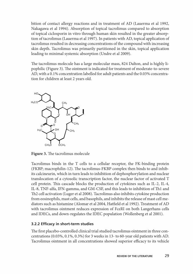
The tacrolimus molecule has a large molecular mass, 824 Dalton, and is highly li-pophilic (Figure 3). The ointment is indicated for treatment of moderate-to-severe AD, with a 0.1% concentration labelled for adult patients and the 0.03% concentra-tion for children at least 2 years old.
Figure 3. The tacrolimus molecule
Tacrolimus binds in the T cells to a cellular receptor, the FK-binding protein (FKBP; macrophilin-12). The tacrolimus-FKBP complex then binds to and inhib-its calcineurin, which in turn leads to inhibition of dephosphorylation and nuclear translocation of a cytosolic transcription factor, the nuclear factor of activated T cell protein. This cascade blocks the production of cytokines such as IL-2, IL-4, IL-8, TNF-alfa, IFN-gamma, and GM-CSF, and this leads to inhibition of Th1 and Th2 cell activation (Luger et al 2008). Tacrolimus also inhibits cytokine production from eosinophils, mast cells, and basophils, and inhibits the release of mast cell me-diators such as histamine (Alomar et al 2004, Hatfield et al 1992). Treatment of AD with tacrolimus ointment reduces expression of FcεRI on both Langerhans cells and IDECs, and down-regulates the IDEC population (Wollenberg et al 2001).
3.2.2 efficacy in short-term studies
The first placebo-controlled clinical trial studied tacrolimus ointment in three con-centrations (0.03%, 0.1%, 0.3%) for 3 weeks in 13- to 60-year old patients with AD. Tacrolimus ointment in all concentrations showed superior efficacy to its vehicle
reVieW of THe liTeraTure
30
from the third treatment day onwards to the end of the study. The only adverse event was a burning sensation in treated areas (Ruzicka et al 1997). These find-ings were confirmed later in both adults and children with moderate-to-severe AD (Reitamo et al 2002a).
Tacrolimus ointment has been compared to pimecrolimus cream in children and adults with AD in three investigator-blinded, randomised, comparative 6-week studies. In the meta-analysis of these studies, tacrolimus ointment was more effec-tive, with an onset of action faster than for pimecrolimus cream, and their safety profiles were similar (Paller et al 2005).
3.2.3 efficacy in long-term studies
The published long-term studies with tacrolimus ointment are summarised in Table 2. Most open-label long-term clinical studies ranging from 6 months to 4 years have used 0.03% or 0.1% tacrolimus ointment twice daily until clearance, and in the case of any new lesions twice daily treatment restarted. These studies have shown sustained, excellent improvement of the eczema, without any signs of tachyphylaxis (Kang et al 2001, Reitamo et al 2000, 2007, 2008, Remitz et al 2007). Two long-term studies suggest that need for tacrolimus ointment decreases over time when the skin heals (Reitamo et al 2000, 2008).
Recently, intermittent (proactive) tacrolimus ointment treatment twice weekly for flare prevention, has been studied. During the initial period it was applied twice daily to all affected areas up to an Investigator Global Assessment (IGA) score of 2 or less. Patients then entered the disease-control period and were randomised to either tacrolimus ointment or vehicle twice weekly for 12 months. Studies in both children and adults showed that intermittent treatment with tacrolimus ointment (0.03% in children and 0.1% in adults) significantly reduced the number of dis-ease exacerbations requiring treatment compared to vehicle treatment (Thaçi et al 2008, Wollenberg et al 2008).
Clinical trials in children aged 2 to 15 with moderate and severe AD have shown significant improvement with both 0.03% and 0.1% tacrolimus ointment (Kang et al 2001, Remitz et al 2007). Infants under 2 (n=12) have been studied once, retrospectively. Improvement in the AD resulted in low tacrolimus blood levels of less than 1.5 ng/mL, measured at a minimum of 1 month after treatment initiation (Patel et al 2003).
reVieW of THe liTeraTure
31
3.2.4 clinical studies comparing topical corticosteroids and tacrolimus oint-ment
Tacrolimus ointment has been compared to several topical corticosteroids in short-term studies. Tacrolimus 0.1% ointment was as effective as hydrocortisone butyrate 0.1% ointment, but tacrolimus 0.03% ointment was less effective in adult patients with moderate-to-severe AD (Reitamo et al 2002b). In 2- to 15-year-old children, both 0.1% and 0.03% doses were more effective than was hydrocortisone acetate ointment (Reitamo et al 2002c).
Tacrolimus ointment has also been compared to 0.005% fluticasone pivalate oint-ment. In treatment of facial eczema, 0.1% tacrolimus was superior to fluticasone in adults in a 3-week study (Doss et al 2009a), while 0.03% tacrolimus showed similar efficacy as did fluticasone for children in a 6-week study (Doss et al 2009b). Another study with 2- to 15-year-old children compared the efficacy of 0.1% methylprednisolone aceponate ointment once daily to that of 0.03% tacrolimus ointment twice daily for 3 weeks in the treatment of a severe to very severe flare of AD. The methylprednisolone aceponate showed better efficacy for the Eczema Area and Severity Index (EASI), sleep, and itch, and the IGA was similar for both treatments (Bieber et al 2007). In one 6-month, double-blind study, tacrolimus 0.1% ointment was compared to a corticosteroid regimen (1% hydrocortisone acetate for head and neck, and 0.1% hydrocortisone butyrate for trunk and limbs) in 972 adult patients with moderate-to-severe AD. In that study, tacrolimus ointment showed efficacy superior to that of the corticosteroid regimen throughout (Reitamo et al 2005).
29
!"#"!$%&&'()(*$'+$,-+./0123$4056'14$
The published long-term studies with tacrolimus ointment are summarised in Table 2. Most open-label long-term clinical studies ranging from 6 months to 4 years have used 0.03% or 0.1% tacrolimus ointment twice daily until clearance, and in the case of any new lesions twice daily treatment restarted. These studies have shown sustained, excellent improvement of the eczema, without any signs of tachyphylaxis (Kang et al 2001, Reitamo et al 2000, 2007, 2008, Remitz et al 2007). Two long-term studies suggest that need for tacrolimus ointment decreases over time when the skin heals (Reitamo et al 2000, 2008).
Recently, intermittent (proactive) tacrolimus ointment treatment twice weekly for flare prevention, has been studied. During the initial period it was applied twice daily to all affected areas up to an Investigator Global Assessment (IGA) score of 2 or less. Patients then entered the disease-control period and were randomised to either tacrolimus ointment or vehicle twice weekly for 12 months. Studies in both children and adults showed that intermittent treatment with tacrolimus ointment (0.03% in children and 0.1% in adults) significantly reduced the number of disease exacerbations requiring treatment compared to vehicle treatment (Thaçi et al 2008, Wollenberg et al 2008).
Clinical trials in children aged 2 to 15 with moderate and severe AD have shown significant improvement with both 0.03% and 0.1% tacrolimus ointment (Kang et al 2001, Remitz et al 2007). Infants under 2 (n=12) have been studied once, retrospectively. Improvement in the AD resulted in low tacrolimus blood levels of less than 1.5 ng/mL, measured at a minimum of 1 month after treatment initiation (Patel et al 2003).
Table 2. Long-term studies (!6 months) with tacrolimus ointment
Study Study setting Nr. of
patients Age
(years) Treatment Duration (months)
Reitamo et al. 2000 Open, non-comparative 316 !18 0.1% tac 6-12 Kang et al 2001 Open, non-comparative 255 2-15 0.1% tac 12 Hanifin et al 2005 Open, non-comparative 799 !2 0.1% tac up to 40 Reitamo et al 2007 Open, non-comparative 672 !18 0.1% tac 24 Remitz et al 2007 Open, non-comparative 466 2-15 0.03% or 0.1% tac 29 Reitamo et al 2008 Open, non-comparative 782 !2 0.1% tac 48 Wollenberg et al 2008 Double-blind, intermittent 257 !18 0.1% tac vs vehicle 12 Thaçi et al 2008 Double-blind, intermittent 267 2-15 0.03% tac vs vehicle 12 Breneman et al 2008 Double-blind, intermittent 197 !2 0.03% or 0.1% tac vs vehicle 10 Paller et al 2008 Double-blind, intermittent 105 2-15 0.03% tac vs vehicle 10 Reitamo et al 2005 Double-blind 972 !18 0.1% tac vs steroid 6 tac=tacrolimus
The safety of tacrolimus ointment has been examined in several short- and long-term clinical studies in both children and adults. A recent review of the long-term safety of tacrolimus ointment revealed no long-term safety problems (Remitz & Reitamo 2009). The most common application-site adverse event is a burning sensation reported in about 60% of the adult and 40% of the paediatric patients (Ruzicka et al 1997, Reitamo et al 2002b). This sensation is usually transient, and when treatment is continued, its incidence decreases over time (Reitamo et al 2000). Other common application-site adverse events are pruritus, skin tingling, hyperaesthesia, and folliculitis. Alcohol intolerance, which is characterised by facial flushing after intake of even a small amount of alcohol, is an adverse event specific for treatment with tacrolimus ointment (Reitamo et al 2000, 2005). Acetylsalicylic acid taken orally one hour before tacrolimus treatment seems to reduce or inhibit symptoms both of this alcohol reaction and of the local burning sensation and pruritus when treatment is initiated (Mandelin et al, unpublished observation).
In the 6-month, double-blind study, Herpes simplex skin infections showed a high-er incidence during the first months in patients treated with tacrolimus ointment vs patients treated with the corticosteroid regimen. Between these groups after 6 months of treatment, incidence was comparable (1.3% in tacrolimus vs 1.0% in steroid-treated patients). No increase in bacterial skin infections was evident in tacrolimus-treated patients (Reitamo et al 2005). In contrast, one uncontrolled 1-year tacrolimus study showed decreased colonisation with S. aureus when skin lesions healed (Remitz et al 2001). Initially, one short-term, double-blind study showed that topical tacrolimus does not reduce collagen synthesis (Reitamo et al 1998). A 1-year study with intermit-tent tacrolimus monotherapy in patients with AD showed an increase in the syn-thesis of PINP and PIIINP in the skin, accompanied by an increase in skin thick-ness measured by an ultra-sound device (Kyllönen et al 2004).
Tacrolimus ointment does not elevate ocular pressure (Remitz et al 2010), and to date there exist no reports of cataract after topical tacrolimus treatment.
Risk for skin cancer is related to systemic immunosuppression and UV radiation. That tacrolimus ointment is a topical immunomodulating agent has inspired inter-est in study of any skin cancer risk. A follow-up study with a maximum observa-tion period of up to 4 years did not report an increased risk for non-melanoma skin cancer in AD patients treated with tacrolimus ointment (Naylor et al 2005). Topical tacrolimus associated with non-melanoma skin cancer in one murine study (Niwa et al 2003). In contrast, several animal studies suggest a decreased risk for skin cancer with tacrolimus treatment, which inhibited development of phorbol ester-induced skin tumours (Jiang et al 1993), and caused activation of the trans-forming growth factor (TGF)-β-1-receptor, playing an important role in wound
reVieW of THe liTeraTure
33
healing and tumour formation (Yao et al 2000). Tacrolimus and pimecrolimus were both able to reduce UVB-induced thymidin-dimer formation, and thus pre-vent DNA damage (Tran et al 2005), and neither accelerated photocarcinogenesis nor induced any dermal carcinogenesis in hairless mice (Lerche et al 2008, 2009). In a large study including almost 300 000 patients the risk for lymphomas in pa-tients treated with topical calcineurin inhibitors was not increased (Arellano et al 2007). The only work suggesting increased risk for lymphoma has been one on T cell lymphoma which showed an increased risk at a median time of 1.4 years from tacrolimus exposure to T cell lymphoma. The mean cumulative amount of tacrolimus prescribed did not significantly differ in patients with lymphoma com-pared to patients without and was less than 100 grams for 0.1% tacrolimus oint-ment. No risk for any other cancer was observable. Unfortunately, this study did not address corticosteroid use by these patients (Hui et al 2009). These findings contradict those of two 4-year safety studies of tacrolimus, showing no increased risk for any cancer (Hanifin et al 2005, Reitamo et al 2008).
3.2.6 pharmacokinetics
All pharmacokinetic studies available to date have indicated that in long-term treatment of AD systemic absorption of topically applied tacrolimus is minimal. When 12 clinical studies with a total of 2015 adult and paediatric patients treat-ed with 0.03 to 0.3% tacrolimus ointment were analysed in combination, results showed low or minimal systemic exposure. The treatment period ranged from 1 week to 12 months, with a total treated area of up to 100%. The maximum tac-rolimus concentration was below 0.5 ng/mL for 60.5% of patients, 0.5-1 ng/mL for 20.6%, 1-<2 ng/mL for 10.9%, 2-<5 ng/mL for 6.7%, and over 5 ng/mL for 1.3%. In any patients with any measurable tacrolimus blood concentration, it was transient, with decreasing concentrations when the skin healed (Reitamo 2002).
The pharmacokinetic profile in children over age 2 has been similar to that in adults. One pharmacokinetic study in children aged 6 to 12 years treated with 0.1% tacrolimus ointment showed minimal systemic exposure, with 92% of the blood samples containing tacrolimus concentrations below 1 ng/mL. Concentrations tended to increase proportionally as the size of the BSA treated increased; ab-sorption decreased with time as the skin lesions healed (Harper et al 2005). A study involving children aged 2 to 5 years showed similar pharmacokinetic results (Krueger et al 2007).
Daily trough tacrolimus levels of 5-20 ng/mL are recommended in transplant re-cipients to avoid allograft rejection. Notably, in patients with Netherton syndrome, a disease with marked skin-barrier impairment which in infants can be mistaken for AD, topical tacrolimus treatment has showed high systemic tacrolimus con-centrations comparable to those after oral treatment (Allen et al 2001), making topical tacrolimus treatment not recommended for Netherton syndrome.
reVieW of THe liTeraTure
34
3.3 pimecrolimus cream
Pimecrolimus (ASM 981) is a semisynthetic derivative of ascomycin and differs from tacrolimus at a single site with an ethyl group instead of the propenyl side-chain residue. Its mechanism of action is similar to that of tacrolimus, with an initial binding to the FKBP (macrophilin-12), which is about three times as weak in vitro as is tacrolimus (Bochelen et al 1999). Its clinical efficacy also seems to be inferior to that of tacrolimus (Paller et al 2005, Abramovits et al 2008). Topical pimecrolimus is indicated for treatment of mild-to-moderate AD, in adults and children of at least 2 years of age.
A 3-week proof-of-concept study of 1% pimecrolimus cream in adults with mod-erate AD showed the superiority of twice-daily treatment to treatment once daily and to vehicle (Van Leent et al 1998). A dose-finding 3-week study in adults with AD compared increasing concentrations of pimecrolimus cream (0.05%, 0.2%, 0.6%, and 1%) to betamethasone 17-valerate cream. The highest concentration was the most effective for pimecrolimus with a median EASI decrease of 47%, while betamethasone 17-valerate cream was more effective with a median de-crease of 78% compared to baseline (Luger et al 2001). Long-term clinical studies in infants, children, and adults have focused on pimecrolimus as a steroid-sparing agent and showed that it, compared to the vehicle, reduces the number of flares needing treatment with topical corticosteroids (Kapp et al 2002, Wahn et al 2002, Meurer et al 2002). One long-term, double-blind controlled study has compared the efficacy of 1% pimecrolimus cream to a corticosteroid regimen (hydrocorti-sone acetate cream for head and neck and 0.1% triamcinolone acetonide cream for trunk and limbs). Efficacy was better in the corticosteroid group, and only 42% of pimecrolimus-treated patients could be maintained without corticosteroids for one year (Luger et al 2004). A meta-analysis which included children aged 2 to 17 from eight randomised controlled trials indicated that patients treated with both 0.03% and 0.1% tacrolimus ointment showed a significantly higher response rate than did those treated with 1% pimecrolimus or 1% hydrocortisone acetate (Yan et al 2008).
Pimecrolimus has been studied in infants with AD under age 2 and showed supe-rior efficacy compared to placebo, and had a good safety profile in one 6-month study (Ho et al 2003). In a 3-week study in infants with AD treated with 1% pime-crolimus cream twice daily, pimecrolimus blood concentrations were minimal (Staab et al 2005).
The safety profile of 1% pimecrolimus cream is very good; pharmacokinetic stud-ies show low systemic exposure (Luger et al 2008). Local adverse events, like skin burning, seem to occur at a similar frequency for topical pimecrolimus and for tacrolimus treatment (Abramovits et al 2008).
reVieW of THe liTeraTure
35
aims of THe sTudY
The main aim of this study was to evaluate the effects of long-term topical tac-rolimus treatment on AD skin and atopic respiratory disease.
The specific aims of this study were to determine:
the effect of long-term tacrolimus ointment treatment in patients with AD on their cell-mediated immunity. the long-term efficacy, safety, and effects on the Th1/Th2 balance in pa-tients with AD treated with tacrolimus ointment or with a corticosteroid regimen.
the 10-year outcome of eczema, respiratory symptoms, and serum IgE le-vels in AD patients initially treated long-term with tacrolimus ointment.
the pharmacokinetics and long-term safety and efficacy of tacrolimus oint-ment in infants under age 2 with AD.
aims of THe sTudY
1)
2)
3)
4)
36
suBJecTs and meTHods
1. patients
Patients in Studies I to III were recruited from the outpatient clinic of the Department of Dermatology, Helsinki University Central Hospital. Patients in-cluded in Study I participated in a 4-year open tacrolimus safety study, and all patients recruited for the 10-year follow-up had also participated in the 4-year study. Thus patients in Studies I and III in part overlapped, but were at baseline in Study I aged ≥16 years and in Study III aged ≥13 years. Patients in Study II were aged ≥18. Study IV was a paediatric, multi-centre study with patient recruitment in Finland, Ireland, the UK, Latvia, and Canada. Patients in Finland were all re-cruited at the Children’s Department of the Skin and Allergy Hospital, Helsinki University Central Hospital, and were aged between 3 and 24 months. Study V was a follow-up and included only patients who participated in Study IV. (Table 3).
35
!"#$%&'!()*+(,%'-.+!((
/0((12345637(
Patients in Studies I to III were recruited from the outpatient clinic of the Department of Dermatology, Helsinki University Central Hospital. Patients included in Study I participated in a 4-year open tacrolimus safety study, and all patients recruited for the 10-year follow-up had also participated in the 4-year study. Thus patients in Studies I and III in part overlapped, but were at baseline in Study I aged !16 years and in Study III aged !13 years. Patients in Study II were aged !18. Study IV was a paediatric, multi-centre study with patient recruitment in Finland, Ireland, the UK, Latvia, and Canada. Patients in Finland were all recruited at the Children’s Department of the Skin and Allergy Hospital, Helsinki University Central Hospital, and were aged between 3 and 24 months. Study V was a follow-up and included only patients who participated in Study IV. (Table 3).
Table 3. Baseline characteristics and reasons for withdrawal. Study N Mean
age (years)
Males/ females
Study duration Reasons for withdrawal (n)
I 48 26.1 17/31 12 months Lack of efficacy (1), unavailable for testing at month 12 (2)
40 29.3 10/30 12 months Lack of efficacy (4), prohibited therapy (2), non-compliance (1), pregnancy (1), lost to follow-up (1)
III 50 33.0 16/34 10-year follow-up
Not applicable
IV 53 1.2 32/21 28 days Withdrawal of consent (1), other (2) V 50 1.2 31/19 48 months Withdrawal of consent (3), adverse event (2),
lost to follow-up (2), non-compliance (1), lack of efficacy (1), other reasons (2)
!"!#$%&'()*+%#,%-#./&'()*+%#&0*1.0*,#
All studies required the patients to have an AD diagnosis according to the Hanifin and Rajka (1980) criteria, and an affected area greater than or equal to 5% of total BSA at baseline. Studies I to III also required patients to have moderate-to-severe AD at baseline based on the grading system of Rajka and Langeland (1989), whereas patients in Study IV had to need AD treatment with mid-
suBJecTs and meTHods
37
1.1 inclusion and exclusion criteria
All studies required the patients to have an AD diagnosis according to the Hanifin and Rajka (1980) criteria, and an affected area greater than or equal to 5% of to-tal BSA at baseline. Studies I to III also required patients to have moderate-to-severe AD at baseline based on the grading system of Rajka and Langeland (1989), whereas patients in Study IV had to need AD treatment with mid-potency topi-cal corticosteroids. Inclusion criteria for Study V were participation in Study IV and having benefitted from treatment with 0.03% tacrolimus ointment, according to the investigator. Wash-out criteria before study enrollment are summarised in Table 4. Exclusion criteria are listed in Table 5.
36
potency topical corticosteroids. Inclusion criteria for Study V were participation in Study IV and having benefitted from treatment with 0.03% tacrolimus ointment, according to the investigator. Wash-out criteria before study enrollment are summarised in Table 4. Exclusion criteria are listed in Table 5.
Table 4. Wash-out criteria before study enrollment. Therapy Study I, III Study II Study IV Study V All medicated topical treatment, including corticosteroids 5 days 3 days 3 days 3 days Antimicrobials n.r. 5 days n.r. n.r. Systemic antihistamines: n.r. 5 days n.r. n.r.
terfenadine 5 days 5 days n.r. n.r. astemizole 6 weeks 5 days n.r. n.r.
Table 5. Exclusion criteria for studies indicated by their Roman numerals (I-V)
Patient has/ Patient is:
• an infection requiring treatment (I-III, V) • a systemic disease, including cancer or history of cancer or AIDS, which would
contraindicate the use of tacrolimus ointment (I-V) • a chronic disease that is unstable or uncontrolled (IV) • diabetes mellitus which is either unstable, poorly controlled, or insulin dependent (I, III) • clinically significant impairment of renal or hepatic function (I-V) • clinically infected AD (I-V) • a history of eczema herpeticum (I, III) • a skin disorder, other than AD, requiring treatment (I-V) • any lesion, other than scalp or mucosa, which in the opinion of the investigator cannot be
treated with study ointment (II, IV, V) • extensive scarring or pigmented lesions in the treatment area, which would interfere with
rating of efficacy parameters (II) • known allergic response to macrolides or any excipient of the ointment (I-V) • a history of more than two courses of systemic corticosteroid treatment (IV) • any form of substance abuse, psychiatric disorder or condition which, in the opinion of the
investigator, may invalidate the communication with the investigator (I-III) • received a vaccination in the last 14 days (28 days in the case of a live attenuated
vaccination) (IV) • previously been treated with tacrolimus or has participated in any Fujisawa (now Astellas)
sponsored trial and received at least one dose of tacrolimus (II) • known to be HIV positive (I-V) • pregnant or breast feeding (I-III) • simultaneously participating in any other drug trial (I-V)
!"#$%&'(&)*+,'+$)-.*(,+*&'/$
During the 4-year open 0.1% tacrolimus study, within which Study I and part of Study III were conducted, the following concomitant medications were prohibited: UV therapy, non-steroidal immunosuppressants, medicated topical agents, astemizole, terfenadine, more than 1 mg/day of intranasal and/or inhaled corticosteroids, or other investigational drugs. The use of systemic and topical corticosteroids was prohibited during the first 6 months, whereafter, rescue therapy for worsening of AD was allowed for a maximum of 2 weeks in every 3 months. Use of non-steroidal anti-inflammatories was permitted for use up to 2 weeks in any 3 months. Emollients were permitted 2 hours prior to or after the use of the study ointment. Study II included all these restrictions and permissions, but in addition all systemic antihistamines, antimicrobials influencing efficacy assessments, and sleeping pills and tranquilisers were
suBJecTs and meTHods
39
1.2 concomitant medications
During the 4-year open 0.1% tacrolimus study, within which Study I and part of Study III were conducted, the following concomitant medications were prohib-ited: UV therapy, non-steroidal immunosuppressants, medicated topical agents, astemizole, terfenadine, more than 1 mg/day of intranasal and/or inhaled corti-costeroids, or other investigational drugs. The use of systemic and topical corti-costeroids was prohibited during the first 6 months, whereafter, rescue therapy for worsening of AD was allowed for a maximum of 2 weeks in every 3 months. Use of non-steroidal anti-inflammatories was permitted for use up to 2 weeks in any 3 months. Emollients were permitted 2 hours prior to or after the use of the study ointment.
Study II included all these restrictions and permissions, but in addition all systemic antihistamines, antimicrobials influencing efficacy assessments, and sleeping pills and tranquilisers were prohibited. Systemic and topical corticosteroids were not allowed for the treatment of AD, and for other indications only for a maximum of 2 weeks in any 3 months.
Prohibited therapies in Study IV included UV therapy, non-steroidal immunosup-pressants, topical and systemic corticosteroids, other investigational drugs, and vaccinations. The use of inhaled and nasal corticosteroids was restricted to <1 mg/day. Study V included the same restrictions, but in addition, other topical immu-nomodulators were prohibited. Treatment with topical corticosteroids for up to 2 weeks in any 3-month period and vaccinations were permitted. In the case of vaccinations, topical tacrolimus treatment had to be discontinued for 5 to 7 weeks, and during this period treatment with topical corticosteroids was allowed.
suBJecTs and meTHods
40
2. study designs and protocols
The study protocols for Studies I to V were approved by the local ethics committee at Helsinki University Central Hospital. All patients or their parents in Studies IV and V, gave written informed consent before the start of the study. The use of study medication, either tacrolimus ointment or the corticosteroid regimen (in Study II) was approved by the National Agency for Medicines (Lääkelaitos).
Studies I and III were open, single-centre studies conducted in patients who were participating or had participated in a long-term multi-centre safety study of 0.1% tacrolimus ointment. Study II was a one-year double-blind randomised controlled study, where patients were treated with either 0.1% tacrolimus ointment or a corti-costeroid regimen. Some of the efficacy and safety data from the first 6 months of the study were included in a large multi-centre study (Reitamo et al 2005). Study IV was a randomised double-blind pharmacokinetic multi-centre study, in which patients were treated with 0.03% tacrolimus ointment either once or twice daily. Following enrollment, patients were allocated to three different groups depending on the affected body surface area (BSA): Group 1, 5 to 20%, Group 2, >20 to 40%, and Group 3: >40%. After allocation, infants were randomised 1:1 to treatment with tacrolimus ointment once or twice daily. Study V was an open 2-year non-comparative multi-centre phase II safety study (Table 6).
39
!"#$%&'(#')*+,-*#.-'#/01%1213*#
The study protocols for Studies I to V were approved by the local ethics committee at Helsinki University Central Hospital. All patients or their parents in Studies IV and V, gave written informed consent before the start of the study. The use of study medication, either tacrolimus ointment or the corticosteroid regimen (in Study II) was approved by the National Agency for Medicines (Lääkelaitos).
Studies I and III were open, single-centre studies conducted in patients who were participating or had participated in a long-term multi-centre safety study of 0.1% tacrolimus ointment. Study II was a one-year double-blind randomised controlled study, where patients were treated with either 0.1% tacrolimus ointment or a corticosteroid regimen. Some of the efficacy and safety data from the first 6 months of the study were included in a large multi-centre study (Reitamo et al 2005). Study IV was a randomised double-blind pharmacokinetic multi-centre study, in which patients were treated with 0.03% tacrolimus ointment either once or twice daily. Following enrollment, patients were allocated to three different groups depending on the affected body surface area (BSA): Group 1, 5 to 20%, Group 2, >20 to 40%, and Group 3: >40%. After allocation, infants were randomised 1:1 to treatment with tacrolimus ointment once or twice daily. Study V was an open 2-year non-comparative multi-centre phase II safety study (Table 6).
Table 6. Study protocols Study Type of study Study treatment Age Duration of
treatment Duration of follow-up
I Open, controlled 0.1% tacrolimus !16 years 12 months 12 months
II Double-blind, randomised 0.1% tacrolimus vs corticosteroid* !18 years 12 months 12 months
III Open, uncontrolled, follow-up 0.1% tacrolimus !13 years 48 months 10 years
IV Double-blind, randomised 0.03% tacrolimus UID vs BID 3-24 months 14 days 18 days
*hydrocortisone acetate for head and neck, hydrocortisone butyrate for trunk and limbs
UID=once daily, BID=twice daily
!"#$%&'()*+(,-$
Patients in Study I were treated with 0.1% tacrolimus ointment for 12 months. Patients in Study III were during the 4-year intervention period treated with 0.1% tacrolimus ointment, but were after the study free to choose any type of AD treatment; these were registered at the 10-year follow-up visit. Study II was double-blind, with patients randomised (1:1) at baseline for either treatment with 0.1% tacrolimus ointment, or a corticosteroid regimen which included 1% hydrocortisone acetate ointment for the head and neck and 0.1% hydrocortisone butyrate ointment for the trunk and limbs. The ointment tubes were identical in appearance in both groups. They were given to the patient and
suBJecTs and meTHods
41
2.1 medication
Patients in Study I were treated with 0.1% tacrolimus ointment for 12 months. Patients in Study III were during the 4-year intervention period treated with 0.1% tacrolimus ointment, but were after the study free to choose any type of AD treat-ment; these were registered at the 10-year follow-up visit. Study II was double-blind, with patients randomised (1:1) at baseline for either treatment with 0.1% tacrolimus ointment, or a corticosteroid regimen which included 1% hydrocor-tisone acetate ointment for the head and neck and 0.1% hydrocortisone butyrate ointment for the trunk and limbs. The ointment tubes were identical in appearance in both groups. They were given to the patient and then returned to the investiga-tor in sealed boxes, and thus the investigator could not assess the ointment bases. In Studies I to III, patients were advised to treat all lesions with study ointment twice daily until the end of itching and of any signs of eczema, and then for an additional 7 days to minimise possible subclinical inflammation. In Studies I and III, treatment was restarted if a new lesion occurred or if the disease recurred on a previously cleared lesion. In Study II, treatment was restarted in case of a flare-up. No restrictions were put on study drug usage.
In Study IV, the area to be treated with study medication was defined on Day 1. During the 14-day study period, the treatment area could not be altered, regardless of any eczema healing or new lesions. At the Day 1 and 14 visits, patients received a single application of study medication in order to obtain full pharmacokinetic profiles. At the Day 4 visit, the morning application was performed in the hospital after drawing of blood, to obtain the trough level. On all other treatment days, the defined areas were treated twice daily. Patients were limited to 5 g or less ointment per application (10 g per day).
In Study V, 0.03% tacrolimus ointment was applied twice daily to all affected skin lesions until healing—for up to 3 weeks. If necessary, treatment continued once daily thereafter. In case of an exacerbation of AD, twice-daily treatment with the study ointment was restarted. Total tacrolimus ointment use was unrestricted.
2.2 study schedules
Study I was performed on some of the patients participating in a long-term open tacrolimus study initiated in November 1998. The first follow-up was one week after initiation of tacrolimus treatment, and then after 1, 3, 6, 9, and 12 months. After this study, visits continued every 6 months up to 48 months. Patients were tested for recall antigen reactions at baseline and after 12 months of treatment. Healthy controls were recruited among medical students and personnel from the Skin and Allergy Hospital and were tested once, within the same time-lines as the AD patients. Retesting was performed in nine randomly chosen controls after one year to investigate whether retesting had any additive effect on test results.
suBJecTs and meTHods
42
Study II was conducted during the years 2001 and 2002. After randomisation, vis-its were performed after 1, 2, and 4 weeks, and then monthly up to 6 months. Then visit intervals increased to 3 months until the 12th month and the end of the study visit.
Study III was performed in patients who had participated in the long-term open tacrolimus study, applying the same study schedule. Patients who had participated in the lung histamine challenge test at baseline were contacted and asked to par-ticipate in the 10-year follow-up study, which required one visit at the Skin and Allergy Hospital. The visits and respiratory assessments were, at baseline, conduct-ed between October 1998 and January 1999, and at the 10-year follow-up, between October 2008 and early March 2009, and thus were not confounded by the pollen season.
Study IV was conducted during 2003 and 2004. Patients attended the hospital four times after the screening visit: at baseline, and at days 4, 14, and 18. Tacrolimus treatment ended at day 14, and day 18 was a follow-up visit.
Patients from Study IV who met the inclusion criteria could continue directly into Study V, which was conducted during 2003 to 2006. Study visits were carried out at baseline and week 2, and then monthly until month 6. Then visits were every 3 months until the 24th month and end of the study visit. 2.3 efficacy and safety assessments
In Studies I and III, treatment response to tacrolimus ointment was assessed by evaluating affected BSA, which in Study I was determined at baseline and at month 12, and in Study III at baseline, month 48, and at the 10-year follow-up. Adverse events were recorded throughout the 4-year tacrolimus study. Laboratory tests, in-cluding haematology and serum chemistry, were assessed at the screening, at week 1, and at month 6, and then every 6 months until study end. At the 10-year follow-up visit, patients were asked for their medical history since the last visit in the open tacrolimus study, with a special focus on events affecting the skin or airways.
In Study II, the treatment response to the study medication was assessed by the EASI, a validated method in clinical studies of AD (Barbier et al. 2004). It is a composite index, including an assessment percentage of BSA involved, and of ery-thema, infiltration or papulation, or both, excoriation, and lichenification, each on a scale of 0 to 3 (Table 7). The modified EASI (mEASI) is similar to the EASI, but also considers the itching during the last 24 hours. From the EASI score, a separate score for the head and neck area, “the eczema score for head and neck”, was calcu-lated. This, and the mEASI score are not yet validated scores. Physician’s and pa-tient’s global evaluation of clinical response, and the response for the head and neck area compared to baseline (cleared, excellent improvement, marked improvement, moderate improvement, slight improvement, no appreciable improvement, worse)
suBJecTs and meTHods
43
were recorded at every study visit. Adverse events were recorded throughout the study. Blood for laboratory tests, including haematology and serum chemistry, was drawn at the screening, and at the month-3, -6, and -12 visits.
In Study IV, the treatment response was at every visit assessed by the EASI score and by the physician’s global evaluation of clinical response compared to baseline (cleared, excellent improvement, marked improvement, moderate improvement, slight improvement, no appreciable improvement, worse). Adverse events were re-corded throughout the study. Laboratory blood tests, including haematology and serum chemistry, were assessed at the screening visit and on days 4 and 18. Blood tacrolimus concentrations were measured to estimate systemic exposure.
In Study V, the treatment response was assessed at every visit by the EASI score, affected BSA, and the physician and parent’s global evaluation of clinical response. Adverse events and laboratory parameters were monitored throughout the study. Tacrolimus blood concentration samples were collected at baseline, week 2, months 1 and 3, and every third month thereafter.
42
Table 7. Eczema area and severity index: calculation for patients 8 years
of age and older1
Body region EASI Score2,3 Head/Neck (H) (E + I + Ex + L) x Area x 0.1 Upper limbs (UL) (E + I + Ex + L) x Area x 0.2 Trunk (T) (E + I + Ex + L) x Area x 0.3 Lower limbs (LL) (E + I + Ex + L) x Area x 0.4 EASI= Sum of the above 4 body region scores 1For children aged 0-7 years, proportionate areas were head/neck, 20%; upper limbs, 20%; trunk, 30%; and lower limbs, 30%. 2E=Erythema, I=induration/papulation, Ex=excoriation, L=lichenification. 3Where area is defined on a 7-point ordinal scale: 0=no eruption; 1=<10%; 2=<10%-29%; 3=<30%-49%; 4=<50%-69%; 5=<70%-89%; 6=>90%-100%. From Hanifin et al, 2001, reprinted with permission of the publisher.
!"#$%&'()*+,-*&.'/$0'1*&$/2))$
TEWL was measured in all patients in Study II at baseline and at months 3, 6, 9, and 12. Measurements were made at nine target regions (three on the upper limbs, two each on the head and trunk, and one on the neck and lower limbs) with an Evaporimeter EP1 (servoMed, Stockholm, Sweden) according to published guidelines (Pinnagoda 1990).
$
!"3$4*5'//$'(1,6*($1*)1,(6$
Recall antigen tests were performed with the Multitest CMI (Institute Mérieux, Lyon, France) applicator, which contained seven antigens: tetanus, diphtheria, Streptococcus group C, tuberculin, Candida albicans, Trichophyton, and Proteus, and glycerin as a negative control. The tests for each study (Studies I, II) came from the same batch. The Multitest applicator had eight prongs containing the antigens and the control, which were pressed into the skin on the volar side of the left forearm. If this site was affected by eczema, the other arm, or dorsal side was chosen. Topical treatment of the test site was not allowed in the morning before testing, nor before the reading of the test result 48 hours later. A positive test reaction was defined as induration of at least 2 mm, and for these, average diameter (mm) was calculated. The results were analysed as the sum of average diameters (Merieux score) and as the number of positive antigens (Kniker et al 1979).
suBJecTs and meTHods
44
2.4 Transepidermal water loss
TEWL was measured in all patients in Study II at baseline and at months 3, 6, 9, and 12. Measurements were made at nine target regions (three on the upper limbs, two each on the head and trunk, and one on the neck and lower limbs) with an Evaporimeter EP1 (servoMed, Stockholm, Sweden) according to published guide-lines (Pinnagoda 1990).
2.5 recall antigen testing
Recall antigen tests were performed with the Multitest CMI (Institute Mérieux, Lyon, France) applicator, which contained seven antigens: tetanus, diphtheria, Streptococcus group C, tuberculin, Candida albicans, Trichophyton, and Proteus, and glycerin as a negative control. The tests for each study (Studies I, II) came from the same batch. The Multitest applicator had eight prongs containing the antigens and the control, which were pressed into the skin on the volar side of the left fore-arm. If this site was affected by eczema, the other arm, or dorsal side was chosen. Topical treatment of the test site was not allowed in the morning before testing, nor before the reading of the test result 48 hours later. A positive test reaction was defined as induration of at least 2 mm, and for these, average diameter (mm) was calculated. The results were analysed as the sum of average diameters (Merieux score) and as the number of positive antigens (Kniker et al 1979).
2.6 serum ige and skin prick testing
In Study II, serum IgE samples were collected at baseline and at months 6 and 12. In Study III, serum IgE samples were collected at baseline and at the 10-year follow-up visit. Samples were analysed by a fluorescence enzyme immunoassay (until the end of 2002: FEIA, CAP system; Pharmacia Diagnostics AB, Uppsala, Sweden; then by ImmunoCAP Total IgE, Phadia AB, Uppsala, Sweden), at the Skin and Allergy Hospital in Helsinki. Age-related references for the upper normal lim-its are: age >17, 110 kU/L; 15 to 17, 160 kU/L; and 13 to 14, 320 kU/L (Björksten & Viander 1987).
SPTs were performed on healthy skin on the forearms. Histamine dihydrochloride 10 mg/ml served as the positive control, and saline as the negative. A test reaction of at least 3 mm was considered positive if the histamine reaction was at least 5 mm and the saline control negative. Antihistamines and antiemetic medications were withheld for 5 days prior to testing. In Study III, tests were performed for birch, timothy, mugwort, cat, dog, horse, cow, house dust mite, Cladosporium herbarum, and natural rubber latex at baseline. At the 10-year follow-up, the same allergens were tested, but only the standardised allergen extracts of timothy, mugwort, cat, dog, horse, and house dust mite served for comparison in the analyses.
suBJecTs and meTHods
45
2.7 respiratory symptoms and findings
Respiratory symptoms were evaluated at baseline and at the 10-year follow-up visit in Study III. Patients had, at the time of lung function testing and of answering the questionnaire, been free from respiratory tract infections for at least 2 weeks.
2.7.1 Questionnaire
All patients answered at baseline a validated questionnaire to estimate whether they had physician-diagnosed asthma or allergic rhinitis (Haahtela et al 1980). New asthma and rhinitis diagnoses were recorded at the follow-up visit. At the baseline and follow-up visits patients reported their degree of asthma (cough, wheesing, dyspnoea) and rhinitis symptoms during the previous month on a visual analogue scale (VAS) from 0 to 10 cm. A result of at least 2 cm was considered to represent active symptoms.
2.7.2 Histamine challenge test
Lung function was measured with a pneumotachograph-based flow-volume spirometer (Masterscreen Pneumo, Jaeger GmbH, Würzburg, Germany). If the baseline forced expiratory volume in one second (FEV1) (Quanjer et al 1993) value was equal to or less than 60% of national predicted values (Viljanen et al 1982), no histamine challenge test was conducted. Use of short- and long-acting beta-agonists was restricted for 1 vs 2 days, and of antihistamines and antiemetic medi-cation for 5 days prior to the histamine challenge test.
BHR was determined with a dosimetric histamine challenge test, where patients inhaled increasing doses of buffered histamine diphosphate (0.025, 0.1, 0.4 and 1.6 mg) (Sovijärvi et al 1993). The dose of inhaled histamine producing a 15% de-crease in FEV1 (PD15FEV1) was determined. Based on this dose, BHR was graded as no (>1.6 mg), mild (1.60-0.41 mg), moderate (0.40-0.11 mg), or severe (<0.10 mg). From the PD15FEV1 dose and the decrease in FEV1, the slope of the dose-response curve of the histamine challenge test was calculated, a continuous and normally distributed index (O’Connor et al 1987).
2.8 Tacrolimus pharmacokinetics
In Study IV, blood concentration-time profiles to define the pharmacokinetics of tacrolimus were collected after the first dose (day 1) and the last dose (day 14). Each profile consisted of serial samples collected before dosing and 2, 4, 8, and 24 hours after the first application. An additional sample was taken at 96 hours after the day-14 application (day 18), and a single pre-dose blood sample was collected on day 4 to provide an estimate of exposure on that day while limiting the number of blood samples taken. The samples comprised 0.25 ml of blood placed in a tube containing EDTA as an anticoagulant, and immediately frozen and stored at -20 ºC before being shipped to a central analytical laboratory.
suBJecTs and meTHods
46
Tacrolimus blood concentration was assayed by high-pressure liquid chromatogra-phy (HPLC) with tandem mass spectrometry detection. Internal standard, a struc-tural analog of tacrolimus and protein precipitation reagent (aqueous zinc sul-fate solution, Sigma, Dorset, UK: methanol, Romil, Cambridge, UK: acetonitrile, Romil 50:30:20), was added to the samples. Solid-phase extraction (using 18C cartridges, Anachem, Luton, UK) of analytes was performed before injection onto the liquid chromatography mass spectrometer (Sciex API III Plus, PerkinElmer, Beaconsfield, UK). The lower limit of quantification for this method was 0.025 ng/ml with a precision—based on the coefficient of variation—of less than 21.3% for the lowest quality control sample and less than 14.7% for all other concentra-tions. All procedures were performed according to the Good Laboratory Practice guidelines.
3. statistical methods
In Studies I to III, analyses were performed with SPSS versions 11.0, 13.0 or 15.0 (SPSS Inc, Chicago, IL, USA). The results are expressed as the mean ± SD for nor-mally distributed variables, and as the median (range or interquartile range) for non-normally distributed variables. Intention-to-treat analyses with the last ob-servation carried forward were carried out where appropriate. A P-value of <0.05 was considered statistically significant.
The material was mainly non-parametrically distributed due to relatively small patient numbers. The Wilcoxon test served to test differences before and after study treatment. Differences between the patients and controls (Study I), or the two different treatment groups (Study II) were tested with the Mann-Whitney test. Fisher’s exact test served for testing associations between binary variables. Binary values (yes/no) in a patient group before and after treatment (anergy, BHR) were compared with the McNemar test. In Study II, correlations between different vari-ables were examined by Spearman’s correlation analysis.
In Study III, the log slope of the dose response curve was tested by the paired t-test.
In Study IV, pharmacokinetic parameters were determined by noncompartmental analysis with the computer program WinNonlin Professional version 4 (Pharsight Corporation). Area under the curve (AUC) calculations were made by the linear/logarithmic trapezoidal rule. The parameters determined were area under the con-centration-time curve between first and last time-points, mean maximum serum concentration, time to attain maximum blood concentration, and terminal half-life. Otherwise the statistical analyses in Studies IV and V were descriptive, and no statistical comparisons were performed.
suBJecTs and meTHods
47
resulTs
1. clinical efficacy
The clinical efficacy of topical tacrolimus was very good in all studies. The im-provement in affected BSA in study patients is presented in Figure 4. In Studies II, IV, and V, EASI scores were assessed in addition.
In Study I, the mean affected BSA decreased from 32 to 8.8% after 12 months of treatment with 0.1% tacrolimus ointment.
In Study II, affected BSA, EASI, and mEASI decreased in both the tacrolimus- and corticosteroid-treated patients at month 6 and month 12. The median EASI score decreased from 21.3 to 3.5 in the tacrolimus-treated patients, and from 20.1 to 6.4 in the corticosteroid-treated patients at month 12. The improvement in all scores was significantly greater (P≤0.05) in the tacrolimus-treated patients at month 6. After month 6, efficacy scores showed a slight improvement in the corticosteroid-treated patients, while the results in the tacrolimus-treated patients stayed approximately at month-6 level. No significant difference in the efficacy parameters between the groups was thus evident at the month-12 visit. At month 12, tacrolimus-treated patients showed an improvement of 91%, and corticosteroid-treated patients an improvement of 79% in median affected BSA. The eczema score for the head and neck was analysed separately from the EASI score, and was significantly lower in patients treated with tacrolimus ointment at both month 6 (P=0.001) and month 12 (P<0.05). This was in accordance with the physician’s evaluation of head and neck clinical response at month 12; in the tacrolimus arm 24 of the 40 patients (60%) compared to 12 of 40 (30%) in the corticosteroid arm were rated as “cleared or excellent” (P<0.01). When the physician’s global evaluation of clinical response (which includes the whole body) at month 12 was done, 23 (57.5%) of tacrolimus-treated, and 17 (42.5%) of the corticosteroid-treated patients were evaluated as “cleared or excellent” (P=0.26, ns). The median number of days in Study II was 363 for the tacrolimus group and 361 for the corticosteroid group, and the median number of treatment days during the study was 255 for the tacrolimus- and 327 for the corticosteroid group.
In Study III, median affected BSA decreased during the 10-year follow-up from 19 to 1.6% (P<0.0001). Patients who responded (≥60% improvement of affected BSA, n=42) to intensive treatment with 0.1% tacrolimus ointment at the 1-year visit in the open safety study showed significantly less affected BSA at both the 4-year visit (P<0.001) and the 10-year follow-up (P=0.04) compared to the non-responders (<60% improvement, n=23). Responders and non-responders did not differ at baseline in terms of age, sex, disease severity, atopic sensitisations, or se-rum IgE levels.
resulTs
48
In Study IV, the median EASI score fell from 14.7 to 5.5. In Study V, the median EASI fell from 8.3 to 0.8, and for both this score and the aff ected BSA a continuous decrease was apparent throughout the study. Th e physician rated the global evalu-ation of clinical response as “cleared” or “excellent” in 63.3% of the patients, and the parents assessed the symptoms as “much better” in 85.7% of the infants. Th e mean number of participation days in Study V was 623, with 28 treatment days during the fi rst month.
Figure 4. Improvement of median aff ected BSA in the diff erent studies. tac=tacrolimus, BSA=body surface area
2. safety
Studies I and III did not investigate the safety of tacrolimus ointment, but were conducted in patients participating in an open 4-year study, which primarily inves-tigated the safety and effi cacy of 0.1% tacrolimus ointment (Reitamo et al 2008).
In Study II, adverse events occurred in 40 patients (100%) in the tacrolimus oint-ment group and in 34 patients (85%) in the corticosteroid group. Th e diff erence between groups was statistically signifi cant (P=0.03) and was mainly due to the higher incidence of skin burning with tacrolimus ointment. During the study, fl u syndrome was reported by 22 tacrolimus-treated and by 16 steroid-treated pa-tients (P=0.26), and folliculitis by 20 vs 17 (P=0.65). No diff erence in infections occurred between groups (P=0.07). During the last 3 months of the study, two patients treated with the corticosteroid regimen showed signs of skin atrophy: one had striae of the legs and one subcutaneous haematomas. No patients showed any clinically signifi cant changes in serum chemistry or haematology; none suff ered from any serious adverse event.
47
85.7% of the infants. The mean number of participation days in Study V was 623, with 28 treatment days during the first month.
Figure 4. Improvement of median affected BSA in the different studies. tac=tacrolimus, BSA=body surface area
!"#$%&'()#
Studies I and III did not investigate the safety of tacrolimus ointment, but were conducted in patients participating in an open 4-year study, which primarily investigated the safety and efficacy of 0.1% tacrolimus ointment (Reitamo et al 2008).
In Study II, adverse events occurred in 40 patients (100%) in the tacrolimus ointment group and in 34 patients (85%) in the corticosteroid group. The difference between groups was statistically significant (P=0.03) and was mainly due to the higher incidence of skin burning with tacrolimus ointment. During the study, flu syndrome was reported by 22 tacrolimus-treated and by 16 steroid-treated patients (P=0.26), and folliculitis by 20 vs 17 (P=0.65). No difference in infections occurred between groups (P=0.07). During the last 3 months of the study, two patients treated with the corticosteroid regimen showed signs of skin atrophy: one had striae of the legs and one subcutaneous haematomas. No patients showed any clinically significant changes in serum chemistry or haematology; none suffered from any serious adverse event.
In Study IV, the most common adverse events were infections such as upper respiratory tract infections and local skin irritations such as skin burning and pruritus. A total of 48 adverse events were reported in 35 patients (66%). Of these, 12 (23%) had at least one causally related adverse
resulTs
49
In Study IV, the most common adverse events were infections such as upper respi-ratory tract infections and local skin irritations such as skin burning and pruritus. A total of 48 adverse events were reported in 35 patients (66%). Of these, 12 (23%) had at least one causally related adverse event and 9 (17%) at least one application-site adverse event. None suff ered from a serious adverse event nor showed any clinically signifi cant changes in their blood chemistry or haematology tests.
In Study V, 47 patients (94%) had at least one adverse event during the whole 2-year study. Th e most common adverse events were infections of the respiratory and gastrointestinal tracts, and the infections of the middle ear commonly seen in this age group. Two patients discontinued due to an adverse event, possibly related to tacrolimus ointment: one with an application-site skin infection and one with thrombocytopenia. A total of 19 serious adverse events occurred in 12 patients; of these one case of eczema herpeticum was considered to be related to treatment. No deaths occurred. Nor did any clinically signifi cant changes in the laboratory parameters occur during the study. 3. Transepidermal water loss
In Study II, both tacrolimus- and steroid-treated patients showed a TEWL de-crease aft er 6 and 12 months of treatment for both the combined sites of head and neck, and trunk and limbs, when compared to baseline (P<0.001). TEWL for the head and neck at both the month-6 and -12 visits were signifi cantly lower in the tacrolimus group than in the steroid group (month 6, P=0.001; month 12, P=0.04), which was in accordance with the lower eczema scores for the head and neck in the tacrolimus group. TEWL for trunk and limbs showed no diff erence between groups.
4. recall antigen testing
Patient characteristics and recall antigen results for Studies I and II are summarised in Table 8. In Study I, both the Merieux score and number of positive antigens were at baseline signifi cantly lower in the AD patients than in the healthy controls. Aft er one year of tacrolimus treatment, a signifi cant increase occurred in the reactions. Healthy controls, retested aft er one year (n=9), showed a decrease both in mean number of positive antigens (from 3.56 to 2.67) and in mean Merieux score (from 13.8 to 7.7).
In Study II, no increase in Mérieux score or in number of antigens was evident aft er 6 months of treatment in either tacrolimus- or corticosteroid-treated pa-tients. Aft er one year of treatment, patients in both groups experienced a signifi -cant increase in Mérieux score compared to baseline (tacrolimus P=0.001, steroid P=0.04). Th e number of positive antigens at month 12 increased signifi cantly only in the tacrolimus ointment group (P=0.003). In the corticosteroid regimen group the increase was numerical, but non-signifi cant (P=0.09). Th e diff erence between the groups at month 12 was non-signifi cant (P=0.46). Figure 5 shows the Merieux
resulTs
50
score and Figure 6 the median number of antigens in tacrolimus-treated patients from Studies I and II.
An inverse correlation between the Merieux score and serum IgE was seen for all patients (n=80) at both baseline (R= -0.40, P<0.001), and month 12 (R= -0.34, P=0.002).
In both Studies I and II, baseline anergy (no positive antigen reactions) was more common in severe disease than in moderate (P<0.01, Fisher’s exact test, and P=0.05, Chi-square test), but neither sex nor age affected anergy. In Study I, the number of anergic patients decreased significantly (P<0.001, McNemar test) after 12 months of treatment. None of the control patients was anergic at baseline. In Study II, the number of anergic patients in the tacrolimus group decreased from 13 at baseline to 6 at month 12, a non-significant decrease (P=0.065, McNemar test). In the corticosteroid group, those patients anergic decreased from 10 at baseline to 7 at month 12 (P=0.45, McNemar test).
49
In both Studies I and II, baseline anergy (no positive antigen reactions) was more common in severe disease than in moderate (P<0.01, Fisher’s exact test, and P=0.05, Chi-square test), but neither sex nor age affected anergy. In Study I, the number of anergic patients decreased significantly (P<0.001, McNemar test) after 12 months of treatment. None of the control patients was anergic at baseline. In Study II, the number of anergic patients in the tacrolimus group decreased from 13 at baseline to 6 at month 12, a non-significant decrease (P=0.065, McNemar test). In the corticosteroid group, those patients anergic decreased from 10 at baseline to 7 at month 12 (P=0.45, McNemar test).
Table 8. Patient characteristics and recall antigen results in Studies I and II. Study 1 Study 2
Tacrolimus
group Control group
Tacrolimus group
Corticosteroid group
Baseline
Number of patients 48a 28 40 40 Age (years), mean 26.1 29.2 29.2 29.3 Gender, male n (%) 17 (35.4) 10 (35.7) 9 (22.5) 10 (25.0) BSA affected, mean % 32.9 NA 54 56 Moderate AD, n (%) 33 (68.8) NA 18 (45.0) 19 (47.5) Severe AD, n (%) 15 (31.3) NA 22 (55.0) 21 (52.5)
Merieux score, median 3.75# 14.75 3 4
Nr. of pos. antigens, median 1.00# 3.00 1 1.5 Anergic patients, n 15 0 13 10 Month 12 Number of patients 45 9 40 40 BSA affected, mean % 8,8 NA 19.7 24.4 Merieux score, median 10.00*** 7.00* 7.00*** 5.00* Nr. of pos. antigens, median 3.00*** 2.00 2.00** 2 Anergic patients, n 1 0 6 7 aOne patient withdrew after 6 months, technical difficulties prevented recall antigen testing in two patients. BSA=body surface area; AD=atopic dermatitis; NA=not applicable.
*P<0.05, **P<0.01, ***P<0.001 vs baseline; # P<0.001 vs control
resulTs
51
Figure 5. Merieux scores in patients treated with 0.1% tacrolimus ointment for one year in the double-blind study (Study II) and open-label study (Study I). * P≤0.001 vs baseline. Th e results are presented as box plots with the median value represented by a line across the box, the mean by a small black box, and minimum and maximum values by bars.
Figure 6. Median number of positive antigens in patients treated with tacrolimus ointment for one year in the double-blind (Study II) and open-label (Study I) stud-ies. #P=0.003 vs baseline, *P<0.0001 vs baseline. Bars represent the interquartile range.
50
Figure 5. Merieux scores in patients treated with 0.1% tacrolimus ointment for one year in the double-blind study (Study II) and open-label study (Study I). * P!0.001 vs baseline. The results are presented as box plots with the median value represented by a line across the box, the mean by a small black box, and minimum and maximum values by bars.
Figure 6. Median number of positive antigens in patients treated with tacrolimus ointment for one year in the double-blind (Study II) and open-label (Study I) studies. #P=0.003 vs baseline, *P<0.0001 vs baseline. Bars represent the interquartile range.
resulTs
52
5. serum ige and skin prick testing
Both Studies II and III showed a decrease in serum IgE in patients with at least 60% improvement of BSA.
In Study II, median serum IgE values differed significantly between the groups at baseline (tacrolimus 659 kU/L, steroid 1523 kU/L, P=0.03), although other baseline characteristics like disease severity and affected BSA were comparable. A decrease in median serum IgE from 666 to 584 kU/L (P=0.02) occurred in patients with a good treatment response (≥60% improvement of BSA, n=53) at the month-12 vis-it, when tacrolimus- and corticosteroid-treated patients were analysed together. In patients with an excellent treatment response (≥90% improvement, n=29), the de-crease was even more significant (median S-IgE 532 vs 383 kU/L, P<0.01) (Figure 7). When the treatment groups where examined separately, the serum IgE decrease approached at best only borderline significance (P=0.05 for corticosteroid treat-ment and ≥60% improvement), probably due to small patient numbers.
Figure 7. Serum IgE levels in patients with good (≥60% improvement) and excel-lent (≥90%) treatment response at month 12 in Study II.
In this study we could show a positive correlation between serum IgE and affected BSA both at baseline (R=0.40, P<0.001) and at month 12 (R=0.49, P<0.001). A neg-ative correlation appeared between serum IgE and recall antigens (Merieux score) at baseline (R=-0.40, P<0.001) and at month 12 (R=-0.34, P=0.002). Responder status (<60% improvement of affected BSA or ≥60% improvement of affected BSA) at month 12 correlated with baseline serum total IgE (R=0.27, P=0.01), but not with affected BSA (R=0.16, P=0.15) or disease severity (R=0.04, P=0.7) at base-line.
*P = 0.02; **P = 0.009 vs. baseline (Wilcoxon test)
resulTs
53
In Study III, when all patients who participated in the follow-up were analysed, median serum IgE decreased compared to baseline (663 to 593 kU/L, P<0.05). When treatment response at month 12 was considered, patients with a good treat-ment response (≥60%) showed a significant drop in serum IgE at the 10-year fol-low-up (926 to 559 kU/L, P=0.002), whereas patients with a poor response (<60%) showed an increase that was non-significant (641 to 913 kU/L, P=0.25).
During follow-up, SPT results increased: the mean number of positive antigens from the baseline 2.72 (median 3.00, n=64) to a follow-up 3.03 (median 3.00, n=50) (P=0.004) and mean sum of positive reactions from 16.65 (median 14.50) to 18.88 (median 17.00) (P<0.001). The histamine reactions remained stable at follow-up. The treatment response at the follow-up visit did not affect the SPT results. 6. respiratory symptoms and findings
6.1 respiratory symptoms
Active asthma symptoms (≥2 cm on the VAS) were reported by 42% (27 of 64) of the patients at baseline and 20% (10 of 50) at follow-up (P=0.04). Active rhinitis symptoms (≥2 cm on the VAS) were reported by 52% (33 of 64) at baseline and by 36% (18 of 50) at follow-up (P=0.24, n.s.). In patients with active symptoms at baseline, the median values decreased significantly during follow-up, for asthma symptoms from 3.80 to 1.10 (P=0.02) and for rhinitis symptoms from 4.80 to 1.70 (P=0.01).
6.2 Histamine challenge test
BHR was at baseline seen in 33 of 64 (52%) patients and at the 10-year follow-up in 21 of 50 (43%). Seven of the patients with BHR at baseline showed no airway reac-tivity at follow-up, whereas four patients without BHR at baseline showed reactiv-ity at follow-up (P=0.5, n.s.). Patients with BHR at baseline showed a significant increase in PD15FEV1 at the 10-year follow-up (P=0.02) indicating less BHR. The slope of the dose-response curve of the histamine challenge test showed a non-significant decrease (indicating less BHR) in median values (0.0093 to 0.0075) (P=0.80). In patients with increased BHR at baseline, the median slope decreased from 0.0247 to 0.0186 (P=0.62, n.s.). FEV1 (l/s) and FEV1 (% of normal value), both decreased during the follow-up (P≤0.001).
7. Tacrolimus pharmacokinetics
In Study IV, 46 of the 53 patients had complete blood concentration-time pro-files; pharmacokinetic analysis is based on their data (Table 9). Tacrolimus blood concentrations after application of 0.03% tacrolimus ointment was low. In 97% of the blood samples the tacrolimus concentrations were below 1 ng/ml, and in 20% of the samples undetectable. Still, samples showed increasing blood levels when treated BSA increased and with twice-daily compared to once-daily application.
resulTs
54
The AUC time and mean maximum serum concentrations for the different groups based on treatment area, and once- vs twice-daily application are presented in Table 9 together with patient demographics. The trough (pre-dose) tacrolimus blood concentrations on day 4 were higher than on day 2, but on day 14 lower than on day 4. This suggests that, following treatment with 0.03% tacrolimus ointment, there occurred no systemic accumulation of tacrolimus. The elimination half-life of tacrolimus was measured after the last application on day 14 and varied among patients, but was, overall, similar in the three treatment groups, with a mean of 80 ± 35 h (range: 25-175 h).
53
were below 1 ng/ml, and in 20% of the samples undetectable. Still, samples showed increasing blood levels when treated BSA increased and with twice-daily compared to once-daily application.
The AUC time and mean maximum serum concentrations for the different groups based on treatment area, and once- vs twice-daily application are presented in Table 8 together with patient demographics. The trough (pre-dose) tacrolimus blood concentrations on day 4 were higher than on day 2, but on day 14 lower than on day 4. This suggests that, following treatment with 0.03% tacrolimus ointment, there occurred no systemic accumulation of tacrolimus. The elimination half-life of tacrolimus was measured after the last application on day 14 and varied among patients, but was, overall, similar in the three treatment groups, with a mean of 80 ± 35 h (range: 25-175 h).
Table 9. Patient demographics (pharmacokinetic evaluable set) and pharmacokinetic parameters of 0.03% tacrolimus ointment Stratification group by treated BSA 5-20% >20-40% >40% Day 1 Number of patients 16 13 17 Age (months), mean ± SD 14.1 ± 5.4 12.2 ±5.9 16.6 ±5.1 Gender, male/female 9/7 8/5 9/8 Height, mean ± SD 77.9 ± 6.1 75.5 ± 5.7 80.5 ± 5.6 Weight, mean ± SD 10.3 ± 1.9 9.5 ± 1.8 10.7 ± 1.7 Caucasian, n (%) 15 (93.8) 12 (92.3) 15 (88.2)
This thesis addresses four central areas in the treatment of AD with tacrolimus ointment. (1) Long-term efficacy and (2) safety are of major importance because AD is a chronic disease requiring long-term treatment strategies. Outcome for both long-term efficacy and safety is, however, dependent on the structural and functional effects of tacrolimus ointment on (3) the skin barrier. Skin barrier func-tion seems also to be of major importance for (4) the development of airway dis-ease in patients with AD.
1. long-term efficacy
All studies (I-V) showed good long-term improvement of AD with topical tac-rolimus treatment in accordance with earlier long-term studies in adults and chil-dren (Remitz et al 2007, Reitamo et al 2008).
Study II is apparently the first double-blind study to compare treatment for one year with topical tacrolimus to treatment with a corticosteroid regimen in adults with AD. Both treatment groups showed excellent improvement of the eczema at month 12: Affected BSA decreased by 91% in tacrolimus-treated, and by 79% in corticosteroid-treated patients. At month 6, tacrolimus treatment was significantly more effective than corticosteroid treatment (P≤0.05), consistent with the results from a 6-month, multi-centre study which included almost 1000 patients (Reitamo et al 2005). Difference in treatment efficacy between the groups was non-significant at month 12, which was due to the continued improvement of eczema in the corticosteroid-treated patients. Although tachyphylaxis is a well-known phenomenon associated with topical corti-costeroid treatment (du Vivier & Stoughton 1975), our findings found no evidence for it. Throughout the study, treatment response for the head and neck was significantly better in the tacrolimus-treated patients. The choice of a mild corticosteroid, hydro-cortisone acetate, for treatment of this area can be criticised, but use of a more potent corticosteroid for one year could potentially lead to adverse events.
In Study V, children under 2 years of age treated long-term with tacrolimus oint-ment showed significant improvement of their eczema: both EASI and affected BSA improved more than 70%. No previous prospective long-term studies with topical tacrolimus treatment have apparently been conducted in this age group, but our efficacy results were similar to those in long-term studies of 2- to 15-year-old children (Kang et al 2001, Remitz et al 2007). Our study was open, without blinding or a control group, which limits the interpretation of the efficacy results, since some of the children will outgrow their AD during their first years of life. Due to the known risks of long-term use of topical corticosteroids, a compara-tive long-term study in this age-group is impossible. A vehicle-controlled study would, in fact, require effective rescue medication, since the children in Study V, before entering the study, needed treatment with at least medium-potency topical corticosteroids.
discussion
56
AD starts before age 1 in 60% of the patients (Kay et al 1994), and these infants usually need treatment repetitively and over rather long periods. Use of emollients and avoidance of provocation factors like allergens and irritatives can help prevent eczema flares, but most will also need topical treatment for the skin inflammation. Infants and small children have higher relative BSA than do adults and enhanced penetration of topical corticosteroids (Turpeinen 1988) and might therefore more easily develop local side-effects like striae and steroid acne. Corticosteroid use in small children thus must be considered carefully, and treatment time restricted as much as possible. This can, on the other hand, lead to under-treatment of the eczema, which directly affects the child’s quality of life and that of the whole fam-ily (Lewis-Jones et al 2001). Long-standing skin inflammation impairs the skin barrier, which may aggravate skin sensitisation to allergens and enable the atopic march.
2. long-term safety
Results of the long-term double-blind study in adults treated with either tacrolimus ointment or a corticosteroid regimen (Study II) showed no long-term safety con-cerns, except clinically visible skin atrophy, striae, and haematomas in two (5%) of the corticosteroid-treated patients. Although clinically visible skin atrophy occurs quite rarely, suppression of collagen synthesis can occur even in healthy controls after one week of treatment with a mild corticosteroid (Haapasaari et al 1995). The recovery of collagen synthesis is slow. When volunteers were treated for 3 days with betamethasone valerate, no recovery of collagen synthesis was evident after one corticosteroid-free week, and 2 weeks provided only 50% recovery (Haapasaari et al 1996). Other factors related to topical corticosteroid treatment, such as up-regulation of proteases and breakdown of the corneodesmosomes, further impair the skin barrier in AD patients, and can lead to the rebound effect often occurring after corticosteroid treatment ends (Cork et al 2009). Treatment with tacrolimus ointment does not reduce skin collagen synthesis (Reitamo et al 1998); on the con-trary, atrophy can be reversed by long-term tacrolimus monotherapy (Kyllönen et al 2004). No evidence reveals any direct effect of tacrolimus on collagen synthesis, so the increase in collagen synthesis is probably due to an effective treatment result or termination of corticosteroid treatment, or both.
In Study II, use of the study medication was unrestricted, and patients with mod-erate-to-severe AD needed topical corticosteroid treatment for 327 of 361 study days to control their disease. Similar treatment has been applied in only one other 1-year study (Luger et al 2004). Real-life treatment is usually more restricted to treat only major flares. In more severe disease, this restriction often leads to poorly controlled AD. Long-term, unrestricted treatment with topical corticosteroids in patients with moderate-to-severe AD leads to reduction in skin collagen synthesis with subsequent skin atrophy (Kyllönen et al 2004).
discussion
57
Other adverse events in the study were the well-known skin burning and alcohol intolerance in patients treated with tacrolimus ointment. Herpes simplex skin in-fections occurred in six tacrolimus-treated patients (15%) compared to two corti-costeroid-treated patients (5%). The difference between the groups was, however, non-significant, probably due to the small number of patients. The large 6-month multi-centre study with the same study setting showed a slight but significant increase in Herpes simplex skin infection incidence during the first months in tacrolimus-treated compared to the incidence in corticosteroid-treated patients. At the end, the incidence decreased and was comparable between these groups (Reitamo et al 2005).
The 2-week pharmacokinetic study (Study IV) and the 2-year safety study (Study V) in children under age 2 revealed no new safety concerns. Systemic exposure to tacrolimus was low in both studies, with no evidence of systemic accumulation with repeated treatment. The 2-week study showed low systemic absorption also in patients with over 40% affected BSA, but a tendency towards increasing absorp-tion when the treated BSA increased. Percutaneous absorption of hydrocortisone causes a higher rise in serum cortisol in children with severe dermatitis and in those under age 18 months (Turpeinen 1988). Absorption of tacrolimus ointment seems to be similar in various age-groups of children and in adults (Harper et al 2005, Krueger et al 2007, Ruzicka et al 1997). Ex vivo studies in human skin have shown that rate of penetration for tacrolimus is low, and that the stratum corneum is the main rate-limiting barrier to its absorption (Undre 2004). This is probably due to the tacrolimus molecule’s highly lipophilic properties and large size (Billich et al 2004). Long-term safety of topical tacrolimus treatment in children under age 2 should be studied in a comparative setting and long-term with a large number of children.
3. structural and functional effects on the skin barrier
Study II showed improvement of the skin barrier function in terms of TEWL after 6 and 12 months of treatment in both the tacrolimus and corticosteroid groups. No significant difference in TEWL was evident between the two treatment groups for the trunk and limbs, but TEWL for the head and neck was lower in patients treated with tacrolimus ointment at months 6 and 12. This was in accordance with the significantly lower eczema score for head and neck than for patients treated with topical corticosteroids. Skin barrier function in AD is impaired by gene-tic factors, but also by active skin inflammation. In a severe flare of AD skin, the sources of proteases include inflammatory cells and S. aureus, both of which im-pair the barrier function. The anti-inflammatory actions of topical corticosteroids can reduce production of these proteases, and will in a flare therefore improve barrier function. Before and after a flare, in fact, the main source of proteases in the stratum corneum is endogenous proteases such as the stratum corneum chy-motryptic enzyme (kallikrein 7), which are further increased by the use of topical corticosteroids (Cork et al 2008). Tape-stripping of the skin in healthy controls
discussion
58
implies that treatment with topical corticosteroids also impairs the function of the corneodesmosomes; more corneocytes are removed from skin treated with topical corticosteroids than from skin treated with vehicle. The more corneocytes removed per tape strip, the higher the TEWL (Kao et al 2003). Even short-term corticosteroid treatment has elevated TEWL in healthy controls (Kolbe et al 2001). It is possible that tape-stripping of the skin before measuring TEWL in the double-blind study could have revealed a greater difference between the tacrolimus- and corticosteroid-treated patients, but this must be confirmed in future studies. Cell-mediated immunity, studied with recall antigens in Study I, showed signifi-cantly impaired reactions in patients with AD compared to reactions of healthy controls at baseline. After one year of tacrolimus treatment, recall antigen reactiv-ity improved significantly and was close to that of the healthy controls. This cannot be due to increased reactivity through repeated exposure to the same antigen, as the reactivity decreased in control patients tested twice. In Study II, treatment with tacrolimus ointment or the corticosteroid regimen improved reactivity in terms of the Merieux score after 12 months, but no increase occurred in either group after 6 months of treatment. Significant improvement of skin inflammation occurred in both treatment groups after 3 months of treatment, which indicates that improve-ment of recall antigen reactivity is not a direct consequence of skin improvement. Effective, long-term anti-inflammatory treatment of AD improves the skin barrier and probably subsequently improves the Th1/Th2 imbalance in the skin, which in these studies was reflected as improved cell-mediated immunity. In contrast, systemic immunosuppressive treatment with ciclosporin reduces cell-mediated immunity (Ellis et al 1991).
The same study also showed an inverse correlation between recall antigen reac-tivity and serum IgE levels and confirmed a correlation between serum IgE levels and affected BSA or EASI, as reported by other authors (Laske & Niggemann 2004, Clendenning 1973). Patients (both tacrolimus- and corticosteroid-treated, analysed together) with at least 60% improvement of affected BSA showed a significant de-crease in serum total IgE levels after one year of treatment. These data suggest that the best results in terms of reduction of serum IgE levels appear when the affected BSA is as small as possible, long-term. Long-term improvement of the skin barrier function in AD led to decreased Th2 activity and a decrease in serum IgE levels.
4. effects on atopic airway disease
Study III included a 6-year follow-up after an initial 4-year intervention with tac-rolimus ointment in patients with moderate-to-severe AD, and showed a signifi-cant decrease in affected BSA, respiratory symptoms, and total serum IgE. The lack of a control group is a limitation of the study, since it is difficult to interpret the role of time and that of the initial 4-year intensive treatment. However, only a few longitudinal studies on respiratory symptoms in patients with AD are available, and this is, to our knowledge, the longest follow-up.
discussion
59
Patients with BHR at baseline revealed at the 10-year follow-up visit an increase in the histamine dose needed to induce a 15% decrease in FEV1, which indicates less BHR. They also reported significant improvement in their clinical asthma and rhinitis symptoms. As the BHR measurements and questionnaires were performed from October to early March, pollen-season effects were absent. These results are in accordance with those seen in the same patients after one year of tacrolimus treatment (Virtanen et al 2007). That study could in addition report a significant decrease in the log slope of the dose response curve, another way to report BHR. An increase in BHR in adults was reported in one 9-year longitudinal follow-up study, which found especially smoking to be a risk factor, but not atopy (Chinn et al 2005).
A murine model of allergic asthma, using natural rubber latex as a model allergen, revealed that the skin is the most efficient route of sensitisation, whereas sensitisa-tion through the airways fails to induce allergic asthma (Lehto et al 2005). Murine models have suggested two mediators involved in the pathogenesis and progres-sion from AD to BHR. TSLP is an interleukin-7-like cytokine overexpressed in skin-barrier defects, with subsequent high systemic levels. This leads, after allergen challenge of the lungs, to BHR which can be blocked by elimination of TSLP signal-ling (Demehri et al 2009). He and co-workers (2007) proposed IL-17 as a mediator of the atopic march; they found that initial cutaneous exposure to ovalbumin in mice induced the production of IL-17, which drove inflammation of the airways. In a murine model for allergic rhinitis, initial skin sensitisation and subsequent intranasal allergen challenge with the same aeroallergen provoked nasal inflam-mation and BHR (Akei et al 2006). These animal models for the “atopic march” imply that development of BHR requires either initial sensitisation through the skin with an allergen, or an impaired skin barrier. By treating AD long-term and effectively with tacrolimus ointment it is possible to improve the skin barrier and most probably reduce sensitisation to allergens through the skin.
5. ideal long-term treatment of atopic dermatitis
In the present studies (I-III), treatment with tacrolimus ointment was performed until all symptoms of AD including the itching had cleared, and thereafter for one additional week. Treatment was reintroduced when new symptoms appeared. This treatment modality differs from both the standard flare and preventive intermittent treatment modalities for AD (Wollenberg et al 2008, Thaçi et al 2008). Standard flare treatment of AD is based on topical corticosteroid treatment and focuses on flare treatment only, whereas preventive intermittent treatment means daily treat-ment until milder disease or even clearing of symptoms is achieved; thereafter treatment of sites of previous lesions is given a few times weekly (Hanifin et al 2002, Berth-Jones et al 2003).
discussion
60
Preventive treatment has shown better efficacy in preventing disease flares with both topical calcineurin inhibitors and corticosteroids in children and adults (Wollenberg et al 2008, Thaçi et al 2008, Breneman et al 2008, Paller et al 2008, Hanifin et al 2002, Berth-Jones et al 2003). Such studies have lasted less than 6 months for corticosteroids, due to their long-term adverse events. Comparison of patients with similar disease severity (moderate to severe, according to the Rajka-Langeland criteria) indicates that long-term results are very good both with the present treatment modality in Studies I to III and with preventive intermittent treatment. The results of standard flare treatment are not equally good regarding topical corticosteroids and tacrolimus ointment (Berth-Jones et al 2003, Reitamo & Allsopp 2009). The conclusion from these studies is to treat the patient until full recovery and then move gradually to intermittent treatment in such a way that no disease relapses occur. The present studies have shown that this approach can re-sult in a reversal of the atopic march with improvement of skin symptoms, recov-ery from the Th1/Th2 imbalance, and improvement of the atopic airway disease symptoms. As treatment with tacrolimus ointment does not seem to be hampered by long-term adverse events, this goal can be achieved with good patient informa-tion and instruction, which leads to better compliance. The present studies in chil-dren under age 2 also suggest that long-term treatment with tacrolimus ointment can be started once their first symptoms of AD appear.
discussion
61
summarY and conclusions
Long-term treatment with tacrolimus ointment in patients with AD shows good efficacy and safety, not only in patients over age 13, but also in infants aged over 3 months. The main advantage of topical tacrolimus monotherapy treatment com-pared to topical corticosteroids for AD is that it causes no skin atrophy and thus allows better improvement of the epidermal barrier function. Improvement of the skin barrier function and reduction in skin inflammation leads to normalisation of the cell-mediated immunity of the skin. This results, over time, in a decreased incidence of bacterial and viral skin infections. Normalisation of the Th1/Th2 im-munity results in a decrease in total serum IgE levels, which probably play a trig-gering role for flares and also for symptoms of asthma. In development of BHR, the importance of skin barrier dysfunction and sensitisation through the skin is supported by several murine studies. Improvement in atopic sensitisation and res-piratory diseases associated with AD can possibly result from effective treatment of the skin barrier dysfunction. It may therefore be important to treat infants with AD effectively to improve barrier function. Future intervention studies should collect more information on the pathogenesis of AD and respiratory disease and should ideally be carried out in a controlled form.
summarY and conclusions
62
acKnoWledgemenTs
This study was carried out during 2001-2009 at the Skin and Allergy Hospital, Departments of Dermatology and of Allergology at Helsinki University Central Hospital and the University of Helsinki. I wish to express my sincere gratitude to the heads of the Departments, Professors Annamari Ranki and Tari Haahtela and Docent Mika Mäkelä for creating great research conditions and a positive atmosphere at the Skin and Allergy Hospital. I also want to thank Professor Annamari Ranki for her support and positive attitude throughout the years and especially in the final phase of this thesis, Professor Tari Haahtela for his invaluable support and interest in the 10-year follow-up study and for his guidance and advice during the writing of this thesis, and Docent Mika Mäkelä for pleasant co-work and sharing his vast knowledge in the paediatric studies.
My supervisor, Docent Sakari Reitamo, deserves thanks for everything: for introducing me to the fascinating worlds of dermatology, ballet, and birds, for all the hours spent educating me, and for believing in and supporting me. Our friendship and interesting discussions throughout the years have brought me much joy!
I also want to express my gratitude to the other members of our team: Docent Anita Remitz for always offering her help and advice, for her friendship, and for being a great example of someone who (almost) never gives up; and Hannele Virtanen for her friend-ship and great support during good as well as bad times, and for her help and advice in finalising this work. Working and travelling with both of you has been fun! Docent Håkan Granlund earns my gratitude for his kind help, guidance, and good company during the first years of this thesis project. I am very grateful to Docents Antti Lauerma and Erkka Valovirta for their valuable comments to improve the manuscript and for their encouraging attitude towards this work.
My other co-authors also deserve sincere gratitude: Docent Pekka Malmberg for his im-portant expertise, help, and support with the 10-year follow-up study, both with setting up the study and with the manuscript; co-authors from the paediatric studies: Professor Andris Rubins, Kristine Cirule, Silvestrs Rubins, Sanita Zigure, Vincent Ho, James Dickinson, and Nasrullah Undre for their invaluable contributions and help. Daniel Booth is acknowledged for editorial assistance and Richard Allsopp for his kind help and support.
My gratitude for pleasant collaboration goes to Eila Granlund, Pia Ralli, and Johanna Sahari-Melasniemi. Pia and Johanna deserve thanks for their support in the intensive paediatric studies and Johanna also for friendship outside the hospital.
I thank the personnel at the Skin- and Allergy Hospital for their positive attitude to-wards our clinical studies and for help with both small and big problems. Special thanks go to Anna-Maija Kukka for good company during the last few years and for her help with many small, but important issues; to Docents Mikael Kuitunen and Soili Mäkinen-Kiljunen, and to Leena Petman for always being willing to share their vast knowledge
acKnoWledgemenTs
63
in their fields of expertise; and nurses at the Department of Clinical Physiology and Outpatient unit for their help and flexibility. Doctors at the Skin- and Allergy Hospital are warmly thanked for their interest in my work and their support during these years.
All study patients, especially the infants participating in the pharmacokinetic study, de-serve my deepest gratitude, as without them this thesis would not exist.
Warm thanks go to Carol Norris for her great, positive attitude towards me and all her students, and for skilful English courses and corrections of the language in this thesis.
I am deeply grateful to my parents Torunn and Heimo for their love and care and for teaching me logical thinking and the value of education. My mother is also thanked for her valuable help with taking care of our children. Heikki and Anne are thanked for their support and care towards our family; my beloved sister for her encouraging and positive attitude throughout my life, and she and my brother-in-law Totte for enjoyable get-togethers.
My parents-in-law Christina and Matti are thanked for including me in their family and for their genuine interest in and support for my thesis. Their help with taking care of our children has also been crucial. Also my sisters-in-law with husbands, Mia and Jussi and Kitty and Mika, are thanked for their interest in my work and for their caring and help towards our family.
Relatives deserve thanks for their interest in my work, with special thanks to Mimi for listening to and supporting me throughout the years of research.
I also want to thank all my dear friends for their interest, patience, and support, also during difficult times of the research. Especially Anna, Daniela, Fia, Micke, My, Nicke, and Susann should be mentioned for their support. Most of all, friends receive thanks for fun and relaxing moments which have been crucial for keeping up one’s spirit!
Last I want to thank the most important persons in my life: the love of my life, Erik, for always being ready to help with anything such as reading a manuscript or statistics even in the middle of the night, for his patience and calmness during all these years, and mainly for being himself. Our great small girls Isabella and Bianca for just existing and bringing so much happiness and joy into our lives. I hope you keep the same enthusi-asm for life and learning forever! This study was financially supported by Finska Läkaresällskapet, the Finnish Dermatological Association, the Wilhelm and Else Stockmann Foundation, The Finnish Medical Foundation, the Stiftelsen Perkléns minne, the Helsinki University Central Hospital Research Funds, Nylands Nation, Liv och Hälsa r.f., and the University of Helsinki.
Helsinki, February 2010 Johanna Mandelin
acKnoWledgemenTs
64
references
Abramovits W, Fleischer AB Jr, Jaracz E, Breneman D. Adult patients with moderate atopic dermatitis: tacrolimus ointment versus pimecrolimus cream. J Drugs Dermatol 7:1153-8, 2008
Akdis CA, Akdis M, Bieber T, Bindslev-Jensen C, Boguniewicz M, Eigenmann P, Hamid Q, Kapp A, Leung DY, Lipozencic J, Luger TA, Muraro A, Novak N, Platts-Mills TA, Rosenwasser L, Scheynius A, Simons FE, Spergel J, Turjanmaa K, Wahn U, Weidinger S, Werfel T, Zuberbier T; European Academy of Allergology; Clinical Immunology/American Academy of Allergy, Asthma and Immunology/PRACTALL Consensus Group. Diagnosis and treatment of atopic dermatitis in children and adults: European Academy of Allergology and Clinical Immunology/American Academy of Allergy, Asthma and Immunology/PRACTALL Consensus Report. Allergy 61:969-87, 2006
Allam JP, Bieber T. A review of recent journal highlights focusing on atopic dermatitis. Clin Exp Dermatol 28:577-8, 2003
Allen A, Siegfried E, Silverman R, Williams ML, Elias PM, Szabo SK, Korman NJ. Significant absorption of topical tacrolimus in 3 patients with Netherton syndrome. Arch Dermatol 137:747-50, 2001
Alomar A, Berth-Jones J, Bos JD, Giannetti A, Reitamo S, Ruzicka T, Stalder JF, Thestrup-Pedersen K; European Working Group on Atopic Dermatitis. The role of topical calcineurin inhibitors in atopic dermatitis. Br J Dermatol 151 (suppl 70):3-27, 2004
Altrichter S, Kriehuber E, Moser J, Valenta R, Kopp T, Stingl G. Serum IgE autoantibodies target keratinocytes in patients with atopic dermatitis. J Invest Dermatol 128:2232-9, 2008
Arellano FM, Wentworth CE, Arana A, Fernández C, Paul CF. Risk of lymphoma following exposure to cal-cineurin inhibitors and topical steroids in patients with atopic dermatitis. J Invest Dermatol 127:808-16, 2007
Arellano FM, Arana A, Wentworth CE, Fernández-Vidaurre C, Schlienger RG, Conde E. Lymphoma among pa-tients with atopic dermatitis and/or treated with topical immunosuppressants in the United Kingdom. J Allergy Clin Immunol 123:1111-6, 2009
Barbier N, Paul C, Luger T, Allen R, De Prost Y, Papp K, Eichenfield LF, Cherill R, Hanifin J. Validation of the Eczema Area and Severity Index for atopic dermatitis in a cohort of 1550 patients from the pimecrolimus cream 1% randomized controlled clinical trials programme. Br J Dermatol 150:96-102, 2004
Barczyk A, Pierzchala W, Sozañska E. Interleukin-17 in sputum correlates with airway hyperresponsiveness to methacholine. Respir Med 97:726-33, 2003
Barrett NA, Austen KF. Innate cells and T helper 2 cell immunity in airway inflammation. Immunity 31:425-37, 2009
Bayrou O, Pecquet C, Flahault A, Artigou C, Abuaf N, Leynadier F. Head and neck atopic dermatitis and malasse-zia-furfur-specific IgE antibodies. Dermatology 211:107-13, 2005
Belloni B, Ziai M, Lim A, Lemercier B, Sbornik M, Weidinger S, Andres C, Schnopp C, Ring J, Hein R, Ollert M, Mempel M. Low-dose anti-IgE therapy in patients with atopic eczema with high serum IgE levels. J Allergy Clin Immunol 120:1223-5, 2007
Berth-Jones J, Damstra RJ, Golsch S, Livden JK, Van Hooteghem O, Allegra F, Parker CA; Multinational Study Group. Twice weekly fluticasone propionate added to emollient maintenance treatment to reduce risk of relapse in atopic dermatitis: randomized, double blind, parallel group study. BMJ 326:1367, 2003
references
65
Bieber T, de la Salle H, Wollenberg A, Hakimi J, Chizzonite R, Ring J, Hanau D, de la Salle C. Human epidermal Langerhans cells express the high affinity receptor for immunoglobulin E (Fc epsilon RI). J Exp Med 175:1285-90, 1992
Bieber T. The pro- anti-inflammatory properties of human antigen-presenting cells expressing the high-affinity receptor for IgE (Fc epsilon RI). Immunobiology 212:499-503, 2007
Bieber T, Vick K, Fölster-Holst R, Belloni-Fortina A, Städtler G, Worm M, Arcangeli F. Efficacy and safety of methylprednisolone aceponate ointment 0.1% compared to tacrolimus 0.03% in children and adolescents with an acute flare of severe atopic dermatitis. Allergy 62:184-9, 2007
Bieber T. Atopic dermatitis. N Engl J Med 358:1483-94, 2008
Billich A, Aschauer H, Aszódi A, Stuetz A. Percutaneous absorption of drugs used in atopic eczema: pimecroli-mus permeates less through skin than corticosteroids and tacrolimus. Int J Pharm 269:29-35, 2004
Björksten F, Viander M. IgE-välitteisen allergian laboratoriotutkimukset – yksiköt muuttuvat, menetelmät kehit-tyvät. Suomen Lääkärilehti 42:18-22, 1987
Bleehen SS, Chu AC, Hamann I, Holden C, Hunter JA, Marks R. Fluticasone propionate 0.05% cream in the treatment of atopic eczema: a multicentre study comparing once-daily treatment and once-daily vehicle cream application versus twice-daily treatment. Br J Dermatol 133:592-7, 1995
Bochelen D, Rudin M, Sauter A. Calcineurin inhibitors FK506 and SDZ ASM 981 alleviate the outcome of focal cerebral ischemic/reperfusion injury. J Pharmacol Exp Ther 288:653-9, 1999
Bousquet J, Khaltaev N, Cruz AA, Denburg J, Fokkens WJ, Togias A, Zuberbier T, Baena-Cagnani CE, Canonica GW, van Weel C, Agache I, Aït-Khaled N, Bachert C, Blaiss MS, Bonini S, Boulet LP, Bousquet PJ, Camargos P, Carlsen KH, Chen Y, Custovic A, Dahl R, Demoly P, Douagui H, Durham SR, van Wijk RG, Kalayci O, Kaliner MA, Kim YY, Kowalski ML, Kuna P, Le LT, Lemiere C, Li J, Lockey RF, Mavale-Manuel S, Meltzer EO, Mohammad Y, Mullol J, Naclerio R, O’Hehir RE, Ohta K, Ouedraogo S, Palkonen S, Papadopoulos N, Passalacqua G, Pawankar R, Popov TA, Rabe KF, Rosado-Pinto J, Scadding GK, Simons FE, Toskala E, Valovirta E, van Cauwenberge P, Wang DY, Wickman M, Yawn BP, Yorgancioglu A, Yusuf OM, Zar H, Annesi-Maesano I, Bateman ED, Ben Kheder A, Boakye DA, Bouchard J, Burney P, Busse WW, Chan-Yeung M, Chavannes NH, Chuchalin A, Dolen WK, Emuzyte R, Grouse L, Humbert M, Jackson C, Johnston SL, Keith PK, Kemp JP, Klossek JM, Larenas-Linnemann D, Lipworth B, Malo JL, Marshall GD, Naspitz C, Nekam K, Niggemann B, Nizankowska-Mogilnicka E, Okamoto Y, Orru MP, Potter P, Price D, Stoloff SW, Vandenplas O, Viegi G, Williams D; World Health Organization; GA(2)LEN; AllerGen. Allergic Rhinitis and its Impact on Asthma (ARIA) 2008 update (in collaboration with the World Health Organization, GA(2)LEN and AllerGen. Allergy 63 (Suppl 86):8-160, 2008
Brehler RB, Mertens M, Luger TA. Atopic dermatitis: the role of funghi. In: Reitamo S, Luger TA, Steinhoff M, eds. Textbook of atopic dermatitis. Informa Healthcare, London, 2008. pp 77-83.
Breneman D, Fleischer AB Jr, Abramovits W, Zeichner J, Gold MH, Kirsner RS, Shull TF, Crowe AW, Jaracz E, Hanifin JM; Tacrolimus Ointment Study Group. Intermittent therapy for flare prevention and long-term disease control in stabilized atopic dermatitis: a randomized comparison of 3-times-weekly applications of tacrolimus ointment versus vehicle. J Am Acad Dermatol 58:990-9, 2008
Briot A, Deraison C, Lacroix M, Bonnart C, Robin A, Besson C, Dubus P, Hovnanian A. Kallikrein 5 induces atopic dermatitis-like lesions through PAR2-mediated thymic stromal lymphopoietin expression in Netherton syndrome. J Exp Med 206:1135-47, 2009
Broberg A, Faergemann J. Topical antimycotic treatment of atopic dermatitis in the head/neck area. A double-blind randomized study. Acta Derm Venereol 75:46-9, 1995
Buchwald P. Glucocorticoid receptor binding: A biphasic dependence on molecular size as revealed by the bilin-ear LinBiExp model. Steroids 73:193-208, 2008
Bunikowski R, Mielke M, Skarabis H, Herz U, Bergmann RL, Wahn U, Renz H. Prevalence and role of serum IgE antibodies to the Staphylococcus aureus-derived superantigens SEA and SEB in children with atopic dermatitis. J Allergy Clin Immunol 103:119-24, 1999
references
66
Chinn S, Jarvis D, Luczynska CM, Ackermann-Liebrich U, Antó JM, Cerveri I, de Marco R, Gislason T, Heinrich J, Janson C, Künzli N, Leynaert B, Neukirch F, Schouten JP, Sunyer J, Svanes C, Wjst M, Burney PG. An increase in bronchial responsiveness is associated with continuing or restarting smoking. Am J Respir Crit Care Med 172:956-61, 2005
Clendenning WE, Clack WE, Ogawa M, Ishizaka K. Serum IgE studies in atopic dermatitis. J Invest Dermatol 61:233-6, 1973
Cogen AL, Nizet V, Gallo RL. Skin microbiota: a source of disease or defence? Br J Dermatol 158:442-55, 2008
Cogen AL, Yamasaki K, Sanchez KM, Dorschner RA, Lai Y, MacLeod DT, Torpey JW, Otto M, Nizet V, Kim JE, Gallo RL. Selective antimicrobial action is provided by phenol-soluble modulins derived from Staphylococcus epidermidis, a normal resident of the skin. J Invest Dermatol 130:192-200, 2010
Comel M. Ichtyosis linearis circumflexa. Dermatologica 98:133-6, 1949
Cork MJ, Danby S, Vasilopoulos Y, Moustafa M, MacGowan A, Varghese J, Duff GW, Tazi-Ahnini R, Ward SJ. Epidermal barrier dysfunction in atopic dermatitis. In: Reitamo S, Luger TA, Steinhoff M, eds. Textbook of atopic dermatitis. Informa Healthcare, London, 2008. pp 35-57.
Cork MJ, Danby SG, Vasilopoulos Y, Hadgraft J, Lane ME, Moustafa M, Guy RH, MacGowan AL, Tazi-Ahnini R, Ward SJ. Epidermal barrier dysfunction in atopic dermatitis. J Invest Dermatol 129:1892-908, 2009
Cornell RC, Stoughton RB. Correlation of the vasoconstriction assay and clinical activity in psoriasis. Arch Dermatol 121:63-7, 1985
Czarnecki D, Zalcberg J, Kulinskáya E, Kay T. Impaired cell-mediated immunity of apparently normal patients who had multiple skin cancers. Cancer 76:228-31, 1995
Darsow U, Laifaoui J, Kerschenlohr K, Wollenberg A, Przybilla B, Wütrich B, Borelli S Jr, Giusti F, Seidenari S, Drzimalla K, Simon D, Disch R, Borelli S, Devillers AC, Oranje AP, De Raeve L, Hachem JP, Dangoisse C, Blondeel A, Song M, Breuer K, Wulf A, Werfel T, Roul S, Taieb A, Bolhaar S, Bruijnzeel-Koomen C, Brönnimann M, Braathen LR, Didierlaurent A, André C, Ring J. The prevalence of positive reactions in the atopy patch test with aeroallergens and food allergens in subjects with atopic eczema: a European multicenter study. Allergy 59:1318-25, 2004
De Benedetto A, Agnihothri R, McGirt LY, Bankova LG, Beck LA. Atopic dermatitis: a disease caused by innate immune defects? J Invest Dermatol 129:14-30, 2009
De Benedictis FM, Franceschini F, Hill D, Naspitz C, Simons FE, Wahn U, Warner JO, de Longueville M; EPAAC Study Group. The allergic sensitization in infants with atopic eczema from different countries. Allergy 64:295-303, 2009
Demehri S, Morimoto M, Holtzman MJ, Kopan R. Skin-derived TSLP triggers progression from epidermal-bar-rier defects to asthma. PLoS Biol 7:e1000067, 2009
Deraison C, Bonnart C, Lopez F, Besson C, Robinson R, Jayakumar A, Wagberg F, Brattsand M, Hachem JP, Leonardsson G, Hovnanian A. LEKTI fragments specifically inhibit KLK5, KLK7, and KLK14 and control desquamation through a pH-dependent interaction. Mol Biol Cell 18:3607-19, 2007
Dickel H, Bruckner TM, Schmidt A, Diepgen TL. Impact of atopic skin diathesis on occupational skin disease incidence in a working population. J Invest Dermatol 121:37-40, 2003
Diepgen TL, Blettner M. Analysis of familial aggregation of atopic eczema and other atopic diseases by ODDS RATIO regression models. J Invest Dermatol 106:977-81, 1996
references
67
Diepgen TL; Early Treatment of the Atopic Child Study Group. Long-term treatment with cetirizine of infants with atopic dermatitis: a multi-country, double-blind, randomized, placebo-controlled trial (the ETAC trial) over 18 months. Pediatr Allergy Immunol 13:278-86, 2002
Dold S, Wjst M, von Mutius E, Reitmeir P, Stiepel E. Genetic risk for asthma, allergic rhinitis, and atopic derma-titis. Arch Dis Child 67:1018-22, 1992
Doss N, Reitamo S, Dubertret L, Fekete GL, Kamoun MR, Lahfa M, Ortonne JP. Superiority of tacrolimus 0.1% ointment compared with fluticasone 0.005% in adults with moderate to severe atopic dermatitis of the face: results from a randomized, double-blind trial. Br J Dermatol 161:427-34, 2009a
Doss N, Kamoun MR, Dubertret L, Cambazard F, Remitz A, Lahfa M, de Prost Y. Efficacy of tacrolimus 0.03% ointment as second-line treatment for children with moderate-to-severe atopic dermatitis: evidence from a ran-domized, double-blind non-inferiority trial vs. fluticasone 0.005% ointment. Pediatr Allergy Immunol, Epub ahead of print, 2009b
Du Vivier A, Stoughton RB. Tachyphylaxis to the action of topically applied corticosteroids. Arch Dermatol 111:581-3, 1975
Durham SR, Ying S, Varney VA, Jacobson MR, Sudderick RM, Mackay IS, Kay AB, Hamid QA. Cytokine mes-senger RNA expression for IL-3, IL-4, IL-5, and granulocyte/macrophage-colony-stimulating factor in the nasal mucosa after local allergen provocation: relationship to tissue eosinophilia. J Immunol 148:2390-4, 1992
Eberlein-König B, Schäfer T, Huss-Marp J, Darsow U, Möhrenslager M, Herbert O, Abeck D, Krämer U, Behrendt H, Ring J. Skin surface pH, stratum corneum hydration, trans-epidermal water loss and skin roughness related to atopic eczema and skin dryness in a population of primary school children. Acta Derm Venereol 80:188-91, 2000
Egelrud T, Brattsand M, Kreutzmann P, Walden M, Vitzithum K, Marx UC, Forssmann WG, Mägert HJ. hK5 and hK7, two serine proteinases abundant in human skin, are inhibited by LEKTI domain 6. Br J Dermatol 153:1200-3, 2005
Eigenmann PA, Sicherer SH, Borkowski TA, Cohen BA, Sampson HA. Prevalence of IgE-mediated food allergy among children with atopic dermatitis. Pediatrics 101:E8, 1998
Elias PM, Menon GK. Structural and lipid biochemical correlates of the epidermal permeability barrier. Adv Lipid Res 24:1-26, 1991
Ellis CN, Fradin MS, Messana JM, Brown MD, Siegel MT, Hartley AH, Rocher LL, Wheeler S, Hamilton TA, Parish TG, Ellis-Madu M, Duell E, Annesley TM, Cooper KD, Voorhees JJ. Cyclosporine for plaque-type psoria-sis. Results of a multidose, double-blind trial. N Engl J Med 324:277-84, 1991
Faergemann J. Atopic dermatitis and funghi. Clin Microbiol Rev 15:545-63, 2002
Gauchat JF, Henchoz S, Mazzei G, Aubry JP, Brunner T, Blasey H, Life P, Talabot D, Flores-Romo L, Thompson J, Kishi K, Butterfield J, Dahinden C, Bonnefoy JY. Induction of human IgE synthesis in B cells by mast cells and basophils. Nature 365:340-3, 1993
Gina Report 2009: Global strategy for asthma management and prevention 2009 (update): www.ginasthma.org
Greer FR, Sicherer SH, Burks AW; American Academy of Pediatrics Committee on Nutrition; American Academy of Pediatrics Section on Allergy and Immunology. Effects of early nutritional interventions on the development of atopic disease in infants and children: the role of maternal dietary restriction, breastfeeding, timing of introduc-tion of complimentary foods, and hydrolyzed formulas. Pediatrics 121:183-91, 2008
Grewe M, Walther S, Gyufko K, Czech W, Schöpf E, Krutmann J. Analysis of the cytokine pattern expressed in situ in inhalant allergen patch test reactions of atopic dermatitis patients. J Invest Dermatol 105:407-10, 1995
references
68
Grimstad O, Sawanobori Y, Vestergaard C, Bilsborough J, Olsen UB, Gronhoj-Larsen C, Matsushima K. Anti-interleukin-31-antibodies ameliorate scratching behaviour in NC/Nga mice: a model of atopic dermatitis. Exp Dermatol 18:35-43, 2009
Grüber C, Meinlschmidt G, Bergmann R, Wahn U, Stark K. Is early BCG vaccination associated with less atopic disease? An epidemiological study in German preschool children with different ethnic backgrounds. Pediatr Allergy Immunol 13:177-81, 2002
Haahtela T, Heiskala M, Suoniemi I. Allergic disorders and immediate skin test reactivity in Finnish adolescents. Allergy 35:433-41, 1980
Haapasaari KM, Risteli J, Koivukangas V, Oikarinen A. Comparison of the effect of hydrocortisone, hydrocorti-sone 17-butyrate and betamethasone on collagen synthesis in human skin in vivo. Acta Derm Venereol 75:269-71, 1995
Haapasaari KM, Risteli J, Oikarinen A. Recovery of human skin collagen synthesis after short-term topical corti-costeroid treatment and comparison between young and old subjects. Br J Dermatol 135:65-9, 1996
Hafeez ZH. Perioral dermatitis: an update. Int J Dermatol 42:514-7, 2003
Hanifin JM, Rajka G. Diagnostic features of atopic dermatitis. Acta Derm Venereol Suppl (Stockh) 92:44-7, 1980
Hanifin JM, Thurston M, Omoto M, Cherill R, Tofte SJ, Graeber M. The eczema area and severity index (EASI): assessment of reliability in atopic dermatitis. EASI Evaluator Group. Exp Dermatol 10:11-8, 2001
Hanifin J, Gupta AK, Rajagopalan R. Intermittent dosing of fluticasone propionate cream for reducing the risk of relapse in atopic dermatitis patients. Br J Dermatol 147:528-37, 2002
Hanifin JM, Paller AS, Eichenfield L, Clark RA, Korman N, Weinstein G, Caro I, Jaracz E, Rico MJ; US Tacrolimus Ointment Study Group. Efficacy and safety of tacrolimus ointment treatment for up to 4 years in patients with atopic dermatitis. J Am Acad Dermatol 53(2 Suppl 2):186-94, 2005
Harper JI, Ahmed I, Barclay G, Lacour M, Hoeger P, Cork MJ, Finlay AY, Wilson NJ, Graham-Brown RA, Sowden JM, Beard AL, Sumner MJ, Berth-Jones J. Cyclosporin for severe childhood atopic dermatitis: short course versus continuous therapy. Br J Dermatol 142:52-8, 2000
Harper J, Smith C, Rubins A, Green A, Jackson K, Zigure S, Boure J, Alomar A, Stevenson P, Foster C, Undre N. A multicenter study of the pharmacokinetics of tacrolimus ointment after first and repeated application to children with atopic dermatitis. J Invest Dermatol 124:695-9, 2005
Hasannejad H, Takahashi R, Kimishima M, Hayakawa K, Shiohara T. Selective impairment of Toll-like receptor 2-mediated proinflammatory cytokine production by monocytes from patients with atopic dermatitis. J Allergy Clin Immunol 120:69-75, 2007
Hatfield SM, Mynderse JS, Roehm NW. Rapamycin and FK506 differentially inhibit mast cell cytokine production and cytokine-induced proliferation and act as reciprocal antagonists. J Pharmacol Exp Ther 261:970-6, 1992
He R, Oyoshi MK, Jin H, Geha RS. Epicutaneous antigen exposure induces a Th17 response that drives airway inflammation after inhalation challenge. Proc Natl Acad Sci USA 104:15817-22, 2007
He R, Oyoshi MK, Garibyan L, Kumar L, Ziegler SF, Geha RS. TSLP acts on infiltrating effector T cells to drive allergic skin inflammation. Proc Natl Acad Sci USA 105:11875-80, 2008
Heine RG, Hill DJ, Hosking CS. Role of food allergens in atopic dermatitis. In: Reitamo S, Luger TA, Steinhoff M, eds. Textbook of atopic dermatitis. Informa Healthcare, London, 2008. pp 85-99.
Henderson J, Northstone K, Lee SP, Liao H, Zhao Y, Pembrey M, Mukhopadhyay S, Smith GD, Palmer CN, McLean WH, Irvine AD. The burden of disease associated with filaggrin mutations: a population-based, longitu-dinal birth cohort study. J Allergy Clin Immunol 121:872-7.e9, 2008
references
69
Hjorth N, Schmidt H, Thomsen K. Fucidic acid plus betamethasone in infected or potentially infected eczema. Pharmatherapeutica 4:126-31, 1985
Ho VC, Gupta A, Kaufmann R, Todd G, Vanaclocha F, Takaoka R, Fölster-Holst R, Potter P, Marshall K, Thurston M, Bush C, Cherill R. Safety and efficacy of nonsteroid pimecrolimus cream 1% in the treatment of atopic derma-titis in infants. J Pediatr 142:155-62, 2003
Hoetzenecker W, Meingassner JG, Ecker R, Stingl G, Stuetz A, Elbe-Bürger A. Corticosteroids but not pimecroli-mus affect viability, maturation and immune function of murine epidermal Langerhans cells. J Invest Dermatol 122:673-84, 2004
Hoetzenecker W, Ecker R, Kopp T, Stuetz A, Stingl G, Elbe-Bürger A. Pimecrolimus leads to an apoptosis-induced depletion of T cells but not Langerhans cells in patients with atopic dermatitis. J Allergy Clin Immunol 115:1276-83, 2005
Hofer MF, Lester MR, Schlievert PM, Leung DY. Upregulation of IgE synthesis by staphylococcal toxic shock syndrome toxin-1 in peripheral blood mononuclear cells from patients with atopic dermatitis. Clin Exp Allergy 25:1218-27, 1995
Howell MD, Gallo RL, Boguniewicz M, Jones JF, Wong C, Streib JE, Leung DY. Cytokine milieu of atopic derma-titis skin subverts the innate immune response to vaccinia virus. Immunity 24:341-8, 2006a
Howell MD, Wollenberg A, Gallo RL, Flaig M, Streib JE, Wong C, Pavicic T, Boguniewicz M, Leung DY. Cathelicidin deficiency predisposes to eczema herpeticum. J Allergy Clin Immunol 117:836-41, 2006b
Howell MD, Kim BE, Gao P, Grant AV, Boguniewicz M, DeBenedetto A, Schneider L, Beck LA, Barnes KC, Leung DY. Cytokine modulation of atopic dermatitis filaggrin skin expression. J Allergy Clin Immunol 120:150-5, 2007
Hui RL, Lide W, Chan J, Schottinger J, Yoshinaga M, Millares M. Association between exposure to topical tacroli-mus or pimecrolimus and cancers. Ann Pharmacother 43:1956-63, 2009
Illi S, von Mutius E, Lau S, Nickel R, Grüber C, Niggemann B, Wahn U; Multicenter Allergy Study Group. The natural course of atopic dermatitis from birth to age 7 years and the association with asthma. J Allergy Clin Immunol 113:925-31, 2004
Jiang H, Yamamoto S, Nishikawa K, Kato R. Anti-tumor-promoting action of FK506, a potent immunosuppres-sive agent. Carcinogenesis 14:67-71, 1993
Jyonouchi H. Delayed-type hypersensitivity. Http://emedicine.medscape.com/article/886393-overview
Kang S, Lucky AW, Pariser D, Lawrence I, Hanifin JM. Long-term safety and efficacy of tacrolimus ointment for the treatment of atopic dermatitis in children. J Am Acad Dermatol 44 (1 Suppl):58-64, 2001
Kao JS, Fluhr JW, Man MQ, Fowler AJ, Hachem JP, Crumrine D, Ahn SK, Brown BE, Elias PM, Feingold KR. Short-term glucocorticoid treatment compromises both permeability barrier homeostasis and stratum corne-um integrity: inhibition of epidermal lipid synthesis accounts for functional abnormalities. J Invest Dermatol 120:456-64, 2003
Kapp A, Papp K, Bingham A, Fölster-Holst R, Ortonne JP, Potter PC, Gulliver W, Paul C, Molloy S, Barbier N, Thurston M, de Prost Y; Flare Reduction in Eczema with Elidel (infants) multicenter investigator study group. Long-term management of atopic dermatitis in infants with topical pimecrolimus, a nonsteroid anti-inflammato-ry drug. J Allergy Clin Immunol 110:277-84, 2002
Kato A, Fukai K, Oiso N, Hosomi N, Murakami T, Ishii M. Association of SPINK5 gene polymorphisms with atopic dermatitis in the Japanese population. Br J Dermatol 148:665-9, 2003
Katoh N, Kraft S, Wessendorf JH, Bieber T. The high-affinity IgE receptor (FcepsilonRI) blocks apoptosis in nor-mal human monocytes. J Clin Invest 105:183-90, 2000
Katsuta M, Takigawa Y, Kimishima M, Inaoka M, Takahashi R, Shiohara T. NK cells and gamma delta+ T cells are phenotypically and functionally defective due to preferential apoptosis in patients with atopic dermatitis. J Immunol 176:7736-44, 2006
references
70
Kay J, Gawkrodger DJ, Mortimer MJ, Jaron AG. The prevalence of childhood atopic eczema in a general popula-tion. J Am Acad Dermatol 30:35-9, 1994
Kerschenlohr K, Decard S, Przybilla B, Wollenberg A. Atopy patch test reactions show a rapid influx of inflam-matory dendritic epidermal cells in patients with extrinsic atopic dermatitis and patients with intrinsic atopic dermatitis. J Allergy Clin Immunol 111:869-74, 2003
King RM, Wilson GV. Use of a diary technique to investigate psychosomatic relations in atopic dermatitis. J Psychosom Res 35:697-706, 1991 Klubal R, Osterhoff B, Wang B, Kinet JP, Maurer D, Stingl G. The high-affinity receptor for IgE is the predominant IgE-binding structure in lesional skin of atopic dermatitis patients. J Invest Dermatol 108:336-42, 1997
Kniker WT, Anderson CT, Roumiantzeff M. The multi-test system: a standardized approach to evaluation of delayed hypersensitivity and cell-mediated immunity. Ann Allergy 43:73-9, 1979
Koga C, Kabashima K, Shiraishi N, Kobayashi M, Tokura Y. Possible pathogenic role of Th17 cells for atopic der-matitis. J Invest Dermatol 128:2625-30, 2008
Kolbe L, Kligman AM, Schreiner V, Stoudemayer T. Corticosteroid-induced atrophy and barrier impairment measured by non-invasive methods in human skin. Skin Res Technol 7:73-7, 2001
Krueger GG, Eichenfield L, Goodman JJ, Krafchik BR, Carlin CS, Pang ML, Croy R, Holum ME, Jaracz E, Sawamoto T, Keirns J. Pharmacokinetics of tacrolimus following topical application of tacrolimus ointment in adult and pediatric patients with moderate to severe atopic dermatitis. J Drugs Dermatol 6:185-93, 2007
Kuitunen M, Kukkonen K, Juntunen-Backman K, Korpela R, Poussa T, Tuure T, Haahtela T, Savilahti E. Probiotics prevent IgE-associated allergy until age 5 years in cesarean-delivered children but not in the total cohort. J Allergy Clin Immunol 123:335-41, 2009
Kukkonen K, Savilahti E, Haahtela T, Juntunen-Backman K, Korpela R, Poussa T, Tuure T, Kuitunen M. Probiotics and prebiotic galacto-oligosaccharides in the prevention of allergic diseases: a randomized, double-blind, place-bo-controlled trial. J Allergy Clin Immunol 119:192-8, 2007
Kyllönen H, Remitz A, Mandelin JM, Elg P, Reitamo S. Effect of 1-year intermittent treatment with topical tac-rolimus monotherapy on skin collagen synthesis in patients with atopic dermatitis. Br J Dermatol 150:1174-81, 2004
Kyllönen H, Malmberg P, Remitz A, Rytilä P, Metso T, Helenius I, Haahtela T, Reitamo S. Respiratory symptoms, bronchial hyper-responsiveness, and eosinophilic airway inflammation in patients with moderate-to-severe atopic dermatitis. Clin Exp Allergy 36:192-7, 2006
Lack G, Fox D, Northstone K, Golding J; Avon Longitudinal Study of Parents and Children Study Team. Factors associated with the development of peanut allergy in childhood. N Engl J Med 348:977-85, 2003
Lammintausta K, Kalimo K, Raitala R, Forsten Y. Prognosis of atopic dermatitis. A prospective study in early adulthood. Int J Dermatol 30:563-8, 1991
Larsen FS, Holm NV, Henningsen K. Atopic dermatitis. A genetic-epidemiologic study in a population-based twin sample. J Am Acad Dermatol 15:487-94, 1986
Laske N, Niggemann B. Does the severity of atopic dermatitis correlate with serum IgE levels? Pediatr Allergy Immunol 15:86-8, 2004
Latvala J, von Hertzen L, Lindholm H, Haahtela T. Trends in prevalence of asthma and allergy in Finnish young men: nationwide study, 1966-2003. BMJ 330:1186-7, 2005
Lau S, Illi S, Sommerfeld C, Niggemann B, Bergmann R, von Mutius E, Wahn U. Early exposure to house-dust mite and cat allergens and development of childhood asthma: a cohort study. M ulticentre Allergy Study Group. Lancet 356:1392-7, 2000
references
71
Lauerma AI, Maibach HI, Granlund H, Erkko P, Kartamaa M, Stubb S. Inhibition of contact allergy reactions by topical FK506. Lancet 340:556, 1992
Lauerma AI, Surber C, Maibach HI. Absorption of topical tacrolimus (FK506) in vitro through human skin: comparison with cyclosporin A. Skin Pharmacol 10:230-4, 1997
Lavker RL. Membrane coating granules: the fate of the discharged lamellae. J Ultrastruct Res 55:79-86, 1976
Lee SW, Morhenn VB, Ilnicka M, Eugui EM, Allison AC. Autocrine stimulation of interleukin-1 alpha and trans-forming growth factor alpha production in human keratinocytes and its antagonism by glucocorticoids. J Invest Dermatol 97:106-10, 1991
Lehto M, Haapakoski R, Wolff H, Majuri ML, Mäkelä MJ, Leino M, Reunala T, Turjanmaa K, Palosuo T, Alenius H. Cutaneous, but not airway, latex exposure induces allergic lung inflammation and airway hyperreactivity in mice. J Invest Dermatol 125:962-8, 2005
Leonard WJ. TSLP: finally in the limelight. Nat Immunol 3:605-7, 2002
Lerche CM, Philipsen PA, Poulsen T, Wulf HC. Topical tacrolimus in combination with simulated solar radiation does not enhance photocarcinogenesis in hairless mice. Exp Dermatol 17:57-62, 2008
Lerche CM, Philipsen PA, Poulsen T, Wulf HC. Topical pimecrolimus and tacrolimus do not accelerate photocar-cinogenesis in hairless mice after UVA or simulated solar radiation. Exp Dermatol 18:246-51, 2009
Leung DY, Gately M, Trumble A, Ferguson-Darnell B, Schlievert PM, Picker LJ. Bacterial superantigens induce T cell expression of the skin-selective homing receptor, the cutaneous lymphocyte-associated antigen, via stimula-tion of interleukin 12 production. J Exp Med 181:747-53, 1995
Leung DY, Bieber T. Atopic dermatitis. Lancet 361:151-60, 2003
Leung DY. Staphylococcus aureus in atopic dermatitis. In: Reitamo S, Luger TA, Steinhoff M, eds. Textbook of atopic dermatitis. Informa Healthcare, London, 2008. pp. 59-68.
Lever R, MacDonald C, Waugh P, Aitchison T. Randomised controlled trial of advice on an egg exclusion diet in young children with atopic eczema and sensitivity to eggs. Pediatr Allergy Immunol 9:13-9, 1998
Lewis-Jones MS, Finlay AY, Dykes PJ. The Infant’s Dermatitis Quality of Life Index. Br J Dermatol 144:104-10, 2001
Leyden JJ, Marples RR, Kligman AM. Staphylococcus aureus in the lesions of atopic dermatitis. Br J Dermatol 90:525-30, 1974
Li XM, Kleiner G, Huang CK, Lee SY, Schofield B, Soter NA, Sampson HA. Murine model of atopic dermatitis associated with food hypersensitivity. J Allergy Clin Immunol 107:693-702, 2001
Liu YJ. Thymic stromal lymphopoietin: master switch for allergic inflammation. J Exp Med 203:269-73, 2006
Lucky AW, Leach AD, Laskarzewski P, Wenck H. Use of an emollient as a steroid-sparing agent in the treatment of mild to moderate atopic dermatitis in children. Pediatr Dermatol 14:321-4, 1997
Luger T, Van Leent EJ, Graeber M, Hedgecock S, Thurston M, Kandra A, Berth-Jones J, Bjerke J, Christophers E, Knop J, Knulst AC, Morren M, Morris A, Reitamo S, Roed-Petersen J, Schoepf E, Thestrup-Pedersen K, Van Der Valk PG, Bos JD. SDZ ASM 981: an emerging safe and effective treatment for atopic dermatitis. Br J Dermatol 144:788-94, 2001
Luger TA, Lahfa M, Fölster-Holst R, Gulliver WP, Allen R, Molloy S, Barbier N, Paul C, Bos JD. Long-term safety and tolerability of pimecrolimus cream 1% and topical corticosteroids in adults with moderate to severe atopic dermatitis. J Dermatolog Treat 15:169-78, 2004
Luger TA, Steinhoff M, Remitz A, Reitamo S. Topical calcineurin inhibitors. In: Reitamo S, Luger TA, Steinhoff M, eds. Textbook of atopic dermatitis. Informa Healthcare, London, 2008. pp 221-35.
references
72
Maibach HI. In vivo percutaneous penetration of corticoids in man and unresolved problems in their efficacy. Dermatologica 152(Suppl 1):11-25, 1976
Male D, Roitt I. Introduction to the immune system. In: Roitt I, Brostoff J, Male D, eds. Immunology 5th edition. Mosby, London, 1998. pp 1-12.
Marenholz I, Nickel R, Rüschendorf F, Schulz F, Esparza-Gordillo J, Kerscher T, Grüber C, Lau S, Worm M, Keil T, Kurek M, Zaluga E, Wahn U, Lee YA. Filaggrin loss-of-function mutations predispose to phenotypes involved in the atopic march. J Allergy Clin Immunol 118:866-71, 2006
Masoli M, Fabian D, Holt S, Beasley R; Global Initiative for Asthma (GINA) Program. The global burden of asthma: executive summary of the GINA Dissemination Committee report. Allergy 59:469-78, 2004
Meding B, Swanbeck G. Prevalence of hand eczema in an industrial city. Br J Dermatol 116:627-34, 1987
Meltzer EO, Blaiss MS, Derebery MJ, Mahr TA, Gordon BR, Sheth KK, Simmons AL, Wingertzahn MA, Boyle JM. Burden of allergic rhinitis: Results from the Pediatric Allergies in America survey. J Allergy Clin Immunol 124 (3 Suppl):43-70, 2009
Meurer M, Fölster-Holst R, Wozel G, Weidinger G, Jünger M, Bräutigam M; CASM-DE-01 study group. Pimecrolimus cream in the long-term management of atopic dermatitis in adults: a six-month study. Dermatology 205:271-7, 2002
Michaëlsson G. Decreased phagocytic capacity of the neutrophil leucocytes in patients with atopic dermatitis. Acta Derm Venereol 53:279-82, 1973
Miedzobrodzki J, Kaszycki P, Bialecka A, Kasprowicz A. Proteolytic activity of Staphylococcus aureus strains isolated from the colonized skin of patients with acute-phase atopic dermatitis. Eur J Clin Microbiol Infect Dis 21:269-76, 2002
Mihm MC Jr, Soter NA, Dvorak HF, Austen KF. The structure of normal skin and the morphology of atopic ec-zema. J Invest Dermatol 67:305-12, 1976
Miyake Y, Arakawa M, Tanaka K, Sasaki S, Ohya Y. Tuberculin reactivity and allergic disorders in schoolchildren, Okinawa, Japan. Clin Exp Allergy 38:486-92, 2008
Morar N, Willis-Owen SA, Moffatt MF, Cookson WO. The genetics of atopic dermatitis. J Allergy Clin Immunol 118:24-34, 2006. Erratum in: J Allergy Clin Immunol 118:559, 2006
Mrabet-Dahbi S, Dalpke AH, Niebuhr M, Frey M, Draing C, Brand S, Heeg K, Werfel T, Renz H. The Toll-like receptor 2 R753Q mutation modifies cytokine production and Toll-like receptor expression in atopic dermatitis. J Allergy Clin Immunol 121:1013-9, 2008
Msika P, De Belilovsky C, Piccardi N, Chebassier N, Baudouin C, Chadoutaud B. New emollient with topical corticosteroid-sparing effect in treatment of childhood atopic dermatitis: SCORAD and quality of life improve-ment. Pediatr Dermatol 25:606-12, 2008
Mücke H, Mohr KT, Rümmler A, Wutzler P. Untersuchungen über den haut-pH-wert der hand nach anwendung von seife, reinigungs- und händedesinfektionsmitteln. Pharmazie 48:468-9, 1993
Naclerio RM, Proud D, Togias AG, Adkinson NF Jr, Meyers DA, Kagey-Sobotka A, Plaut M, Norman PS, Lichtenstein LM. Inflammatory mediators in late antigen-induced rhinitis. N Engl J Med 313:65-70, 1985
Nakae S, Komiyama Y, Nambu A, Sudo K, Iwase M, Homma I, Sekikawa K, Asano M, Iwakura Y. Antigen-specific T cell sensitization is impaired in IL-17-deficient mice, causing suppression of allergic cellular and humoral re-sponses. Immunity 17:375-87, 2002
Nakagawa H, Etoh T, Ishibashi Y, Higaki Y, Kawashima M, Torii H, Harada S. Tacrolimus ointment for atopic dermatitis. Lancet 344:883, 1994
references
73
Naylor M, Elmets C, Jaracz E, Rico JM. Non-melanoma skin cancer in patients with atopic dermatitis treated with topical tacrolimus. J Dermatolog Treat 16:149-53, 2005
Neis MM, Peters B, Dreuw A, Wenzel J, Bieber T, Mauch C, Krieg T, Stanzel S, Heinrich PC, Merk HF, Bosio A, Baron JM, Hermanns HM. Enhanced expression levels of IL-31 correlate with IL-4 and IL-13 in atopic and aller-gic contact dermatitis. J Allergy Clin Immunol 118:930-7, 2006
Niebuhr M, Langnickel J, Sigel S, Werfel T. Dysregulation of CD36 upon TLR-2 stimulation in monocytes from patients with atopic dermatitis and the TLR2 R753Q polymorphism. Exp Dermatol, Epub ahead of print, 2009
Nishio Y, Noguchi E, Shibasaki M, Kamioka M, Ichikawa E, Ischikawa K, Umebayashi Y, Otsuka F, Arinami T. Association between polymorphisms in the SPINK5 gene and atopic dermatitis in the Japanese. Genes Immun 4:515-7, 2003
Niwa Y, Terashima T, Sumi H. Topical application of the immunosuppressant tacrolimus accelerates carcinogene-sis in mouse skin. Br J Dermatol 149:960-7, 2003
Niyonsaba F, Ogawa H, Nagaoka I. Human beta-defensin-2 functions as a chemotactic agent for tumour necrosis factor-alpha-treated human neutrophils. Immunology 111:273-81, 2004
Nomura I, Tanaka K, Tomita H, Katsunuma T, Ohya Y, Ikeda N, Takeda T, Saito H, Akasawa A. Evaluation of the staphylococcal exotoxins and their specific IgE in childhood atopic dermatitis. J Allergy Clin Immunol 104:441-6, 1999
Nomura I, Goleva E, Howell MD, Hamid QA, Ong PY, Hall CF, Darst MA, Gao B, Boguniewicz M, Travers JB, Leung DY. Cytokine milieu of atopic dermatitis, as compared to psoriasis, skin prevents induction of innate im-mune response genes. J Immunol 171:3262-9, 2003
Novak N, Bieber T. Allergic and nonallergic forms of atopic diseases. J Allergy Clin Immunol 112:252-62, 2003
Novak N, Valenta R, Bohle B, Laffer S, Haberstok J, Kraft S, Bieber T. FcepsilonRI engagement of Langerhans cell-like dendritic cells and inflammatory dendritic epidermal cell-like dendritic cells induces chemotactic signals and different T-cell phenotypes in vitro. J Allergy Clin Immunol 113:949-57, 2004
Novak N, Kwiek B, Bieber T. The mode of topical immunomodulators in the immunological network of atopic dermatitis. Clin Exp Dermatol 30:160-4, 2005
Novak N, Yu CF, Bussmann C, Maintz L, Peng WM, Hart J, Hagemann T, Diaz-Lacava A, Baurecht HJ, Klopp N, Wagenpfeil S, Behrendt H, Bieber T, Ring J, Illig T, Weidinger S. Putative association of a TLR9 promoter poly-morphism with atopic eczema. Allergy 62:766-72, 2007
O’Connor G, Sparrow D, Taylor D, Segal M, Weiss S. Analysis of dose-response curves to methacholine. An ap-proach suitable for population studies. Am Rev Respir Dis 136:1412-7, 1987
Odhiambo JA, Williams HC, Clayton TO, Robertson CF, Asher MI; ISAAC Phase Three Study Group. Global variations in prevalence of eczema symptoms in children from ISAAC Phase Three. J Allergy Clin Immunol 124:1251-8.e23, 2009
Oikarinen A, Autio P, Kiistala U, Risteli L, Risteli J. A new method to measure type I and III collagen synthesis in human skin in vivo: demonstration of decreased collagen synthesis after topical glucocorticoid treatment. J Invest Dermatol 98:220-5, 1992
Ong PY, Ohtake T, Brandt C, Strickland I, Boguniewicz M, Ganz T, Gallo RL, Leung DY. Endogenous antimicro-bial peptides and skin infections in atopic dermatitis. N Engl J Med 347:1151-60, 2002
Ou LS, Goleva E, Hall C, Leung DY. T regulatory cells in atopic dermatitis and subversion of their activity by superantigens. J Allergy Clin Immunol 113:756-63, 2004
references
74
Oyoshi MK, Murphy GF, Geha RS. Filaggrin-deficient mice exhibit TH17-dominated skin inflammation and permissiveness to epicutaneous sensitization with protein antigen. J Allergy Clin Immunol 124:485-93, 2009
Paller AS, Lebwohl M, Fleischer AB Jr, Antaya R, Langley RG, Kirsner RS, Blum RR, Rico MJ, Jaracz E, Crowe A, Linowski GJ; US/Canada Tacrolimus Ointment Study Group. Tacrolimus ointment is more effective than pime-crolimus cream with a similar safety profile in the treatment of atopic dermatitis: results from 3 randomized, comparative studies. J Am Acad Dermatol 52:810-22, 2005
Paller AS, Eichenfield LF, Kirsner RS, Shull T, Jaracz E, Simpson EL; US Tacrolimus Ointment Study Group. Three times weekly tacrolimus ointment reduces relapse in stabilized atopic dermatitis: a new paradigm for use. Pediatrics 122:e1210-8, 2008
Palmer CN, Irvine AD, Terron-Kwiatkowski A, Zhao Y, Liao H, Lee SP, Goudie DR, Sandilands A, Campbell LE, Smith FJ, O’Regan GM, Watson RM, Cecil JE, Bale SJ, Compton JG, DiGiovanna JJ, Fleckman P, Lewis-Jones S, Arseculeratne G, Sergeant A, Munro CS, El Houate B, McElreavey K, Halkjaer LB, Bisgaard H, Mukhopadhyay S, McLean WH. Common loss-of-function variants of the epidermal barrier protein filaggrin are a major predispos-ing factor for atopic dermatitis. Nat Genet 38:441-6, 2006
Patel RR, Vander Straten MR, Korman NJ. The safety and efficacy of tacrolimus therapy in patients younger than 2 years with atopic dermatitis. Arch Dermatol 139:1184-6, 2003
Pinnagoda J, Tupker RA, Agner T, Serup J. Guidelines for transepidermal water loss (TEWL) measurement. A re-port from the Standardization Group of the European Society of Contact Dermatitis. Contact Dermatitis 22:164-78, 1990
Porsbjerg C, von Linstow ML, Ulrik CS, Nepper-Christensen S, Backer V. Risk factors for onset of asthma: a 12-year prospective follow-up study. Chest 129:309-16, 2006
Proksch E, Fölster-Holst R, Jensen JM. Skin barrier function, epidermal proliferation and differentiation in ec-zema. J Dermatol Sci 43:159-69, 2006
Quanjer PH, Tammeling GJ, Cotes JE, Pedersen OF, Peslin R, Yernault JC. Lung volumes and forced ventilatory flows. Report Working Party Standardization of Lung Function Tests, European Community for Steel and Coal. Official Statement of the European Respiratory Society. Eur Resp J Suppl 16:5-40, 1993
Rajka G, Langeland T. Grading of the severity of atopic dermatitis. Acta Derm Venereol Suppl (Stockh) 144:13-4, 1989
Ramsay CA, Savoie JM, Gilbert M, Gidon M, Kidson P. The treatment of atopic dermatitis with topical fucidic acid and hydrocortisone acetate. J Eur Acad Dermatol Venereol 7 (Suppl):15-22, 1996
Rawlings AV. Trends in stratum corneum research and the management of dry skin conditions. Int J Cosmet Sci 25:63-95, 2003
Reitamo S, Rissanen J, Remitz A, Granlund H, Erkko P, Elg P, Autio P, Lauerma AI. Tacrolimus ointment does not affect collagen synthesis: results of a single-center randomized trial. J Invest Dermatol 111:396-8, 1998
Reitamo S, Wollenberg A, Schöpf E, Perrot JL, Marks R, Ruzicka T, Christophers E, Kapp A, Lahfa M, Rubins A, Jablonska S, Rustin M. Safety and efficacy of 1 year of tacrolimus ointment monotherapy in adults with atopic dermatitis. The European Tacrolimus Ointment Study Group. Arch Dermatol 136:999-1006, 2000
Reitamo S. Topical macrolide immunomodulators for therapy of atopic dermatitis. In: Bieber T, Leung DY, eds. Atopic dermatitis. Marcel Dekker, Inc, New York, 2002. pp 541-65.
Reitamo S, Remitz A, Kyllönen H, Saarikko J. Topical noncorticosteroid immunomodulation in the treatment of atopic dermatitis. Am J Clin Dermatol 3:381-8, 2002a
Reitamo S, Rustin M, Ruzicka T, Cambazard F, Kalimo K, Friedmann PS, Schöpf E, Lahfa M, Diepgen TL, Judodihardjo H, Wollenberg A, Berth-Jones J, Bieber T; European Tacrolimus Ointment Study Group. Efficacy and safety of tacrolimus ointment compared with that of hydrocortisone butyrate ointment in adult patients with atopic dermatitis. J Allergy Clin Immunol 109:547-55, 2002b
references
75
Reitamo S, Van Leent EJ, Ho V, Harper J, Ruzicka T, Kalimo K, Cambazard F, Rustin M, Taïeb A, Gratton D, Sauder D, Sharpe G, Smith C, Jünger M, de Prost Y; European/Canadian Tacrolimus Ointment Study Group. Efficacy and safety of tacrolimus ointment compared with that of hydrocortisone acetate ointment in children with atopic dermatitis. J Allergy Clin Immunol 109:539-46, 2002c
Reitamo S, Ortonne JP, Sand C, Cambazard F, Bieber T, Fölster-Holst R, Vena G, Bos JD, Fabbri P, Groenhoej Larsen C; European Tacrolimus Ointment Study Group. A multicentre, randomized, double-blind, controlled study of long-term treatment with 0.1% tacrolimus ointment in adults with moderate to severe atopic dermatitis. Br J Dermatol 152:1282-9, 2005
Reitamo S, Ortonne JP, Sand C, Bos J, Cambazard F, Bieber T, Gronhoj-Larsen C, Rustin M, Fölster-Holst R, Schuttelaar M; European Tacrolimus Ointment Study Group. Long-term treatment with 0.1% tacrolimus oint-ment in adults with atopic dermatitis: results of a two-year, multicentre, non-comparative study. Acta Derm Venereol 87:406-12, 2007
Reitamo S, Rustin M, Harper J, Kalimo K, Rubins A, Cambazard F, Brenninkmeijer EE, Smith C, Berth-Jones J, Ruzicka T, Sharpe G, Taieb A; 0.1% Tacrolimus Ointment Long-term Follow-up Study Group. A 4-year follow-up study of atopic dermatitis therapy with 0.1% tacrolimus ointment in children and adult patients. Br J Dermatol 159:942-51, 2008
Reitamo S, Allsopp R; European Tacrolimus Ointment Study Group. Treatment with twice-weekly tacrolimus ointment in patients with moderate-to-severe atopic dermatitis: Results from two randomized, multicentre, com-parative studies. J Dermatolog Treat 21:34-44, 2010
Remitz A, Kyllönen H, Granlund H, Reitamo S. Tacrolimus ointment reduces staphylococcal colonization of atopic dermatitis lesions. J Allergy Clin Immunol 107:196-7, 2001
Remitz A, Harper J, Rustin M, Goldschmidt WF, Palatsi R, van der Valk PG, Sharpe G, Smith CH, Dobozy A, Turjanmaa K; European Tacrolimus Ointment Study Group. Long-term safety and efficacy of tacrolimus oint-ment for the treatment of atopic dermatitis in children. Acta Derm Venereol 87:54-61, 2007
Remitz A, Reitamo S. Long-term safety of tacrolimus ointment in atopic dermatitis. Expert Opin Drug Saf 8:501-6, 2009
Remitz A, Virtanen HM, Reitamo S, Kari O. Tacrolimus ointment in atopic blepharoconjunctivitis does not seem to elevate intraocular pressure. Acta Ophtalmologica, Epub ahead of print, 2010
Rentenaar RJ, van Diepen FN, Meijer RT, Surachno S, Wilmink JM, Schellekens PT, Pals ST, van Lier RA, ten Berge IJ. Immune responsiveness in renal transplant recipients: mycophenolic acid severely depresses humoral immunity in vivo. Kidney Int 62:319-28, 2002
Roll A, Cozzio A, Fischer B, Schmid-Grendelmeier P. Microbial colonization and atopic dermatitis. Curr Opin Allergy Clin Immunol 4:373-8, 2004
Ruiz RG, Kemeny DM, Price JF. Higher risk of infantile atopic dermatitis from maternal atopy than from paternal atopy. Clin Exp Allergy 22:762-6, 1992
Ruzicka T, Bieber T, Schöpf E, Rubins A, Dobozy A, Bos JD, Jablonska S, Ahmed I, Thestrup-Pedersen K, Daniel F, Finzi A, Reitamo S. A short-term trial of tacrolimus ointment for atopic dermatitis. European Tacrolimus Multicenter Atopic Dermatitis Study Group. N Engl J Med 337:816-21, 1997
Rystedt I. Long term follow-up in atopic dermatitis. Acta Derm Venereol Suppl (Stockh) 114:117-20, 1985
Sampson HA. Food allergy. Part 1: immunopathogenesis and clinical disorders. J Allergy Clin Immunol 103:717-28, 1999
Schuller E, Oppel T, Bornhövd E, Wetzel S, Wollenberg A. Tacrolimus ointment causes inflammatory dendrit-ic epidermal cell depletion but no Langerhans cell apoptosis in patients with atopic dermatitis. J Allergy Clin Immunol 114:137-43, 2004
references
76
Schultz Larsen F. Atopic dermatitis: a genetic-epidemiologic study in a population-based twin sample. J Am Acad Dermatol 28:719-23, 1993
Schultz Larsen F, Hanifin J. Epidemiology of atopic dermatitis. Immunol Allergy Clin North Am 22:1-24, 2002
Seguchi T, Cui CY, Kusuda S, Takahashi M, Aisu K, Tezuka T. Decreased expression of filaggrin in atopic skin. Arch Dermatol Res 288:442-6, 1996
Seidenari S, Giusti G. Objective assessment of the skin of children affected by atopic dermatitis: a study of pH, capacitance and TEWL in eczematous and clinically uninvolved skin. Acta Derm Venereol 75:429-33, 1995
Shaaban R, Zureik M, Soussan D, Antó JM, Heinrich J, Janson C, Künzli N, Sunyer J, Wjst M, Burney PG, Neukirch F, Leynaert B. Allergic rhinitis and onset of bronchial hyperresponsiveness: a population based study. Am J Respir Crit Care Med 176:659-66, 2007
Shaaban R, Zureik M, Soussan D, Neukirch C, Heinrich J, Sunyer J, Wjst M, Cerveri I, Pin I, Bousquet J, Jarvis D, Burney PG, Neukirch F, Leynaert B. Rhinitis and onset of asthma: a longitudinal population-based study. Lancet 372:1049-57, 2008 Shahidullah M, Raffle EJ, Rimmer AR, Frain-Bell W. Transepidermal water loss in patients with dermatitis. Br J Dermatol 81:722-30, 1969
Sheu HM, Lee JY, Chai CY, Kuo KW. Depletion of stratum corneum intercellular lipid lamellae and barrier func-tion abnormalities after long-term topical corticosteroids. Br J Dermatol 136:884-90, 1997
Shirakawa T, Enomoto T, Shimazu S, Hopkin JM. The inverse association between tuberculin responses and atopic disorder. Science 275:77-9, 1997
Simon HU, Yousefi S, Dibbert B, Hebestreit H, Weber M, Branch DR, Blaser K, Levi-Schaffer F, Anderson GP. Role for tyrosine phosphorylation and Lyn tyrosine kinase in fas receptor-mediated apoptosis in eosinophils. Blood 92:547-57, 1998
Smith FJ, Irvine AD, Terron-Kwiatkowski A, Sandilands A, Campbell LE, Zhao Y, Liao H, Evans AT, Goudie DR, Lewis-Jones S, Arseculeratne G, Munro CS, Sergeant A, O’Regan G, Bale SJ, Compton JG, DiGiovanna JJ, Presland RB, Fleckman P, McLean WH. Loss-of-function mutations in the gene encoding filaggrin cause ichtyosis vulgaris. Nat Genet 38:337-42, 2006 Sonkoly E, Muller A, Lauerma AI, Pivarcsi A, Soto H, Kemeny L, Alenius H, Dieu-Nosjean MC, Meller S, Rieker J, Steinhoff M, Hoffmann TK, Ruzicka T, Zlotnik A, Homey B. IL-31: a new link between T cells and pruritus in atopic skin inflammation. J Allergy Clin Immunol 117:411-7, 2006
Soumelis V, Reche PA, Kanzler H, Yuan W, Edward G, Homey B, Gilliet M, Ho S, Antonenko S, Lauerma A, Smith K, Gorman D, Zurawski S, Abrams J, Menon S, McClanahan T, de Waal-Malefyt Rd R, Bazan F, Kastelein RA, Liu YJ. Human epithelial cells trigger dendritic cell mediated allergic inflammation by producing TSLP. Nat Immunol 3:673-80, 2002
Sovijärvi AR, Malmberg LP, Reinikainen K, Rytilä P, Poppius H. A rapid dosimetric method with controlled tidal breathing for histamine challenge. Repeatability and distribution of bronchial reactivity in a clinical material. Chest 104:164-70, 1993
Spergel JM, Mizoguchi E, Brewer JP, Martin TR, Bhan AK, Geha RS. Epicutaneous sensitization with protein antigen induces localized allergic dermatitis and hyperresponsiveness to methacholine after single exposure to aerosolized antigen in mice. J Clin Invest 101:1614-22, 1998
Spergel JM, Paller AS. Atopic dermatitis and the atopic march. J Allergy Clin Immunol 112 (6 Suppl):118-27, 2003
references
77
Sprecher E, Chavanas S, DiGiovanna JJ, Amin S, Nielsen K, Prendiville JS, Silverman R, Esterly NB, Spraker MK, Guelig E, de Luna ML, Williams ML, Buehler B, Siegfried EC, Van Maldergem L, Pfendner E, Bale SJ, Uitto J, Hovnanian A, Richard G. The spectrum of pathogenic mutations in SPINK5 in 19 families with Netherton syndrome: implications for mutation detection and first case of prenatal diagnosis. J Invest Dermatol 117:179-87, 2001
Staab D, Pariser D, Gottlieb AB, Kaufmann R, Eichenfield LF, Langley RG, Scott G, Ebelin ME, Barilla D, Schmidli H, Burtin P. Low systemic absorption and good tolerability of pimecrolimus, administered as 1% cream (Elidel) in infants with atopic dermatitis – a multicenter, 3-week, open-label study. Pediatr Dermatol 22:465-71, 2005
Steinert PM, Cantieri JS, Teller DC, Lonsdale-Eccles JD, Dale BA. Characterization of a class of cationic proteins that specifically interact with intermediate filaments. Proc Natl Acad Sci USA 78:4097-101, 1981
Stenger D, Delbrück H, Krumrey K, Zaun H. Standardisierte Recall-Antigentestung bei Patienten mit malignem Melanoma, endogenen Ekzematikern und Gesunden. Z Hautkr 58:293-304, 1983
Strachan DP. Hay fever, hygiene, and household size. BMJ 299:1259-60, 1989
Ständer S, Steinhoff M. Pathophysiology of pruritus in atopic dermatitis: an overview. Exp Dermatol 11:12-24, 2002
Stücker M, Pieck C, Stoerb C, Niedner R, Hartung J, Altmeyer P. Topical vitamin B12 – a new therapeutic ap-proach in atopic dermatitis – evaluation of efficacy and tolerability in a randomized placebo-controlled multi-centre clinical trial. Br J Dermatol 150:977-83, 2004
Sulzberger MB, Witten VH. The effect of topically applied compound F in selected dermatoses. J Invest Dermatol 19:101-2, 1952
Suzuki Y, Nomura J, Koyama J, Horii I. The role of proteases in stratum corneum: involvement in stratum cor-neum desquamation. Arch Dermatol Res 286:249-53, 1994
Söderhäll C, Marenholz I, Kerscher T, Rüschendorf F, Esparza-Gordillo J, Worm M, Gruber C, Mayr G, Albrecht M, Rohde K, Schulz H, Wahn U, Hubner N, Lee YA. Variants in a novel epidermal collagen gene (COL29A1) are associated with atopic dermatitis. PLoS Biol 5:e242, 2007
Taha RA, Leung DY, Ghaffar O, Boguniewicz M, Hamid Q. In vivo expression of cytokine receptor mRNA in atopic dermatitis. J Allergy Clin Immunol 102:245-50, 1998
Taylor B, Wadsworth J, Wadsworth M, Peckham C. Changes in the reported prevalence of childhood eczema since the 1939-45 war. Lancet 2:1255-7, 1984
Taylor AL, Dunstan JA, Precott SL. Probiotic supplementation for the first 6 months of life fails to reduce the risk of atopic dermatitis and increases the risk of allergen sensitization in high-risk children: a randomized controlled trial. J Allergy Clin Immunol 119:184-91, 2007
Tesmer LA, Lundy SK, Sarkar S, Fox DA. Th17 cells in human disease. Immunol Rev 223:87-113, 2008
Thaçi D, Reitamo S, Gonzalez Ensenat MA, Moss C, Boccaletti V, Cainelli T, van der Valk P, Buckova H, Sebastian M, Schuttelaar ML, Ruzicka T; European Tacrolimus Ointment Study Group. Proactive disease management with 0.03% tacrolimus ointment for children with atopic dermatitis: results of a randomized, multicentre, comparative study. Br J Dermatol 159:1348-56, 2008
Tran C, Lübbe J, Sorg O, Doelker L, Carraux P, Antille C, Grand D, Leemans E, Kaya G, Saurat JH. Topical cal-cineurin inhibitors decrease the production of UVB-induced thymine dimers from hairless mouse epidermis. Dermatology 211:341-7, 2005
Trautmann A, Akdis M, Bröcker EB, Blaser K, Akdis CA. New insights into the role of T cells in atopic dermatitis and allergic contact dermatitis. Trends Immunol 22:530-2, 2001
Tupker RA, De Monchy JG, Coenraads PJ, Homan A, van der Meer JB. Induction of atopic dermatitis by inhala-tion of house dust mite. J Allergy Clin Immunol 97:1064-70, 1996
references
78
Turpeinen M. Influence of age and severity of dermatitis on the percutaneous absorption of hydrocortisone in children. Br J Dermatol 118:517-22, 1988
Turpeinen M, Mashkilleyson N, Björkstén F, Salo OP. Percutaneous absorption of hydrocortisone during exacer-bation and remission of atopic dermatitis in adults. Acta Derm Venereol 68:331-5, 1988
Undre NA, Moloney FJ, Ahmadi S, Stevenson P, Murphy GM. Skin and systemic pharmacokinetics of tacrolimus following topical application of tacrolimus ointment in adults with moderate to severe atopic dermatitis. Br J Dermatol 160:665-9, 2009
Van den Oord RA, Sheikh A. Filaggrin gene defects and risk of developing allergic sensitisation and allergic dis-orders: systematic review and meta-analysis. BMJ 339:b2433, 2009
Van Leent EJ, Gräber M, Thurston M, Wagenaar A, Spuls PI, Bos JD. Effectiveness of the ascomycin macrolactam SDZ ASM 981 in the topical treatment of atopic dermatitis. Arch Dermatol 134:805-9, 1998
Van Reijsen FC, Felius A, Wauters EA, Bruijnzeel-Koomen CA, Koppelman SJ. T-cell reactivity for a peanut-derived epitope in the skin of a young infant with atopic dermatitis. J Allergy Clin Immunol 101:207-9, 1998
Varjonen E, Kalimo K, Lammintausta K, Terho P. Prevalence of atopic disorders among adolescents in Turku, Finland. Allergy 47:243-8, 1992
Verhagen J, Akdis M, Traidl-Hoffmann C, Schmid-Grendelmeier P, Hijnen D, Knol EF, Behrendt H, Blaser K, Akdis CA. Absence of T-regulatory cell expression and function in atopic dermatitis skin. J Allergy Clin Immunol 117:176-83, 2006
Virtanen H, Remitz A, Malmberg P, Rytilä P, Metso T, Haahtela T, Reitamo S. Topical tacrolimus in the treatment of atopic dermatitis – does it benefit the airways? A 4-year open follow-up. J Allergy Clin Immunol 120:1464-6, 2007
Von Hertzen LC, Savolainen J, Hannuksela M, Klaukka T, Lauerma A, Mäkelä MJ, Pekkanen J, Pietinalho A, Vaarala O, Valovirta E, Vartiainen E, Haahtela T. Scientific rationale for the Finnish Allergy Programme 2008-2018: emphasis on prevention and endorsing tolerance. Allergy 64:678-701, 2009
Von Mutius E. The environmental predictors of allergic disease. J Allergy Clin Immunol 105:9-19, 2000
Wahn U, Bos JD, Goodfield M, Caputo R, Papp K, Manjra A, Dobozy A, Paul C, Molloy S, Hultsch T, Graeber M, Cherill R, de Prost Y; Flare Reduction in Eczema with Elidel (Children) Multicenter Investigator Study Group. Efficacy and safety of pimecrolimus cream in the long-term management of atopic dermatitis in children. Pediatrics 110:e2, 2002
Walley AJ, Chavanas S, Moffatt MF, Esnouf RM, Ubhi B, Lawrence R, Wong K, Abecasis GR, Jones EY, Harper JI, Hovnanian A, Cookson WO. Gene polymorphism in Netherton and common atopic disease. Nat Genet 29:175-8, 2001
Wang B, Rieger A, Kilgus O, Ochiai K, Maurer D, Födinger D, Kinet JP, Stingl G. Epidermal Langerhans cells from normal human skin bind monomeric IgE via Fc epsilon RI. J Exp Med 175:1353-65, 1992
Weidinger S, Illig T, Baurecht H, Irvine AD, Rodriguez E, Diaz-Lacava A, Klopp N, Wagenpfeil S, Zhao Y, Liao H, Lee SP, Palmer CN, Jenneck C, Maintz L, Hagemann T, Behrendt H, Ring J, Nothen MM, McLean WH, Novak N. Loss-of-function variations within the filaggrin gene predispose for atopic dermatitis with allergic sensitizations. J Allergy Clin Immunol 118:214-9, 2006
references
79
Weidinger S, Baurecht H, Wagenpfeil S, Henderson J, Novak N, Sandilands A, Chen H, Rodriquez E, O’Regan GM, Watson R, Liao H, Zhao Y, Barker JN, Allen M, Reynolds N, Meggitt S, Northstone K, Smith GD, Strobl C, Stahl C, Kneib T, Klopp N, Bieber T, Behrendt H, Palmer CN, Wichmann HE, Ring J, Illig T, McLean WH, Irvine AD. Analysis of the individual and aggregate genetic contributions of previously identified serine peptidase inhibitor Kazal type 5 (SPINK5), kallikrein-related peptidase 7 (KLK7), and filaggrin (FLG) polymorphisms to eczema risk. J Allergy Clin Immunol 122:560-8.e4, 2008. Erratum in: J Allergy Clin Immunol 122:976, 2008
Werfel T, Breuer K. Role of food allergy in atopic dermatitis. Curr Opin Allergy Clin Immunol 4:379-85, 2004
White MI, Jenkinson D, Lloyd DH. The effect of washing on the thickness of the stratum corneum in normal and atopic individuals. Br J Dermatol 116:525-30, 1987
Wilhelm KP, Scholermann A. Efficacy and tolerability of a topical preparation containing 10% urea in patients with atopic dermatitis. Aktuel Dermatol 24:26-30, 1998
Williams HC, Burney PG, Pembroke AC, Hay RJ. The U.K. Working Party’s Diagnostic Criteria for Atopic Dermatitis. III. Independent hospital validation. Br J Dermatol 131:406-16, 1994
Williams HC, Wüthrich B. The natural history of atopic dermatitis. In: Atopic dermatitis: the epidemiology, causes, and prevention of atopic eczema, ed. Williams HC. Cambridge University Press, Cambridge, 2000. pp 41-59
Williams H, Flohr C. How epidemiology has challenged 3 prevailing concepts about atopic dermatitis. J Allergy Clin Immunol 118:209-13, 2006
Williams H, Stewart A, von Mutius E, Cookson W, Anderson HR; International Study of Asthma and Allergies in Childhood (ISAAC) Phase One and Three Study Groups. Is eczema really on the increase worldwide? J Allergy Clin Immunol 121:947-54.e15, 2008
Wolkewitz M, Rothenbacher D, Löw M, Stegmaier C, Ziegler H, Radulescu M, Brenner H, Diepgen TL. Lifetime prevalence of self-reported atopic diseases in a population-based sample of elderly subjects: results of the ESTHER study. Br J Dermatol 156:693-7, 2007
Wollenberg A, Wen S, Bieber T. Langerhans cell phenotyping: a new tool for differential diagnosis of inflamma-tory skin diseases. Lancet 346:1626-7, 1995
Wollenberg A, Kraft S, Hanau D, Bieber T. Immunomorphological and ultrastructural characterization of Langerhans cells and a novel, inflammatory dendritic epidermal cell (IDEC) population in lesional skin of atopic eczema. J Invest Dermatol 106:446-53, 1996
Wollenberg A, Sharma S, von Bubnoff D, Geiger E, Haberstok J, Bieber T. Topical tacrolimus (FK506) leads to profound phenotypic and functional alterations of epidermal antigen-presenting dendritic cells in atopic derma-titis. J Allergy Clin Immunol 107:519-25, 2001
Wollenberg A, Wagner M, Günther S, Towarowski A, Tuma E, Moderer M, Rothenfusser S, Wetzel S, Endres S, Hartmann G. Plamacytoid dendritic cells: a new cutaneous dendritic cell subset with distinct role in inflamma-tory skin diseases. J Invest Dermatol 119:1096-102, 2002
Wollenberg A, Reitamo S, Girolomoni G, Lahfa M, Ruzicka T, Healy E, Giannetti A, Bieber T, Vyas J, Deleuran M; European Tacrolimus Ointment Study Group. Proactive treatment of atopic dermatitis in adults with 0.1% tacrolimus ointment. Allergy 63:742-50, 2008
Wong AW, Hon EK, Zee B. Is topical antimycotic treatment useful as adjuvant therapy for flexural atopic derma-titis: randomized, double-blind, controlled trial using one side of the elbow or knee as a control. Int J Dermatol 47:187-91, 2008
Wood LC, Elias PM, Calhoun C, Tsai JC, Grunfeld C, Feingold KR. Barrier disruption stimulates interleukin-1 alpha expression and release from a pre-formed pool in murine epidermis. J Invest Dermatol 106:397-403, 1996
Wood LC, Stalder AK, Liou A, Campbell IL, Grunfeld C, Elias PM, Feingold KR. Barrier disruption increases gene expression of cytokines and the 55 kD TNF receptor in murine skin. Exp Dermatol 6:98-104, 1997
references
80
Wütrich B, Schmid-Grendelmeier P. Definition and diagnosis of intrinsic versus extrinsic atopic dermatitis. In: Atopic dermatitis, ed. Bieber T, Leung DY. Marcel Dekker Inc, New York, 2002. pp 1-20.
Yan J, Chen SL, Wang XL, Zhou W, Wang FS. Meta-analysis of tacrolimus ointment for atopic dermatitis in pedi-atric patients. Pediatr Dermatol 25:117-20, 2008
Yang D, Chertov O, Oppenheim JJ. Participation of mammalian defensins and cathelicidins in anti-microbial im-munity: receptors and activities of human defensins and cathelicidin (LL-37). J Leukoc Biol 69:691-7, 2001
Yao D, Doré JJ Jr, Leof EB. FKBP12 is a negative regulator of transforming growth factor-beta receptor internaliza-tion. J Biol Chem 275:13149-54, 2000
Ying S, O’Connor B, Ratoff J, Meng Q, Mallet K, Cousins D, Robinson D, Zhang G, Zhao J, Lee TH, Corrigan C. Thymic stromal lymphopoietin expression is increased in asthmatic airways and correlates with expression of Th2-attracting chemokines and disease severity. J Immunol 174:8183-90, 2005
Yousef GM, Scorilas A, Magklara A, Soosaipillai A, Diamandis EP. The KLK7 (PRSS6) gene, encoding for the stratum corneum chymotryptic enzyme is a new member of the human kallikrein gene family – genomic charac-terization, mapping, tissue expression and hormonal regulation. Gene 254:119-28, 2000
Ziegler SF, Liu YJ. Thymic stromal lymphopoietin in normal and pathogenic T cell development and function. Nat Immunol 7:709-14, 2006
Zonneveld IM, De Rie MA, Beljaards RC, Van Der Rhee HJ, Wuite J, Zeegelaar J, Bos JD. The long-term safety and efficacy of cyclosporin in severe refractory atopic dermatitis: a comparison of two dosage regimens. Br J Dermatol 135 (Suppl 48):15-20, 1996