r Human Brain Mapping 33:2920–2931 (2012) r Tagging Cortical Networks in Emotion: A Topographical Analysis Andreas Keil, 1 * Vincent Costa, 1 J. Carson Smith, 2 Dean Sabatinelli, 3 E. Menton McGinnis, 1 Margaret M. Bradley, 1 and Peter J. Lang 1 1 Center for the Study of Emotion and Attention, University of Florida, Gainesville, Florida 2 Department of Kinesiology, University of Maryland, College Park, Maryland 3 Department of Psychology, University of Georgia, Athens, Georgia r r Abstract: Viewing emotional pictures is associated with heightened perception and attention, indexed by a relative increase in visual cortical activity. Visual cortical modulation by emotion is hypothesized to reflect re-entrant connectivity originating in higher-order cortical and/or limbic structures. The pres- ent study used dense-array electroencephalography and individual brain anatomy to investigate func- tional coupling between the visual cortex and other cortical areas during affective picture viewing. Participants viewed pleasant, neutral, and unpleasant pictures that flickered at a rate of 10 Hz to evoke steady-state visual evoked potentials (ssVEPs) in the EEG. The spectral power of ssVEPs was quanti- fied using Fourier transform, and cortical sources were estimated using beamformer spatial filters based on individual structural magnetic resonance images. In addition to lower-tier visual cortex, a network of occipito-temporal and parietal (bilateral precuneus, inferior parietal lobules) structures showed enhanced ssVEP power when participants viewed emotional (either pleasant or unpleasant), compared to neutral pictures. Functional coupling during emotional processing was enhanced between the bilateral occipital poles and a network of temporal (left middle/inferior temporal gyrus), parietal (bilateral parietal lobules), and frontal (left middle/inferior frontal gyrus) structures. These results con- verge with findings from hemodynamic analyses of emotional picture viewing and suggest that view- ing emotionally engaging stimuli is associated with the formation of functional links between visual cortex and the cortical regions underlying attention modulation and preparation for action. Hum Brain Mapp 33:2920–2931, 2012. V C 2011 Wiley Periodicals, Inc. Key words: dense-array EEG; emotion; motivation; arousal; picture perception r r INTRODUCTION Heightened perceptual sensitivity when processing cues related to threat or reward is adaptive for both humans and animals. Studies using hemodynamic imaging in humans have provided evidence for activity in large-scale functional circuits during appetitive and aversive process- ing, whose activity is related to other measures of emo- tional engagement [Costa et al., 2010; Lang and Bradley, 2009]. For instance, research using functional magnetic res- onance imaging (fMRI) has established that viewing pic- tures with emotionally arousing content (pleasant or unpleasant) is associated with greater activation in extras- triate areas of the occipital cortex, compared to viewing neutral pictures [Lang et al., 1998]. Signal increases during emotional processing are also evident in occipital and pari- etal cortex [Bradley et al., 2003] and the amygdala [Sabati- nelli et al., 2005], as well as in the inferotemporal cortex and anterior cingulate cortex [Sabatinelli et al., 2007; *Correspondence to: Andreas Keil, Center for the Study of Emo- tion and Attention, University of Florida, Gainesville, Florida, USA. E-mail: akeil@ufl.edu Received for publication 3 March 2011; Revised 30 May 2011; Accepted 24 June 2011 DOI: 10.1002/hbm.21413 Published online 23 September 2011 in Wiley Online Library (wileyonlinelibrary.com). V C 2011 Wiley Periodicals, Inc.
Transcript
r Human Brain Mapping 33:2920–2931 (2012) r
Tagging Cortical Networks in Emotion:A Topographical Analysis
Andreas Keil,1* Vincent Costa,1 J. Carson Smith,2 Dean Sabatinelli,3
E. Menton McGinnis,1 Margaret M. Bradley,1 and Peter J. Lang1
1Center for the Study of Emotion and Attention, University of Florida, Gainesville, Florida2Department of Kinesiology, University of Maryland, College Park, Maryland
3Department of Psychology, University of Georgia, Athens, Georgia
r r
Abstract: Viewing emotional pictures is associated with heightened perception and attention, indexedby a relative increase in visual cortical activity. Visual cortical modulation by emotion is hypothesizedto reflect re-entrant connectivity originating in higher-order cortical and/or limbic structures. The pres-ent study used dense-array electroencephalography and individual brain anatomy to investigate func-tional coupling between the visual cortex and other cortical areas during affective picture viewing.Participants viewed pleasant, neutral, and unpleasant pictures that flickered at a rate of 10 Hz to evokesteady-state visual evoked potentials (ssVEPs) in the EEG. The spectral power of ssVEPs was quanti-fied using Fourier transform, and cortical sources were estimated using beamformer spatial filtersbased on individual structural magnetic resonance images. In addition to lower-tier visual cortex, anetwork of occipito-temporal and parietal (bilateral precuneus, inferior parietal lobules) structuresshowed enhanced ssVEP power when participants viewed emotional (either pleasant or unpleasant),compared to neutral pictures. Functional coupling during emotional processing was enhanced betweenthe bilateral occipital poles and a network of temporal (left middle/inferior temporal gyrus), parietal(bilateral parietal lobules), and frontal (left middle/inferior frontal gyrus) structures. These results con-verge with findings from hemodynamic analyses of emotional picture viewing and suggest that view-ing emotionally engaging stimuli is associated with the formation of functional links between visualcortex and the cortical regions underlying attention modulation and preparation for action. Hum BrainMapp 33:2920–2931, 2012. VC 2011 Wiley Periodicals, Inc.
Heightened perceptual sensitivity when processing cuesrelated to threat or reward is adaptive for both humansand animals. Studies using hemodynamic imaging in
humans have provided evidence for activity in large-scalefunctional circuits during appetitive and aversive process-ing, whose activity is related to other measures of emo-tional engagement [Costa et al., 2010; Lang and Bradley,2009]. For instance, research using functional magnetic res-onance imaging (fMRI) has established that viewing pic-tures with emotionally arousing content (pleasant orunpleasant) is associated with greater activation in extras-triate areas of the occipital cortex, compared to viewingneutral pictures [Lang et al., 1998]. Signal increases duringemotional processing are also evident in occipital and pari-etal cortex [Bradley et al., 2003] and the amygdala [Sabati-nelli et al., 2005], as well as in the inferotemporal cortexand anterior cingulate cortex [Sabatinelli et al., 2007;
*Correspondence to: Andreas Keil, Center for the Study of Emo-tion and Attention, University of Florida, Gainesville, Florida,USA. E-mail: [email protected]
Received for publication 3 March 2011; Revised 30 May 2011;Accepted 24 June 2011
DOI: 10.1002/hbm.21413Published online 23 September 2011 in Wiley Online Library(wileyonlinelibrary.com).
VC 2011 Wiley Periodicals, Inc.
Vuilleumier and Driver, 2007]. Using ultrarapid fMRIscanning, activation of the bilateral amygdala, and infero-temporal cortex in emotional processing were found tooccur earlier than extrastriate visual cortex activation[Sabatinelli et al., 2009]. This finding supports a hypothesisof re-entrant facilitation of perceptual analysis during emo-tional engagement that is consistent with both animal[Desimone, 1996] and human [McMains et al., 2007] stud-ies of selective attention. Computational models of re-entryexist, which predict changes in lower-tier visual cortexsensitivity to locations and features by feedback projec-tions originating in more anterior structures such as thefrontal and parietal cortex [Hamker, 2005; Yantis, 2008].
Here, we examine functional coupling between visualand higher-order extra-visual cortices during emotionalprocessing. Non-invasive methods of measurement, suchas human electroencephalography (EEG) or magnetoence-phalography (MEG) are well suited to examine functionallinks between cortical regions as they provide a bettertime resolution than hemodynamic methods, at the cost ofspatial specificity. In particular, the use of steady-state vis-ual evoked potentials (ssVEPs), a continuous brainresponse elicited by flickering visual stimuli, allows theassessment of large-scale neural interactions between vis-ual cortex and other cortical structures. Steady-state VEPsare recorded on the scalp as an oscillatory waveform thathas the same fundamental frequency as the flickering stim-ulus [Regan, 1989]. Because the ssVEP is narrowly definedin the frequency domain (i.e., as the portion of the brain’selectrical response at the driving frequency), it can beextracted as a clean and reliable signal from noisy neuro-physiological time series, or from single trials [Keil et al.,2008].
Although the ssVEP primarily reflects sensoryresponses, it may be sensitive to higher-order processes aswell, depending on the duration of the stimulus cycle[Herrmann, 2001]; with slower stimulation (<15 Hz), thescalp-recorded ssVEP probably includes electrocortical sig-nals originating in secondary and higher-order corticalareas to which the activity spreads until the next excitationoccurs [Di Russo et al., 2006]. Thus, topographical analysesof ssVEPs highlight cortical regions that are engaged reli-ably during each cycle of the flickering stimulus train.Importantly, regions that are coupled with visual corticalareas at a different temporal rate, or not engaged repeti-tively with each cycle of the stimulus, will not be indexedin this analysis.
Previous studies have found that the amplitudes ofssVEPs are sensitive to affective and attentional features ofexperimental tasks, including spatial selective attention[Muller et al., 2003], executive control [Silberstein et al.,1995], learned motivational relevance [Moratti and Keil,2005, 2009], and emotional stimulus content [Keil et al.,2003; Kemp et al., 2002]. Granger causality analyses ofssVEP signals during picture viewing provided evidencefor re-entrant modulation of early visual processing as afunction of the pictures’ emotional content [Keil et al.,
2009]. Extending this previous work, the present studyused the information contained in individual neuroanat-omy and dense-array ssVEP recordings to identify rapidlyand coherently engaged cortical networks during emo-tional processing.
The topographical distribution of the amplitude andphase response to a flickering stimulus at a defined fre-quency can be used to tag brain locations that are involvedin processing that stimulus at the specified frequency.Measures of coherence or phase-locking (temporal stabil-ity) of the signal over time can be quantified by examininghow stimulus-locked averaging across multiple epochs orwithin a viewing period affects the phase and amplitudeof the signal [Lachaux et al., 2003]. Scalp regions with highamplitudes at the tagging frequency are interpreted asbeing coactive and can be further examined using coeffi-cients of inter-site coherency at the stimulus frequency.This method of identifying coherently active cortical net-works depends on a valid spatial representation of the sig-nal, which minimizes spurious inter-site coherence, e.g.,due to dipolar projection of the same underlying source,or to volume conduction effects [Michel et al., 2004].
In the present study, individual structural MR imageswere used to construct realistic volume conductor modelsfor beamformer source projection [Ward et al., 1999] of thessVEP evoked by affective pictures flickering at 10 Hz.Beamformers can be regarded as spatial bandpass filtersthat select activity relative to a location in the brain vol-ume. By scanning multiple, densely localized sites in thebrain volume, tomography-like source projections can beobtained. Because the spatial filters rely on the temporalor spectral covariance (coherence) of spatially separatebrain regions, beamformer-based methods suppress effectsof sources that are highly correlated in time, which helpsto reduce effects of volume conduction on the estimatedsource configuration. As a consequence, beamformers canbe effectively used for spatio-temporal analysis of activityin coherently active neural networks in EEG, where falsepositive coherencies are a concern [Gross et al., 2001]. Forthe present purpose, this approach is particularly advanta-geous because (1) it relies on individual head geometryand sources can be constrained to the cortical gray matter,which is the tissue of interest in this study, (2) it does notrequire fitting of a dipole model with limited sources, thenumber, orientation, or location of which needs to beknown a-priori, and (3) in our implementation, it usesspectral density estimates for the dominant current direc-tion at a given source location [see Gross et al., 2001] andthus does not introduce a systematic bias on the phaseinformation.
Previous work has used parameters of scalp-recordedoscillatory brain activity to describe the spatio-temporalproperties of cortical networks involved in affective per-ception [Aftanas et al., 2004]. For instance, phase syn-chrony has been measured between sites in a linearlyestimated (minimum-norm) source space when viewingemotional pictures [Keil et al., 2007]. Regression-based
r Tagging Cortical Networks in Emotion r
r 2921 r
studies of oscillatory activity with MEG have pointed tomodulation of widespread dorsal and ventral cortical areasof visual cortex, linearly varying with peripheral and be-havioral measures of emotional reactivity [Moratti andKeil, 2005, 2009]. Network analyses based on Granger cau-sality have provided directional descriptions of cortico-cortical connections, suggesting re-entrant ventral and dor-sal projections into medio-occipital visual cortex whenviewing emotionally arousing stimuli [Keil et al., 2009].Although providing support for the hypothesis that cor-tico-cortical re-entry has a role in modulating visual cortexin emotional perception, previous studies could not sys-tematically address questions related to the location of theefferent areas of re-entry, or whether re-entry is present ineach cycle of the ssVEP. If reliable amplitude and coher-ence modulations of the ssVEP were observed outsidelower tier visual areas, this would imply that a functionalnetwork of visual and potentially extravisual structuresoscillates at the same rate, entrained by the visual flicker.
These questions were examined here by comparing thessVEP topography when viewing pleasant, neutral, andunpleasant pictures in a beamformer source estimationbased on individual head and brain geometry. Amplitudeand inter-site coherence measures were used to describethe location and timing of the functional cortico-corticalnetworks that co-engage in time with every on/off cycleof a stimulus in a repetitive stream. On the basis of previ-ous research, we expected to find greater amplitude inextended visual cortex for emotionally arousing comparedto neutral pictures. We also expected that higher-orderareas, particularly in the parietal, anterior temporal, andfrontal cortex, which underlie the rapid allocation of visualresources to emotional cues on a trial-by-trial basis, wouldbe tagged using this method, and would be more stronglyengaged when viewing emotionally arousing, compared toneutral pictures.
METHODS
Participants
Eleven right-handed students (five male) with normal orcorrected-to-normal vision ranging in age from 18 to 21years (mean age 18 years, 10 month) gave informed con-sent to participate in the study and were given class creditfor participation.
Stimuli and Design
Sixty color pictures were selected from the InternationalAffective Picture System [Lang et al., 2008] to form sixcontent categories. Pleasant pictures consisted of 10 eroticcouples and 10 cute animals; neutral pictures consisted of10 persons and 10 scenes with daily activities; unpleasantpictures consisted of 10 mutilated bodies and 10 animalthreat scenes. The IAPS picture numbers are given in theAppendix. Normative ratings of hedonic valence for pic-tures in these categories differed (pleasant: 7.37, neutral:
5.08, unpleasant: 2.69), as did normative ratings of emo-tional arousal (pleasant: 5.38, neutral: 3.40, unpleasant:6.24). The pictures were projected on a screen using a digi-tal projector, connected to the graphics card of a controlcomputer and set to a vertical frame rate of 60 Hz. Pic-tures subtended a visual angle of 10� horizontally and of7� vertically. A fixation point was marked in the center ofthe screen and was present throughout the experiment.Each picture was presented for 6,000-ms flickering at arate of 10 Hz (thus containing 60 on/off cycles), with thepicture shown for 33.33 ms, followed by 66.67-ms blackscreen during each cycle. Each inter-trial interval lasted atleast 12 s.
Electrophysiological Recordings
EEG was recorded continuously from 257 electrodesusing an Electrical GeodesicsTM system digitized at a rateof 250 Hz, using Cz as a recording reference. Impedanceswere kept below 50 kX, as recommended for the ElectricalGeodesics high input-impedance amplifiers. A subset ofEGI net electrodes located at the outer canthi as well asabove and below the right eye was used to determine thehorizontal and vertical electrooculogram. All channelswere preprocessed on-line by means of 0.1 Hz high-passand 100 Hz low-pass filtering.
Structural MRI
T1-weighted anatomical volumes were acquired for eachparticipant in a separate session using a Siemens 3T Alle-gra MR scanner. The prescription specified 160 sagittal sli-ces, with 1-mm isotropic voxels in a 256-mm field of view.Offline, the anatomical volumes were manually trans-formed to Talairach-Tournoux coordinates by identifyingfiducial markers that aligned the anterior–posterior axis tothe anterior and posterior commissures and the inferior-superior axis to the mid-sagittal fissure. The aligned ana-tomical volume was then registered to the Montreal Neu-rological Institute (MNI) brain atlas. This procedure yieldsalignments comparable to those obtained using automatedmethods based on multi-parameter affine transformation.
Procedure
Participants were greeted and informed about the exper-imental procedures. They participated in two sessions, oneMRI session, in which structural volumes were collectedas described above, and one EEG session. The two ses-sions were scheduled in a counter-balanced order acrossparticipants. In the EEG session, the sensor net wasapplied and participants viewed two blocks of pictures,each consisting of the same 60 pictures. During the firstblock, the EEG was recorded; during the second block,affective ratings of valence and arousal were obtainedusing the Self Assessment Manikin [SAM, Lang, 1980].
r Keil et al. r
r 2922 r
The order of the stimuli within each block was pseudo-randomized with the restriction that no more than threepictures in the same affective category could occur in arow. A central fixation point was present throughout thestudy to aid participants in maintaining their gaze in thecenter of each picture. They were instructed to avoid eyemovements and eye blinks and to view the pictures whilethey were on the screen.
Data Reduction and Artifact Control
Continuous data were low-pass filtered at a frequencyof 40 Hz (48 dB/octave) and reduced using a proceduredeveloped by [Junghofer et al., 2000] for segmentation andartifact correction. Epochs of 12,000 ms (6,000 ms pre-,6,000 ms post-onset of the flickering pictures) wereobtained from the continuously recorded EEG, whichensured comparable signal-to-noise and identical fre-quency resolution for the frequency-domain estimates inthe pre- (i.e., baseline) and poststimulus segments. The ar-tifact rejection procedure used distributions of amplitudes,standard deviations, and change values to identify chan-nels and trials that contain artifacts. Recording artifactswere first detected using the recording reference (i.e., Cz),and global artifacts subsequently detected using the aver-age reference. In a next interactive step, distinct sensorsfrom particular trials were removed based on the distribu-tion. Data at eliminated electrodes were replaced with astatistically weighted spherical spline interpolation fromthe full channel set [Junghofer et al., 1997]. Spline interpo-lation may make channels less independent and thusreduce the rank of the data matrix subjected to the beam-former estimate. As a consequence, we ensured that thethree conditions (pleasant, neutral, unpleasant pictures)were comparable with respect to the amount and locationof channels interpolated. The maximum number ofapproximated sensors in any trial, across conditions andparticipants, was 28. The SCADS procedure requires thatinterpolated channels are not in the same location in trialsentering an average. Thus, topographies with rejected sen-sors were checked against a test topography to avoid inter-polation artifacts, e.g., due to a region with consistentlybad sensors. Spherical spline interpolation was used bothfor approximation of sensors and illustration of voltagemaps [Junghofer et al., 1997]. Single trials with excessiveeye-movements and blinks, or with more than 28 channelscontaining artifacts were discarded. Trials that showedremaining ocular artifacts, based on visual inspection ofthe vertical and horizontal EOG computed from a subsetof the net sensors, were dismissed at this step of the analy-sis. Subsequently, data were arithmetically transformed tothe average reference, which was used for all analyses.Following artifact correction, �70% of the 60 trials wereretained. No significant differences in the number ofretained trials or channels were observed when comparingthe three affective contents.
Extraction of ssVEPs
Single epochs of 12,000 ms were averaged for each pic-ture category separately, resulting in a time series contain-ing significant amounts of stimulus-locked spectral powerin the 10-Hz band. To further increase the sensitivity ofthe analysis to the 10-Hz oscillations that were evoked bythe stimulus, a window procedure was applied to theaveraged potential. A 500-ms Hanning window (contain-ing five cycles of ssVEP) was shifted across the epoch insteps of 100 ms, and two types of analyses were conductedwith the moving windows.
First, each window segment was transformed into thefrequency domain using Discrete Fourier Transform (DFT)on 125 data points. The presence of significant power ofevoked 10 Hz ssVEP signal was tested by means of the cir-cular T-square statistic [Victor and Mast, 1991] applied tothe Fourier coefficients of each window segment, at a sen-sor located at the occipital pole. The circular T-squarealgorithm is based on the ratio of circular variances calcu-lated for the residuals with respect to a population meanand the empirical average. When setting the populationmean to zero, this is a test for presence of a time-lockedsignal at the frequency of interest [here, 10 Hz using an F-distributed test statistic; Mast and Victor, 1991].
Second, window segments were successively averagedin the time domain, resulting in a representative time se-ries of the ssVEP estimates with a high signal-to-noise ra-tio. From this time series, the cross-spectral density (CSD)matrix was computed for all sensor pairs and used in thebeamformer estimation of the sources underlying thessVEP. Both analyses were repeated for the baseline seg-ment, which was used as a comparison to ensure that dif-ferences found in the ssVEP epoch were specific to thevisual brain being driven by an external stimulus, and notto averaging spontaneous oscillatory activity.
Forward Model and Beamformer Source
Estimation
A beamforming approach was chosen, combined withspatial coherence analysis as described by Gross et al.[2001], and implemented in the fieldtrip open source Mat-lab toolbox (http://www.ru.nl/fcdonders/fieldtrip). In thisprocedure, a spatial filter (beamformer) is calculated foreach point p on a seeded grid in a realistic brain, based onthe cross-spectral density matrix and the lead field of thissource location. The beamformer is designed to capturethe specific contribution of the source location p to themeasured electric field, and suppress contributions fromother source locations. The estimated power at p can beobtained by passing the multichannel EEG data throughthe spatial filter. Individually repeating the above proce-dure for each location on a three-dimensional grid inthe brain results in a tomographic map of brain activity[Hillebrand and Barnes, 2005].
r Tagging Cortical Networks in Emotion r
r 2923 r
In the current study, a volume conduction model andleadfield (i.e., the solution of the forward model for spe-cific orthogonal dipoles at the source locations describedbelow) was calculated from individual Talairach-trans-formed MR images, which were segmented into scalp,skull, and brain compartments using the segmentationalgorithm implemented in SPM 5 (http://www.fil.ion.u-cl.ac.uk/spm/doc/). This resulted in an anatomically real-istic three-shell model for each participant [Gross et al.,2001]. Source locations were seeded regularly at 4-mm dis-tance of each other inside the brain compartment, and theleadfield was calculated as the solution of the forwardmodel for three orthogonal dipoles at each of the gridlocations source locations. The source space was restrictedto the gray matter. Using this pre-defined space, thesource activity at each grid point (i.e., each source locationwith three orthogonal model dipoles), in the dominantdirection was estimated for the 10-Hz Fourier componentsextracted from the averaged ssVEP time series, using thebeamformer spatial filters [see Gross et al., 2001, Appen-dix, for a detailed description of the procedure]. Regulari-zation was applied to the cross-spectral density (CSD)matrix during inversion, as recommended by Gross et al.[2001]. A regularization parameter of 0.005*lambda1(where lambda1 is the largest singular value of the CSD)was found to provide the best trade-off between spatial re-solution and sensitivity to noise. The baseline segment andpost-stimulus segment of each epoch were submitted tothe beamformer algorithm separately, resulting in tomo-graphical 10-Hz power representations for the baselineand viewing period in each condition, respectively.
In addition to power at each grid point, the spectral co-herence across grid points was calculated [Gross et al.,2001]. Coherence between two time series is defined as thesquared modulus of the time series’ cross spectrum di-vided by the product of the power spectra of the time se-ries. It thus represents correlation in the frequency domainand reaches high values (maximum of 1) when two timeseries have a constant phase difference, and 0 if they areuncoupled (i.e., temporally independent). Because thefocus of this study was sensory-extrasensory coupling, wecomputed the spectral coherence between two referencelocations at the bilateral occipital poles of each subject andevery other grid location in the source volume. This proce-dure results in tomographical representations of coherencein which only areas are highlighted that are functionallyrelated (i.e., showing high coherency) with each occipitalpole.
Within each participant and condition, these two coher-ence maps were averaged to result in a single coherencemap highlighting coupling between visual cortex and theother areas. Coherence between a reference area and itselfis by definition 1 and was set to 0 manually to avoid dis-tortion of the scale when plotting coherence maps. Thismeasure does not indicate that there is high coherencyacross trials at the location itself, but that the phase is sta-ble across the locations compared. The rationale for select-
ing reference locations in lower-tier visual cortex to studyinter-site coherency over a period of 6,000 ms was as fol-lows: on the basis of previous work, we assumed that thessVEP should primarily drive early visual cortex. The ref-erence points at the occipital poles thus helped us to (a)measure sensory-extrasensory coherency based on signalshaving high signal-to-noise ratios and (b) test our hypothe-sis of temporally sustained coupling between visual cortexand higher-order cortices at a fixed temporal rate, i.e., 10Hz. This also implies that regions that exhibit couplingwith the visual reference areas at a rate slower (or faster)than 10/s will not be visible in this analysis.
Statistical Parametric Mapping
The spectral power associated with picture viewing wascompared to 10-Hz power at baseline for each grid loca-tion and participant by dividing the total power (baselineþ ssVEP) by the ssVEP power, resulting in a normalizedmeasure of change in power associate with picture view-ing, bounded between 0 and 1. Coherence was likewiseexpressed as change from baseline. In this representationof the data, doubling of the power/coherence over base-line (corresponding to the 3 dB point) results in a value of0.67, and values below 0.5 indicate a reduction below thebaseline level.
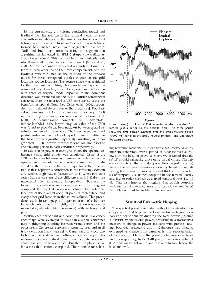
Figure 1.
Grand mean (n ¼ 11) ssVEP time series at electrode site Poz,
located just superior to the occipital pole. The three panels
show the time domain averages over the entire viewing period
(6,000 ms) for pleasant (top), neutral (middle), and unpleasant
(bottom) pictures.
r Keil et al. r
r 2924 r
For group analysis, each individual source space wastransformed to an average brain obtained for the 11 partic-ipants using a linear transformation implemented in thefieldtrip toolbox. To evaluate effects of picture content onssVEP amplitude across participants, paired t tests wereperformed on normalized power change and coherence ateach grid point, comparing the three content conditions ina pair-wise fashion (pleasant vs. neutral, unpleasant vs.neutral, pleasant vs. unpleasant). Significance thresholdswere determined by calculating a permutation distributionof t values for shuffled data to avoid alpha error accumu-lation [for a similar procedure see Keil et al., 2005]. Inbrief, 5,000 t-maps for each of the comparisons betweenthe three contents (i.e., pleasant vs. neutral, unpleasant vs.neutral, pleasant vs. unpleasant) were calculated fromdata in which the conditions were randomly shuffledwithin each participant, and the maximum values of eachmap entered the permutation distribution. The 2.5 and97.5% tails of the resulting distribution were then used toestablish thresholds for t values indicating significant dif-ferences between picture contents. A mass univariate t-mapping approach was chosen here over factorial or mul-tivariate designs, because this allows easier comparisonwith previous work on ssVEPs elicited by IAPS pictures[Keil et al., 2005], and the direction of any significant dif-ferences is more immediately evident, compared to an F-test.
For plotting purposes, the statistical maps were alignedwith standard brain surfaces representing the cortical sur-face based on the MNI template brain transformed to fitnative Talairach space and viewed using the AFNI surfacemapper, SUMA [Saad et al., 2004]. Cortical structures withsignificant effects are reported in MNI coordinates.Because we did not observe differences between unpleas-ant and pleasant picture content (see results), t-maps thatcompared emotionally arousing (i.e., pleasant and unpleas-ant) and neutral content are illustrated.
RESULTS
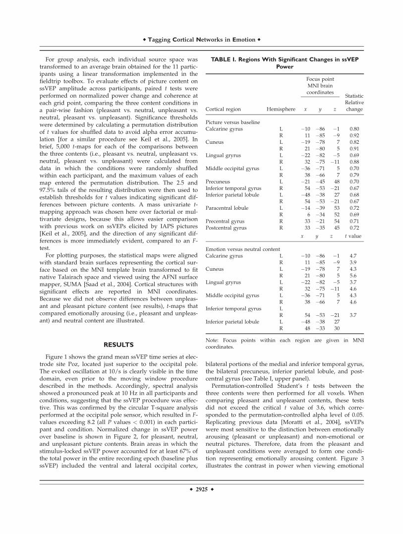
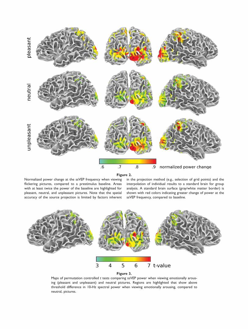
Figure 1 shows the grand mean ssVEP time series at elec-trode site Poz, located just superior to the occipital pole.The evoked oscillation at 10/s is clearly visible in the timedomain, even prior to the moving window proceduredescribed in the methods. Accordingly, spectral analysisshowed a pronounced peak at 10 Hz in all participants andconditions, suggesting that the ssVEP procedure was effec-tive. This was confirmed by the circular T-square analysisperformed at the occipital pole sensor, which resulted in F-values exceeding 8.2 (all P values < 0.001) in each partici-pant and condition. Normalized change in ssVEP powerover baseline is shown in Figure 2, for pleasant, neutral,and unpleasant picture contents. Brain areas in which thestimulus-locked ssVEP power accounted for at least 67% ofthe total power in the entire recording epoch (baseline plusssVEP) included the ventral and lateral occipital cortex,
bilateral portions of the medial and inferior temporal gyrus,the bilateral precuneus, inferior parietal lobule, and post-central gyrus (see Table I, upper panel).
Permutation-controlled Student’s t tests between thethree contents were then performed for all voxels. Whencomparing pleasant and unpleasant contents, these testsdid not exceed the critical t value of 3.6, which corre-sponded to the permutation-controlled alpha level of 0.05.Replicating previous data [Moratti et al., 2004], ssVEPswere most sensitive to the distinction between emotionallyarousing (pleasant or unpleasant) and non-emotional orneutral pictures. Therefore, data from the pleasant andunpleasant conditions were averaged to form one condi-tion representing emotionally arousing content. Figure 3illustrates the contrast in power when viewing emotional
TABLE I. Regions With Significant Changes in ssVEP
Power
Cortical region Hemisphere
Focus pointMNI braincoordinates
Statistic
x y z
Relativechange
Picture versus baselineCalcarine gyrus L �10 �86 �1 0.80
R 11 �85 �9 0.92Cuneus L �19 �78 7 0.82
R 21 �80 5 0.91Lingual gryrus L �22 �82 �5 0.69
R 32 �75 �11 0.88Middle occipital gyrus L �36 �71 5 0.70
R 38 �66 7 0.79Precuneus L �21 �45 48 0.70Inferior temporal gyrus R 54 �53 �21 0.67Inferior parietal lobule L �48 �38 27 0.68
R 54 �53 �21 0.67Paracentral lobule L �14 �39 53 0.72
R 6 �34 52 0.69Precentral gyrus R 33 �21 54 0.71Postcentral gyrus R 33 �35 45 0.72
x y z t value
Emotion versus neutral contentCalcarine gyrus L �10 �86 �1 4.7
R 11 �85 �9 3.9Cuneus L �19 �78 7 4.3
R 21 �80 5 5.6Lingual gryrus L �22 �82 �5 3.7
R 32 �75 �11 4.6Middle occipital gyrus L �36 �71 5 4.3
R 38 �66 7 4.6Inferior temporal gyrus L
R 54 �53 �21 3.7Inferior parietal lobule L �48 �38 27
R 48 �33 30
Note: Focus points within each region are given in MNIcoordinates.
r Tagging Cortical Networks in Emotion r
r 2925 r
Figure 2.
Normalized power change at the ssVEP frequency when viewing
flickering pictures, compared to a prestimulus baseline. Areas
with at least twice the power of the baseline are highlighted for
pleasant, neutral, and unpleasant pictures. Note that the spatial
accuracy of the source projection is limited by factors inherent
in the projection method (e.g., selection of grid points) and the
interpolation of individual results to a standard brain for group
analysis. A standard brain surface (gray/white matter border) is
shown with red colors indicating greater change of power at the
ssVEP frequency, compared to baseline.
Figure 3.
Maps of permutation controlled t tests comparing ssVEP power when viewing emotionally arous-
ing (pleasant and unpleasant) and neutral pictures. Regions are highlighted that show above
threshold difference in 10-Hz spectral power when viewing emotionally arousing, compared to
neutral, pictures.
(pleasant and unpleasant) pictures, compared to viewingneutral pictures, thresholded at the critical t value of 3.6(P < 0.05 corrected). Stimulus-locked ssVEP power wasenhanced for emotionally arousing pictures in lateral occi-pital cortices and in the same parietal areas—bilateral pre-cuneus and intraparietal lobules—where spectral powerwas generally enhanced following picture onset (see TableI, lower panel).
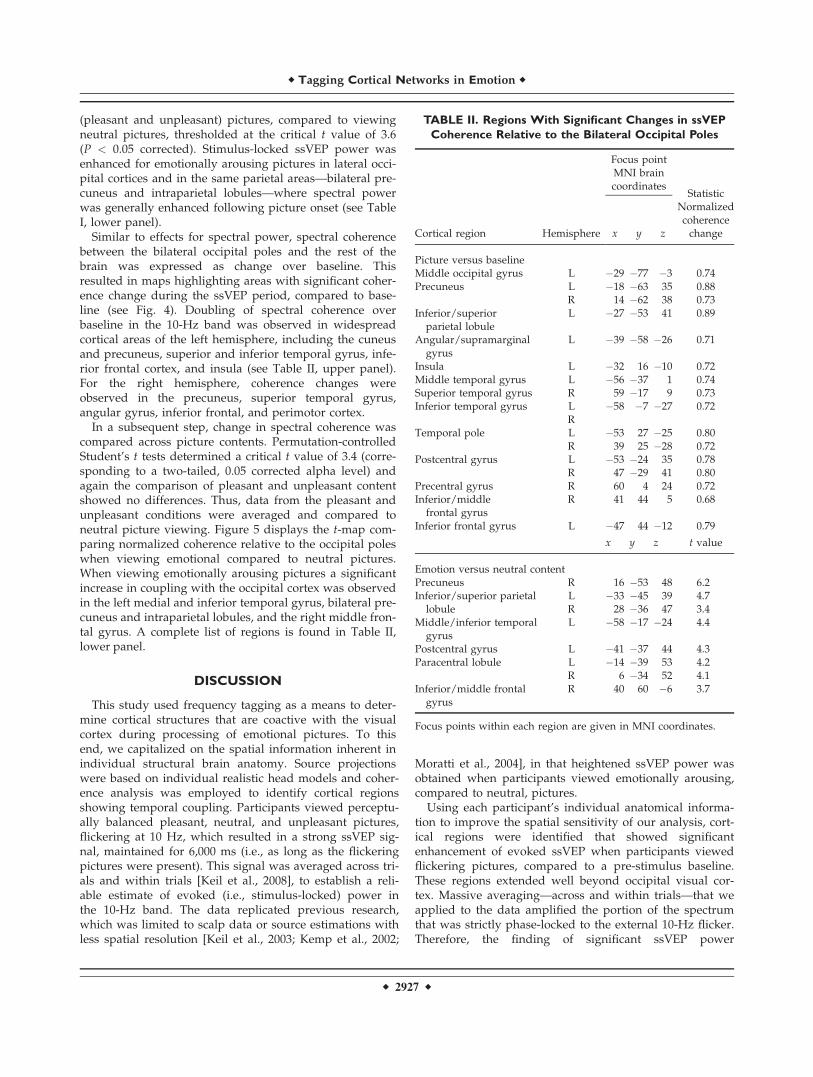
Similar to effects for spectral power, spectral coherencebetween the bilateral occipital poles and the rest of thebrain was expressed as change over baseline. Thisresulted in maps highlighting areas with significant coher-ence change during the ssVEP period, compared to base-line (see Fig. 4). Doubling of spectral coherence overbaseline in the 10-Hz band was observed in widespreadcortical areas of the left hemisphere, including the cuneusand precuneus, superior and inferior temporal gyrus, infe-rior frontal cortex, and insula (see Table II, upper panel).For the right hemisphere, coherence changes wereobserved in the precuneus, superior temporal gyrus,angular gyrus, inferior frontal, and perimotor cortex.
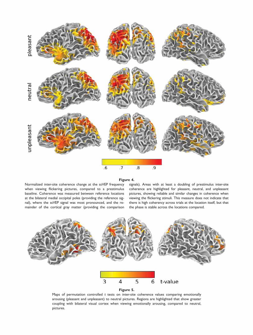
In a subsequent step, change in spectral coherence wascompared across picture contents. Permutation-controlledStudent’s t tests determined a critical t value of 3.4 (corre-sponding to a two-tailed, 0.05 corrected alpha level) andagain the comparison of pleasant and unpleasant contentshowed no differences. Thus, data from the pleasant andunpleasant conditions were averaged and compared toneutral picture viewing. Figure 5 displays the t-map com-paring normalized coherence relative to the occipital poleswhen viewing emotional compared to neutral pictures.When viewing emotionally arousing pictures a significantincrease in coupling with the occipital cortex was observedin the left medial and inferior temporal gyrus, bilateral pre-cuneus and intraparietal lobules, and the right middle fron-tal gyrus. A complete list of regions is found in Table II,lower panel.
DISCUSSION
This study used frequency tagging as a means to deter-mine cortical structures that are coactive with the visualcortex during processing of emotional pictures. To thisend, we capitalized on the spatial information inherent inindividual structural brain anatomy. Source projectionswere based on individual realistic head models and coher-ence analysis was employed to identify cortical regionsshowing temporal coupling. Participants viewed perceptu-ally balanced pleasant, neutral, and unpleasant pictures,flickering at 10 Hz, which resulted in a strong ssVEP sig-nal, maintained for 6,000 ms (i.e., as long as the flickeringpictures were present). This signal was averaged across tri-als and within trials [Keil et al., 2008], to establish a reli-able estimate of evoked (i.e., stimulus-locked) power inthe 10-Hz band. The data replicated previous research,which was limited to scalp data or source estimations withless spatial resolution [Keil et al., 2003; Kemp et al., 2002;
Moratti et al., 2004], in that heightened ssVEP power wasobtained when participants viewed emotionally arousing,compared to neutral, pictures.
Using each participant’s individual anatomical informa-tion to improve the spatial sensitivity of our analysis, cort-ical regions were identified that showed significantenhancement of evoked ssVEP when participants viewedflickering pictures, compared to a pre-stimulus baseline.These regions extended well beyond occipital visual cor-tex. Massive averaging—across and within trials—that weapplied to the data amplified the portion of the spectrumthat was strictly phase-locked to the external 10-Hz flicker.Therefore, the finding of significant ssVEP power
TABLE II. Regions With Significant Changes in ssVEP
Coherence Relative to the Bilateral Occipital Poles
Cortical region Hemisphere
Focus pointMNI braincoordinates
Statistic
x y z
Normalizedcoherencechange
Picture versus baselineMiddle occipital gyrus L �29 �77 �3 0.74Precuneus L �18 �63 35 0.88
R 14 �62 38 0.73Inferior/superior
parietal lobuleL �27 �53 41 0.89
Angular/supramarginalgyrus
L �39 �58 �26 0.71
Insula L �32 16 �10 0.72Middle temporal gyrus L �56 �37 1 0.74Superior temporal gyrus R 59 �17 9 0.73Inferior temporal gyrus L �58 �7 �27 0.72
RTemporal pole L �53 27 �25 0.80
R 39 25 �28 0.72Postcentral gyrus L �53 �24 35 0.78
R 47 �29 41 0.80Precentral gyrus R 60 4 24 0.72Inferior/middle
frontal gyrusR 41 44 5 0.68
Inferior frontal gyrus L �47 44 �12 0.79
x y z t value
Emotion versus neutral contentPrecuneus R 16 �53 48 6.2Inferior/superior parietal
lobuleL �33 �45 39 4.7R 28 �36 47 3.4
Middle/inferior temporalgyrus
L �58 �17 �24 4.4
Postcentral gyrus L �41 �37 44 4.3Paracentral lobule L �14 �39 53 4.2
R 6 �34 52 4.1Inferior/middle frontal
gyrusR 40 60 �6 3.7
Focus points within each region are given in MNI coordinates.
r Tagging Cortical Networks in Emotion r
r 2927 r
Figure 4.
Normalized inter-site coherence change at the ssVEP frequency
when viewing flickering pictures, compared to a prestimulus
baseline. Coherence was measured between reference locations
at the bilateral medial occipital poles (providing the reference sig-
nal), where the ssVEP signal was most pronounced, and the re-
mainder of the cortical gray matter (providing the comparison
signals). Areas with at least a doubling of prestimulus inter-site
coherence are highlighted for pleasant, neutral, and unpleasant
pictures, showing reliable and similar changes in coherence when
viewing the flickering stimuli. This measure does not indicate that
there is high coherency across trials at the location itself, but that
the phase is stable across the locations compared.
Figure 5.
Maps of permutation controlled t tests on inter-site coherence values comparing emotionally
arousing (pleasant and unpleasant) to neutral pictures. Regions are highlighted that show greater
coupling with bilateral visual cortex when viewing emotionally arousing, compared to neutral,
pictures.
modulation in inferior temporal and dorsal parietal visualcortex suggests that the flicker systematically and rhythmi-cally engaged visual association cortex, resonating with thebrightness-modulated stimulus train at a frequency of 10 Hz.This suggests a close functional relationship between earlyvisual and temporo-parietal areas during rapid serial visualprocessing. The present data imply that these functional linksoperate at a steady repetitive rate, suggesting rapid connec-tions between visual and extra-visual areas. In terms of later-ality, earlier work reported that the left hemisphere shows apreponderance of ssVEP power during stimulation withcomplex IAPS pictures, whereas phase synchrony is oftenmore pronounced on the right [Keil et al., 2005].
The fact that many visual associative structures showedresonance with the 10-Hz flicker and coherent temporalrelationships with occipital visual cortex is consistent withhypotheses that visual representations formed duringssVEP stimulation are not established independently andde novo for each cycle, but that information from previouscycles is retained in a functionally coupled neural networkincluding sensory as well as higher-order areas [Harleet al., 2004; Keil et al., 2007]. In the present study, oscilla-tory network communication was established as a distrib-uted set of connected regions coherently engaged anddisengaged in synchrony with the reference location in thevisual cortex, at a rate of 10 oscillations s�1. Coordinatedactivity in such functional networks may assist in the for-mation of predictions regarding the nature of incomingstimuli in a given context [Engel et al., 2001]. Although itis possible that widespread coordinated activity is specificto complex stimuli such as IAPS pictures and is not seenwith more simple stimuli (faces, geometric shapes) oftenused in the cognitive neuroscience laboratory, photographsas used here are most similar to the visual scenes proc-essed in daily life. In the laboratory, on the other hand,simplistic grating stimuli receiving motivational relevancethrough classical conditioning have been shown to beassociated with more circumscribed connectivity that isconstrained to the occipital lobe [Keil et al., 2007]. Thissuggests that more ecologically valid, complex visualscenes engage a broader array of cortical structures in acoherent fashion, which probably reflects an integrativeneural mechanism linking complex visual information tosemantic content and behavioral response representationsthat are important for relevant action.
To further examine the spatio-temporal structure of suchtightly coupled cortical networks, inter-area coupling wasquantified by means of coherence analysis [Nunez et al.,1997]. It is crucial for spatio-temporal analysis of brainelectric activity that false positives due to volume conduc-tion, choice of reference electrodes, etc., are avoided[Nunez et al., 1999]. In the current study, we adopted mul-tiple strategies to reduce effects leading to spurious differ-ences in coherence: first, the beamforming approachenhances spatially independent activity [Gross et al., 2001]by actively suppressing the contributions of other, tempo-rally identical, sources to a given location in the source
model (see Methods). Second, we examined differencesbetween picture contents, thus keeping any residual, non-specific, influences on the measurement of coherence con-stant. This approach highlights content-related changes ofpower and coherence that were under experimental control,as recommended by Nunez et al. [1999]. A further meth-odological improvement over earlier work concerns theconstraints of source projections. Beamforming depends toa large extent on the validity of the volume conductormodel and its relationship with the sensor space, i.e., theleadfield [Huang et al., 2004], and here we used realistichead models derived from individual anatomy as reflectedin the high resolution structural MRI data.
In terms of large-scale neurophysiology, the oscillatoryfunctional network observed in this study may provide re-entrant feedback to visual areas, originating from higherorder visual cortices, and following an initial bottom-upcascade of visual analysis in each cycle of processing [Mar-tinez et al., 1999]. Work examining classical conditioningwith simple visual stimuli has suggested that local connec-tivity within the extended visual cortex increases with pro-longed exposure to the learning regime [Keil et al., 2007].Thus, one could speculate that re-entrant modulation fromanterior, distant structures is reduced over trials, giving riseto more effective, highly connected local networks in theoccipital lobe [Gruber et al., 2004]. Work in experimentalanimals has demonstrated strong re-entrant connectionsinto visual cortex originating in the amygdala [Amaralet al., 2003]. The present study supports that such feedbackmay also arise from higher-order (parietal and frontal) cort-ical areas and may act to enhance sensitivity of visual neu-rons coding for motivationally relevant features present inthe field of view, as hypothesized in the context of spatialattention [Hamker, 2005] as well as classical and instrumen-tal conditioning [Keil, 2004]. In this vein, the ssVEP as ameasure of ongoing stimulus processing in visual cortexmight be able to reflect biasing signals from higher-ordercortical areas [Kastner and Ungerleider, 2000], changing thesensitivity of visual cortical neurons in favor of featuresassociated with emotionally arousing content.
The present data provide evidence that (i) increasedcoupling for arousing content occurs for large-scale (neuralmass) electrical activity on the cortical level, and (ii) thecortical regions tagged as elements in such a functionalnetwork are consistent with regions identified by means offunctional imaging. Taken together, this suggests that cort-ical structures interact in a rapid fashion, establishing net-works linking perceptual features to attentive behaviorand ultimately action preparation.
REFERENCES
Aftanas LI, Reva NV, Varlamov AA, Pavlov SV, Makhnev VP(2004): Analysis of evoked EEG synchronization and desynch-ronization in conditions of emotional activation in humans:Temporal and topographic characteristics. Neurosci BehavPhysiol 34:859–867.
r Tagging Cortical Networks in Emotion r
r 2929 r
Amaral DG, Behniea H, Kelly JL (2003): Topographic organizationof projections from the amygdala to the visual cortex in themacaque monkey. Neuroscience 118:1099–1120.
Bradley MM, Sabatinelli D, Lang PJ, Fitzsimmons JR, King W,Desai P (2003): Activation of the visual cortex in motivatedattention. Behav Neurosci 117:369–380.
Costa VD, Lang PJ, Sabatinelli D, Versace F, Bradley MM (2010):Emotional imagery: Assessing pleasure and arousal in thebrain’s reward circuitry. Hum Brain Mapp 31:1446–1457.
Desimone R (1996): Neural mechanisms for visual memory andtheir role in attention. Proc Natl Acad Sci USA 93:13494–13499.
Di Russo F, Pitzalis S, Aprile T, Spitoni G, Patria F, Stella A,Spinelli D, Hillyard SA (2007): Spatiotemporal analysis of thecortical sources of the steady-state visual evoked potential.Hum Brain Mapp 28:323–334.
Engel AK, Fries P, Singer W (2001): Dynamic predictions: Oscilla-tions and synchrony in top-down processing. Nat Rev Neuro-sci 2:704–716.
Gross J, Kujala J, Hamalainen M, Timmermann L, Schnitzler A,Salmelin R (2001): Dynamic imaging of coherent sources:Studying neural interactions in the human brain. Proc NatlAcad Sci USA 98:694–699.
Gruber T, Malinowski P, Muller MM (2004): Modulation of oscil-latory brain activity and evoked potentials in a repetition pri-ming task in the human EEG. Eur J Neurosci 19:1073–1082.
Hamker FH (2005): The reentry hypothesis: The putative interac-tion of the frontal eye field, ventrolateral prefrontal cortex, andareas V4, IT for attention and eye movement. Cereb Cortex15:431–447.
Harle M, Rockstroh BS, Keil A, Wienbruch C, Elbert TR (2004):Mapping the brain’s orchestration during speech comprehen-sion: Task-specific facilitation of regional synchrony in neuralnetworks. BMC Neurosci 5:40.
Herrmann CS (2001): Human EEG responses to 1–100 Hz flicker:Resonance phenomena in visual cortex and their potential cor-relation to cognitive phenomena. Exp Brain Res 137:346–353.
Hillebrand A, Barnes GR (2005): Beamformer analysis of MEGdata. Int Rev Neurobiol 68:149–171.
Huang MX, Shih JJ, Lee RR, Harrington DL, Thoma RJ, WeisendMP, Hanlon F, Paulson KM, Li T, Martin K, Millers GA, Can-ive JM (2004): Commonalities and differences among vector-ized beamformers in electromagnetic source imaging. BrainTopogr 16:139–158.
Junghofer M, Elbert T, Leiderer P, Berg P, Rockstroh B (1997):Mapping EEG-potentials on the surface of the brain: A strategyfor uncovering cortical sources. Brain Topogr 9:203–217.
Junghofer M, Elbert T, Tucker DM, Rockstroh B (2000): Statisticalcontrol of artifacts in dense array EEG/MEG studies. Psycho-physiology 37:523–532.
Kastner S, Ungerleider LG (2000): Mechanisms of visual attentionin the human cortex. Annu Rev Neurosci 23:315–341.
Keil A (2004): The role of human prefrontal cortex in motivatedperception and behavior: A macroscopic perspective. In: OtaniS, editor. Prefrontal Cortex: From Synaptic Plasticity to Cogni-tion. New York: Kluwer. pp 245–267.
Keil A, Gruber T, Muller MM, Moratti S, Stolarova M, BradleyMM, Lang PJ (2003): Early modulation of visual perception byemotional arousal: Evidence from steady-state visual evokedbrain potentials. Cogn Affect Behav Neurosci 3:195–206.
Keil A, Moratti S, Sabatinelli D, Bradley MM, Lang PJ (2005): Addi-tive effects of emotional content and spatial selective attentionon electrocortical facilitation. Cereb Cortex 15:1187–1197.
Keil A, Stolarova M, Moratti S, Ray WJ (2007): Adaptation in vis-ual cortex as a mechanism for rapid discrimination of aversivestimuli. Neuroimage 36:472–479.
Keil A, Smith JC, Wangelin B, Sabatinelli D, Bradley MM, Lang PJ(2008): Electrocortical and electrodermal responses co-vary as afunction of emotional arousal: A single-trial analysis. Psycho-physiology 45:511–515.
Keil A, Sabatinelli D, Ding M, Lang PJ, Ihssen N, Heim S (2009):Re-entrant projections modulate visual cortex in affective per-ception: Directional evidence from granger causality analysis.Hum Brain Mapp 30:532–540.
Kemp AH, Gray MA, Eide P, Silberstein RB, Nathan PJ (2002):Steady-state visually evoked potential topography during proc-essing of emotional valence in healthy subjects. Neuroimage17:1684–1692.
Lachaux JP, Chavez M, Lutz A (2003): A simple measure of corre-lation across time, frequency and space between continuousbrain signals. J Neurosci Methods 123:175–188.
Lang PJ (1980): Behavioral treatment and bio-behavioral assess-ment: Computer applications. In: Sidowski JB, Johnson JH,William TA, editors. Technology in Mental Health Care Deliv-ery Systems. Norwood, NJ: Ablex. pp 119–137.
Lang PJ, Bradley MM (2010): Emotion and the motivational brain.Biol Psychol 83:437–450.
Lang PJ, Bradley MM, Fitzsimmons JR, Cuthbert BN, Scott JD,Moulder B, Nangia V (1998): Emotional arousal and activationof the visual cortex: An fMRI analysis. Psychophysiology35:199–210.
Lang PJ, Bradley MM, Cuthbert BN (2005): International AffectivePicture System: Technical Manual and Affective Ratings. Gain-esville, FL: NIMH Center for the Study of Emotion andAttention.
Martinez A, Anllo-Vento L, Sereno MI, Frank LR, Buxton RB,Dubowitz DJ, Wong EC, Hinrichs H, Heinze HJ, Hillyard SA(1999): Involvement of striate and extrastriate visual corticalareas in spatial attention. Nat Neurosci 2:364–369.
Mast J, Victor JD (1991): Fluctuations of steady-state VEPs: Interac-tion of driven evoked potentials and the EEG. Electroencepha-logr Clin Neurophysiol 78:389–401.
McMains SA, Fehd HM, Emmanouil TA, Kastner S (2007): Mecha-nisms of feature- and space-based attention: Response modula-tion and baseline increases. J Neurophysiol 98:2110–2121.
Michel CM, Murray MM, Lantz G, Gonzalez S, Spinelli L, Gravede Peralta R (2004): EEG source imaging. Clin Neurophysiol115:2195–2222.
Moratti S, Keil A (2005): Cortical activation during Pavlovian fearconditioning depends on heart rate response patterns: AnMEG study. Brain Res Cogn Brain Res 25:459–471.
Moratti S, Keil A (2009): Not what you expect: Experience but notexpectancy predicts conditioned responses in human visualand supplementary cortex. Cereb Cortex 19:2803–2809.
Moratti S, Keil A, Stolarova M (2004): Motivated attention in emo-tional picture processing is reflected by activity modulation incortical attention networks. Neuroimage 21:954–964.
Muller MM, Malinowski P, Gruber T, Hillyard SA (2003): Sus-tained division of the attentional spotlight. Nature 424:309–312.
Regan D (1989): Human Brain Electrophysiology: Evoked Poten-tials and Evoked Magnetic Fields in Science and Medicine.New York: Elsevier.
Saad ZS, Reynolds RC, Argall BD, Japee S, Cox RW (2004):SUMA:an interface for surface-based intra- and inter-subjectanalysis with AFNI. In: Proceedings of the 2004 IEEE Interna-tional Symposium on Biomedical Imaging, Arlington, VA.New York: IEEE .p 1510–1513.
Sabatinelli D, Bradley MM, Fitzsimmons JR, Lang PJ (2005): Paral-lel amygdala and inferotemporal activation reflect emotionalintensity and fear relevance. Neuroimage 24:1265–1270.
Sabatinelli D, Lang PJ, Keil A, Bradley MM (2007): Emotional per-ception: Correlation of functional MRI and event related poten-tials. Cereb Cortex 17:1066–1073.
Sabatinelli D, Lang PJ, Bradley MM, Costa VD, Keil A (2009): Thetiming of emotional discrimination in human amygdala andventral visual cortex. J Neurosci 29:14864–14868.
Silberstein RB, Ciorciari J, Pipingas A (1995): Steady-state visuallyevoked potential topography during the Wisconsin card sort-ing test. Electroencephalogr Clin Neurophysiol 96:24–35.
Victor JD, Mast J (1991): A new statistic for steady-stateevoked potentials. Electroencephalogr Clin Neurophysiol 78:378–388.
Vuilleumier P, Driver J (2007): Modulation of visual processing byattention and emotion: Windows on causal interactionsbetween human brain regions. Philos Trans R Soc Lond B BiolSci 362:837–855.
Ward DM, Jones RD, Bones PJ, Carroll GJ (1999): Enhancement ofdeep epileptiform activity in the EEG via 3-D adaptive spatialfiltering. IEEE Trans Biomed Eng 46:707–716.
Yantis S (2008): The neural basis of selective attention: Corticalsources and targets of attentional modulation. Curr Dir Psy-chol Sci 17:86–90.