Tailored cancer immunotherapy using combinations of chemotherapy and a mixture of antibodies against EGF-receptor ligands Moshit Lindzen a , Sara Lavi a , Orith Leitner b , and Yosef Yarden a,1 Departments of a Biological Regulation and b Biological Services, The Weizmann Institute of Science, Rehovot 76100, Israel Communicated by Michael Sela, The Weizmann Institute of Science, Rehovot, Israel, June 12, 2010 (received for review January 24, 2010) Growth factors are implicated in several processes essential for cancer progression. Specifically, growth factors that bind to ErbB family receptors have been implicated in cell proliferation and in resistance of solid tumors to chemotherapy. We quantified ligand secretion by several human cancer cell lines, and generated mAbs against two ligands, namely TGF-α and heparin-binding EGF-like growth factor. These growth factors are frequently secreted by pan- creatic tumor cell lines, including BxPC3 cells. The monoclonal anti- bodies were tested for their antigen specificity and ability to inhibit growth of BxPC3 cells in vitro. Combining the two antibodies resulted in enhanced inhibition of BxPC3 cell growth, both in vitro and in tumor-bearing animals. Hence, we combined the two antibodies with gemcitabine, an effective chemotherapeutic drug commonly used to treat pancreatic cancer patients. Because treatment with a combination of two monoclonal antibodies enhanced the ability of chemotherapy to inhibit BxPC3 tumors in mice, we propose a gen- eral cancer therapeutic strategy that entails profiling the repertoire of growth factors secreted by a tumor, and combining with chemo- therapy several antibodies capable of blocking autocrine ligands. cancer therapy | growth factor | monoclonal antibody | signal transduction T he ErbB family of receptors and their ligands play important roles in development, as well as in tissue remodeling, through- out adulthood. Ligand-binding is followed by receptor dimeriz- ation and phosphorylation, and results in various cellular pro- cesses, including proliferation (1). There are four ErbB receptors. ErbB-1 (EGFR) binds seven growth factors, including the EGF, TGF-α, heparin-binding EGF-like growth factor (HB-EGF), and amphiregulin (AR) (2). ErbB-3 and ErbB-4 bind a distinct group of growth factors called neuregulins (NRGs) (3), but ErbB-2/HER2 has no known ligand (4, 5). All ErbB-family ligands share a 50 to 60 amino acid-long sequence containing six cysteines (6). EGF-like ligands bind and activate receptors on distant cells, neighboring cells, or on the cells of their origin, mechanisms termed endocrine, paracrine, or autocrine, respectively. ErbB proteins are involved in several types of human cancer. Clinical studies indicate that overexpression of one or more ligands correlates with decreased patient survival; for example, expression of TGF-α in colorectal tumors is associated with a greater than 50- fold increased risk of liver metastases (7). In bladder cancer, the elevated expression of a number of ligands is linked to decreased patient survival (8). Likewise, increased expression of TGF-α in head and neck tumors is correlated with decreased survival (9). Moreover, tumor cell expression of some ligands is associated with resistance to chemotherapeutic drugs (10, 11). Despite these observations, the currently approved drugs for the treatment of cancers driven by the ErbB family target the receptors, rather than the ligands, and they include either monoclonal antireceptor antibodies, or tyrosine kinase inhibitors (TKIs) (12, 13). Acquired resistance, often associated with up-regulation of ligands (14–16) or other receptors (17–20), limits efficacy of anti- ErbB drugs. Human breast-cancer cells selected in vivo for re- sistance to trastuzumab overexpress EGFR and ErbB ligands (18). Because of resistance and moderate clinical efficacies of anti- receptor antibodies and TKIs, it is worthwhile considering alter- native strategies. For example, fractions of colorectal and other tumors respond to bevacizumab, an antibody that binds the vas- cular endothelial growth factor (21, 22). The multiplicity of EGF- like ligands, along with extensive receptor-receptor interactions, are potential impediments to similar applications of antiligand antibodies. Nevertheless, because the repertoires of autocrine growth factors secreted by individual tumors is often limited to two to three ligands, we assumed that it would be feasible to profile the spectrum of ligands secreted by specific carcinomas, combine the respective antigrowth factor antibodies, and test their combination with chemotherapy. Here, we focused on TGF-α and HB-EGF. In parallel to the generation of antagonistic antibodies, we screened human tumor cell lines and identified a pancreatic line that secretes both growth factors. We describe in vitro assays and tests in animals, collectively supporting the feasibility of a “tailored” immunotherapeutic strategy able to enhance the effect of che- motherapy. This study opens the way for combinations of antibody pairs with receptor antagonists, such as kinase inhibitors. Results As a prelude to testing our working hypothesis, we examined se- cretion of different EGF-like ligands by tumor cell lines. For this purpose, we used an immunological kit able to detect EGF, TGF-α, HB-EGF, and AR. The assay was performed on media condi- tioned by 13 carcinoma cell lines of a wide variety of tumors, such as ovary, breast, lung, and pancreas (Table 1). As expected, the assay detected distinct combinations of growth factors and wide ranges of expression levels. Notably, unlike HB-EGF, AR, and TGF-α, which were abundantly secreted by several tumor cell lines, EGF was either absent or very low. Because an alternative assay that used real-time PCR and mRNA isolated from BxPC3 cells confirmed high expression of TGF-α and HB-EGF, but did not detect comparable levels of AR, NRG, betacellulin, and epi- regulin, we next aimed at the generation of mAbs capable of blocking the action of HB-EGF and TGF-α. Cloning, Expression, and Biological Activity of EGF-Like Ligands. It is known that highly conserved three-disulfide bonds are responsible for the correct folding and activity of the EGF-like domain of all ErbB ligands (23). Therefore, we chose to express the EGF-like domain of EGFR-specific ligands as fusion proteins, linked to the thioredoxin protein (TRX) (Fig. 1A). The fused protein also contained a histidine repeat, as well as a factor Xa cleavage site, to enable specific release of the EGF-like domain. Following ex- pression in bacteria, the two ligands were purified using a metal column (Fig. 1B), and their biological activity was verified by confirming their ability to induce EGFR phosphorylation (Fig. 1C). The results we obtained ensured that the recombinant Author contributions: M.L., S.L., O.L., and Y.Y. designed research; M.L., S.L., and O.L. performed research; M.L. and Y.Y. analyzed data; and M.L. and Y.Y. wrote the paper. The authors declare no conflict of interest. 1 To whom correspondence should be addressed. E-mail: [email protected]. www.pnas.org/cgi/doi/10.1073/pnas.1006218107 PNAS | July 13, 2010 | vol. 107 | no. 28 | 12559–12563 CELL BIOLOGY
Transcript
Tailored cancer immunotherapy using combinationsof chemotherapy and a mixture of antibodiesagainst EGF-receptor ligandsMoshit Lindzena, Sara Lavia, Orith Leitnerb, and Yosef Yardena,1
Departments of aBiological Regulation and bBiological Services, The Weizmann Institute of Science, Rehovot 76100, Israel
Communicated by Michael Sela, The Weizmann Institute of Science, Rehovot, Israel, June 12, 2010 (received for review January 24, 2010)
Growth factors are implicated in several processes essential forcancer progression. Specifically, growth factors that bind to ErbBfamily receptors have been implicated in cell proliferation and inresistance of solid tumors to chemotherapy. We quantified ligandsecretion by several human cancer cell lines, and generated mAbsagainst two ligands, namely TGF-α and heparin-binding EGF-likegrowth factor. These growth factors are frequently secreted by pan-creatic tumor cell lines, including BxPC3 cells. The monoclonal anti-bodies were tested for their antigen specificity and ability to inhibitgrowthofBxPC3cells invitro.Combining thetwoantibodies resultedin enhanced inhibition of BxPC3 cell growth, both in vitro and intumor-bearing animals. Hence, we combined the two antibodieswith gemcitabine, an effective chemotherapeutic drug commonlyused to treat pancreatic cancer patients. Because treatment witha combination of two monoclonal antibodies enhanced the abilityof chemotherapy to inhibit BxPC3 tumors inmice, we propose a gen-eral cancer therapeutic strategy that entails profiling the repertoireof growth factors secreted by a tumor, and combining with chemo-therapy several antibodies capable of blocking autocrine ligands.
cancer therapy | growth factor | monoclonal antibody | signal transduction
The ErbB family of receptors and their ligands play importantroles in development, as well as in tissue remodeling, through-
out adulthood. Ligand-binding is followed by receptor dimeriz-ation and phosphorylation, and results in various cellular pro-cesses, including proliferation (1). There are four ErbB receptors.ErbB-1 (EGFR) binds seven growth factors, including the EGF,TGF-α, heparin-binding EGF-like growth factor (HB-EGF), andamphiregulin (AR) (2). ErbB-3 and ErbB-4 bind a distinct groupof growth factors called neuregulins (NRGs) (3), but ErbB-2/HER2has no known ligand (4, 5). All ErbB-family ligands share a 50 to60 amino acid-long sequence containing six cysteines (6). EGF-likeligands bind and activate receptors on distant cells, neighboringcells, or on the cells of their origin, mechanisms termed endocrine,paracrine, or autocrine, respectively.ErbB proteins are involved in several types of human cancer.
Clinical studies indicate that overexpression of one ormore ligandscorrelates with decreased patient survival; for example, expressionof TGF-α in colorectal tumors is associated with a greater than 50-fold increased risk of liver metastases (7). In bladder cancer, theelevated expression of a number of ligands is linked to decreasedpatient survival (8). Likewise, increased expression of TGF-α inhead and neck tumors is correlated with decreased survival (9).Moreover, tumor cell expression of some ligands is associated withresistance to chemotherapeutic drugs (10, 11). Despite theseobservations, the currently approved drugs for the treatment ofcancers driven by the ErbB family target the receptors, rather thanthe ligands, and they include either monoclonal antireceptorantibodies, or tyrosine kinase inhibitors (TKIs) (12, 13).Acquired resistance, often associated with up-regulation of
ligands (14–16) or other receptors (17–20), limits efficacy of anti-ErbB drugs. Human breast-cancer cells selected in vivo for re-sistance to trastuzumab overexpress EGFR and ErbB ligands (18).Because of resistance and moderate clinical efficacies of anti-
receptor antibodies and TKIs, it is worthwhile considering alter-native strategies. For example, fractions of colorectal and othertumors respond to bevacizumab, an antibody that binds the vas-cular endothelial growth factor (21, 22). The multiplicity of EGF-like ligands, along with extensive receptor-receptor interactions,are potential impediments to similar applications of antiligandantibodies. Nevertheless, because the repertoires of autocrinegrowth factors secreted by individual tumors is often limited to twoto three ligands, we assumed that it would be feasible to profile thespectrum of ligands secreted by specific carcinomas, combine therespective antigrowth factor antibodies, and test their combinationwith chemotherapy. Here, we focused on TGF-α and HB-EGF. Inparallel to the generation of antagonistic antibodies, we screenedhuman tumor cell lines and identified a pancreatic line thatsecretes both growth factors. We describe in vitro assays and testsin animals, collectively supporting the feasibility of a “tailored”immunotherapeutic strategy able to enhance the effect of che-motherapy. This study opens the way for combinations of antibodypairs with receptor antagonists, such as kinase inhibitors.
ResultsAs a prelude to testing our working hypothesis, we examined se-cretion of different EGF-like ligands by tumor cell lines. For thispurpose, we used an immunological kit able to detect EGF, TGF-α,HB-EGF, and AR. The assay was performed on media condi-tioned by 13 carcinoma cell lines of a wide variety of tumors, suchas ovary, breast, lung, and pancreas (Table 1). As expected, theassay detected distinct combinations of growth factors and wideranges of expression levels. Notably, unlike HB-EGF, AR, andTGF-α, which were abundantly secreted by several tumor celllines, EGF was either absent or very low. Because an alternativeassay that used real-time PCR and mRNA isolated from BxPC3cells confirmed high expression of TGF-α and HB-EGF, but didnot detect comparable levels of AR, NRG, betacellulin, and epi-regulin, we next aimed at the generation of mAbs capable ofblocking the action of HB-EGF and TGF-α.
Cloning, Expression, and Biological Activity of EGF-Like Ligands. It isknown that highly conserved three-disulfide bonds are responsiblefor the correct folding and activity of the EGF-like domain of allErbB ligands (23). Therefore, we chose to express the EGF-likedomain of EGFR-specific ligands as fusion proteins, linked to thethioredoxin protein (TRX) (Fig. 1A). The fused protein alsocontained a histidine repeat, as well as a factor Xa cleavage site, toenable specific release of the EGF-like domain. Following ex-pression in bacteria, the two ligands were purified using a metalcolumn (Fig. 1B), and their biological activity was verified byconfirming their ability to induce EGFR phosphorylation (Fig.1C). The results we obtained ensured that the recombinant
Author contributions: M.L., S.L., O.L., and Y.Y. designed research; M.L., S.L., and O.L.performed research; M.L. and Y.Y. analyzed data; and M.L. and Y.Y. wrote the paper.
The authors declare no conflict of interest.1To whom correspondence should be addressed. E-mail: [email protected].
ligands represented the respective functionally active conforma-tion. Hence, mice were subsequently immunized with the activeTRX-fused ligands.
Generation of an Antagonistic Antibody Directed Against the EGF-Like Domain of TGF-α. Following four injections of theTRX–TGF-αfusion protein, sera were obtained from mice and examined forantiligand responses. To facilitate screening of antisera and hy-bridoma supernatants,weestablishedaCHOcell line that expressesthe EGF-like domain of TGF-α at the plasma membrane. For thispurpose, theEGF-likedomainwas fused to a signal peptide, anHA-peptide tag, and aGPI (glycosyl phosphatidylinositol) anchormotif,which is responsible for lipid-based anchoring at the plasma mem-brane (Fig. 1D). Antisera of immunized mice were tested for theirability to inhibit ligand-induced EGFR phosphorylation (Fig. 1E).Subsequently, the spleens of two mice were used to establishhybridomas, which were screened for their ability to recognize cellsurface-exposed GPI–TGF-α fusion protein. To functionally char-acterize a selected anti–TGF-αmAb, denoted mAb-551, we testedits specificity using anELISAassay. This assay confirmedbinding toTGF-α but not to six other ligandswe tested (Fig. 2A). Furthermore,
the anti–TGF-α mAb could immunopercipatate a commercialpreparation of TGF-α (Fig. 2B) and specifically inhibit TGF-α–in-ducedEGFRphosphorylation (Fig. 2C).Notably, neitherEGF-norHB-EGF–induced activation of EGFR were inhibited. Taken to-gether, these results establish suitability ofmAb-551 for attempts tointercept TGF-α-mediated autocrine loops.
Generation of an Antagonistic Antibody Directed Against the EGF-Like Domain of HB-EGF. To enable combination treatments, wesimilarly generated mAbs to another major growth-stimulatingligand, HB-EGF. Using three mAbs to HB-EGF and an immo-bilized HB-EGF, we confirmed antigen specificity, along withabsence of cross-reactivity (Fig. 3A). In the next step, we con-firmed the ability of two positive hybridoma clones to immuno-precipitate a commercial preparation of HB-EGF (Fig. 3B). Inaddition, by testing HB-EGF-induced tyrosine phosphorylationin HeLa and in T47D mammary cancer cells, we concluded thatthe three mAbs to HB-EGF variably inhibited ligand-inducedreceptor activation (Fig. 3C). These functional assays selectedmAb-898 for further studies in vitro and in animals.
Table 1. In vitro secretion of EGF-like ligands by human cancer cell lines
Cell line Tumor type TGF-α EGF HB-EGF AR
MDA-MB-231 Breast 111 ± 1 — 617 ± 28 1607 ± 171MDA-MB-468 Breast — — 64 ± 7 —
The indicated cell lines (1 × 10⁶) were seeded in 10-cm plates, covered with 8 mL medium, and incubated for4 d. Media were collected and ligand quantified by using the Duoset kit from R&D Systems. Ligand concen-trations are indicated in pg/mL± SDs.
TGFα
HB- EGF
31.536.5
596791
169112
B DA
119
125
130 1801
TRX 6XHis EGF like
domainFactor Xa
HA
GPI
130 SP
94
135
EGF likedomain
80
plasmamembrane
CIBAb to:P-Tyr
TGFα HB-EGFEGF -TRX-LIGAND
180
ETGFα: + - + + + + + + +antiserum: - - control
IBAb to:P-Tyr180
Fig. 1. Construction, expression and biological activity of recombinant EGF-like ligands. (A) A scheme of an EGF-like chimeric protein, including a thioredoxin(TRX) domain, a histidine box (6XHis), a flanking Factor Xa cleavage site, and a carboxyl-terminal EGF-like domain. Residue numbers are indicated. (B)Coomassie blue staining of an acrylamide gel showing purified TGF-α and HB-EGF prepared using bacterial expression and purified on a NiNTA column.Molecular weight markers are indicated in kilodaltons. (C) Cells were seeded in a 24-well plate, washed, and incubated with increasing concentrations of thepurified fusion proteins (TRX–TGF-α: 2, 20, 200 ng/mL; TRX–HB-EGF: 3, 30, 300 ng/mL). EGF (10 ng/mL) was used as a positive control. After a 10-min longincubation, the cells were lysed, and cleared extracts immunoblotted (IB) with an antiphosphotyrosine (P-Tyr) antibody. (D) A scheme presenting the domainstructure of a generic GPI-fusion protein comprising the HER2’s signal peptide (SP), the EGF-like domain of TGF-α (or HB-EGF), an HA-peptide tag, and a GPI-lipid anchor. Residue numbers refer to TGF-α. Cysteine residues of the EGF-like domain are highlighted. (E) Cells were seeded in a 24-well plate, washed, andcoincubated with or without TGF-α (5 ng/mL), and an antiserum from TGF-α-immunized mice (0.2, 2, 4, 6, 8, and 10 μL of serum diluted in 120 μL total; control:10 μL serum from a naive animal). An antiphosphotyrosine mAb was used to detect phosphorylated EGFRs.
12560 | www.pnas.org/cgi/doi/10.1073/pnas.1006218107 Lindzen et al.
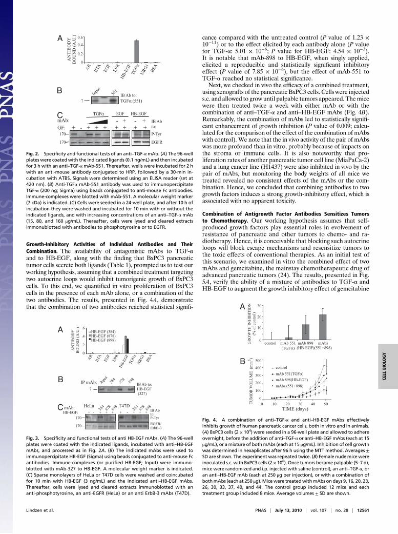
Growth-Inhibitory Activities of Individual Antibodies and TheirCombination. The availability of antagonistic mAbs to TGF-αand to HB-EGF, along with the finding that BxPC3 pancreatictumor cells secrete both ligands (Table 1), prompted us to test ourworking hypothesis, assuming that a combined treatment targetingtwo autocrine loops would inhibit tumorigenic growth of BxPC3cells. To this end, we quantified in vitro proliferation of BxPC3cells in the presence of each mAb alone, or a combination of thetwo antibodies. The results, presented in Fig. 4A, demonstratethat the combination of two antibodies reached statistical signifi-
cance compared with the untreated control (P value of 1.23 ×10⁻¹¹) or to the effect elicited by each antibody alone (P valuefor TGF-α: 5.01 × 10⁻⁶; P value for HB-EGF: 4.54 × 10⁻³).It is notable that mAb-898 to HB-EGF, when singly applied,elicited a reproducible and statistically significant inhibitoryeffect (P value of 7.85 × 10⁻⁶), but the effect of mAb-551 toTGF-α reached no statistical significance.Next, we checked in vivo the efficacy of a combined treatment,
using xenografts of the pancreatic BxPC3 cells. Cells were injecteds.c. and allowed to grow until palpable tumors appeared. Themicewere then treated twice a week with either mAb or with thecombination of anti–TGF-α and anti–HB-EGF mAbs (Fig. 4B).Remarkably, the combination of mAbs led to statistically signifi-cant enhancement of growth inhibition (P value of 0.009; calcu-lated for the comparison of the effect of the combination of mAbswith control). We note that the in vivo activity of the pair of mAbswas more profound than in vitro, probably because of impacts onthe stroma or immune cells. It is also noteworthy that pro-liferation rates of another pancreatic tumor cell line (MiaPaCa-2)and a lung cancer line (H1437) were also inhibited in vivo by thepair of mAbs, but monitoring the body weights of all mice wetreated revealed no consistent effects of the mAbs or the com-bination. Hence, we concluded that combining antibodies to twogrowth factors induces a strong growth-inhibitory effect, which isassociated with no apparent toxicity.
Combination of Antigrowth Factor Antibodies Sensitizes Tumorsto Chemotherapy. Our working hypothesis assumes that self-produced growth factors play essential roles in evolvement ofresistance of pancreatic and other tumors to chemo- and ra-diotherapy. Hence, it is conceivable that blocking such autocrineloops will block escape mechanisms and resensitize tumors tothe toxic effects of conventional therapies. As an initial test ofthis scenario, we examined in vitro the combined effect of twomAbs and gemcitabine, the mainstay chemotherapeutic drug ofadvanced pancreatic tumors (24). The results, presented in Fig.5A, verify the ability of a mixture of antibodies to TGF-α andHB-EGF to augment the growth inhibitory effect of gemcitabine
EGFR/ErbB-3170
HB-EGF: + - + + + + - + + +P-Tyr
IB Abto:
mAb: T47DHeLa878898
384
878898
384
170
A
0
.2
.4
.6HB-EGF (384)HB-EGF (878)HB-EGF (898)
AR EGF
HB-EGF
TGFα
NRG1 BSA
EPR
BTA
IBAb to:HB-EGF(327)
IP mAb: 878
898
Input
7384B
C
ANTIBODY
BOUND(A.U.)
Fig. 3. Specificity and functional tests of anti HB-EGF mAbs. (A) The 96-wellplates were coated with the indicated ligands, incubated with anti–HB-EGFmAbs, and processed as in Fig. 2A. (B) The indicated mAbs were used toimmunopercipitate HB-EGF (Sigma) using beads conjugated to anti-mouse Fcantibodies. Immune-complexes (or purified HB-EGF; Input) were immuno-blotted with mAb-327 to HB-EGF. A molecular weight marker is indicated.(C) Sparse monolayers of HeLa or T47D cells were washed and coincubatedfor 10 min with HB-EGF (3 ng/mL) and the indicated anti–HB-EGF mAbs.Thereafter, cells were lysed and cleared extracts immunoblotted with ananti-phosphotyrosine, an anti-EGFR (HeLa) or an anti ErbB-3 mAbs (T47D).
controlmAb 551(TGFα)mAb 898(HB-EGF)mAbs (551+898)
100
200
400
300
500
0
TUMORVOLUME(mm3 )
0 10 20 30 40 50
B
TIME (days)
GROWTH
INHIBITION
(%ofcontrol)
10
20
30
0control mAbs
(551+898)mAb 898(HB-EGF)
A
mAb 551(TGFα)
Fig. 4. A combination of anti–TGF-α and anti–HB-EGF mAbs effectivelyinhibits growth of human pancreatic cancer cells, both in vitro and in animals.(A) BxPC3 cells (2 × 10⁴) were seeded in a 96-well plate and allowed to adhereovernight, before the addition of anti–TGF-α or anti–HB-EGFmAbs (each at 15μg/mL), or amixture of bothmAbs (each at 15 μg/mL). Inhibition of cell growthwas determined in hexaplicates after 96 h using the MTT method. Averages ±SD are shown. The experiment was repeated twice. (B) Female nudemice wereinoculated s.c. with BxPC3 cells (2× 10⁶). Once tumors becamepalpable (5–7 d),mice were randomized and i.p. injected with saline (control), an anti–TGF-α, oran anti–HB-EGF mAb (each at 250 μg per injection), or with a combination ofbothmAbs (each at 250 μg).Micewere treatedwithmAbs on days 9, 16, 20, 23,26, 30, 33, 37, 40, and 44. The control group included 12 mice and eachtreatment group included 8 mice. Average volumes ± SD are shown.
A
AR EGF
HB-EGF
TGFα
NRG1 BSA
EPR
BTA
ANTIBODY
BOUND(A.U.)
0
0.2
0.40.6
EGFR
170 P-Tyr
IBAbto:+ - + + + + + + +GF:
TGFα EGF HB-EGFmAb: - + - +C
B7
IBAb to:TGFα (551)
551
Input
170
Fig. 2. Specificity and functional tests of an anti–TGF-αmAb. (A) The 96-wellplates were coated with the indicated ligands (0.1 ng/mL) and then incubatedfor 3 h with an anti–TGF-αmAb-551. Thereafter, wells were incubated for 2 hwith an anti-mouse antibody conjugated to HRP, followed by a 30-min in-cubation with ATBS. Signals were determined using an ELISA reader (set at420 nm). (B) Anti-TGFα mAb-551 antibody was used to immunopercipitateTGF-α (200 ng; Sigma) using beads conjugated to anti-mouse Fc antibodies.Immune-complexes were blotted with mAb-551. A molecular weight marker(7 kDa) is indicated. (C) Cells were seeded in a 24-well plate, and after 10 h ofincubation they were washed and incubated for 10 min with or without theindicated ligands, and with increasing concentrations of an anti–TGF-α mAb(15, 80, and 160 μg/mL). Thereafter, cells were lysed and cleared extractsimmonublotted with antibodies to phosphotyrosine or to EGFR.
Lindzen et al. PNAS | July 13, 2010 | vol. 107 | no. 28 | 12561
CELL
BIOLO
GY
on cultured BxPC3 cells. Hence, our next experiment examined inanimals the effect of a triple combination. Cells were injected s.c.and allowed to grow until palpable tumors appeared. Thereafter,the mice were treated twice (days 16 and 21) with gemcitabine(150 mg/kg body weight), and with or without the two mAbs. Theresults presented in Fig. 5B demonstrate that two injections ofgemcitabine resulted in >85% inhibition of tumor growth, but re-peated injections of a mixture of twomAbs consistently augmentedthe cytotoxic effects of the chemotherapeutic agent.In summary, by concentrating on BxPC3 pancreatic tumor cells
that maintain several autocrine loops involving EGFR and at leasttwo ligands, TGF-α andHB-EGF, we provide evidence supportingthe notion that autocrine loops help tumors to evade the cytotoxicaction of chemotherapeutic drugs like gemcitabine. If verified inadditional tumor models, these observations offer a scenario ofindividualized cancer therapy. Accordingly, the autocrine loopsoperating in a specific tumor are first characterized using immu-nological or other assays. In the next step, antibodies capable ofblocking the specific growth factors are combined with chemo-therapy in a way that sensitizes tumors to cytotoxicity and delaysonset of chemoresistance.
DiscussionThe work presented here offers a general strategy for effectiveblockade of the tumorigenic action of ErbB-specific ligands, takinginto account ligand multiplicity, tumor-specific distinct repertoiresof ligands, and the roles played by such growth factors in frequentemergence of resistance to chemotherapy. Chemoresistance isa multifactorial phenomenon and a major clinical obstacle, whichobliterates successful treatment of cancer patients. Under chem-ical stress, or upon irradiation, tumor cells overexpress severalgrowth factors, including fibroblast growth factors, the macro-phage growth factor (CSF-1), and members of the EGF family(25). Moreover, up-regulation of EGF-like ligands is consideredan inherent part of the cellular response to growth factors, whichestablishes an auto-stimulatory feedback loop (26). Accordingly,
tumors driven by dysregulated ErbB proteins, such as brain tumorsexpressing the EGFRvIII mutant (27) or RAS-transformed cells,which secrete large amounts of TGF-α and HB-EGF, may alsodisplay sensitivity to the triple drug combination we propose.Motivated by a working hypothesis that attributes chemo-
resistance to secretion of several distinct EGF-like ligands, wedesigned an experimental three-step therapeutic strategy andtested it in animals. In the first step, the repertoire of EGF-likeligands secreted by an individual tumor is determined using PCRor paraffin-embedded tissue microarrays (28), which enables thenext step, namely tailoring a combination of mAbs specific to therespective growth factors. In the last step, the selected mixture ofantibodies is combined with a chemotherapeutic agent represent-ing the mainstay of the respective clinical indication. For example,the study we reported focused on a pancreatic cancer cell line,hence we used gemcitabine, a chemotherapeuitc agent often usedto treat pancreatic tumors (29). Although the clinical feasibility ofthe protocol we propose here is a matter of future studies, it isworthwhile noting that a combination of chemotherapy with ananti-VEGF antibody, bevacizumab, is clinically effective in co-lorectal (22) and in other carcinomas, and antibodies to anothermember of the VEGF family, placenta growth factor, showedpromising results in animal models (30).Several recent reports interested in mechanisms underlying
evolvement of resistance to ErbB-targeted therapies (e.g., mAbsand TKIs), have identifiedEGF-like ligands as potential targets fortreatments aimed at delaying the onset of resistance. For example,trastuzumab-resistant breast cancer cells exhibited higher levels ofEGFR/ErbB-1, TGF-α, HB-EGF, and NRG (18). Similarly, it wasshown that several chemoresistant cell lines were more sensitive togefetinib, an anti–ErbB-1 TKI, and this correlated with alteredligand expression, as well as with constitutive phosphorylation ofErbB-2/HER2 and ErbB-3 (31). Another study characterizedmRNA expression of EGF-like ligands before and followingtreatment of mammary tumor cells with an EGFR-specific TKI,and correlated resistance with ligand expression (32). Taken to-gether, these studies predict that resistance to antibodies and TKIsdirected at ErbB proteins is because of production of autocrinegrowth factors; hence, the combination protocol we propose mayprevent not only chemoresistance, but also resistance to moleculartargeted therapies.In summary, we envision a tailor-made strategy for cancer
treatment, which combines with chemotherapy two or more anti-bodies to tumor-specific autocrine growth factors. Accurate de-termination of the autocrine growth factors specific to each tumoris crucial for successful delay of resistance. Future studies willexamine other types of carcinomas, along with antibodies to ad-ditional EGF-like peptides. Likewise, we are interested in thepossibility raised by some recent observations (33, 34) that thetreatment we envision may overcome not only resistance tochemo- and molecular-targeted therapy, but also to radiotherapy.
Materials and MethodsMaterials and Cells. Growth factors were from PeproTech Asia. NiNTA beadswere from Novagen. ATBS [2,2’-Azino-bis (3-ethylbenzthiozoline-6-sulfonicacid)] and MTT [3-(4,5-Dimethylthiazol-2-yl)-2,5-diphenyltetrazolium bro-mide] were purchased from Sigma. Duo-set kits were from R&D Systems.Female athymic NCr-nudemice and female BALB/c mice were purchased fromHarlen. Monoclonal antibodies to EGFR and ErbB-3 were generated in ourlaboratory. HRP-conjugated anti-mouse antibody was from Jackson ImmunoResearch Laboratories. Human cancer cell lines were purchased from theAmerican Type Culture Collection. All animal procedures were approved bythe Institute’s Review Board.
Cloning and Expression of EGF-Like Ligands in Bacteria and in Mammalian Cells.EGF-like domains were cloned into the pET32b vector, and expressed asC-terminal TRX fusion proteins with a Factor Xa cleavage site flanking the N-terminal residue of the EGF-like domain. The fusion proteins were expressedin Escherichia coli (BL21) using standard procedures. Following sonication,
A
PROLIFERATION
(%ofcontrol)
Gemc.control mAbs(551+898)
Gemc.+mAbs
020406080100
control
Gemcitabine
mAbs(551+898)
Gemc. +mAbs
TIME (days)15 20 25 30 35
mAbs:Gemc.:
0200400600
800
10001200
TUMORVOLUME(mm3 )B
Fig. 5. A combination of chemotherapy and two mAbs to growth factors ef-fectively inhibits pancreatic cancer cells, both in vitro and in animal. (A) BxPC3cells (2×10⁴)were treatedas in Fig. 4A, except thatgemcitabine (0.5ng/mL)wasused, either alone or in combination with a mixture of anti–TGF-α and anti–HB-EGF mAbs (each at 10 μg/mL). Averages and SDs (bars) of hexaplicates areshown. The experiment was repeated twice. (B) Female nude mice (6 wk old)were s.c. inoculated with 2 × 10⁶ BxPC3 cells. Once tumors became palpable(5–7 d), mice were randomized into groups. The first group (8 mice) wasi.p. injected with a mixture of mAbs to TGF-α and to HB-EGF (each at 120 μgper injection; arrows). A second group (11 mice) was injected i.p. on days 16and 21 with gemcitabine (150 mg/kg body weight; arrows), and a third group(6 mice) was treated with a combination of gemcitabine and mAbs. The con-trol group (11 mice) was similarly treated with saline. Averages ± SD (bars) oftumor volume are shown.
12562 | www.pnas.org/cgi/doi/10.1073/pnas.1006218107 Lindzen et al.
cleared extracts were transfered to pre-equilibrated NiNTA beads. The beadswere washed and then eluted with 300 mM immidazole. Construction offusion proteins comprising a GPI motif was performed in two steps. The firstPCR was performed on the GPI signal of the rat contactin-1. The 5′ primerintroduced a NsiI cleavage site, which was followed by an HA tag, and the 3′primer introduced a NotI site. The product was cloned into the pIRES-Hygvector using NsiI and NotI restriction enzymes. The second step employedoverlapping PCR. The first reaction used the signal peptide of HER2 asa template, and a 3′ primer that included the 5′ sequence of the respectiveEGF-like domain. The second PCR employed the respective EGF-like domainas a template, and a 5′ primer that included the 3′ end of the HER2 signalpeptide. The products of both reactions served as templates for another PCR.The final PCR product was cloned into pIRES-Hyg-GPI, by using BamHI andNsiI cleavage sites. To establish clones of CHO cells, we transfected thecorresponding pIERS-Hyg using Lipofectamine (Invitrogen) and selectedclones using hygromycin (2 μg/mL).
Generation of Monoclonal Antibodies. Five Female BALB/cmice (3moold)wereinjected s.c. and into the foot pad with 30 μg protein in complete Freund’sadjuvant (Tifco). Three weeks later, a second injection was performed in in-complete Freund’s adjuvant. This injection was followed by three to fiveinjections at intervals of 3 wk. A month after the last boost, the twomice withthe highest titer received two more injections on two consecutive days. Fourdays after the last boost, cells from each spleenwere fusedwith 20 × 106 NS0/1myeloma line as described (35). Following fusion, cellswere distributed into 96-well plates, at concentration of 2 × 10⁴ viable myloma cells per well. Hybridcells were selected for growth in the presence of HAT. Positive hybrid cultureswere weaned out of HAT, cloned and recloned in limiting dilution.
In Vitro Tests of mAb Binding to EGF-Like Ligands. The 96-well plates werecoated with the indicated ligands and incubated for 3 h at 37 °C. Plates were
washed twice and blocked with 1% albumin for 1 h at 37 °C, followed by a 3-h long incubation at room temperature with an antibody (1 μg/mL) or withsaline. Thereafter, wells were incubated for 120 min with an anti-mouse HRPantibody, followed by incubation with ATBS (Sigma). Signals were de-termined using an ELISA reader (420 nm).
Determination of Ligand Concentration in Conditioned Medium. Human cancercell lines (1 × 10⁶) were seeded in 10-cm plates, covered with 8 mL medium,and incubated for 4 d. Media were then collected and ligand quantifiedusing the DuoSet ELISA kit (R&D Systems).
Cell Proliferation Assays. Cells were plated on 96-well plates (2,000 cells perwell) in hexaplicates. Proliferation was measured after 24, 48, and 72 h usingthe MTT method. MTT was added to the wells, and 2 h later the cells weredissolved in 4 mM HCl (in isopropanol), and absorbance was determinedat 570 nm.
Determination of Antitumor Activity of mAbs in Animals. Female atymic NCr-nudemice (6wkold) were inoculated s.c. with 2× 10⁶ human cancer cells. Oncetumors became palpable (5–7 d), mice were randomized into groups andinjected i.p. at the indicated timepoints with amAb, chemotherapy, or variouscombinations. Tumor volumes were monitored twice a week.
ACKNOWLEDGMENTS. We thank members of our group for insightfulcomments. Y.Y. is the incumbent of the Harold and Zelda GoldenbergProfessorial Chair. This work is supported by research grants from the USNational Cancer Institute (CA072981), the Israel Science Foundation, Dr.Miriam and Sheldon G. Adelson Medical Research Foundation, the M.D.Moross Cancer Institute, and the Marc Rich Foundation for Education,Culture, and Welfare (the Linda de Picciotto Program).
1. Yarden Y, Sliwkowski MX (2001) Untangling the ErbB signalling network. Nat Rev MolCell Biol 2:127–137.
2. Harris RC, Chung E, Coffey RJ (2003) EGF receptor ligands. Exp Cell Res 284:2–13.3. Falls DL (2003) Neuregulins: Functions, forms, and signaling strategies. Exp Cell Res
284:14–30.4. Klapper LN, et al. (1999) The ErbB-2/HER2 oncoprotein of human carcinomas may
function solely as a shared coreceptor for multiple stroma-derived growth factors.Proc Natl Acad Sci USA 96:4995–5000.
5. Kochupurakkal BS, et al. (2005) Epigen, the last ligand of ErbB receptors, revealsintricate relationships between affinity and mitogenicity. J Biol Chem 280:8503–8512.
6. Jorissen RN, et al. (2003) Epidermal growth factor receptor: Mechanisms of activationand signalling. Exp Cell Res 284:31–53.
7. Barozzi C, et al. (2002) Relevance of biologic markers in colorectal carcinoma: Acomparative study of a broad panel. Cancer 94:647–657.
8. Thøgersen VB, et al. (2001) A subclass of HER1 ligands are prognostic markers forsurvival in bladder cancer patients. Cancer Res 61:6227–6233.
9. Grandis JR, Chakraborty A, Zeng Q, Melhem MF, Tweardy DJ (1998) Downmodulationof TGF-alpha protein expression with antisense oligonucleotides inhibits proliferationof head and neck squamous carcinoma but not normal mucosal epithelial cells. J CellBiochem 69:55–62.
10. Wang F, et al. (2007) Heparin-binding EGF-like growth factor is an early responsegene to chemotherapy and contributes to chemotherapy resistance. Oncogene 26:2006–2016.
11. Eckstein N, et al. (2008) Epidermal growth factor receptor pathway analysis identifiesamphiregulin as a key factor for cisplatin resistance of human breast cancer cells.J Biol Chem 283:739–750.
12. Britten CD (2004) Targeting ErbB receptor signaling: A pan-ErbB approach to cancer.Mol Cancer Ther 3:1335–1342.
13. Weiner LM, Borghaei H (2006) Targeted therapies in solid tumors: Monoclonalantibodies and small molecules. Hum Antibodies 15(3):103–111.
14. Ishikawa N, et al. (2005) Increases of amphiregulin and transforming growth factor-alpha in serum as predictors of poor response to gefitinib among patients withadvanced non-small cell lung cancers. Cancer Res 65:9176–9184.
15. Zhou BB, et al. (2006) Targeting ADAM-mediated ligand cleavage to inhibit HER3 andEGFR pathways in non-small cell lung cancer. Cancer Cell 10:39–50.
17. Engelman JA, et al. (2007) MET amplification leads to gefitinib resistance in lungcancer by activating ERBB3 signaling. Science 316:1039–1043.
18. Ritter CA, et al. (2007) Human breast cancer cells selected for resistance totrastuzumab in vivo overexpress epidermal growth factor receptor and ErbB ligandsand remain dependent on the ErbB receptor network. Clin Cancer Res 13:4909–4919.
19. Bianchi S, et al. (2006) ErbB-receptors expression and survival in breast carcinoma: A15-year follow-up study. J Cell Physiol 206:702–708.
20. Karamouzis MV, Badra FA, Papavassiliou AG (2007) Breast cancer: The upgraded roleof HER-3 and HER-4. Int J Biochem Cell Biol 39:851–856.
21. Shih T, Lindley C (2006) Bevacizumab: An angiogenesis inhibitor for the treatment ofsolid malignancies. Clin Ther 28:1779–1802.
22. Hurwitz H, et al. (2004) Bevacizumab plus irinotecan, fluorouracil, and leucovorin formetastatic colorectal cancer. N Engl J Med 350:2335–2342.
23. Van Zoelen EJ, Stortelers C, Lenferink AE, Van de Poll ML (2000) The EGF domain:Requirements for binding to receptors of the ErbB family. Vitam Horm 59:99–131.
24. Mackenzie RP, McCollum AD (2009) Novel agents for the treatment of adenocarcinomaof the pancreas. Expert Rev Anticancer Ther 9:1473–1485.
25. Harari PM, Wheeler DL, Grandis JR (2009) Molecular target approaches in head andneck cancer: Epidermal growth factor receptor and beyond. Semin Radiat Oncol 19:63–68.
26. Citri A, Yarden Y (2006) EGF-ERBB signalling: Towards the systems level. Nat Rev MolCell Biol 7:505–516.
27. Ramnarain DB, et al. (2006) Differential gene expression analysis reveals generationof an autocrine loop by a mutant epidermal growth factor receptor in glioma cells.Cancer Res 66:867–874.
28. Abdeen A, et al. (2009) Correlation between clinical outcome and growth factorpathway expression in osteogenic sarcoma. Cancer 115:5243–5250.
29. Heinemann V (2001) Gemcitabine: Progress in the treatment of pancreatic cancer.Oncology 60:8–18.
30. Fischer C, et al. (2007) Anti-PlGF inhibits growth of VEGF(R)-inhibitor-resistant tumorswithout affecting healthy vessels. Cell 131:463–475.
31. Servidei T, Riccardi A, Mozzetti S, Ferlini C, Riccardi R (2008) Chemoresistant tumorcell lines display altered epidermal growth factor receptor and HER3 signaling andenhanced sensitivity to gefitinib. Int J Cancer 123:2939–2949.
32. Ferrer-Soler L, et al. (2007) An update of the mechanisms of resistance to EGFR-tyrosine kinase inhibitors in breast cancer: Gefitinib (Iressa) -induced changes in theexpression and nucleo-cytoplasmic trafficking of HER-ligands (Review). Int J Mol Med20:3–10.
33. Bianco C, et al. (2002) Enhancement of antitumor activity of ionizing radiation bycombined treatment with the selective epidermal growth factor receptor-tyrosinekinase inhibitor ZD1839 (Iressa). Clin Cancer Res 8:3250–3258.
34. Raben D, Helfrich BA, Chan D, Johnson G, Bunn PA, Jr (2002) ZD1839, a selectiveepidermal growth factor receptor tyrosine kinase inhibitor, alone and in combinationwith radiation and chemotherapy as a new therapeutic strategy in non-small cell lungcancer. Semin Oncol 29(1, Suppl 4):37–46.
35. Eshhar Z, Ofarim M, Waks T (1980) Generation of hybridomas secreting murinereaginic antibodies of anti-DNP specificity. J Immunol 124:775–780.
Lindzen et al. PNAS | July 13, 2010 | vol. 107 | no. 28 | 12563