+ All Categories
Home > Documents > Talus fractures Outline - Orthopaedic Trauma … · 9/22/13 1 Talus fractures Philipp Leucht, MD"...

Talus fractures Outline - Orthopaedic Trauma … · 9/22/13 1 Talus fractures Philipp Leucht, MD"...

Date post: 14-Sep-2018
Category:
Upload: dinhduong
View: 218 times
Download: 0 times
Share this document with a friend
7
9/22/13 1 Talus fractures Philipp Leucht, MD Stanford University School of Medicine Orthopaedic Trauma course for NP/PAs OTA 2013 Outline Anatomy Surgical Approaches Fixation strategies Outcomes and complications Anatomy Bone 60-70% articular cartilage No muscular attachments Complex articulations Anatomy Vascular 1. Posterior Tibial Artery of the Tarsal Canal Deltoid Artery (really off the Art of the Tarsal Canal) 2. Anterior Tibial 3. Perforating Peroneal Artery of the Tarsal Sinus
Transcript

9/22/13  

1  

Talus fractures

Philipp Leucht, MD"Stanford University School of Medicine

Orthopaedic Trauma course for NP/PAs OTA 2013

Outline

•  Anatomy

•  Surgical Approaches

•  Fixation strategies

•  Outcomes and complications

Anatomy"Bone

•  60-70% articular cartilage

•  No muscular attachments

•  Complex articulations

Anatomy"Vascular

1.  Posterior Tibial –  Artery of the Tarsal Canal –  Deltoid Artery (really off

the Art of the Tarsal Canal)

2.  Anterior Tibial

3.  Perforating Peroneal –  Artery of the Tarsal Sinus

9/22/13  

2  

Anatomy"Vascular

Inferior Anastomotic Sling

-  Artery of the Tarsal Canal (Posterior Tibial)

-  Artery of the Tarsal Sinus (Perforating Peroneal)

•  Send numerous branches into the inferior talar neck

•  Main supply of the talar body is from the artery of the tarsal canal

Anatomy"Vascular

Injury mechanism

•  Forced Dorsiflexion –  Dorsiflexion causes

tibiotalar impingement, leads to neck fracture

–  Dorsomedial comminution

–  not reproduced biomechanically

•  Shear Force

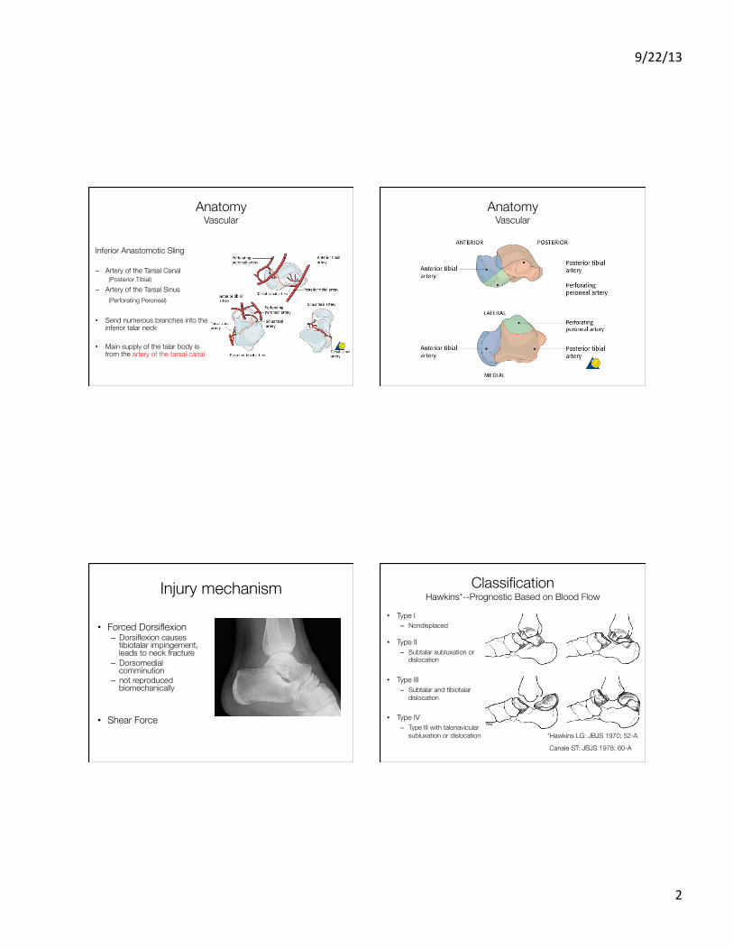
Classification"Hawkins*--Prognostic Based on Blood Flow

•  Type I –  Nondisplaced

•  Type II –  Subtalar subluxation or

dislocation

•  Type III –  Subtalar and tibiotalar

dislocation

•  Type IV –  Type III with talonavicular

subluxation or dislocation *Hawkins LG: JBJS 1970; 52-A

Canale ST: JBJS 1978; 60-A

9/22/13  

3  

Radiographic Evaluation •  Ankle Series

•  Foot Series

•  Canale View

•  CT Scans –  Consider for head,

body, and lateral process fractures

Treatment"Closed Methods

•  Non-operative (rare)

–  For truly undisplaced fractures

•  Closed Reduction –  Realignment of gross displacement or

dislocation –  important for soft tissues –  becomes increasingly more difficult

with severity of fracture

Treatment Principles •  Accurate alignment of talar neck – Re-establish hindfoot mechanics

•  Stable fixation – Maximize revascularization potential – Allow early ROM

Treatment"Exposure

•  Surgical Approaches

•  Combined anteromedial and anterolateral

*Mayo KA:Fractures of the talus: Principles of management and techniques of treatment. Tech Orthop 1987;2

9/22/13  

4  

Anteromedial approach"

Anterolateral approach"

Operative Considerations

•  Radiolucent table •  Small clamps •  Small distractor or external fixator •  Small/mini-fragment fixation

–  1.5mm –  2.0mm –  2.4mm –  2.7mm & 3.5mm –  Mini-fragment plates occasionally

9/22/13  

5  

Intra-operative techniques

•  Reduce and judge alignment through both approaches

•  Lateral cortex usually provides good read (tension failure)

•  Medial comminution (no lag screw fixation) –  Cancellous bone graft/ structural bone graft

•  Avoid stripping •  K-wire through talar head as reduction aid

Reduction of a Dislocated Talar Body

•  Infrequently works in the ER… but worth a try. •  GA for paralysis •  Flex the knee, traction on the the hindfoot, direct pressure

on the talar body •  For open reduction: Dual exposure •  Medial and lateral distractors (ex-fix) with a transcalcaneal

pin •  Malleolar osteotomy

The body is usually between the posteromedial aspect of the tibia and the Achilles tendon, immediately adjacent to the

posteromedial neurovascular bundle

INFRA-TECTAL

Medial malleolar osteotomy

•  Drill screw before osteotomy –  Easier reduction

•  Allows visualization of entire medial talus

9/22/13  

6  

Post-operative rehabilitation

•  Early ROM exercises once skin allows •  NWB for 12 weeks •  in cases with ligamentous instability, longer

cast immobilization might be considered

•  Delayed union •  Nonunion •  Malunion

–  Particularly varus •  Subtalar and ankle arthritis •  Osteonecrosis

Complications

-  AVN most common complication, followed by infection, mal-/non-union

-  Hawkins sign is good predictor of appropriate blood supply at 6-8 weeks

Outcome Outcome •  Sanders et al.

–  Excellent functional outcomes if pt did not require 2° reconstructive procedure

–  Development of varus and/or ST arthritis leads to poor functional outcomes

–  Most common indication for 2° surgery was ST arthritis. –  Risk factors for 2° surgery were comminution, high

Hawkin’s type, and associated L/E fractures. •  All markers of higher-energy injuries

–  Time between injury and surgery did not correlate with outcome

–  12% AVN rate (much lower than most other reports).

Sanders, DW, et al. J Orthop Trauma. 18;5, 2004  

9/22/13  

7  

Summary

•  Delayed fixation has no effect on outcome, union or AVN

•  Outcomes worse with comminution, open injuries, varus

•  Post-traumatic arthritis is common

•  AVN may be partial and not lead to collapse

•  Set expectations


Recommended