Targeting MET kinase with the small-moleculeinhibitor amuvatinib induces cytotoxicity inprimary myeloma cells and cell linesCornel Joseph Phillip1,6†, Shadia Zaman1†, Shujun Shentu1, Kumudha Balakrishnan1,6, Jiexin Zhang2,Veera Baladandayuthapani3,6, Pietro Taverna7, Sanjeev Redkar7, Michael Wang5, Christine Marie Stellrecht1,6†
and Varsha Gandhi1,4,6*†
Abstract
Background: MET is a receptor tyrosine kinase that is activated by the ligand HGF and this pathway promotes cellsurvival, migration, and motility. In accordance with its oncogenic role, MET is constitutively active, mutated, orover-expressed in many cancers. Corollary to its impact, inhibition of MET kinase activity causes reduction of thedownstream signaling and demise of cells. In myeloma, a B-cell plasma malignancy, MET is neither mutated norover-expressed, however, HGF is increased in plasma or serum obtained from myeloma patients and this wasassociated with poor prognosis. The small-molecule, amuvatinib, inhibits MET receptor tyrosine kinase. Based on thisbackground, we hypothesized that targeting the HGF/MET signaling pathway is a rational approach to myelomatherapy and that myeloma cells would be sensitive to amuvatinib.
Methods: Expression of MET and HGF mRNAs in normal versus malignant plasma cells was compared duringdisease progression. Cell death and growth as well as MET signaling pathway were assessed in amuvatinib treatedprimary myeloma cells and cell lines.
Results: There was a progressive increase in the transcript levels of HGF (but not MET) from normal plasma cells torefractory malignant plasma cells. Amuvatinib readily inhibited MET phosphorylation in primary CD138+ cells frommyeloma patients and in concordance, increased cell death. A 48-hr amuvatinib treatment in high HGF-expressingmyeloma cell line, U266, resulted in growth inhibition. Levels of cytotoxicity were time-dependent; at 24, 48, and72 h, amuvatinib (25 μM) resulted in 28%, 40%, and 55% cell death. Consistent with these data, there was anamuvatinib-mediated decrease in MET phosphorylation in the cell line. Amuvatinib at concentrations of 5, 10, or25 μM readily inhibited HGF-dependent MET, AKT, ERK and GSK-3-beta phosphorylation. MET-mediated effects werenot observed in myeloma cell line that has low MET and/or HGF expression.
Conclusions: These data suggest that at the cellular level MET/HGF pathway inclines with myeloma diseaseprogression. Amuvatinib, a small molecule MET kinase inhibitor, is effective in inducing growth inhibition and celldeath in myeloma cell lines as well as primary malignant plasma cells. These cytostatic and cytotoxic effects wereassociated with an impact on MET/HGF pathway.
* Correspondence: [email protected]†Equal contributors1Departments of Experimental Therapeutics, The University of Texas MDAnderson Cancer Center, Houston, Texas, USA4Leukemia, The University of Texas MD Anderson Cancer Center, Houston,Texas, USAFull list of author information is available at the end of the article
IntroductionMultiple myeloma (MM) is an indolent B-cell diseasethat develops in the bone marrow and is associatedwith osteolytic lesions in the advanced stages [1]. Des-pite progress in prolonging myeloma patient survival,current therapies are not curative; thus, it is imperativethat new treatments be developed for this debilitatingdisease [2,3].Survival and proliferation of myeloma cells are dependent
on the presence of a permissive microenvironment, whichincludes bone marrow stroma and soluble cytokines [4-9]such as IL-6 and HGF [8,10]. HGF is the ligand for METreceptor tyrosine kinase. When HGF binds to and activatesMET, MET is autophosphorylated on Tyr1230, Tyr1234and Tyr1235 located in the activation loop [11-14]. Inaddition, MET has a multisubstrate docking site that is acti-vated at Tyr1349 and Tyr1356. The phosphorylation of thisregion results in the induction of MET signaling throughthe activation of several downstream target pathways, in-cluding the mitogen-activated protein kinase (MAPK) andAKT signaling pathways [11]. HGF/MET-induced MAPKsignaling has been shown to be essential for proliferation,migration and invasion [7,11,15,16] while the induction ofAKT signaling promotes tumor cell survival [17].HGF/MET signaling is increasingly recognized as an
important contributor to the pathogenesis of myeloma.Expression of both HGF and MET has been demonstratedin most myeloma cell lines and primary patient samples[18,19]. Studies correlating HGF levels with MM clinicalparameters such as diagnosis [20-23] disease stage, aggres-siveness [22,24,25], prognosis [22,23,26], and response[26-29]. Besides its effects on the malignant myelomacells, HGF is involved in the pathogenesis of myeloma-related bone disease. HGF levels are increased in patientswith extensive bone lesions, and correlates with expressionof osteoclast stimulating cytokines [24]. IL-11 secretionfrom osteoblasts is induced by HGF [30], and HGF in-hibits bone morphogenetic protein-induced osteoblasto-genesis [31].Taken together, these clinical findings strongly support
our hypothesis that targeting the HGF/MET signalingpathway is a rational approach to myeloma therapy. Inline with this postulate, our laboratory studies demon-strated that genetically knocking down MET in myelomacell lines using short hairpin RNA and ribozyme ap-proaches resulted in growth inhibition and demise of themyeloma cells [32,33]. Consistent with these observations,a decline in MET transcript and protein levels inducedby treatment with any of the transcription inhibitorsflavopiridol, cordycepin, or 8-chloro-adenosine, promotedmyeloma cell death [32-34]. Collectively, these data dem-onstrate MET’s pivotal role in myeloma cell biology andunderscore the importance of MET targeting as a thera-peutic strategy in MM [35].
While these genetic and pharmacologic strategies sug-gest utility of MET/HGF inhibition as therapeutic tar-gets, these interventions are not pragmatic for clinicaluse. Amuvatinib (previously known as MP470, AstexPharmaceuticals, Inc.) is a synthetic carbothioamide thatinhibits MET, cKIT and platelet derived growth factorreceptor (PDGFR). This small-molecule inhibitor competeswith ATP for binding at the catalytic site. In solid cancers,amuvatinib has been shown to be effective in inhibitingMET at low micromolar concentrations (IC50 ~5 μM) [36].Amuvatinib is a well-tolerated, orally bioavailable drugcurrently in phase II clinical trials [37,38]. Availability of aclinical candidate, its inhibitory potential for MET kinase,and the role of MET in myeloma cell survival providedcompelling rationales for testing the effects of amuvatinibon myeloma cells.In the present study, we compared mRNA levels of
MET and HGF in normal and primary myeloma plasmacells. We investigated amuvatinib’s actions and cytotoxiceffects in primary plasma cells obtained from patientswith myeloma. To elucidate in more detail the mechan-ism of action of amuvatinib in myeloma cells, we evalu-ated its effect on MET activity and downstream signalingin the myeloma cell line U266, which over-expressesHGF. Our data demonstrate that MET receptor tyrosinekinase may be targeted in myeloma and support the in-vestigation of small-molecule inhibitors such as amuvati-nib as possible therapeutic agents against this disease.
ResultsExpression levels of MET and HGF mRNA in bone marrowplasma cells of healthy donors and patientsPreviously studies have correlated plasma HGF levels withMM clinical parameters such as diagnosis [20-23] diseasestage, aggressiveness [22,24,25], prognosis [22,23,26], andresponse [26-29]. While expression of both HGF andMET transcripts has been shown to be present in mye-loma cells [18,19] and HGF mRNA has also been demon-strated to be expressed in bone marrow stromal cells [39]the levels of HGF and MET mRNA in patient plasma cellshave not been well evaluated nor correlated with diseasestatus.To determine the levels of MET and HGF gene expres-
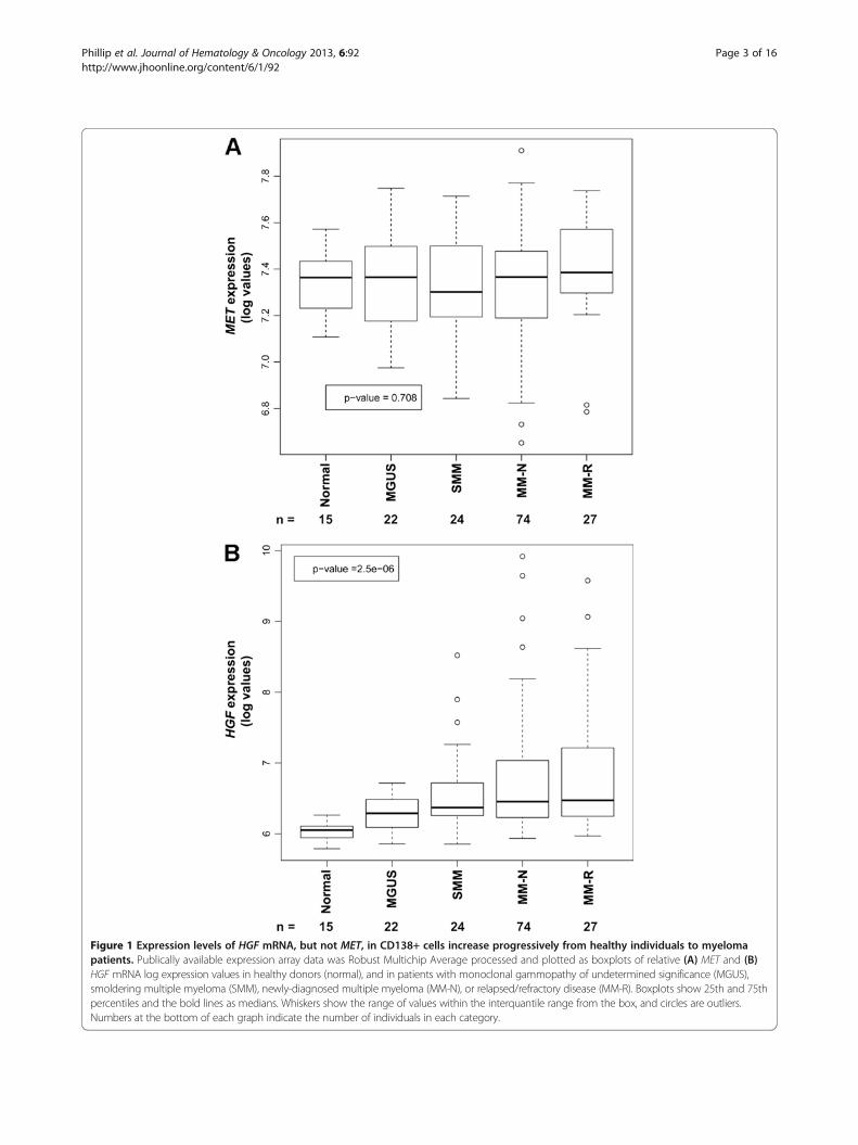
sion in malignant and normal plasma cells, we analyzeddata from the Mayo Clinic Patient Dataset available inthe public domain [40,41]. The 162 samples evaluatedrepresented 15 healthy individuals (normal), 22 patientswith monoclonal gammopathy of undetermined signifi-cance (MGUS), 24 with smoldering MM (SMM), 74 withnewly diagnosed MM (MM-N), and 27 with relapsed/re-fractory MM (MM-R). Among these five groups, therewas no significant difference (P = 0.708) in the expres-sion of MET in the CD138+ cells (Figure 1A). In con-trast, there was a significant trend (P = 2.5 × 10-06) for
Phillip et al. Journal of Hematology & Oncology 2013, 6:92 Page 2 of 16http://www.jhoonline.org/content/6/1/92
Figure 1 Expression levels of HGF mRNA, but not MET, in CD138+ cells increase progressively from healthy individuals to myelomapatients. Publically available expression array data was Robust Multichip Average processed and plotted as boxplots of relative (A) MET and (B)HGF mRNA log expression values in healthy donors (normal), and in patients with monoclonal gammopathy of undetermined significance (MGUS),smoldering multiple myeloma (SMM), newly-diagnosed multiple myeloma (MM-N), or relapsed/refractory disease (MM-R). Boxplots show 25th and 75thpercentiles and the bold lines as medians. Whiskers show the range of values within the interquantile range from the box, and circles are outliers.Numbers at the bottom of each graph indicate the number of individuals in each category.
Phillip et al. Journal of Hematology & Oncology 2013, 6:92 Page 3 of 16http://www.jhoonline.org/content/6/1/92
increases in HGF mRNA levels in CD138+ plasma cells,with progressive severity of disease from healthy donorsto patients with relapsed or refractory MM (Figure 1B).Within each group, there was heterogeneity in HGFexpression as evinced by the 75th percentile mark. It isinteresting to note that even samples with lower HGFmRNA levels in the plasma cells, typically had higherlevels than the samples from healthy individuals; the25th percentile for the myeloma patient HGF levels was >the 75th percentile for healthy individuals.
Induction of apoptosis by amuvatinib in primary CD138+and CD138– CellsGene array analysis along with numerous other studiesby us [32,33] and others [22,23,26], identified the HGF/MET axis as a therapeutic target in myeloma. To testthis, we assessed the sensitivity of primary myeloma cellsto the MET-kinase inhibitor, amuvatinib. CD138+ (mye-loma plasma cells) and CD138– (non-malignant cells)cells were isolated from bone marrow samples obtainedfrom eight myeloma patients (Table 1) and treated with25 μM amuvatinib for 24 h. This dose was chosen basedon findings that >95% of the compound is bound and se-questered by serum proteins (unpublished data) as wellas a small preliminary screen in myeloma cell lines (datanot shown). CD138+ cells from six out of eight patientsamples showed a cell death induction in the amuvatinibtreated cells compared to time-matched dimethyl sulfox-ide (DMSO)-treated control cells as measured byannexin V/propidium iodide (PI) staining (Figure 2A).Cell death in these six samples increased by 20% to 67%compared to time-matched controls; patients 3 and 5showed <10% increase in cell death.An assessment of clinical characteristics of the mye-
loma patients did not reveal any correlations withamuvatinib-induced cell death, including prior treat-ment, patient age, or cellularity of the bone marrow(Table 1). We were able to measure the HGF levels inplasma samples from patients 2, 4, and 5 which were
11.9, 1.7, and 1.4 ng/mL, respectively. Although thenumber of total samples is small, there seems to be arelationship between levels of HGF and amuvatinib-induced apoptosis. Additional studies are needed to de-termine if there is any correlation between HGF leveland sensitivity of CD138+ cells to MET inhibition.In contrast to the sensitivity of CD138+ cells to amu-
vatinib, CD138– cells did not show any sizeable induc-tions (all, <10%) of death compared to time-matchedcontrols (Figure 2B), suggesting that non-malignant bonemarrow cells are not affected by amuvatinib. Overall, theseresults indicate that treatment of CD138+ MM cells witha MET inhibitor is detrimental to their survival.
Effect of Amuvatinib on MET signaling in CD138+ andCD138- cellsTo determine whether death of CD138+ cells was associ-ated with an effect on the target, total and phosphorylated-MET (p-MET) expression levels were determined by flowcytometry. There were sufficient numbers of cells from pa-tients 6 and 8 to perform this assessment. In both samples,MET phosphorylation was reduced on Tyr 1234/1235 inthe CD138+ cells by 40% and 50%, respectively, as com-pared to the time-matched controls (Figure 3A and B).In contrast, there was no detectable level of p-MET inCD138– cells from patient 8 as compared to isotype con-trol (Figure 3C). The lack of p-MET in CD138– cells likelyexplains why this population of cells was not affected byamuvatinib treatment in any of the eight patient samples.
Effect of Amuvatinib on Growth InhibitionSince the number of the primary CD138+ cells in eightpatient samples were insufficient for performing a de-tailed investigation of HGF/MET signaling, we decidedto further investigate the effects of amuvatinib in a mye-loma cell line. Because our results in the patient samplessuggested that higher levels of HGF may be associatedwith an increased sensitivity to MET inhibition, we usedthe U266 cell line which expresses high levels of HGF
Table 1 Myeloma patient characteristics
Pt # Age (yrs) Sex Ethnicity WBCa (103/μl) Plasma cells (%) BMb aspirate done Outcome of treatment
1 62 Male White 3.6 7 At diagnosis NAc
2 53 Female Black 2.0 74 Post bortezomib + doxorubicin + lenalidomide +dexamethasone
Progressive
3 53 Female White 6.3 4 Post 3 cycles bortezomib + dexamethasone Near complete remission
4 76 Female White 4.4 2 Post cyclophosphamide + lenalidomide +dexamethasone
Progressive
5 58 Female Hispanic 9.1 8 At diagnosis NA
6 64 Female White 12.5 49 At diagnosis NA
7 71 Male White 3.9 32 NA NA
8 76 Male White 4.9 36 post 1 yr auto stem cell transplant Progressiveawhite blood cells; bbone marrow; cnot applicable.
Phillip et al. Journal of Hematology & Oncology 2013, 6:92 Page 4 of 16http://www.jhoonline.org/content/6/1/92
[19]. Compared to DMSO-treated control cells,amuvatinib-treated U266 cells showed a dose- and time-dependent decrease in growth (Figure 4A). The growthinhibition was ~40% at a dose of 5 μM after 48 and 72 hof incubation and 50% at a dose of ~7 μM at 72 h.
Effect of Amuvatinib on cell cycle arrest and DNAsynthesisWe also tested whether the observed amuvatinib-induced growth inhibition was associated with an alter-ation in the cell cycle. At low micromolar doses (3 and5 μM), U266 cells were arrested at G1 after 48 and 72 h(Figure 4B and C). In the DMSO-treated controls, ap-proximately 65% of cells were in G1 phase at 72 h, whilecells treated 3 μM amuvatinib significantly increased to75% in G1 phase at the same time point (p < 0.05). Incu-bation with a higher level of amuvatinib (25 μM) re-sulted in a lower percentage of cells in G1 phase(Figure 4C), with a concomitant increase in the subG1
fraction (data not shown).
To determine whether the observed cell cycle changeswere associated with an effect on DNA synthesis, wemeasured incorporation of thymidine in total DNA.Compared to DMSO-treated (control cells), amuvatinib-treated cells had decreased thymidine incorporation atdoses of both 5 and 25 μM (Figure 4D), which was sig-nificantly higher for cells treated with 5 μM amuvatinibfor 24 and 72 h, (P = 0.008 and P = 0.048, respectively).At 24 h, the inhibition of thymidine incorporation wasgreater than 50% with 5 μM amuvatinib. Additionally, asexpected, the decrease in the cells’ S-phase DNA replica-tive capacity was discernible 24 h before there was ameasurable change in the cell cycle profile as the doub-ling time for U266 cells is ~36 hours.
Induction of Apoptosis by AmuvatinibSimilar to what was seen in the primary CD138+ cells,U266 cells treated with 25 μM amuvatinib exhibited sig-nificantly greater cell death (28%, 40%, and 54%) thanDMSO-treated controls (6%, 7%, and 7%) for 24, 48, and
Figure 2 Amuvatinib induces apoptosis in CD138+, but not CD138– cells. (A) CD138+ and (B) CD138– cells isolated and purified from bonemarrow samples of eight myeloma patients were stained with annexin V/PI and assessed by flow cytometry initially after isolation (white bars) orafter 24 h of treatment with vehicle (gray bars) or 25 μM amuvatinib (black bars).
Phillip et al. Journal of Hematology & Oncology 2013, 6:92 Page 5 of 16http://www.jhoonline.org/content/6/1/92
72 h, respectively (Figure 5A and B) (P = 0.045, 0.015,and 0.018 respectively). This apoptotic induction wasblocked by a pan-caspase inhibitor, ZVAD, suggesting arole for caspases in amuvatinib-mediated cell death (datanot shown). Consistent with the annexin V/PI stainingresults was our finding that amuvatinib induced polyADP ribose polymerase (PARP) cleavage in these cells ina dose-dependent manner. Under full serum conditions(10% fetal bovine serum (FBS)), an induction of PARP
cleavage was seen after 24 h with doses as low as 3 μMamuvatinib with the highest level of cleaved PARP prod-uct was seen at 25 μM (Figure 5C).Because 95% of amuvatinib binds serum proteins and
is unavailable to the cells, we also examined the efficacyof amuvatinib under low serum (0.1% FBS) conditions.When cells were treated with 5 μM amuvatinib for16 hours in the presence of low serum, but in the pres-ence of endogenous autocrine HGF, we found maximuminduction of PARP cleavage (Figure 5D). To further con-firm the cytotoxic effects of amuvatinib in myeloma cellsis associated with inhibiting the HGF/MET signalingaxis, we compared the efficacy of amuvatinib to induceapoptosis in U266 cells versus RPMI-8226/S, a myelomacell line which express 75% lower levels of HGF and 95%lower levels of MET (Additional file 1: Figure S1). As ex-pected, there was a significant amuvatinib dose-dependent apoptosis-induction in the U266 cells after48 h treatment (Figure 5E) (25 μM versus 0, 2.5, 5, and10; P = 0.022, 0.018, 0.013, and 0.029, respectively). Incontrast there was only a minor apoptosis induction inthe RPMI-8226/S cells which was not statistically signifi-cant (P = 0.053, 0.300, 0.427, and 0.503, respectively).These results suggest that the apoptosis induction is dueto targeting MET kinase which U266 cells are addictedto while RPMI-8226/S cells are not.
Tumoricidal Effects of Amuvatinib in Myeloma CellsGrown in a Protective Stromal EnvironmentBone marrow stroma provides a protective environmentfor MM cells [42]; thus, it is important to assess the effi-cacy of therapeutic agents in the context of a stromalenvironment. To assess this, we treated U266 cells co-cultured with and without stromal cells with amuvatinibfor 48 h and measured viability by using flow cytometryanalysis of annexin V/PI staining. Under these condi-tions, the U266 do not attach to the stromal cells, butare protected by them through both cell to cell contactand by various soluble factors produced by the stromalcells [43]. Amuvatinib induced ~50% cell killing duringthis time period and co-culture with the stromal cellsprovided no protection from this effect (Figure 5F). Incontrast, these stromal cells were able to protect U266cells from bortezomib treatment as they reduced theamount of bortezomib-induced apoptosis from ~75%to ~40% (Additional file 1: Figure S2A). To determinewhether amuvatinib had an effect on the survival of stro-mal cells, stromal cells cultured alone were treated withamuvatinib, harvested by trypsinization, and similarlyassessed for viability. Interestingly, amuvatinib had avery minimal effect on the survival of this population ofcells (Figure 5F), though they express MET (Additionalfile 1: Figure S2B). These results indicate that the tumor-icidal action of amuvatinib was largely restricted to the
Figure 3 Amuvatinib treatment decreases phospho-MET pro-tein in freshly obtained CD138+ cells. (A) Flow cytometricanalysis of the levels of p-MET (Tyr1234/Tyr1235) in CD138+ cellsfrom patients 6 and 8 after 24 h treatment with (thick black line) orwithout (gray shaded peak) 25 μM amuvatinib. (B) p-MET and totalMET were quantified in untreated (white bars) and 25 μM amuvati-nib treated cells (hashed bars) from panel (A) along with total METlevels in untreated (black bars) and 25 μM amuvatinib treated cells(diagonal bars). (C) Plot represents the levels of p-MET (Tyr1234/Tyr1235) in CD138– cells from patient 8 24 h after treatment with(thick black line) or without (gray shaded peak) 25 μM amuvatinib,and secondary only control (dashed line).
Phillip et al. Journal of Hematology & Oncology 2013, 6:92 Page 6 of 16http://www.jhoonline.org/content/6/1/92
U266 myeloma cells, whereas the stromal cells, which arenot addicted to MET, are not affected by this inhibitor.Furthermore, the stromal cells were not able to protectthe U266 cells from amuvatinib’s tumoricidal activity.Since amuvatinib also inhibits PDGFR and KIT, we vali-
dated MET kinase inhibition as the primary cause of celldeath by using imatinib as a negative control. In additionto ABL, imatinib is known to also inhibit PDGFR and KITbut not MET [44]. In contrast to amuvatinib, 25 μM ima-tinib did not induce significant cell death (Figure 5G; P =0.07) indicating that amuvatinib-mediated cell death is notdue to its effects on PDGFR and KIT.
Effect of Amuvatinib on MET ProteinTo further investigate the effect of amuvatinib on METsignaling, we first measured MET receptor tyrosine kin-ase activity in U266 cells by flow cytometry. Similar tothe results seen with the CD138+ cells from the patientsamples, a reduction in MET phosphorylation was de-tected in cells treated for 24 hr with 25 μM amuvatinibwhen cells are grown in normal growth conditions (10%FBS) (Figure 6A and B). This decrease of p-MET was as-sociated with cell death as cell death was induced with25 μM amuvatinib when cells are grown in normalgrowth conditions (10% FBS) (Figure 5B).
Figure 4 Amuvatinib inhibits growth, induces cell cycle arrest, and inhibits DNA synthesis in myeloma cell line U266. (A) U266 cellswere treated with amuvatinib at various concentrations for 24 (■), 48 (▲), and 72 h (▼). Cells were counted using a Coulter counter, and theeffect of the treatment on cell growth was determined. The data are presented in a time- and dose-dependent manner as percentages oftime-matched controls. Data are representative of three independent experiments and presented as Mean ± SEM, n = 3, *P < 0.05. (B) Todetermine the effect of amuvatinib on the cell cycle, U266 cells were treated with 3 or 5 μM amuvatinib or DMSO for 72 h. Flow cytometry wasused to assess the DNA content of cells stained with PI, regions labeled M1, M2, M3, and M4 represent cells in Sub-G1, G1, S, and G2/M phases ofthe cell cycle, respectively. (C) Cell cycle analyses were performed as in panel (B), with treatments for 24 (white bars), 48 (gray bars), and 72 h(black bars) and the percentage of cells in the G1 phase of the cell cycle were plotted. Data are representative of three independent experimentsand presented as Mean ± SEM, n = 3, *P < 0.05. (D) The effect of amuvatinib on DNA synthesis was determined by treating U266 cells with DMSOor 5 and 25 μM amuvatinib for 24 (white bars), 48 (gray bars), and 72 h (black bars) and quantifying thymidine incorporation. The data arepresented as percentages of time-matched controls. Data are representative of three independent experiments and presented as Mean ± SEM,n = 3, *P < 0.05, **P < 0.01.
Phillip et al. Journal of Hematology & Oncology 2013, 6:92 Page 7 of 16http://www.jhoonline.org/content/6/1/92
To assess the effects of amuvatinib on HGF-specific sig-naling, protein lysates from U266 cells serum starved in0.1% FBS for 16 h with and without various concentra-tions of amuvatinib followed with 15 min HGF stimula-tion were examined by immunoblot analysis. The resultsshowed that under serum starved conditions, treatmentwith 5 μM amuvatinib, decreased phosphorylation of theprocessed ~140 kDa MET β-chain at Tyr1349 by ~60%(Figure 6C and D). Because of the autocrine stimulation ofMET by the endogenous HGF produced in these cells,
MET was phosphorylated under serum-starved con-ditions even without the addition of exogenous HGF.Furthermore, an amuvatinib-dependent decrease of totalMET levels of ~30% was also observed. A ~170 kDaphosphorylated MET band was detected at ~2 foldhigher levels than the 140 kDa band in untreated U266cells. A comparison with total MET shows both bandswere present but the levels of the total 140 kDa bandwas ~4 times greater than the levels of the 170 kDaband. Although unprocessed pro-MET, containing both
Figure 5 Amuvatinib is tumoricidal to U266 myeloma cells. Apoptosis was quantified by staining cells with annexin V/PI and measuringstaining positivity using flow cytometry. (A) A representative annexin V/PI staining profile 72 h after treatment with 25 μM amuvatinib or DMSO isshown. (B) U266 cells were treated with DMSO or various concentrations of amuvatinib for 24 (white bars), 48 (gray bars), and 72 h (black bars)and percent of annexin V/PI positivity is presented. Data are representative of three independent experiments and presented as Mean ± SEM,n = 3, *P < 0.05. Immunoblot analysis demonstrating cleavage of PARP protein after treatment with amuvatinib at indicated concentrations underconditions of (C) full serum (10% FBS) for 24 h, or (D) serum starved conditions with endogenous HGF for 16 h, (E) U266 and RPMI-8226/S cellswere treated with various concentrations of amuvatinib for 48 h and percent of annexin V/PI positivity is presented. Data are representative ofthree independent experiments and presented as Mean ± SEM, n = 3, *P < 0.05. (F) U266 cells cultured alone (black bars), or on NK-tert stromalcells (gray bars), or stromal cells alone (white bars), were treated with or without 25 μM amuvatinib for 48 h and assessed by flow cytometry forannexin V/PI staining. (G) Cells were treated with various concentrations of imatinib and annexin V/PI positivity is presented as percentage oftime-matched controls. Data are representative of three independent experiments and presented as Mean ± SEM, n = 3, *P < 0.05.
Phillip et al. Journal of Hematology & Oncology 2013, 6:92 Page 8 of 16http://www.jhoonline.org/content/6/1/92
the α and β subunits, has been detected by SDS PAGEas a 170 kDa band, it has not been associated with kinaseactivity. Conversely, a splice variant of MET containing anadditional 54 nt of exon 10 has been reported to beexpressed at low levels [45]. This splice form produces a
MET isoform that has kinase activity, though it cannot beprocessed into α and β subunits. In U266 cells, amuvatinibinhibited phosphorylation of a 170 kDa MET by ~70%(Figure 6C and D, B). Again, the decrease of HGF-specificphosphorylation of both isoforms of MET under low
Figure 6 Amuvatinib suppresses MET receptor tyrosine kinase activity. (A) Flow cytometry analysis of p-MET (Tyr1234/1235) levels in U266cells were treated for 24 hrs with DMSO (gray shaded) and 25 μM amuvatinib (dark black line). (B) Quantification of phospho (black bars) andtotal (gray bars) MET staining in U266 cells treated with the indicated concentrations of amuvatinib (Rx) from quadruplet experiments as in (A).(C) U266 cells were serum starved and treated with the indicated concentrations of amuvatinib or DMSO and stimulated with 50 ng/ml HGF for15 min. Cell lysates were subjected to immunoblot analysis to assess MET (Y1349) phosphorylation. (D) The 140 kDa (solid bars) and the 170 kDa(speckled bars) phospho (black bars) and total (gray bars) MET bands from triplicate experiment as in (C) were quantitated and normalized toGAPDH levels. The results are presented as percentages of the HGF-stimulated DMSO controls. Data are representative of three independentexperiments and presented as Mean ± SEM, n = 3, *P < 0.05, **P < 0.01.
Phillip et al. Journal of Hematology & Oncology 2013, 6:92 Page 9 of 16http://www.jhoonline.org/content/6/1/92
serum conditions is associated with cell death under lowserum conditions (Figure 5D). The lower concentration ofamuvatinib needed to decrease MET phosphorylationunder 0.1% serum versus 10% serum conditions is inagreement with binding of the drug by serum proteins.Additionally, the concentration of amuvatinib required todecrease MET phosphorylation correlates with the concen-tration required to induce cell killing under either growthconditions. These results suggest the amuvatinib-inducedcell death was associated with reduced METactivity.
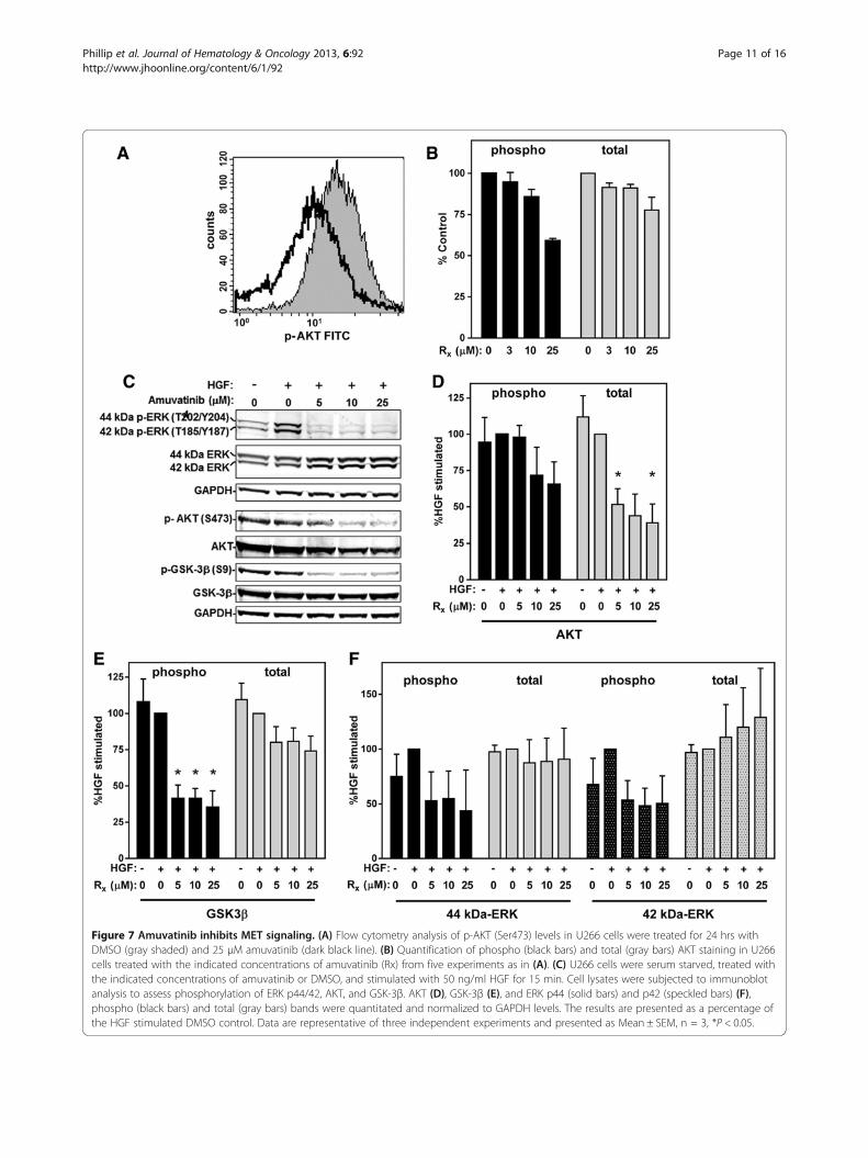
Effect of Amuvatinib on downstream targets of METPrevious studies have shown that inhibition of METcauses a reduction in the phosphorylation of both AKTand extracellular signal-regulated kinases (ERK)1/2 inthe MAPK signaling pathway [11]. The regulation ofAKT activity by MET plays a prominent role in promot-ing cell survival. Moreover, MET regulation of the ERKpathway is important for proliferation and both theERK1/2 and AKT pathways are involved in MET-induced cell spreading and motility. To examine AKTactivity, p-AKT (S473) levels were measured in U266cells by flow cytometry. Similar to the results seen withp-MET, a reduction in AKT phosphorylation was de-tected in cells treated for 24 hr with 25 μM amuvatinibwhen cells were grown in normal growth conditions(10% FBS) (Figure 7A and B). An assessment of amuvati-nib’s effects on HGF-specific signaling was also per-formed in the U266 cells cultured in 0.1% FBS for 16 hwith and without various concentrations of amuvatinibfollowed with 15 min HGF. Immunoblot analysis againshowed that lower concentrations amuvatinib is neededto decreased AKT phosphorylation at Ser473 (Figure 7Cand D), even though in these cells the levels were lowand difficult to detect. Interestingly, total AKT decreasedby 60% with amuvatinib treatment. To better assess theeffect of amuvatinib on the AKT pathway, we examinedthe phosphorylation of an AKT target, glycogen synthasekinase 3 β (GSK3β) on Ser9. Amuvatinib-treated cellsshowed, in addition to reduction of AKT, a 65% decreasein phosphorylation of GSK3β, with a 24% decrease intotal GSK3β (Figure 7C and E).A similar assessment of phospho-ERK1/2 levels under
HGF specific signaling demonstrated that amuvatinibinhibited phosphorylation of both the 44-kDa and the42-kDa ERK isoforms by 55% and 50%, respectively,while total ERK1/2 levels did not significantly change(Figure 7C and F). These results demonstrate that amu-vatinib treatment inhibits both ERK1/2 and AKT signal-ing through the MET pathway.
DiscussionMET is a receptor tyrosine kinase that is activated bythe ligand HGF and has been shown to be constitutively
expressed, mutated, or over-expressed in many differentcancer cell types. It serves as an important factor for cellsurvival, migration, and motility [7,11,15]. Corollary tothat, inhibition of MET kinase activity causes reductionof the downstream signaling that is necessary for thesecells to maintain their oncogenic properties [46]. Pre-vious studies in our laboratory showed that while METreceptor tyrosine kinase acts as a survival factor formyeloma cells [32,33], it is neither mutated nor, for themost part, over-expressed in MM. However, its ligandHGF is increased in plasma or serum obtained frommyeloma patients and higher HGF level has been associ-ated with poor prognosis [18,20,22,26]. Furthermore,HGF not only promotes growth, migration, and survivalof myeloma cells, it also potentiates IL-6 effects [46].While levels of plasma HGF have been associated with
myeloma, levels of HGF and MET mRNA in patientplasma cells have not been well evaluated nor correlatedwith disease status. Our analyses of mRNA array data[40,41] demonstrated autocrine expression of HGF inCD138+ plasma cells from MM patients. This was con-sistent with previous report in 7 myeloma patient sam-ples [18]. Our results further elucidated that the level ofthe HGF expression was directly associated with diseaseprogression.Together, these findings provide a rationale for target-
ing the HGF/MET signaling axis in myeloma. TargetingHGF directly may prove difficult, since therapeutic tar-geting of HGF would need to be effective at elevatedlevels to successfully compete and inhibit the high serumHGF concentrations in myeloma patients. Therefore,using a small-molecule MET suppressor such as amuva-tinib may be a viable option to target the HGF/METpathway. Additionally, several MET inhibitors are avail-able for clinical testing [11].Amuvatinib is an orally available drug that is currently
in clinical trials for the treatment of solid tumors[37,38,47]. This compound was designed, developed,and selected via a computation-driven in silico processwhereby drug scaffolds were screened, docked, and fittedagainst a homologous model of KIT. After additionalscreening in biochemical and cell-based assays, amuvati-nib was selected as a tyrosine kinase inhibitor with activ-ity against wild-type and mutant KIT, MET, RET, FLT3and PDGFRα [48,49]. Later, amuvatinib inhibition ofMET activity was found to lead to reduction of RAD51expression and to radiosensitization of tumor cells [50].Since amuvatinib is a small-molecule inhibitor that
suppresses MET activity, we tested this agent as a proof-of-concept to therapeutically target MET in myeloma.Our study demonstrated that amuvatinib was effective ininhibiting growth and DNA synthesis at low micromolarconcentrations in cell lines grown under normal condi-tions (10% FBS). Moreover, amuvatinib treatment resulted
Phillip et al. Journal of Hematology & Oncology 2013, 6:92 Page 10 of 16http://www.jhoonline.org/content/6/1/92
Figure 7 Amuvatinib inhibits MET signaling. (A) Flow cytometry analysis of p-AKT (Ser473) levels in U266 cells were treated for 24 hrs withDMSO (gray shaded) and 25 μM amuvatinib (dark black line). (B) Quantification of phospho (black bars) and total (gray bars) AKT staining in U266cells treated with the indicated concentrations of amuvatinib (Rx) from five experiments as in (A). (C) U266 cells were serum starved, treated withthe indicated concentrations of amuvatinib or DMSO, and stimulated with 50 ng/ml HGF for 15 min. Cell lysates were subjected to immunoblotanalysis to assess phosphorylation of ERK p44/42, AKT, and GSK-3β. AKT (D), GSK-3β (E), and ERK p44 (solid bars) and p42 (speckled bars) (F),phospho (black bars) and total (gray bars) bands were quantitated and normalized to GAPDH levels. The results are presented as a percentage ofthe HGF stimulated DMSO control. Data are representative of three independent experiments and presented as Mean ± SEM, n = 3, *P < 0.05.
Phillip et al. Journal of Hematology & Oncology 2013, 6:92 Page 11 of 16http://www.jhoonline.org/content/6/1/92
in cell death in U266 myeloma cell line dependent onMET/HGF signaling, as measured by annexin V/PI stain-ing and PARP cleavage. This cytotoxic effect remainedeven when these MET-addicted cells were grown on bonemarrow stromal cells. In contrast, the drug did not induceapoptosis in another myeloma cell line (RPMI-8226/S)that is not dependent on the MET/HGF signaling axis dueto lower levels of HGF (75% less) andMET (95% less).Because amuvatinib also impairs KIT and PDGFR sig-
naling, we tested impact of imatinib (an established KITand PDGFR inhibitor) in myeloma cells. Imatinib in-duced no significant amount of cell death in U266 cellsdemonstrating that amuvatinib’s effect was due to METinhibition. This statement was in line with the dataregarding decreased phosphorylation of MET after amu-vatinib treatment. Because >95% of the compound isbound and sequestered by serum proteins (Unpublisheddata), the dose required to achieve maximum inhibitionof MET phosphorylation in serum starved conditionswas lower than the dose to induce apoptosis in fullserum conditions. Likewise, under serum starved condi-tions, the maximum induction of apoptosis was seen atthe same dose which achieved maximum inhibition ofMET phosphorylation. As expected, in imatinib treatedcells, there was no reduction of p-MET (data not shown)as well as no significant reduction in survival. These cor-relation data suggest that amuvatinib mediated growthinhibition and cell death is due to its action on MET andnot its action on KIT or PDGFR.In conjunction with a decrease in MET phosphoryl-
ation, there was a decline in HGF-dependent ERK1/2and AKT phosphorylation as well as the phosphorylationof the AKT targets GSK3β and caspase-9 (data notshown). Diminution of phosphorylated MET and associ-ated decreases in ERK1/2 and AKT phosphorylation hasbeen shown to be important in growth, migration andcell survival pathways for other cancer cell types [11].Amuvatinib proved to be effective in inducing cell death
not only in a MET dependent myeloma cell line but alsoin primary CD138+ malignant plasma cells obtained frompatients with myeloma. In contrast, amuvatinib did notcause cell death in normal CD138– cells obtained from thesame individuals (Figure 2). These data provide evidence ofthe selectivity of amuvatinib, suggesting that it may be usedspecifically for myeloma treatment without impairing othernormal hematological cells in the bone marrow. In linewith this selective cytotoxic effect on CD138 plasma cells,MET phosphorylation was reduced by amuvatinib treat-ment in primary plasma cells but not CD138– cells.The effects of amuvatinib described here provide
proof-of-concept that MET is important for the survivalof myeloma cells and that reduction of its kinase activitymay prove to be an effective targeted therapy. The 25 μMdose of amuvatinib needed to robustly induce apoptosis in
cell lines and plasma cells under full serum conditionsmay not be achievable in vivo. Pharmacokinetic studies ofamuvatinib during a phase I trial indicated that plasmalevels reached between 1 and 2 μM [51]. Hence, newergeneration and more potent MET tyrosine kinase inhibi-tors are needed [11]. ARQ 197 (tivantinib) is a small-molecule, non-ATP-competitive inhibitor which is highlyspecific for MET [52,53]. This drug is well tolerated inclinical trials and has shown efficacy in solid tumors[54-58]. Pharmacodynamic studies from a phase I trial in-dicated that at an oral dosing of 360 mg, twice daily, ARQ197 reached steady-state plasma concentrations of 6–7 μM[55]. This correlated with decreases in total MET andphospho-FAK (Tyr861) and increases in TUNEL-positivecells in patients’ tumors.Our results with amuvatinib provided the impetus to
pursue testing of ARQ 197 in myeloma cells. Our preclin-ical studies indicated that treatment with ARQ 197 for48 hours was cytotoxic to myeloma cell lines (≥ 60%increase in annexin V/PI-positive cells) at clinically achiev-able doses [59]. Moreover, these studies provided thefoundation for a Cancer Therapy Evaluation Program,National Cancer Institute sponsored phase 2 clinical trialof ARQ 197 in myeloma patients, which is currently un-derway at MD Anderson Cancer Center [60].
ConclusionsOur finding provides proof-of-principle that MET is im-portant for the survival of myeloma cells and using aMET inhibitor such as amuvatinib may prove to be aneffective strategy for treatment of MM. Amuvatinib ex-hibited tumoricidal activity in myeloma cells which wasassociated with inhibition of MET signaling. Amuvatinib’slack of effect on CD138– cells from the same patients fur-ther establishes the selectivity of this agent. The clinicalsuccess of other targeted therapeutics for cytoplasmic andreceptor tyrosine kinases, further underscores a need fortesting a small-molecule inhibitor that targets MET kinaseactivity for patients with myeloma.
MethodsMaterialsAmuvatinib (MP470) was obtained from Astex Pharma-ceuticals, Inc. (Dublin, CA) and was dissolved in DMSO(Sigma Aldrich, St. Louis, MO). Because of stabilityconstraints, amuvatinib solution was prepared fresh foreach experiment. Imatinib was purchased from Novartis(St. Louis, MO) and was dissolved in DMSO and storedin aliquots at -20°C. [3H] thymidine (60 Ci/mmol) wasobtained from Moravek Biochemical Inc. (Brea, CA).
Cell culture and growth analysisThe myeloma cell lines U266 [61] and RPMI-8226/S[62] were obtained from Dr. William Dalton at H. Lee
Phillip et al. Journal of Hematology & Oncology 2013, 6:92 Page 12 of 16http://www.jhoonline.org/content/6/1/92
Moffitt Cancer Center (Tampa, FL). NK-tert humanbone marrow stromal cells were obtained from Dr. JanBurger at UT MD Anderson Cancer Center [63]. Thecell lines were maintained as described [32,63] and rou-tinely tested for Mycoplasma infection and authenticatedby short tandem repeat analysis by UT MD AndersonCancer Center’s Characterized Cell Line Core facility.Myeloma cell-stromal co-cultures were performed using
U266 cells and NK-tert cells at a ratio of 20 to 1. Stromalcells were plated at a concentration of ~2 × 10² cells/mm2
surface area 5 hours before adding U266 cells at a 20 foldhigher concentration. The cells were co-cultured for 2 hprior to treatment with or without amuvatinib or bortezo-mib for 48 h. At the end of incubation, the U266 cells,which are free floating in these cultures, were carefullyremoved for analysis, leaving the adherent stromal layerundisturbed. Additionally, the stromal cells were also har-vested by trypsinization and similarly assessed.The effect of amuvatinib treatment on cell growth
inhibition was measured in exponentially growingU266 cells. Cells were counted using a Coulter counter(Beckman Coulter, Fullerton, CA). DNA synthesis wasmeasured using [3H]thymidine incorporation as de-scribed [64].
Gene expression array analysesExpression data from 162 CD138+ bone marrow plasmacell samples from healthy individuals as well as patientswith MGUS, SMM, MM-N, and MM-R, which were mea-sured by using Affymetrix U133A microarrays, were down-loaded from GEO (GSE6477) [40,41]. Robust MultichipAverage (RMA) algorithm was used for normalization/quantification of the data. The maximal values for the re-spective probe-sets of MET and HGF were used for geneexpression profiling. The Kruskal-Wallis test was appliedto assess whether expression of MET and HGF was associ-ated with defined clinical groups, and results are presentedas box-plots.
Isolation of CD138+ AND CD138– cells from primary bonemarrow aspirates from MM patientsPrimary samples were obtained from both male andfemale myeloma patients being treated at MD AndersonCancer Center (Table 1). Patient samples were obtainedusing an MD Anderson Cancer Center InstitutionalReview Board approved protocol. All patients signed aninformed consent form to provide peripheral blood andbone marrow samples. After collection of bone marrowsamples, CD138+ cells were isolated as described [65],suspended in RPMI 1640 with 10% human AB serum(Cambrex Biosciences, East Rutherford, NJ) and usedimmediately for experiments. Peripheral blood sampleswere collected from patients 2, 4, and 5 for assessmentof plasma HGF levels.
Immunoblot analysisThe effects of amuvatinib on HGF dependent signalingwere assessed in U266 cells that had been serum starvedfor 24 h in RPMI 1640 containing 0.1% FBS; for the last16 h of starvation the cells were treated with variousconcentrations of amuvatinib or DMSO. They were thentreated with 50 ng/ml HGF for 15 min to stimulateMET. Amuvatinib-mediated induction of PARP cleavagewas performed on U266 cells cultured in full serum(10% FBS) as well as under low serum conditions (0.1%FBS). Protein lysates and immunoblots were prepared aspreviously described [66]. Experiments were performedin triplicates, and bands were quantified by using anOdyssey Infrared Imaging System (LI-COR Biosciences,Lincoln, NE). Primary antibodies were: mouse monoclo-nal antibodies to MET clone 3D4 (Invitrogen, Carlsbad,CA); GSK-3β clone 7/GSK-3b, PARP clone C2-10,cleaved PARP Asp 214 clone F21-852, AKT clone 9Q7(BD Biosciences Pharmingen, San Diego, CA); GAPDHclone 6C6 (Abcam, Inc, Cambridge, MA); phospho-ERK1/2 (Thr202/Tyr204) clone E10 (Cell SignalingTechnology, Danvers, MA); β-actin clone AC-15 (SigmaAldrich); rabbit monoclonal antibodies to phospho-GSK-3β (Ser9) clone 5B3 (Cell Signaling Technology); rabbitpolyclonal antibodies to phospho-MET (Tyr1349) (Invi-trogen); ERK1/2, and phospho-AKT (Ser473) (Cell Sig-naling Technology).
Flow cytometryIntracellular protein expression in U266 cells was mea-sured using BD Cytofix/Cytoperm Fixation/Perme-abilization Kit (BD Biosciences). Primary antibodiesused were anti-phospho-HGF R/c-MET (Tyr1234/1235) (R&D Systems, Minneapolis, MN), MET (C-12):sc-10 (Santa Cruz Biotechnology, Santa Cruz, CA),phospho-AKT (Ser473), AKT antibody (Cell SignalingTechnology); and caspase-9 (Ser196) (Santa CruzBiotechnology). Secondary antibody was a fluoresceinisothiocyanate–conjugated (FITC) Affinipure goat anti-rabbit (Jackson ImmunoResearch, West Grove, PA). Cellcycle analysis and annexin V/propidium iodide (PI) stain-ing were performed, respectively, as described [32,33]. Allflow cytometry analysis was performed using a BectonDickinson FACSCalibur flow cytometer (San Jose, CA,USA). Statistical significance of changes was assessed bypaired t-test analysis using Prism software (Graphpad, SanDiego, CA).
Enzyme linked-immuno-sorbent assay for HGF levelsHGF levels in primary patient plasma were determinedusing the Human HGF Immunoassay Kit as per themanufacturer’s protocol (Invitrogen). The absorbance ofthis horseradish-peroxidase based assay was measured at450 nm. Each sample was assayed in triplicate.
Phillip et al. Journal of Hematology & Oncology 2013, 6:92 Page 13 of 16http://www.jhoonline.org/content/6/1/92
Additional file
Additional file 1: Figure S1. MET and HGF expression. Real-time RT-PCRanalysis of HGF and MET transcript levels in U266 and RPMI-8226/S cells.Figure S2. (A) NK-tert cells protect U266 cells from bortezomib-inducedcytotoxicity. (B) MET expression and HGF-dependent activity in serumstarved NK-tert cells.
Competing interestSR and PT are employed by Astex Pharmaceuticals, Inc., Dublin, CA. For theremaining authors, none was declared.
Authors’ contributionsCJP, SZ, KB, and CMS designed and performed experiments; CJP, SZ, JZ, VB,and CMS analyzed data; SS performed experiments; PT and SR providedamuvatinib; MW provide sorted bone marrow samples; CJP and CMS wrotethe manuscript; CMS and VG directed study; VG provided laboratoryresources; All authors critically read and approved manuscript.
Authors’ informationChristine Marie Stellrecht and Varsha Gandhi contributed equally as seniorauthors.
AcknowledgementsThis work was supported in part by grants from the National Institutes ofHealth [Minority Graduate Student Supplement NIH RO1-CA85915 to CJP],the National Cancer Institute [Cancer Center Support Grant NIH P50CA16672; and MD Anderson Cancer Center SPORE in Multiple MyelomaResearch NIH P50 CA142509 to VB and MW], the Center for Targeted Therapyto CMS and VG, and Multiple Myeloma Research Foundation Senior ResearchAward to CMS.The authors are grateful to David Graber for processing the bone marrowsamples and separating the CD138+ cells and to Jairo A Matthews forcoordinating patient sample collection distributing these samples to ourlaboratory.
Author details1Departments of Experimental Therapeutics, The University of Texas MDAnderson Cancer Center, Houston, Texas, USA. 2Bioinformatics andComputational Biology, The University of Texas MD Anderson Cancer Center,Houston, Texas, USA. 3Biostatistics, The University of Texas MD AndersonCancer Center, Houston, Texas, USA. 4Leukemia, The University of Texas MDAnderson Cancer Center, Houston, Texas, USA. 5Lymphoma/Myeloma, TheUniversity of Texas MD Anderson Cancer Center, Houston, Texas, USA.6Graduate School of Biomedical Sciences, The University of Texas HealthScience Center, Houston, Texas, USA. 7Astex Pharmaceuticals, Inc., Dublin,California, USA.
Received: 3 November 2013 Accepted: 2 December 2013Published: 10 December 2013
References1. Hallek M, Bergsagel PL, Anderson KC: Multiple myeloma: increasing
evidence for a multistep transformation process. Blood 1998, 91:3–21.2. Piazza FA, Gurrieri C, Trentin L, Semenzato G: Towards a new age in the
treatment of multiple myeloma. Ann Hematol 2007, 86:159–172.3. Mahindra A, Laubach J, Raje N, Munshi N, Richardson PG, Anderson K:
Latest advances and current challenges in the treatment of multiplemyeloma. Nat Rev Clin Oncol 2012, 9:135–143.
4. Raab MS, Podar K, Breitkreutz I, Richardson PG, Anderson KC: Multiplemyeloma. Lancet 2009, 374:324–339.
5. Roodman GD: Targeting the bone microenvironment in multiplemyeloma. J Bone Miner Metab 2010, 28:244–250.
6. Podar K, Anderson KC: The pathophysiologic role of VEGF in hematologicmalignancies: therapeutic implications. Blood 2005, 105:1383–1395.
7. Derksen PW, De Gorter DJ, Meijer HP, Bende RJ, Van Dijk M, Lokhorst HM,Bloem AC, Spaargaren M, Pals ST: The hepatocyte growth factor/Metpathway controls proliferation and apoptosis in multiple myeloma.Leukemia 2003, 17:764–774.
9. Hideshima T, Podar K, Chauhan D, Anderson KC: Cytokines and signaltransduction. Best Pract Res Clin Haematol 2005, 18:509–524.
10. Song L, Li Y, Sun YX, Yu M, Shen BF: IL-6 inhibits apoptosis of humanmyeloma cell line XG-7 through activation of JAK/STAT pathway andup-regulation of Mcl-1. Ai Zheng 2002, 21:113–116.
11. Stellrecht CM, Gandhi V: MET receptor tyrosine kinase as a therapeuticanticancer target. Cancer Lett 2009, 280:1–14.
12. Naldini L, Vigna E, Ferracini R, Longati P, Gandino L, Prat M, Comoglio PM:The tyrosine kinase encoded by the MET proto-oncogene is activated byautophosphorylation. Mol Cell Biol 1991, 11:1793–1803.
13. Naldini L, Vigna E, Narsimhan RP, Gaudino G, Zarnegar R, Michalopoulos GK,Comoglio PM: Hepatocyte growth factor (HGF) stimulates the tyrosinekinase activity of the receptor encoded by the proto-oncogene c-MET.Oncogene 1991, 6:501–504.
14. Rodrigues GA, Park M: Autophosphorylation modulates the kinase activityand oncogenic potential of the Met receptor tyrosine kinase. Oncogene1994, 9:2019–2027.
15. Derksen PW, Keehnen RM, Evers LM, van Oers MH, Spaargaren M, Pals ST:Cell surface proteoglycan syndecan-1 mediates hepatocyte growthfactor binding and promotes Met signaling in multiple myeloma. Blood2002, 99:1405–1410.
16. Tjin EPM, Groen RWJ, Vogelzang I, Derksen PWB, Klok MD, Meijer HP, vanEeden S, Pals ST, Spaargaren M: Functional analysis of HGF/MET signalingand aberrant HGF-activator expression in diffuse large B-cell lymphoma.Blood 2006, 107:760–768.
17. Hennessy BT, Smith DL, Ram PT, Lu Y, Mills GB: Exploiting the PI3K/AKTpathway for cancer drug discovery. Nat Rev Drug Discov 2005,4:988–1004.
18. Borset M, Hjorth-Hansen H, Seidel C, Sundan A, Waage A: Hepatocytegrowth factor and its receptor c-met in multiple myeloma. Blood 1996,88:3998–4004.
19. Borset M, Lien E, Espevik T, Helseth E, Waage A, Sundan A: Concomitantexpression of hepatocyte growth factor/scatter factor and thereceptor c-MET in human myeloma cell lines. J Biol Chem 1996,271:24655–24661.
20. Seidel C, Borset M, Turesson I, Abildgaard N, Sundan A, Waage A: Elevatedserum concentrations of hepatocyte growth factor in patients withmultiple myeloma. The nordic myeloma study group. Blood 1998,91:806–812.
21. Seidel C, Borset M, Hjertner O, Cao D, Abildgaard N, Hjorth-Hansen H,Sanderson RD, Waage A, Sundan A: High levels of soluble syndecan-1 inmyeloma-derived bone marrow: modulation of hepatocyte growthfactor activity. Blood 2000, 96:3139–3146.
22. Iwasaki T, Hamano T, Ogata A, Hashimoto N, Kitano M, Kakishita E: Clinicalsignificance of vascular endothelial growth factor and hepatocytegrowth factor in multiple myeloma. Br J Haematol 2002, 116:796–802.
23. Seidel C, Lenhoff S, Brabrand S, Anderson G, Standal T, Lanng-Nielsen J,Turesson I, Borset M, Waage A: Hepatocyte growth factor in myelomapatients treated with high-dose chemotherapy. Br J Haematol 2002,119:672–676.
24. Alexandrakis MG, Passam FH, Sfiridaki A, Kandidaki E, Roussou P, KyriakouDS: Elevated serum concentration of hepatocyte growth factor inpatients with multiple myeloma: correlation with markers of diseaseactivity. Am J Hematol 2003, 72:229–233.
25. Kara IO, Sahin B, Gunesacar R, Unsal C: Clinical significance of hepatocytegrowth factor, platelet-derived growth factor-AB, and transforminggrowth factor-alpha in bone marrow and peripheral blood of patientswith multiple myeloma. Adv Ther 2006, 23:635–645.
Phillip et al. Journal of Hematology & Oncology 2013, 6:92 Page 14 of 16http://www.jhoonline.org/content/6/1/92
26. Sezer O, Jakob C, Eucker J, Niemoller K, Gatz F, Wernecke K, Possinger K:Serum levels of the angiogenic cytokines basic fibroblast growth factor(bFGF), vascular endothelial growth factor (VEGF) and hepatocytegrowth factor (HGF) in multiple myeloma. Eur J Haematol 2001,66:83–88.
27. Pour L, Svachova H, Adam Z, Mikulkova Z, Buresova L, Kovarova L, Buchler T,Penka M, Vorlicek J, Hajek R: Pretreatment hepatocyte growth factor andthrombospondin-1 levels predict response to high-dose chemotherapyfor multiple myeloma. Neoplasma 2010, 57:29–34.
28. Ludek P, Hana S, Zdenek A, Martina A, Dana K, Tomas B, Lucie K, Marta K,Jaroslav M, Miroslav P, et al: Treatment response to bortezomib inmultiple myeloma correlates with plasma hepatocyte growth factorconcentration and bone marrow thrombospondin concentration. Eur JHaematol 2010, 84:332–336.
29. Pour L, Svachova H, Adam Z, Almasi M, Buresova L, Buchler T, Kovarova L,Nemec P, Penka M, Vorlicek J, Hajek R: Levels of angiogenic factors inpatients with multiple myeloma correlate with treatment response.Ann Hematol 2010, 89:385–389.
30. Hjertner O, Torgersen ML, Seidel C, Hjorth-Hansen H, Waage A, Borset M,Sundan A: Hepatocyte growth factor (HGF) induces interleukin-11secretion from osteoblasts: a possible role for HGF in myeloma-associatedosteolytic bone disease. Blood 1999, 94:3883–3888.
31. Standal T, Abildgaard N, Fagerli UM, Stordal B, Hjertner O, Borset M,Sundan A: HGF inhibits BMP-induced osteoblastogenesis: possibleimplications for the bone disease of multiple myeloma. Blood 2007,109:3024–3030.
32. Stellrecht CM, Phillip CJ, Cervantes-Gomez F, Gandhi V: Multiple myelomacell killing by depletion of the MET receptor tyrosine kinase. Cancer Res2007, 67:9913–9920.
33. Phillip CJ, Stellrecht CM, Nimmanapalli R, Gandhi V: Targeting METtranscription as a therapeutic strategy in multiple myeloma. CancerChemother Pharmacol 2009, 63:587–597.
34. Chen LS, Stellrecht CM, Gandhi V: RNA-directed agent, cordycepin,induces cell death in multiple myeloma cells. Br J Haematol 2008,140:682–691.
35. Mahtouk K, Tjin EP, Spaargaren M, Pals ST: The HGF/MET pathway as targetfor the treatment of multiple myeloma and B-cell lymphomas. BiochimBiophys Acta 1806, 2010:208–219.
36. Qi W, Cooke L, Calaluce R, Bearss D, Mahadevan D: Dual inhibition ofreceptor tyrosine kinases of PDGFR and EGFR abolishes prostate cancercell growth in a mouse xenograft model by completedephosphorylation of PKB/Akt. Proc Am Assoc Cancer Res 2007, 48:5421.
37. Tibes R, Berk GI, Fine GD, Choy GS, Tolcher AW: A phase-1 study of MP-470, anovel orally bioavailable small molecule with Rad51 suppression activity.Proc Am Assoc Cancer Res 2008, 49:4083.
38. Tolcher AW, Berk GI, Fine GD, Choy GD, Bearss DJ, Redkar S, Tibes R: MP470,a potent oral Rad51 suppressor is safe and tolerable in first-in-humanstudy. Proc Am Assoc Cancer Res 2008, 49:4083.
39. Mahtouk K, Moreaux J, Hose D, Reme T, Meissner T, Jourdan M, Rossi JF,Pals ST, Goldschmidt H, Klein B: Growth factors in multiple myeloma: acomprehensive analysis of their expression in tumor cells and bonemarrow environment using Affymetrix microarrays. BMC Cancer 2010,10:198.
40. Chng WJ, Kumar S, Vanwier S, Ahmann G, Price-Troska T, Henderson K,Chung TH, Kim S, Mulligan G, Bryant B, et al: Molecular dissection ofhyperdiploid multiple myeloma by gene expression profiling. Cancer Res2007, 67:2982–2989.
41. Keats JJ, Fonseca R, Chesi M, Schop R, Baker A, Chng WJ, Van Wier S,Tiedemann R, Shi CX, Sebag M, et al: Promiscuous mutations activate thenoncanonical NF-kappaB pathway in multiple myeloma. Cancer Cell 2007,12:131–144.
42. Meads MB, Hazlehurst LA, Dalton WS: The bone marrowmicroenvironment as a tumor sanctuary and contributor to drugresistance. Clin Cancer Res 2008, 14:2519–2526.
43. Zhang W, Huang P: Cancer-stromal interactions: role in cell survival,metabolism and drug sensitivity. Cancer Biol Ther 2011, 11:150–156.
44. Druker BJ: Imatinib as a paradigm of targeted therapies. Adv Cancer Res2004, 91:1–30.
45. Rodrigues GA, Naujokas MA, Park M: Alternative splicing generatesisoforms of the met receptor tyrosine kinase which undergo differentialprocessing. Mol Cell Biol 1991, 11:2962–2970.
46. Hov H, Holt RU, Ro TB, Fagerli UM, Hjorth-Hansen H, Baykov V, ChristensenJG, Waage A, Sundan A, Borset M: A selective c-met inhibitor blocks anautocrine hepatocyte growth factor growth loop in ANBL-6 cells andprevents migration and adhesion of myeloma cells. Clin Cancer Res 2004,10:6686–6694.
47. Tolcher AW, Mita M, Gordon M, Rosen L, Patnaik A, Mita A, Fine GD, ChoyGS, Berk GI: Clinical responses in highly refractory solid tumor patientswith oral MP-470, a multi-targeted tyrosine kinase inhibitor, incombination with standard of care chemotherapy regimens: preliminaryreport from a multi-institutional phase-Ib clinical trial. Proc Am Assoc CancerRes 2008, 49:403.
48. Mahadevan D, Cooke L, Riley C, Swart R, Simons B, Della Croce K, Wisner L,Iorio M, Shakalya K, Garewal H, et al: A novel tyrosine kinase switch is amechanism of imatinib resistance in gastrointestinal stromal tumors.Oncogene 2007, 26:3909–3919.
49. Trevor K, Combs D, Mahadevan D, Bearss D, Cranmer L: Activity of themulti-targeted, receptor tyrosine kinase inhibitor MP470 against synovialsarcoma cells. Proc Am Assoc Cancer Res 2008, 49:4891.
51. Tibes R, Fine G, Choy G, Redkar S, Taverna P, Oganesian A, Sahai A, Azab M,Tolcher AW: A phase I, first-in-human dose-escalation study of amuvatinib, amulti-targeted tyrosine kinase inhibitor, in patients with advanced solidtumors. Cancer Chemother Pharmacol 2013, 71:463–471.
52. Munshi N, Jeay S, Li Y, Chen CR, France DS, Ashwell MA, Hill J, Moussa MM,Leggett DS, Li CJ: ARQ 197, a novel and selective inhibitor of the humanc-Met receptor tyrosine kinase with antitumor activity. Mol Cancer Ther2010, 9:1544–1553.
53. Eathiraj S, Palma R, Volckova E, Hirschi M, France DS, Ashwell MA, Chan TC:Discovery of a novel mode of protein kinase inhibition characterized bythe mechanism of inhibition of human mesenchymal-epithelial transitionfactor (c-Met) protein autophosphorylation by ARQ 197. J Biol Chem2011, 286:20666–20676.
54. Sequist LV, von Pawel J, Garmey EG, Akerley WL, Brugger W, Ferrari D, ChenY, Costa DB, Gerber DE, Orlov S, et al: Randomized phase II study oferlotinib plus tivantinib versus erlotinib plus placebo in previouslytreated non-small-cell lung cancer. J Clin Oncol 2011, 29:3307–3315.
55. Yap TA, Olmos D, Brunetto AT, Tunariu N, Barriuso J, Riisnaes R, Pope L,Clark J, Futreal A, Germuska M, et al: Phase I trial of a selective c-METinhibitor ARQ 197 incorporating proof of mechanism pharmacodynamicstudies. J Clin Oncol 2011, 29:1271–1279.
56. Goldman JW, Laux I, Chai F, Savage RE, Ferrari D, Garmey EG, Just RG, RosenLS: Phase 1 dose-escalation trial evaluating the combination of theselective MET (mesenchymal-epithelial transition factor) inhibitortivantinib (ARQ 197) plus erlotinib. Cancer 2012, 118:5903–5911.
57. Previdi S, Abbadessa G, Dalo F, France DS, Broggini M: Breast cancer-derivedbone metastasis can be effectively reduced through specific c-MET inhibitortivantinib (ARQ 197) and shRNA c-MET knockdown. Mol Cancer Ther 2012,11:214–223.
58. Wagner AJ, Goldberg JM, Dubois SG, Choy E, Rosen L, Pappo A, Geller J,Judson I, Hogg D, Senzer N, et al: Tivantinib (ARQ 197), a selective inhibitorof MET, in patients with microphthalmia transcription factor-associatedtumors: results of a multicenter phase 2 trial. Cancer 2012,118:5894–5902.
59. Zaman S, Stellrecht CM, Shentu S, Orlowski R, Gandhi V: Targeting thepro-survival protein c-MET with ARQ 197 inhibits growth of multiplemyeloma cells. Proc Am Assoc Cancer Res 2012, 53:844.
60. Dorkhom SJ, Zaman S, Thomas SK, Alexanian R, Shah JJ, Weber DM, WangM, Anderson ML, Baladandayuthapani V, Lin YH, et al: Phase II study of thec-Met inhibitor ARQ 197 (tivantinib) in patients with relapsed multiplemyeloma. Blood (ASH Annual Meeting Abstracts) 2012, 120:2976.
61. Catlett-Falcone R, Landowski TH, Oshiro MM, Turkson J, Levitzki A, Savino R,Ciliberto G, Moscinski L, Fernandez-Luna JL, Nunez G, et al: Constitutiveactivation of Stat3 signaling confers resistance to apoptosis in humanU266 myeloma cells. Immunity 1999, 10:105–115.
62. Bellamy WT, Dalton WS, Gleason MC, Grogan TM, Trent JM: Developmentand characterization of a Melphalan-resistant human multiple myelomacell line. Cancer Res 1991, 51:995–1002.
63. Manshouri T, Estrov Z, Quintas-Cardama A, Burger J, Zhang Y, Livun A, KnezL, Harris D, Creighton CJ, Kantarjian HM, Verstovsek S: Bone marrow
Phillip et al. Journal of Hematology & Oncology 2013, 6:92 Page 15 of 16http://www.jhoonline.org/content/6/1/92
stroma-secreted cytokines protect JAK2(V617F)-mutated cells from theeffects of a JAK2 inhibitor. Cancer Res 2011, 71:3831–3840.
64. Gandhi V, Ayres M, Halgren RG, Krett NL, Newman RA, Rosen ST:8-chloro-cAMP and 8-chloro-adenosine act by the same mechanism inmultiple myeloma cells. Cancer Res 2001, 61:5474–5479.
65. Bjorklund CC, Ma W, Wang ZQ, Davis RE, Kuhn DJ, Kornblau SM, Wang ML,Shah JJ, Orlowski RZ: Evidence of a role for activation of Wnt/b-cateninsignaling in the resistance of plasma cells to lenalidomide. J Biol Chem2011, 286:11009–11020.
66. Stellrecht CM, Ayres M, Arya R, Gandhi V: A unique RNA-directednucleoside analog is cytotoxic to breast cancer cells and depletes cyclinE levels. Breast Cancer Res Treat 2010, 121:355–364.
doi:10.1186/1756-8722-6-92Cite this article as: Phillip et al.: Targeting MET kinase with thesmall-molecule inhibitor amuvatinib induces cytotoxicity in primarymyeloma cells and cell lines. Journal of Hematology & Oncology 2013 6:92.
Submit your next manuscript to BioMed Centraland take full advantage of:
• Convenient online submission
• Thorough peer review
• No space constraints or color figure charges
• Immediate publication on acceptance
• Inclusion in PubMed, CAS, Scopus and Google Scholar
• Research which is freely available for redistribution
Submit your manuscript at www.biomedcentral.com/submit
Phillip et al. Journal of Hematology & Oncology 2013, 6:92 Page 16 of 16http://www.jhoonline.org/content/6/1/92