6

Proc. Nail. Acad. Sci. USAVol. 88, pp. 10018-10022, November 1991Biochemistry
Tat transactivation of the human immunodeficiency virus type 1promoter is influenced by basal promoter activity and thesimian virus 40 origin of DNA replication
(replication origin/transcrlption/transactivator)
MARK KESSLER AND MICHAEL B. MATHEWS*Cold Spring Harbor Laboratory, 1 Bungtown Road, P.O. Box 100, Cold Spring Harbor, NY 11724-2208
Communicated by James D. Watson, July 15, 1991 (received for review May 28, 1991)
ABSTRACT We examined the activation of transcriptionfrom the human immunodeficiency virus type 1 (HIV-1) pro-moter by the viral Tat protein in a transient expression system.Plasmids contained a HIV-reporter gene cassette and a simianvirus 40 origin ofDNA replication. Run-on assays of transcrip-tion complex distribution and analysis of cytoplasmic RNAaccumulation confirmed that Tat is able to activate transcrip-tion by two mechanisms: by increasing the rate of transcrip-tional initiation and the efficiency of transcriptional elongation.The degree to which Tat stimulated initiation is determined bythe basal level of HIV-directed transcription, which is influ-enced by the position of the simian virus 40 replication orwin.Tat functions primarily to increase the efficiency of elongationwhen the origin is located downstream from the HIV-reportercassette and the basal level oftranscription is high. On the otherhand, Tat functions primarily to increase the rate of initiationwhen the origin is upstream from the cassette and the basallevel oftranscription is 10-fold lower. These studies suggest thatthe site of integration of the virus into the cellular genome maysignificantly affect the level of expression from the H1V pro-moter and consequently the pathobiology of the virus.
The human immunodeficiency virus type 1 (HIV-1) Tatprotein is a potent transactivator of HIV-1 gene expressionand plays an essential role in the infectious process of thevirus (1, 2), but its mechanism of action has not been fullyresolved. It has been-proposed that Tat functions at a numberof different levels, including transcriptional initiation, elon-gation of nascent RNA chains, posttranscriptional process-ing, and protein synthesis (reviewed in ref. 3). The target forTat-mediated transactivation is a sequence within the Rregion of the long terminal repeat (LTR) designated thetrans-acting responsive element (TAR), minimally, residues+14 to +44 relative to the transcriptional initiation site (4-8).The TAR element must be situated near the transcriptionstart site in the correct orientation (7). Tat interacts withRNA transcribed from the TAR region, which forms a bulgedstem-loop structure (9), and recent work suggests that TARfunctions largely, if not exclusively, to draw Tat to thevicinity of the transcription start site (10-12).
Studies of Tat's effect on transcription have typicallyemployed model systems in which LTR-directed transcrip-tion of a reporter gene, generally the chloramphenicol ace-tyltransferase (CAT) gene, is compared in the presence andabsence of Tat. Run-on transcription analysis revealed thatTat modulates reporter gene expression by increasing the rateof transcription from the HIV LTR. This conclusion is drawnfrom transient expression assays, in which the reporter genewas transfected into cells as plasmid DNA (4, 13-16), andfrom infection assays, in which the reporter gene was deliv-
ered via an adenovirus vector (17, 18). Further analysis oftranscription complex distribution in a transient expressionsystem suggested that Tat modulates LTR-directed transcrip-tion by relieving a block to transcriptional elongation in theTAR region (14). In this study, the transcription complexeswere determined to be distributed equally between the pro-moter proximal region [339 nucleotides (nt)] and the adjacentregion (520 nt) in the presence of Tat. In the absence of Tat,however, the density of transcription complexes was un-changed in the promoter proximal region, but no complexeswere detected in the promoter distal region. A similar studyconducted in our laboratory using the adenovirus systemreached somewhat different conclusions (19). When tran-scription complex distribution was examined over shortintervals within the promoter proximal region, most com-plexes were observed within the immediately promoter-proximal region (23 or 83 nt) when transcription was carriedout in the absence of Tat. Tat affected transcription in twoways: it increased the efficiency of transcriptional elongationby suppressing polarity, and it also increased the rate oftranscriptional initiation at the HIV promoter.To resolve the differences between these two studies we
examined transcription from the HIV-1 LTR in the transientexpression system. We found that the basal transcription rateis the determinative feature and that this is influenced by thestructure of the template, in particular by the location of thesimian virus 40 (SV40) replication origin. Placement of theorigin region downstream from the HIV LTR-CAT cassettepermitted 10-fold more transcription from the LTR than whenthe origin was placed upstream from the LTR. When theorigin was in the upstream location, Tat increased both therate of initiation and the efficiency of elongation; on the otherhand, when the origin was downstream Tat primarily func-tioned to increase the efficiency of transcriptional elongation.We infer that Tat can stimulate both processes but theincrease in initiation is apparent only when the basal level ofLTR-directed transcription is low.
MATERIALS AND METHODSPlasmid and Probe Construction. The structures of the
LTR-CAT plasmids are diagramed in Fig. 1. Plasmid pHicontains the Xho I-BamHI fragment of pU3RIII (20) flankedby Xba I linkers and cloned into pUC19. The SV40 origin ofreplication (SV40 fragment nt 5171-128) was inserted betweenthe polylinker HindIII and Sph I restriction sites situatedupstream ofthe LTR. Plasmids pH2 and pH2.1 are identical topH1 except that the origin fragment, flanked with BamHI
Abbreviations: HIV-1, human immunodeficiency virus type 1; SV40,simian virus 40; LTR, long terminal repeat; CAT, chloramphenicolacetyltransferase; TAR, trans-acting responsive element; nt, nucle-otide(s).*To whom reprint requests should be addressed.
10018
The publication costs of this article were defrayed in part by page chargepayment. This article must therefore be hereby marked "advertisement"in accordance with 18 U.S.C. §1734 solely to indicate this fact.

Proc. Natl. Acad. Sci. USA 88 (1991) 10019
linkers, was inserted in either orientation within the BamHIsite downstream of the LTR-CAT cassette. To constructpBAH1CAT, the HindIll fragment of pH1 (containing theorigin region and HIV LTR) was cloned into the HindIII siteof pBACAT (21). Plasmid pSt5 was derived from pHIVCAT(ref. 14; agiftofB. M. Peterlin) by insertion ofthe SV40origin,flanked by BamHI linkers, into the BamHI site downstreamfrom the LTR-CAT cassette. pSt4 was derived from pSt5 bydeleting the sequences +83 to +220 relative to the transcrip-tion start site. pStS.1 was prepared by replacing the Kpn I-Bgl
II fragment (containing the LTR region from -450 to +21) ofpStS with that ofpHi. DNA probes I, II, and III and the RNAprobe used in the RNase protection analysis are as describedby Laspia et al. (19). Probe 2 was prepared by subcloning theHindIll fragment (+83 to +194) of pStS into M13mpl8. TheTat expression vector pRSVtat (pBC12/RSV/t23; ref. 22) wasprovided by B. R. Cullen. Plasmids pU2/-247/RA.2, encodinga P-globin transcript, and pU2/RA.2/142, used to generate anantisense probe (23), were provided by S. M. Lobo.
Transient Assays. DNA transfections (1.5 ,kg of reportergene and 1.13 pug of pRSVtat or carrier DNA per 106 COS-1cells) were carried out using the DEAE-dextran method (24).Run-on transcription assays were conducted at 72 hr post-transfection using 2.5 x 10⁷ cells per assay. Cell permeabi-lization by digitonin and run-on transcription assays followedthe procedure of Ucker and Yamamoto (25). Reaction mix-tures contained 1 AM UTP and 330 pCi of [a-32P]UTP (1 Ci= 37 GBq). The RNA was purified and hybridized to slotblots according to the protocol of Greenberg (26). Cytoplas-mic RNA was prepared at 48 hr posttransfection (27). RNaseprotection assays were carried out according to Melton et al.(28) with 2 ,&g of RNA per assay. The quantity of plasmidDNA in transfected cells was determined by extractionaccording to Hirt (29); this was followed by alkali treatment(1-hr incubation at 680C in 0.3 M NaOH), serial dilution, andslot blotting onto nitrocellulose filters. The filter was probedwith sense-strand RNA probe corresponding to sequences+1 to +83. Hybridization was quantified with the AMBISBeta Scan system (AMBIS Systems, San Diego). For CATenzyme assays, cell extracts were prepared at 48 hr post-transfection and assayed as described (17).
RESULTS
Differential Effects of Tat on Transcriptional Initiation andElongation. Kao et al. (14) suggested that Tat functions as an
antiterminator without increasing the amount oftranscriptioninitiated from the HIV LTR, whereas Laspia et al. (19)concluded that Tat modulates transcriptional initiation as
well as elongation. In principle, the discrepancy betweenthese two sets of results might reflect any of the severaldifferences that exist between the model systems used inthese two studies. Laspia et al. (19) introduced the HIVLTR-reporter gene cassette into HeLa cells and HeLa/Tatcells (human cells that stably express the Tat protein) byinfection with an adenovirus vector (17). The LTR was
derived from the U3RIII clone (20) ofthe HXB2 virus isolate.Cells were maintained under nonreplicating conditions andrun-on analysis of transcription was conducted in isolatednuclei. In contrast, Kao et al. (14) used recombinant plasmidscontaining the reporter gene under the direction of the LTRfrom the SFI1 (ARV II) virus isolate. The plasmids carried anSV40 origin allowing them to replicate when introduced bytransfection into COS cells (monkey cells stably expressingSV40 large tumor antigen). Tat was supplied by cotransfec-tion of a Tat expression vector and run-on analysis was
conducted in cells permeabilized by treatment with digitonin.We set out to discover whether the discrepant results
stemmed from inherent methodological differences betweenthese two systems or had a more interesting underlying
cause. As a first step, plasmid pH1, containing the pU3RIII-CAT cassette cloned into pUC19 with a SV40 origin region(Fig. 1), was transfected into COS cells in the presence or
absence of pRSVtat. Nascent RNA transcripts were labeledand hybridized to nitrocellulose filters containing single-stranded DNA probes corresponding to the sense and anti-sense strands of the first 83 nt of the HIV leader and the next151 nt of the CAT transcription unit (fragments I and II,
respectively; Fig. 1B). Hybridization to f3-actin was alsomonitored to ensure that RNA recoveries were equivalent.
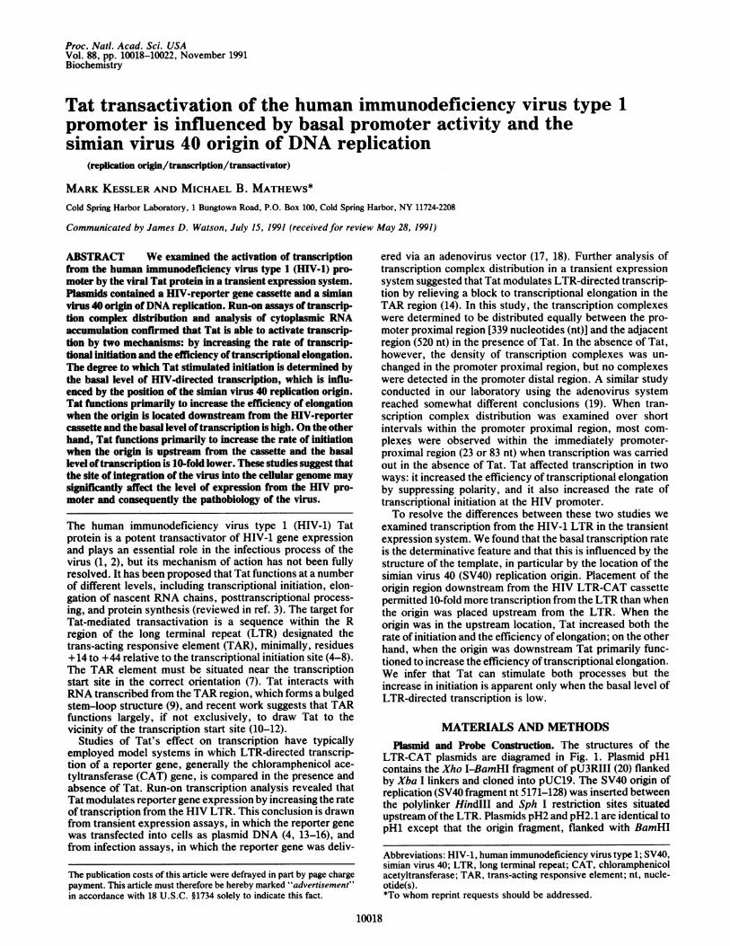
Fig. 2 shows the patterns of hybridization obtained withRNA labeled for various periods of time in run-on transcrip-tion assays. In the absence of Tat, hybridization to fragmentsI and II was observed after 3 min of labeling. Taking intoaccount the greater length and uridine content of fragment II(Fig. 1B), the data indicate that following 3 min and 6 min oflabeling the majority of transcriptional complexes were infragment I. By 12 min oftranscription the hybridization signalin fragment II exceeded that in fragment I, consistent withmovement of complexes from fragment I into fragment II. Inthe presence of Tat, RNA hybridizing to fragments I and II
was detected after only 1.5 min of labeling. Again, hybrid-ization to fragment I predominated with short labeling peri-ods but was exceeded by hybridization to fragment II withlonger labeling times, emphasizing that the shortest practicallabeling times should be used to give an accurate represen-tation of polymerase density. The uniform increase in hy-bridization to fragments I and II indicates that Tat has a
greater effect on transcriptional initiation than on elongation,especially with short labeling periods. Inclusion of sarkosyl(30) or a-amanitin demonstrated that most, if not all, of thelabeling is due to elongation ofpreviously initiated complexescontaining RNA polymerase II (not shown).
A or net T R CAl SV-pApHi pCia9- f~ i
pSt5 1 i- {
puttsrho! -- --{ L41 _ iJ- 07
ps:t 5.1 pEe.-- -- 9 ; ot_
pH2 p- -I i
pH2.1 pIlCiP - j311 l _ _
pBAHICAT DPRA -- L3iRI _
B
FIG. 1. Map of HIV CAT reporter constructs. (A) Schematicdiagram of HIV CAT constructs showing the location of the SV40origin (ori), HIV-1 nefgene sequence (striped box), LTR (open box),CAT coding sequences (black box), and SV40 splicing and poly(A)signals (hatched box). The orientation of the origin region, relative tothe direction of early (E) and late (L) transcription, is noted as is thesource of the viral sequences. The bacterial vector used to constructeach plasmid is also noted. (The nef region marked in the diagramdesignates the portion ofthe nefgene lying upstream ofthe LTR.) (B)Schematic diagram depicting the structure of the LTR-CAT cassettein pSt5. The regions corresponding to probes I, 2, II, and III used inthe run-on assays are also shown.
Biochemistry: Kessler and Mathews
10020 Biochemistry: Kessler and Mathews
A
.; H -
F., '. V t
FIG. 2. Run-on transcription analysis of pH1 in the presence andabsence of Tat. COS-1 cells were mock transfected or transfectedwith pHi, pRSVtat, or pH1 and pRSVtat, and run-on transcriptionreactions were carried out for 1.5, 3, 6, and 12 min. Labeled RNA wasisolated and hybridized to blots containing 5 ,ug of sense (+) orantisense (-) DNA probe corresponding to nt -18 to +83 (I), +83to +229 (II), 13-actin, M13mpl8, and M13mpl9. The mock andpRSVtat assays represent 12-min run-on reactions.
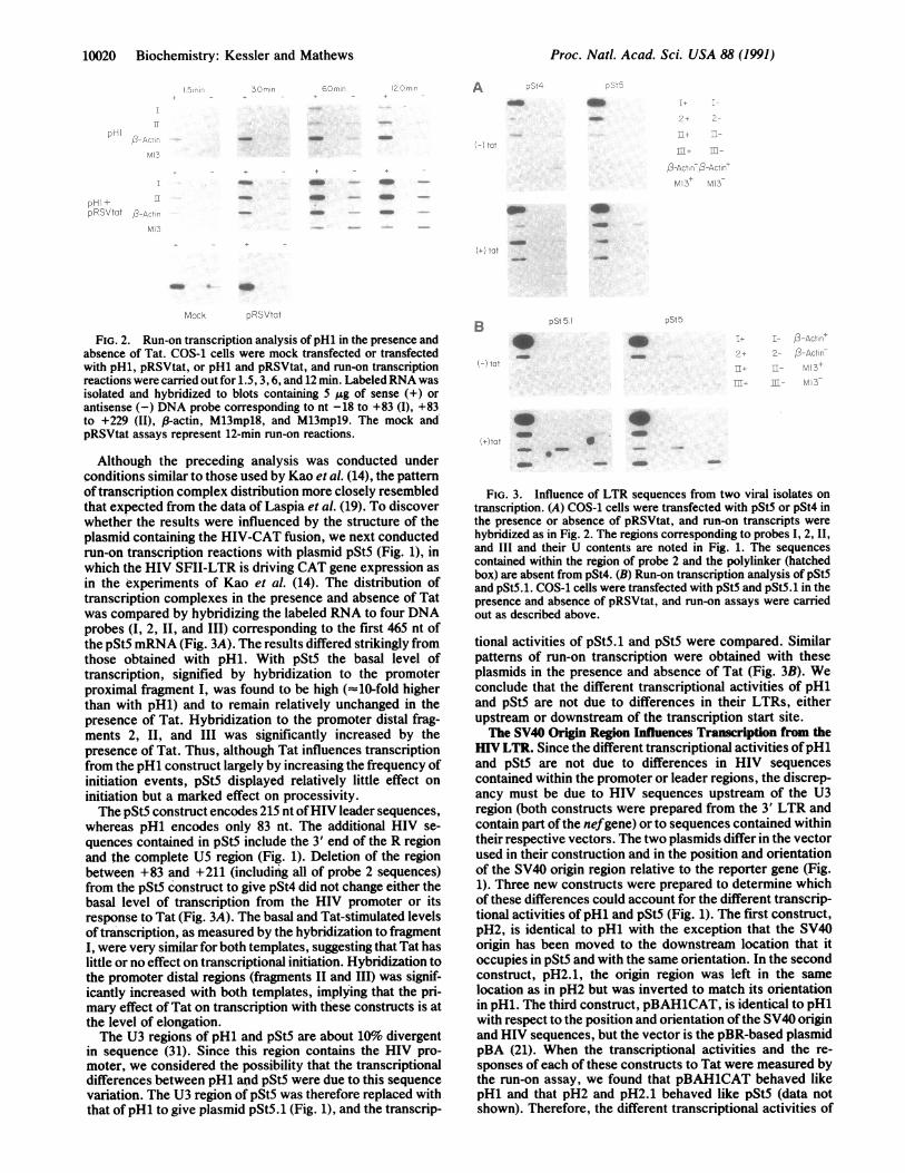
Although the preceding analysis was conducted underconditions similar to those used by Kao et al. (14), the patternof transcription complex distribution more closely resembledthat expected from the data of Laspia et al. (19). To discoverwhether the results were influenced by the structure of theplasmid containing the HIV-CAT fusion, we next conductedrun-on transcription reactions with plasmid pStS (Fig. 1), inwhich the HIV SFII-LTR is driving CAT gene expression asin the experiments of Kao et al. (14). The distribution oftranscription complexes in the presence and absence of Tatwas compared by hybridizing the labeled RNA to four DNAprobes (I, 2, II, and III) corresponding to the first 465 nt ofthe pStS mRNA (Fig. 3A). The results differed strikingly fromthose obtained with pH1. With pSt5 the basal level oftranscription, signified by hybridization to the promoterproximal fragment I, was found to be high (l10-fold higherthan with pHi) and to remain relatively unchanged in thepresence of Tat. Hybridization to the promoter distal frag-ments 2, II, and III was significantly increased by thepresence of Tat. Thus, although Tat influences transcriptionfrom the pH1 construct largely by increasing the frequency ofinitiation events, pSt5 displayed relatively little effect oninitiation but a marked effect on processivity.The pStS construct encodes 215 nt ofHIV leader sequences,
whereas pH1 encodes only 83 nt. The additional HIV se-quences contained in pStS include the 3' end of the R regionand the complete U5 region (Fig. 1). Deletion of the regionbetween +83 and +211 (including all of probe 2 sequences)from the pStS construct to give pSt4 did not change either thebasal level of transcription from the HIV promoter or itsresponse to Tat (Fig. 3A). The basal and Tat-stimulated levelsof transcription, as measured by the hybridization to fragmentI, were very similar for both templates, suggesting that Tat haslittle or no effect on transcriptional initiation. Hybridization tothe promoter distal regions (fragments II and III) was signif-icantly increased with both templates, implying that the pri-mary effect of Tat on transcription with these constructs is atthe level of elongation.The U3 regions of pH1 and pStS are about 10% divergent
in sequence (31). Since this region contains the HIV pro-moter, we considered the possibility that the transcriptionaldifferences between pH1 and pSt5 were due to this sequencevariation. The U3 region of pSt5 was therefore replaced withthat ofpH1 to give plasmid pSt5.1 (Fig. 1), and the transcrip-
DSb tB
-
FIG. 3. Influence of LTR sequences from two viral isolates ontranscription. (A) COS-1 cells were transfected with pSt5 or pSt4 inthe presence or absence of pRSVtat, and run-on transcripts werehybridized as in Fig. 2. The regions corresponding to probes I, 2, II,and III and their U contents are noted in Fig. 1. The sequencescontained within the region of probe 2 and the polylinker (hatchedbox) are absent from pSt4. (B) Run-on transcription analysis of pSt5and pSt5.1. COS-1 cells were transfected with pSt5 and pSt5.1 in thepresence and absence of pRSVtat, and run-on assays were carriedout as described above.
tional activities of pSt5.1 and pSt5 were compared. Similarpatterns of run-on transcription were obtained with theseplasmids in the presence and absence of Tat (Fig. 3B). Weconclude that the different transcriptional activities of pH1and pStS are not due to differences in their LTRs, eitherupstream or downstream of the transcription start site.The SV40 Origin Region Influences Transcription from the
HIV LTR. Since the different transcriptional activities ofpH1and pStS are not due to differences in HIV sequencescontained within the promoter or leader regions, the discrep-ancy must be due to HIV sequences upstream of the U3region (both constructs were prepared from the 3' LTR andcontain part of the nefgene) or to sequences contained withintheir respective vectors. The two plasmids differ in the vectorused in their construction and in the position and orientationof the SV40 origin region relative to the reporter gene (Fig.1). Three new constructs were prepared to determine whichof these differences could account for the different transcrip-tional activities of pH1 and pStS (Fig. 1). The first construct,pH2, is identical to pH1 with the exception that the SV40origin has been moved to the downstream location that itoccupies in pSt5 and with the same orientation. In the secondconstruct, pH2.1, the origin region was left in the samelocation as in pH2 but was inverted to match its orientationin pH1. The third construct, pBAH1CAT, is identical to pH1with respect to the position and orientation ofthe SV40 originand HIV sequences, but the vector is the pBR-based plasmidpBA (21). When the transcriptional activities and the re-sponses of each of these constructs to Tat were measured bythe run-on assay, we found that pBAH1CAT behaved likepH1 and that pH2 and pH2.1 behaved like pSt5 (data notshown). Therefore, the different transcriptional activities of
Proc. Natl. Acad. Sci. USA 88 (1991)
_ _ _
_ __Q
00

Proc. Nati. Acad. Sci. USA 88 (1991) 10021
pH1 and pSt5 are due to the position, but not orientation, ofthe SV40 origin region relative to the HIV LTR.RNase protection analysis of cytoplasmic HIV LTR-
directed RNA confirmed these conclusions. With an antisenseprobe extending from +83 to -117, two classes of protectedfragments were anticipated: a series of short fragments, 55-59nt in length, presumably resulting from premature terminationof transcription, and a longer fragment (83 nt) representingfull-length mRNA (14, 19, 32). In the absence of Tat, pH1 andpBAH1CAT gave rise to very little RNA (Fig. 4A, lanes 1 and5). The protected species corresponding to the long and shortRNA populations were observed in roughly equal abundance.In the presence of Tat little change was observed in thequantity of short RNAs, but the abundance of long transcriptswas greatly increased (lanes 2 and 6). By contrast, pH2 andpSt4 generated large amounts of short transcripts in theabsence ofTat (lanes 3 and 7). Full-length RNA was observed,but as a minor population of the protected RNA. In thepresence of Tat, these two constructs gave rise to largeamounts ofthe full-length RNA (lanes 4 and 8). Similar resultswere obtained with pSt5 (data not shown), confirming theobservations of Kao et al. (14). Although the quantity of shorttranscripts was reduced, the reduction did not appear to besufficient to account for the increase in full-length RNA andwas not observed consistently (see Fig. 4C). RNA transcribedfrom pH2.1 in the presence and absence ofTat (Fig. 4C, lanes1 and 2) was similar to that transcribed from pSt4 (lanes 3 and4). Thus, the positioning of the SV40 origin upstream of theLTR gives rise to a lower basal level of HIV-promotedtranscripts and allows for a large effect of Tat on cytoplasmicRNA accumulation. Corresponding results were obtained bymeasurement of CAT enzyme activity (Table 1).We also considered the possibility that the different quan-
tities of cytoplasmic RNA observed following transfection ofthese plasmids were the result of differences in the efficiencyof transfection or replication of the plasmid DNA. To controlfor transfection efficiency and RNA recovery, a plasmidcontaining 3-globin sequences under the direction of the U2snRNA promoter was included in each transfection. Asshown in Fig. 4B, the differences in transfection efficiency,reflected in differences in the amount of protected f3-globinprobe, cannot account for the observed differences in HIVLTR-directed transcription. To measure the relative quantityof plasmid present at the time ofRNA extraction, DNA was
isolated from the transfected cells and quantified by hybrid-ization. From the data ofTable 1 it is apparent that there werevariations in the amount ofDNA, but the differences (2-foldoverall) were too small to account for the differences ob-served at the level of cytoplasmic RNA.
DISCUSSIONIn a transient expression system, the HIV-1 transactivatorprotein Tat is capable of activating transcription from theHIV-1 LTR by increasing the rate of transcriptional initiationand by increasing the efficiency of transcriptional elongation.The relative extent to which it performs each ofthese functionsis determined by the basal level ofLTR-directed transcription.If the basal level of transcription is high, Tat functions pri-marily to increase the elongation rate, whereas if the basallevel is low Tat primarily increases the rate of initiation. Thisfinding is in accord with previous studies in which we utilizedrecombinant adenovirus containing the HIV LTR-CAT cas-sette to study Tat-mediated activation of HIV transcription.Tat increased the rate of transcriptional initiation and stabi-lized elongation in HeLa cells infected with the virus but onlystabilized elongation when transcription was carried out in thepresence of the general transcriptional activator ElA (19, 33).These observations may help to reconcile the results of
Kao et al. (14), who concluded that Tat acts as an antiter-
A
..do ..~~* a
-A..I
VW
B
C
|:~~~~~frrof.O
.....l
D Short
-117 +1 +59 +83
Fuzz Probe83nt
59nt Protected Fragments
FIG. 4. Effect of vector and SV40 origin sequences on HIVLTR-directed RNA expression. (A) RNase protection analysis ofcytoplasmic HIVCAT RNA following transfection of COS-1 cellswith pH1 (lanes 1 and 2), pH2 (lanes 3 and 4), pI3AH1CAT (lanes 5and 6), or pSt4 (lanes 7 and 8) or without an LTR-CAT plasmid (lane9) in the presence (lanes 2, 4, 6, 8, and 9) or absence (lanes 1, 3, 5,and 7) of pRSVtat. Lane M contains pBR322/Hpa II markers.Locations of the long and short transcripts are marked. (B) RNaseprotection analysis of .8-globin transcripts present in the cytoplasmicRNA preparations used in A. Plasmid pU2/-247/RA.2 was cotrans-fected with the HIVCAT reporter constructs and RNase protectionanalysis was carried out using a riboprobe transcribed from EcoRI-digested pU2/RA.2/142. A 142-nt fragment is protected. (C) RNaseprotection analysis of cytoplasmic HIVCAT RNA following trans-fection of COS-1 cells with pH2.1 (lanes 1 and 2) or pSt4 (lanes 3 and4) or without a LTR-CAT plasmid (lane 5) in the presence (lanes 2,4, and 5) or absence (lanes 1 and 3) of pRSVtat. Lane M containspBR322/Hpa II markers. Locations of the long and short transcriptsare noted. (D) Schematic diagram of the mapping strategy depictingthe probe, RNA transcripts, and protected fragments.
minator to increase transcriptional elongation, with resultsfrom our laboratory indicating that Tat also acts to increasethe rate of initiation (19). The most likely explanation wouldbe that Kao et al. (14) studied Tat transactivation in thecontext of a high basal level of HIV-directed transcription,whereas Laspia et al. (19) examined transactivation underconditions where the promoter exhibited a low basal level of
Biochemistry: Kessler and Mathews

10022 Biochemistry: Kessler and Mathews
Table 1. Quantitation of plasmid DNA and HIV-directed CATactivity in transfected cells
DNA level* CAT activitytWithout Tat With Tat Without Tat With Tat
pH1 149 99 12 2300pBAH1CAT 130 74 26 1120pH2 166 160 312 6000pSt4 202 106 120 6720pSt4- 2None§ 5*Values represent the standardized cpm hybridizing to two dilutionsof blotted DNA.
tPercent conversion of chloramphenicol substrate to its acetylatedforms.*Control, not alkali-treated.§DNA extracted from cells transfected with pRSVtat only.
transcription. Consistent with this view, Kao et al. (14) useda plasmid with the SV40 origin downstream from the HIV-reporter gene cassette for their analysis of RNA accumula-tion. Unfortunately for this simple explanation, the plasmidused in their run-on transcription analysis contained the SV40origin region upstream from the HIV LTR. This construct isanalogous to pH1, which displayed low basal level of activityin our study and which therefore would be expected to exhibitincreased initiation in the presence of Tat. However, directcomparison is not possible because the distance of the originregion from the LTR and the size of the SV40 fragmentcontaining the origin sequences differed from pH1, and it ispossible that these factors change the influence of the originregion on basal transcription levels.
It is not clear why the SV40 origin region influencestranscription from the HIV LTR. Since the origin regioncontains, in addition to sequences directingDNA replication,promoter elements involved in the regulation ofearly and lateviral gene expression, its effect could be due to occlusion ofthe HIV promoter by transcription from the origin region.This explanation seems unlikely, however, since RNA tran-scripts containing sequences 5' to the LTR start site weredetected only as a minor population with plasmids expressingelevated basal levels of transcription, pH2 and pSt4, and notwith plasmids expressing low basal levels (result not shown).An alternative explanation is that the replication processinfluences transcription from the HIV promoter, but todifferent degrees depending on the distance of the origin ofreplication from the promoter. In several instances, replica-tion has been shown to activate transcription (34-38) and theHIV LTR may belong to this class of promoters (N. Proud-foot and J. Monks, personal communication).The transition from viral latency to growth, as well as from
noncytopathic to cytopathic infection, appears to require thecoordinated and sequential expression of the viral regulatoryand structural proteins (39-41). Viral transcriptional ratesdirectly influence this pathway (40). Activation of the HIVpromoter as a result ofDNA replication may explain, at leastin part, why nonresting T cells are permissive for HIV-1infection, whereas Go cells are not (42). Interestingly, analysisof retroviral replication in resting and actively growing cellsrevealed the proviral DNA to be integrated only in cellularDNA replicated during infection (43). Whether cellular originregions are preferred sites for viral integration is unknown.However, if the effect of the SV40 origin region on HIV geneexpression is typical, viral integration near a cellular originmay contribute significantly to the pathobiology of the virus.
We thank M. Laspia and A. Rice for discussions, N. Hernandezand W. Herr for critical review of the manuscript, and R. Packer for
cell work. These studies were supported by National Institutes ofHealth Grants CA13106 and A127270 and by National Institutes ofHealth Fellowship A107877 to M.K.
1. Dayton, A. I., Sodroski, J. G., Rosen, C. A., Goh, W. C. & Ha-seltine, W. A. (1986) Cell 44, 941-947.
2. Fisher, A. G., Feinberg, S. F., Josephs, S. F., Harper, M. E.,Marselle, L. M., Reyes, G., Gonda, M. A., Aldovini, A., Dibouck,C., Gallo, R. C. & Wong-Staal, F. (1986) Nature (London) 320, 367.
3. Jones, K. (1990) New Biol. 1, 127-135.4. Jakobovits, A., Smith, D. H., Jakobovits, E. B. & Capon, D. J.
(1988) Mol. Cell. Biol. 8, 2555-2561.5. Hauber, J. & Cullen, B. R. (1988) J. Virol. 63, 1181-1187.6. Berkhout, B. & Jeang, K.-T. (1989) J. Virol. 63, 5501-5504.7. Selby, M. J., Bain, E. S., Luciw, P. A. & Peterlin, B. M. (1989)
Genes Dev. 3, 547-558.8. Garcia, J. A., Harrich, D., Mitsuyasu, R. & Gaynor, R. B. (1988)
EMBO J. 7, 3143-3147.9. Muesing, M. A., Smith, D. H. & Capon, D. J. (1987) Cell 48, 691-701.
10. Berkhout, B., Gatignol, A., Rabson, A. B. & Jeang, K.-T. (1990)Cell 62, 757-767.
11. Selby, M. J. & Peterlin, B. M. (1990) Cell 62, 769-776.12. Southgate, C., Sapp, M. L. & Green, M. R. (1990) Nature (London)
345, 640-642.13. Hauber, J., Perkins, A., Heimer, E. P. & Cullen, B. R. (1987) Proc.
Nat!. Acad. Sci. USA 84, 6364-6368.14. Kao, S. Y., Calman, A. F., Luciw, A. P. & Peterlin, B. M. (1987)
Nature (London) 330, 489-493.15. Jeang, K.-T., Shank, P. R. & Kumar, A. (1988) Proc. Nat!. Acad.
Sci. USA 85, 8291-8295.16. Sadaie, M. R., Benter, T. & Wong-Staal, F. (1988) Science 239,
910-913.17. Rice, A. P. & Mathews, M. B. (1988) Proc. Natl. Acad. Sci. USA
85, 4200-4204.18. Rice, A. P. & Mathews, M. B. (1988) Nature (London) 332, 551-553.19. Laspia, M. F., Rice, A. P. & Mathews, M. B. (1989) Cell 59, 283-292.20. Sodroski, J. G., Rosen, C. A., Wong-Staal, F., Salahuddin, S. Z.,
Popovic, M., Arya, S. & Gallo, R. C. (1985) Science 227, 171-173.21. DMry, C. V., Herrmann, C. H. & Mathews, M. B. (1987) Oncogene
2, 15-23.22. Cullen, B. R. (1986) Cell 46, 973-982.23. Lobo, S. M. & Hernandez, N. (1989) Cell 58, 55-67.24. Cullen, B. R. (1987) Methods Enzymol. 152, 684-704.25. Ucker, D. S. & Yamamoto, K. R. (1984) J. Biol. Chem. 259,
7416-7420.26. Greenberg, M. E. (1989) in Current Protocols in Molecular Biology,
eds. Ausubel, F. M., Brent, R., Kingston, R. E., Moore, D. D.,Seidman, J. G., Smith, J. A. & Struhl, K. (Wiley-Interscience, NewYork), pp. 4.10.1-4.10.4.
27. Gilman, M. (1989) in Current Protocols in Molecular Biology, eds.Ausubel, F. M., Brent, R., Kingston, R. E., Moore, D. D., Sei-dman, J. G., Smith, J. A. & Struhl, K. (Wiley-Interscience, NewYork), pp. 4.1.2-4.1.6.
28. Melton, D., Krieg, P. A., Rebagliati, M. R., Maniatis, T., Zinn, K.& Green, M. R. (1984) Nucleic Acids Res. 12, 7035-7056.
29. Hirt, B. (1967) J. Mol. Biol. 26, 365-369.30. Gariglio, P., Buss, J. & Green, M. H. (1974) FEBS Lett. 44, 330-333.31. Myers, G., Berzofsky, J. A., Rabson, A. B., Smith, T. F. & Wong-
Staal, F. (1990) Human Retroviruses and Aids Database (LosAlamos National Laboratory, Los Alamos, NM).
32. Roy, S., Parkin, N. T., Rosen, C., Itouitch, J. & Sonnenberg, N.(1990) J. Virol. 64, 1402-1406.
33. Laspia, M. F., Rice, A. P. & Mathews, M. B. (1990) Genes Dev. 4,2397-2408.
34. Thomas, P. G. & Mathews, M. B. (1980) Cell 22, 523-533.35. Treisman, R., Green, M. R. & Maniatis, T. (1983) Proc. Natl. Acad.
Sci. USA 80, 7428-7432.36. Grass, D. S., Read, D., Lewis, E. D. & Manley, J. L. (1987) Genes
Dev. 1, 1065-1074.37. Enver, T., Brewer, A. C. & Patient, R. K. (1988) Mol. Cell. Biol. 8,
1301-1308.38. Yamaguchi, M. & Matsukage, A. (1990) New Biol. 2, 343-350.39. Cullen, B. R. & Greene, W. C. (1989) Cell 58, 423-426.40. Ma, X., Koji, S., Sinangil, F., Golub, E. & Volsky, D. J. (1990)
Virology 176, 184-194.41. Pomerantz, R. J., Trono, D., Feinberg, M. B. & Baltimore, D.
(1990) Cell 61, 1271-1276.42. Zagury, D., Bernard, J., Leonard, R., Cheynier, R., Feldman, M.,
Sarin, P. S. & Gallo, R. C. (1986) Science 231, 850-853.43. Varmus, H. E., Padgett, T., Heasley, S., Simon, G. & Bishop,
J. M. (1977) Cell 11, 307-319.
Proc. Natl. Acad. Sci. USA 88 (1991)


