Tau’s role in the developing brain: implications for intellectual disability Tamar Sapir 1 , Michael Frotscher 2 , Talia Levy 1 , Eva-Maria Mandelkow 3 and Orly Reiner 1, ∗ 1 Department of Molecular Genetics, Weizmann Institute of Science, 76100 Rehovot, Israel, 2 Institute for Structural Neurobiology, Center for Molecular Neurobiology Hamburg, Martinistr. 52, D-20246 Hamburg, Germany and 3 DZNE (German Ctr. Neurodeg. Diseases) and CAESAR Research Center, Ludwig-Erhard-Allee 2, 53175 Bonn, Germany Received September 27, 2011; Revised November 24, 2011; Accepted December 15, 2011 Microdeletions encompassing the MAPT (Tau) locus resulting in intellectual disability raised the hypothesis that Tau may regulate early functions in the developing brain. Our results indicate that neuronal migration was inhibited in mouse brains following Tau reduction. In addition, the leading edge of radially migrating neu- rons was aberrant in spite of normal morphology of radial glia. Furthermore, intracellular mitochondrial trans- port and morphology were affected. In early postnatal brains, a portion of Tau knocked down neurons reached the cortical plate. Nevertheless, they exhibited far less developed dendrites and a striking reduction in connectivity evident by the size of boutons. Our novel results strongly implicate MAPT as a dosage-sen- sitive gene in this locus involved in intellectual disability. Furthermore, our results are likely to impact our understanding of other diseases involving Tau. INTRODUCTION Tau is a microtubule-associated protein (MAP), well known for its involvement in a group of neurodegenerative diseases collectively known as tauopathies (reviewed in 1 – 4). This is probably the reason that most tau-related studies involve over- expression of the protein in relation to the adult brain. The most common tauopathy is Alzheimer’s disease where hyper- phosphorylated tau accumulates within paired helical fila- ments. Mutations within the MAPT (Tau) locus result in frontotemporal dementia with Parkinsonism (5,6), whereas microdeletions of a region encompassing the MAPT gene result in moderate intellectual disability with associated dys- morphic features (7 – 11). The frequency of the microdeletion syndrome was estimated to be 1:13 000 to 1:20 000, thus sug- gesting it to be a common underlying cause for intellectual disability. Today, we know that copy number variations are an important component of the molecular mechanism under- lying many brain diseases, such as intellectual disability, autism and schizophrenia (12 – 22). Yet, in only a handful of examples, the identity of the dosage-sensitive gene(s) within the disease locus has been revealed. MAPT is one of the few genes within the microdeleted locus, it is strongly expressed in the developing brain (23,24) and it has been suggested to play a role in neuronal migration. There are multiple examples indicating a strong link between intellectual disability and ab- normalities in neuronal migration (25,26). Nevertheless, so far the role of Tau in neuronal migration has not been proven un- equivocally. Mice deleted for Tau exhibited a reduction in microtubule density in small caliber axons (27), as well as muscle weakness and memory disturbances (28). It has been proposed that developmental functional redundancy by increased expression of other MAPs may explain the relatively mild phenotype (27). This hypothesis was corroborated by the observed neuronal migration phenotype in mice mutated for both tau and MAP1b, where the single MAP1b mutant mice already demonstrate a neuronal migration phenotype (29). We investigated the role of tau in the developing mouse brain using in utero electroporation which has been shown to circumvent gene redundancy as previously demonstrated in case of the Dcx family of proteins (30,31) and in case of the MARK family of proteins (32). Our results show several novel and important roles for tau in the developing cerebral cortex, which have not been previously demonstrated. We show for the first time that reduction in Tau inhibits neuronal migration in the developing cortex. The leading edge of radi- ally migrating neurons was aberrant in spite of normal morph- ology of radial glia. Furthermore, we discovered a new role for ∗ To whom correspondence should be addressed. Tel: +972 89344927; Fax: +972 89344108; Email: [email protected]# The Author 2011. Published by Oxford University Press. All rights reserved. For Permissions, please email: [email protected]Human Molecular Genetics, 2012, Vol. 21, No. 8 1681–1692 doi:10.1093/hmg/ddr603 Advance Access published on December 21, 2011 Downloaded from https://academic.oup.com/hmg/article/21/8/1681/621290 by University of Bologna user on 25 February 2022
Transcript
Tau’s role in the developing brain: implicationsfor intellectual disability
Tamar Sapir1, Michael Frotscher2, Talia Levy1, Eva-Maria Mandelkow3 and Orly Reiner1,∗
1Department of Molecular Genetics, Weizmann Institute of Science, 76100 Rehovot, Israel, 2Institute for Structural
Neurobiology, Center for Molecular Neurobiology Hamburg, Martinistr. 52, D-20246 Hamburg, Germany and 3DZNE
(German Ctr. Neurodeg. Diseases) and CAESAR Research Center, Ludwig-Erhard-Allee 2, 53175 Bonn, Germany
Received September 27, 2011; Revised November 24, 2011; Accepted December 15, 2011
Microdeletions encompassing the MAPT (Tau) locus resulting in intellectual disability raised the hypothesisthat Tau may regulate early functions in the developing brain. Our results indicate that neuronal migrationwas inhibited in mouse brains following Tau reduction. In addition, the leading edge of radially migrating neu-rons was aberrant in spite of normal morphology of radial glia. Furthermore, intracellular mitochondrial trans-port and morphology were affected. In early postnatal brains, a portion of Tau knocked down neuronsreached the cortical plate. Nevertheless, they exhibited far less developed dendrites and a striking reductionin connectivity evident by the size of boutons. Our novel results strongly implicate MAPT as a dosage-sen-sitive gene in this locus involved in intellectual disability. Furthermore, our results are likely to impact ourunderstanding of other diseases involving Tau.
INTRODUCTION
Tau is a microtubule-associated protein (MAP), well knownfor its involvement in a group of neurodegenerative diseasescollectively known as tauopathies (reviewed in 1–4). This isprobably the reason that most tau-related studies involve over-expression of the protein in relation to the adult brain. Themost common tauopathy is Alzheimer’s disease where hyper-phosphorylated tau accumulates within paired helical fila-ments. Mutations within the MAPT (Tau) locus result infrontotemporal dementia with Parkinsonism (5,6), whereasmicrodeletions of a region encompassing the MAPT generesult in moderate intellectual disability with associated dys-morphic features (7–11). The frequency of the microdeletionsyndrome was estimated to be 1:13 000 to 1:20 000, thus sug-gesting it to be a common underlying cause for intellectualdisability. Today, we know that copy number variations arean important component of the molecular mechanism under-lying many brain diseases, such as intellectual disability,autism and schizophrenia (12–22). Yet, in only a handful ofexamples, the identity of the dosage-sensitive gene(s) withinthe disease locus has been revealed. MAPT is one of the fewgenes within the microdeleted locus, it is strongly expressedin the developing brain (23,24) and it has been suggested to
play a role in neuronal migration. There are multiple examplesindicating a strong link between intellectual disability and ab-normalities in neuronal migration (25,26). Nevertheless, so farthe role of Tau in neuronal migration has not been proven un-equivocally. Mice deleted for Tau exhibited a reduction inmicrotubule density in small caliber axons (27), as well asmuscle weakness and memory disturbances (28). It has beenproposed that developmental functional redundancy byincreased expression of other MAPs may explain the relativelymild phenotype (27). This hypothesis was corroborated by theobserved neuronal migration phenotype in mice mutated forboth tau and MAP1b, where the single MAP1b mutant micealready demonstrate a neuronal migration phenotype (29).We investigated the role of tau in the developing mousebrain using in utero electroporation which has been shownto circumvent gene redundancy as previously demonstratedin case of the Dcx family of proteins (30,31) and in case ofthe MARK family of proteins (32). Our results show severalnovel and important roles for tau in the developing cerebralcortex, which have not been previously demonstrated. Weshow for the first time that reduction in Tau inhibits neuronalmigration in the developing cortex. The leading edge of radi-ally migrating neurons was aberrant in spite of normal morph-ology of radial glia. Furthermore, we discovered a new role for
∗To whom correspondence should be addressed. Tel: +972 89344927; Fax: +972 89344108; Email: [email protected]
# The Author 2011. Published by Oxford University Press. All rights reserved.For Permissions, please email: [email protected]
Human Molecular Genetics, 2012, Vol. 21, No. 8 1681–1692doi:10.1093/hmg/ddr603Advance Access published on December 21, 2011
Dow
nloaded from https://academ
ic.oup.com/hm
g/article/21/8/1681/621290 by University of Bologna user on 25 February 2022
tau in regulation of mitochondria; both intracellular mitochon-drial transport and morphology were severely affected follow-ing tau knockdown. In early postnatal brains, a portion of Tauknocked down neurons reached the cortical plate (CP). Never-theless, they exhibited smaller somas, far less-developed den-drites and a striking reduction in connectivity. Collectively,our results support the previously raised hypothesis thatTau’s deletion contributes to the pathophysiology of the17q21.31 microdeletion syndrome.
RESULTS
Tau levels are effectively reduced in vivo
To study the effect of reduced Tau levels on cortical develop-ment, we introduced gene-specific shRNA sequences inprimary cortical neurons. Brains of E14 mouse embryoswere co-electroporated with Tau shRNA and green fluorescentprotein (GFP) (3:1 ratio) ex utero. Immediately followingelectroporation, primary cultures were prepared from thetreated cortices. The cells were fixed and stained after 2days growth in vitro. Four shRNA sequences were tested fortheir ability to reduce Tau expression (Fig. 1). Three of thetested sequences (shRNA A, B and D) almost eliminatedTau1 immunoreactivity in the axons of the cultured neurons(Fig. 1A–L). The ability of shRNA A to knockdown Taulevels in vivo was further evaluated using an independentmethod. Two and 4 days post-electroporation, theGFP-positive areas were dissected out from 3 to 5 differentelectroporated brains. Brain lysates were pooled and separated
by sodium dodecyl sulfate polyacrylamide gel electrophoresis(SDS–PAGE) followed by western blot analysis (Fig. 1M andSupplementary Material, Fig. S1). Tau levels decreased asdemonstrated using either Tau1 (recognizing a relativelywide repertoire of Tau species), K9JA (pan-Tau antibody) orPHF-1 (recognizing phosphorylated Tau at Ser396/Ser404)antibodies. Constant actin or emerin levels indicated equalloading. Furthermore, we noted equal amount of LIS1 andDCX, two additional MAPs as well as a slight elevation inthe levels of MAP-2. Thus, we concluded that three shRNAsequences successfully reduced endogenous Tau. We thentested whether Tau knockdown affects the dynamics of micro-tubules in cultured cortical neurons by tracking microtubulesusing the plus-tip binding protein EB1-mCherry. We notedthat microtubules were more dynamic in primary corticalneurons with reduced Tau levels (Supplementary Material,Fig. S2).
Tau reduction impairs radial neuronal migration
Taking into consideration the role of Tau in regulation of micro-tubule dynamics and regulation of microtubule-associatedmotors, we questioned whether reduction in the levels of theTau protein affects radial neuronal migration in the developingcortex. Developing embryonic brains were electroporated atE14.5 by in utero electroporation together with a GFP expres-sion plasmid, and the position of the electroporated cells wasanalyzed 4 days later. Our results indicate that control pyramidalneurons born at E14.5 migrate to the CP and typically occupylayers II/III (Fig. 2A). Notably, all Tau shRNA sequences
Figure 1. Knock down of Tau in cortical neurons. E14 mouse embryos were co-electroporated ex utero with GFP and Tau shRNA plasmids. Cortical neuronswere prepared from the electroporated brains and after 2 DIV fixed and stained with Tau1 antibodies. Control shRNA (A–C), Tau in red merged with the greencell (A, D, G, J), electroporated neurons identified by GFP (B, E, H, K) and immunoreactivity of Tau in black (C, F, I, L). A marked reduction in Tau levels inthe somatodendritic and the axonal compartments was noted for all shRNA sequences tested (D–K) in comparison to control shRNA (A–C). (M) Tau1 andPHF-1 show reduced reactivity in lysates extracted from Tau shRNA A-electroporated regions of E18 brains, 4 days after electroporation. b-Actin levels indicatesimilar loading (lower panel).
1682 Human Molecular Genetics, 2012, Vol. 21, No. 8
Dow
nloaded from https://academ
ic.oup.com/hm
g/article/21/8/1681/621290 by University of Bologna user on 25 February 2022
effectively inhibited the ability of the GFP-positive-treated cellsto reach the outer layers of the CP (Fig. 2). Two-way analysis ofvariance (ANOVA) detected significant differences in the pro-portion of shRNA neurons located in the different bins in com-parison with control neurons. The most severe effect was notedfor Tau shRNA A (3/8 bins P , 0.001, 2/8 bins P , 0.01, n ¼8). To exclude off-target events, human Tau (hTau) wasco-introduced with Tau shRNA. Human and mouse Tau arefunctionally conserved; however. hTau is resistant to TaushRNA D sequence. For this experiment, we used hTau23,which is the most abundant isoform in the developing brain con-taining three microtubule-binding repeats (33). Following add-ition of hTau, some of the observed neuronal migration defectswere alleviated (Fig. 2E and E′), and this treatment significantlyimproved neuronal migration of the Tau shRNA D-treatedneurons (1/8 bins P , 0.01, 1/8 bins P , 0.05, n ¼ 8). Acloser examination of the stalled neurons revealed a cell autono-mous morphological defect (Fig. 3). The leading edge of cellswith reduced Tau levels was crooked and thinner than incontrol cells (Fig. 3A–F). This observation was further strength-ened by quantitative analyses of the straightness and thicknessof the leading edge. Tau shRNA A and D significantlyreduced the straightness of the leading edge, whereas the reduc-tion observed following treatment with Tau shRNA B was notstatistically different (one-way ANOVA, P , 0.01 consideredvery significant in case of Tau A and D, n ¼ 41, 31, 60, 37 forcontrol, shRNA A, B and D, respectively) (Fig. 3E). To estimatethe thickness of the leading edge, we measured a cross-section ina distance of 15 mm from the cell body (Fig. 3F). The cross-sections surfaces of the control neurons were significantlylarger (.60%) than the TauA shRNA-treated neurons (two-tailed t-test, P ¼ 0.04, control 7+ 1.05 mm2, TauA shRNA4.90+ 0.76 mm2, n ¼ 16). To rule out the possibility that the
deformation of the leading edge of migrating neurons is a sec-ondary effect, radial glia fibers were examined. Some radialglia cells were GFP-positive following in utero electroporation.These cells were readily identified by the location of their cellbodies and their typical elongated processes that can be followedalong the thickness of the cortex (Fig. 3G and H). No morpho-logical differences in GFP-positive radial glia fibers werenoted in any of the treatments. Additionally, radial glia in thearea of the stalled cells appeared normal as judged by DiO back-fills from the cortex outer surface (Fig. 3I). The axons extendingfrom Tau shRNA-treated neurons were somewhat shorter thanthe control at E16 (Supplementary Material, Fig. S3).However, this phenotype may be secondary due to their inhib-ited migration. Collectively, our findings indicate that knock-down of Tau impairs neuronal migration and affects themorphology of the leading edge in a cell autonomous manner.
Tau knockdown affects the morphology and thedistribution of mitochondria
Increased expression of Tau has been demonstrated to affectthe transport of mitochondria in the axon of culturedneurons (34). Therefore, we speculated that mitochondriamay be affected following Tau knockdown. To test this hy-pothesis, we examined the motility, distribution and morph-ology of mitochondria in migrating neurons where Tau hadbeen knocked down (Fig. 4). The mitochondria were labeledwith either mCherry or DsRed fused to mitochondria local-ization sequence and imaged in live organotypic slices(Fig. 4A–H) as well as in fixed slices (Fig. 4I–L). Incontrol neurons, the mitochondria found in the cell somaand the leading edge (Fig. 4A–C) exhibited high motilityas seen in a kymograph presentation (Fig. 4D). The
Figure 2. Acute reduction in Tau levels inhibits neuronal migration. E14 mouse embryos were co-electroporated with control (A) or Tau shRNAs (B–D) andGFP plasmids. The location of GFP-positive cells was examined 4 days after electroporation. The proportion of cells located in eight arbitrary bins along thecortex was measured and plotted (A′ –D′). (E and E′) Partial rescue of the neuronal migration phenotype following Tau knockdown is achieved by introduction ofhTau, resistant to Tau shRNA D.
Human Molecular Genetics, 2012, Vol. 21, No. 8 1683
Dow
nloaded from https://academ
ic.oup.com/hm
g/article/21/8/1681/621290 by University of Bologna user on 25 February 2022
labeled mitochondria in Tau shRNA-treated neurons loca-lized primarily to the cell body and poorly penetrated theabnormal leading edge (Fig. 4E–G). Moreover, the motilityof the mitochondria that did penetrate the apical aspect ofthe leading edge was reduced in comparison with controlneurons as demonstrated in time-lapse images (Fig. 4A–C and E–G) and typical kymographs obtained fromcontrol and Tau shRNA A-treated cells, respectively(Fig. 4D and H). Examination of fixed neurons substantiatedthe live imaging observations (Fig. 4I and J compared withK and L). We cannot exclude the possibility that the abilityof mitochondria to enter the leading edge was impaired atleast in part due to the reduced thickness of the leadingedge. Nevertheless, even in control cells with a relativelythin leading edge, the mitochondria were transported to agreater distance than in Tau shRNA-treated neurons,which exhibited rather thick leading processes (Supplemen-tary Material, Fig. S4). These findings prompted us toexamine the mitochondria at a higher resolution using elec-tron microscopy (Fig. 4M–R). Immunogold staining forGFP identified the Tau shRNA A or the control shRNA-treated cells. While similar numbers of mitochondria cross-sections were examined in the EM images, Tau shRNA-treated cells contained a considerable proportion of abnor-mal mitochondria (22% Fig. 4S). Abnormal mitochondriaincluded both enlarged rounded mitochondria that occasion-ally engulfed cytoplasmic components (Fig. 4M and N) andmitochondria with unusually enlarged vacuoles (Fig. 4O
and P). These abnormal mitochondria were not undergoingmitophagy since they were not engulfed in a double mem-brane. Control neurons contained mitochondria with anormal structure (Fig. 4Q and R). Our results indicate thatthe knockdown of Tau reduces the mobility of mitochondriainto the leading edge and impairs their structure.
Postnatal consequences of reduced Tau levels duringembryonic development
To estimate the outcome of the developmental deficitsdescribed, we analyzed postnatal (P8) brains of E14 treatedembryos. At P8, the control neurons were located in theouter layers of the CP, layers II/III (Fig. 5A), and the majorityof the stalled cells observed at E18 had reached a similar pos-ition. However, the distribution of Tau shRNA GFP-positivecells included also the less superficial layer IV (Fig. 5B). Add-itionally, a small number of cells in the Tau shRNA-treatedbrains remained in an ectopic position deep in the whitematter, where usually only axons are located (Fig. 5C andD). We did not observe an increase in apoptotic cells in TaushRNA-treated brain sections (Supplementary Material,Fig. S5). Interestingly, ectopic cells were able to extendaxons across the midline to contralateral targets (Supplemen-tary Material, Fig. S3). Similar results were obtained usingtwo different Tau shRNA sequences (data not shown).
Despite of the finding that many of the Tau shRNA-treatedpyramidal neurons were located in the CP at P8, many
Figure 3. Abnormal morphology of bipolar migrating neurons with reduced Tau levels. (A–D) GFP-positive cells treated with Tau shRNA (B–D) exhibit a thinand crooked leading edge in comparison to control cells (A). The straightness of the leading edge was measured using Imaris and plotted in (E). (F) Cross-sections of leading edge of neurons were taken at a distance of 15 mm from the cell body. The position of the end of the cell body is marked with a redline, the position of the cross-section is marked with a yellow line, the z-stack of the position at the yellow line is on the right panel and the sections arelabeled with a yellow arrowhead. (G–I) Radial glia in the vicinity of GFP-positive cells in either control (G) or Tau shRNA-treated brains (H) are indistinguish-able. DiO Backfills of radial glia in Tau shRNA-treated brains (I) reveals no obvious morphological abnormalities.
1684 Human Molecular Genetics, 2012, Vol. 21, No. 8
Dow
nloaded from https://academ
ic.oup.com/hm
g/article/21/8/1681/621290 by University of Bologna user on 25 February 2022
abnormalities were noted. The cross-sectional areas of the cellsoma of the Tau shRNA-treated neurons located in the som-atosensory cortex exhibited a significant reduction of 15%(P ¼ 0.0063, unpaired t-test, control 385+ 12.65 mm2, n ¼18 in comparison to Tau shRNA 325.9+ 14.98 mm2, n ¼24) (Fig. 5E and F). A more striking morphological featurewas observed when individual cells were traced (representa-tive images are shown in Fig. 6A–C). Tau shRNA-treatedcells displayed a much simpler, less branched and shorterapical dendrite in comparison with control cells (Fig. 6Aversus B and C). This impression was reflected using theSholl analysis (Fig. 6D). The shRNA-treated neurons differedvery significantly from the controls (two-way ANOVA ana-lysis P ¼ 0.0068, n ¼ 12). Based on the spectacular differ-ences in neuronal morphology, we postulated that theconnectivity might also be impaired in these neurons. Con-nectivity was evaluated by co-electroporation of a fluorescentsynaptic protein expression plasmid (synaptic vesicle proteinSV2a) with Tau shRNA A, B or control plasmids. In P8brains, positive cells were analyzed in the somatosensorycortex. A reduction in the number of SV2a-positive punctadecorating the apical dendrite as well as the basal dendriteswas noted in Tau knocked down neurons (Fig. 6E–J). Thesize and distribution of the SV2a-positive boutons on descend-ing axons was measured and plotted. Small dots were notincluded in the analysis since they most likely reflect traffick-ing packets (35,36). The identity of the boutons used for theanalysis was validated by immunostaining with additional
synaptic markers (Supplementary Material, Fig. S6). Allaxons displayed similar densities of boutons; the averagenumber of boutons along 10 mm was 3.8, 3.8 and 2.9 incontrol, Tau shRNA and Tau shRNA B, respectively.However, Tau knocked down axons (using either TaushRNA A or shRNA B) exhibited an extremely significantsmaller size boutons (Fig. 6K–N) (one-way ANOVA P ,0.0001, n ¼ 111, 103, 106 for control, Tau shRNA A andTau shRNA B, respectively).
Our data clearly define novel functions for Tau in the devel-oping brain. We demonstrated that reduced Tau levels result inslower radial migration of layer II/III pyramidal neurons to thesuperficial layers of the cortex, which is accompanied by a de-fective leading edge in which mitochondria are partially ab-normal and less motile. In postnatal brains, some neuronsremain in ectopic positions, while others reach their approxi-mate correct position in CP. Nevertheless, these neuronsexhibit a smaller cell soma with simpler dendritic trees andless synaptic connections.
DISCUSSION
Tau and neuronal migration
The importance of microtubules in neuronal migration wasunderscored by the severe neuronal migration abnormalitiesresulting in lissencephaly (‘smooth brain’) observed in caseof mutations of some MAPs such as LIS1 and DCX
Figure 4. Tau affects mitochondria. Mitochondrial motility and distribution (A–L). Organotypic slices from control (A–D) and shRNA TauA (E–H) in uteroelectroporated brain sections were imaged. GFP identifies the treated cells and mitochondria were labeled in red using a mito-dsRed plasmid. (A–C) and (E andF) show mitochondria (volumes of dsRed positive puncta) along the shaft of the leading edge at selected times in control (A–C) and Tau shRNA-treated cells(E–G). (D) and (H) show kymographs of control and treated cells, respectively. (I–L) Distribution of mitochondria in control (I and J) and Tau shRNA-treatedbipolar cells (K and L). (M–R) Electron micrographs of mitochondria within control (Q and R) or Tau shRNA migrating neurons (O–R). (M′ –R′) Lower mag-nification images of the cells from which the mitochondrial images M–R are shown, demonstrating that the cells are positive for GFP (black puncta). (S) Leftpanel, the number of mitochondria in the sections were counted and did not differ between treatments. Right panel, the percentage of abnormal mitochondria innormal and Tau shRNA-treated cells in E16 brains is shown.
Human Molecular Genetics, 2012, Vol. 21, No. 8 1685
Dow
nloaded from https://academ
ic.oup.com/hm
g/article/21/8/1681/621290 by University of Bologna user on 25 February 2022
(37–45), as well as mutations in one of the main tubulin iso-forms, alpha-tubulin (TUBA1A) (46–49). The spectrum ofdiseases associated with abnormal neuronal migration includesschizophrenia (DISC1) (50–53), intellectual disability (dupli-cation and triplication in LIS1) (54), intellectual disability and/or autism (14-3-3 epsilon/YWHAE) (55–59). Furthermore,neuronal motility has been implicated as one of the majorpathways underlying autism (15). Most of these diseases willnot be diagnosed using current imaging techniques, yet thepathophysiology involves developmental processes occurringin the cerebral cortex. In this respect, our findings that neuron-al migration is impaired during development, yet many of theneurons are found in the CP in the postnatal brain, are highlyrelevant. Such a phenotype has not been reported in Tauknockout mice, yet it is possible that a subtle phenotypeexists and has not been detected so far. Alternatively, generedundancy mechanisms may differ between knockdown andknockout mice. Tau may be inhibiting neuronal migrationthrough its activity as a MAP (60–63). In this context, it is
entirely plausible that the knockdown of Tau affects the inter-action of other MAPs to the microtubules. For example, Tauand doublecortin (DCX) may compete for binding to microtu-bules (64) and Tau regulates the access of the microtubulesevering protein katanin to microtubules (65). Alternative spli-cing generates several isoforms of Tau, which differ in theirmicrotubule-binding properties. The most abundant isoformin the developing brain exhibits three microtubule-bindingsites, whereas later in life, an isoform containing fourmicrotubule-binding sites is more predominant (33,66).However, it is possible that the effects we observe followingTau reduction in the embryonic brain are not exclusivelydue to the microtubule-stabilization activity of Tau. Taumay influence neuronal migration by affecting microtubule-associated motors such as kinesin and cytoplasmic dynein(34,67). In this respect, it should be noted that tau23 was amore potent inhibitor for both molecular motors (67). Further-more, Tau has been shown to interact directly with dynactin,which regulates dynein activity, but can also regulate kinesin(68–71). Tau shRNA-treated migrating neurons exhibit an ab-normal leading edge. Such abnormalities have been observedfollowing knockdown of several genes involved in cytoskel-etal remodeling. Most pertinent for this study is the knock-down of Mark2/Par-1, one of the main Tau kinases involvedin microtubule remodeling (32). In humans, MARK1, aMARK2 paralog, has been suggested as a susceptibility genefor autism (72). Remodeling of microtubules is not the onlycomponent required for proper formation of the leadingedge; actin is involved as well. Tau activity affects the actincytoskeleton as well as microtubules (73–75, reviewed in76). Proper stabilization of actin in the leading edge can beregulated by a variety of factors, for example by phosphoryl-ation of Cofilin mediated by reelin-induced LIM-kinase acti-vation (77). In the absence of reelin, the anchorage of theleading edge to the marginal zone does not take place, andmany apical dendrites lose their proper orientation (77). Muta-tions in reelin severely impair neuronal migration in humansand in mice (reviewed in 78). Reelin has also been considereda susceptibility gene for schizophrenia (reviewed in 79).
Mitochondrial abnormalities in migrating neurons
Tau has been suggested to affect the transport of mitochondriain axons via molecular motors and its effect on microtubules(34,80–83). Our studies demonstrate a novel finding followingTau knockdown. We show that not only the axonal transport inmature neurons is affected but also the mitochondria enteringthe leading edge of migrating neurons are less motile. In add-ition, electron microscopy images clearly demonstrate the ex-istence of a population of abnormal mitochondria.Abnormalities in mitochondrial structure and transport arestrongly associated with neurodegenerative diseases (reviewedin 84), yet their role in diseases with a known developmentalbasis has not been strongly established. It has been suggestedthat some autism patients suffer from mitochondrial malfunc-tioning (85,86). In addition, a recent study has identified‘translation in mitochondria’ as one of the biological processesaffected in mice displaying neuronal migration abnormalities,mutated in Lis1, Dcx, Ywhae or Ndel1 (87). These genes, asmentioned above, are associated with human brain diseases,
Figure 5. Ectopic location and reduced somal size of pyramidal neurons fol-lowing reduction in Tau levels in the postnatal brain. (A and B) Control (A)and Tau shRNA-treated cells (B) show similar positions at postnatal day8. Tau-treated cells are in layers II/III but are also visible in the deeperlayer V. (C and D) Ectopic cells are often found in the white matter of TaushRNA-treated brains (D) but not in control brains (C). (E and F) Larger mag-nification of GFP-positive cells of control (E) and Tau shRNA-treated brains.Cells with smaller somata are often visible in the treated brains (arrowheadsshowing typical cells somata).
1686 Human Molecular Genetics, 2012, Vol. 21, No. 8
Dow
nloaded from https://academ
ic.oup.com/hm
g/article/21/8/1681/621290 by University of Bologna user on 25 February 2022
such as lissencephaly, autism and intellectual disability. De-tection of mitochondrial abnormalities further support ournotion that there is a tight connection and utilization ofsimilar pathways in neurodegenerative and neuronal migrationprocesses (88).
The role of Tau in the early postnatal brain
Tau has been considered mainly as an axonal protein(89–93); nevertheless, Tau may affect sorting of various pro-teins to dendrites, and it has been suggested to be involved inpostsynaptic targeting of the Src kinase Fyn (94). Other
studies have demonstrated an active crosstalk between Tauand Fyn (95,96). We noticed a reduced intensity of Fyn inthe dendritic field of postnatal brains treated with TaushRNA (data not shown). Furthermore, a very significant re-duction in the dendritic complexity of neurons was noted,thus our results suggest that Tau participates in shaping thecomplexity of the dendritic tree, which has not been previ-ously reported.
A smaller cell body and less extensive axodendritic tree is acommon feature detected in schizophrenic patients (reviewedin 97), autistic patients with Rett syndrome and in a condition-al mouse model for this disease (98,99), as well as in patients
Figure 6. Tau reduction affects connectivity of cortical pyramidal neurons in the somatosensory cortex. (A–D) Sholl analysis of the complexity of control (A)and Tau shRNA (B and C) treated pyramidal neurons in P8 brains. Tracing of typical pyramidal cells is shown (A–C). The number of crossings in concentricareas around the cells bodies is plotted in (D). (F–N) Electroporation of SV2a-GFP with control (E, H, K) and Tau shRNA-treated cells (F, G, I, J, L, M) allowedthe visualization of GFP-positive puncta along various areas of the treated pyramidal neurons at P8. (N) Diagram of apical dendrites (E–G) basal dendrites (H–J)and ascending axons (K–M). (O) The size of boutons along the axons of control and Tau shRNA-treated cells was measured and plotted. Size markers are 5 mm.
Human Molecular Genetics, 2012, Vol. 21, No. 8 1687
Dow
nloaded from https://academ
ic.oup.com/hm
g/article/21/8/1681/621290 by University of Bologna user on 25 February 2022
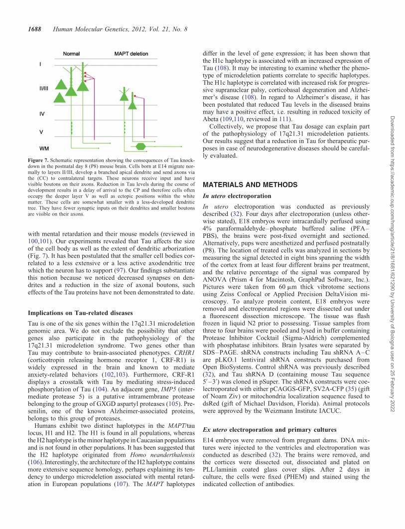
with mental retardation and their mouse models (reviewed in100,101). Our experiments revealed that Tau affects the sizeof the cell body as well as the extent of dendritic arborization(Fig. 7). It has been postulated that the smaller cell bodies cor-related to a less extensive or a less active axodendritic treewhich the neuron has to support (97). Our findings substantiatethis notion because we noticed decreased synapses on den-drites and a reduction in the size of axonal boutons, sucheffects of the Tau proteins have not been demonstrated to date.
Implications on Tau-related diseases
Tau is one of the six genes within the 17q21.31 microdeletiongenomic area. We do not exclude the possibility that othergenes also participate in the pathophysiology of the17q21.31 microdeletion syndrome. Two genes other thanTau may contribute to brain-associated phenotypes. CRHR1(corticotropin releasing hormone receptor 1, CRF-R1) iswidely expressed in the brain and known to mediateanxiety-related behaviors (102,103). Furthermore, CRF-R1displays a crosstalk with Tau by mediating stress-inducedphosphorylation of Tau (104). An adjacent gene, IMP5 (inter-mediate protease 5) is a putative intramembrane proteasebelonging to the group of GXGD aspartyl proteases (105). Pre-senilin, one of the known Alzheimer-associated proteins,belongs to this group of proteases.
Humans exhibit two distinct haplotypes in the MAPT/taulocus, H1 and H2. The H1 is found in all populations, whereasthe H2 haplotype is the minor haplotype in Caucasian populationsand is not found in other populations. It has been suggested thatthe H2 haplotype originated from Homo neanderthalensis(106). Interestingly, the architecture of the H2 haplotype containsmore extensive sequence homology, perhaps explaining its ten-dency to undergo microdeletion associated with mental retard-ation in European populations (107). The MAPT haplotypes
differ in the level of gene expression; it has been shown thatthe H1c haplotype is associated with an increased expression ofTau (108). It may be interesting to examine whether the pheno-type of microdeletion patients correlate to specific haplotypes.The H1c haplotype is correlated with increased risk for progres-sive supranuclear palsy, corticobasal degeneration and Alzhei-mer’s disease (108). In regard to Alzheimer’s disease, it hasbeen postulated that reduced Tau levels in the diseased brainsmay have a positive effect, i.e. resulting in reduced toxicity ofAbeta (109,110, reviewed in 111).
Collectively, we propose that Tau dosage can explain partof the pathophysiology of 17q21.31 microdeletion patients.Our results suggest that a reduction in Tau for therapeutic pur-poses in case of neurodegenerative diseases should be careful-ly evaluated.
MATERIALS AND METHODS
In utero electroporation
In utero electroporation was conducted as previouslydescribed (32). Four days after electroporation (unless other-wise stated), E18 embryos were intracardially perfused using4% paraformaldehyde–phosphate buffered saline (PFA–PBS), the brains were post-fixed overnight and sectioned.Alternatively, pups were anesthetized and perfused postnatally(P8). The location of treated cells was analyzed in sections bymeasuring the signal detected in eight bins spanning the widthof the cortex from at least four different brains per treatment,and the relative percentage of the signal was compared byANOVA (Prism 4 for Macintosh, GraphPad Software, Inc.).Pictures were taken from 60 mm thick vibrotome sectionsusing Zeiss Confocal or Applied Precision DeltaVision mi-croscopy. To analyze protein content, E18 embryos wereremoved and electroporated regions were dissected out undera fluorescent dissection microscope. The tissue was flashfrozen in liquid N2 prior to possessing. Tissue samples fromthree to four brains were pooled and lysed in buffer containingProtease Inhibitor Cocktail (Sigma-Aldrich) complementedwith phosphatase inhibitors. Brain lysates were separated bySDS–PAGE. shRNA constructs including Tau shRNA A–Care pLKO.1 lentiviral shRNA constructs purchased fromOpen BioSystems. Control shRNA was previously described(32), and Tau shRNA D (containing mouse Tau sequence5′ –3′) was cloned in pSuper. The shRNA constructs were coe-lectroporated with either pCAGGS-GFP, SV2A-CFP (35) (giftof Noam Ziv) or mitochondria localization sequence fused todsRed (gift of Michael Davidson, Florida). Animal protocolswere approved by the Weizmann Institute IACUC.
Ex utero electroporation and primary cultures
E14 embryos were removed from pregnant dams. DNA mix-tures were injected to the ventricles and electroporation wasconducted as described (32). The brains were removed, andthe cortices were dissected out, dissociated and plated onPLL/laminin coated glass cover slips. After 2 days inculture, the cells were fixed (PHEM) and stained using theindicated collection of antibodies.
Figure 7. Schematic representation showing the consequences of Tau knock-down in the postnatal day 8 (P8) mouse brain. Cells born at E14 migrate nor-mally to layers II/III, develop a branched apical dendrite and send axons viathe (CC) to contralateral targets. These neurons receive input and havevisible boutons on their axons. Reduction in Tau levels during the course ofdevelopment results in a delay of arrival to the CP and therefore cells oftenoccupy the deeper layer V as well as ectopic positions within the whitematter. These cells are somewhat smaller with a less-developed dendritictree. They have fewer synaptic inputs on their dendrites and smaller boutonsare visible on their axons.
1688 Human Molecular Genetics, 2012, Vol. 21, No. 8
Dow
nloaded from https://academ
ic.oup.com/hm
g/article/21/8/1681/621290 by University of Bologna user on 25 February 2022
Live imaging
Electroporated brains were harvested 2–3 days after electropor-ation (E16–E17). Brains were excised in ice-cold L-15 mediasupplemented with glucose (0.6%), Gentamicin (0.02 mg/ml)and oxygen. Brains were embedded in 3.5% low-melt agarosedissolved in L-15 and cut to 300 mm thick slices by a vibrotome.The explants were placed onto Millicell-CM inserts floating onthe neurobasal medium supplemented with B27 and N2, 2 mM
GlutaMax, 0.5% glucose and Gentamicin. Images were taken2 h after plating in Applied Precision DeltaVision microscopyequipped with an environmental chamber. Images were ana-lyzed using Imaris software (Bitplane).
Electron microscopy
Two days after electroporation (E16), the brains were removedand immersed in fixation solution [0.1 M PB; pH 7.4; 4% PFA;0.1% glutaraldehyde EM grade (Polyscience, Warrington, PA,USA), and 15% saturated picric acid]. The brains were post-fixed for 24 h in glutaraldehyde-free fixative to prevent add-itional loss of antigenicity. The tissue was thoroughly rinsedin PB and cut on a vibratome to 50 mm thick slices. These vibra-tome sections were freeze-thawed, blocked (normal goat serum,NGS; 20%) and incubated in an antibody against GFP (Invitro-gen); 1:500 in Tris–buffered saline, containing 2% NGS. Thiswas followed by thoroughly washing the sections, which werethen incubated in 1.4 nm gold-coupled secondary antibodies(goat anti-rabbit; 1:100; Nanoprobes), followed by silver in-tensification (HQ-silver; Nanoprobes). Finally, the sectionswere osmicated and stained with uranyl acetate, dehydratedand flat-embedded in Durcupan (Fluca; Sigma). Thin sections(60 nm) were viewed in an electron microscope (LEO 906E).
Immunohistochemistry
The following antibodies were used to stain primary neuronalcultures and brain sections: Tau1 (Chemicon), anti-Tau PHF-1(E-M Mandelkow), goat anti-GFP-biotinylated (Vector labs)and rabbit anti-Fyn (FYN3) (Santa Cruz, SC-16). Floating sec-tions or cover slips containing fixed cells were blocked inblocking solution (PBS, 0.1% Triton X-100, 10% HS,10%FCS) for 30 min. Antibodies were incubated in blockingsolution over night at 48C. After washing, appropriate second-ary antibodies (Jackson ImmunoResearch) were diluted inblocking solution, and incubated for 30 min at room tempera-ture. Slices were mounted onto glass slides using Aqua Poly/mount (Polyscience).
SUPPLEMENTARY MATERIAL
Supplementary Material is available at HMG online.
ACKNOWLEDGEMENTS
We thank current and previous lab members for their contribu-tions and Drs Michael Davidson and Noam Ziv for plasmids.O.R. is an Incumbent of the Berstein-Mason professorial chairof Neurochemistry. M.F. is Hertie Senior Research Professorof the Hertie Foundation.
FUNDING
Our research has been supported in part by the Israel ScienceFoundation (grant no. 47/10), BSF grant 2007081, the LegacyHeritage Biomedical Program of the Israel Science Foundation(grant no. 1062/08), Minerva foundation with funding fromthe Federal German Ministry for Education and Research,ERANET-NEURON (DISCover, IMOS 3-00000-6785), theBenoziyo Center for Neurological diseases, the Helen andMartin Kimmel Stem Cell Research Institute, Sylvia SchaeferAlzheimer’s Research Fund and the David and Fela ShapellFamily Center for Genetic Disorders Research.
REFERENCES
1. Hernandez, F. and Avila, J. (2007) Tauopathies. Cell Mol. Life Sci., 64,2219–2233.
2. Avila, J., Lucas, J.J., Perez, M. and Hernandez, F. (2004) Role of tauprotein in both physiological and pathological conditions. Physiol. Rev.,84, 361–384.
3. Mandelkow, E.M. and Mandelkow, E. (1998) Tau in Alzheimer’sdisease. Trends Cell Biol., 8, 425–427.
4. Rademakers, R., Cruts, M. and van Broeckhoven, C. (2004) The role oftau (MAPT) in frontotemporal dementia and related tauopathies. Hum.Mutat., 24, 277–295.
5. Hutton, M., Lendon, C.L., Rizzu, P., Baker, M., Froelich, S., Houlden,H., Pickering-Brown, S., Chakraverty, S., Isaacs, A., Grover, A. et al.(1998) Association of missense and 5′-splice-site mutations in tau withthe inherited dementia FTDP-17. Nature, 393, 702–705.
6. D’Souza, I., Poorkaj, P., Hong, M., Nochlin, D., Lee, V.M., Bird, T.D.and Schellenberg, G.D. (1999) Missense and silent tau gene mutationscause frontotemporal dementia with parkinsonism-chromosome 17 type,by affecting multiple alternative RNA splicing regulatory elements.Proc. Natl Acad. Sci. USA, 96, 5598–5603.
7. Shaw-Smith, C., Pittman, A.M., Willatt, L., Martin, H., Rickman, L.,Gribble, S., Curley, R., Cumming, S., Dunn, C., Kalaitzopoulos, D. et al.(2006) Microdeletion encompassing MAPT at chromosome 17q21.3 isassociated with developmental delay and learning disability. Nat. Genet.,38, 1032–1037.
8. Sharp, A.J., Hansen, S., Selzer, R.R., Cheng, Z., Regan, R., Hurst, J.A.,Stewart, H., Price, S.M., Blair, E., Hennekam, R.C. et al. (2006)Discovery of previously unidentified genomic disorders from theduplication architecture of the human genome. Nat. Genet., 38,1038–1042.
9. Koolen, D.A., Vissers, L.E., Pfundt, R., de Leeuw, N., Knight, S.J.,Regan, R., Kooy, R.F., Reyniers, E., Romano, C., Fichera, M. et al.(2006) A new chromosome 17q21.31 microdeletion syndrome associatedwith a common inversion polymorphism. Nat. Genet., 38, 999–1001.
10. Varela, M.C., Krepischi-Santos, A.C., Paz, J.A., Knijnenburg, J., Szuhai,K., Rosenberg, C. and Koiffmann, C.P. (2006) A 17q21.31 microdeletionencompassing the MAPT gene in a mentally impaired patient. CytogenetGenome Res., 114, 89–92.
11. Koolen, D.A., Sharp, A.J., Hurst, J.A., Firth, H.V., Knight, S.J.,Goldenberg, A., Saugier-Veber, P., Pfundt, R., Vissers, L.E., Destree, A.et al. (2008) Clinical and molecular delineation of the 17q21.31microdeletion syndrome. J. Med. Genet., 45, 710–720.
12. Schaaf, C.P. and Zoghbi, H.Y. (2011) Solving the autism puzzle a fewpieces at a time. Neuron, 70, 806–808.
13. Sanders, S.J., Ercan-Sencicek, A.G., Hus, V., Luo, R., Murtha, M.T.,Moreno-De-Luca, D., Chu, S.H., Moreau, M.P., Gupta, A.R., Thomson,S.A. et al. (2011) Multiple recurrent de novo CNVs, includingduplications of the 7q11.23 Williams Syndrome Region, are stronglyassociated with Autism. Neuron, 70, 863–885.
14. Levy, D., Ronemus, M., Yamrom, B., Lee, Y.H., Leotta, A., Kendall, J.,Marks, S., Lakshmi, B., Pai, D., Ye, K. et al. (2011) Rare de novo andtransmitted copy-number variation in autistic spectrum disorders.Neuron, 70, 886–897.
15. Gilman, S.R., Iossifov, I., Levy, D., Ronemus, M., Wigler, M. andVitkup, D. (2011) Rare de novo variants associated with autism
Human Molecular Genetics, 2012, Vol. 21, No. 8 1689
Dow
nloaded from https://academ
ic.oup.com/hm
g/article/21/8/1681/621290 by University of Bologna user on 25 February 2022
implicate a large functional network of genes involved in formation andfunction of synapses. Neuron, 70, 898–907.
16. Brunetti-Pierri, N., Berg, J.S., Scaglia, F., Belmont, J., Bacino, C.A.,Sahoo, T., Lalani, S.R., Graham, B., Lee, B., Shinawi, M. et al. (2008)Recurrent reciprocal 1q21.1 deletions and duplications associated withmicrocephaly or macrocephaly and developmental and behavioralabnormalities. Nat. Genet., 40, 1466–1471.
17. Xu, B., Roos, J.L., Levy, S., van Rensburg, E.J., Gogos, J.A. andKarayiorgou, M. (2008) Strong association of de novo copy numbermutations with sporadic schizophrenia. Nat. Genet., 40, 880–885.
18. Walsh, T., McClellan, J.M., McCarthy, S.E., Addington, A.M., Pierce,S.B., Cooper, G.M., Nord, A.S., Kusenda, M., Malhotra, D., Bhandari,A. et al. (2008) Rare structural variants disrupt multiple genes inneurodevelopmental pathways in schizophrenia. Science, 320, 539–543.
19. Stone, J.L., O’Donovan, M.C., Gurling, H., Kirov, G.K., Blackwood,D.H., Corvin, A., Craddock, N.J., Gill, M., Hultman, C.M., Lichtenstein,P. et al. (2008) Rare chromosomal deletions and duplications increaserisk of schizophrenia. Nature, 455, 237–241.
20. Stefansson, H., Rujescu, D., Cichon, S., Pietilainen, O.P., Ingason, A.,Steinberg, S., Fossdal, R., Sigurdsson, E., Sigmundsson, T.,Buizer-Voskamp, J.E. et al. (2008) Large recurrent microdeletionsassociated with schizophrenia. Nature, 455, 232–236.
21. Madrigal, I., Rodriguez-Revenga, L., Armengol, L., Gonzalez, E.,Rodriguez, B., Badenas, C., Sanchez, A., Martinez, F., Guitart, M.,Fernandez, I. et al. (2007) X-chromosome tiling path array detection ofcopy number variants in patients with chromosome X-linked mentalretardation. BMC Genomics, 8, 443.
22. Guilmatre, A., Dubourg, C., Mosca, A.L., Legallic, S., Goldenberg, A.,Drouin-Garraud, V., Layet, V., Rosier, A., Briault, S., Bonnet-Brilhault,F. et al. (2009) Recurrent rearrangements in synaptic andneurodevelopmental genes and shared biologic pathways inschizophrenia, autism, and mental retardation. Arch. Gen. Psychiatry, 66,947–956.
23. Bullmann, T., Holzer, M., Mori, H. and Arendt, T. (2009) Pattern of tauisoforms expression during development in vivo. Int. J. Dev. Neurosci.,27, 591–597.
24. Takuma, H., Arawaka, S. and Mori, H. (2003) Isoforms changes of tauprotein during development in various species. Brain Res. Dev. Brain
Res., 142, 121–127.25. Reiner, O. and Sapir, T. (2009) Polarity regulation in migrating neurons
in the cortex. Mol. Neurobiol., 40, 1–14.26. Ayala, R., Shu, T. and Tsai, L.H. (2007) Trekking across the brain: the
journey of neuronal migration. Cell, 128, 29–43.27. Harada, A., Oguchi, K., Okabe, S., Kuno, J., Terada, S., Ohshima, T.,
Sato-Yoshitake, R., Takei, Y., Noda, T. and Hirokawa, N. (1994) Alteredmicrotubule organization in small-calibre axons of mice lacking tau
protein. Nature, 369, 488–491.
28. Ikegami, S., Harada, A. and Hirokawa, N. (2000) Muscle weakness,hyperactivity, and impairment in fear conditioning in tau-deficient mice.Neurosci. Lett., 279, 129–132.
29. Takei, Y., Teng, J., Harada, A. and Hirokawa, N. (2000) Defects inaxonal elongation and neuronal migration in mice with disrupted tau andmap1b genes. J. Cell Biol., 150, 989–1000.
30. Bai, J., Ramos, R.L., Ackman, J.B., Thomas, A.M., Lee, R.V. andLoTurco, J.J. (2003) RNAi reveals doublecortin is required for radialmigration in rat neocortex. Nat. Neurosci., 6, 1277–1283.
31. Koizumi, H., Tanaka, T. and Gleeson, J.G. (2006) Doublecortin-likekinase functions with doublecortin to mediate fiber tract decussation andneuronal migration. Neuron, 49, 55–66.
32. Sapir, T., Sapoznik, S., Levy, T., Finkelshtein, D., Shmueli, A., Timm,T., Mandelkow, E.M. and Reiner, O. (2008) Accurate balance of thepolarity kinase MARK2/Par-1 is required for proper cortical neuronalmigration. J. Neurosci., 28, 5710–5720.
33. Goedert, M., Spillantini, M.G., Jakes, R., Rutherford, D. and Crowther,R.A. (1989) Multiple isoforms of human microtubule-associated proteintau: sequences and localization in neurofibrillary tangles of Alzheimer’sdisease. Neuron, 3, 519–526.
34. Mandelkow, E.M., Thies, E., Trinczek, B., Biernat, J. and Mandelkow,E. (2004) MARK/PAR1 kinase is a regulator of microtubule-dependenttransport in axons. J. Cell Biol., 167, 99–110.
35. Zhai, R.G., Vardinon-Friedman, H., Cases-Langhoff, C., Becker, B.,Gundelfinger, E.D., Ziv, N.E. and Garner, C.C. (2001) Assembling the
presynaptic active zone: a characterization of an active one precursorvesicle. Neuron, 29, 131–143.
36. Ziv, N.E. and Garner, C.C. (2004) Cellular and molecular mechanisms ofpresynaptic assembly. Nat. Rev. Neurosci., 5, 385–399.
37. Reiner, O., Carrozzo, R., Shen, Y., Whenert, M., Faustinella, F., Dobyns,W.B., Caskey, C.T. and Ledbetter, D.H. (1993) Isolation of aMiller-Dieker lissencephaly gene containing G protein b-subunit-likerepeats. Nature, 364, 717–721.
38. Sapir, T., Elbaum, M. and Reiner, O. (1997) Reduction of microtubulecatastrophe events by LIS1, platelet-activating factor acetylhydrolasesubunit. EMBO J., 16, 6977–6984.
39. Coquelle, F.M., Caspi, M., Cordelieres, F.P., Dompierre, J.P., Dujardin,D.L., Koifman, C., Martin, P., Hoogenraad, C.C., Akhmanova, A.,Galjart, N. et al. (2002) LIS1, CLIP-170’s key to the dynein/dynactinpathway. Mol. Cell Biol., 22, 3089–3102.
40. des Portes, V., Pinard, J.M., Billuart, P., Vinet, M.C., Koulakoff, A.,Carrie, A., Gelot, A., Dupuis, E., Motte, J., Berwald-Netter, Y. et al.
(1998) A novel CNS gene required for neuronal migration and involvedin X-linked subcortical laminar hetrotropia and lissencephaly syndrome.Cell, 92, 51–61.
41. Gleeson, J.G., Allen, K.M., Fox, J.W., Lamperti, E.D., Berkovic, S.,Scheffer, I., Cooper, E.C., Dobyns, W.B., Minnerath, S.R., Ross, M.E.et al. (1998) Doublecortin, a brain-specific gene mutated in humanX-linked lissencephaly and double cortex syndrome, encodes a putativesignaling protein. Cell, 92, 63–72.
42. Francis, F., Koulakoff, A., Boucher, D., Chafey, P., Schaar, B., Vinet,M.C., Friocourt, G., McDonnell, N., Reiner, O., Kahn, A. et al. (1999)Doublecortin is a developmentally regulated, microtubule-associatedprotein expressed in migrating and differentiating neurons. Neuron, 23,247–256.
43. Gleeson, J.G., Lin, P.T., Flanagan, L.A. and Walsh, C.A. (1999)Doublecortin is a microtubule-associated protein and is expressed widelyby migrating neurons. Neuron, 23, 257–271.
44. Horesh, D., Sapir, T., Francis, F., Caspi, M., Grayer Wolf, S., Elbaum,M., Chelly, J. and Reiner, O. (1999) Doublecortin, a stabilizer ofmicrotubules. Hum. Mol. Genet., 8, 1599–1610.
45. Sapir, T., Horesh, D., Caspi, M., Atlas, R., Burgess, H.A., Grayer Wolf,S., Francis, F., Chelly, J., Elbaum, M., Pietrokovski, S. et al. (2000)Doublecortin mutations cluster in evolutionary conserved functionaldomains. Hum. Mol. Genet., 5, 703–712.
46. Keays, D.A., Tian, G., Poirier, K., Huang, G.J., Siebold, C., Cleak, J.,Oliver, P.L., Fray, M., Harvey, R.J., Molnar, Z. et al. (2007) Mutations inalpha-tubulin cause abnormal neuronal migration in mice andlissencephaly in humans. Cell, 128, 45–57.
47. Poirier, K., Keays, D.A., Francis, F., Saillour, Y., Bahi, N., Manouvrier,S., Fallet-Bianco, C., Pasquier, L., Toutain, A., Tuy, F.P. et al. (2007)Large spectrum of lissencephaly and pachygyria phenotypes resultingfrom de novo missense mutations in tubulin alpha 1A (TUBA1A).Hum. Mutat., 48, 1055–1064.
49. Tian, G., Jaglin, X.H., Keays, D.A., Francis, F., Chelly, J. and Cowan,N.J. (2010) Disease-associated mutations in TUBA1A result in aspectrum of defects in the tubulin folding and heterodimer assemblypathway. Hum. Mol. Genet., 19, 3599–3613.
50. Kamiya, A., Kubo, K., Tomoda, T., Takaki, M., Youn, R., Ozeki, Y.,Sawamura, N., Park, U., Kudo, C., Okawa, M. et al. (2005) Aschizophrenia-associated mutation of DISC1 perturbs cerebral cortexdevelopment. Nat. Cell Biol., 7, 1167–1178.
51. Duan, X., Chang, J.H., Ge, S., Faulkner, R.L., Kim, J.Y., Kitabatake, Y.,Liu, X.B., Yang, C.H., Jordan, J.D., Ma, D.K. et al. (2007)Disrupted-In-Schizophrenia 1 regulates integration of newly generatedneurons in the adult brain. Cell, 130, 1146–1158.
52. Meyer, K.D. and Morris, J.A. (2009) Disc1 regulates granule cellmigration in the developing hippocampus. Hum. Mol. Genet., 18,3286–3297.
53. Millar, J.K., Wilson-Annan, J.C., Anderson, S., Christie, S., Taylor,M.S., Semple, C.A., Devon, R.S., St Clair, D.M., Muir, W.J.,Blackwood, D.H. et al. (2000) Disruption of two novel genes by a
1690 Human Molecular Genetics, 2012, Vol. 21, No. 8
Dow
nloaded from https://academ
ic.oup.com/hm
g/article/21/8/1681/621290 by University of Bologna user on 25 February 2022
translocation co-segregating with schizophrenia. Hum. Mol. Genet., 9,1415–1423.
54. Bi, W., Sapir, T., Shchelochkov, O.A., Zhang, F., Withers, M.A., Hunter,J.V., Levy, T., Shinder, V., Peiffer, D.A., Gunderson, K.L. et al. (2009)Increased LIS1 expression affects human and mouse brain development.Nat. Genet., 41, 168–177.
55. Toyo-oka, K., Shionoya, A., Gambello, M.J., Cardoso, C., Leventer, R.,Ward, H.L., Ayala, R., Tsai, L.H., Dobyns, W., Ledbetter, D. et al.
(2003) 14-3-3epsilon is important for neuronal migration by binding toNUDEL: a molecular explanation for Miller-Dieker syndrome. Nat.
Genet., 34, 274–285.56. Bruno, D.L., Anderlid, B.M., Lindstrand, A., van Ravenswaaij-Arts, C.,
Ganesamoorthy, D., Lundin, J., Martin, C.L., Douglas, J., Nowak, C.,Adam, M.P. et al. (2010) Further molecular and clinical delineation ofco-locating 17p13.3 microdeletions and microduplications that showdistinctive phenotypes. J. Med. Genet., 47, 299–311.
57. Shimojima, K., Sugiura, C., Takahashi, H., Ikegami, M., Takahashi, Y.,Ohno, K., Matsuo, M., Saito, K. and Yamamoto, T. (2010) Genomiccopy number variations at 17p13.3 and epileptogenesis. Epilepsy Res.,89, 303–309.
58. Mignon-Ravix, C., Cacciagli, P., El-Waly, B., Moncla, A., Milh, M.,Girard, N., Chabrol, B., Philip, N. and Villard, L. (2010) Deletion ofYWHAE in a patient with periventricular heterotopias and pronouncedcorpus callosum hypoplasia. J. Med. Genet., 47, 132–136.
59. Nagamani, S.C., Zhang, F., Shchelochkov, O.A., Bi, W., Ou, Z., Scaglia,F., Probst, F.J., Shinawi, M., Eng, C., Hunter, J.V. et al. (2009)Microdeletions including YWHAE in the Miller-Dieker syndrome regionon chromosome 17p13.3 result in facial dysmorphisms, growthrestriction, and cognitive impairment. J. Med. Genet., 46, 825–833.
60. Weingarten, M.D., Lockwood, A.H., Hwo, S.Y. and Kirschner, M.W.(1975) A protein factor essential for microtubule assembly. Proc. Natl
Acad. Sci. USA, 72, 1858–1862.61. Cleveland, D.W., Hwo, S.Y. and Kirschner, M.W. (1977) Purification of
tau, a microtubule-associated protein that induces assembly ofmicrotubules from purified tubulin. J. Mol. Biol., 116, 207–225.
62. Drubin, D.G. and Kirschner, M.W. (1986) Tau protein function in livingcells. J. Cell Biol., 103, 2739–2746.
63. Drechsel, D.N., Hyman, A.A., Cobb, M.H. and Kirschner, M.W. (1992)Modulation of the dynamic instability of tubulin assembly by themicrotubule-associated protein tau. Mol. Biol. Cell, 3, 1141–1154.
64. Tint, I., Jean, D., Baas, P.W. and Black, M.M. (2009) Doublecortinassociates with microtubules preferentially in regions of the axondisplaying actin-rich protrusive structures. J. Neurosci., 29,10995–11010.
65. Qiang, L., Yu, W., Andreadis, A., Luo, M. and Baas, P.W. (2006) Tauprotects microtubules in the axon from severing by katanin. J. Neurosci.,26, 3120–3129.
66. Himmler, A., Drechsel, D., Kirschner, M.W. and Martin, D.W. (1989)Tau consists of a set of proteins with repeated C-terminalmicrotubule-binding domains and variable N-terminal domains. Mol.
Cell Biol., 9, 1381–1388.67. Dixit, R., Ross, J.L., Goldman, Y.E. and Holzbaur, E.L. (2008)
Differential regulation of dynein and kinesin motor proteins by tau.Science, 319, 1086–1089.
68. Magnani, E., Fan, J., Gasparini, L., Golding, M., Williams, M., Schiavo,G., Goedert, M., Amos, L.A. and Spillantini, M.G. (2007) Interaction oftau protein with the dynactin complex. EMBO J., 26, 4546–4554.
69. Berezuk, M.A. and Schroer, T.A. (2007) Dynactin enhances theprocessivity of kinesin-2. Traffic, 8, 124–129.
70. Kardon, J.R. and Vale, R.D. (2009) Regulators of the cytoplasmic dyneinmotor. Nat. Rev. Mol. Cell Biol., 10, 854–865.
72. Maussion, G., Carayol, J., Lepagnol-Bestel, A.M., Tores, F., Loe-Mie,Y., Milbreta, U., Rousseau, F., Fontaine, K., Renaud, J., Moalic, J.M.et al. (2008) Convergent evidence identifying MAP/microtubuleaffinity-regulating kinase 1 (MARK1) as a susceptibility gene for autism.Hum. Mol. Genet., 17, 2541–2551.
73. Amano, M., Kaneko, T., Maeda, A., Nakayama, M., Ito, M., Yamauchi,T., Goto, H., Fukata, Y., Oshiro, N., Shinohara, A. et al. (2003)Identification of Tau and MAP2 as novel substrates of Rho-kinase andmyosin phosphatase. J. Neurochem., 87, 780–790.
74. Matenia, D., Griesshaber, B., Li, X.Y., Thiessen, A., Johne, C., Jiao, J.,Mandelkow, E. and Mandelkow, E.M. (2005) PAK5 kinase is aninhibitor of MARK/Par-1, which leads to stable microtubules anddynamic actin. Mol. Biol. Cell, 16, 4410–4422.
75. Sharma, V.M., Litersky, J.M., Bhaskar, K. and Lee, G. (2007) Tauimpacts on growth-factor-stimulated actin remodeling. J. Cell Sci., 120,748–757.
76. Timm, T., Matenia, D., Li, X.Y., Griesshaber, B. and Mandelkow, E.M.(2006) Signaling from MARK to tau: regulation, cytoskeletal crosstalk,and pathological phosphorylation. Neurodegener. Dis., 3, 207–217.
77. Chai, X., Forster, E., Zhao, S., Bock, H.H. and Frotscher, M. (2009)Reelin stabilizes the actin cytoskeleton of neuronal processes byinducing n-cofilin phosphorylation at serine3. J. Neurosci., 29, 288–299.
78. Tissir, F. and Goffinet, A.M. (2003) Reelin and brain development. Nat.Rev. Neurosci., 4, 496–505.
79. Fatemi, S.H. (2005) Reelin glycoprotein: structure, biology and roles inhealth and disease. Mol. Psychiatry, 10, 251–257.
80. Jimenez-Mateos, E.M., Gonzalez-Billault, C., Dawson, H.N., Vitek,M.P. and Avila, J. (2006) Role of MAP1B in axonal retrograde transportof mitochondria. Biochem. J., 397, 53–59.
81. Stamer, K., Vogel, R., Thies, E., Mandelkow, E. and Mandelkow, E.M.(2002) Tau blocks traffic of organelles, neurofilaments, and APP vesiclesin neurons and enhances oxidative stress. J. Cell Biol., 156, 1051–1063.
82. Arawaka, S., Usami, M., Sahara, N., Schellenberg, G.D., Lee, G. andMori, H. (1999) The tau mutation (val337met) disrupts cytoskeletalnetworks of microtubules. Neuroreport, 10, 993–997.
83. Ebneth, A., Godemann, R., Stamer, K., Illenberger, S., Trinczek, B. andMandelkow, E. (1998) Overexpression of tau protein inhibitskinesin-dependent trafficking of vesicles, mitochondria, and endoplasmicreticulum: implications for Alzheimer’s disease. J. Cell Biol.,143, 777–794.
84. Schon, E.A. and Przedborski, S. (2011) Mitochondria: the next(neurode)generation. Neuron, 70, 1033–1053.
85. Palmieri, L., Papaleo, V., Porcelli, V., Scarcia, P., Gaita, L., Sacco, R.,Hager, J., Rousseau, F., Curatolo, P., Manzi, B. et al. (2010) Alteredcalcium homeostasis in autism-spectrum disorders: evidence frombiochemical and genetic studies of the mitochondrial aspartate/glutamatecarrier AGC1. Mol. Psychiatry, 15, 38–52.
86. Giulivi, C., Zhang, Y.F., Omanska-Klusek, A., Ross-Inta, C., Wong, S.,Hertz-Picciotto, I., Tassone, F. and Pessah, I.N. (2010) Mitochondrialdysfunction in autism. JAMA, 304, 2389–2396.
87. Pramparo, T., Libiger, O., Jain, S., Li, H., Youn, Y.H., Hirotsune, S.,Schork, N.J. and Wynshaw-Boris, A. (2011) Global developmental geneexpression and pathway analysis of normal brain development andmouse models of human neuronal migration defects. PLoS Genet., 7,e1001331.
88. Reiner, O., Shmueli, A. and Sapir, T. (2009) Neuronal migrationand neurodegeneration: 2 sides of the same coin. Cereb. Cortex,19(Suppl. 1), i42–i48.
89. Binder, L.I., Frankfurter, A. and Rebhun, L.I. (1985) The distribution oftau in the mammalian central nervous system. J. Cell Biol., 101,1371–1378.
90. Kanai, Y. and Hirokawa, N. (1995) Sorting mechanisms of tau andMAP2 in neurons: suppressed axonal transit of MAP2 and locallyregulated microtubule binding. Neuron, 14, 421–432.
91. Black, M.M., Slaughter, T., Moshiach, S., Obrocka, M. and Fischer, I.(1996) Tau is enriched on dynamic microtubules in the distal region ofgrowing axons. J. Neurosci., 16, 3601–3619.
92. Hirokawa, N., Funakoshi, T., Sato-Harada, R. and Kanai, Y. (1996)Selective stabilization of tau in axons and microtubule-associated protein2C in cell bodies and dendrites contributes to polarized localization ofcytoskeletal proteins in mature neurons. J. Cell Biol., 132, 667–679.
93. Kempf, M., Clement, A., Faissner, A., Lee, G. and Brandt, R. (1996) Taubinds to the distal axon early in development of polarity in amicrotubule- and microfilament-dependent manner. J. Neurosci., 16,5583–5592.
94. Ittner, L.M., Ke, Y.D., Delerue, F., Bi, M., Gladbach, A., van Eersel, J.,Wolfing, H., Chieng, B.C., Christie, M.J., Napier, I.A. et al. (2010)Dendritic function of tau mediates amyloid-beta toxicity in Alzheimer’sdisease mouse models. Cell, 142, 387–397.
95. Williamson, R., Scales, T., Clark, B.R., Gibb, G., Reynolds, C.H., Kellie,S., Bird, I.N., Varndell, I.M., Sheppard, P.W., Everall, I. et al. (2002)Rapid tyrosine phosphorylation of neuronal proteins including tau and
Human Molecular Genetics, 2012, Vol. 21, No. 8 1691
Dow
nloaded from https://academ
ic.oup.com/hm
g/article/21/8/1681/621290 by University of Bologna user on 25 February 2022
focal adhesion kinase in response to amyloid-beta peptide exposure:involvement of Src family protein kinases. J. Neurosci., 22, 10–20.
96. Williamson, R., Usardi, A., Hanger, D.P. and Anderton, B.H. (2008)Membrane-bound beta-amyloid oligomers are recruited into lipid raftsby a fyn-dependent mechanism. FASEB J., 22, 1552–1559.
97. Harrison, P.J. and Weinberger, D.R. (2005) Schizophrenia genes, geneexpression, and neuropathology: on the matter of their convergence. Mol.Psychiatry, 10, 40–68; image 45.
98. Marchetto, M.C., Carromeu, C., Acab, A., Yu, D., Yeo, G.W., Mu, Y.,Chen, G., Gage, F.H. and Muotri, A.R. (2010) A model for neuraldevelopment and treatment of Rett syndrome using human inducedpluripotent stem cells. Cell, 143, 527–539.
99. Li, H., Radford, J.C., Ragusa, M.J., Shea, K.L., McKercher, S.R.,Zaremba, J.D., Soussou, W., Nie, Z., Kang, Y.J., Nakanishi, N. et al.(2008) Transcription factor MEF2C influences neural stem/progenitorcell differentiation and maturation in vivo. Proc. Natl Acad. Sci. USA,105, 9397–9402.
100. Huttenlocher, P.R. (1991) Dendritic and synaptic pathology in mentalretardation. Pediatr. Neurol., 7, 79–85.
101. Dierssen, M. and Ramakers, G.J. (2006) Dendritic pathology in mentalretardation: from molecular genetics to neurobiology. Genes, BrainBehav., 5(Suppl. 2), 48–60.
102. Stenzel, P., Kesterson, R., Yeung, W., Cone, R.D., Rittenberg, M.B. andStenzel-Poore, M.P. (1995) Identification of a novel murine receptor forcorticotropin-releasing hormone expressed in the heart. Mol.Endocrinol., 9, 637–645.
103. Timpl, P., Spanagel, R., Sillaber, I., Kresse, A., Reul, J.M., Stalla, G.K.,Blanquet, V., Steckler, T., Holsboer, F. and Wurst, W. (1998) Impairedstress response and reduced anxiety in mice lacking a functionalcorticotropin-releasing hormone receptor 1. Nat. Genet., 19, 162–166.
104. Rissman, R.A., Lee, K.F., Vale, W. and Sawchenko, P.E. (2007)Corticotropin-releasing factor receptors differentiallyregulate stress-induced tau phosphorylation. J. Neurosci., 27,6552–6562.
105. Weihofen, A. and Martoglio, B. (2003) Intramembrane-cleavingproteases: controlled liberation of proteins and bioactive peptides.Trends Cell Biol., 13, 71–78.
106. Hardy, J., Pittman, A., Myers, A., Gwinn-Hardy, K., Fung, H.C., deSilva, R., Hutton, M. and Duckworth, J. (2005) Evidence suggesting thatHomo neanderthalensis contributed the H2 MAPT haplotype to Homosapiens. Biochem. Soc. Trans., 33, 582–585.
107. Zody, M.C., Jiang, Z., Fung, H.C., Antonacci, F., Hillier, L.W., Cardone,M.F., Graves, T.A., Kidd, J.M., Cheng, Z., Abouelleil, A. et al. (2008)Evolutionary toggling of the MAPT 17q21.31 inversion region. Nat.
Genet., 40, 1076–1083.
108. Myers, A.J., Pittman, A.M., Zhao, A.S., Rohrer, K., Kaleem, M.,Marlowe, L., Lees, A., Leung, D., McKeith, I.G., Perry, R.H. et al.
(2007) The MAPT H1c risk haplotype is associated with increasedexpression of tau and especially of 4 repeat containing transcripts.
Neurobiol. Dis., 25, 561–570.
109. Roberson, E.D., Scearce-Levie, K., Palop, J.J., Yan, F., Cheng, I.H., Wu,T., Gerstein, H., Yu, G.Q. and Mucke, L. (2007) Reducing endogenoustau ameliorates amyloid beta-induced deficits in an Alzheimer’s disease
mouse model. Science, 316, 750–754.
110. Vossel, K.A., Zhang, K., Brodbeck, J., Daub, A.C., Sharma, P.,Finkbeiner, S., Cui, B. and Mucke, L. (2010) Tau reduction prevents
Abeta-induced defects in axonal transport. Science, 330, 198.
111. Morris, M., Maeda, S., Vossel, K. and Mucke, L. (2011) The many facesof tau. Neuron, 70, 410–426.
1692 Human Molecular Genetics, 2012, Vol. 21, No. 8
Dow
nloaded from https://academ
ic.oup.com/hm
g/article/21/8/1681/621290 by University of Bologna user on 25 February 2022