INTRODUCTIONDuring embryonic heart development, the formation and mitogenicgrowth of the heart are sustained through contribution from theepicardium, which is a specialized layer of cells derived from amesothelial precursor structure, the proepicardial organ (PEO).Subpopulations of epicardial cells undergo epithelial-mesenchymaltransition (EMT) and migrate into the sub-epicardial space andmyocardium, where they differentiate into interstitial cells, cardiacfibroblasts and smooth muscle cells (reviewed by Lie-Venema etal., 2007; Ratajska et al., 2008; van Wijk et al., 2009; Pérez-Pomaresand de la Pompa, 2011; Gittenberger-de Groot et al., 2012; von Giseand Pu, 2012). Moreover, during cardiac repair post-injury in theadult, epicardial cells reestablish their developmental geneticprogram, migrate into wounded myocardium and differentiate intothe required cell lineages to provide support to regenerating hearttissue (Lepilina et al., 2006; Limana et al., 2007; Winter et al., 2007;Winter et al., 2009; Limana et al., 2010).
Basic helix-loop-helix (bHLH) transcription factors play crucialroles in cell fate specification and differentiation during organdevelopment, including the heart (Massari and Murre, 2000;Conway et al., 2010). Transcription factor 21 (Tcf21; also known asPod1, Epicardin, Capsulin) is expressed in the mesenchyme ofdeveloping organs, including the branchial arches, kidney, lung,spleen, gonads and in the PEO and epicardium (Lu et al., 1998;Quaggin et al., 1998; Tamura et al., 2001; Simrick et al., 2005;Serluca, 2008). Tcf21 mouse mutants display severe developmental
defects and die shortly after birth due to pulmonary hyperplasia(Quaggin et al., 1999; Lu et al., 2000). Moreover, depletion of Tcf21leads to defects in epithelial differentiation and branching in thekidney and lung, a phenotype that is thought to arise from disruptedepithelial-mesenchymal interactions (Quaggin et al., 1999). Morerecently, Tcf21 function has been linked to epicardial EMT anddifferentiation (Acharya et al., 2012; Braitsch et al., 2012).
Here, we characterized the morphological, molecular and cellulardevelopment of the epicardium in a new vertebrate model, Xenopus.We further define an essential role for Tcf21 in epicardialdevelopment and show that, in the absence of Tcf21, epicardial cellsfail to form a cohesive polarized sheet and instead are retained in aproepicardial (PE) precursor state. This is in contrast to recent dataand thus provides unique insight into the Tcf21-null mousephenotype, which may result from deleterious epicardialorganization at an earlier stage of development. Using a proteomicsapproach we identify Tcf21 protein interactions, determining thatTcf21 associates with co-factors involved in transcriptionalrepression including HDAC2, Pbx1 and Ctbp2, the latter two havingestablished requirements in cardiac development. Furthermore,using high-throughput sequencing analysis we define a unique setof nine genes that are expressed in the PEO and are repressed byTcf21, and observe a persistent and increased expression of thesemarkers in epicardial cells depleted of Tcf21. Altogether, our datademonstrate that Tcf21 functions as a transcriptional repressor toregulate processes of proepicardial specification and epicardialmaturation.
MATERIALS AND METHODSCell transfection and immunoaffinity purification (IP) of Tcf21 andinteraction partnersHuman embryonic kidney 293 (HEK293) cells were plated onto two sets of10×15 cm culture dishes and transfected at 40% confluence with FuGENE6 (Roche) at a 3:1 ratio, using 20 µg EGFP control plasmid (pEGFP_N1) orTcf21-EGFP plasmid (EGFP_N1-xlTcf21cds, NP_001085957). Cells weregrown to confluence, harvested, frozen in liquid nitrogen and processed asdescribed (Greco et al., 2011; Conlon et al., 2012). Cell powders were
1University of North Carolina McAllister Heart Institute, University of North Carolinaat Chapel Hill, Chapel Hill, NC 27599-3280, USA. 2Department of Genetics,University of North Carolina at Chapel Hill, Chapel Hill, NC 27599-3280, USA.3Department of Molecular Biology, Princeton University, NJ 08544-1014, USA.4Department of Biology, University of North Carolina at Chapel Hill, Chapel Hill, NC 27599-3280, USA.
SUMMARYThe epicardium is a mesothelial cell layer essential for vertebrate heart development and pertinent for cardiac repair post-injury inthe adult. The epicardium initially forms from a dynamic precursor structure, the proepicardial organ, from which cells migrate ontothe heart surface. During the initial stage of epicardial development crucial epicardial-derived cell lineages are thought to bedetermined. Here, we define an essential requirement for transcription factor Tcf21 during early stages of epicardial developmentin Xenopus, and show that depletion of Tcf21 results in a disruption in proepicardial cell specification and failure to form a matureepithelial epicardium. Using a mass spectrometry-based approach we defined Tcf21 interactions and established its association withproteins that function as transcriptional co-repressors. Furthermore, using an in vivo systems-based approach, we identified a panelof previously unreported proepicardial precursor genes that are persistently expressed in the epicardial layer upon Tcf21 depletion,thereby confirming a primary role for Tcf21 in the correct determination of the proepicardial lineage. Collectively, these studies leadus to propose that Tcf21 functions as a transcriptional repressor to regulate proepicardial cell specification and the correct formationof a mature epithelial epicardium.
KEY WORDS: Tcf21, Proepicardial organ, Heart development
Tcf21 regulates the specification and maturation ofproepicardial cellsPanna Tandon1,2, Yana V. Miteva3, Lauren M. Kuchenbrod1,2,4, Ileana M. Cristea3 and Frank L. Conlon1,2,4,*
DEVELO
PMENT
2410
suspended in prechilled optimized lysis buffer [20 mM HEPES-KOH pH7.4, 0.1 M potassium acetate, 2 mM MgCl2, 0.1% Tween 20 (v/v), 1 μMZnCl2, 1 μM CaCl2, 0.5% Triton X-100 (v/v), 150 mM NaCl, 4 μg/mlDNase, 1/100 (v/v) Protease Inhibitor Cocktail and 1/100 PhosphataseInhibitor Cocktail] using 5 ml lysis buffer/g cell powder. Lysates werehomogenized, subjected to centrifugation, and supernatants incubated for 1hour with 7 mg magnetic beads (M270 Epoxy Dynabeads, Invitrogen)conjugated with anti-EGFP antibodies (Cristea et al., 2005). Proteins wereeluted by incubation for 10 minutes (70°C) in 30 µl 1× LDS sample buffer(Invitrogen) containing 1× Reducing Agent (Invitrogen), followed byshaking at room temperature for 10 minutes.
In-solution digestion, mass spectrometry analysis and dataprocessingProtein IP eluates were prepared as described (Conlon et al., 2012; Grecoet al., 2011; Greco et al., 2012; Tsai et al., 2012). Briefly, IP eluates weremixed with 8 M urea in aqueous 0.1 M Tris-HCl pH 8.0, applied toultrafiltration Vivacon 500 units (Sartorius Stedim), and centrifuged at14,000 g for 40 minutes at 20°C. Samples were washed, alkylated anddigested with trypsin (Promega) overnight at 37°C. Resulting peptides werecollected by centrifugation, acidified with trifluoroacetic acid, concentratedby vacuum centrifugation, and desalted using Empore C18 StageTips(Rappsilber et al., 2007; Greco et al., 2012). Peptides were analyzed bynLC-MS/MS using a Dionex Ultimate 3000 RSLC system coupled onlineto an LTQ-Orbitrap Velos mass spectrometer (ThermoFisher Scientific)(Greco et al., 2012; Tsai et al., 2012). Peptides were fragmented by collision-induced dissociation (CID) and the MS/MS spectra were extracted byProteome Discoverer (ThermoFisher Scientific) and searched by SEQUESTagainst a database containing Xenopus, human and mouse UniProt Swiss-Prot sequences, including common contaminants and reversed sequences.Results were validated in Scaffold (Proteome Software) usingPeptideProphet and ProteinProphet. Co-isolated proteins were considered asTcf21-specific interactions if absent or enriched by at least 3-fold comparedwith controls. Proteins were clustered into functional subgroups accordingto biological roles based on Gene Ontology (GO) annotations.
RNA extraction and cDNA librariesXenopus embryos, injected with ConMO or Tcf21-MO (40 ng), were grownto stage 45. Hearts were collected (n=70-100 per condition) and processedfor RNA extraction (1 µg), purified using Sera-Mag magnetic oligo(dT)beads (Thermo Scientific), fragmented to ~300 bp at 70°C for 4 minutes,and cDNA generated (Superscript II, Invitrogen). For Solexa cDNAlibraries, 10 ng cDNA was blunted and paired-end adapters(Illumina/Solexa) ligated before purification on AMPure-XP (SPRI) beads(Agencourt). cDNA was amplified (PfuUltra II Fusion HS DNApolymerase, Stratagene), analyzed for purity and fragmentation size usinga 2100 Bioanalyzer (Agilent) and sequenced using an Illumina GenomeAnalyzer II system (High-Throughput Sequencing Facility, UNC).
RNA-seqTo reduce redundancy and isolate high-quality reads, 8879 cDNA sequencesannotated as RefSeq sequences in XenBase (Bowes et al., 2008) (May, 01,2011) were used for expression analysis (http://www.marcottelab.org/index.php/Xenopus_reference; Taejoon Kwon, Marcotte lab., University ofTexas, Austin). Alignments were performed on Bowtie2 v2.0.0.6 (JohnsHopkins University) and normalized for total counts to determine foldchange between control and Tcf21-depleted cardiac cDNA datasets (GEOseries GSE45786). Secondary screens were based on fold change (seesupplementary material Table S3) and GO term analysis performed usingGOrilla (updated 12/08/2012) (Eden et al., 2007; Eden et al., 2009) (seesupplementary material Tables S1 and S2) and ReviGO (updated04/02/2012) (Supek et al., 2011), and validated using RT-PCR fromindependent biological replicates (supplementary material Table S4, Fig.S7).
Xenopus transgenicsLinearized DNA (CMV:dsRED, NotI; gift from Enrique Amaya,Manchester University, UK) and CAG:KikumeGR (SalI) (Nowotschin andHadjantonakis, 2009) were introduced into Xenopus using trangenesis
procedures (Kroll and Amaya, 1996; Mandel et al., 2010). Fluorescentembryos were sorted and housed until adulthood, when germlinetransmission was tested. Stage 39-40 CAG:KikumeGR transgenic embryoswere placed in low-melting-point agarose (0.8%) in 0.33× Marc’s ModifiedRinger’s (MMR) cooled to room temperature. Embryos were positionedventral side down on a coverslip-based dish in agarose, submerged in 0.1×Modified Barth’s Saline (MBS) containing 0.01% tricaine. Localizedbleaching of the septum transversum (ST) was performed using a UV laser(Zeiss 710 confocal, seven cycles, 100 iterations, scan speed 10, excitation405 nm at 100%). Embryos were excised and recovered in 0.1× MBS beforeimaging (Leica MZ16F, Retiga 4000RV camera) (supplementary materialFig. S1).
Xenopus manipulationsXenopus embryos were staged according to Nieuwkoop and Faber(Nieuwkoop and Faber, 1967; Brown et al., 2005). An EST cDNA IMAGEX. laevis clone (ID 8077326, Open Biosystems) was sequenced andidentified as full-length Tcf21 (Simrick et al., 2005). Two non-overlappingtranslation-blocking morpholinos (MOs) were designed against the start siteof Tcf21 and upstream 5�UTR region (Tandon et al., 2012) as determined byRLM-RACE (Invitrogen, Gene Tools) (supplementary material Fig. S3);40 ng Tcf21-MO1 and Tcf21-MO2 were injected at the one-cell stage(Tandon et al., 2012) (see supplementary material Table S5 for MOsequences).
In situ hybridizationWhole-mount in situ hybridization (ISH) was carried out as described(Harland, 1991), the pericardial cavity membrane in late tadpole stageembryos being removed postfixation to improve resolution. Embryos wereprocessed for vibratome sectioning (30 µm) (Gessert and Kühl, 2009). TheWt1 probe was kindly provided by Peter Vize (Carroll and Vize, 1996); allother probes were generated by PCR (supplementary material Table S6) orreported previously (Brown et al., 2005; Goetz et al., 2006; Langdon et al.,2007).
ImmunohistochemistryAntibody staining was conducted as reported (Brown et al., 2005; Christineand Conlon, 2008; Mandel et al., 2010; Langdon et al., 2012)(supplementary material Table S7), then incubated in DAPI (200 ng/ml inPBS) and processed for agarose vibratome sectioning (150-200 µm)(Wallingford, 2010) or cryosectioning (10 µm) (Brown et al., 2005). Imageswere taken on an Olympus IX 81-ZDC inverted fluorescence microscope orZeiss LSM710.
Electron microscopyThe pericardial cavity membrane was excised from embryos anaesthetizedin 0.1% (w/v) tricaine and transmission (TEM) and scanning (SEM)electron microscopy conducted as reported using a Zeiss EM 910 and aZeiss Supra 25 FESEM, respectively (Microscope Services Laboratory,UNC) (Brown et al., 2007).
Live imaging of epicardial explantsStage 40 embryos were incubated in 0.1× MBS containing 25 µg/mlgentamycin and 0.1% iodine for 2 hours at room temperature, andsubsequently maintained in 0.1× MBS containing 0.006% iodine and25 µg/ml gentamycin. Rat tail collagen I [3 mg/ml collagen I, 40 mMsodium bicarbonate in 1× Dulbecco’s Modified Eagle Medium (DMEM)pH 8, on ice] was added at 10 µl per well of a 24-well plate, and allowed togel at room temperature for 30 minutes. Wells were rinsed with 1× Barth’ssolution and incubated with Barth’s+ (70 µg/ml gentamycin, 50 U/mlnystatin, 0.006% iodine and 10% heat-inactivated fetal bovine serum),rocking for 1 hour at room temperature. Hearts were excised fromanaesthetized embryos and placed in Barth’s on 1% agarose dishes on ice,then placed onto air-dried collagen cushions and allowed to adhere for 20minutes at room temperature before the addition of Barth’s+, then culturedat 23°C in a humidified chamber for 24-48 hours, fixed in 4%paraformaldehyde and processed for immunostaining as described, orvisualized on an Olympus IX70. Live images were captured every 30minutes over a 24- to 48-hour period at room temperature and analyzed
RESEARCH ARTICLE Development 140 (11)
DEVELO
PMENT
using ImageJ (NIH) and Imaris (Bitplane) software. Double-transgenicembryos (CMV:dsRED, CA:GFP) were used to enable better visualizationand cell migration tracking by Imaris software (supplementary material Fig.S9). Two-tailed unpaired non-parametric Mann-Whitney statistical test wasused to determine significance (GraphPad Prism 6, JMP).
RESULTSConserved epicardial development in XenopusTo gain new insights into the essential role of the epicardium duringdevelopment, we turned to a non-mammalian vertebrate modelsystem that is highly effective for modeling cardiac development.Molecular mechanisms of heart development in the frog Xenopusare highly conserved with those of mouse and human (Brown et al.,2003; Gormley and Nascone-Yoder, 2003; Mohun et al., 2003;Brown et al., 2005; Garriock et al., 2005; Goetz et al., 2006; Bartlettet al., 2007; Langdon et al., 2007; Warkman and Krieg, 2007;Bartlett and Weeks, 2008; Afouda and Hoppler, 2009; Evans et al.,2010; Mandel et al., 2010; Kaltenbrun et al., 2011; Langdon et al.,2012). We have confirmed that, similar to other vertebrates,Xenopus epicardium develops as a specialized layer of cellssurrounding the heart and is derived from a mesothelial precursorstructure, the PEO, located on the ST (Jahr et al., 2008). SEManalysis of Xenopus verifies that by late tadpole stages – roughlyequivalent to E9.5 in mouse, HH17 in chick and day 20 in humanembryonic development – the precursor epicardial structure andepicardium display prominent phenotypic changes as they migrateonto and over the ventricular surface (supplementary material Fig.S1) (Jahr et al., 2008). The PEO forms on the right-hand side on theST and attaches to the heart at the atrioventricular sulcus (AVS)close to the outflow tract (OFT) junction by stage 41(supplementary material Fig. S1B-D) (Jahr et al., 2008).Interestingly, at these stages this region of the Xenopus heartexpresses BMP2 (supplementary material Fig. S1K-P), a factorshown to influence correct PEO attachment and migration (Ishii etal., 2010). At slightly later stages, the PEO bridge is maintained(supplementary material Fig. S1D) and the epicardium develops asa distinct smooth epithelial-like sheet that gradually progresses overthe cobblestone-like cardiac surface (supplementary material Fig.S1C-F) (Jahr et al., 2008). To confirm that cells of the Xenopus PEOare similar to those of mammals and undergo similar cellmovements, we generated transgenic Xenopus embryosubiquitously expressing the photoconvertible fluorescent proteinKikumeGR (Nowotschin and Hadjantonakis, 2009; Griswold et al.,2011; Ridelis et al., 2012). Photoconversion of cells within the STand PEO (stage 39/40) confirms that these cells migrate, attach andform an epicardial sheet over the surface of the heart (stage 43,supplementary material Fig. S1G-J).
To determine whether the molecular underpinnings of epicardialdevelopment are conserved in Xenopus, we examined expressionpatterns of the epicardial-associated transcription factors Tbx18(Kraus et al., 2001; Begemann et al., 2002; Haenig and Kispert,2004; Jahr et al., 2008), Wilm’s tumor 1 (Wt1) (Moore et al., 1998;Carmona et al., 2001) and Tcf21 (Quaggin et al., 1998; Simrick etal., 2005; Ishii et al., 2007; Serluca, 2008) (Fig. 1). We observedthat all three are evolutionarily conserved in expression with regardsto the ST/PEO and epicardium. However, we also note a distinctspatiotemporal discrepancy, with Tbx18 being expressed in the STfirst (stage 31-32, Fig. 1A), followed by Wt1 and Tcf21 (stage 36,Fig. 1E,F), suggesting a potential hierarchical organization of thesegenes or subfunctionalization within the PEO, as postulated inmouse and chick (Mikawa and Gourdie, 1996; Braitsch et al., 2012).Expression of these markers becomes more spatially restricted to
the PEO at later stages, prior to migration onto the heart (stage 40,Fig. 1G-L), with Tcf21 displaying a more confined distribution(Fig. 1H,K). Thus, the cellular and molecular hallmarks ofepicardium formation are conserved from Xenopus to mouse.
Tcf21 is required for epicardial attachmentSince studies have reported that null mutations in Tbx18 have littleeffect on epicardial development (Bussen et al., 2004; Christoffelset al., 2006), we sought to establish the function of Wt1 and Tcf21during this process in Xenopus. Consistent with studies in mouse(Martínez-Estrada et al., 2010; Moore et al., 1999), depletion of Wt1in Xenopus, although resulting in severe pericardial edema, did notaffect initial PEO outgrowth, migration or formation of the epithelialepicardial sheet (supplementary material Fig. S2). By contrast, weobserved an essential requirement for Tcf21 in Xenopus epicardiumformation. Marker analysis showed that Tcf21 is not essential for theinitiation of cardiac mesoderm (supplementary material Fig. S3) orPEO specification because Tcf21, Wt1 and Tbx18 expression isdetected and maintained in Tcf21-depleted embryos (see Fig. 7S-V; data not shown). Detailed ultrastructural analysis using SEM andTEM confirmed the presence of a PEO in Tcf21-depleted embryosand further indicated that cells from the PEO were able to traverseonto the myocardial surface between stages 41 and 43 (Fig. 2).Strikingly, at stage 44 the epicardial layer of Tcf21-depletedembryos lacked adhesive connections to the underlyingmyocardium (Fig. 2Q-T�) and epicardial cells displayed a morerounded, bleb-like morphology as compared with the smooth
2411RESEARCH ARTICLETcf21 and epicardial development
Fig. 1. Conservation of epicardial markers in Xenopus. (A-L) In situhybridization (ISH) for Tbx18 (A,D,G,J), Tcf21 (B,E,H,K) and Wt1 (C,F,I,L) indeveloping Xenopus embryos, showing ventral views of the anteriorregion (anterior left) (A-I) or transverse vibratome sections through theheart (anterior top) (J-L). Tbx18 expression was observed in the septumtransversum (ST) (red arrowheads in A,D) earlier than that of Tcf21 (redarrowhead in E) and Wt1 (red arrowhead in F). Expression of all genesbecomes restricted to proepicardial organ (PEO) by stage 40 (greenarrowheads in G-L). cg, cement gland; h, heart. Scale bars: 2 mm in A-I;100 μm in J-L.
DEVELO
PMENT
2412
epithelial sheet in controls (Fig. 2I-P; supplementary material Fig.S1). Collectively, these data define a requirement for Tcf21 forcorrect epicardial layer morphology and the ability of theepicardium to adhere to the myocardial surface of the heart.
Tcf21 associates with repressor complex proteinsand is phosphorylated at multiple sitesOur findings are consistent with an evolutionarily conserved rolefor Tcf21 in epicardium formation. However, the molecularmechanisms by which Tcf21 acts to regulate transcription are yetto be established. Therefore, we sought to determine the generaltranscription factor complex proteins that interact with Tcf21. Weused immunoaffinity purification (IP) and quantitative massspectrometry to characterize Tcf21 interactions (Fig. 3A).Optimization of IP conditions [performed as described (Conlon et
al., 2012; Greco et al., 2011; Tsai et al., 2012)] allowed us tosuccessfully isolate transfected EGFP-tagged Xenopus Tcf21 fromHEK293 cells (Fig. 3B; supplementary material Fig. S4, TableS8). In the absence of a confirmed cardiac/epicardial cell line, theHEK293 cell line was chosen to perform this screen as itendogenously expresses Tcf21 and is routinely used to study Tcf21transcriptional activity. Interestingly, epicardial and nephric tissuemay share an evolutionary origin and therefore have conservedmechanisms of transcriptional regulation (Pombal et al., 2008)(see Discussion). This approach is supported by the observationthat the C-terminal EGFP tag on Tcf21 has little or no effect onTcf21 activity in bioassays (e.g. injection of EGFP-tagged Tcf21into Xenopus gives the same phenotype as control Tcf21, data notshown).
As expected, among the proteins unique to the Tcf21 isolationversus the control IP was the transcriptional regulator Tcf12(supplementary material Fig. S4A), a known Tcf21heterodimerization partner (Hidai et al., 1998; Lu et al., 1998;Arab et al., 2011). Interestingly, our results from two biologicalreplicate experiments identified an association of Tcf21 withHDAC2 (supplementary material Fig. S4B-D), a class I histonedeacetylase involved in transcriptional regulation and chromatinremodeling, as well as with the pre-B-cell leukemia transcriptionfactor 1 (Pbx1), which is known to be crucial in cardiovasculardevelopment (Chang et al., 2008). In addition, C-terminal-bindingprotein 2 (Ctbp2) was identified, a protein that is involved in BH3-only gene expression, p53-independent apoptosis, as well ascardiac development and transcriptional repression via interactionwith the E1A and E-box repressor ZEB (Hildebrand and Soriano,2002; Zhao et al., 2006; Kovi et al., 2010). The Ctbp2 interactionwith Tcf21 was validated by independent immunoaffinity isolation(Fig. 3C; supplementary material Fig. S4C). GO analysis ofputative Tcf21 interactions also highlighted proteins involved inDNA repair: RecQl, Lig3 and Msh6 (Fig. 3E; supplementarymaterial Table S8). All genes isolated were identified in Xenopuscardiac tissue by RNA-seq analysis and hence corroborate apotential conservation of Tcf21 transcriptional regulation andprotein interactions (Fig. 3D).
The IP of Tcf21 further provided an enrichment that allowed the identification of previously unreported post-translationalmodifications on Tcf21. Within the 59% amino acid sequencecoverage obtained, three phosphorylation sites were identified withhigh confidence on Tcf21 on the serine residues S37, S48 and S67(Fig. 3F; supplementary material Figs S5 and S6). Interestingly,Tcf21 phosphorylation sites were localized within the N-terminalregion (Fig. 3F; supplementary material Fig. S5A-E, Fig. S6) onresidues that are evolutionarily conserved across species, implyinga functional role for these post-translational modifications.
Identification of unique PE genesHaving established that Tcf21 is required for PEO/epicardiumformation and can interact with transcriptional co-repressors, wesought to identify genes downregulated by Tcf21 during epicardialdevelopment using high-throughput sequence analysis to determinethe cardiac transcriptome in Tcf21-depleted embryos versus controls(stage 45, supplementary material Fig. S1; Fig. 2). Consistent withour proteomic data we observed a trend whereby the 146 genes thatwere upregulated at least 1.8-fold (supplementary material TablesS8-S10) were significantly enriched for functions involvingextracellular matrix (ECM), cell adhesion and locomotion by GOterm analysis (GOrilla), in concurrence with cellular processesknown to be involved with epicardium formation (Kálmán et al.,
RESEARCH ARTICLE Development 140 (11)
Fig. 2. Tcf21 is required for epicardial integrity and adhesion to themyocardial surface. (A-P) SEM images of hearts from control and Tcf21-depleted Xenopus embryos (A,C,E,G,I,K,M,O) with magnified views of theventricular region (B,D,F,H,J,L,N,P). Blue dashed line indicates the extent ofepicardial sheet migration over the ventricular surface. Ventral views ofpericardial cavity with anterior at top (A-P). (Q-T�) Toluidine Blue staining(Q,R) (dorsal at top) and TEM images (S-T�) of transverse sections throughthe heart region demonstrating adhesions, or lack thereof, to theunderlying myocardial surface (red arrows). The boxed regions in Q,R aremagnified in S-T�. a, atrium; en, endocardium; ep, epicardium; m,myocardium; oft, outflow tract; peo, proepicardial organ; v/ven, ventricle.Scale bars: 50 μm in A,C,E,G,I,K,M,O; 25 μm in B,D,F,H,J,L,N,P; 5 μm in S,T;2 μm in S�,T�.
DEVELO
PMENT
1995; Nahirney et al., 2003; Hirose et al., 2006; Pae et al., 2008;Martínez-Estrada et al., 2010) (supplementary material Fig. S7A).
Top candidate genes were validated by RT-PCR fromindependent biological replicates (supplementary material Fig. S7B,Table S9) and whole-embryo spatiotemporal expression analysis byISH (Fig. 4; supplementary material Table S10). Candidate geneswere selected based on potential roles during epicardialdevelopment, cell adhesion, migration or interactions with the ECM.From the 25 genes analyzed by ISH, 15 out of 18 genes from theupregulated dataset showed increased expression throughout theembryo, with 12 genes displaying augmented expression in the PEOand migrating epicardial cells in control and Tcf21-depletedembryos, therefore identifying nine genes as unique markers of thePEO (Fig. 4A-P,CC,DD; supplementary material Table S10). Fromthe downregulated gene set, of the seven genes examined by ISHonly one showed expression in the PEO: PDGFRα, a knownepicardial marker (Kang et al., 2008).
A subset of the upregulated genes was analyzed further by ISH atearlier stages to validate them as PE cell markers. At these stages ofepicardial development we detected a punctate region of expressionusing these markers, bearing close resemblance to Tcf21 expression(Fig. 4Q-FF) and therefore clarifying them as markers of PE cells.
Significantly, at both early and later stages of epicardialdevelopment (stage 40 PEO attachment and stage 45 epicardiallayer formation) we detected an expansion in PE marker expressionin the absence of Tcf21 function (Fig. 4). This suggests that Tcf21functions within a transcriptional pathway as a repressor to regulateeither the transcription of PE genes or the specification of theprecursor PE cells at the initial stages of epicardial development.This is in contrast to Tcf21 functioning during later processes ofepicardial-derived cell (EPDC) EMT or differentiation, as has beenproposed in the mouse model (Acharya et al., 2012; Braitsch et al.,2012).
Tcf21 is required for epicardial maturationOur TEM and SEM analysis and transcriptional profiling of Tcf21-depleted cardiac tissue are all consistent with Tcf21 being requiredfor PEO maturation. One of the hallmarks of PE maturation is thetransition to a more epithelial character. Consistently, we find thatTcf21 depletion leads to a dramatic increase in the mesenchymalmarker vimentin (stage 46; Fig. 5C-D�) (Dent et al., 1989; Torpey etal., 1992; Shook and Keller, 2003; Compton et al., 2006; Ramos et al.,2010; Smith et al., 2011), in line with a failure of Tcf21-depleted cellsto undergo epithelialization. We further observe alterations in the
2413RESEARCH ARTICLETcf21 and epicardial development
Fig. 3. Tcf21 associates with co-repressor proteins and isphosphorylated at multiple sites.(A) Experimental workflow for Tcf21immunoaffinity purification (IP), andidentification of Tcf21 interactions andpost-translational modifications (PTMs).(B) Identification of Tcf21 peptide by CIDMS/MS analysis. ‘ΔM, ppm’ refers to thedifference between experimental andtheoretical peptide masses in parts permillion. (C) IP validation of interactionbetween Tcf21 and Ctbp2 in HEK293cells. Both proteins run at ~50 kDa. WB,western blot. (D) Expression of Tcf21protein interaction candidates, asidentified by mass spectrometry, inXenopus stage 45 cardiac tissue analyzedby RNA-seq read counts. Huwe1 andTcf12 were not included in the RefSeqlibrary used for alignment. (E) Enrichedputative Tcf21 interactions, color-codedaccording to their GO annotations forbiological processes. (F) Map ofidentified Tcf21 phosphorylation sites onserine residues.
DEVELO
PMENT
2414
apical-basal polarity of Tcf21-depleted epicardial cells, as visualizedby expression of the apical marker atypical protein kinase C ζ (aPKC)(Izumi et al., 1998; Hirose et al., 2006; Ghosh et al., 2008; Munsonet al., 2008) (Fig. 5A-B�). Specifically, aPKC is localized along theapical aspect of the epicardium and shows sparse intracellular stainingin control epicardial cells. By contrast, Tcf21-depleted cells, whileretaining a degree of staining along the apical aspect, also displayeda distinct and dramatic accumulation of aPKC within cell stroma andto some extent in the nuclei (Fig. 5B�,B�), which resembles the aPKCdistribution in the attached PEO (supplementary material Fig. S8).These findings were further verified by laminin staining, which wasused to visualize the distribution of this basally localized protein in thefully polarized epicardial sheet on the surface of control myocardium(Fig. 5E-E�). Concomitantly, we observed alterations in this basalmembrane marker in Tcf21-depleted hearts, where it displayeddiscontinuous staining, appearing as accumulated deposits and, insome instances, in sparse contact with the underlying myocardium(Fig. 5F-F�).
Collectively, these data indicate that Tcf21 is required for theintegrity of the polarized epithelial epicardial layer, furthercorroborating that, in the absence of Tcf21, epicardial cells remainin the mesothelial state associated with migratory precursor PEcells.
Tcf21 depletion leads to retention of themigratory precursor stateThe inability of Tcf21-depleted epicardial cells to properly adhereor form a polarized epithelium showed that Tcf21 is required forthe initiation of proper epicardial integrity and, ultimately, theformation of a mature epicardium. Our observations in Xenopus(supplementary material Fig. S1B; Fig. 2A,C) and those from othervertebrate models (Komiyama et al., 1987; Kálmán et al., 1995;Nahirney et al., 2003; Schulte et al., 2007; Jahr et al., 2008; Serluca,2008; Liu and Stainier, 2010) have identified PE cells as having a
rounded clustered morphology that is indicative of mesenchymalcharacteristics that are functionally linked to their ability to migrateonto the heart surface.
We sought to directly assess the migratory potential of Tcf21-depleted epicardial cells to confirm their precursor PE cell-likephenotype. We developed a novel Xenopus quantitative cardiacexplant assay to monitor epicardial cell behavior and allow accuratevisualization of epicardial cells. For these studies we generated anew Xenopus transgenic line that ubiquitously expresses stabledsRED, and crossed this line with the transgenic line cardiacactin:GFP (Latinkić et al., 2002), from which double-transgenichearts were excised at the stage when we first observed theepicardial defect in Tcf21-depleted embryos (Fig. 6; supplementarymaterial Fig. S9). Explants were cultured on a collagen gel matrix,allowing epicardial cells to migrate off the ventricular surface.Migrating epicardial cells were analyzed for cell trajectory, cellvelocity and distance traveled over a 48-hour period. From theseanalyses, we observed a dramatic increase in the migratory speed ofTcf21-depleted epicardial cells relative to controls (n=1382 controlcells versus n=3329 Tcf21-depleted cells, three independentexperiments, P<0.0001). Moreover, Tcf21-depleted epicardial cellswere able to migrate a greater distance (n=29 control hearts versusn=28 Tcf21-depleted hearts, three independent experiments,P<0.0001) and showed an enrichment in F-actin stress fibers nearthe heart, which is indicative of a more migratory state (Fig. 6;supplementary material Movies 1 and 2, Fig. S10). However,epicardial cells derived from control and Tcf21-depleted embryoswere indistinguishable in their cellular trajectory. Thus, Tcf21 isrequired for the molecular and cellular events associated with PEcell maturation.
Tcf21 is required for proepicardial specificationOur data thus far have implied that Tcf21 depletion results inincreased detection of PE cell markers on the heart surface as well as
RESEARCH ARTICLE Development 140 (11)
Fig. 4. Markers of epicardial precursor cells identified by high-throughput sequence analysis. (A-P) ISH of selected genes from the RNA-sequpregulated dataset showing expression within the PEO (arrowheads). Magnified lateral images of the heart region are shown (anterior right). (Q-FF) ISH of unique and known PE cell markers showing conserved expression patterns at earlier stages (stage 40) of epicardial development (ventralviews, anterior top). cg, cement gland. Scale bars: 1 mm.
DEVELO
PMENT
epicardial cells that are more rounded, more migratory and exhibitlittle resemblance to the polarized epithelial epicardial layer that weobserve in control embryos. These characteristics are all reminiscentof the precursor PE cells as they initially migrate onto the surface ofthe heart. To assess whether Tcf21 is involved in the retention orincreased specification of PE cells we assessed additional knownmarkers of the PEO. LIM homeobox 9 (Lhx9) and Integrin alpha 4(Itga4) have both been associated with epicardial development,interestingly with Lhx9 expression being specifically downregulatedas the epicardial layer matures (Pinco et al., 2001; Dettman et al.,2003; Kirschner et al., 2006; Smagulova et al., 2008). Both Lhx9 and
Itga4 were found to be upregulated in our high-throughput sequenceanalysis of the Tcf21-depleted cardiac transcriptome as comparedwith controls (3.77-fold and 2.79-fold, respectively), and whole-embryo ISH showed increased expression specifically in the area ofPEO attachment to the heart in Tcf21-depleted embryos, suggestingan increased PE cell specification (Fig. 7G-J�). This was corroboratedwith cytokeratin staining, a marker of intermediate filaments and cellsof the PEO (Vrancken Peeters et al., 1995), which was visualized ina punctate manner in the precursor PEO structure but found to bemaintained in Tcf21-depleted epicardial cells migrating onto the heartsurface, although only detected in the attached PEO cells in controls(Fig. 7A-F�). These findings corroborate that migrating Tcf21-depleted epicardial cells retain a PEO-like state.
Since it is possible to analyze the spatiotemporal expression ofTcf21 RNA in embryos depleted of Tcf21 protein by translation-blocking MOs, we further validated the role of Tcf21 in promotingepicardial maturation. In control embryos during epicardialdevelopment, Tcf21 expression was undetectable as epicardialcells migrated over the heart surface from the attached PEO to
2415RESEARCH ARTICLETcf21 and epicardial development
Fig. 5. Tcf21 functions to promote epicardial maturation andepithelialization. Transverse sections (dorsal to the top) through thecardiac region of stage 46 CA:GFP transgenic embryos showingmyocardium (green), cell nuclei stained with DAPI (blue) andimmunohistochemical stains for (A-B�) aPKC, (C-D�) vimentin and (E-F�)laminin in red. Arrowheads highlight epicardial cells with increased ormislocalized staining compared with controls. Boxes indicates the regionsmagnified in A�-F� and A�-F�. a, atrium; oft, outflow tract; ven, ventricle.Scale bars: 50 μm in A,C,E; 10 μm in A�,C�,E�; 5 μm in C�; 2.5 μm in A�,E�.
Fig. 6. Epicardial cell migration is regulated by Tcf21. (A,B) Brightfieldimages of control (A) and Tcf21-depleted (B) cardiac explant on collagengel. The dashed line indicates the extent of epicardial outgrowth after16 hours of culture. (C,D) Imaris tracking software depicts individualepicardial cell movements during culture, comparing control (C) andTcf21-depleted (D) epicardial behavior. (E,F) A significantly increased rateof epicardial cell migration (E) and total cell outgrowth (F) in a 48-hourculture period was observed in Tcf21-depleted as compared with controlembryos. ****P<0.0001, two-tailed unpaired non-parametric Mann-Whitney test. Mean ± s.d. from a total of three independent experimentswith the number of (E) migrating cells from 27 control and 34 Tcf21-depleted explants or (F) hearts assayed indicated in parentheses. epic,epicardial cells; oft, outflow tract; ven, ventricle. Scale bar: 200 μm.
DEVELO
PMENT
2416
form the mature epithelial epicardial layer (as indicated by Tbx18expression, Fig. 7K-N), while being retained in the attachedprecursor structure, suggesting that Tcf21, like Lhx9, is a markerof the precursor PEO structure (Fig. 7O,Q). Similarly, Tcf21, asdetected by ISH, appeared to decrease as the epicardial layermatures in mouse from E11.5-E15.5 (Acharya et al., 2012).Strikingly, in Tcf21-depleted embryos, Tcf21 expression was moreevident as epicardial cells covered the ventricular surface(Fig. 7P,R). These data strongly suggest that, in the absence ofTcf21, PEO cells maintain their immature characteristics as theymigrate over the heart. Furthermore, the fact that Tcf21-depletedepicardial cells are unable to mature into an epicardial layerimplies that Tcf21 has a role in restricting the specification ofproepicardial cells. Moreover, and consistent with the expansionor duplication of the PEO region of expression at earlier stages(Fig. 4Q-FF; Fig. 7S-V), we observed a statistically higher number
of migrating epicardial cells from Tcf21-depleted hearts versuscontrols (Fig. 6E; Fig. 7W), which is indicative of an increasednumber of precursor cells. This increase was not due to an increasein the mitotic index as judged by phospho-Histone H3 staining(Fig. 7X-Y�).
The data presented here therefore show an increased number ofproepicardial-like cells on the heart surface in Tcf21-depletedembryos, as identified by previously known and novel PEO genesidentified in this study, and thus imply a role for Tcf21 in regulating,most likely restricting, the specification of the PEO at earlier stagesof epicardial development (Fig. 8).
DISCUSSIONThis study has characterized in detail the dynamic transformationsthat are involved during the formation of the crucially importantepicardial cell layer. This structure forms from a source of
RESEARCH ARTICLE Development 140 (11)
Fig. 7. Tcf21 is required for correct specification of precursor PE cells and for epicardial maturation. (A-F�) Transverse sections through thecardiac region of stage 46 CA:GFP transgenic Xenopus embryos stained for cytokeratin (red) and with DAPI (blue), with the myocardium expressing GFPunder the cardiac actin promoter. Punctate cytokeratin staining is observed in migrating PE cells (arrowheads). (G-J�) ISH of stage 46 embryos showingthe PEO markers (red arrowheads) Lhx9 and Itga4; lateral views with head facing right. G�-J� are magnifications from G-J. (K-R) ISH of Tbx18 and Tcf21 instage 46 embryos, showing lateral magnified views of hearts (K,L,O,P; anterior right) and transverse gelatin vibratome sections (M,N,Q,R; dorsal to top).Red arrowheads indicate PEO expression, green arrowheads migrating epicardial cell expression. Note the thickened and more rounded appearance ofthe Tbx18-expressing layer in Tcf21-depleted embryos (N). (S-V) ISH of Tbx18 and Tcf21; ventral images of younger, stage 40 embryos (anterior right).Arrowheads indicate PEO expression and duplication/expansion thereof in Tcf21-depleted embryos (U,V). (W) The number of migrating epicardial cellsfrom Tcf21-depleted cardiac explants (17 control hearts, 23 Tcf21-depleted hearts, two independent experiments) is significantly increased comparedwith control (****P<0.0001, two-tailed unpaired non-parametric Mann-Whitney test). Mean ± s.d. (X-Y�) The increased number of DAPI-stained nuclei inTcf21-depleted explants, compared with controls, is not due to an increase in proliferation as shown by the absence of phospho-Histone H3 staining. a,atrium; oft, outflow tract; peo, proepicardium; v, ventricle. Scale bars: 50 μm in A; 10 μm in C�; 500 μm in K; 100 μm in M; 2 mm in G,S; 1 mm in G�,X.
DEVELO
PMENT
pluripotent precursor cells, the PEO, which our work in Xenopushas demonstrated to be a highly dynamic structure during itsmaturation into the epicardium. The cellular changes associatedwith maturation involve a transition between a migratorymesothelial-like PE cell to a more mature polarized and adherentepithelial epicardial sheet. To better understand these processes wehave investigated the transcriptional regulation of epicardiumformation. Our cellular, biochemical and molecular data are allconsistent with a role for Tcf21 in PE specification and maturation.
Tcf21 and epicardial cell fateFate mapping of the epicardium has demonstrated that it can give riseto cardiac fibroblasts and smooth muscle cells (Vrancken Peeters etal., 1999; Olivey et al., 2006; Acharya et al., 2011; Kikuchi et al.,2011; Acharya et al., 2012; Braitsch et al., 2012). Recent reports usingthe mouse model implied a role for Tcf21 in cell fate decisions,demonstrating a requirement for Tcf21 in the cardiac fibroblastlineage (Acharya et al., 2012; Braitsch et al., 2012). Consistent withour findings, both groups showed a morphological defect in epicardialintegrity in the absence of Tcf21. However, dissimilar theories wereput forward to explain the preferential loss of the cardiac fibroblastlineage, with the absence being attributed to either a defect inepicardial EMT (Acharya et al., 2012) or a premature differentiationof epicardial cells into smooth muscle at the heart surface (Braitschet al., 2012).
Based on our cellular, biochemical and molecular findings wefavor an alternative model by which Tcf21 plays an earlier role inthe correct specification of PE cells. In this model, in the absence ofTcf21 function, epicardial cell number is increased, an observationthat bares resemblance to the findings in mouse of an increasednumber of Tcf21-null lineage-traced epicardial cells (Acharya et al.,2012), as well as an increase in rounded cell condensatessurrounding the uteric bud in Tcf21-null mice (Quaggin et al., 1999;Cui et al., 2003), suggesting a conserved mechanism between thetissues. Furthermore, the Tcf21-depleted cells remain in animmature precursor state, which is reflected by the cells exhibitinga more PE-like and migratory state. As such, Tcf21-depletedepicardial cells fail to attain complete contact with, or to adhere to,
the surface of the heart. This in turn leads to a failure of Tcf21-depleted cells to receive the necessary instructive signals to invadethe myocardium and to fully differentiate into respective terminalcardiac cell types, including cardiac fibroblasts.
In addition, although the depth of analysis provided in the mousemodels was extensive, we are cautious that markers of smoothmuscle, primarily actin-binding proteins, have also been identified inother cardiac populations and migratory mesenchymal cell types(Tarin and Sturdee, 1971; Li et al., 1996; Miano and Olson, 1996;Nakajima et al., 1997; Langlois et al., 2010; Thompson et al., 2012),which could include the precursor PE cells. Our data thereforehighlight a unique earlier mechanism whereby precursor epicardialcell specification is coordinated by Tcf21, with its depletion resultingin the phenotypes described in both Xenopus and mouse and leadingultimately to the later observed deficiencies in EPDC lineages.
Tcf21 interactions with transcriptional regulatoryproteinsTranscriptional assays and electrophoretic mobility shift assayshave shown Tcf21 to have transcriptional activity, both activatingand repressive, in a number of in vitro systems (Hidai et al., 1998;Miyagishi et al., 2000; Funato et al., 2003; Cui et al., 2005; Honget al., 2005; Plotkin and Mudunuri, 2008). However, only threepotential direct transcriptional targets of Tcf21 have been reported,namely the genes encoding Muscle creatine kinase, Androgenreceptor and Kisspeptin-1 (Kiss1) (Funato et al., 2003; Hong etal., 2005; Arab et al., 2011). Interestingly, Tcf21 silencing and itseffects on Kiss1 transcription have been linked to cancer cellmetastasis and increased cell migration (Arab et al., 2011). Ourmass spectrometry experiments confirmed the interaction of Tcf21with Tcf12, a known class I bHLH heterodimeric partner of Tcf21(Hidai et al., 1998; Lu et al., 1998; Arab et al., 2011) that has beenimplicated in cancer metastasis and the maintenance ofpluripotency (Uittenbogaard and Chiaramello, 2002; Lee et al.,2012). Interestingly, our results also reveal interactions withHDAC2, Pbx1 and Ctbp2, suggesting that Tcf21 can act as atranscriptional repressor by functioning to locally remodelchromatin. The finding that HDAC2 associates with Tcf21 mightindicate that Tcf21 can interact with HDAC-containing complexesin a context-dependent manner (Zupkovitz et al., 2006; Dovey etal., 2010; Kurosawa et al., 2010; Jurkin et al., 2011).
Additionally, our data show an association of Tcf21 with Pbx1and Ctbp2, two proteins with an established role in heartdevelopment and disease (Katsanis and Fisher, 1998; Hildebrandand Soriano, 2002; Chinnadurai, 2003; Chang et al., 2008;Stankunas et al., 2008; Arrington et al., 2012). Moreover, Pbx1,which encodes a homeodomain transcription factor, has beendemonstrated to genetically interact with Tcf21 to control spleendevelopment (Brendolan et al., 2005) and, in complex with thebHLH transcription factor Tcf3, has been implicated in acutelymphoblastic leukemia (McWhirter et al., 1997; Waurzyniak et al.,1998; Knoepfler et al., 1999). Thus, our data suggest that a commonset of Tcf21-interacting proteins might function in a broad set ofdevelopmental processes and disease states.
Proepicardial specification and competence todifferentiateAn understanding of the mechanisms that govern the specificationof the PEO is of increasing importance with regards to epicardialcell pluripotency and their ability to repopulate and repair aninfarcted adult heart. Given the paucity in known epicardial genesand useful epicardial-specific enhancers, and the lack of Tcf21
2417RESEARCH ARTICLETcf21 and epicardial development
Fig. 8. Model for the role of Tcf21 in proepicardial cell specificationand maturation. In control embryos, PE cells migrate onto the heartsurface and mature into the epithelial-like epicardium (top panel). In theabsence of Tcf21, PE cell numbers are increased and these cells retaintheir precursor cell characteristics upon migration onto the heart (bottompanel).
DEVELO
PMENT
2418
transcriptional targets (direct or non-direct) in the epicardium, it isinteresting to speculate about the potential role of the nine geneticmarkers of the proepicardial lineage identified in our study.Compellingly, these genes have previously been ascribed roles inmodulating ECM remodeling, cell adhesion, migration andepithelial-mesenchymal interactions, and frequently in the contextof human malignancies and diseases, including Alzheimer’s,Noonan syndrome and cardiovascular disorders (Jones and Jomary,2002; Ny et al., 2002; Lin et al., 2005; Plaisier et al., 2005; Lin etal., 2006; Hu et al., 2007; Wei et al., 2009; Lee et al., 2010; Liu etal., 2010; Mehta and Parker, 2010; Fan et al., 2011; Tsai et al., 2011).Furthermore, with current data showing that differentialtranscriptional competency within the PEO can give rise to variousEPDC populations (Mikawa and Gourdie, 1996; Männer, 1999;Jenkins et al., 2005; Guadix et al., 2006; Smith et al., 2011; Acharyaet al., 2012; Katz et al., 2012) and recent findings that adult residentcardiac stem cells have a PEO origin (Chong et al., 2011), furtherinvestigation into the function of these genes during epicardiumformation might provide a better understanding of their roles duringnormal cardiac development and human disease.
AcknowledgementsWe are extremely grateful to the Faculty of Microscopy Services Laboratory atUNC for help with microscopy; John Wallingford for helpful discussions andcritical reading of manuscript; and Nirav Amin, Erin Osborne, Hemant Kelkar,James Minchin and Taejoon Kwon for guidance in RNA-seq and for statisticalanalysis advice. The antibodies against vimentin and cytokeratin type II(developed by M. Klymkowsky) and tropomyosin (developed by Jim Jung-Ching Lin) were obtained from the Developmental Studies Hybridoma Bank,developed under the auspices of the NICHD and maintained by the Universityof Iowa, Department of Biological Sciences, Iowa City, IA 52242, USA.
FundingWe are thankful for funding from the National Institutes of Health [R01 HL112618-01 to F.L.C. and National Institute on Drug Abuse grantDP1DA026192 to I.M.C.]; and the Human Frontier Science ProgramOrganization [award RGY0079/2009-C to I.M.C.]. Deposited in PMC forrelease after 12 months.
Competing interests statementThe authors declare no competing financial interests.
Supplementary materialSupplementary material available online athttp://dev.biologists.org/lookup/suppl/doi:10.1242/dev.093385/-/DC1
ReferencesAcharya, A., Baek, S. T., Banfi, S., Eskiocak, B. and Tallquist, M. D. (2011).
Efficient inducible Cre-mediated recombination in Tcf21 cell lineages in theheart and kidney. Genesis 49, 870-877.
Acharya, A., Baek, S. T., Huang, G., Eskiocak, B., Goetsch, S., Sung, C. Y.,Banfi, S., Sauer, M. F., Olsen, G. S., Duffield, J. S. et al. (2012). The bHLHtranscription factor Tcf21 is required for lineage-specific EMT of cardiacfibroblast progenitors. Development 139, 2139-2149.
Afouda, B. A. and Hoppler, S. (2009). Xenopus explants as an experimentalmodel system for studying heart development. Trends Cardiovasc. Med. 19,220-226.
Arab, K., Smith, L. T., Gast, A., Weichenhan, D., Huang, J. P., Claus, R.,Hielscher, T., Espinosa, A. V., Ringel, M. D., Morrison, C. D. et al. (2011).Epigenetic deregulation of TCF21 inhibits metastasis suppressor KISS1 inmetastatic melanoma. Carcinogenesis 32, 1467-1473.
Arrington, C. B., Dowse, B. R., Bleyl, S. B. and Bowles, N. E. (2012). Non-synonymous variants in pre-B cell leukemia homeobox (PBX) genes areassociated with congenital heart defects. Eur. J. Med. Genet. 55, 235-237.
Bartlett, H. L. and Weeks, D. L. (2008). Lessons from the lily pad: using Xenopusto understand heart disease. Drug Discov. Today Dis. Models 5, 141-146.
Bartlett, H. L., Sutherland, L., Kolker, S. J., Welp, C., Tajchman, U.,Desmarais, V. and Weeks, D. L. (2007). Transient early embryonic expressionof Nkx2-5 mutations linked to congenital heart defects in human causes heartdefects in Xenopus laevis. Dev. Dyn. 236, 2475-2484.
Begemann, G., Gibert, Y., Meyer, A. and Ingham, P. W. (2002). Cloning ofzebrafish T-box genes tbx15 and tbx18 and their expression during embryonic
development. Mech. Dev. 114, 137-141.Bowes, J. B., Snyder, K. A., Segerdell, E., Gibb, R., Jarabek, C., Noumen, E.,
Pollet, N. and Vize, P. D. (2008). Xenbase: a Xenopus biology and genomicsresource. Nucleic Acids Res. 36, D761-D767.
Braitsch, C. M., Combs, M. D., Quaggin, S. E. and Yutzey, K. E. (2012).Pod1/Tcf21 is regulated by retinoic acid signaling and inhibits differentiation ofepicardium-derived cells into smooth muscle in the developing heart. Dev.Biol. 368, 345-357.
Brendolan, A., Ferretti, E., Salsi, V., Moses, K., Quaggin, S., Blasi, F., Cleary,M. L. and Selleri, L. (2005). A Pbx1-dependent genetic and transcriptionalnetwork regulates spleen ontogeny. Development 132, 3113-3126.
Brown, D. D., Binder, O., Pagratis, M., Parr, B. A. and Conlon, F. L. (2003).Developmental expression of the Xenopus laevis Tbx20 orthologue. Dev.Genes Evol. 212, 604-607.
Brown, D. D., Martz, S. N., Binder, O., Goetz, S. C., Price, B. M., Smith, J. C.and Conlon, F. L. (2005). Tbx5 and Tbx20 act synergistically to controlvertebrate heart morphogenesis. Development 132, 553-563.
Brown, D. D., Christine, K. S., Showell, C. and Conlon, F. L. (2007). Small heatshock protein Hsp27 is required for proper heart tube formation. Genesis 45,667-678.
Bussen, M., Petry, M., Schuster-Gossler, K., Leitges, M., Gossler, A. andKispert, A. (2004). The T-box transcription factor Tbx18 maintains theseparation of anterior and posterior somite compartments. Genes Dev. 18,1209-1221.
Carmona, R., González-Iriarte, M., Pérez-Pomares, J. M. and Muñoz-Chápuli, R. (2001). Localization of the Wilm’s tumour protein WT1 in avianembryos. Cell Tissue Res. 303, 173-186.
Carroll, T. J. and Vize, P. D. (1996). Wilms’ tumor suppressor gene is involved inthe development of disparate kidney forms: evidence from expression in theXenopus pronephros. Dev. Dyn. 206, 131-138.
Chang, C. P., Stankunas, K., Shang, C., Kao, S. C., Twu, K. Y. and Cleary, M. L.(2008). Pbx1 functions in distinct regulatory networks to pattern the greatarteries and cardiac outflow tract. Development 135, 3577-3586.
Chinnadurai, G. (2003). CtBP family proteins: more than transcriptionalcorepressors. BioEssays 25, 9-12.
Chong, J. J., Chandrakanthan, V., Xaymardan, M., Asli, N. S., Li, J., Ahmed, I.,Heffernan, C., Menon, M. K., Scarlett, C. J., Rashidianfar, A. et al. (2011).Adult cardiac-resident MSC-like stem cells with a proepicardial origin. Cell StemCell 9, 527-540.
Christine, K. S. and Conlon, F. L. (2008). Vertebrate CASTOR is required fordifferentiation of cardiac precursor cells at the ventral midline. Dev. Cell 14,616-623.
Christoffels, V. M., Mommersteeg, M. T., Trowe, M. O., Prall, O. W., de Gier-de Vries, C., Soufan, A. T., Bussen, M., Schuster-Gossler, K., Harvey, R. P.,Moorman, A. F. et al. (2006). Formation of the venous pole of the heart froman Nkx2-5-negative precursor population requires Tbx18. Circ. Res. 98, 1555-1563.
Compton, L. A., Potash, D. A., Mundell, N. A. and Barnett, J. V. (2006).Transforming growth factor-beta induces loss of epithelial character andsmooth muscle cell differentiation in epicardial cells. Dev. Dyn. 235, 82-93.
Conlon, F. L., Miteva, Y., Kaltenbrun, E., Waldron, L., Greco, T. M. andCristea, I. M. (2012). Immunoisolation of protein complexes from Xenopus.Methods Mol. Biol. 917, 369-390.
Conway, S. J., Firulli, B. and Firulli, A. B. (2010). A bHLH code for cardiacmorphogenesis. Pediatr. Cardiol. 31, 318-324.
Cristea, I. M., Williams, R., Chait, B. T. and Rout, M. P. (2005). Fluorescentproteins as proteomic probes. Mol. Cell. Proteomics 4, 1933-1941.
Cui, S., Schwartz, L. and Quaggin, S. E. (2003). Pod1 is required in stromal cellsfor glomerulogenesis. Dev. Dyn. 226, 512-522.
Cui, S., Li, C., Ema, M., Weinstein, J. and Quaggin, S. E. (2005). Rapid isolationof glomeruli coupled with gene expression profiling identifies downstreamtargets in Pod1 knockout mice. J. Am. Soc. Nephrol. 16, 3247-3255.
Dent, J. A., Polson, A. G. and Klymkowsky, M. W. (1989). A whole-mountimmunocytochemical analysis of the expression of the intermediate filamentprotein vimentin in Xenopus. Development 105, 61-74.
Dettman, R. W., Pae, S. H., Morabito, C. and Bristow, J. (2003). Inhibition ofalpha4-integrin stimulates epicardial-mesenchymal transformation and altersmigration and cell fate of epicardially derived mesenchyme. Dev. Biol. 257,315-328.
Dovey, O. M., Foster, C. T. and Cowley, S. M. (2010). Histone deacetylase 1(HDAC1), but not HDAC2, controls embryonic stem cell differentiation. Proc.Natl. Acad. Sci. USA 107, 8242-8247.
Eden, E., Lipson, D., Yogev, S. and Yakhini, Z. (2007). Discovering motifs inranked lists of DNA sequences. PLoS Comput. Biol. 3, e39.
Eden, E., Navon, R., Steinfeld, I., Lipson, D. and Yakhini, Z. (2009). GOrilla: atool for discovery and visualization of enriched GO terms in ranked gene lists.BMC Bioinformatics 10, 48.
Evans, S. M., Yelon, D., Conlon, F. L. and Kirby, M. L. (2010). Myocardial lineagedevelopment. Circ. Res. 107, 1428-1444.
RESEARCH ARTICLE Development 140 (11)
DEVELO
PMENT
Fan, C., Fu, Z., Su, Q., Angelini, D. J., Van Eyk, J. and Johns, R. A. (2011).S100A11 mediates hypoxia-induced mitogenic factor (HIMF)-induced smoothmuscle cell migration, vesicular exocytosis, and nuclear activation. Mol. Cell.Proteomics 10, M110.000901.
Funato, N., Ohyama, K., Kuroda, T. and Nakamura, M. (2003). Basic helix-loop-helix transcription factor epicardin/capsulin/Pod-1 suppresses differentiationby negative regulation of transcription. J. Biol. Chem. 278, 7486-7493.
Garriock, R. J., D’Agostino, S. L., Pilcher, K. C. and Krieg, P. A. (2005). Wnt11-R,a protein closely related to mammalian Wnt11, is required for heartmorphogenesis in Xenopus. Dev. Biol. 279, 179-192.
Gessert, S. and Kühl, M. (2009). Comparative gene expression analysis and fatemapping studies suggest an early segregation of cardiogenic lineages inXenopus laevis. Dev. Biol. 334, 395-408.
Ghosh, S., Marquardt, T., Thaler, J. P., Carter, N., Andrews, S. E., Pfaff, S. L.and Hunter, T. (2008). Instructive role of aPKCzeta subcellular localization inthe assembly of adherens junctions in neural progenitors. Proc. Natl. Acad. Sci.USA 105, 335-340.
Gittenberger-de Groot, A. C., Winter, E. M., Bartelings, M. M., Goumans, M.J., DeRuiter, M. C. and Poelmann, R. E. (2012). The arterial and cardiacepicardium in development, disease and repair. Differentiation 84, 41-53.
Goetz, S. C., Brown, D. D. and Conlon, F. L. (2006). TBX5 is required forembryonic cardiac cell cycle progression. Development 133, 2575-2584.
Gormley, J. P. and Nascone-Yoder, N. M. (2003). Left and right contributions tothe Xenopus heart: implications for asymmetric morphogenesis. Dev. GenesEvol. 213, 390-398.
Greco, T. M., Yu, F., Guise, A. J. and Cristea, I. M. (2011). Nuclear import ofhistone deacetylase 5 by requisite nuclear localization signal phosphorylation.Mol. Cell. Proteomics 10, M110.004317.
Greco, T. M., Miteva, Y., Conlon, F. L. and Cristea, I. M. (2012). Complementaryproteomic analysis of protein complexes. Methods Mol. Biol. 917, 391-407.
Griswold, S. L., Sajja, K. C., Jang, C. W. and Behringer, R. R. (2011). Generationand characterization of iUBC-KikGR photoconvertible transgenic mice for livetime-lapse imaging during development. Genesis 49, 591-598.
Guadix, J. A., Carmona, R., Muñoz-Chápuli, R. and Pérez-Pomares, J. M.(2006). In vivo and in vitro analysis of the vasculogenic potential of avianproepicardial and epicardial cells. Dev. Dyn. 235, 1014-1026.
Haenig, B. and Kispert, A. (2004). Analysis of TBX18 expression in chickembryos. Dev. Genes Evol. 214, 407-411.
Harland, R. M. (1991). In situ hybridization: an improved whole-mount methodfor Xenopus embryos. Methods Cell Biol. 36, 685-695.
Hidai, H., Bardales, R., Goodwin, R., Quertermous, T. and Quertermous, E. E.(1998). Cloning of capsulin, a basic helix-loop-helix factor expressed inprogenitor cells of the pericardium and the coronary arteries. Mech. Dev. 73,33-43.
Hildebrand, J. D. and Soriano, P. (2002). Overlapping and unique roles for C-terminal binding protein 1 (CtBP1) and CtBP2 during mouse development.Mol. Cell. Biol. 22, 5296-5307.
Hirose, T., Karasawa, M., Sugitani, Y., Fujisawa, M., Akimoto, K., Ohno, S.and Noda, T. (2006). PAR3 is essential for cyst-mediated epicardialdevelopment by establishing apical cortical domains. Development 133, 1389-1398.
Hong, C. Y., Gong, E. Y., Kim, K., Suh, J. H., Ko, H. M., Lee, H. J., Choi, H. S. andLee, K. (2005). Modulation of the expression and transactivation of androgenreceptor by the basic helix-loop-helix transcription factor Pod-1 throughrecruitment of histone deacetylase 1. Mol. Endocrinol. 19, 2245-2257.
Hu, K., Wu, C., Mars, W. M. and Liu, Y. (2007). Tissue-type plasminogenactivator promotes murine myofibroblast activation through LDL receptor-related protein 1-mediated integrin signaling. J. Clin. Invest. 117, 3821-3832.
Ishii, Y., Langberg, J. D., Hurtado, R., Lee, S. and Mikawa, T. (2007). Inductionof proepicardial marker gene expression by the liver bud. Development 134,3627-3637.
Ishii, Y., Garriock, R. J., Navetta, A. M., Coughlin, L. E. and Mikawa, T. (2010).BMP signals promote proepicardial protrusion necessary for recruitment ofcoronary vessel and epicardial progenitors to the heart. Dev. Cell 19, 307-316.
Izumi, Y., Hirose, T., Tamai, Y., Hirai, S., Nagashima, Y., Fujimoto, T., Tabuse,Y., Kemphues, K. J. and Ohno, S. (1998). An atypical PKC directly associatesand colocalizes at the epithelial tight junction with ASIP, a mammalianhomologue of Caenorhabditis elegans polarity protein PAR-3. J. Cell Biol. 143,95-106.
Jahr, M., Schlueter, J., Brand, T. and Männer, J. (2008). Development of theproepicardium in Xenopus laevis. Dev. Dyn. 237, 3088-3096.
Jenkins, S. J., Hutson, D. R. and Kubalak, S. W. (2005). Analysis of theproepicardium-epicardium transition during the malformation of theRXRalpha–/– epicardium. Dev. Dyn. 233, 1091-1101.
Jones, S. E. and Jomary, C. (2002). Clusterin. Int. J. Biochem. Cell Biol. 34, 427-431.Jurkin, J., Zupkovitz, G., Lagger, S., Grausenburger, R., Hagelkruys, A.,
Kenner, L. and Seiser, C. (2011). Distinct and redundant functions of histonedeacetylases HDAC1 and HDAC2 in proliferation and tumorigenesis. Cell Cycle10, 406-412.
Kálmán, F., Virágh, S. and Módis, L. (1995). Cell surface glycoconjugates andthe extracellular matrix of the developing mouse embryo epicardium. Anat.Embryol. (Berl.) 191, 451-464.
Kaltenbrun, E., Tandon, P., Amin, N. M., Waldron, L., Showell, C. and Conlon,F. L. (2011). Xenopus: An emerging model for studying congenital heartdisease. Birth Defects Res. A Clin. Mol. Teratol. 91, 495-510.
Kang, J., Gu, Y., Li, P., Johnson, B. L., Sucov, H. M. and Thomas, P. S. (2008).PDGF-A as an epicardial mitogen during heart development. Dev. Dyn. 237,692-701.
Katsanis, N. and Fisher, E. M. (1998). A novel C-terminal binding protein(CTBP2) is closely related to CTBP1, an adenovirus E1A-binding protein, andmaps to human chromosome 21q21.3. Genomics 47, 294-299.
Katz, T. C., Singh, M. K., Degenhardt, K., Rivera-Feliciano, J., Johnson, R. L.,Epstein, J. A. and Tabin, C. J. (2012). Distinct compartments of theproepicardial organ give rise to coronary vascular endothelial cells. Dev. Cell22, 639-650.
Kikuchi, K., Gupta, V., Wang, J., Holdway, J. E., Wills, A. A., Fang, Y. and Poss,K. D. (2011). tcf21+ epicardial cells adopt non-myocardial fates duringzebrafish heart development and regeneration. Development 138, 2895-2902.
Kirschner, K. M., Wagner, N., Wagner, K. D., Wellmann, S. and Scholz, H.(2006). The Wilms tumor suppressor Wt1 promotes cell adhesion throughtranscriptional activation of the alpha4integrin gene. J. Biol. Chem. 281, 31930-31939.
Knoepfler, P. S., Bergstrom, D. A., Uetsuki, T., Dac-Korytko, I., Sun, Y. H.,Wright, W. E., Tapscott, S. J. and Kamps, M. P. (1999). A conserved motif N-terminal to the DNA-binding domains of myogenic bHLH transcription factorsmediates cooperative DNA binding with pbx-Meis1/Prep1. Nucleic Acids Res.27, 3752-3767.
Komiyama, M., Ito, K. and Shimada, Y. (1987). Origin and development of theepicardium in the mouse embryo. Anat. Embryol. (Berl.) 176, 183-189.
Kovi, R. C., Paliwal, S., Pande, S. and Grossman, S. R. (2010). An ARF/CtBP2complex regulates BH3-only gene expression and p53-independentapoptosis. Cell Death Differ. 17, 513-521.
Kraus, F., Haenig, B. and Kispert, A. (2001). Cloning and expression analysis ofthe mouse T-box gene Tbx18. Mech. Dev. 100, 83-86.
Kroll, K. L. and Amaya, E. (1996). Transgenic Xenopus embryos from spermnuclear transplantations reveal FGF signaling requirements duringgastrulation. Development 122, 3173-3183.
Kurosawa, K., Lin, W. and Ohta, K. (2010). Distinct roles of HDAC1 and HDAC2in transcription and recombination at the immunoglobulin loci in the chickenB cell line DT40. J. Biochem. 148, 201-207.
Langdon, Y. G., Goetz, S. C., Berg, A. E., Swanik, J. T. and Conlon, F. L. (2007).SHP-2 is required for the maintenance of cardiac progenitors. Development134, 4119-4130.
Langdon, Y., Tandon, P., Paden, E., Duddy, J., Taylor, J. M. and Conlon, F. L.(2012). SHP-2 acts via ROCK to regulate the cardiac actin cytoskeleton.Development 139, 948-957.
Langlois, D., Hneino, M., Bouazza, L., Parlakian, A., Sasaki, T., Bricca, G. andLi, J. Y. (2010). Conditional inactivation of TGF-β type II receptor in smoothmuscle cells and epicardium causes lethal aortic and cardiac defects.Transgenic Res. 19, 1069-1082.
Latinkić, B. V., Cooper, B., Towers, N., Sparrow, D., Kotecha, S. and Mohun, T.J. (2002). Distinct enhancers regulate skeletal and cardiac muscle-specificexpression programs of the cardiac alpha-actin gene in Xenopus embryos.Dev. Biol. 245, 57-70.
Lee, Y. C., Lin, K. P., Chang, M. H., Liao, Y. C., Tsai, C. P., Liao, K. K. and Soong,B. W. (2010). Cellular characterization of MPZ mutations presenting withdiverse clinical phenotypes. J. Neurol. 257, 1661-1668.
Lee, C. C., Chen, W. S., Chen, C. C., Chen, L. L., Lin, Y. S., Fan, C. S. and Huang,T. S. (2012). TCF12 protein functions as transcriptional repressor of E-cadherin,and its overexpression is correlated with metastasis of colorectal cancer. J. Biol.Chem. 287, 2798-2809.
Lepilina, A., Coon, A. N., Kikuchi, K., Holdway, J. E., Roberts, R. W., Burns, C.G. and Poss, K. D. (2006). A dynamic epicardial injury response supportsprogenitor cell activity during zebrafish heart regeneration. Cell 127, 607-619.
Li, L., Miano, J. M., Cserjesi, P. and Olson, E. N. (1996). SM22 alpha, a marker ofadult smooth muscle, is expressed in multiple myogenic lineages duringembryogenesis. Circ. Res. 78, 188-195.
Lie-Venema, H., van den Akker, N. M., Bax, N. A., Winter, E. M., Maas, S.,Kekarainen, T., Hoeben, R. C., deRuiter, M. C., Poelmann, R. E. andGittenberger-de Groot, A. C. (2007). Origin, fate, and function of epicardium-derived cells (EPDCs) in normal and abnormal cardiac development.ScientificWorldJournal 7, 1777-1798.
Limana, F., Zacheo, A., Mocini, D., Mangoni, A., Borsellino, G., Diamantini,A., De Mori, R., Battistini, L., Vigna, E., Santini, M. et al. (2007).Identification of myocardial and vascular precursor cells in human and mouseepicardium. Circ. Res. 101, 1255-1265.
Limana, F., Bertolami, C., Mangoni, A., Di Carlo, A., Avitabile, D., Mocini, D.,Iannelli, P., De Mori, R., Marchetti, C., Pozzoli, O. et al. (2010). Myocardial
2419RESEARCH ARTICLETcf21 and epicardial development
DEVELO
PMENT
2420
infarction induces embryonic reprogramming of epicardial c-kit(+) cells: roleof the pericardial fluid. J. Mol. Cell. Cardiol. 48, 609-618.
Lin, J., Patel, S. R., Cheng, X., Cho, E. A., Levitan, I., Ullenbruch, M., Phan, S.H., Park, J. M. and Dressler, G. R. (2005). Kielin/chordin-like protein, a novelenhancer of BMP signaling, attenuates renal fibrotic disease. Nat. Med. 11, 387-393.
Lin, J., Patel, S. R., Wang, M. and Dressler, G. R. (2006). The cysteine-richdomain protein KCP is a suppressor of transforming growth factor beta/activinsignaling in renal epithelia. Mol. Cell. Biol. 26, 4577-4585.
Liu, J. and Stainier, D. Y. (2010). Tbx5 and Bmp signaling are essential forproepicardium specification in zebrafish. Circ. Res. 106, 1818-1828.
Liu, X. G., Wang, X. P., Li, W. F., Yang, S., Zhou, X., Li, S. J., Li, X. J., Hao, D. Y.and Fan, Z. M. (2010). Ca2+-binding protein S100A11: a novel diagnosticmarker for breast carcinoma. Oncol. Rep. 23, 1301-1308.
Lu, J., Richardson, J. A. and Olson, E. N. (1998). Capsulin: a novel bHLHtranscription factor expressed in epicardial progenitors and mesenchyme ofvisceral organs. Mech. Dev. 73, 23-32.
Lu, J., Chang, P., Richardson, J. A., Gan, L., Weiler, H. and Olson, E. N. (2000).The basic helix-loop-helix transcription factor capsulin controls spleenorganogenesis. Proc. Natl. Acad. Sci. USA 97, 9525-9530.
Mandel, E. M., Kaltenbrun, E., Callis, T. E., Zeng, X. X., Marques, S. R., Yelon,D., Wang, D. Z. and Conlon, F. L. (2010). The BMP pathway acts to directlyregulate Tbx20 in the developing heart. Development 137, 1919-1929.
Männer, J. (1999). Does the subepicardial mesenchyme contributemyocardioblasts to the myocardium of the chick embryo heart? A quail-chickchimera study tracing the fate of the epicardial primordium. Anat. Rec. 255,212-226.
Martínez-Estrada, O. M., Lettice, L. A., Essafi, A., Guadix, J. A., Slight, J.,Velecela, V., Hall, E., Reichmann, J., Devenney, P. S., Hohenstein, P. et al.(2010). Wt1 is required for cardiovascular progenitor cell formation throughtranscriptional control of Snail and E-cadherin. Nat. Genet. 42, 89-93.
Massari, M. E. and Murre, C. (2000). Helix-loop-helix proteins: regulators oftranscription in eucaryotic organisms. Mol. Cell. Biol. 20, 429-440.
McWhirter, J. R., Goulding, M., Weiner, J. A., Chun, J. and Murre, C. (1997). Anovel fibroblast growth factor gene expressed in the developing nervoussystem is a downstream target of the chimeric homeodomain oncoproteinE2A-Pbx1. Development 124, 3221-3232.
Mehta, P. and Parker, R. I. (2010). Imbalance of plasminogen activator inhibitortype-1 (PAI-1) and tissue plasminogen activator (t-PA) activity in patients withNoonan syndrome. J. Pediatr. Hematol. Oncol. 32, 532-536.
Miano, J. M. and Olson, E. N. (1996). Expression of the smooth muscle cellcalponin gene marks the early cardiac and smooth muscle cell lineages duringmouse embryogenesis. J. Biol. Chem. 271, 7095-7103.
Mikawa, T. and Gourdie, R. G. (1996). Pericardial mesoderm generates apopulation of coronary smooth muscle cells migrating into the heart alongwith ingrowth of the epicardial organ. Dev. Biol. 174, 221-232.
Miyagishi, M., Hatta, M., Ohshima, T., Ishida, J., Fujii, R., Nakajima, T. andFukamizu, A. (2000). Cell type-dependent transactivation or repression ofmesoderm-restricted basic helix-loop-helix protein, POD-1/Capsulin. Mol. Cell.Biochem. 205, 141-147.
Mohun, T., Orford, R. and Shang, C. (2003). The origins of cardiac tissue in theamphibian, Xenopus laevis. Trends Cardiovasc. Med. 13, 244-248.
Moore, A. W., Schedl, A., McInnes, L., Doyle, M., Hecksher-Sorensen, J. andHastie, N. D. (1998). YAC transgenic analysis reveals Wilms’ tumour 1 geneactivity in the proliferating coelomic epithelium, developing diaphragm andlimb. Mech. Dev. 79, 169-184.
Moore, A. W., McInnes, L., Kreidberg, J., Hastie, N. D. and Schedl, A. (1999).YAC complementation shows a requirement for Wt1 in the development ofepicardium, adrenal gland and throughout nephrogenesis. Development 126,1845-1857.
Munson, C., Huisken, J., Bit-Avragim, N., Kuo, T., Dong, P. D., Ober, E. A.,Verkade, H., Abdelilah-Seyfried, S. and Stainier, D. Y. (2008). Regulation ofneurocoel morphogenesis by Pard6 gamma b. Dev. Biol. 324, 41-54.
Nahirney, P. C., Mikawa, T. and Fischman, D. A. (2003). Evidence for anextracellular matrix bridge guiding proepicardial cell migration to themyocardium of chick embryos. Dev. Dyn. 227, 511-523.
Nakajima, Y., Mironov, V., Yamagishi, T., Nakamura, H. and Markwald, R. R.(1997). Expression of smooth muscle alpha-actin in mesenchymal cells duringformation of avian endocardial cushion tissue: a role for transforming growthfactor beta3. Dev. Dyn. 209, 296-309.
Nieuwkoop, P. D. and Faber, J. (1967). Normal Table of Xenopus laevis (Daudin).Amsterdam: North-Holland Publishing.
Nowotschin, S. and Hadjantonakis, A. K. (2009). Use of KikGR aphotoconvertible green-to-red fluorescent protein for cell labeling andlineage analysis in ES cells and mouse embryos. BMC Dev. Biol. 9, 49.
Ny, T., Wahlberg, P. and Brändström, I. J. (2002). Matrix remodeling in theovary: regulation and functional role of the plasminogen activator and matrixmetalloproteinase systems. Mol. Cell. Endocrinol. 187, 29-38.
Olivey, H. E., Mundell, N. A., Austin, A. F. and Barnett, J. V. (2006).Transforming growth factor-beta stimulates epithelial-mesenchymaltransformation in the proepicardium. Dev. Dyn. 235, 50-59.
Pae, S. H., Dokic, D. and Dettman, R. W. (2008). Communication betweenintegrin receptors facilitates epicardial cell adhesion and matrix organization.Dev. Dyn. 237, 962-978.
Pérez-Pomares, J. M. and de la Pompa, J. L. (2011). Signaling duringepicardium and coronary vessel development. Circ. Res. 109, 1429-1442.
Pinco, K. A., Liu, S. and Yang, J. T. (2001). alpha4 integrin is expressed in asubset of cranial neural crest cells and in epicardial progenitor cells duringearly mouse development. Mech. Dev. 100, 99-103.
Plaisier, E., Mougenot, B., Verpont, M. C., Jouanneau, C., Archelos, J. J.,Martini, R., Kerjaschki, D. and Ronco, P. (2005). Glomerular permeability isaltered by loss of P0, a myelin protein expressed in glomerular epithelial cells.J. Am. Soc. Nephrol. 16, 3350-3356.
Plotkin, M. and Mudunuri, V. (2008). Pod1 induces myofibroblastdifferentiation in mesenchymal progenitor cells from mouse kidney. J. Cell.Biochem. 103, 675-690.
Pombal, M. A., Carmona, R., Megías, M., Ruiz, A., Pérez-Pomares, J. M. andMuñoz-Chápuli, R. (2008). Epicardial development in lamprey supports anevolutionary origin of the vertebrate epicardium from an ancestral pronephricexternal glomerulus. Evol. Dev. 10, 210-216.
Quaggin, S. E., Vanden Heuvel, G. B. and Igarashi, P. (1998). Pod-1, amesoderm-specific basic-helix-loop-helix protein expressed in mesenchymaland glomerular epithelial cells in the developing kidney. Mech. Dev. 71, 37-48.
Quaggin, S. E., Schwartz, L., Cui, S., Igarashi, P., Deimling, J., Post, M. andRossant, J. (1999). The basic-helix-loop-helix protein pod1 is criticallyimportant for kidney and lung organogenesis. Development 126, 5771-5783.
Ramos, C., Becerril, C., Montaño, M., García-De-Alba, C., Ramírez, R., Checa,M., Pardo, A. and Selman, M. (2010). FGF-1 reverts epithelial-mesenchymaltransition induced by TGF-beta1 through MAPK/ERK kinase pathway. Am. J.Physiol. Lung Cell. Mol. Physiol. 299, L222-L231.
Rappsilber, J., Mann, M. and Ishihama, Y. (2007). Protocol for micro-purification, enrichment, pre-fractionation and storage of peptides forproteomics using StageTips. Nat. Protoc. 2, 1896-1906.
Ratajska, A., Czarnowska, E. and Ciszek, B. (2008). Embryonic development ofthe proepicardium and coronary vessels. Int. J. Dev. Biol. 52, 229-236.
Ridelis, I., Schmidt, A., Teichmann, A., Furkert, J., Wiesner, B. and Schülein,R. (2012). Use of Kikume green-red fusions to study the influence ofpharmacological chaperones on trafficking of G protein-coupled receptors.FEBS Lett. 586, 784-791.
Schulte, I., Schlueter, J., Abu-Issa, R., Brand, T. and Männer, J. (2007).Morphological and molecular left-right asymmetries in the development ofthe proepicardium: a comparative analysis on mouse and chick embryos. Dev.Dyn. 236, 684-695.
Serluca, F. C. (2008). Development of the proepicardial organ in the zebrafish.Dev. Biol. 315, 18-27.
Shook, D. and Keller, R. (2003). Mechanisms, mechanics and function ofepithelial-mesenchymal transitions in early development. Mech. Dev. 120,1351-1383.
Simrick, S., Massé, K. and Jones, E. A. (2005). Developmental expression of Pod1 in Xenopus laevis. Int. J. Dev. Biol. 49, 59-63.
Smagulova, F. O., Manuylov, N. L., Leach, L. L. and Tevosian, S. G. (2008).GATA4/FOG2 transcriptional complex regulates Lhx9 gene expression inmurine heart development. BMC Dev. Biol. 8, 67.
Smith, C. L., Baek, S. T., Sung, C. Y. and Tallquist, M. D. (2011). Epicardial-derived cell epithelial-to-mesenchymal transition and fate specificationrequire PDGF receptor signaling. Circ. Res. 108, e15-e26.
Stankunas, K., Shang, C., Twu, K. Y., Kao, S. C., Jenkins, N. A., Copeland, N.G., Sanyal, M., Selleri, L., Cleary, M. L. and Chang, C. P. (2008). Pbx/Meisdeficiencies demonstrate multigenetic origins of congenital heart disease. Circ.Res. 103, 702-709.
Supek, F., Bošnjak, M., Škunca, N. and Šmuc, T. (2011). REVIGO summarizesand visualizes long lists of gene ontology terms. PLoS ONE 6, e21800.
Tamura, M., Kanno, Y., Chuma, S., Saito, T. and Nakatsuji, N. (2001). Pod-1/Capsulin shows a sex- and stage-dependent expression pattern in themouse gonad development and represses expression of Ad4BP/SF-1. Mech.Dev. 102, 135-144.
Tandon, P., Showell, C., Christine, K. and Conlon, F. L. (2012). Morpholinoinjection in Xenopus. Methods Mol. Biol. 843, 29-46.
Tarin, D. and Sturdee, A. P. (1971). Early limb development of Xenopus laevis. J.Embryol. Exp. Morphol. 26, 169-179.
Thompson, O., Moghraby, J. S., Ayscough, K. R. and Winder, S. J. (2012).Depletion of the actin bundling protein SM22/transgelin increases actindynamics and enhances the tumourigenic phenotypes of cells. BMC Cell Biol.13, 1.
Torpey, N. P., Heasman, J. and Wylie, C. C. (1992). Distinct distribution ofvimentin and cytokeratin in Xenopus oocytes and early embryos. J. Cell Sci.101, 151-160.
RESEARCH ARTICLE Development 140 (11)
DEVELO
PMENT
Trausch-Azar, J. S., Lingbeck, J., Ciechanover, A. and Schwartz, A. L. (2004).Ubiquitin-proteasome-mediated degradation of Id1 is modulated by MyoD. J.Biol. Chem. 279, 32614-32619.
Tsai, F. M., Wu, C. C., Shyu, R. Y., Wang, C. H. and Jiang, S. Y. (2011).Tazarotene-induced gene 1 inhibits prostaglandin E2-stimulated HCT116colon cancer cell growth. J. Biomed. Sci. 18, 88.
Tsai, Y. C., Greco, T. M., Boonmee, A., Miteva, Y. and Cristea, I. M. (2012).Functional proteomics establishes the interaction of SIRT7 with chromatinremodeling complexes and expands its role in regulation of RNA polymerase Itranscription. Mol. Cell. Proteomics 11, 60-76.
Uittenbogaard, M. and Chiaramello, A. (2002). Expression of the bHLHtranscription factor Tcf12 (ME1) gene is linked to the expansion of precursorcell populations during neurogenesis. Brain Res. Gene Expr. Patterns 1, 115-121.
van Wijk, B., van den Berg, G., Abu-Issa, R., Barnett, P., van der Velden, S.,Schmidt, M., Ruijter, J. M., Kirby, M. L., Moorman, A. F. and van den Hoff,M. J. (2009). Epicardium and myocardium separate from a common precursorpool by crosstalk between bone morphogenetic protein- and fibroblastgrowth factor-signaling pathways. Circ. Res. 105, 431-441.
von Gise, A. and Pu, W. T. (2012). Endocardial and epicardial epithelial tomesenchymal transitions in heart development and disease. Circ. Res. 110,1628-1645.
Vrancken Peeters, M. P., Mentink, M. M., Poelmann, R. E. and Gittenberger-de Groot, A. C. (1995). Cytokeratins as a marker for epicardial formation in thequail embryo. Anat. Embryol. (Berl.) 191, 503-508.
Vrancken Peeters, M. P., Gittenberger-de Groot, A. C., Mentink, M. M. andPoelmann, R. E. (1999). Smooth muscle cells and fibroblasts of the coronaryarteries derive from epithelial-mesenchymal transformation of the epicardium.Anat. Embryol. (Berl.) 199, 367-378.
Wallingford, J. B. (2010). Preparation of fixed Xenopus embryos for confocalimaging. Cold Spring Harb. Protoc. 2010, pdb prot5426.
Warkman, A. S. and Krieg, P. A. (2007). Xenopus as a model system forvertebrate heart development. Semin. Cell Dev. Biol. 18, 46-53.
Waurzyniak, B. J., Heerema, N., Sensel, M. G., Gaynon, P. S., Kraft, P., Sather,H. N., Chelstrom, L., Reaman, G. H. and Uckun, F. M. (1998). Distinct in vivoengraftment and growth patterns of t(1;19)+/E2A-PBX1+ and t(9;22)+/BCR-ABL+ human leukemia cells in SCID mice. Leuk. Lymphoma 32, 77-87.
Wei, L., Xue, T., Wang, J., Chen, B., Lei, Y., Huang, Y., Wang, H. and Xin, X.(2009). Roles of clusterin in progression, chemoresistance and metastasis ofhuman ovarian cancer. Int. J. Cancer 125, 791-806.
Winter, E. M., Grauss, R. W., Hogers, B., van Tuyn, J., van der Geest, R., Lie-Venema, H., Steijn, R. V., Maas, S., DeRuiter, M. C., deVries, A. A. et al.(2007). Preservation of left ventricular function and attenuation of remodelingafter transplantation of human epicardium-derived cells into the infarctedmouse heart. Circulation 116, 917-927.
Winter, E. M., van Oorschot, A. A., Hogers, B., van der Graaf, L. M.,Doevendans, P. A., Poelmann, R. E., Atsma, D. E., Gittenberger-de Groot,A. C. and Goumans, M. J. (2009). A new direction for cardiac regenerationtherapy: application of synergistically acting epicardium-derived cells andcardiomyocyte progenitor cells. Circ. Heart Fail. 2, 643-653.
Zhao, L. J., Subramanian, T. and Chinnadurai, G. (2006). Changes in C-terminal binding protein 2 (CtBP2) corepressor complex induced by E1A andmodulation of E1A transcriptional activity by CtBP2. J. Biol. Chem. 281, 36613-36623.
Zupkovitz, G., Tischler, J., Posch, M., Sadzak, I., Ramsauer, K., Egger, G.,Grausenburger, R., Schweifer, N., Chiocca, S., Decker, T. et al. (2006).Negative and positive regulation of gene expression by mouse histonedeacetylase 1. Mol. Cell. Biol. 26, 7913-7928.
2421RESEARCH ARTICLETcf21 and epicardial development
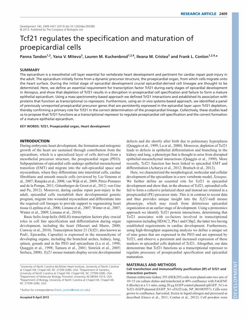


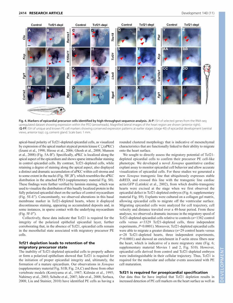


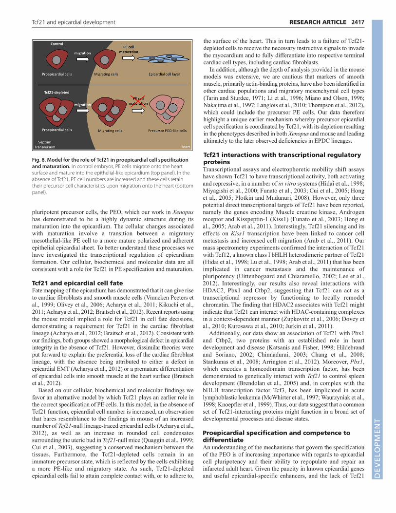


![Supplemental Information Supplemental Experimental Procedures · high-salt buffer (20 mM HEPES-KOH [pH 7.4], 500 mM NaCl, 1 mM EDTA, 0.5% NP-40), and once with 20 mM HEPES-KOH, pH](https://static.documents.pub/doc/80x56/5f291d72048adb291e413862/supplemental-information-supplemental-experimental-procedures-high-salt-buffer-20.jpg)


