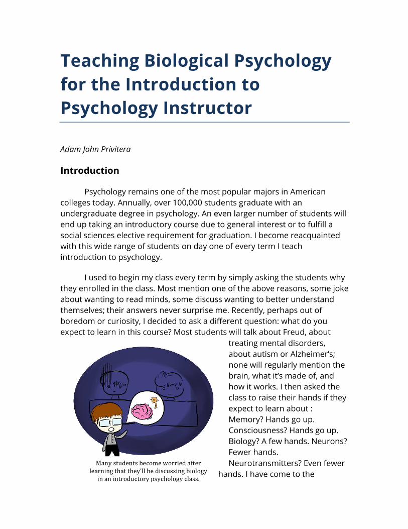
Many students become worried after learning that they’ll be discussing biology in an introductory psychology class. Teaching Biological Psychology for the Introduction to Psychology Instructor Adam John Privitera Introduction Psychology remains one of the most popular majors in American colleges today. Annually, over 100,000 students graduate with an undergraduate degree in psychology. An even larger number of students will end up taking an introductory course due to general interest or to fulfill a social sciences elective requirement for graduation. I become reacquainted with this wide range of students on day one of every term I teach introduction to psychology. I used to begin my class every term by simply asking the students why they enrolled in the class. Most mention one of the above reasons, some joke about wanting to read minds, some discuss wanting to better understand themselves; their answers never surprise me. Recently, perhaps out of boredom or curiosity, I decided to ask a different question: what do you expect to learn in this course? Most students will talk about Freud, about treating mental disorders, about autism or Alzheimer’s; none will regularly mention the brain, what it’s made of, and how it works. I then asked the class to raise their hands if they expect to learn about : Memory? Hands go up. Consciousness? Hands go up. Biology? A few hands. Neurons? Fewer hands. Neurotransmitters? Even fewer hands. I have come to the
Teaching Biological Psychology for the Introduction to Psychology Instructor
Adam John Privitera Introduction
Psychology remains one of the most popular majors in American
colleges today. Annually, over 100,000 students graduate with an undergraduate degree in psychology. An even larger number of students will end up taking an introductory course due to general interest or to fulfill a social sciences elective requirement for graduation. I become reacquainted with this wide range of students on day one of every term I teach introduction to psychology.
I used to begin my class every term by simply asking the students why they enrolled in the class. Most mention one of the above reasons, some joke about wanting to read minds, some discuss wanting to better understand themselves;their answers never surprise me. Recently, perhaps out of boredom or curiosity, I decided to ask a different question: what do you expect to learn in this course? Most students will talk about Freud, about
treating mental disorders, about autism or Alzheimer’s; none will regularly mention the brain, what it’s made of, and how it works. I then asked the class to raise their hands if they expect to learn about : Memory? Hands go up. Consciousness? Hands go up. Biology? A few hands. Neurons? Fewer hands. Neurotransmitters? Even fewer
hands. I have come to the
conclusion that the typical introduction to psychology student has no idea how important a discussion of biology is to this topic and because of this, they are not prepared.
Why aren’t students thinking about the biology of the brain when they take a psychology course? My suspicion is that they have spent their entire life being exposed to the stereotypes of what psychology supposedly is, not what it actually is. It is a huge wakeup call for an undergraduate to hear that part of studying human behavior and mental processes is learning about the organ that creates behavior and mental processes. Truly, the prospect of learning biology in a social sciences course scares the hell out of them.
Students are not alone in their fear of biology in a psychology course.
Regularly, I meet faculty that are openly uncomfortable discussing the biological aspects of behavior and mental processes during an introductory course. I’ve met some that completely skip this section of the course! Why is that? Many instructors are simply not comfortable covering a topic they didn’t explore in depth during their own graduate training. It seems to be the case that most of the anxiety associated with covering this topic in an introductory psychology class comes from one of two general misconceptions: 1. A discussion of the biological basis of behavior and mental processes is an optional topic during an introductory course.
2. An instructor needs to be trained extensively in biological psychology in order to cover the topic in an introductory class.
Given the huge shift in research focus over the last few decades toward a more “hard-science” form of psychology, especially after the “decade of the brain”, it is no longer the case that a discussion of biological psychology is an optional topic. Truly, it was never an optional topic. Without a discussion of biological psychology students would leave with a tremendous knowledge gap that is crucial for success in later courses, degree programs, and careers. Additionally, students would lose out on crucial knowledge and experience needed to critically evaluate the myriad articles that present biological psychological research in a scientifically inaccurate way. Many instructors can think of at least one article from the Internet mentioned by a student that made them cringe.
Instructor Qualifications
Regarding qualification to teach this topic, the ability to discuss biological psychology at a level that is appropriate in an introductory course simply requires that an instructor be comfortable with the very basic tenets of the field. I look to the Society for Neuroscience for guidance on what the main learning objectives of a discussion of biological psychology should be. Some of the proposed learning objectives include understanding the following:
1. The brain is the body’s most complex organ 2. Communication within the brain is electrical and chemical 3. Experience changes the nervous system
An instructor need not teach themselves an entire new field in order
to cover a chapter in an introductory course as this topic can be framed within the instructor’s field of expertise. It is expected that an instructor reviews current research trends in order to update their course content, but this does not constitute a complete relearning of what was covered during graduate school. Simply put, you are renewing your knowledge about biological psychology in a way that can fit into your area of expertise. Clinical background? Discuss how drugs can be used to treat mental disorders by manipulating the chemicals the brain uses in communication. Social psychologist? Why not frame a discussion about experience changing the nervous system around the negative effects of social isolation? Neuroscientist? You’ve likely already infused biological psychology into every topic discussed in an introductory course. Bravo.
Once it is clear that a baseline level of knowledge has been established and updated, the instructor should spend their time describing these complex processes in a way that is relatable for the student, and in a language they can understand. In any introductory course there is a new vocabulary that students must learn in order to discuss the topics appropriately. While this is no different in psychology, a number of terms that are used when describing biological psychology are very intuitive and are easy to get the hang of after a little bit of practice. Additionally, there are many terms that aren’t truly needed to introduce in order for the broader concepts to be discussed and mastered. Providing an appropriate amount of new vocabulary is a good first step before introducing a framework in which the students can use and master these new terms.
In order to accommodate this excursion into uncharted territory I encourage the use of a short preparatory writing assignment in which students are given a list of key terms and asked to define them using their own words. The assignment can be as simple as the following: To prepare for our discussion of biological psychology I would like you to conduct some background research (online and in your book) on specific areas of focus we will cover in class. Please briefly summarize the following topics in your own words.
1. The neuron.
2. How communication occurs within a neuron.
3. How communication occurs between neurons.
4. The subdivisions of the nervous system and what they do (e.g. CNS, PNS, ANS, etc.).
5. Pick one area of the brain (e.g. hippocampus) and describe what it is involved in.
Reliably, a good chunk of these assignments will have huge flaws in
the use of terminology, descriptions of processes, and in demonstrating a clear understanding of how brain and behavior are connected. This should be expected; this is why these students are taking your class! However, you
have just done yourself and your students a huge favor: they have just engaged with a number of new concepts and terms that will not be new to them during the subsequent class discussion.
Anyone can cut and paste a definition from a book or a website but this does nothing to help in the understanding of new vocabulary. It is much easier to work within the mind of the individual student so they can frame things in a way that makes sense to them. Normally, I have students share their definitions in class so that everyone gets exposed to a range of perspectives on this new, scary vocabulary. I’ve learned that I can describe something like a neurotransmitter five different ways and still have one or two students not understand what it is. Students, in my experience, are the best translators of complex terms into their own language. Why not use that to your advantage?
After students are tracking on new terminology, it’s time to begin the difficult task of describing what these things do and, more importantly, how they all interact in a way to create behavior and mental processes. Luckily, the majority of introductory topics in biological psychology can be easily demonstrated during class time in a way that is interactive, educational, and fun! Here are some of the most important topics to cover and strategies to help students understand them. Structure of the Neuron
One of the biggest misconceptions I address immediately when discussing biological psychology relates to the structure of the neuron. Most students I encounter believe that neurons are plastic, rigid structures that are inflexible. Part of this might be related to a discussion of cytoskeletal structure, which could prime thoughts of bones found elsewhere in the body. I blame the way in which neurons are depicted in popular textbooks for this misconception. While some books do discuss the bi-lipid membrane nature of the neuron, very few will describe what lipids are like. They aren’t plastic. They aren’t rigid. I find that illustrating how lipids interact with water can give the students a more realistic understanding of what the neuron is like structurally. While it might not be the most scientifically accurate way of depicting this interaction, using a pipette or straw to inject a drop of cooking oil in a bottle or glass of water can show students what happens when a lipid based solution is added into an environment filled with water. Additionally,
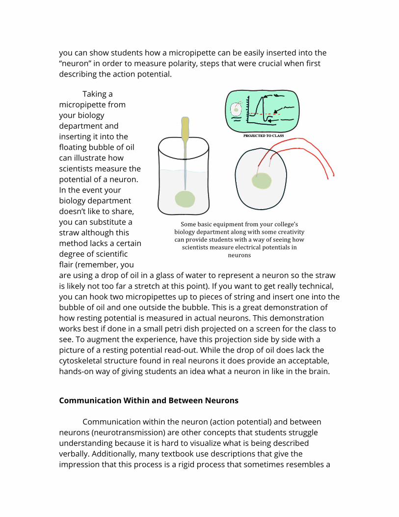
you can show students how a micropipette can be easily inserted into the “neuron” in order to measure polarity, steps that were crucial when first describing the action potential.
Taking a micropipette from your biology department and inserting it into the floating bubble of oil can illustrate how scientists measure the potential of a neuron. In the event your biology department doesn’t like to share, you can substitute a straw although this method lacks a certain degree of scientific flair (remember, you are using a drop of oil in a glass of water to represent a neuron so the straw is likely not too far a stretch at this point). If you want to get really technical, you can hook two micropipettes up to pieces of string and insert one into the bubble of oil and one outside the bubble. This is a great demonstration of how resting potential is measured in actual neurons. This demonstration works best if done in a small petri dish projected on a screen for the class to see. To augment the experience, have this projection side by side with a picture of a resting potential read-out. While the drop of oil does lack thecytoskeletal structure found in real neurons it does provide an acceptable, hands-on way of giving students an idea what a neuron in like in the brain.Communication Within and Between Neurons Communication within the neuron (action potential) and between neurons (neurotransmission) are other concepts that students struggle understanding because it is hard to visualize what is being described verbally. Additionally, many textbook use descriptions that give the impression that this process is a rigid process that sometimes resembles a
key being inserted into a lock. These are lipids and proteins! There is no solid, rigid lock and key to be found! A helpful demonstration can help remove some of these misconceptions and really drive home what the process is like.
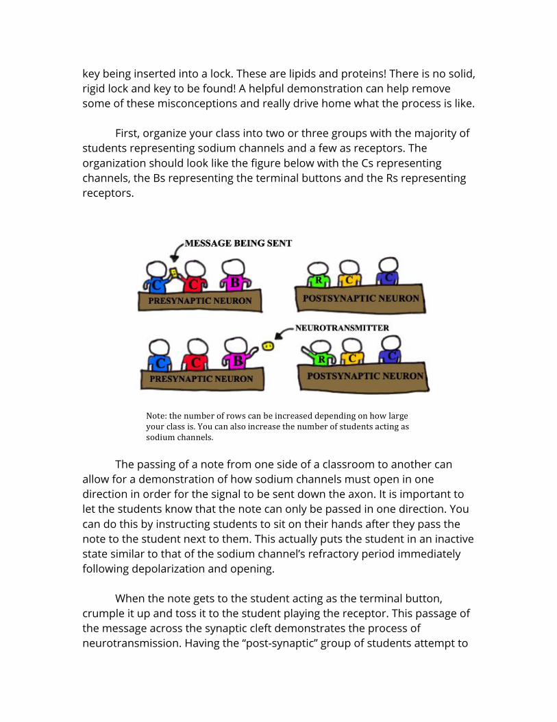
First, organize your class into two or three groups with the majority of students representing sodium channels and a few as receptors. The organization should look like the figure below with the Cs representing channels, the Bs representing the terminal buttons and the Rs representing receptors.
The passing of a note from one side of a classroom to another can allow for a demonstration of how sodium channels must open in one direction in order for the signal to be sent down the axon. It is important to let the students know that the note can only be passed in one direction. You can do this by instructing students to sit on their hands after they pass the note to the student next to them. This actually puts the student in an inactive state similar to that of the sodium channel’s refractory period immediately following depolarization and opening.
When the note gets to the student acting as the terminal button, crumple it up and toss it to the student playing the receptor. This passage of the message across the synaptic cleft demonstrates the process of neurotransmission. Having the “post-synaptic” group of students attempt to
catch the crumpled up note with their hands fixed into a fist, an open hand that can’t close, and other positions can illustrate not only that the receptors need to be specialized, but that the receptors and neurotransmitters need to be able to mold to one another. Remember, these are globular proteins. Neurotransmission is like a handshake between members of the same species, not really like a lock and key.
This demonstration can be changed slightly to include coverage of inhibitory postsynaptic potentials (IPSPs) and excitatory postsynaptic potentials (EPSPs). Written on the note that is being passed from one side of the classroom to the other can be the name of a known excitatory (e.g. glutamate) or inhibitory (e.g. GABA) neurotransmitter. It is then up to the student who is playing the role of the first channel whether or not they are going to open or do nothing. This decision-making element continues to the next group of students after the throwing of the note to the student playing the role of the receptor. What will that student tell the student sodium channel next to them to do? Open? Do nothing? This will be up to the student to decide based on the neurotransmitter they are told they are receiving. If you really want to make things interesting, you can say that the students are located on neurons in a specific region of the brain and then ask them to predict what changes in behavior or mental processes might occur as a consequence of the activity they acted out.
The first question you likely have as an instructor after this demonstration is, “did my students get it?” I used to give a short, formative assessment after this demonstration in the form of a short quiz. I quickly learned that this is a great way to assess whether or not students are good at taking surprise quizzes. Recently, I began having a discussion with the class centered on the following, or similar, questions:
• To the students who were sodium channels: what role did you play?
What was your job? • What was the passing of the note meant to represent? • Why could you only pass the note in one direction? • What happened when the note got to the end of the row of
“channels”? • To the students who were receptors: what role did you play? What was
your job? • What did it mean when you caught the crumbled up note?
• What happened when you were forced to hold your hand in a rigid shape?
Additional questions can be added depending on how many different
concepts you add to this demonstration. Ask the students to explain why they opened their channels or did nothing. Have them explain why they predicted a certain behavioral change would occur. Ask the students playing receptors why they told the student sodium channels next to them to open or do nothing. This normally provides a very interesting conversation that will allow you to see if any of this made sense to your students. You can end this activity by showing a video of the action potential and neurotransmission in a computer generated neuron and framing a discussion around how what was shown in the video relates to the class activity. The Role Myelin Plays in the Action Potential
An effective way to illustrate this concept in any size class is by having the students engage in a modified “wave”. Covering this after introducing how the action potential works will allow for the students to make the connection between their movement “up” in the wave and the opening of a sodium channel during the propagation of an action potential. Having one student keep track of how long it takes for the wave to move from one side of the class to another will give them a quantifiable thing to compare with the “myelinated” example.
In order to do this demonstration, select a student that has a stopwatch or cellphone with stopwatch capabilities and have them come to the front of the room. It is important that the student be able to see the entire class. After this, explain to the students that they are going to be performing “the wave” in class. For those that are unfamiliar, “the wave” is a popular activity at sporting events. During this activity a column of spectators will all stand up and lift their arms at around the same time.
Upon seeing this, the column of people seated next to this group will lift their arms and stand. The process is repeated until movement has gone from one side of a stadium to the other. The movement, when performed correctly, gives off the appearance of a wave. This is a fitting demonstration as the movement of electrical current down the axon of a neuron can be described as being wavelike. However, this wave will be slightly modified to illustrate an important concept with the action potential: the refractory period. Students will stand and lift their arms but will be instructed to sit on their hands after the completion of their movement. The student “channels” are in a refractory state and are unable to stand and lift their arms again. This illustrates two points:
• The action potential can only travel in one direction; and • Sodium channels close and cannot reopen during their refractory
state.
After the class is on the same page about what is expected of them, tell the first column of students to begin on your signal. It is helpful to use a countdown to ensure that the timekeeper knows exactly when to start the clock. I would do a practice wave before actually recording the times; students will reliably botch their first attempt. After the class completes the first round, write the first time on the board and repeat the process two more times; use the average of the three scores.
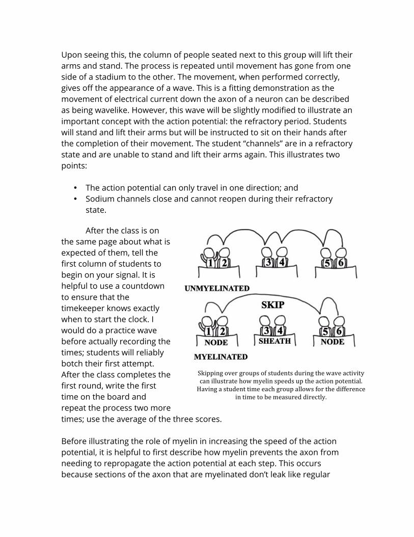
Before illustrating the role of myelin in increasing the speed of the action potential, it is helpful to first describe how myelin prevents the axon from needing to repropagate the action potential at each step. This occurs because sections of the axon that are myelinated don’t leak like regular
sections. Additionally, there are no sodium channels in the membrane of axon sections that are myelinated. This demonstration is essentially the same as the previous with one difference: “the wave” will be skipping over a few columns of students at a time. Normally, I will tell the first, fourth, and seventh columns of students to participate in “the wave” as though the columns in-between them didn’t exist. This can be adjusted depending on the size of the class you are teaching. To reduce the likelihood thatstudents willaccidentally participate, I have those columns of students lay their arms flat on their desk. Again, having a student time this can give you a measurement to compare with the first time. I suggest using an average of three measurements as well. Reliably, the example with fewer columns (the myelinated axon) will take less time because fewer “sodium channels” have to open. This is the way in which myelin increases the speed of the action potential.
Before moving on, I have a discussion with the class centered on a few main points to check that this demonstration achieved the desired goals:
• What did it represent when you stood up during “the wave”? • What ion was allowed to enter the neuron when your channel was
open? • What happened to the channel next to you after your channel opened? • What happened to you after you stood up during “the wave”? What did
it represent? • Could the wave move in more than one direction? Why/why not? • What happened when the axon was myelinated? Why did this happen?
During this discussion students will be given a chance to explain what
makes sense to them and what doesn’t. In my experience, I don’t end up doing a lot of teaching during this discussion because the students who understand the example are very willing to speak up and describe how it makes sense to them; many students enjoy helping each other. By the end, most students report being comfortable with the idea of how the action potential moves down an axon and how myelination increases the speed. Development of the Nervous System
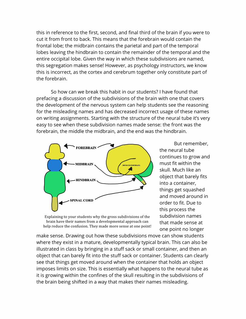
Most textbooks will describe the brain using three conventional subdivisions: forebrain, midbrain and hindbrain. Most students assume that
this in reference to the first, second, and final third of the brain if you were to cut it from front to back. This means that the forebrain would contain the frontal lobe; the midbrain contains the parietal and part of the temporal lobes leaving the hindbrain to contain the remainder of the temporal and the entire occipital lobe. Given the way in which these subdivisions are named, this segregation makes sense! However, as psychology instructors, we know this is incorrect, as the cortex and cerebrum together only constitute part of the forebrain.
So how can we break this habit in our students? I have found that prefacing a discussion of the subdivisions of the brain with one that covers the development of the nervous system can help students see the reasoning for the misleading names and has decreased incorrect usage of these names on writing assignments. Starting with the structure of the neural tube it’s very easy to see when these subdivision names made sense: the front was the forebrain, the middle the midbrain, and theend was the hindbrain.
But remember,
the neural tube continues to grow and must fit within the skull. Much like an object that barely fits into a container, things get squashed and moved around in order to fit. Due to this process the subdivision names that made sense at one point no longer
make sense. Drawing out how these subdivisions move can show students where they exist in a mature, developmentally typical brain. This can also be illustrated in class by bringing in a stuff sack or small container, and then an object that canbarely fit into the stuff sack or container. Students can clearly see that things get moved around when the container that holds an object imposes limits on size. This is essentially what happens to the neural tube as it is growing within the confines of the skull resulting in the subdivisions of the brain being shifted in a way that makes their names misleading.
Following the discussion of brain subdivisions up with a few questions can allow you to informally assess whether or not your students are on the same page. I find it helpful to just ask students to explain what subdivision a random brain region belongs to and to have the students articulate why this is the case. Normally, seeing the neural tube fold in order to fit into the skull is sufficient to break down this misconception.Neuroanatomy and Brain Function
Now that we have gotten over the misleading names of the subdivisions of the brain, it is possible to open a discussion about different areas in the brain and their function. It is important to avoid an approach that leads the student down a path believing that the brain is made of distinct, independent “centers” that are specialized to do a single function. While it is true that there is a degree of specialization for individual brain regions, it is not absolute.
A great example of this emerges when discussing the fusiform face area (FFA). It is tempting to assign every aspect of facial recognition to this one region; this is what students tend to do. How soon they forget about the large collection of brain regions involved in the processing of visual information at a very basic level. Describing an area like this requires a discussion of the other regions that coordinate to provide the needed information to this structure. While it isn’t the case that the instructor needs to be able to articulate the specific role that each region plays in the creation of complex visual perceptions, it is important that an attempt be made to solidify an understanding of how interconnected the brain is and how many regions interact to produce behavior and mental processes.
A helpful metaphor to illustrate how information is processed in the brain involves describing a car going from one point to another on a map. Cars cannot teleport from one place to another automatically, they need to drive on roads, across bridges, through tunnels, and eventually they get to their destination. If the car represents the image of your best friend’s face processed by the retina in the back of the eye, and a random location on the map represents where that information needs to go in order for it to be processed, students can clearly see that a number of different steps are involved outside of just the visual information (the car) and the FFA (the car’s destination). Will the car be able to get to its destination if a road it needs to
cross is out? No. What about if a bridge is out? No. What about if a tunnel the car doesn’t need to go through is out? Yes. In order for the car to get to its destination, all essential paths need to be intact. However, non-essential paths can be closed without impacting the car at all. This is the same for the brain. In order for visual information to go from the retina to the FFA it must pass through a number of interconnected regions until it gets to its final destination. If any of those paths are cut, the transmission of information is impacted just like if a bridge a car needs to cross is out.
Just like the initial framing of a discussion of biological psychology can be within the realm of an instructor’s area of expertise, introducing neuroanatomy can involve sticking to your strengths. The important things to discuss are less related to any specific region of the brain and more about the general way the brain coordinates and integrates signals across many regions and circuits. It’s never a good idea to overload a student with different brain regions during an introductory course. Focusing on developing an understanding of how any specific region works as part of an integrated whole can prepare the student to apply what they have learned to any region of the brain that is brought up during a later discussion or course. Behavioral Genetics
A topic that has somewhat recently been introduced into the biological psychology section of introductory textbooks is a section on behavioral genetics. This is another somewhat recent hot topic in light modern advances in the ability to gather genetic information about oneself (e.g. 23andme, etc.). Similar to the popular “science” articles covering breakthroughs in biological psychology research, the role that genes play in behavior tends to be greatly exaggerated by the media.
Regularly, students will ask me about the “cheating” gene, or the “aggression” gene, or some other gene that purportedly controls an entire behavior. Truthfully, as a student, I at one point assumed this is how genes worked because of how often I read about them working in this manner. Unfortunately, as we all know, this is not how geneswork.
As a psychology instructor, you might be the first and perhaps only exposure to the central dogma of molecular biology that a student encounters in college. A brief discussion of this topic can really drive a wedge between genes and behavior for students. While a discussion of DNA and RNA might throw a wrench in an otherwise stress-free discussion, an instructor can make a clear distinction between genes and behavior using a less scary approach. A past instructor of mine, Dr. Mark Kristal, used three simple statements in order to illustrate the role that genes play in behavior:
1. Genes have a profound effect on behavior. 2. Genes do not have a direct effect on behavior. 3. Genes do not have an exclusive effect on behavior.
Regardless of whether or not a student understands the central dogma these statements give them a very clear understanding of how genes influence behavior. It isn’t the case that there is a single gene for any behavior. Behavior is far too complicated. But, there are single genes that can influence behavior tremendously, although indirectly. Students need to remember that there are a number of steps that exist between gene and protein so logically, there are even more steps between gene and behavior.
Another misunderstanding students have has to do with the somewhat recently expanding field of epigenetics. Most students I encounter assume that all genes are always on, all the time. This ignores the huge body of research investigating environmental factors that influence the expression of genes. The way I describe genes in my class uses the analogy of a light switch. Lights are not always on or off. If I want a light to be on I can simply flip a switch with my hand in order to turn it on. I can do that same thing and have the light turn off as well. In this analogy my hand acts as an environmental trigger that can influence gene expression. This trigger has the potential to turn on or up-regulate (turning on the light) or turn off or down-regulate (turning off the light) the expression of a gene.
There are a number of fun studies using transgenic, bioluminescent animals that can be used to illustrate how genes are not always on or off. I cover this in class by discussing the work of Contag et al. (1997) which evaluated substrate activated luciferase expression in mice. When the gene associated with this photoprotein was turned on, it would glow under certain wavelengths of light. What this paper showed is that the lungs of the transgenic mice would luminesce only when the mouse had the specific gene and was injected with a certain chemical signal; having the gene alone was necessary but not sufficient for the expression of the bioluminescent photoprotein. Not only is it the case that environmental factors can influence genes on the short term, but processes such as methylation have the potential to influence genes in a manner than is heritable. In Closing
Biological psychology is easily the most intimidating branch of psychology for both students and instructors to tackle. However, it doesn’t take a neuroscientist to teach a classroom of undergraduates the basics of this topic in a way that is appropriate for an introductory course. Remember, your introduction to psychology class might be the only exposure some of these students ever get to the field. Given the rising tide of inaccurately represented biological psychology research findings presented in the popular media, it is more important than ever to ensure that students are provided with an opportunity to have their assumptions about the nervous system challenged and to explore this fascinating topic in a way that cultivates their scientific critical thinking. By putting these complex concepts into a language that students can understand, and by providing examples and demonstrations that can add a hands-on component to these discussions, both students and faculty will have a more enjoyable experience learning about this fascinating topic. References Contag, C. H., Spilman, S. D., Contag, P. R., Oshiro, M., Eames, B., Dennery, P.,
Stevenson, D. K., & Benaron, D. A. (1997). Visualizing gene expression in living mammals using a bioluminescent reporter. Photochemistry and photobiology, 66(4), 523-531.