55
Techniques Of Harvesting Cartilage Graft For Cartilage Tympanoplasty Dr. Erami M.D. ENT Resident Department Of ENT Shahid Sadoghi Hospital Yazd Iran
| Date post: | 09-Jan-2017 |
| Category: |
Health & Medicine |
| Upload: | mderami |
| View: | 83 times |
| Download: | 0 times |
Techniques Of Harvesting Cartilage Graft For Cartilage Tympanoplasty
Dr. Erami M.D.ENT Resident
Department Of ENTShahid Sadoghi Hospital
Yazd Iran

• Cummings 2015 (141 | TYMPANOPLASTY AND OSSICULOPLASTY)
• The tissue rigidity of cartilage and its resistance to retraction, even in the setting of ongoing eustachian tube dysfunction, has led to the growing acceptance of its use in middle ear reconstruction.
• Some of indications:1. Cartilage grafts placed between the TM and an ossicular
prosthesis decrease extrusion risk and can augment the prosthesis-tissue interface, which allows for better long-term hearing results.
• Cummings 2015 (141 | TYMPANOPLASTY AND OSSICULOPLASTY)
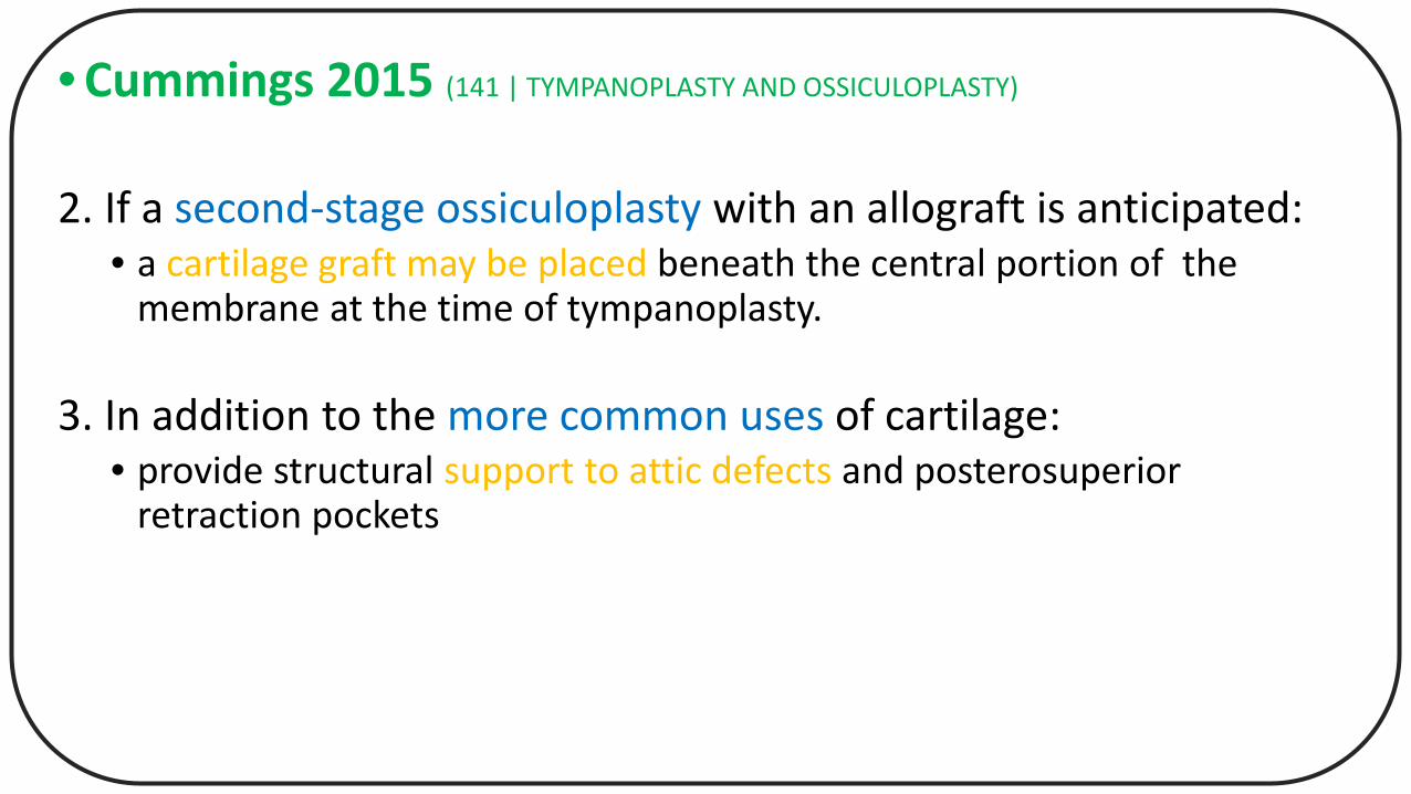
2. If a second-stage ossiculoplasty with an allograft is anticipated:• a cartilage graft may be placed beneath the central portion of the
membrane at the time of tympanoplasty.
3. In addition to the more common uses of cartilage:• provide structural support to attic defects and posterosuperior
retraction pockets
• Cummings 2015 (141 | TYMPANOPLASTY AND OSSICULOPLASTY)
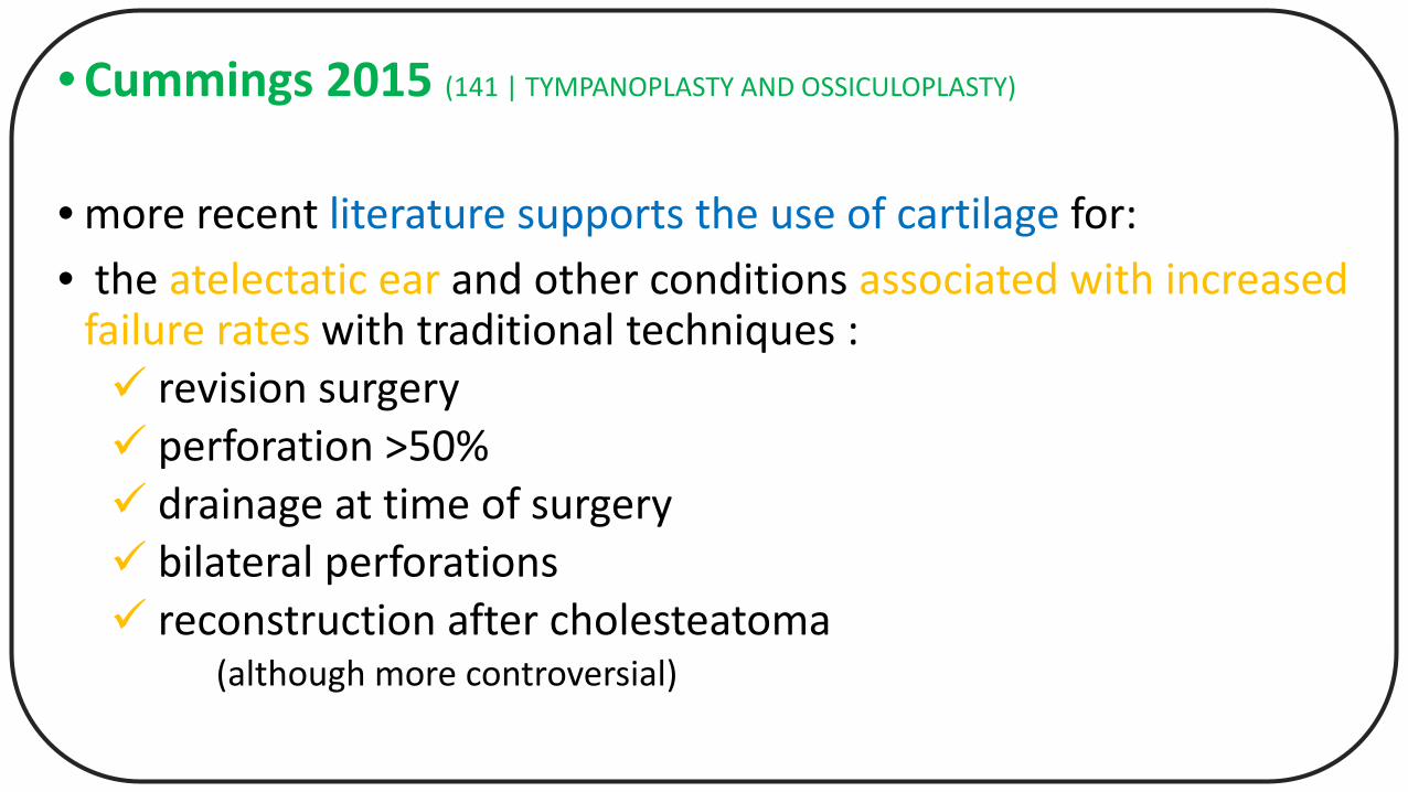
• more recent literature supports the use of cartilage for:• the atelectatic ear and other conditions associated with increased
failure rates with traditional techniques : revision surgery perforation >50% drainage at time of surgery bilateral perforations reconstruction after cholesteatoma
(although more controversial)
• Cummings 2015 (141 | TYMPANOPLASTY AND OSSICULOPLASTY)
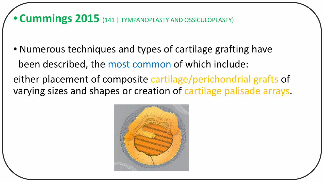
• Numerous techniques and types of cartilage grafting havebeen described, the most common of which include:
either placement of composite cartilage/perichondrial grafts of varying sizes and shapes or creation of cartilage palisade arrays.
• Cummings 2015 (141 | TYMPANOPLASTY AND OSSICULOPLASTY)
• Cartilage may be harvested with its attached perichondriumfrom the tragus or the concha.
• Tragal cartilage than conchal cartilage: • Thicker• flatter
• Tragal cartilage may be :• more suitable for larger perforations
• Cummings 2015 (141 | TYMPANOPLASTY AND OSSICULOPLASTY)
• The cartilaginous graft can be made quite thin and of a small enough diameter to bolster only the weakened portion of the involved TM, or it may be shaped to fill a pantympanicperforation.
• Reducing cartilage thickness optimizes:• the acoustic-transfer properties
• with thicknesses of 500 μm or less resulting in a highly favorable vibratory transfer.
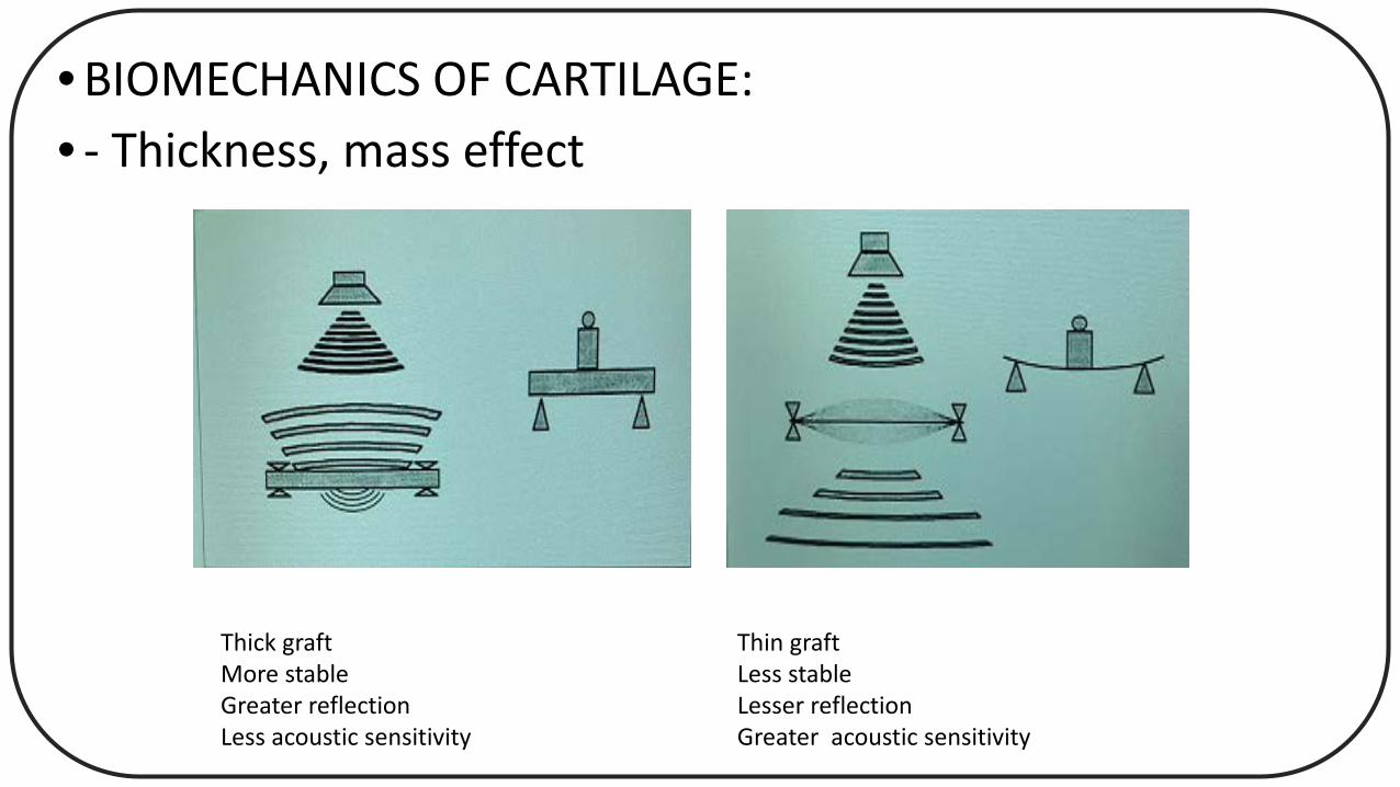
• BIOMECHANICS OF CARTILAGE:• - Thickness, mass effect
Thick graftMore stableGreater reflectionLess acoustic sensitivity
Thin graftLess stableLesser reflection Greater acoustic sensitivity
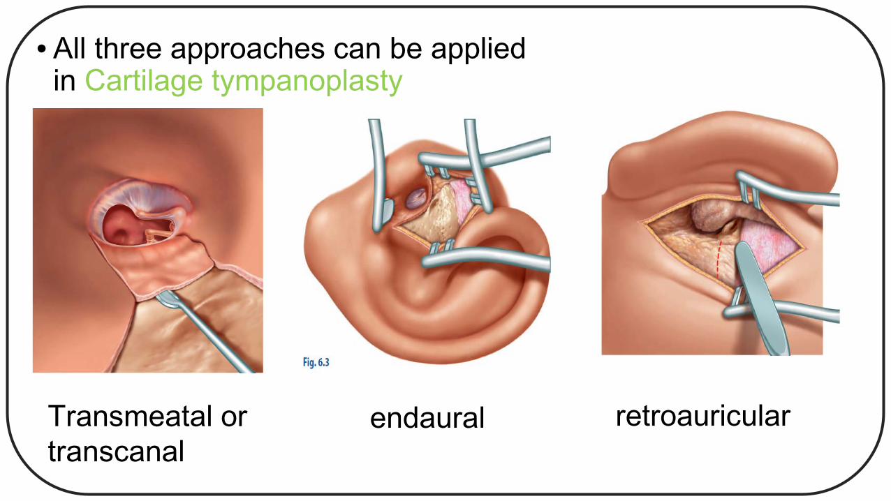
• All three approaches can be appliedin Cartilage tympanoplasty
Transmeatal ortranscanal
endaural retroauricular
• In mastoidectomy, antrotomy, and atticotomy:
• most surgeons will employ a retroauricular approach.
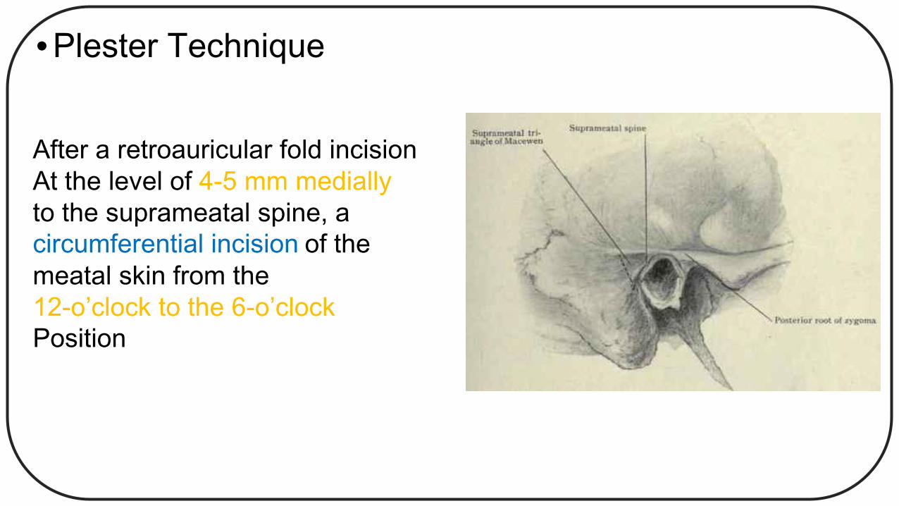
• Plester Technique
After a retroauricular fold incisionAt the level of 4-5 mm medially to the suprameatal spine, a circumferential incision of the meatal skin from the 12-o’clock to the 6-o’clock Position
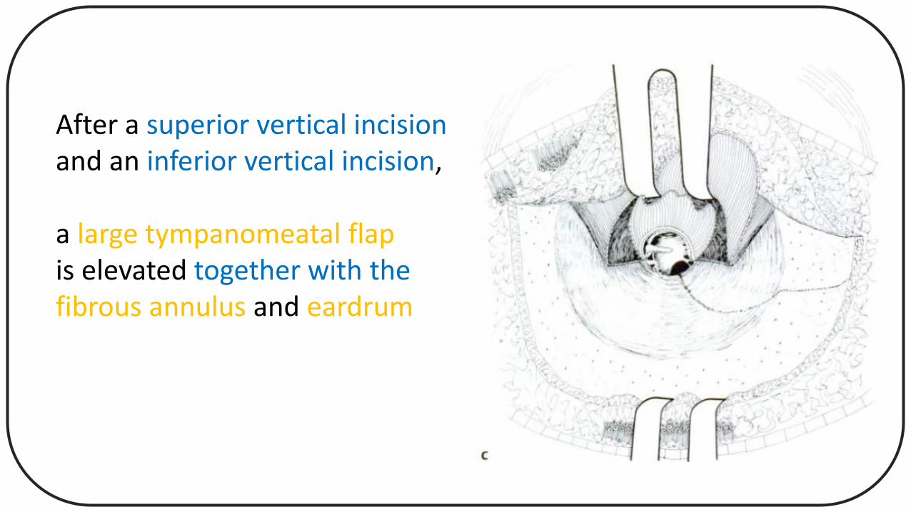
After a superior vertical incision and an inferior vertical incision,
a large tympanomeatal flapis elevated together with thefibrous annulus and eardrum
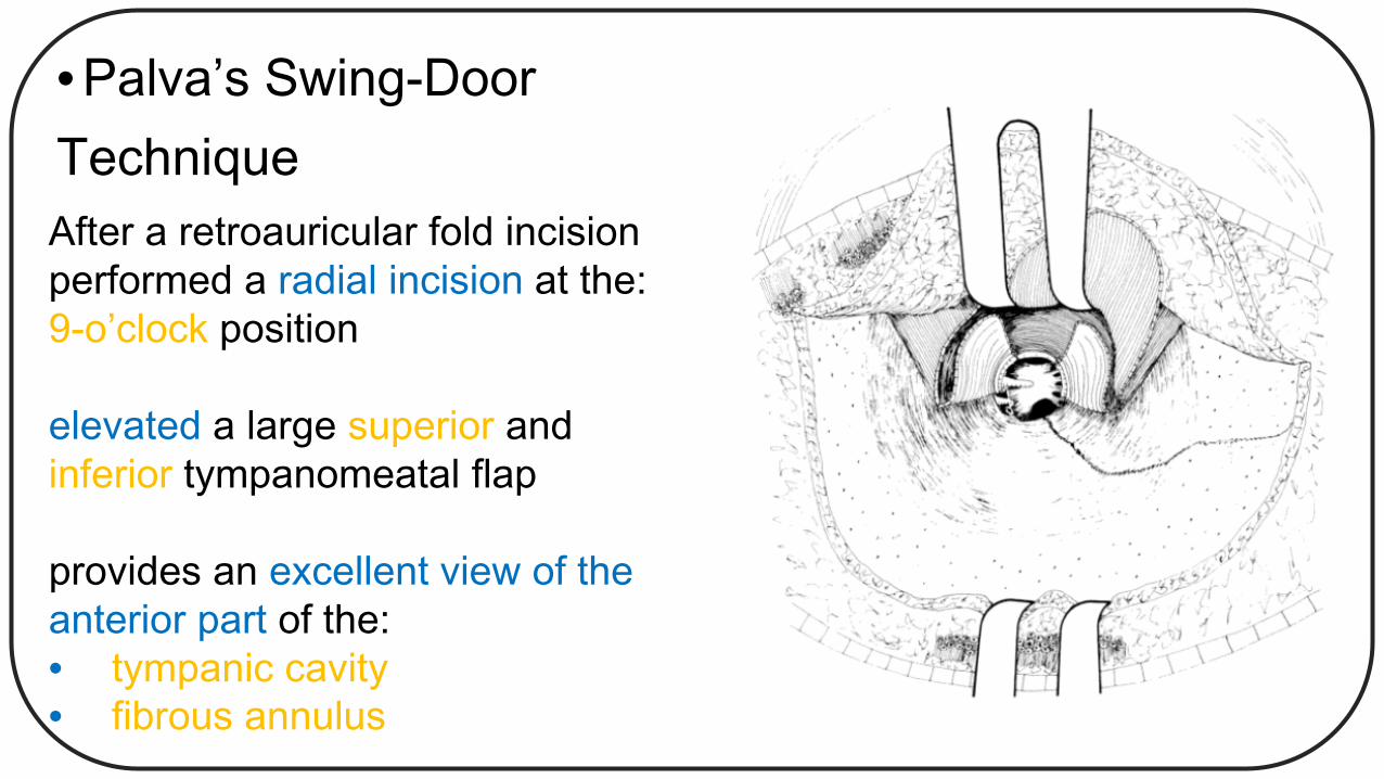
• Palva’s Swing-DoorTechniqueAfter a retroauricular fold incision performed a radial incision at the:9-o’clock position
elevated a large superior andinferior tympanomeatal flap
provides an excellent view of theanterior part of the:• tympanic cavity• fibrous annulus
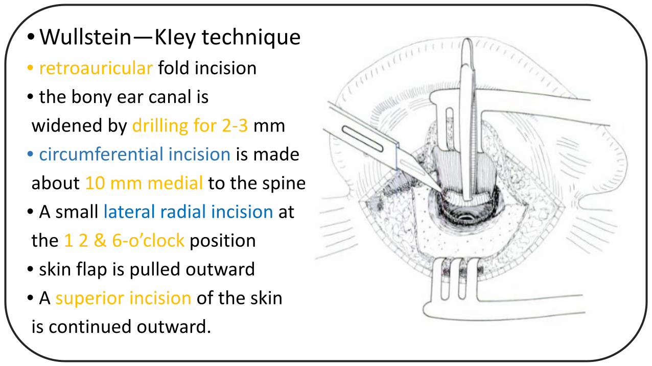
• Wullstein—KIey technique • retroauricular fold incision• the bony ear canal iswidened by drilling for 2-3 mm• circumferential incision is madeabout 10 mm medial to the spine• A small lateral radial incision atthe 1 2 & 6-o’clock position• skin flap is pulled outward• A superior incision of the skinis continued outward.
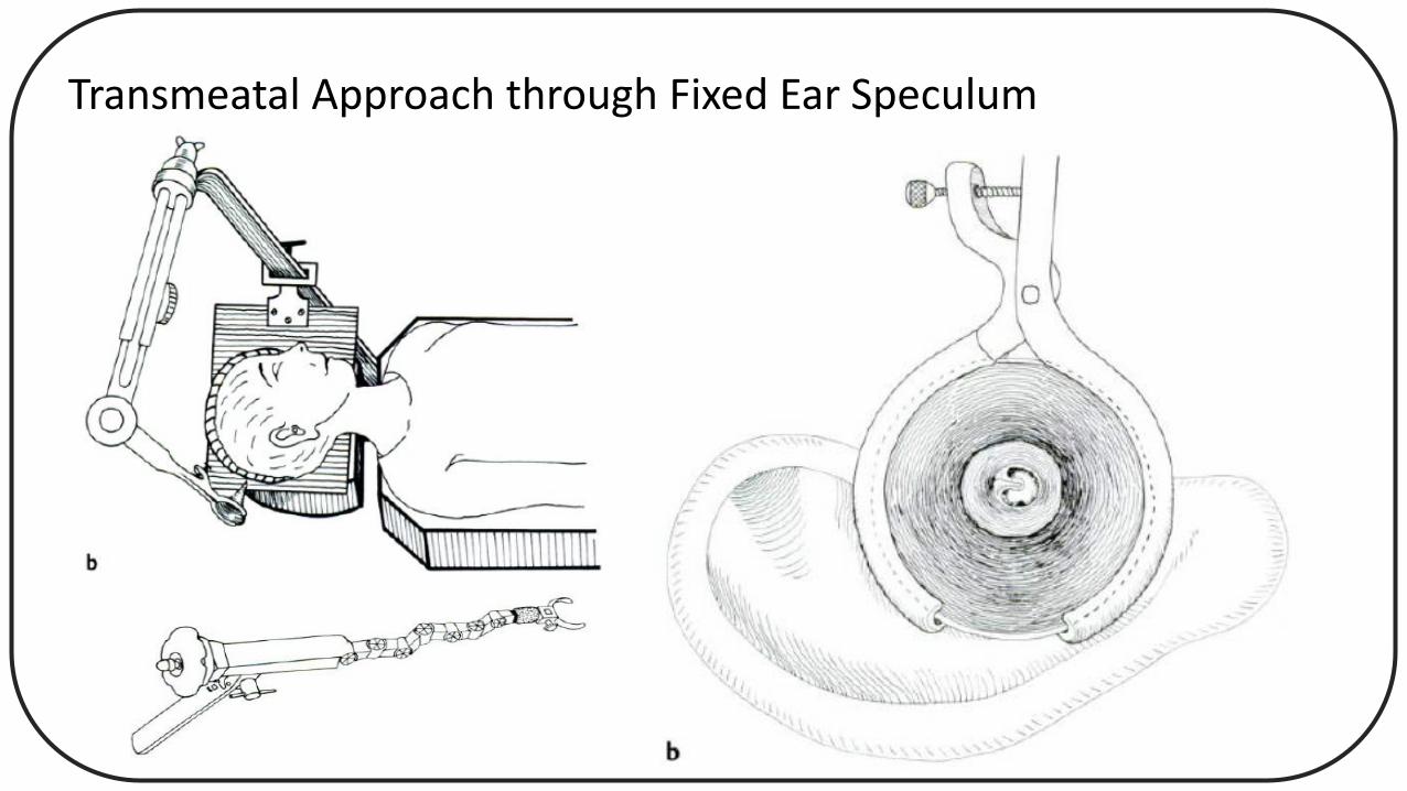
Transmeatal Approach through Fixed Ear Speculum
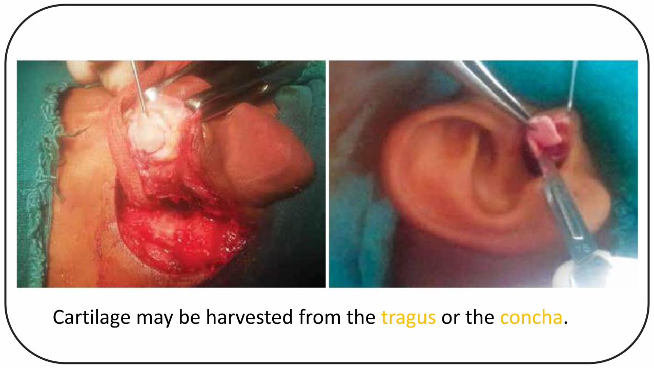
Cartilage may be harvested from the tragus or the concha.
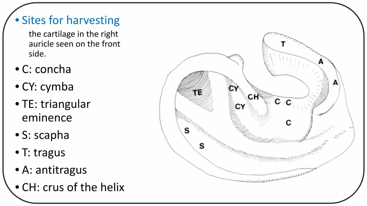
• Sites for harvestingthe cartilage in the right auricle seen on the front side.
• C: concha• CY: cymba• TE: triangular
eminence• S: scapha• T: tragus• A: antitragus• CH: crus of the helix
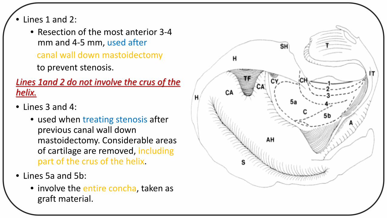
• Lines 1 and 2:• Resection of the most anterior 3-4
mm and 4-5 mm, used aftercanal wall down mastoidectomyto prevent stenosis.
Lines 1and 2 do not involve the crus of the helix.• Lines 3 and 4:
• used when treating stenosis after previous canal wall down mastoidectomy. Considerable areas of cartilage are removed, including part of the crus of the helix.
• Lines 5a and 5b:• involve the entire concha, taken as
graft material.

• Harvesting of Tragal Cartilage:
• Tragal cartilage seems to be used more often than conchal cartilage, mainly because :
• it is harvested along the same route as that of the transcanal and the endaural approaches.
• tragal cartilage is less convex than conchal cartilage.
• For cosmetic reasons, the incision is most often made:• 2-3 mm medial to the tragal dome
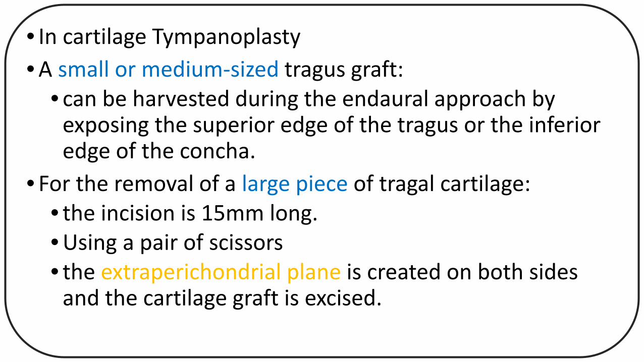
• In cartilage Tympanoplasty • A small or medium-sized tragus graft:
• can be harvested during the endaural approach by exposing the superior edge of the tragus or the inferior edge of the concha.
• For the removal of a large piece of tragal cartilage:• the incision is 15mm long.• Using a pair of scissors• the extraperichondrial plane is created on both sides
and the cartilage graft is excised.

Harvesting a large tragal cartilage graft with a 15 mm incision 2 mm medial to the dome.
The large incision is made in one sweep through the skin and the cartilage with the perichondrium
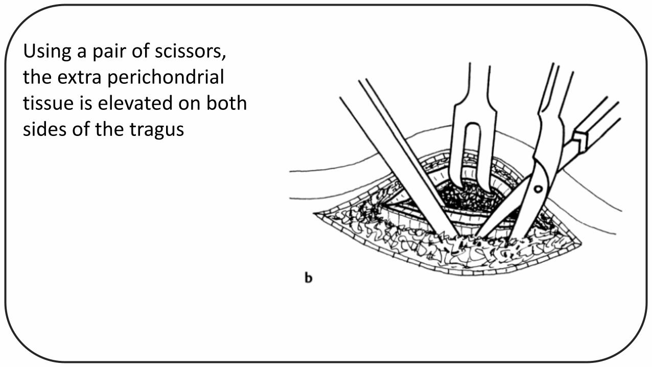
Using a pair of scissors,the extra perichondrialtissue is elevated on both sides of the tragus
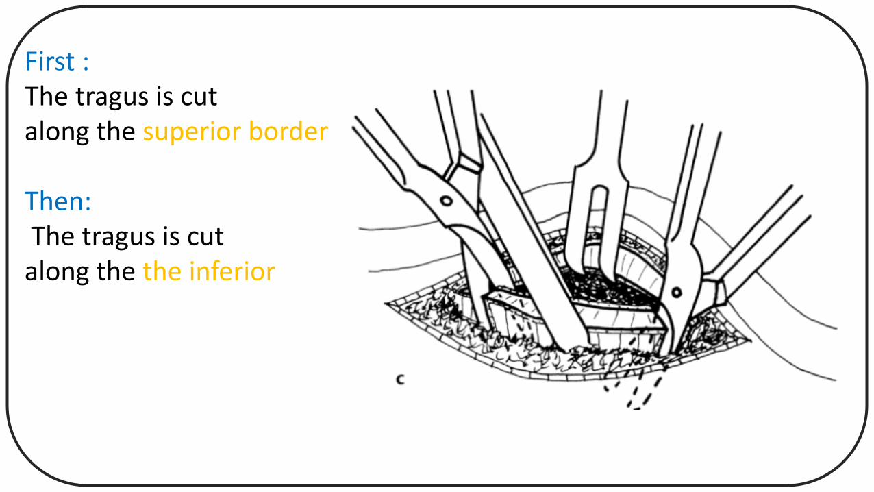
First :The tragus is cutalong the superior border
Then:The tragus is cutalong the the inferior
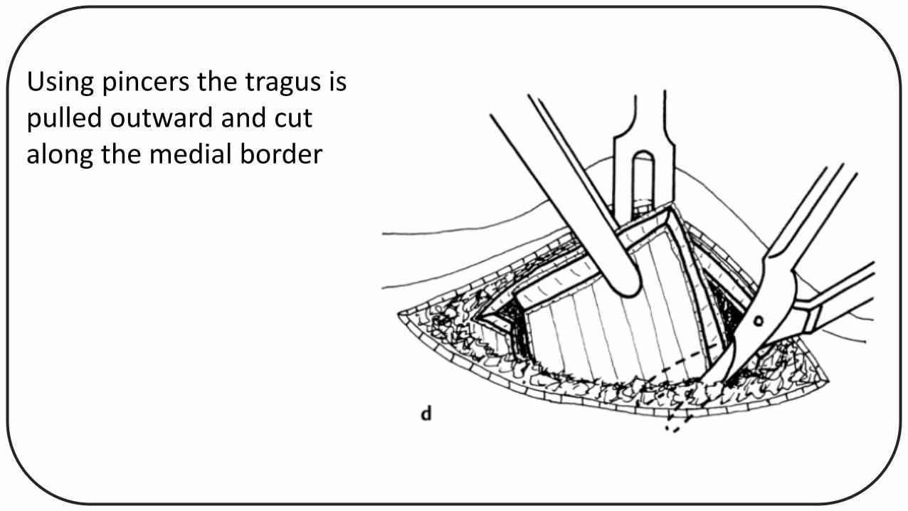
Using pincers the tragus is pulled outward and cut along the medial border

• Harvesting the tragus cartilage:
• Heermann endaural approach (through the intercartilaginousincision)
• The superior edge of the tragus is first separated from the fibrous tissue of the intercartilaginous region
• tragus is grasped and pulled in superior direction
• subcutaneous tissue is elevated from the dome anteriorly and posteriorly
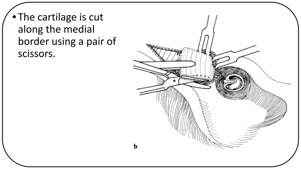
• The cartilage is cut along the medial border using a pair of scissors.
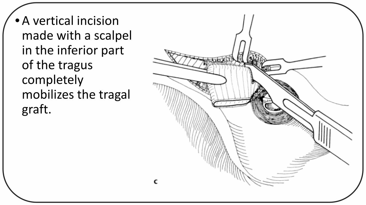
• A vertical incision made with a scalpel in the inferior part of the tragus completely mobilizes the tragal graft.
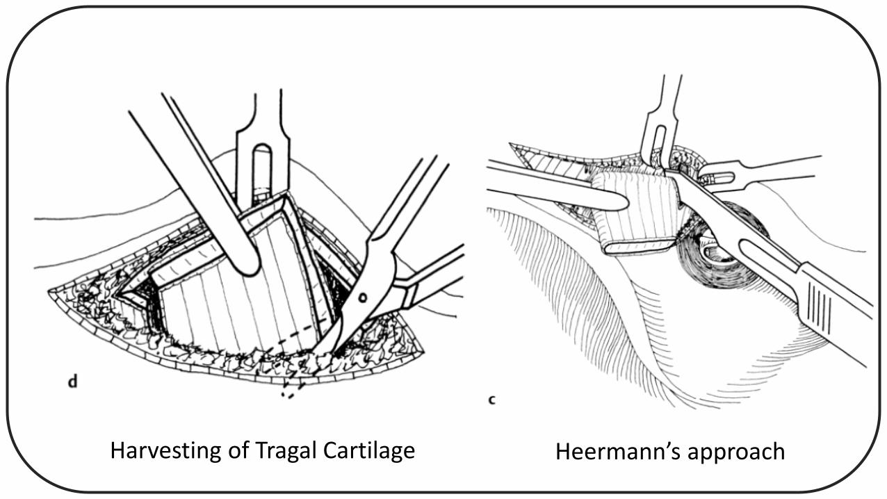
Heermann’s approachHarvesting of Tragal Cartilage
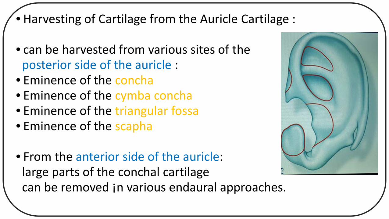
• Harvesting of Cartilage from the Auricle Cartilage :
• can be harvested from various sites of theposterior side of the auricle :
• Eminence of the concha• Eminence of the cymba concha• Eminence of the triangular fossa• Eminence of the scapha
• From the anterior side of the auricle:large parts of the conchal cartilagecan be removed ¡n various endaural approaches.
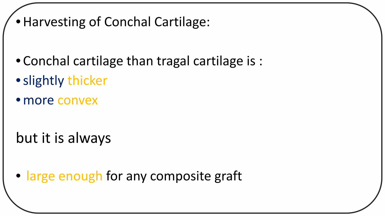
• Harvesting of Conchal Cartilage:
• Conchal cartilage than tragal cartilage is :• slightly thicker • more convex
but it is always
• large enough for any composite graft
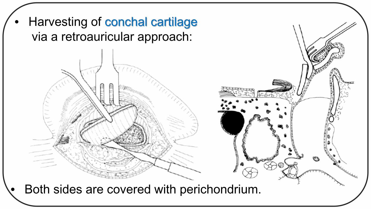
• Harvesting of conchal cartilagevia a retroauricular approach:
• Both sides are covered with perichondrium.

• Harvesting of the cymba cartilage and the fossa triangulariscartilage:
• Incision of the skin is:slightly superior to theeminence of the concha.
• Subcutaneous tissue:is elevated
• The perichondrium is exposed( of the superior part of theeminence of the concha andof the cymba cartilage).
• With a circular incision:The most convex part—the cymba cartilage—is cut and removed.
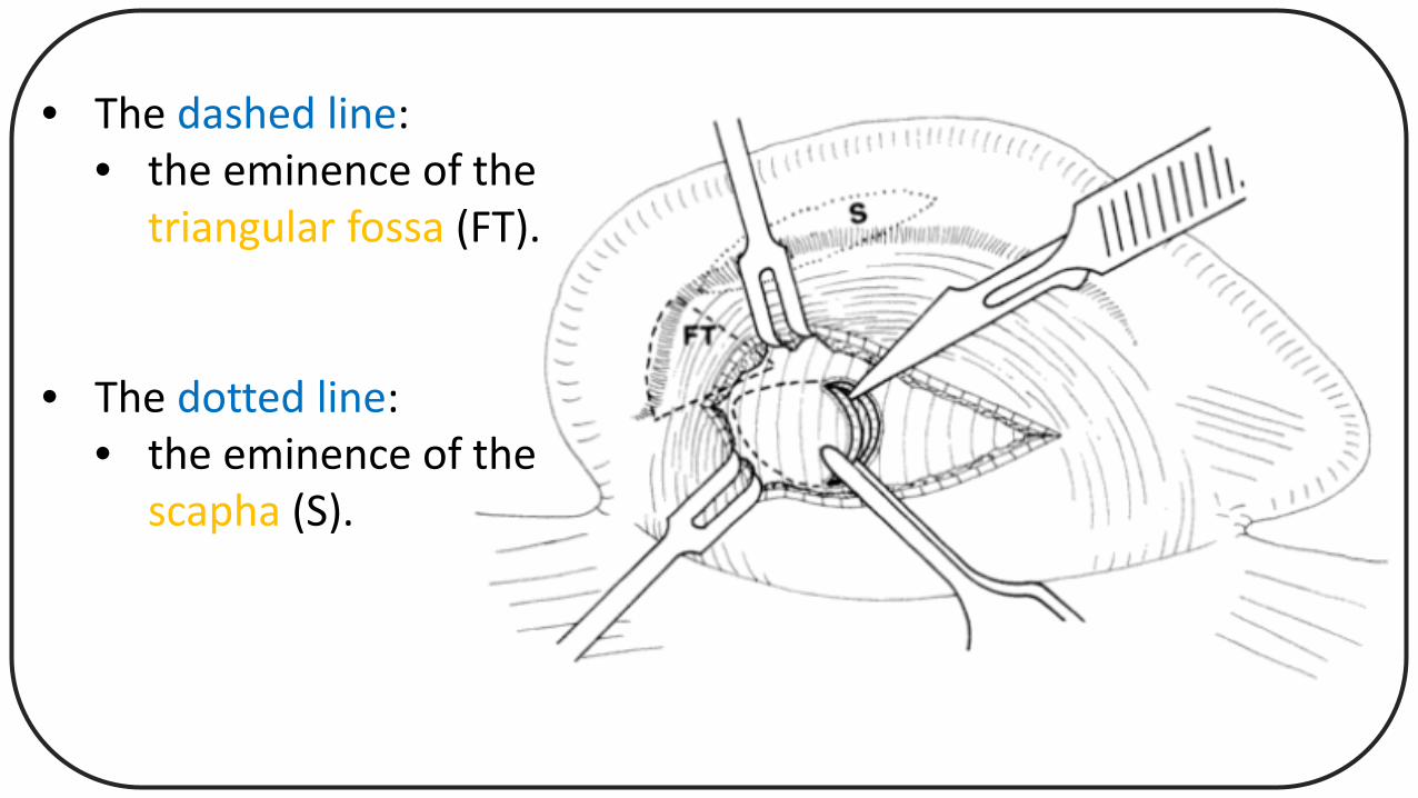
• The dashed line:• the eminence of the
triangular fossa (FT).
• The dotted line:• the eminence of the
scapha (S).
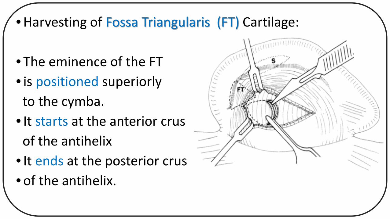
• Harvesting of Fossa Triangularis (FT) Cartilage:
• The eminence of the FT • is positioned superiorlyto the cymba.
• It starts at the anterior crusof the antihelix
• It ends at the posterior crus• of the antihelix.
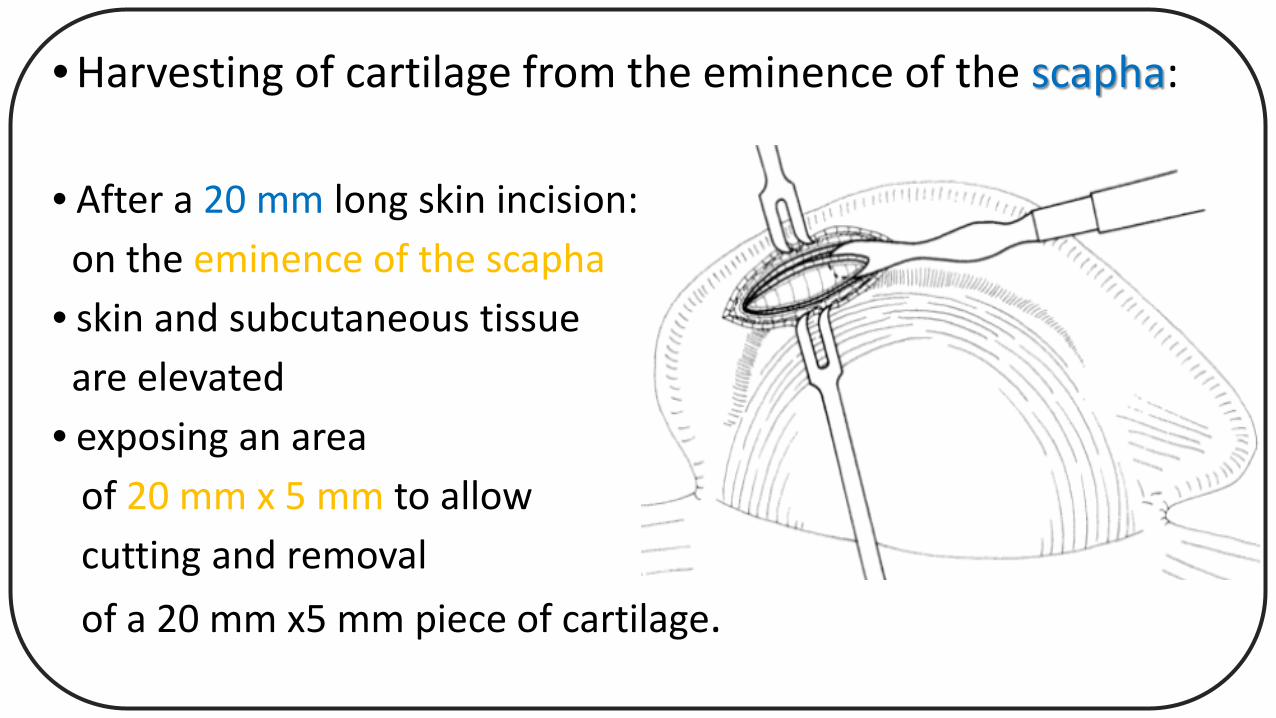
• Harvesting of cartilage from the eminence of the scapha:
• After a 20 mm long skin incision:on the eminence of the scapha
• skin and subcutaneous tissueare elevated
• exposing an areaof 20 mm x 5 mm to allowcutting and removal of a 20 mm x5 mm piece of cartilage.
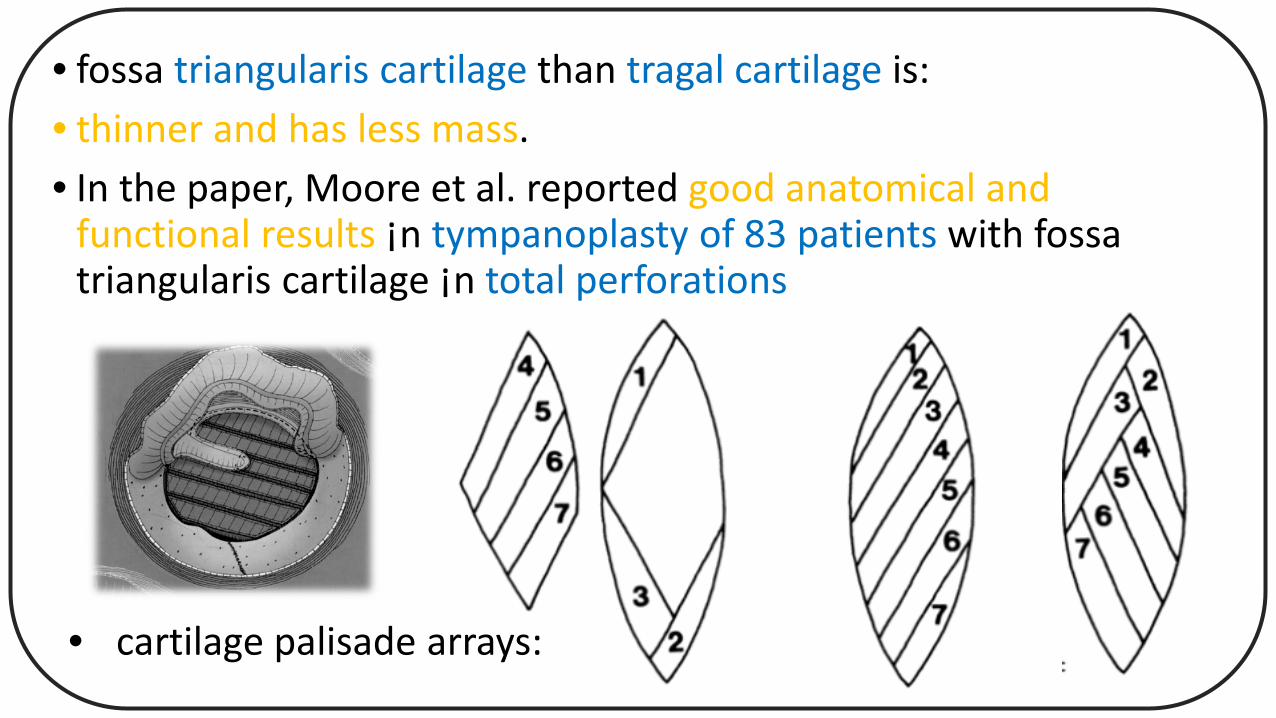
• fossa triangularis cartilage than tragal cartilage is:• thinner and has less mass.• In the paper, Moore et al. reported good anatomical and
functional results ¡n tympanoplasty of 83 patients with fossa triangularis cartilage ¡n total perforations
• cartilage palisade arrays:
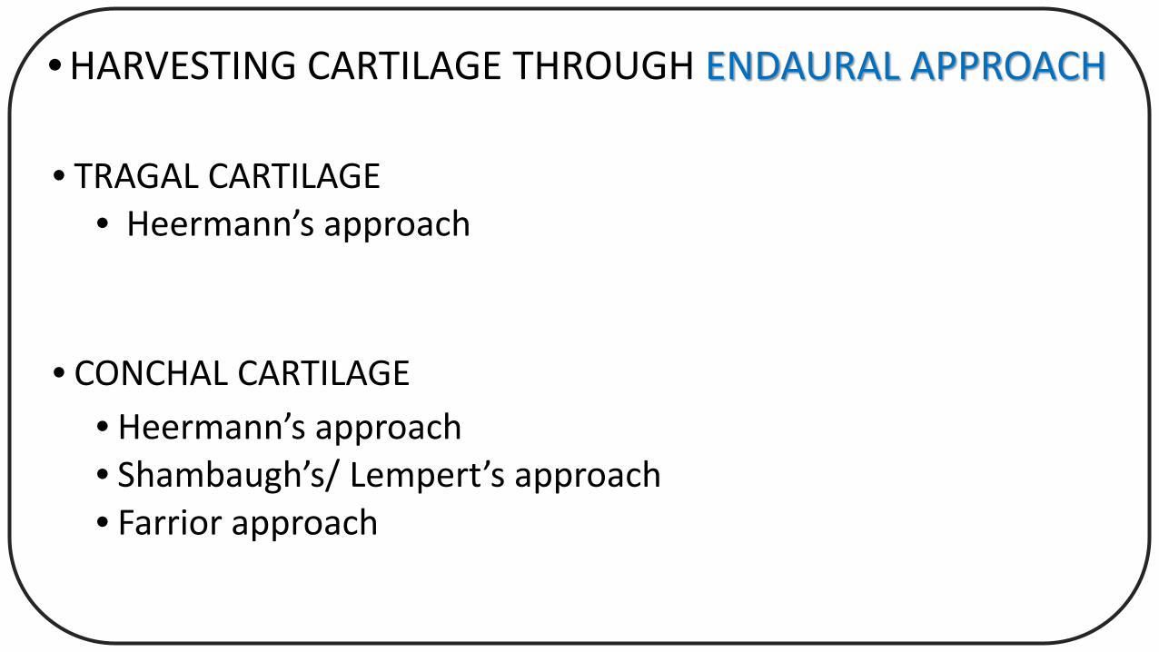
• HARVESTING CARTILAGE THROUGH ENDAURAL APPROACH
• TRAGAL CARTILAGE• Heermann’s approach
• CONCHAL CARTILAGE • Heermann’s approach• Shambaugh’s/ Lempert’s approach• Farrior approach
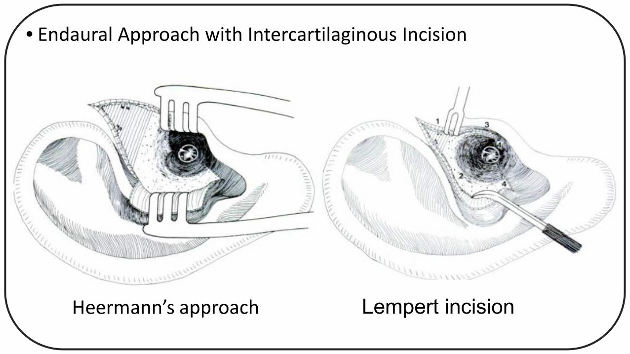
• Endaural Approach with Intercartilaginous Incision
Lempert incision
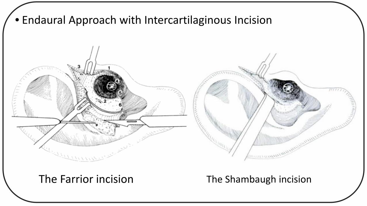
• Endaural Approach with Intercartilaginous Incision
The Farrior incision The Shambaugh incision
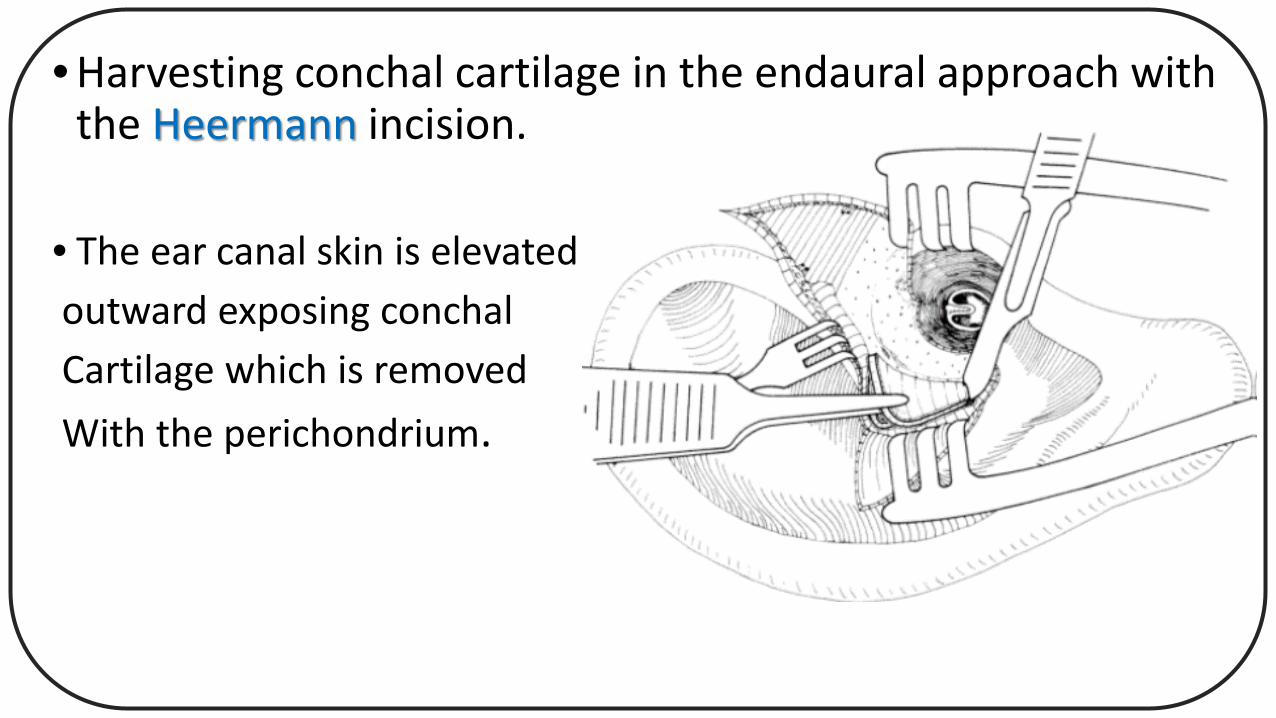
• Harvesting conchal cartilage in the endaural approach with the Heermann incision.
• The ear canal skin is elevatedoutward exposing conchalCartilage which is removedWith the perichondrium.
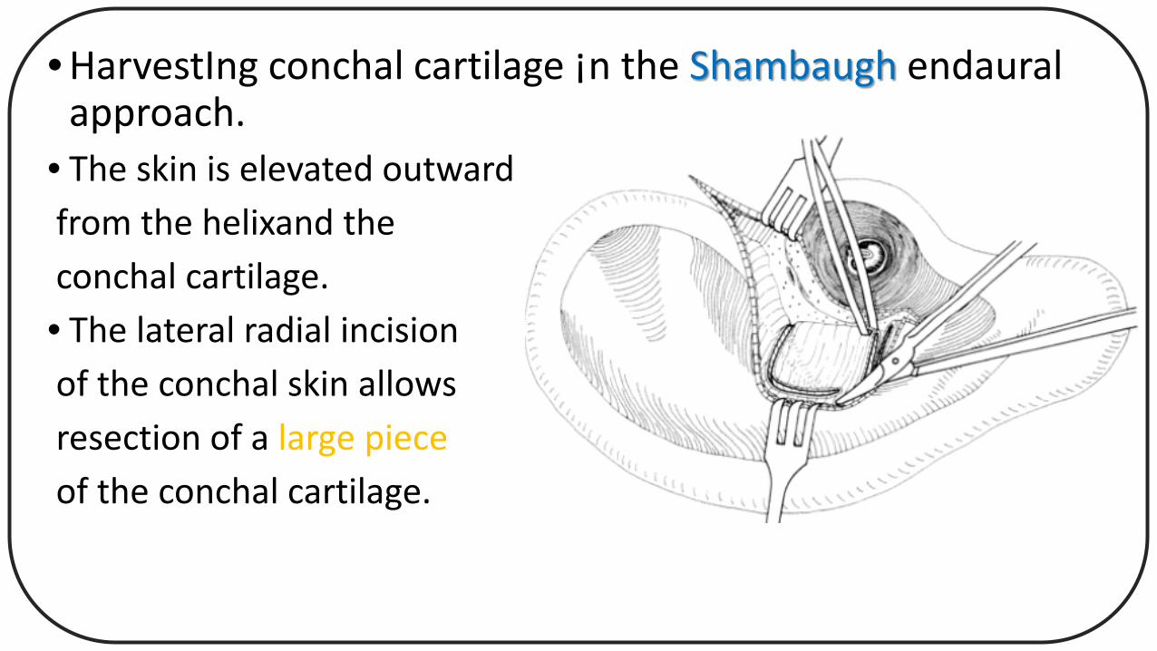
• HarvestIng conchal cartilage ¡n the Shambaugh endauralapproach.
• The skin is elevated outwardfrom the helixand theconchal cartilage.• The lateral radial incisionof the conchal skin allowsresection of a large pieceof the conchal cartilage.
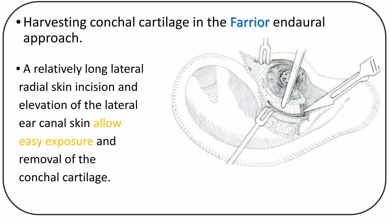
• Harvesting conchal cartilage in the Farrior endauralapproach.
• A relatively long lateralradial skin incision andelevation of the lateralear canal skin alloweasy exposure andremoval of theconchal cartilage.
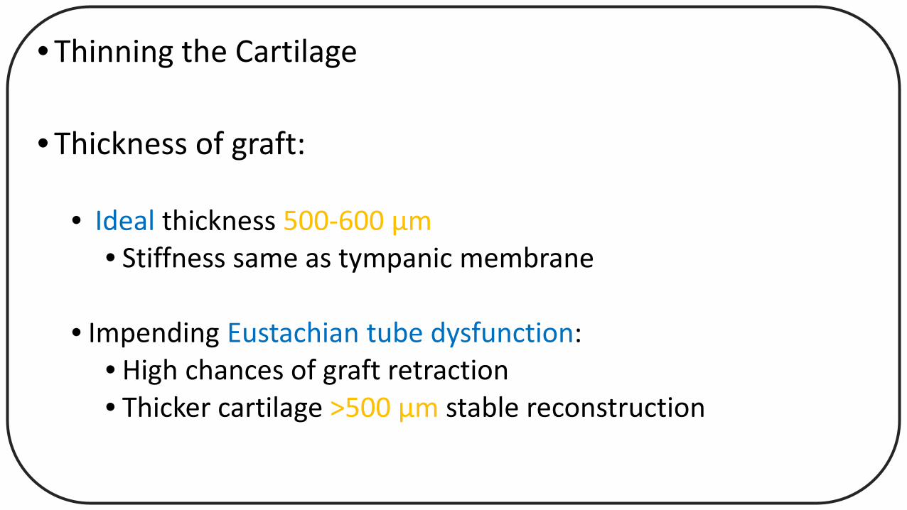
• Thinning the Cartilage
• Thickness of graft:
• Ideal thickness 500-600 µm• Stiffness same as tympanic membrane
• Impending Eustachian tube dysfunction:• High chances of graft retraction• Thicker cartilage >500 µm stable reconstruction
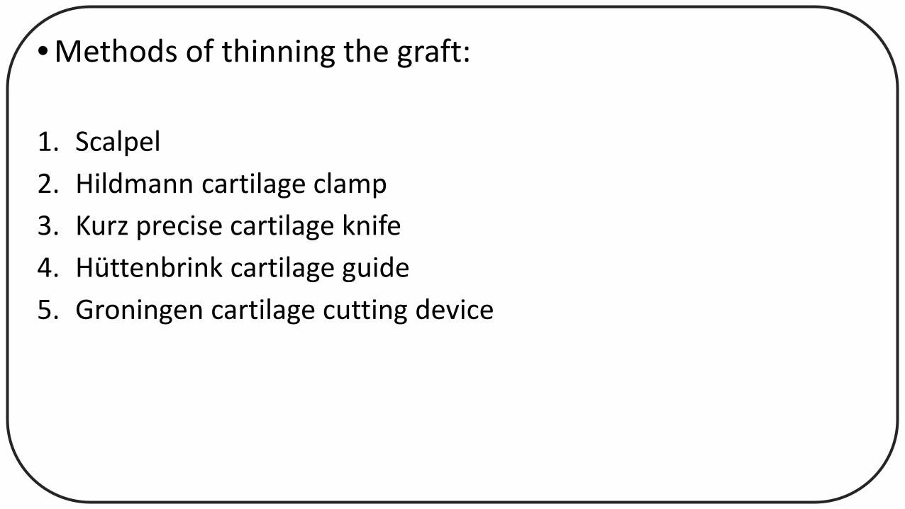
• Methods of thinning the graft:
1. Scalpel 2. Hildmann cartilage clamp3. Kurz precise cartilage knife4. Hüttenbrink cartilage guide5. Groningen cartilage cutting device
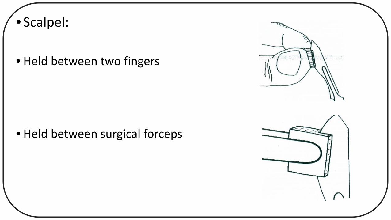
• Scalpel:
• Held between two fingers
• Held between surgical forceps
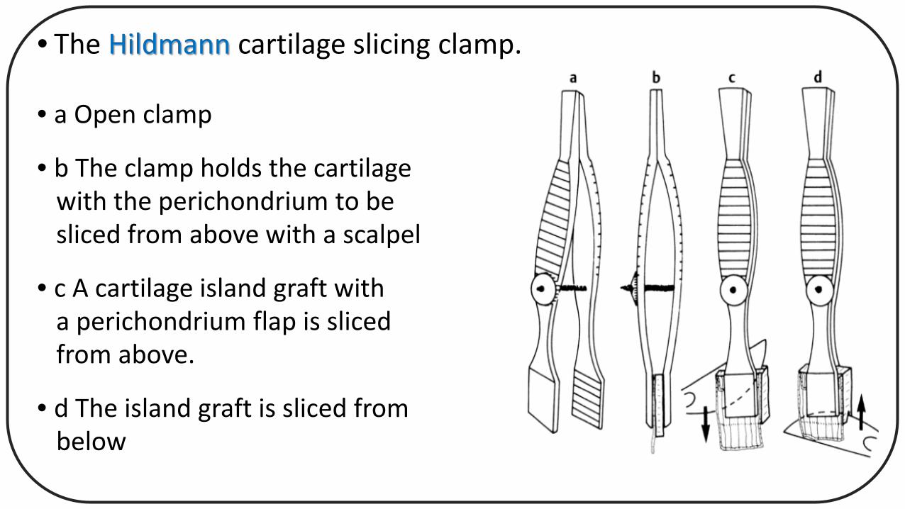
• The Hildmann cartilage slicing clamp.
• a Open clamp
• b The clamp holds the cartilagewith the perichondrium to besliced from above with a scalpel
• c A cartilage island graft witha perichondrium flap is slicedfrom above.
• d The island graft is sliced frombelow
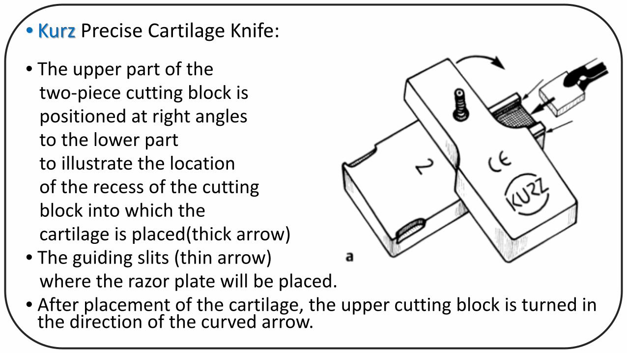
• Kurz Precise Cartilage Knife:
• The upper part of thetwo-piece cutting block ispositioned at right anglesto the lower partto illustrate the locationof the recess of the cuttingblock into which thecartilage is placed(thick arrow)
• The guiding slits (thin arrow)where the razor plate will be placed.
• After placement of the cartilage, the upper cutting block is turned in the direction of the curved arrow.

• The razor blade is fixed¡n the blade holder with a screw.
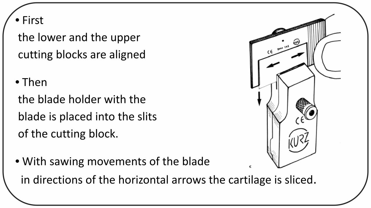
• Firstthe lower and the uppercutting blocks are aligned
• Thenthe blade holder with theblade is placed into the slitsof the cutting block.
• With sawing movements of the bladein directions of the horizontal arrows the cartilage is sliced.
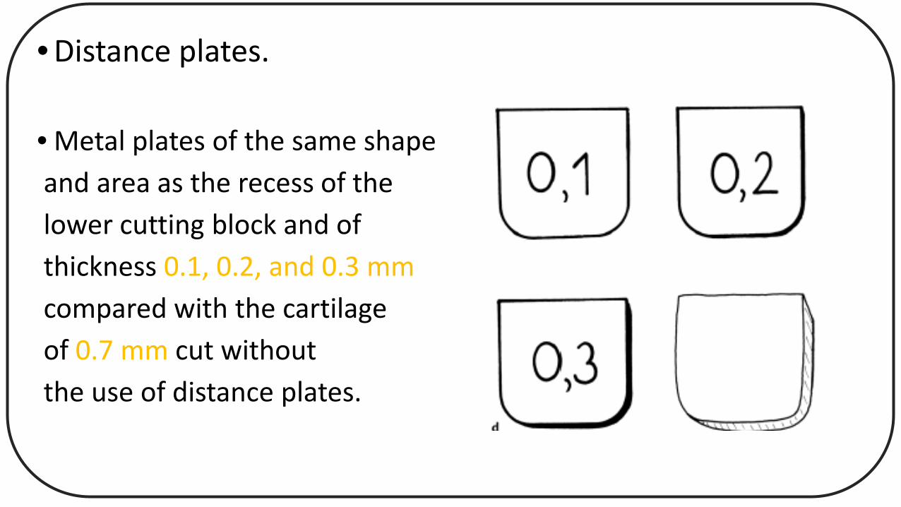
• Distance plates.
• Metal plates of the same shapeand area as the recess of thelower cutting block and ofthickness 0.1, 0.2, and 0.3 mmcompared with the cartilageof 0.7 mm cut withoutthe use of distance plates.
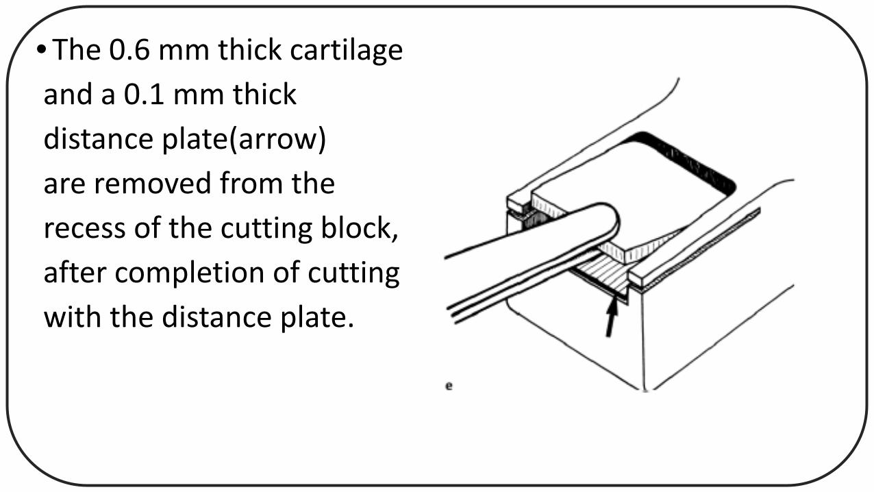
• The 0.6 mm thick cartilageand a 0.1 mm thickdistance plate(arrow)are removed from therecess of the cutting block,after completion of cuttingwith the distance plate.
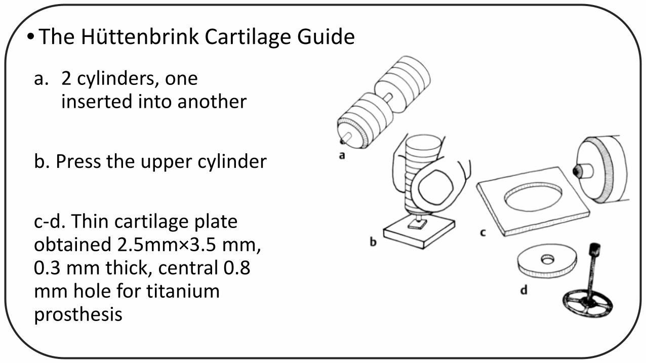
• The Hüttenbrink Cartilage Guide
a. 2 cylinders, one inserted into another
b. Press the upper cylinder
c-d. Thin cartilage plate obtained 2.5mm×3.5 mm, 0.3 mm thick, central 0.8 mm hole for titanium prosthesis
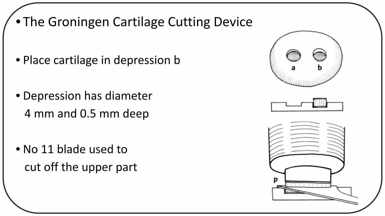
• The Groningen Cartilage Cutting Device
• Place cartilage in depression b
• Depression has diameter 4 mm and 0.5 mm deep
• No 11 blade used tocut off the upper part


![Cartilage - facultymembers.sbu.ac.irfacultymembers.sbu.ac.ir/rajabi/ppt toPDF/Cartilage [Compatibility Mode].pdfFibrocartilage • Fibrous Cartilage • is a form of connective tissue](https://static.documents.pub/doc/80x56/6012989a4318862a0e5813ae/cartilage-topdfcartilage-compatibility-modepdf-fibrocartilage-a-fibrous.jpg)


