In many ways, the hollow fibre bioreactor may be considered the original
three‐dimensional (3D) cell culture system (Figure 14.1). Hollow fibre‐based
cell culture was first developed by Richard Knazek at the NIH in 1972. He was
searching for a way to culture adrenal tumour cells under in vivo‐like conditions
to study hormone secretion in response to drug stimuli. His reports that response
curves can be generated by assaying the amount of hormone secreted in response
to biochemical stimuli were the original dynamic in vitro cell‐based assay (Knazek
et al., 1972). Hollow fibre bioreactors offer a method by which cells can be
cultured at tissue‐like densities over long periods of time. Hollow fibres act as
‘artificial capillaries’ and act much as capillaries do in the human body. Hollow
fibre bioreactors hit their peak of popularity in the late 1970s to 1980s when
they were employed in the bio‐manufacturing of monoclonal antibodies
(Tharakan & Chau, 1986).
The controlled molecular weight cut‐off (MWCO) of the fibres allowed
inhibitory cytokines such as TGF‐β to diffuse away from the hybridoma cells,
while entrapping the secreted antibody to high concentration within the small
volume of the extracapillary space. Poor flux in the particular fibres available at
the time, coupled with a lack of available technology for the scale‐up of hollow
fibre bioreactors, limited their application and the method fell into disfavour,
except for small‐scale research applications.
In the past 10 years, advances such as new high‐flux fibre materials, better
system engineering and an improved understanding of hollow fibre cell culture
methods have given new life to the use of hollow fibre bioreactors. It had been
thought that the benefits of hollow fibre bioreactors were restricted to simply
higher secreted protein concentrations, the ability to reduce serum levels or
more easily adapt cultures to commercially available serum free media. Much
Three‐dimensional cell‐based assays in hollow fibre bioreactorsJohn J. S. Cadwell1 and William G. Whitford2
1 FiberCell Systems Inc., Frederick, Maryland, USA2 GE Healthcare, Life Sciences, Cell Culture, Logan, Utah, USA
CHAPTER 14
328 Chapter 14
new information has been gathered regarding hollow fibre bioreactors and it is
now understood that they represent a fundamentally different and more in vivo‐
like way to culture cells. There is no question that the conditions under which
cells are cultured have a profound effect on their behaviour, and cell culture
conditions are fundamentally different in a hollow fibre bioreactor (Stanness
et al., 1997).
The hollow fibre bioreactor
The biomimetic hollow fibre (HF) bioreactor is a high‐density continuous
perfusion culture system. It presents many unique distinctions from the com-
monly employed non‐porous plastic surfaces, for example flasks, microcarrier
beads and discs or roller bottles. A HF bioreactor includes a cartridge containing
thousands of semi‐permeable hollow fibres in a parallel array within a tubular
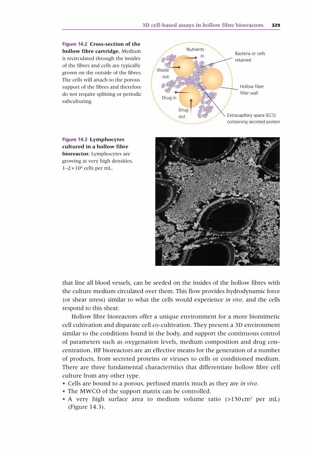
housing fitted with inlet and outlet ports (Figure 14.2). These fibre bundles are
potted at each end so that any liquid entering the ends of the cartridge will
necessarily flow through the interior of the fibres. Cells are generally seeded
within the cartridge but outside the hollow fibres in what is referred to as the
extracapillary space (ECS).
In this configuration, culture media is pumped inside the hollow fibres, allow-
ing nutrients and waste products to diffuse both ways across the fibre walls. Having
passed through the cartridge, the culture medium is oxygenated and recirculated
to the cartridge. There are, however, a few other possible configurations and
alterations of this basic implementation and flow circuit. Endothelial cells, the cells
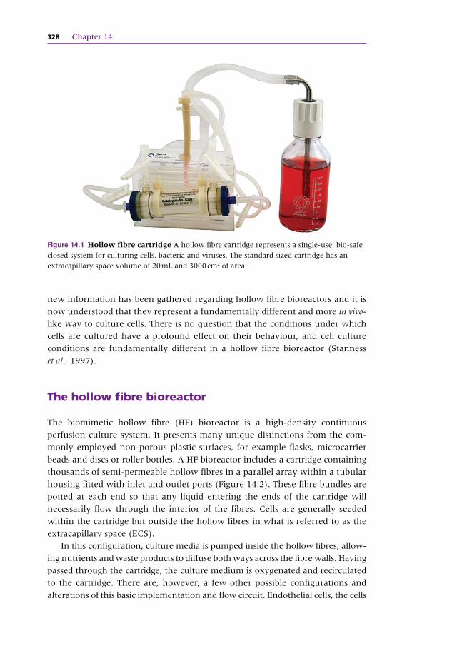
Figure 14.1 Hollow fibre cartridge A hollow fibre cartridge represents a single‐use, bio‐safe
closed system for culturing cells, bacteria and viruses. The standard sized cartridge has an
extracapillary space volume of 20 mL and 3000 cm2 of area.
3D cell‐based assays in hollow fibre bioreactors 329
that line all blood vessels, can be seeded on the insides of the hollow fibres with
the culture medium circulated over them. This flow provides hydrodynamic force
(or shear stress) similar to what the cells would experience in vivo, and the cells
respond to this shear.
Hollow fibre bioreactors offer a unique environment for a more biomimetic
cell cultivation and disparate cell co‐cultivation. They present a 3D environment
similar to the conditions found in the body, and support the continuous control
of parameters such as oxygenation levels, medium composition and drug con-
centration. HF bioreactors are an effective means for the generation of a number
of products, from secreted proteins or viruses to cells or conditioned medium.
There are three fundamental characteristics that differentiate hollow fibre cell
culture from any other type. • Cells are bound to a porous, perfused matrix much as they are in vivo. • The MWCO of the support matrix can be controlled. • A very high surface area to medium volume ratio (>150 cm2 per mL) (Figure 14.3).
Hollow �ber�lter wall
Extracapillary space (ECS)containing secreted protein
Drugout
Drug in
Wasteout
Nutrientsin Bacteria or cells
retained
Figure 14.2 Cross‐section of the hollow fibre cartridge. Medium
is recirculated through the insides
of the fibres and cells are typically
grown on the outside of the fibres.
The cells will attach to the porous
support of the fibres and therefore
do not require splitting or periodic
subculturing.
Figure 14.3 Lymphocytes cultured in a hollow fibre bioreactor. Lymphocytes are
growing at very high densities,
1–2 × 10⁸ cells per mL.
330 Chapter 14
Cells bound to a porous supportThe fact that the cells are bound to a porous support provides a number of
distinct features. One is the support of a continuous system where there is no
requirement to split the cells. Cultures in this system can maintain viability and
production‐relevant metabolism in a postconfluent manner for extended peri-
ods of time – months or longer. For example, one hybridoma cell line was
reported to maintain productivity in a HF bioreactor‐based culture for over one
year. It has been reported that a contributing factor here is that more in vivo‐like
growth conditions afforded by HF bioreactors result in significantly reduced
apoptosis (Hirschel et al., 2011). Another advantage is that, due to the extremely
low shear generated with the cartridge, the majority of cells that become necrotic
will not release significant cytoplasmic proteins or DNA into the culture medium.
This provides features such as an improved culture environment, more accurate
culture parameter monitoring and a raw harvest that is cleaner and easier to
handle for downstream assays and purification.
Controlled molecular weight cut‐offThrough the selection of fibre porosity, desired products can be retained to
significantly higher concentrations and the location/effects of cytokines can also
be controlled. This is well illustrated in the case of hybridoma culture where the
inhibitory cytokine TGF‐β can be selectively removed from the culture while
secreted antibody is concurrently retained (Dennler et al., 2002). Secreted
recombinant proteins can be selectively retained and concentrated and cytokines
and other factors that facilitate cell‐cell interactions can be concentrated as well.
Small molecule drugs can easily exchange across the fibre and rapidly reach
equilibrium.
High surface‐to‐volume ratioThe small diameter of the fibres (of the order of 200 microns) generates an
extremely high surface area‐to‐cartridge volume ratio in the range of 100–
200 cm2/mL. In conjunction with the high gross filtration rate of polysulfone
fibres, the exchange of primary and secondary metabolites is high enough to
support cell densities of 1–2 × 10⁸/mL, approaching in vivo tissue‐like densities.
A 20 mL cartridge will easily support the cell mass provided by a standard 2 L
suspension culture or 20–40 roller bottles. High cell densities produce more
protein per area of reactor footprint and facilitate adaptation to both lower
serum concentrations and simplified serum‐free culture, such as employing the
FiberCell® Systems’ CDM HD perfusion‐optimised serum replacement. The use
of such a defined and protein‐free medium results in much cleaner product harvests
and more simplified purification as well as a more defined culture environment
compared to the presence of fetal bovine serum (FBS) (Figure 14.4).
Operation of a HF bioreactor, in its most simplified form, begins by seeding
a prepared cartridge with either suspension or harvested adherent cells.
3D cell‐based assays in hollow fibre bioreactors 331
The reactor cartridge is connected to an external reservoir and the medium
recirculated from the reservoir through the cartridge. In one popular imple-
mentation, the recirculation is accomplished using a positive pressure displace-
ment pump designed specifically for this purpose. Here, a piston compresses a
short piece of pump tubing and one‐way check valves on either side produce
flow, in the same manner as the human heart. This frictionless pumping mech-
anism can generate 100 mL/minute of flow rate without wear on peristaltic
pump tubing. Mass transfer of gases can be accomplished in a variety of ways,
one being diffusive exchange through a loop of gas‐permeable silicone tubing
prior to the medium entering the bioreactor itself. The medium is constantly
recirculated, providing a supply of oxygen and nutrients as well as removal of
CO2 and secondary metabolites. Medium recirculation rate and culture feeding
can be linked to any number of culture parameters, and a number of control
and automation options have been explored. A basic approach is to manually
monitor the glucose and replace the medium as the concentration approaches
50% of the original.
Several cartridges sizes and fibre types are available, including those com-
posed of polysulfone (PS) and polyvinylidene fluoride (PVDF). The MWCO in
the PS fibres includes 5 KD or 20 KD and the pore size of PVDF is 0.1 micron. The
PVDF fibre is of particular interest as various protein matrices, antibodies or
growth factors can be readily bound to its surface via hydrophobic interaction.
Cartridges come presterilised, assembled and ready to use and are intended for
a single use.
Figure 14.4 FiberCell Duet pump in a CO2 incubator. Temperature and gas control is
provided by the incubator and a thin cord is used to go through the incubator door without
affecting gas composition.
332 Chapter 14
During this renaissance of perfusion in general, and of the HF bioreactor in
particular, several characteristics of hollow fibre cell culture have recently been
identified. • Reduction in apoptosis (Weeraphan et al., 2012). • Consistency of culture over long periods of time (G. Pavlakis, personal com-munication, 2013).
• More in vivo‐like growth conditions resulting in improved cell function (Bennet et al., 2007).
• Facilitation of the use of serum‐free, protein‐free and chemically defined media formulations (Whitford & Cadwell, 2011).
Hollow fibre bioreactors provide many particular (and some unique) culture
characteristics due to a number of physical and ambient chemical conditions
provided by the system, including: • perfused medium flow and porous support permits long‐term culture • high cell density culture increases cell‐cell contact • selectable MWCO of fibres concentrates interactive cytokines • selectable MWCO of fibres segregates cells, metabolites and products • directional flow establishes gentle interstitial gradients within the cell mass • hydrodynamic (shear) force on endothelial cells required for proper physiology • long‐term high‐density culture on porous support facilitates development of cell‐cell interactions over time.
Reduced apoptosisIt has long been recognised that reduced proteolysis and contamination by
intracellular proteins and DNA are characteristics of the HF bioreactor. During
extremely long culture periods of several months or longer, no degradation of
secreted products has been reported, even when using serum‐free and protein‐
free media. P.C. Liao’s group has identified a potential mechanism for this (Wu
et al., 2009). Their research focused on the culture of primary cancer cells to
identify potential secreted biomarkers. Cultures were initiated in FBS and then
switched to a protein‐free, chemically defined medium in order to reduce the
amount of extraneous protein that might obscure the secretome of the cells.
They found a significant reduction in apoptosis of up to 90%. The cells would
die, but not lyse and go through the typical process of releasing proteolytic
enzymes, intracellular proteins and DNA (Chang et al., 2009). This, and the
absence of significant shear forces inside the HF cartridge, results in dramatically
reduced postsecretion alterations and cleaner harvests (Srisomsap et al., 2010).
Culture consistencyIn a HF bioreactor, the cells are bound to a porous support, not a non‐porous
plastic surface. Cell division rate and generation number are reduced, cultures
do not require splitting, passage number is irrelevant, and cells grow in multiple
layers in a ‘postconfluent’ fashion. Cultures can be maintained for many months
and up to a year or longer (personal communication). Culture conditions remain
3D cell‐based assays in hollow fibre bioreactors 333
highly consistent during this time, as does cell physiology. In an example of this,
provided by Dr George Pavlakis (NCI), 293 T cells were transformed to produce
a very complex protein. The cytokine product described was more than 40%
carbohydrate and somewhat labile due to the fact that the two subunits are held
together only with hydrophobic interactions. Attempts at producing this protein
in standard flask or spinner cultures were unsuccessful. It was demonstrated
that consistent production of dimerised product with complete and consistent
post‐translational modifications was maintained for over 140 days (Figure 14.5).
In vivo‐like growth conditionsIn vivo, most animal cells grow in 3D at very high density under tightly defined
and highly controlled conditions. There is very little variation in oxygen tension,
pH, glucose levels, etc. These parameters can have a wide range in flasks and
other culture devices. It has been recently observed that ‘The 2D cell culture
systems used so far have several drawbacks: the morphology, proliferation,
metabolism and expression profiles of cells grown in 2D systems are very differ-
ent to cells in living tissues’ (Tecan Group, 2012). It has been shown that in a HF
bioreactor, consistency of culture conditions has a direct effect on cell physiology
and product generation. One researcher found that transformed CHO cells
expressing a hexamerised IgG responded favourably to HF bioreactor culture.
The researcher first cultured the CHO cells in standard T150 flasks. Protein fold-
ing and post‐translational modifications from this condition were incomplete,
resulting in nearly 40% of the protein being expressed as an incompletely
assembled monomeric subunit. When these same cells were removed from the
glycosylated protein in CDM HD protein‐free medium. The HPLC demonstrates no changes in
post‐translational modifications over 137 days of continuous culture.
334 Chapter 14
flask and seeded into a HF bioreactor, within one week the expressed protein
product was discovered to be 95% properly folded hexamer (J. Arthos, personal
communication, 2013). The more biomemetic cell culture conditions had
resulted in improved cell physiology and correct protein expression (Figure 14.6).
Single‐use flow path systemsThe culture‐contact surfaces of many HF bioreactor instruments are composed
of entirely single‐use (SU) materials. These provide a number of benefits in the
establishment of manufacturing and assay systems. They present reduced cross-
contamination risks for many reasons, including that the materials have never
seen living agents before and that they are sterilised by validated γ‐irradiation
systems. They provide lower initial investment costs as the SU housing appara-
tus is less expensive than full reuseable instrumentation. Lower facility and
operating costs are supported by a reduced requirement for services such as
autoclaves and process water as well as reduced operator requirements. Process
efficiency and flexibility are provided by the ability to easily reroute the fluid
flow path, and to order variant culture‐contact materials or custom‐designed
assemblies from the manufacturer.
Volume
Abs
280
Abs
280
Fibercell polysulphone cartridge
Volume
150 cm2. tissue culture �ask
C.
A.
B.
Figure 14.6 A CHO cell line expressing a hexamerised IgG protein construct. When
these cells are cultured in flasks, 40% of the protein is expressed as a monomeric subunit. The
exact same cells, when removed from the flask and seeded into the hollow fibre bioreactor,
now express 95% of the protein as a properly folded hexamer.
3D cell‐based assays in hollow fibre bioreactors 335
Protein‐free, chemically defined culture mediumBesides providing many positive factors, animal serum presents a number of
detrimental features including foaming, risk of contamination and variability in
both production and cell‐based assay performance. There are a number of non‐
culture‐related limitations as well, including high and variable cost and the fact
that high protein concentration can interfere with culture analysis. Cells in vivo
are exposed to serum only during injury, and many cells then respond by
particular activations. HF bioreactor culture characteristics, such as the very high
cell density, allow for a reduction in serum concentration and facilitate adapta-
tion to commercially available serum‐free media. This has been taken one step
further with the introduction of CDM HD, a commercially available serum
replacement. CDM HD is a protein‐free, chemically defined (CD), animal com-
ponent‐free serum replacement that is simplified and optimised for high‐density
cell culture.
Applications for cell‐based assays
Many drug effects are dependent not only on concentration but also time. Some
researchers believe cell‐based assays and in vitro testing methods are a useful,
time‐ and cost‐effective tool for drug discovery. However, it is generally accepted
that many of the available assays are not effective for examining the effects of
both time and concentration, and do not closely mimic physiological kinetics.
More specifically, such tests poorly report pharmacodynamic actions (what a
drug does to the body) and pharmacokinetic actions (what the body does to a
drug). Static cell‐based assays in plates, flasks or other formats do not readily
permit changes in drug concentration, as would be seen in humans from admin-
istration, uptake, distribution, distal metabolism and elimination effects. Animal
models generally do not provide the same drug kinetics as would be found in
humans, many infections cannot be supported with an animal model and often
the bacterial load is not high enough to reveal the emergence of resistance. HF
bioreactor cartridges have continuous medium circulation supporting dynamic
control of drug concentration over time and resulting in the mimicking of
naturally occurring gradients in tissue drug concentration. A high surface area‐
to‐volume ratio permits extremely rapid exchange of metabolites and pharma-
coactive molecules between the central reservoir and cells growing in the
relatively small ECS of the cartridge. Furthermore, the volume of this central
reservoir can be easily adjusted to permit rapid and reproducible changes in drug
concentration (Figure 14.7).
Simulation of the kinetics of multiple drugs can also be accomplished so
drug‐drug interactions and combination therapies, as well as transport and
efflux, can readily be modelled. The system is compact enough that multiple
cartridges can be conveniently manipulated in a relatively small space, providing
336 Chapter 14
multiplexed or parallel and higher throughput activities. Such systems can be
configured for cell‐based assays employing either a single‐cell type or multicell
in co‐cultivation.
Examples of hollow fibre‐based cell assays
Hollow fibre‐based assays are inherently more complex and costly to design and
set up than conventional cell‐based assays. They require a higher degree of cell
culture technical ability, more laboratory space and are more suited for higher
value, more complex questions rather than smaller and truly high‐throughput
screening type assays. It can take up to a week before the culture is established
and data can be generated. They are most effective when either using suspen-
sion cell types (permits the sampling of a small population of cells) or when
measuring something the cells are secreting over time, such as a specific protein
marker or virus particles.
However, these assays can generate data that is not available in any other
manner, and can bridge an important gap between animal studies and phase I
clinical trials. Large numbers of cells can be assayed over a pharmacologically
Hollow �brecapillaries
Fibrecell systemsduet pump
Pump
Centralreservoir
Dosingport
Diluentreservoir
Filteredvent
Pump
Sample port for access toextracapillary compartment
Elimination reservoir
BIOREACTOR LOOP
Mixer
Figure 14.7 The set‐up for controlling drug concentration to mimic human PK/PD. Medium or broth is constantly recirculating through the cartridge from the central reservoir.
Drug can be introduced into the central reservoir at a defined rate representing the absorption
curve. Diluent is then added and removed from the central reservoir at a controlled rate to
model the elimination curve. Drug concentration in the extracapillary space of the cartridge
equals the drug concentration in the central reservoir and the volume of the central reservoir
remains constant.
3D cell‐based assays in hollow fibre bioreactors 337
relevant period of time. Drug concentrations can be controlled in a dynamic
fashion and both adsorption and elimination curves can be modelled. Multiple
tests can be performed on the same cell population. 3D cultures of multiple cell
types can model complex processes such as virus infections in tissues, haemat-
opoiesis, cancer cell propagation, cancer cell metastasis and the blood–brain
barrier. Furthermore, nuclear magnetic resonance (NMR) and other imaging
methods have demonstrated potential as non‐invasive ways to monitor the
culture (Mancuso et al., 1990).
Assays using only one cell typeThe simplest type of 3D cell‐based assays performed in HF bioreactors consist
of only one cell type, seeded in the ECS. A popular implementation of this
approach is in the study of antimicrobial compounds on bacteria. While outside
the purview of this chapter, it is mentioned because it is the most common
application of this format (Inloes et al., 2004). The primary cell‐based assays
based upon this method are virus culture using a mammalian host cell and
cancer cell‐based assays.
There are many advantages for assaying drug effects on viral activity in a
HF bioreactor. The rapid delivery of nutrients, removal of waste products and
equilibration of drugs across the fibres facilitates the culture of cells at high
densities. It is the only cell culture method that can support cells at physiological
densities and provide for in vivo‐like viral infection and pharmacokinetics at
these densities. While both adherent and suspension cells can be cultured in a
HF bioreactor, it is notable that adherent cells bound to a porous support do not
require periodic splitting and can be maintained for extended periods of time.
Suspension cells can also be supported for extended periods of time due to the
same constant feeding and removal of metabolites.
The ability to add medium with or without drugs is particularly important for
dose fractionation pharmacodynamic studies where compounds are added to
the system over a short period of time and then removed by dilution with drug‐
free medium without disturbing the cells or their environment. Both absorption
and elimination can be simulated and the small volume of the ECS contributes
to the rapidity and economy with which this equilibrium takes place. Lastly, and
perhaps most importantly, because of their large size both viruses and virus‐
infected cells are retained in the small volume of the ECS. These infectious
agents cannot cross the fibres into the medium. The system is completely closed
to the external environment and provides an added biosafety component pro-
tecting laboratory personnel from exposure.
Antiviral pharmacodynamicsPharmacodynamics is the science that links drug exposure to response. A key
element here involves identification of the best dosing and scheduling. This is
derived from the principle that the drug concentration‐time curve is a factor in
a drug’s effectiveness (Eagle et al., 2004). For example, the duration that a drug
338 Chapter 14
concentration remains above inhibitory levels may be closely linked to the
antiviral effect. In this case, continuous or defined episodic intervals of drug
exposure may lead to desired effects. On the other hand, peak concentrations of
drug may be more important for antiviral activity. Here, brief doses at maximal
concentrations may be most effective. It might also be demonstrated that the
mode of dosing does not significantly affect antiviral activity. There are four
essential pillars to the pharmacodynamic index (dose and schedule) of a particu-
lar entity for a particular virus:
1 The EC50
/EC95
values of the compound for the particular virus.
2 Binding (and therefore removal) of the compound to unreactive tissue and
serum factors.
3 Identification of the compound’s pharmacokinetics (and any variability of this
within the population).
4 Identification of optimal exposure and administration schedule of the com-
pound for the virus in question.
The high‐density culture within HF reactors supports the efficient (biomimetic)
cell‐to‐cell spread of virus in either matrixed or suspension cells. In either case,
released virus and virus‐infected cells accumulate in the ECS over time.
Computer‐controlled pumps administer drug and diluent into the central reser-
voir to model any schedule of drug exposure.
Antiviral effect can be determined by sampling the contents of the ECS
through the sampling ports. This supports many divergent assays, beginning
with virus‐infected cell identification and counting by, for example, FACS anal-
ysis following treatment with a fluorochrome‐labelled viral antigen‐specific
monoclonal antibody. The effect of drug on the yield of free infective virus par-
ticles can be determined by means such as plaque assay of harvested cell‐free
medium or by ELISA of viral antigen. The dosing regimen providing inhibition
of viral replication and/or cell‐to‐cell spread of virus can be determined by
sequential analysis of cell and ambient media viral titre and drug concentration
from identified points later validated by, for example, LC/MSn. Values for both
drug exposure antiviral effects allow construction of an exposure–response rela-
tionship curve. Accuracy in this relationship is facilitated by the fact that eight or
more evaluations can be performed simultaneously (McSharry et al., 2011).
HIVNucleoside analoguesNucleoside analogues are versions of the building blocks of DNA that almost act
like normal nucleosides in DNA synthesis. But when these faulty building blocks
are used, the new DNA cannot be built correctly. This in turn prevents the cell
from producing new infective virus. Nucleotide analogue‐based drugs also exist
and are technically different from nucleoside analogues but act in much the
same way. HF‐based pharmacodynamic models have been employed in the
examination of such HIV‐1 antivirals for quite some time.
3D cell‐based assays in hollow fibre bioreactors 339
The nucleoside analogue 2′,3′‐didehydro‐3′deoxythymidine (d4T) was
examined using HF infection models (HFIM) (Drusano et al., 2002). Here, three
HF units containing HIV‐infected and uninfected CEM cells were set up. One
unit was continuously infused with medium without drug and served as a no‐
drug control. A second unit received drug as a continuous infusion. A third unit
received a bolus of d4T every 12 hours followed by a no‐drug washout to model
a one‐hour pharmacokinetic half‐life. In the absence of d4T there was a fourfold
increase in p24 antigen over the nine days of the experiment. In the presence of
d4T, delivered either as a continuous infusion or a twice‐daily bolus, the amount
of p24 antigen did not increase. In separate experiments, HF units infected with
the same amount of virus and treated in the same way, but with d4T at half these
doses, failed to completely inhibit virus replication. The results indicated the
dosing required as either a bolus or continuous infusion to inhibit virus replica-
tion. The HFIM system predicted that the minimum effective dose of d4T to treat
patients infected with HIV was approximately 0.5 mg/kg/day administered twice
a day. This prediction was confirmed in a clinical study (Anderson et al., 1992).
Protease inhibitorsThe precursor Gag polyprotein is processed by a viral protease to many HIV
and leads to an irreversible loss of the infectivity of the virus. A HF system was
successfully used to determine the minimum concentration of the protease
inhibitor A77003 that inhibits the replication of HIV in CEM cells (Preston
et al., 2003).
Vaccinia virusBecause routine smallpox vaccination among the public stopped after the
disease was eradicated, the majority of the world’s citizens are susceptible to
infection with variola major virus. After the events of September and October
2001, however, the US government established goals for developing newer
vaccines. Smallpox vaccine is made from vaccinia, a virus related to smallpox.
Most smallpox vaccine contains live, replication‐competent, vaccinia virus,
not dead or attenuated virus like many other vaccines. For that reason, the
vaccination site must be cared for to prevent the virus from spreading to other
portions of the patient’s body or other people. Also, the vaccine can cause
undesirable side effects and complications.
The main challenge in developing a new, safer vaccine is that its effectiveness
cannot be tested on humans, and other animals do not naturally contract small-
pox. Monkeys or ferrets infected with other poxviruses are often used, but tests
on animals that are artificially modelled with related disease often give false or
misleading results. For improved pharmacodynamic studies on vaccinia virus,
some researchers have employed HF‐based models. One group developed such
a model based upon used HeLa‐S3 cells because they have been used to grow
340 Chapter 14
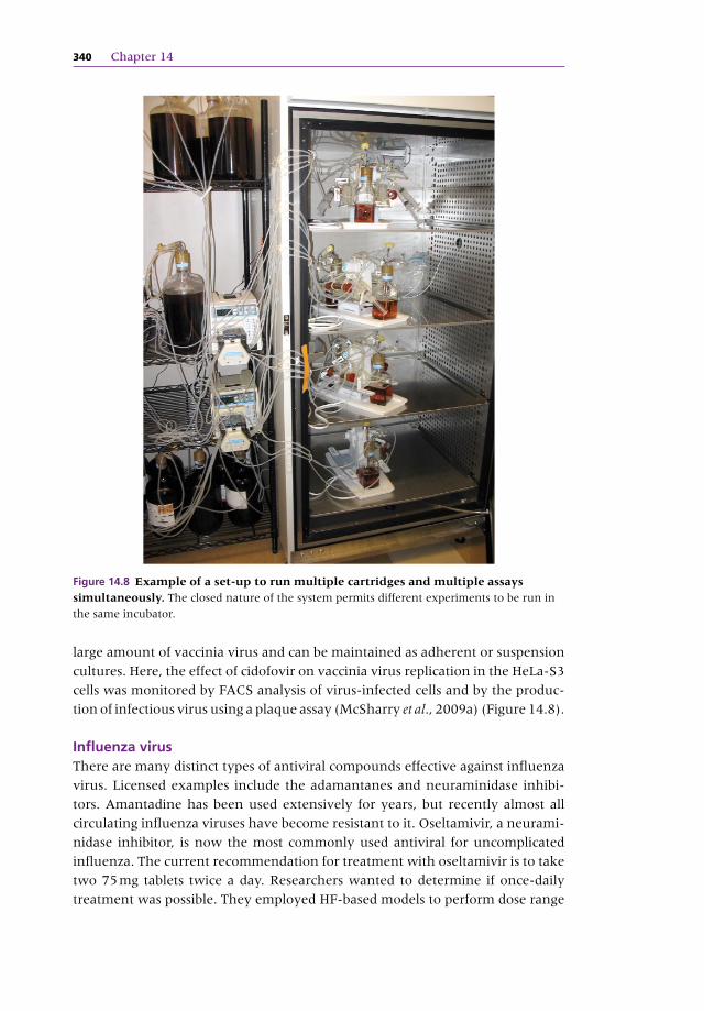
large amount of vaccinia virus and can be maintained as adherent or suspension
cultures. Here, the effect of cidofovir on vaccinia virus replication in the HeLa‐S3
cells was monitored by FACS analysis of virus‐infected cells and by the produc-
tion of infectious virus using a plaque assay (McSharry et al., 2009a) (Figure 14.8).
Influenza virusThere are many distinct types of antiviral compounds effective against influenza
virus. Licensed examples include the adamantanes and neuraminidase inhibi-
tors. Amantadine has been used extensively for years, but recently almost all
circulating influenza viruses have become resistant to it. Oseltamivir, a neurami-
nidase inhibitor, is now the most commonly used antiviral for uncomplicated
influenza. The current recommendation for treatment with oseltamivir is to take
two 75 mg tablets twice a day. Researchers wanted to determine if once‐daily
treatment was possible. They employed HF‐based models to perform dose range
Figure 14.8 Example of a set‐up to run multiple cartridges and multiple assays simultaneously. The closed nature of the system permits different experiments to be run in
the same incubator.
3D cell‐based assays in hollow fibre bioreactors 341
and dose fractionation experiments (McSharry et al., 2009b). As is the case for
poxviruses, there are no cell lines that continuously produce influenza virus
infection. The standard cell line used for influenza viruses is the Madin–Darby
canine kidney (MDCK), which grows well on glass and plastic surfaces and is
permissive to various strains of influenza virus, but not with high efficiency.
To perform pharmacodynamic studies for oseltamivir and influenza viruses,
this group used a derivative of MDCK that expresses higher levels of a particular
cell surface glycoprotein than the original line. It was determined that the R292
strain of influenza virus grows well in these cells in the HF units. To perform
dose‐ranging studies in the HFIM system, six HF units containing influenza A
virus‐infected AX‐4 cells were mixed with uninfected AX‐4 cells and continu-
ously infused with various concentrations of oseltamivir. The effect of the drug
on virus replication was determined daily by plaque and haemagglutination
assay. This provided an EC50
value in the HFIM system for oseltamivir with this
isolate of the R292 strain of influenza virus. To determine the pharmacodynami-
cally linked index, a dose fractionation study was designed to mimic a variety of
half‐lives of the drug in application. Here, one HF unit received an appropriate
exposure delivered by continuous infusion; a second unit received the same
exposure once a day followed by a no‐drug washout; a third unit received the
same exposure delivered twice a day followed by a no‐drug washout; and a
fourth unit received the same exposure delivered three times a day followed by
a no‐drug washout.
The data showed that in the absence of drug, the virus grew well in the HFIM
system. The continuous dose and all three fractionated doses gave the same
amount of inhibition of virus replication. This group concluded that the pharma-
codynamically linked index for oseltamivir for the R292 strain of influenza A
virus is the AUC/EC50
/95
ratio. This means that the model indicates that at the
appropriate dose, oseltamivir could be given once a day.
The demonstration that adherent cells can be used to grow virus in the HFIM
system opens this system up to the pharmacodynamic analysis of antiviral com-
pounds for a wide variety of viruses. As long as the virus‐infected cells produce
virus that is released into the ECS, then the effect of antiviral compounds can be
measured, such as in H1N1 influenza (Brown et al., 2011).
Anticancer agentsAnother example of a 3D cell‐based assay using only one cell type in the ECS of
the HF cartridge is for the analysis and characterisation of anticancer agents.
Anticancer agents also exhibit both time‐ and concentration‐dependent efficacy
and the goal of cell‐based assays can be twofold. The first is to ascertain the most
effective dosage profile for cytotoxicity of cancer cells while leaving normal cells
in an acceptable state. The second is to provide an indication of the compound’s
robustness and application breadth by testing different types of cancer cell lines
under identical conditions.
342 Chapter 14
Kirstein reported on the use of the HF model for anticancer drug evaluations.
In this case, gemcitabine was examined in the anchorage‐dependent MDA‐
MB‐231 breast cancer cell line (Kirstein et al., 2006). Longer‐term exposures are
possible in continuous culture HF systems since the cells do not require splitting
when a confluence is reached. More accurate results are obtained for a few rea-
sons. One is that because the multilayer, 3D organisation of these continually
perfused cells more closely reflects the in vivo structure of the tumour, they have
an increased relevance for assays of anticancer agents. Another is that the cell
division rates in static cultures are commonly artificially high, and this can ren-
der them more sensitive to some chemotherapeutic agents then their natural
counterpart (Kirstein et al., 2008).
Assays using more than one cell type
Hollow fibre and 3D co‐cultivation for cell‐based assaysHistorically, the science of cell biology was intended to reduce complex systems
to their most fundamental components in order to better understand them.
However, in this age of systems biology, we are appreciating the limitations to
such reductionist research, and better understanding the implications of organ-
isms and tissues being made up of more than one type of cell. From the conse-
quences of complex physical interactions to the signalling between multiple cell
types, we are seeing the behaviour of cells in the context of an organisation or
ensemble of heterotypes. HF bioreactor‐based culture is one of the few in vitro
techniques providing large numbers of cells in close enough proximity and suf-
ficiently high density to observe a number of tissue‐like behaviours (Figure 14.9).
These behaviours include: • signalling and direct interactions between different cell types • co‐ordinate (or synergistic) activity upon the ambient media • co‐ordinate (or synergistic) activity upon a compound or drug • mixed cell type‐specific effect (+/−) upon viral infections.
Hollow fibre cell culture permits the recapitulation of natural structures con-
taining more than one cell type in a defined, controlled and more biomimetic
environment. Endothelial cells are the only cells that can be easily cultured on
the insides of the fibres under flow. There are two types of cell co‐cultivation
that can be performed in a HF bioreactor. They are one or more cell types: • on both the inside and the outside of the fibres (Davis, 2007) • on only one side of the fibres (typically on the ECS).
An example of the first type of cellular co‐cultivation is the use of HF bioreactors
to culture endothelial cells on the insides of the fibres while culturing a different
cell type on the outside to provide cell signalling. Endothelial cells comprise the
inner lining of all blood vessels, and the endothelium can actually be considered
a discrete organ, rather than simply a cell type. Endothelial cells were first
3D cell‐based assays in hollow fibre bioreactors 343
cultured on the insides of hollow fibres by Dr Barbara Ballerman (Ballerman &
Ott, 1995). When cultured on the insides of a hollow fibre (the intracapillary
space) and exposed to hydrodynamic shear stress from medium flow, endothelial
cells behave differently (more in vivo‐like) than they do in static, flask culture.
Under shear, endothelial cells lay down flat as a monolayer, stop dividing, form
tight junctions and express different genes than when cultured under static
conditions in flasks (Figure 14.10). Differences in shear stress throughout the
body may explain both the different functions and characteristics of endothelial
cells in those areas (Redmond et al., 1995).
The concept of ‘organ recapitulation’ in hollow fibres was first applied by
Jorg Gerlach using liver tissue (Gridelli et al., 2012). The system used was a
complex HF bioreactor with two different fibre types and three separate bundles
Figure 14.9 Co‐cultivation of mixed cell population derived from collagenase digestion of a human placenta. This results in the formation of three‐dimensional
structures capable of generating suspension cells with stem‐like phenotypes. Source: Courtesy
of FiberCell Systems.
Figure 14.10 Cross‐section of a PVDF fibre with an endothelial cell monolayer. This monolayer
forms on the inside of the fibre
under the influence of shear stress.
344 Chapter 14
of fibres. Liver tissue was simply digested with collagenase and this mixture of
cells seeded into the HF bioreactor. Primary function was maintained for four
weeks. It appeared that endothelial cells provided a necessary component of
the cell mixture and were involved in the formation of 3D structures (Hoffman
et al., 2012).
Bone marrow modelPerhaps the most rigorous application of HF systems for single‐compartment
cell co‐cultivation is in the area of stromal cell/suspension cell interactions.
Contrary to popular belief, it is possible under specific conditions to harvest
large numbers of healthy, intact cells from a HF bioreactor. In fact, the most
powerful applications for HF systems involve longer‐term cultures with peri-
odic harvesting over time. There are a number of recent examples of this effec-
tiveness presenting some very interesting results. In demonstration of the
second type of co‐culture, Dr Mayasari Lim at University Hospital, Hong Kong,
published an article on the co‐cultivation of a human bone marrow stromal
cell line with a leukaemic T cell line (Usuludin et al., 2012). The standard
approach to this is generally accomplished in flask culture, but here she
reported the establishment of a stromal line cultured to fairly high density
within one week of culture initiation in the HF cartridge. Once the glucose
uptake rate of standard media in the cartridge reached 1 g per day, the leukae-
mic T cell line was seeded into the cartridge. When co‐cultured in flasks, the
T cells would typically undergo a 10× expansion. When cultured in the HF
cartridge, the T cells underwent a 4000‐fold expansion, much more in line
with what occurs in vivo (Figure 14.11).
Hollow �bres
Stromal cells
Suspension cellsNutrients
in
Wasteout
Figure 14.11 Schematic drawing of adherent stromal cells interacting with suspension cells. Since the cells are bound to a porous support, they do not require splitting
and the culture can be maintained for several weeks to months.
3D cell‐based assays in hollow fibre bioreactors 345
The bone marrow microenvironment plays an integral role in the regulation
of haematopoiesis. Residing stromal cells and the various extracellular matrixes
in the bone marrow provide biological signals that control haematopoietic stem
cell (HSC) function. In this study, a biomimetic co‐culture platform using the HF
bioreactor was developed for the ex vivo expansion of HSCs. The efficacy of such
a platform was evaluated in comparison to standard cultures performed on tis-
sue culture polystyrene (TCP). A human stromal cell line (HS‐5) was employed
as a co‐cultured stromal support of lineage cell‐depleted human cord blood cells.
This was accomplished in serum‐free medium supplemented with a cytokine
cocktail (Xue et al., 2014) (Figure 14.12).
Results showed that the performance of the HF bioreactor in supporting total
cell and CD34+ progenitor cell expansion was comparable to that of cultures on
TCP, while cells harvested from the HF bioreactor had a higher clonogenic
ability. The performance of ex vivo‐expanded cells from the HF bioreactor upon
haematopoietic reconstitution in humanised mice was also comparable to that of
the TCP control. Scanning electron microscopy revealed that stroma cell growth
inside the HF bioreactor created a three‐dimensional cell matrix. These findings
demonstrate the feasibility of utilising an HF bioreactor for creating a complex
matrix architecture, which may provide good in vitro mimicry of the bone
marrow supporting large‐scale expansion of HSCs.
(a) (b)
(d)(c)
10 kV X25 1 mm 0001 18 40 SEI 10 kV X100 100 μm 0001 18 40 SEI
10 kV X200 100 μm 0001 18 40 SEI 10 kV X500 50 μm 0001 18 40 SEI
Figure 14.12 Scanning electron micrographs of bone marrow stroma cells bound to the surface of the 5 KD MWCO polysulfone fibres. These cells can interact with
haematopoietic stem cells or leukaemic lymphocytes or other cell types. Images (a–d) are of
increasing magnification showing the general structure of the polysulfone fibres with 3D cell
growth in between.
346 Chapter 14
Asymmetric co‐culture using endothelial cellsThree‐dimensional cell‐based assays utilising co‐culture of multiple cell types in
HF bioreactors can recapitulate more complex structures than those with a single
cell type. One type of cellular co‐cultivation, as described above, is the use of HF
bioreactors to culture endothelial cells on the insides of the fibres and a different
cell type on the outside. A number of such examples have been reported, includ-
ing some with significantly more complex asymmetric cell co‐cultivation. For
example, Paul Cahill cultured bovine aortic endothelial cells on the insides of the
fibres and vascular smooth muscle cells on the outside of the fibres. As altering
the flow rate changed the shear stress, G‐protein formation and endothelin
receptor expression were directly modulated, even though there was no physical
contact between the two cell types (Redmond et al., 1995) (Figure 14.13).
Blood–brain barrier modelEndothelial cells at the blood–brain barrier (BBB) demonstrate tight intercellular
junctions, the absence of fenestrations and reduced pinocytotic vesicles. The
in vitro study of the BBB has progressed rapidly of late as improved monitoring
technologies and new or enhanced cell culture models have become available.
Developments in in vitro models promise the design of new drugs for treatment
of CNS disorders. These models play an important role in investigating the ability
of such compounds to cross the BBB. There are many in vitro approaches to
modelling BBB physical and biochemical behaviour, but most fail to represent
its three‐dimensional nature and do not support the associated exposure of
endothelial cells to such complex influences as exist in vivo (Janke et al., 2013).
To answer this challenge, Janigro developed a new, dynamic, in vitro BBB
model (NDIV‐BBB) designed to allow for extensive pharmacological, morpho-
logical and physiological studies (Stanness et al., 1999). He first reported that
brain microvascular endothelial cells developed robust growth and differen-
tiation when cultured alone. In the presence of glial cells, they developed
measurable properties allowing monitoring of the system and experimental
Figure 14.13 Scanning electron micrograph of bovine aortic endothelial cells. These cells were
cultured on the insides of the fibres and
vascular smooth muscle on the outside
of the fibre. Note the 3D structure
formed by the vascular smooth muscle
cells. Source: Courtesy of Dr. Paul Cahill
and Dr. Eileen Redmond.
3D cell‐based assays in hollow fibre bioreactors 347
perturbations and physical manipulations. For example, it was noted that flow
rates played an essential role in endothelial cell differentiation and concomitant
decreased cell division. His new dynamic model of the BBB allows for longitu-
dinal studies of the effects of flow and co‐culture in a controlled and fully
recyclable environment. It also permits visual inspection of the abluminal com-
partment and manipulation of individual capillaries. This model and testing
services are available from FloCel (www.flocel.com).
In perhaps the ultimate embodiment of state‐of‐the‐art HF‐based cell assays,
Dr Chris Pepper and co‐workers have developed a model for primary investiga-
tions and assaying for cancer metastasis (Walsby et al., 2014). It utilises endothelial
cells on the inside of the fibre subjected to shear stress as well as their interaction
with circulating leukaemic lymphocytes (Figures 14.14 and 14.15). There is
Figure 14.14 Endothelial cells attached to the inside of a PVDF hollow fibre with a short period of shear stress. Cells have begun to flatten in response to the shear.
Figure 14.15 Circulating leukaemic lymphocytes on the outside of the fibres. This was
taken after the transmigration of lymphocytes and their specific interaction with endothelial
cells on the insides of the fibres.
348 Chapter 14
growing evidence that lymphocyte trafficking contributes to the clinical course of
chronic lymphocytic leukaemia (CLL) but to date only static in vitro cultures have
been used to study these phenomena. To address this, a dynamic in vitro model
was developed in which CLL cells experience shear forces equivalent to those in
capillary beds and are made to flow through capillary‐like hollow fibres lined with
endothelial cells. CLL cells treated in this way increased their expression of CD62L,
CXCR4, CD49d and CD5 directly as a result of the shear force. Furthermore, CLL
cells migrated through the endothelium into the ‘extravascular space’ (EVS).
Migrated CLL cells had significantly higher expression of CD49d, MMP‐9, CD38,
CD80 and CD69 compared with CLL cells that remained in the circulation. The
degree of migration observed strongly correlated with CD49d expression, and
treatment with the CD49d blocking antibody natalizumab resulted in significantly
decreased migration. Taken together, these data provide evidence for a novel,
dynamic and reproducible in vitro model of lymphocyte migration and cancer
metastasis. This model might also prove useful in the study of stem cell homing
and trafficking.
Conclusion
As the original 3D culture approach, HF systems have for decades supported
the biomimetic culture of many cell types, at tissue‐like densities, for extended
periods of time. Novel material features supporting a renaissance in the tech-
nology include fibres composed of new materials, new surface derivatisations
and porosities providing improved binding, flux and flow rates. Enhanced
process control features include innovative culture monitoring approaches,
automated feed and recirculation control as well as improved process under-
standing leading to new closed‐loop control parameters. New and innovative
application development has kept pace with the availability of these new
technologies, expanding the scope of applicability of 3D HF cell‐based assays.
The HF bioreactor represents a more in vivo‐like method by which to culture
cells for assays. The new fields of metabolomics and systems biology are
supporting the development of HF systems based upon multicell co‐culture
and defined serum‐free media. The result is a robust and flexible technology
with diverse applications. Such applications range from protein biological pro-
duction providing ultra‐clean harvest and simplified purification to improved
in vitro viral infection models providing more accurate drug candidate
pharmacokinetic/pharmacodynamic modelling.
The 3D HF‐based cell assay system may not replace either animal models or
human testing but does represent an important methodology for the testing of
clinical compounds. It provides human kinetics and pathogen load levels before
or in conjunction with human testing and bridges an important gap between
animal and human testing.
3D cell‐based assays in hollow fibre bioreactors 349
References
Anderson, R. E., Meyer, W., Balch, F. et al. (1992) Antiviral effects of stavudine (d4T) therapy.
8th International Conference on AIDS, 19–24 July, Amsterdam, The Netherlands. Abstract
WeB 1010.
Ballerman, B. & Ott, M. (1995) Adhesion and differentiation of endothelial cells by exposure to


![[DDMC 2014] The Role of Online 3D Platforms in Open Innovation with Customers](https://static.documents.pub/doc/80x56/54b732174a795916198b498a/ddmc-2014-the-role-of-online-3d-platforms-in-open-innovation-with-customers.jpg)


