A glimpse of biodegradable polymers and their biomedical applicationshttps://doi.org/10.1515/epoly-2019-0041Received December 04, 2018; accepted March 29, 2019.
Abstract: Over the past two decades, biodegradable polymers (BPs) have been widely used in biomedical applications such as drug carrier, gene delivery, tissue engineering, diagnosis, medical devices, and antibac-terial/antifouling biomaterials. This can be attributed to numerous factors such as chemical, mechanical and physiochemical properties of BPs, their improved pro-cessibility, functionality and sensitivity towards stimuli. The present review intended to highlight main results of research on advances and improvements in terms of synthesis, physical properties, stimuli response, and/or applicability of biodegradable plastics (BPs) during last two decades, and its biomedical applications. Recent literature relevant to this study has been cited and their developing trends and challenges of BPs have also been discussed.
* Corresponding author: Dilip V. Vasava, Department of Chemistry, School of Sciences, Gujarat University, Ahmedabad, Gujarat- 380009, India, email: [email protected] V. Shah, Department of Chemistry, School of Sciences, Gujarat University, Ahmedabad, Gujarat- 380009, India.
Self-dual Leonard pairshttps://doi.org/10.1515/spma-2019-0001Received May 8, 2018; accepted September 22, 2018
Abstract: Let F denote a �eld and let V denote a vector space over Fwith �nite positive dimension. Considera pair A, A∗ of diagonalizable F-linear maps on V, each of which acts on an eigenbasis for the other one in anirreducible tridiagonal fashion. Such a pair is called a Leonard pair. We consider the self-dual case in whichthere exists an automorphismof the endomorphismalgebra ofV that swapsA andA∗. Such anautomorphismis unique, and called the duality A ↔ A∗. In the present paper we give a comprehensive description of thisduality. Inparticular,wedisplay an invertibleF-linearmap T onV such that themap X �→ TXT−1 is thedualityA ↔ A∗. We express T as a polynomial in A and A∗. We describe how T acts on 4 �ags, 12 decompositions,and 24 bases for V.
Keywords: Leonard pair, tridiagonal matrix, self-dual
Classi�cation: 17B37, 15A21
1 IntroductionLet F denote a �eld and let V denote a vector space over F with �nite positive dimension. We consider apair A, A∗ of diagonalizable F-linear maps on V, each of which acts on an eigenbasis for the other one in anirreducible tridiagonal fashion. Such a pair is called a Leonard pair (see [13, De�nition 1.1]). The Leonard pairA, A∗ is said to be self-dual whenever there exists an automorphism of the endomorphism algebra of V thatswaps A and A∗. In this case such an automorphism is unique, and called the duality A ↔ A∗.
The literature containsmany examples of self-dual Leonardpairs. For instance (i) the Leonardpair associ-atedwith an irreduciblemodule for the Terwilliger algebra of the hypercube (see [4, Corollaries 6.8, 8.5]); (ii) aLeonard pair of Krawtchouk type (see [10, De�nition 6.1]); (iii) the Leonard pair associatedwith an irreduciblemodule for the Terwilliger algebra of a distance-regular graph that has a spin model in the Bose-Mesner alge-bra (see [1, Theorem], [3, Theorems 4.1, 5.5]); (iv) an appropriately normalized totally bipartite Leonard pair(see [11, Lemma 14.8]); (v) the Leonard pair consisting of any two of a modular Leonard triple A, B, C (see [2,De�nition 1.4]); (vi) the Leonard pair consisting of a pair of opposite generators for the q-tetrahedron alge-bra, acting on an evaluationmodule (see [5, Proposition 9.2]). The example (i) is a special case of (ii), and theexamples (iii), (iv) are special cases of (v).
Let A, A∗ denote a Leonard pair on V. We can determine whether A, A∗ is self-dual in the following way.By [13, Lemma 1.3] each eigenspace of A, A∗ has dimension one. Let {θi}di=0 denote an ordering of the eigen-values of A. For 0 ≤ i ≤ d let vi denote a θi-eigenvector for A. The ordering {θi}di=0 is said to be standardwhenever A∗ acts on the basis {vi}di=0 in an irreducible tridiagonal fashion. If the ordering {θi}di=0 is standardthen the ordering {θd−i}di=0 is also standard, and no further ordering is standard. Similar comments apply toA∗. Let {θi}di=0 denote a standard ordering of the eigenvalues of A. Then A, A∗ is self-dual if and only if {θi}di=0is a standard ordering of the eigenvalues of A∗ (see [7, Proposition 8.7]).
*Corresponding Author: Kazumasa Nomura: Tokyo Medical and Dental University, Ichikawa, 272-0827, Japan,E-mail: [email protected] Terwilliger: Department of Mathematics, University of Wisconsin, Madison, WI53706, USA, E-mail:[email protected]
This work is licensed under the Creative CommonsAttribution alone 4.0 License.
1 IntroductionPetroleum-based polymers (commonly known as Plastics) have been used since a long time due to its handiness, durability, flexibility and less reactivity towards the water and other chemicals, also these products are easy and cheaper to process. These properties allow synthetic plastics to be broadly used in many fields (1). The demand for plastic is boosting day by day, the production of plastic was between 280-290 million tons in 2012, exceeded 300 million tons in 2014. It is alleged that only half of this plastic, has been collected for recycling or reuse, while others have been remained littered or thrown away in the oceans (2-4). Most of the petroleum-derived polymers are resistant to degradation and many of them release toxic gases, upon incineration, they trigger the pollution and global warming. Whereas, sites to bury this waste are also limited. Thus, upon being discarded, they put a serious impact on the environment and surroundings (5-9). However, to conquer the drawbacks of the synthetic plastics, researchers are intensifying their efforts in the development of biodegradable plastics (BPs). These polymers are vulnerable by variation in pH and temperature, enzymatic and non-enzymatic actions of micro-organisms, oxidation, reduction, light which scissions them in smaller portions which are further degraded into simpler and non-harmful products such as CO2, CH4, water, and hummus. BPs can be used for packaging materials, disposable non-woven, hygiene products, consumer goods, agricultural tools, and much more (10-13).
386 T.V. Shah and D.V. Vasava: A glimpse of biodegradable polymers and their biomedical applications
Figure 1: Classification of biodegradable plastics.
Figure 2: General formula of PHA.
BPs can be classified into three major classes (3,10,14) (Figure 1):
(I) Natural polymers: These are the polymers which are derived directly from the natural sources
(II) Semi-synthetic polymers: These are the polymers in which raw material is obtained from nature, but polymerization takes place after some chemical modification.
(III) Synthetic polymers: These are purely synthetic polymers derived from chemical synthesis.
Owing to their outstanding biocompatibility, low toxicity and chemically tunable properties, biodegradable polymers have become excellent materials for biomedical applications and by observing the recent literature, it is not exaggerating to say that, BPs have rife applications in therapeutics (15-17). These applications embrace skillful release of vaccine, drugs, and proteins (18-20), tissue engineering and scaffolds (21-23), cardiology (24), urology, and orthopedics (25-27). However, all BPs does not possess the characteristics to be used directly; but intense research has been done and is still going on for the development of properties of BPs by applying synthetic tactics to amend the mechanical properties, degradation rates, surface properties, glass transition temperatures (21,28-33).
The primary intention of this study is to provide the insights into the properties of the biodegradable polymers used for biomedical applications along with the chemical and synthetic developments accomplished so far to mitigate the inadequacies possessed by the virgin polymer. Selected chemical, as well as physical vital parameters of BPs which are decisive for BPs to be used for specific biomedical applications are also summarized and discussed. The various BPs, their blends, composites and nanoscale materials reported
by authors over the globe with their claimed biomedical uses have been communicated in detail. Drug delivery, orthopedics and couple of other applications and trends of degradable polymers are also highlighted.
2 Common polymers used for biomedical applications
2.1 Polyhydroxyalkanoates (PHA)
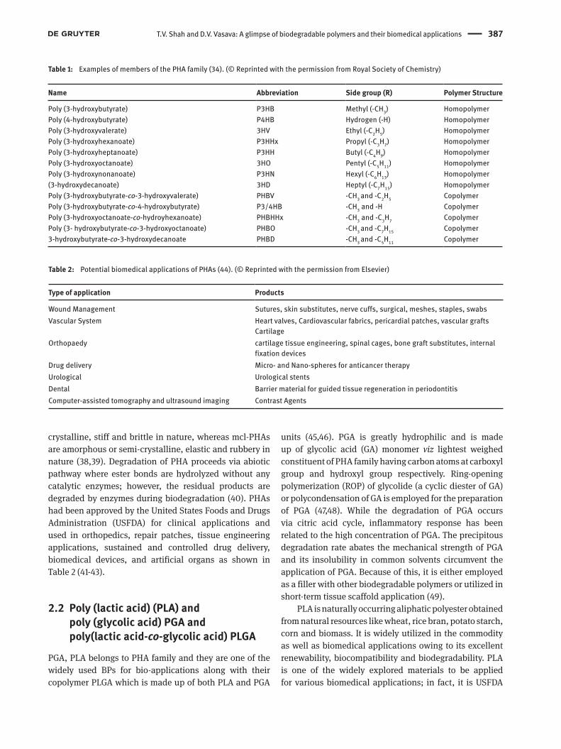
Polyhydroxyalkanoates are the versatile class of biodegrad-able and biocompatible polyesters produced by micro-organisms as an energy storage material via fermentation process under stressed conditions. A number of bacterial species from both gram-positive and gram-negative species are capable of producing PHA ranging from 30-80% of their cellular dry weight. The general formula of PHA is given in Figure 2, and till date more than 150 types of different PHA monomers have been identified and classified, thus allowing a possibility of preparing an extensive range of polymers and copolymers with different properties (34-37).
Few common monomers of this family are presented in Table 1.
Monomer units in PHA possess D (-) configuration and the molecular mass of PHA depending upon the source, ranges from 50,000 to 1,000,000 Da. In general, PHAs contain 1 to 14 carbon atoms at C-3 position, and thus can also be classified into three subcategories depending upon number of carbons at C-3 position as:
a) short chain length PHAs (scl-PHA) (up to 5 carbon atoms),
b) medium chain length PHAs (mcl-PHA) (6 to 14 carbon atoms), and
c) long chain length PHA (lcl-PHA) (>14 carbon atoms).
The scl-PHAs are brittle in nature and resemble conventional plastics in terms of properties and are
T.V. Shah and D.V. Vasava: A glimpse of biodegradable polymers and their biomedical applications 387
crystalline, stiff and brittle in nature, whereas mcl-PHAs are amorphous or semi-crystalline, elastic and rubbery in nature (38,39). Degradation of PHA proceeds via abiotic pathway where ester bonds are hydrolyzed without any catalytic enzymes; however, the residual products are degraded by enzymes during biodegradation (40). PHAs had been approved by the United States Foods and Drugs Administration (USFDA) for clinical applications and used in orthopedics, repair patches, tissue engineering applications, sustained and controlled drug delivery, biomedical devices, and artificial organs as shown in Table 2 (41-43).
2.2 Poly (lactic acid) (PLA) and poly (glycolic acid) PGA and poly(lactic acid-co-glycolic acid) PLGA
PGA, PLA belongs to PHA family and they are one of the widely used BPs for bio-applications along with their copolymer PLGA which is made up of both PLA and PGA
units (45,46). PGA is greatly hydrophilic and is made up of glycolic acid (GA) monomer viz lightest weighed constituent of PHA family having carbon atoms at carboxyl group and hydroxyl group respectively. Ring-opening polymerization (ROP) of glycolide (a cyclic diester of GA) or polycondensation of GA is employed for the preparation of PGA (47,48). While the degradation of PGA occurs via citric acid cycle, inflammatory response has been related to the high concentration of PGA. The precipitous degradation rate abates the mechanical strength of PGA and its insolubility in common solvents circumvent the application of PGA. Because of this, it is either employed as a filler with other biodegradable polymers or utilized in short-term tissue scaffold application (49).
PLA is naturally occurring aliphatic polyester obtained from natural resources like wheat, rice bran, potato starch, corn and biomass. It is widely utilized in the commodity as well as biomedical applications owing to its excellent renewability, biocompatibility and biodegradability. PLA is one of the widely explored materials to be applied for various biomedical applications; in fact, it is USFDA
CartilageOrthopaedy cartilage tissue engineering, spinal cages, bone graft substitutes, internal
fixation devicesDrug delivery Micro- and Nano-spheres for anticancer therapyUrological Urological stentsDental Barrier material for guided tissue regeneration in periodontitisComputer-assisted tomography and ultrasound imaging Contrast Agents
388 T.V. Shah and D.V. Vasava: A glimpse of biodegradable polymers and their biomedical applications
approved material used extensively for therapeutics applications including surgical implants, tissue culture, resorbable surgical sutures, wound closure, controlled release systems, and prosthetic devices (50-52).
The constituent monomer of PLA is 2-hydroxypro-pionic acid, it is a chiral molecule existing in D- and L-enantiomeric form and properties of the polymer depend on stereoregularity of monomers in polymeric backbone; thus, polymer with required properties can be achieved by varying amount of enantiomeric stereocenters (D- or L-) in resulting PLA (53-56). The general properties of lactic acid polymers are depicted in Table 3.
PLA can be synthesized from lactic acid monomers by various processes like polycondensation, ROP of lactone ring (dimer of lactic acid known as lactide) or direct methods like azeotropic dehydration and enzymatic polymerization (Figure 3). Among this, polycondensation is the cheapest method. However, it does not produce high molecular weight PLA. To achieve high molecular
weight PLA, azeotropic dehydrative condensation or ROP is generally being employed (57,58). Azeotropic polycondensation method includes removal of water molecules using appropriate azeotropic solvents, which manipulates the equilibrium of polymerization process to produce relatively high molecular weight polymers at a temperature lower than melting point temperature of the polymer (59). Kim and Woo (60) obtained high molecular weight PLA using Dean-Stark trap and molecular sieve (3 Å) to remove the water molecules formed during the polymerization process.
The low to intermediate molecular weight PLA obtained by polycondensation can be converted to high molecular weight PLA (>30,000 gmol-1) either using chain extending agents such as diols or dicarboxylic acids or esterification adjuvants such as diisocyanates (61-63). The polar oxygen linkages in PLA are accountable for its hydrophilic nature, however, the presence of methyl group side chain imparts hydrophobicity to this polymer and
T.V. Shah and D.V. Vasava: A glimpse of biodegradable polymers and their biomedical applications 389
thus PLA mildly decomposes under humid conditions. The degradation of PLA embarks with reduction of molecular weight (<10 kDa) by hydrolysis followed by random cleavage of the ester bond into the fragments of lactic acid, oligomers and other water-soluble products. Microorganisms then consume these products to degrade it into CO2, water and biomass (64). The hydrolytic degradation of PLA proceeds either via surface erosion or bulk erosion and degradation rate is dependent on molecular weight along with the temperature and pH of the medium (65). Many factors such as- polymer properties (molecular structure, polydispersity index, hydrophilic or hydrophobic character, crystallinity, chemical stability of polymer chains, trace amounts of catalyst, additives, pollutants and softening agents), geometry of implants, and condition of implant affects the degradation of PLA (66). Depending on the chirality and composition, polylactide undergoes mass loss within 6-16 months (67).
Despite having desired properties, applications of PLA are stymied by its poor crystallization rate, low crystallinity, low heat distortion temperature (HDT) and relatively high cost. Due to its slow crystallization, PLA products prepared via practical processing methods such as injection molding and extrusion exhibit poor mechanical strength and stiffness above its glass transition (Tg ~60°C) (68-70). Thus, it is necessary to improve the crystallization kinetics of PLA for its practical use. In fact, several methods such as blending (71,72), adding nucleating agents (73,74), chemical modification (68) and/or adding plasticizing agents (75) have been employed to improve the crystallization kinetics of PLA (69). Addition of nucleating agents enhances crystallization temperature and degree of crystallinity. The effect of the addition of talc (76,77), poly (D- Lactic acid) (PDLA), Zinc phenyl phosphonate, carbon nanotubes, PLA inclusion complex, natural fibers as a nucleating agent to PLA have been investigated (73,78). Vestena et al. (74) investigated an impact on crystallization kinetics of PLA matrix with the inclusion of Cellulose Nanocrystals (CNCs). It has been found that the CNC acts as a nucleating agent, which not only decreases the crystallization rate of the PLA, but also the size of spherulite and degree of crystallinity has been found to be decreasing. Jiang and group (69) have presented a comprehensive review on crystallization modification of PLA by nucleating agents and streocomplexation. In addition to nucleating agents, various plasticizers such as PEG, oligomeric lactic acid, citrate esters have been used to improve the thermal and mechanical properties of PLA (75). The plasticizing effect of biodegradable isosorbitidediester on the crystallization kinetics of PLA and its stereocomplex with PDLA has been
investigated by Srithep and Pholharn (79). The studies revealed that the stereocomplex structure elevated melting and crystallization temperature, as well as crystallization and flexibility have also been improved. In another work PLA was toughened by melt-blending with rubber grade ethylene-co -vinyl acetate (PLA/EVM) without any compatibilizer. The comparison with other PLA-based composites revealed that the toughness of the prepared composite surpassed to composites reported earlier. Also, the addition of EVM increased the elongation at break of PLA by 50 times (80).
PLGA is also sanctioned for medical use by USFDA and it is obtained by the amalgamation of PLA and PGA and is one of the commonly employed materials for the biomedical application. Copolymerization of PLA and PGA by changing the monomer ratio, allow us to tune the degradation rate as well as mechanical and chemical properties without affecting biodegradability and biocompatibility (21,81). Degradation of PLGA proceeds by hydrolysis of ester bond to yield their constituent monomer LA and GA which are found in the human body and thus safe for consumption (82).
2.3 Polysaccharides
Polysaccharides are naturally originated BPs and contain a number of functional groups which allows them to be used for various bio-applications. When compared to synthetic polymers, they are having the advantage of being biocompatible, biodegradable, non-poisonous, low cost, environment-friendly and ubiquitously available. Polysaccharides included for discussion herein are starch, cellulose, guar gum, and their derivatives.
2.3.1 Cellulose
Cellulose is one of the most abundantly obtained polysaccharides having reactive hydroxyl groups in backbone, it has promising use in the advanced polymeric material. Cellulose and its derivatives are environment-friendly materials and are extensively used due to its renewability, degradability and compatibility. However, the application of cellulose is impeded by its insolubility in common organic and inorganic solvents which lead to poor processing (83-86). Furthermore, hydrophilic nature cellulose deteriorates the mechanical properties by absorbing moisture. Thus, developments of cellulose derivatives by means of chemical modification involving reagents capable of forming a bond with reactive -OH
390 T.V. Shah and D.V. Vasava: A glimpse of biodegradable polymers and their biomedical applications
group of cellulose is one of the extensively studied topics in industry and academy which can ameliorate solubility and mechanical properties by abating hydrophilicity of cellulose (87,88).
Cellulose is a linear polymer of β-1,4 linked D-Glucose units (Figure 4) which forms a highly crystalline structure with high aspect-ratio particles which are insoluble in water but capable of H-bonding. Cellulosic material upon hydrolysis via sulfuric acid isolates the rigid, rod-shaped crystalline nanoparticles from the disordered particles. Nowadays these cellulose nanoscale materials including cellulose nanofibrils (CNFs), bacterial nanocellulose and cellulose nanocrystals (CNCs) have attained more interest (89,90). Unlike hemicellulose- which is heterogeneous chemical composition of cellulose, CNC is chemically homogenous. The degradation rate of cellulose by enzyme varies from several hours to thousands of years and is an incomplete process. The crystalline cellulose can be hydrolyzed only by a cluster of simultaneously present, interacting enzymes, or alternatively by a multienzyme complex is known as Cellulase. The cellulases hydrolyze the β–1,4–glucosidic bond between glucosyl moieties via general acid catalysis. The products are released with overall retention of inversion of the anomeric configuration at C1 (91). Entcheva et al. (92) studied the in vitro hydrolytic and enzymatic degradation of cellulose derivatives using cellulase. It was manifested from the results that the degradation byproduct was glucose and degradation rate is dependent on hydrolysis temperature and time. In addition to that, the degradation effect of cellulases was cell type specific. However, the adverse effect of cellulase during in vivo was undetermined.
2.3.2 Starch
Starch is water-soluble natural polysaccharide, it is made up of linear amylose and branched amylopectin (Figure 5) and available copiously in plant sources such as wheat,
maize, potatoes and other plants (93,94). Non-cytotoxic starch has been widely used for a variety of biomedical applications including bone and tissue engineering and drug delivery. However, virgin starch cannot be used for biomedical applications, as it is brittle in nature and degrades before its melting temperature, which makes it a poor candidate in terms of mechanical properties and processability. To overcome this, starch is usually modified physically or chemically with other BPs to improve its functionality and properties. However, there are good papers of starch modification and blends to improve its property and processability (95,96).
Starch is totally biodegradable and both the amylose and amylopectin components are readily hydrolyzed at acetal α-1-4 link by amylase and glucosidase enzyme hydrolyzes α-1-6 link of amylopectin. The endoamylase are inactive towards the hydrolysis of the branched points of amylopectin, whereas the exoamylases can hydrolyze both the branched as well as main chains to generate glucose or dimer (maltose) or trimer (maltose triose) by attacking the non-reducing end of the starch molecules. Most often, the enzyme remains associated with fragmented chain and can catalyze the hydrolysis reaction after the first cleavage (97).
2.3.3 Guar gum (GG)
Guar gum (Figure 6) is a high molecular weight natural polysaccharide having linear β-D-(1-4) linked mannose units by α-D-galactopyranose units with their linkage point at (1-6) position (98). It is extracted from Cyamopsis tetragonolobus seeds of the leguminous plant. It is USFDA permited and generally regarded as safe to use as an emulsifier, thickener, and stabilizer in food, therapeutic and cosmetic products. Besides this, it is certified in the United States Pharmacopeia (USP). Owing to its non-toxicity, biodegradability, bioavailability and biocompatibility guar gum and its derivatives are widely studied for various therapeutic applications such as drug delivery. It is a potential material for oral drug delivery systems having stability over wide pH range, also it is prone to enzymes exuded from colonic microflora in the colon (98-101). It has a considerable number of hydroxyl groups in the backbone, it shows high solubility in water which thwarts its biomedical applications. Thus, to circumvent this and improve the mechanical properties of guar gum for use in biomedical applications is one of the important objectives (102). In fact, various functional modifications of guar gum- such as esterification (103), hydroxyalkylation (104), and carboxymethylation (105) have been reported in the literature. The review article Figure 4: Structure of cellulose.
T.V. Shah and D.V. Vasava: A glimpse of biodegradable polymers and their biomedical applications 391
published by Archana George et al. (106) underscores the properties, modifications, and application of guar gum in drug delivery in detail.
2.3.4 Poly (amino acid)
This class of polymers being structurally analogous with protein has widely investigated for the biomedical applications. However, these polymers exhibit
immunogenicity and poor mechanical properties lead to the development of pseudo amino acids: an amino acid linked through non-amide linkages such as esters, imino-carbonates and carbonates. Pseudo amino acids can be obtained by reacting amino acids with other synthetic polymers, block polymers, and PEG, or copolymerization with other monomers (107,108). While amide bonds are stable hydrolytically, body possesses the wide-array of proteases can degrade proteins rapidly. These polymers have been utilized as biomaterials in sutures, scaffolds, and drug delivery devices and are mainly classified into three classes as follows (49):
(II) Poly (basic amino acid): polylysine, polyarginine, poly L-histidine
(III) Poly (neutral amino acid)
Tyrosine-based polycarbonates, polyarylates and polyesters have been derived with differing physical and mechanical properties. The polycarbonates and polyarylates show exceptional strength, superior
Figure 5: Structure of amylose and amylopectin.
Figure 6: Structure of guar gum.
392 T.V. Shah and D.V. Vasava: A glimpse of biodegradable polymers and their biomedical applications
compatibility and flexibility and elasticity respectively. PEG containing copolymers exhibit water solubility and self-assembly properties. These polymers undergo degradation by hydrolysis of non-amide bonds, to degrade into amino acid used in the polymerization. Owing to its high mechanical strength and stiffness poly (desaminotyrosinetyrosylhexyl ester carbonates) which is another pseudo-poly(amino acid) has been studied for orthopedic applications. Some other amino acids are also explored for biomedical applications including poly(L-glutamic acid) for adhesive and hemostat applications, poly(aspartic acid) as a plasma expander and Some polymers have also been investigated for their dental tissue engineering (107,109).
3 BPs for controlled delivery systems
Oral consumption is the conventional and convenient route of drug administration. However, few drugs such as protein-based drugs are susceptible to degradation by enzymes in the gastrointestinal tract (GIT). Hence they have to be administered via other routes such as infusions, injections, and implants. Therefore, for such drugs formulations have to be developed to make these drugs suitable for oral consumption. In addition to this, drugs administered by means of injection or mouth are not always best; as whole drug concentration is loaded in the body at the time which results in irritating effect and induces toxicity. To circumvent these effects and maintain optimal drug concentration, multiple administrations of drugs is often required at regular intervals. However, nowadays another approach is becoming more famous, viz designing novel drug delivery systems: Process of releasing bioactive agents at the precise rate at the exact site and simultaneously maintaining the optimum level of drug to avail maximum efficacy and minimize associated side effects can be termed drug delivery in simple terms. Due to new developments and rapid market changes in the pharmaceutical industry, drug excipients are now being used as a performance enhancer in terms of drug stability, release, and bioavailability (110-115). In this context, polymers are versatile materials for the development of drug delivery systems owing to their ability to improving pharmacokinetics by prolonging drug circulation and capability of tissue targeting. Hence, they are one of the most exploited materials to design and develop new drug delivery systems by coalescing pharmaceutical science and polymer science (116,117). Owing to its degradation into biocompatible by-products, the biodegradable polymers
formulated with drug constituents can be incorporated as used as drug delivery vehicles, which releases the drug in a controlled manner. Due to this drug concentration at the targeted site can be maintained within a therapeutic window. In addition to this, the rate of drug release can also be maneuvered with BPs. BPs including polyesters, poly (orthoesters), polyanhydrides, polyaminoacid, poly (alkyl cyanoacrylates), polyphosphazene, copolymers of PLA/PGA and aspartate are widely used to control the rate of drug release since a long time, and several of them have been successfully applied on humans. Yet, their difficult processability and irreproducible release kinetics hamper their use (111,118,119).
In the past decade, however, there has been marked an increase in the development of stimuli-responsive polymers by using smart materials which show a quick response to environmental changes. The few important (but not limited) responses include variation in temperature, pH, reduction capability, ionic strength, and magnetic field (11,120,121). The conformation, solubility and hydrophobic/ hydrophilic ratio or release of an active molecule of these polymers vary in response to external stimuli (e.g. change in temperature). In spite of this, it is also possible to develop materials which respond to two or more stimuli simultaneously (116). These polymers are advanced in terms of targeted drug delivery- as it takes advantage of the difference between milieu of the damaged and healthy cell. For instance, the pH of cancerous cells is acidic than healthier one, also they possess a high concentration of glutathione (GSH) triphosphate than extracellular fluids- which are responsible for high redox potential in cytosol and nuclei (119).
3.1 Conductive polymers
Conductive polymers (CPs) are polymers not only having inorganic metals and semiconductors like electrical and optical properties but also, they are easy to process and synthesize as conventional polymers. It has been widely used in electronic industries such as, microelectronics, photovoltaic devices and LEDs until it has been shown that CPs such as polypyrrole (PPy), polyaniline (PANi), polythiophene and their derivatives are biocompatible and biodegradable in both in vivo and in vitro, since then paradigm has been shifted to utilize these polymers in biomedical applications. However, the interest in the development of biomedical applications of these polymers had been intensified in the mid-90s when it has been shown that via electrical stimulation CPs can modulate cellular activities. CPs are good candidates for tissue
T.V. Shah and D.V. Vasava: A glimpse of biodegradable polymers and their biomedical applications 393
engineering as its biomedical applications revolve around cells responsive to electrical impulses such as nerve, bone, muscles and cardiac cells (122,123). The blending of conventional polymers with conductive polymers has been succeeded in attaining good mechanical properties and conductivity (124). Guar gum-acrylic acid- polyaniline based biodegradable, antimicrobial and electrically conductive hydrogels were prepared by Kaith et al. (125) using two-step polymerization process. Recently PHB based biodegradable semi-conductive composite embedded with in-situ polymerized acid doped PANi nanofibers have been reported (126). The presence of nano-rods of HCl-PANi significantly increased the conductivity of the non-conductive PHB films without any interaction with films. However, the biodegradability of the films was found to be decreased due to the presence of non-biodegradable HCl-PANi.
3.2 Thermo-responsive polymers
Presence of pathogens and pyrogens deviates the normal body temperature (37°C), and this variation in temperature can be utilized as a trigger to release drug particles by incorporation of various thermo-responsive drug delivery systems. In fact, thermo-responsive systems can provide an extensive range of therapeutic applications, from the field of tissue engineering to drug delivery and from artificial organs to detecting (127,128). These polymers’ solvation state rapidly alters in response to hydration state which in turn results in volume phase transition at a certain temperature. Upon the solubility condition (i.e. phase transition or sol-gel transition), these polymeric systems are broadly bifurcated into two sections as shown in Figure 7 (129-131).
a) Polymers which form a gel when the temperature is increased (two-phase region) and when the temperature is lowered below its cloud point temperature (CPT), they become soluble (single-phase region), these are Lower critical solution temperature (LCST) polymers.
b) Contrary to LCST polymers, these polymers on heating become soluble, and when cooled they again precipitates in the form of a gel, these type of polymers are Upper critical temperature (UCST) polymer.
CPT of the polymer is experimentally determined temperature where the separation of phase commences. Polymers possessing LCST behavior are mostly hydrophilic in nature than the UCST polymers which are more likely to dissolve in organic solvents. Thus, for biomedical
applications, polymers belonging to LCST class are more convenient than those of UCST polymers. However, for a given polymer-solvent system, the LCST (and thus CPT) of a given polymer can be maneuvered by adjusting the hydrophilic/hydrophobic ratio of the monomers; a number of hydrophobic polymers tend to lower CPT, while the number of hydrophilic monomers increases CPT compared to the original monomer (132). In addition to this, drug release and degradation were also controlled by varying stoichiometric ratio of monomers/blocks in polymers. This has been further countenanced from studies conducted by Jeong et al. (133) group- who have reported hydrogels made up of PLLA and PEO units linked using hexamethylene diisocyanate (HMDI) as a coupling agent. Due to the hydrophobic interaction of PLLA blocks, both diblock (PLLA-PEO) and triblock polymers (PEO-PLLA-PEO), polymers formed micelles at a lower concentration; but it was further evidence that the transition was functioned not only of concentration but was also of the composition of block polymers. The release profile of this UCST class hydrogels suggested that the %release of the model drug was a function of initial loading and hydrophobicity of drug as well as polymer concentration and degradation also affected the release of the drug. In another study (134) they investigated the prowess of PEG-PLGA-PEG triblock copolymer linked with urethane linkage, which showed LCST behavior. The critical gel concentration of this polymer was found to be 16% by thermal studies, as below this temperature no gel formation was observed. Besides this, in vitro release and degradation profile of star-shaped microspheres prepared from PEO-PLA block copolymers was also delineated- it was found that drug release and degradation profile was dependent on molecular geometry (i.e. a number of arms in a star) in later phase.
Monomers of commonly used thermo-sensitive polymers are shown in Table 4.
394 T.V. Shah and D.V. Vasava: A glimpse of biodegradable polymers and their biomedical applications
Biocompatible thermo-responsive nanoparticles of poly(ethylene glycol) methyl ether methacrylate (PEGMA) and vinyl pyrrolidone which was crosslinked with disulfide
crosslinker bis(2-acryloyloxyethyl) disulfide (DSDA) using surfactant-free polymerization method. The materials showed no cytotoxicity in vitro studies. To study the drug
Table 4: Monomers of commonly used thermo-sensitive polymers in biomedical applications with their phase transition temperature (130).
Behavior Monomer Structure/ Name of Polymer Phase transition temperature (in aqueous solution)
Application Reference
LCST
30-34°C Drug DeliveryWound Healing
(135,136)(137,138)
32-34°C Drug Delivery (139,140)
30-50°C Drug Delivery (141-143)
Tissue Enginee-ring
(144)
37°C Drug Delivery (145,146)
PEO-b-PPO 20-85°C Drug Delivery (147,148)Poly(pentapeptide) of elastin 28-30°C Drug Delivery (149,150)
UCST 25°C
Drug Delivery (98,151)Bone Cement (152-154)
Drug DeliveryBone and Tissue Engineering
(155-157)(158)
T.V. Shah and D.V. Vasava: A glimpse of biodegradable polymers and their biomedical applications 395
release prowess of these nanoparticles, two different molecular sized dyes were loaded. Interestingly, smaller sized dye releases just at body temperature while larger sized dye was not released due to smaller pore size and a decrease in diameter: the macro dye was only released when reducing agent has been introduced (11). To control protein adsorption, thermal-responsive polymers of PHA copolymer consisting of 3HD and 3-hydroxy-9-decanoate (3H9D) grafted with poly(2-dimethylamino-ethyl methacrylate) (PDMAEMA) have been reported by a group of Yao et al. (12). This non-toxic stimuli-responsive comb-like materials showed an increase in protein absorption above LCST (~ 47.5°C), but below LCST they did not absorb protein. Moreover, it was observed that the grafting of PDMAEMA increased thermal stability, in addition to protein absorption was a function of PDMAEMA content in the polymer.
Among thermosensitive polymers, PNIPAM based polymers are one of the extensively used polymers. Phase transition of these polymers is observed due to the formation of inter/intra- H-bonds above LCST in water (127,128). The CPT of PNIPAM in water is found to be around 32°C viz very close to body temperature, and thus it has been used widely for thermo-sensitive drug delivery systems such as micelles and hydrogels (159). Despite having alluring properties, PNIPAM based materials did not achieve clinical success in biomedical application owing to its non-degradability. To unravel this difficulty, various strategies have been employed to prepare biodegradable or semi-degradable PNIPAM-based materials (135,160). In addition to polymeric materials, Aluminum oxide, and superparamagnetic Fe2O3 nanoparticles have also been successfully modified with PNIPAM and reported for drug release applications (161,162). Protein release studies via PNIPAM and PEG-functionalized mesoporous silica nanoparticles reported recently (163). Release study of protein revealed the function of PNIPAM was to act as switches for protein release at body temperature, whereas the PEGlytion was carried out at the interior side of particle pores to mitigate conformational deformation of entrapped protein, and improving protein release by doing so.
3.3 pH-sensitive BPs
Amidst all stimuli, pH is the most enticing stimuli for drug targeting because of the variance in pH between normal tissues and the pathological tissue site. For instance, it has been found that tumorous cells are quite acidic (pH 6.8-7.0) than normal physiological pH (7.4). At the cellular
level, the greater difference in pH can be detected where lysosomes and endosomes of intracellular compartments are much acidic (pH 4.5-6.5) than extracellular milieu. Thus, this variation in pH can be used to steer a drug at a specific site and trigger release at an optimum rate (164,165). This can be illustrated by work carried reported by Jin-Zhi Du and coworkers (120), who synthesized pH-responsive conjugate for the release of doxorubicin. The release studies revealed that % drug release can be adjusted by varying the pH and at pH 5.0, 75% of drug release was observed. The similar results were obtained by cellular uptake behavior at different pH. Furthermore, guar gum-based pH-sensitive microparticles for colon-specific drug carriers have been prepared and were found innocuous as indicated by MTT essay studies for cytotoxicity. The drug release studies of Ibuprofen revealed that the % release of Ibuprofen was dependent on pH and drug release was found to increase with increment in pH and vice versa, whereas the neat GG showed no such effect on release (98,166,167).
3.4 Multi-stimuli responsive BPs
In addition to thermal sensitivity, dual and tri-responsive polymers are also possible to synthesize and not uncommon in the literature (119,120). These polymers, for example in addition to thermal sensitivity, they may also sensitive towards other stimuli such as pH or redox. These multi-responsive polymers allow attuning therapeutic efficacy and drug release profile. Dual thermo- and pH-sensitive hydrogels of bacterial cellulose have been prepared by grafting it with AA using electron beam irradiation technique. The morphological study exposed the highly porous structure of these hydrogels, while swelling was found to be increased as a medium was shifted towards basic. In addition to pH, the swelling % was reduced at body temperature (168). Mahdi Rahimi and group (169) have grafted chitosan on methoxylated PEG and coated it with magnetic Fe3O4 to prepare biocompatible magnetic nanocarriers for the delivery of doxorubicin and methotrexate -anticancer drugs. The in vitro studies revealed nanocarriers’ encapsulation efficiency for both the drugs were very high, furthermore, these carriers showed pH-dependent release behavior. The free drugs show taxing effects on heart and kidney, however, when these drugs with nanocarriers even at high dosage they muted the toxic effects in vivo. More recently tri-responsive micelles made up of PNIPAM and poly(2-spiropyranpropyl methacrylate)- a pH and light-sensitive moiety which undergoes a reversible transition
396 T.V. Shah and D.V. Vasava: A glimpse of biodegradable polymers and their biomedical applications
between ring-closed spiropyran state and ring-opening merocyanine state, has been carried out using reversible addition fragment transfer (RAFT) polymerization by the group of Yuan Zhang (170).
4 BPs for bone and tissue applications
Bone is complex living tissue which not only provides mechanical chassis to tissues and anchor to muscles but also acts as a calcium storehouse. It consists of 60% of minerals which are based on calcium phosphate (BCP), 30% of the collagen-based matrix and 10% of water. BCP is a mixture of β-tricalcium phosphate [TCP-Ca3(PO 4)2] and hydroxyapatite [HA- Ca10(PO4)6(OH)2] provides rigidity and tensile strength with Young’s modulus of 6-17 MPa (171-173). Regeneration of critical bone defects caused by trauma, fractures and tumors is limited due to problems associated with regular autogenic and allogenic bone grafting methods. Due to the limitation of these techniques, tissue engineering substitutes like ceramics, polymers, metals and organic bone substitutes are of mounting demand (118,174). The function of scaffolds is to provide temporary support for cells and to encourage cell separation and proliferation toward the formation of the desired new tissue. A perfect scaffold should degrade when support is no longer needed likewise it must be biocompatible, having satisfactory pore size to allow small biomolecules to pass through, mechanically robust and innocuous to host tissue. A variety of alloys such as steel and titanium along with nondegradable porous and malleable composites have been used to heal the damaged bone by providing temporary support. Apart from being nondegradable, they often necessitate a second surgery for the removal of the implant which in turn may cause fracture (26,123,175,176). Ceramics, despite having good osteoconductivity and bone-bonding, possess brittleness and poor processing (177). There are several advantages of using BPs as implant instead of their metal counterparts such as:
a) no distortion in magnetic resonance imaging (MRI)b) no need of subsequent surgery for removal of the
implant as it disintegrates after the desired duration of time,
c) this reduces the cost and risk to the patient, d) these implants are easy to sterile and prepare.
Pins and rods made up of BPs are available to cure the fractured bone, plates and screws are used to cure maxillofacial repair. Several biodegradable polymers
including PLA, PGA are commercially available and used for the orthopedic applications/ implants or devices (15,25). In addition to this, these materials are concurrently used to cure infections and growth factors by disseminating therapeutic agents. This was demonstrated by Hu et al. (178) and Rodriguez et al. (176) who prepared chitosan/HA nanocomposites by in situ hybridization and HA-PLGA polymer with biocompatible, porous and cell adhesive reticulated vitreous carbon (RVC) foam respectively. These composites showed excellent mechanical properties and cell adhesion properties to be used in bone defects as well as fixation materials (Figure 8).
Conventionally, magnesium-based materials have been used in orthopedic applications as they are ductile and their mechanical properties are analogous to cortical bone. However, the rapid corrosion rate of Mg and resulting hydrogen formation which promotes gas voids and granulation tissues around the site of the implant (23,179). To address this challenge, PLGA coatings have been applied on magnesium alloys to delay its degradation. The cell viability (ratio of living and dead cells) was performed and it was observed that coatings improved the biocompatibility and cell proliferation. However, the corrosion potential and current density studies were evident that the coatings acted as a barrier for corrosion to some degree but at high concentration, degradation was found to be aggravated due to hydrolyzation of polymer (175).
Figure 8: Common BPs used for the orthopedic implants.
T.V. Shah and D.V. Vasava: A glimpse of biodegradable polymers and their biomedical applications 397
PLA can also be used in the orthopedic application and its fracture behavior is verisimilar to stiff polymers such as polypropylene and polystyrene. PCL, on the other hand, is semi-crystalline and elastic in nature; in addition to this, the processing temperature of PCL is also similar to the PLA and thus it is a perfect plasticizing agent for PLA not only in order to improve its elasticity but also to modulate its degradation rate (180-182). Pyzik and group (183) have prepared biodegradable and asymmetric membranes of PLA-PCL for guided bone regeneration. PLA matrix was prepared via electrospinning and PCL was fabricated using freeze drying method. Where the larger pores of PCL allowed adhesion and proliferation of osteoblasts, smaller pores of PLA inhibited the overgrowth of soft tissues and facilitate neovascularization and nutrition of the bone tissue forming in place of the defect. In addition, the obtained membranes were stable for 6-weeks of incubation in distilled water and had improved mechanical strength than the native membrane. The study carried out on methacrylated PLA-PCL shows the versatility of this copolymer for orthopedic application, and it has been seen that the mechanical and degradation properties of this polymer can be modulated by varying the monomer ratio in the polymer matrix. However, the change in monomer ratio did not significantly affect the pore size, besides these highly porous copolymers were capable of being sterilized by gamma irradiation as it shows no significant alteration on gamma-ray exposure, which otherwise causes chain scission and crosslinking (184). The nanocomposite of HA and a triblock copolymer of PCL and PLA have been prepared for tissue scaffold application; morphological studies transpired that this composite was porous in nature and HA particles were well spread into the polymer matrix. The MIT assay revealed that these nanocomposites accelerated the cell growth and from in vitro degradation studies, it can be extrapolated that this nanocomposite will require up to two years to degrade completely (173). In order to ameliorate mechanical properties, carbon nanotubes (CNTs) (182) and boron nitride nanotubes (BNNTs) (185) have been encapsulated in PLA-PCL copolymer matrix by liquid dispersion method. Incorporation of CNT altered porosity of films sporadically, whereas, BNNTs gradually decreased the porosity; however, both nano-reinforcements increased elastic modulus, tensile strength, and ductility of films. There was no discrepancy in fluorescent microscopy images of cell osteoblast suggesting the biocompatibility, moreover, the cell viability of composite films was appreciably greater than those of pure PLA-PCL films.
Apart from scaffolds, BPs can also be used as bone cement which on degradation promotes bone growth by providing micropores on degradation. These polymers are often used with calcium orthophosphate which improves osteoblast, adhesion and proliferation of bone cells (174,186). Acrylics such as poly (methyl methacrylate) based materials are long been used for dental and bone cavities owing to its biostability and good mechanical properties. However, these materials possess no adhesion capacity and thus fixation of these materials in bone is quite problematic and over a long time causes loosening of fixation. This can be solved by incorporating it with BPs as shown by Espigares et al. (152) by mixing starch/cellulose acetate blends solid phase with acrylic acid and methyl methacrylate liquid phase with HA filler. The maximum temperature (Tmax) of polymerization was in agreement with that stipulated by ASTM legislation. Furthermore, the mechanical tests revealed that compressibility was ameliorated and the tensile strength of the material was parallel to the commercial acrylic bone cement. Similarly, hydroxyalkanoates such as PHB and PHBV along with a bioactive glass (silicate and borate) have been introduced to an acrylic acid matrix which enhanced mechanical properties. The degradation time for PHB and PHBV was measured by keeping the sample in phosphate buffer solution (PBS) and maximum weight loss was obtained on the 7th and 21st day of immersion respectively without altering the pH. Furthermore, the SEM micrographs of the samples showed increased cavities. This observation was supported by the MTT essay and CLSM studies which revealed a significant increase in osteoblasts especially materials with silicate glass (187). However, the aforementioned polymers are formulated by using an initiator and activator which produces free radical and can last for several weeks giving adverse side effects. To circumvent this Méndez et al. (153) incorporated vitamin E as an antioxidizing agent. Though mechanical properties and cell proliferation profile of material did not alter significantly with that of PMMA.
Injectable scaffolds are gaining more interest as it obviates the surgical procedure for the treatment of irregular shaped bone and bone regeneration. HA-coated PLGA microspheres have been prepared and found useful for the bone rejuvenation in preliminary in vivo studies carried out on mice (177). Citrate-based biodegradable, water-soluble and cross-linkable hydrogels have been reported and were found to have adequate mechanical properties. Simulated body fluid was used to carry out in vitro studies and it was found that composite allows nucleation and growth of calcium phosphate, moreover in ex vivo studies the injected composite
398 T.V. Shah and D.V. Vasava: A glimpse of biodegradable polymers and their biomedical applications
acquired the voids in bone in situ; these results clearly endorses the injectability and crosslinking capacity of this nanocomposite (188). The hyaluronic acid-based injectable hydrogels have been reported recently via click chemistry (189). These four-armed hydrogels showed low cytotoxicity and biocompatibility in vitro studies performed on chondrocytes, whereas in vivo studies showed that these composites are capable of cartilage regeneration, however, this regeneration was low and unevenly distributed due to low-stiffness of the material. In addition to these materials, injectable hydrogels made up of dextran and chitosan have also been reported by Rong Jin et al. (190,191) in which, gelation time, mechanical properties, as well as degradation rate, were dependent on the polymer concentration in the solution.
5 The modern method of synthesis of BPs
The BPs are designed such that it follows maximum principles of Green chemistry suggested by P. Anastas (192). However, ROP and Polycondensation which are the commonest routes of polymerization of PLA, PGA, and PCL generally require the use of poisonous solvents during synthesis. Use of these solvents deviates the moto of BPs and generates a paradox with the principles of green chemistry. Several methods have been developed in an attempt to synthesize BPs using green chemistry. Wang et al. (193) have used cheap, idle, unharmful, and non-flammable supercritical carbon dioxide (scCO2) to supplant the toxic solvents which have been used for the ROP of PGA in suspension polymerization. In addition to this, the elimination of scCO2 can be done simply by system depressurization and also improves enzyme activity and stability (194). This property of scCO2 has been used for ROP of macrolactone globalide, which otherwise is a difficult process and results in low molecular weight polymers. High molecular weight copolymer of PCL and globalide (Mn up to 25,000 Da) has been obtained using enzymatic ROP in scCO2 (195). There are few excellent review articles which the modern method of preparation and post modification of polymers prepared via ROP (67,196).
An alternative approach for the polymerization process is the use of enzymes as the catalysts, which provides control over reaction specificity such as enantioselectivities, chemoselectivities, regioselectivities, stereoselectivities, and choroselectivities. This selectivity of catalysis reduces unwanted side reactions and leads
to easier separation of products. Moreover, the enzymes ability to operate at milder temperature and pressure are also favorable for the “green chemistry” (197,198). There are excellent reviews on enzyme catalysis in polymer synthesis in the literature which depicts the various enzymes used for synthesis (199,200). The six major types of enzymes classified by their Enzyme Commission (EC) numbers and their capabilities in polymeric processes are formulated in Table 5.
The enzyme catalysis can be utilized for the ROP of lactones, which otherwise requires organometallic catalysts. The problem with these conventional catalysts is that they are difficult to remove, and thus, may get accumulated and causes toxic effects after polymer degradation. The enzymes, on the other hand, are natural catalysts and thus not only provide “green synthesis” but also obviates the problem of toxicity (202). Matsumura and co-workers (203) have reported ROP of TMC with lipase and have obtained polymers with MW 169000. It was noteworthy that up to 96% conversion was obtained with catalyst concentration as low as 0.25 wt% and the reaction was carried out for 24 h at 100°C, whereas under similar conditions without catalyst no conversion was observed.
Hyperbranched polymers are considered the fourth major class of polymer architecture followed by linear, crossed-linked and branched architecture. They are generally formulated by one-pot synthesis and owing highly branched structure with a number of functional groups, they possess unique physical as well as chemical properties (204-206). The six major types of hyperbranched or dendritic polymers are shown in Figure 9.
However, biocompatibility plays a major role in the designing of the ideal polymeric material as a material with low or inadequate biocompatibility and biodegradability can induce toxicity and problems in controlled release. In this regard, hyperbranched BPs have shown great potential in the field of biomedical applications. The hyperbranched
Table 5: Enzymes involved in polymerization (201).
EC Number
Biocatalyst type Polymer Synthesis
Polymer Modification
Polymer Hydrolyses
1 Oxidoreductase 2 Transferase
3 Hydrolase 4 Lyase 5 Isomerase
6 Ligase
= Yes, can be applied in polymer reaction shown = No, cannot be applied in polymer reaction shown
T.V. Shah and D.V. Vasava: A glimpse of biodegradable polymers and their biomedical applications 399
BPs can be further classified into seven sub-classes as (1) hyperbranched polyether, (2) hyperbranched polyester, (3) hyperbranched polyphosphate, (4) hyperbranched polysaccharide, (5) hyperbranched polypeptide, (6) hyperbranched polyamide, and (7) others (207). There is a number of excellent reviews describing the synthesis and medicinal use of hyperbranched BPs in the literature (205, 206, 208-210).
Wolf and Frey (211) have prepared hyperbranched polymers of PLLA copolymers by AB/ABB’-type ring opening polymerization (ROP) of lactone inimer and 5-hydroxyethyl-1,4-dioxane-2on for the drug delivery applications. Hyperbranched polymers of PLA (33) and PGA (212) with 2,2’-Bis (hydroxymethyl)-butyric acid (BHB) have been prepared by combining AB2-ROP polycondensation and possesses good thermal and rheological properties (Schemes 1 and 2). Furthermore, the terminal hydroxyl groups of these molecules can be further modified post-synthesis.
Similarly, hyperbranched polymers from functionalized caprolactone have also been reported using ROP by Liu and coworkers. Though the toxicity and compatibility of this polymer were not studied, it gave an approach for the synthesis of PCL based hyperbranched BPs (213). Zheng et al. (214) have synthesized PCL based hyperbranched BPs by combining reversible addition
fragmentation chain transfer polymerization (RAFT) and ROP polymerization.
It is also possible to functionalize hyperbranched polymers with BPs and several studies have reported such approaches in the literature (215). For example, Chen et al. (216) have used PCL and PEG to functionalize commercially available hyperbranched aliphatic polyester Boltorn H40 as a core. Subsequently, folic acid was further coupled to this polymer to achieve tumor targeting property and their drug release properties and targeting in vitro studies have been conducted with two antineoplastic drugs, 5-fluorouracil, and paclitaxel. In the different study, Wang and coworkers (217) have synthesized hyperbranched poly (amine esters) and then functionalized it with PCL using ROP and its applicability on the release of anticancer drug doxorubicin have been studied. Hyperbranched poly (ester-amide) was successfully synthesized using a series of AB3 type of monomers modified from natural gallic acid and amino acids by Li and colleagues (218). The degradation study revealed that the polymers were hydrolytically degraded and structures of degraded products were close to the starting materials and not to the monomers. Moreover, proteinase K was found to catalyze the degradation rate of polymers.
poly(N-(2-hydroxypropyl) methacrylamide) (PHPMA) is an important water-soluble and biocompatible polymer,
Figure 9: Schematic description of dendritic polymers.
400 T.V. Shah and D.V. Vasava: A glimpse of biodegradable polymers and their biomedical applications
and have shown potential to supplant the PEG (219). The hyperbranched polymers were prepared by RAFT polymerization of HPMA and butyl 2-cyanopropan-2-yl-carbontrithioate as a chain transfer agent by Alfurhood et al. (220). Such stimuli-responsive hyperbranched polymers possess great potential to be used as drug carriers if made from BPs. In fact, there are papers present discussing the potential of biodegradable hyperbranched polyglycerols functionalized with acid cleavable groups and hyperbranched polymer-Gadolinium complexes for biomedical applications (221,222).
“Click” chemistry has gained much attention recently due to the fact that they provide high yields, regiospecific and stereospecific, are immune to oxygen and water, requires milder reactions conditions and many more. The procedures satiating most (if not all) these conditions, are to be classified under click reactions. From many such click reactions, copper-catalyzed azide/alkyne click reactions, in particular, have become a puissant tool and gained much interest in the synthesis of polymers (223-225). This tool is so versatile that it is often used to functionalize BPs.
For example, Kose et al. (226) have prepared a series of biodegradable dendrimers with “clickable” end groups. Similarly, hydrophobic bio-inactive lanthanide doped nanoparticles, iron oxide nanoparticles, manganese oxide nanoparticles’ surface were modified to hydrophilic bioactive particles using thiol-ene click chemistry (225).
Recently more adaptable polymers have been prepared by using click chemistry with other polymerization methods. Such polymers not only are easy to prepare but also exhibit excellent properties to being used in various biomedical applications. Agyeefi and Scholz (227) have reported a post-bio-chemical modification of bromo and alkyl functionalized PHAs. Which were subsequently modified using copper-catalyzed cycloaddition reactions with propargyl benzoate and methyl-2-azido acetate. Similarly, Pal and Pal (228) have reported pH-sensitive PEG-b-poly (acrylamide)-b -PLA amphiphilic copolymers by combining ATRP, ROP and click chemistry. On the other hand, Zhao et al. (229) have used ATRP and RAFT in accordance with click chemistry to prepare polymeric brushes with PEO and PCL as side chains.
Scheme 2: Synthesis of dendritic polymer from L-lactide and 2,2’-bis (hydroxymethyl) butyric acid.
Scheme 1: Mechanistic scheme of preparation of soluble hyperbranched glycolide with 2,2’- Bis(hydroxymethyl) butyric acid using St(Oct)2 as a catalyst.
T.V. Shah and D.V. Vasava: A glimpse of biodegradable polymers and their biomedical applications 401
5.1 Fibers of BPs obtained via electrospinning
Electrospinning was patented in the mid-1930s for the production of textile threads and it has gained considerable interest in late 20th century after realizing the potential of this method in the medical field by the fabrication scaffolds with long, porous fibers with thin diameters which rendered them high surface to volume ratio, good mechanical properties through entanglement, high flexibility, low weight and cost (230,231). The typical electrospinning systems consists a conductive spinneret to infuse polymer solution, and a conductive grounded collector having high voltage power supply to generate a strong electric field between them as shown in Figure 10 (232,233).
At the outlet tip of the syringe, electrically charged pendant droplet of the polymer solution deforms into a conical shape known as “Taylor Cone”. As a result of the higher electrical field (which is greater than surface tension on the polymer droplet), the jet of the polymer solution is ejected from the cone. The solvent is evaporated during the travel which stretches and accelerates the polymer jet resulting in exceptionally thin polymer fibers on the collector (233). Electrospinning has attained interest owing to its ability to fabricate the fibers with required properties by varying the several parameters. The review articles published by Haider et al. (234) and Feltz et al. (230) provides comprehensive details on how electrospinning, solution, and environmental parameters affect the resultant fibers and
techniques to manipulate them. A variety of natural and synthetic polymers and also the blends of these polymers have electrospun for the numerous applications such as filtration membranes, cosmetic masks, military protective clothing, Nanosensor, energy applications, wound dressings, drug delivery, enzyme immobilization, and tissue engineering (235,236).
Arbeiter and coworkers (237) have investigated the influence of addition of formic acid, tetraethylammonium chloride (TEAC), and either 1 %wt or 12.5 %wt of Triton X-100 in PLLA solution and its fibers obtained from electrospinning. Though the morphological, mechanical, and thermal properties were found to adequate for tissue engineering applications, the biocompatibility of these fibers was not investigated in the studies. Zeng Jun and his team (238) have carried out studies on the effects of solution viscosity and electroconductivity on the diameter and morphology of electrospun PLA nanofibers with pyridinium formiate (PF) as an additive. It was noteworthy that the additive used was volatile and was evacuated simultaneously with the solvent evaporation in the electrospinning process, thus its addition did not affect the PLA fiber characteristics. The electrospun fibers of PLA and blends of PLA have been investigated for drug release (148,239) and tissue engineering applications (240,241), in which the drugs or active agents were embedded into fibers via direct addition to the polymer solution.
Gao and his team (242) have obtained fibrous scaffolds from PCL and modified it with oregano selenium
402 T.V. Shah and D.V. Vasava: A glimpse of biodegradable polymers and their biomedical applications
modified polyethyleneimine and heparin via layer- by-layer approach to facilitate nitric oxide (NO) generation. The tensile strength of these fibers was found to adequate for its use in vascular tissue engineering. Furthermore, the modified materials manifested a good catalyzing ability for the formation of NO in vivo and in vitro. In a similar experiment carried out by Li group (243), electrospun fiber scaffolds prepared from amphiphilic block copolymers of poly (caprolactone)-b-poly (sulphobetaine methacrylate) (PCL-b- PSBMA) prepared via combination ROP and ATRP. It was revealed that the hydrophilicity of these polymers was higher than that of pristine PCL, which augmented the cytocompatibility of the modified block copolymer fibers as transpired from MTT assay. The porosity of biomaterial enhances cell adhesion, migration, propagation and extracellular production within the scaffold matrix at a defected site, and is a vital criterion for tissue engineering application (244). In order to increase the porosity of electrospun fibers, myriad methods have been developed. Among these methods, phase separation technique- in which polymer is dissolved in a mixture of good and poor solvent during the electrospinning to induce thermodynamic instability within the solution. This unsteadiness in the polymer solution separates the solution into two phases: polymer-rich phase which forms solid matrix and polymer poor phase forms pores by the discharge of solvent and non-solvent out of the system (245). Porous scaffolds of PCL were prepared
via phase separation at the Loughborough University using chloroform, dichloromethane, tetrahydrofuran, and formic acid as a good solvent and its mixture with poor solvent dimethyl sulfoxide and bead free porous fibers were prepared using chloroform/ DMSO solvent at 12.5% w/v PCL concentration (246). The nano- to micro-structured electrospun fibers of PLA-co -PCL copolymers were prepared by Kwon and colleagues (247) and its cell adhesion properties have been evaluated. Human umbilical vein endothelial cells (HUVECs) cultured on these electrospun fibers for 7 days revealed that highly dense fibers (Figures 11a and 11b) favored the cell adhesion and proliferation whereas fibers with large diameter size did not promote cell adhesion (Figure 11c).
In another study reported by Milosevic et al. (248) have prepared synthetic scaffold for the artificial blood vessel grafting in which outer layer of suture was prepared from electrospun PCL on the inner layer of porous PCL-PEG copolymer. The biomechanical properties, the pore size of these scaffolds was found to adequate for scaffold materials, however, the authors did not cultured cells to corroborate the potential of this material in vivo.
The electrospun nanofibers prepared from a block copolymer of bacterial PHB have been reported by Liu and group (249). In this work, the authors polymerized PHB and of PEG using hexamethylene diisocyanate and the resultant polyurethane was subsequently electrospun without any additional treatment. Addition
T.V. Shah and D.V. Vasava: A glimpse of biodegradable polymers and their biomedical applications 403
of PEG segment to PHBV not increased the hydrophilicity of the copolymer, but also enhanced ductility of the fiber. In addition, due to enhanced hydrophilicity of material, the mineralization capability of mats prepared from these electrospun fibers in simulated body fluid (SBF) exhibited superior calcium phosphate mineralization than that of PHBV.
6 ConclusionBiodegradable polymer certainly possesses the potential in biomedical applications. Currently, the biodegradable polymers have been exploited in various biomedical applications such as drug delivery, orthopedics, gene transfection, protein delivery, bioimaging and diagnosis, tissue engineering, biomedical devices and many more. The BPs not only display specific functions, but also can undergo a dynamic regulation of structure, morphology, and function under exposure to diverse external stimuli, thereby offering a flexible and robust platform for the design and preparation of smart polymeric materials that perfectly combine dynamics and molecular order to realize multiple functions. In addition, BPs have some unusual superiority including degradability, compatibility, non-toxicity, sterility and smart responsiveness to various physiological stimuli, thereby showing immense potential in a wide range of applications in the biomedical field.
The future prospect of BPs seems promising. However, this area still faces several key challenges. Firstly, the mechanical properties of current BPs are still relatively poor, and thereby, the design and development of multiple and sophisticated polymers have become vital and indispensable. Secondly, a large number of systems constructed from non-degradable polymers and metals have been used in a wide range of applications in the biomedical field, whereas the biomedical application of BPs has not been well achieved up to now, owing to their high cost and weak mechanical properties. Thirdly, the majority of BPs have only been used for in vitro biomedical applications, and thus there is still a long way to go before their application in clinical diagnosis and rehabilitation can be attained in the future.
Acknowledgment: This research did not receive any specific grant from funding agencies in the public, commercial, or not-for-profit sectors. Authors would like to acknowledge the Department of Chemistry, Gujarat University, Ahmedabad for providing necessary facilities. The authors also acknowledge the anonymous reviewers who enhanced the overall quality of this review.
References 1. Kaczmarek H., Vukovic-Kwiatkowska I., Preparation and charac-
terization of interpenetrating networks based on polyacrylates and poly(lactic acid). Express Polym. Lett., 2012, 6(1), 78-94.
2. Jambeck J.R., Geyer R., Wilcox C., Siegler T.R., Perryman M., Andrady A., et al., Plastic waste inputs from land into the ocean. Science, 2015, 347(6223), 768-771.
3. Lackner M., Bioplastics. Kirk-Othmer Encyclopedia of Chemical Technology, 2015. p. 1-41.
4. Rochman C.M., Browne M.A., Halpern B.S., Hentschel B.T., Hoh E., Karapanagioti H.K., et al., Policy: Classify plastic waste as hazardous. Nature, 2013, 494(7436), 169-171.
5. Sinha Ray S., Yamada K., Okamoto M., Ogami A., Ueda K., New Polylactide/Layered Silicate Nanocomposites. 3. High-Performance Biodegradable Materials. Chem. Mater., 2003, 15(7), 1456-1465.
6. Ten E., Vermerris W., Recent developments in polymers derived from industrial lignin. J. Appl. Polym. Sci., 2015, 132(24).
7. Laist D.W., Overview of the biological effects of lost and discarded plastic debris in the marine environment. Mar. Pollut. Bull., 1987, 18(6), 319-326.
8. Derraik J.G., The pollution of the marine environment by plastic debris: a review. Mar. Pollut. Bull., 2002, 44(9), 842-52.
9. Ortega-Toro R., Collazo-Bigliardi S., Talens P., Chiralt A., Influence of citric acid on the properties and stability of starch-polycaprolactone based films. J. Appl. Polym. Sci., 2016, 133(2).
11. Ulasan M., Yavuz E., Bagriacik E.U., Cengeloglu Y., Yavuz M.S., Biocompatible thermoresponsive PEGMA nanoparticles crosslinked with cleavable disulfide-based crosslinker for dual drug release. J. Biomed. Mater. Res. A, 2015, 103(1), 243-51.
12. Yao H., Wei D., Che X., Cai L., Tao L., Liu L., et al., Comb-like temperature-responsive polyhydroxyalkanoate-graft-poly(2-dimethylamino-ethylmethacrylate) for controllable protein adsorption. Polym. Chem., 2016, 7(38), 5957-5965.
13. Gross R.A., Kalra B., Biodegradable polymers for the environment. Science, 2002, 297(5582), 803-807.
14. Mohanty A.K., Misra M., Hinrichsen G., Biofibres, biodegradable polymers and biocomposites: An overview. Macromol. Mater. Eng., 2000, 276-277(1), 1-24.
16. Leite Á.J., Mano J.F., Biomedical applications of natural-based polymers combined with bioactive glass nanoparticles. J. Mater. Chem. B, 2017, 5(24), 4555-4568.
17. Radwan-Pragłowska J., Piątkowski M., Janus Ł., Bogdał D., Matysek D., Biodegradable, pH-responsive chitosan aerogels for biomedical applications. RSC Adv., 2017, 7(52), 32960-32965.
18. Zhou S., Deng X., Li X., Jia W., Liu L., Synthesis and charac-terization of biodegradable low molecular weight aliphatic polyesters and their use in protein-delivery systems. J. Appl. Polym. Sci., 2004, 91(3), 1848-1856.
19. Gavasane A.J., Synthetic Biodegradable Polymers Used in Controlled Drug Delivery System: An Overview. Clin. Pharmacol. Biopharm., 2014, 3(2).
404 T.V. Shah and D.V. Vasava: A glimpse of biodegradable polymers and their biomedical applications
20. Tu Y., Peng F., Andre A.A., Men Y., Srinivas M., Wilson D.A., Biodegradable Hybrid Stomatocyte Nanomotors for Drug Delivery. ACS Nano, 2017, 11(2), 1957-1963.
21. Lih E., Park K.W., Chun S.Y., Kim H., Kwon T.G., Joung Y.K., et al., Biomimetic Porous PLGA Scaffolds Incorporating Decellularized Extracellular Matrix for Kidney Tissue Regeneration. ACS Appl. Mater. Inter., 2016, 8(33), 21145-21154.
22. Liu X., Holzwarth J.M., Ma P.X., Functionalized synthetic biodegradable polymer scaffolds for tissue engineering. Macromol. Biosci., 2012, 12(7), 911-919.
23. Farraro K.F., Kim K.E., Woo S.L., Flowers J.R., McCullough M.B., Revolutionizing orthopaedic biomaterials: The potential of biodegradable and bioresorbable magnesium-based materials for functional tissue engineering. J. Biomech., 2014, 47(9), 1979-1986.
24. Singh B., Garg T., Goyal A.K., Rath G., Recent advancements in the cardiovascular drug carriers. Artif. Cell Nanomed. B, 2016, 44(1), 216-225.
25. Weiler A., Hoffmann R.F., Stahelin A.C., Helling H.J., Sudkamp N.P., Biodegradable implants in sports medicine: the biological base. Arthroscopy, 2000, 16(3), 305-321.
30. Kallinteri P., Higgins S., Hutcheon G.A., St Pourcain C.B., Garnett M.C., Novel functionalized biodegradable polymers for nanoparticle drug delivery systems. Biomacromolecules, 2005, 6(4), 1885-1894.
31. Peesan M., Supaphol P., Rujiravanit R., Preparation and characterization of hexanoyl chitosan/polylactide blend films. Carbohyd. Polym., 2005, 60(3), 343-350.
32. Wang Z., Yu L., Ding M., Tan H., Li J., Fu Q., Preparation and rapid degradation of nontoxic biodegradable polyurethanes based on poly(lactic acid)-poly(ethylene glycol)-poly(lactic acid) andl-lysine diisocyanate. Polym. Chem., 2011, 2(3), 601-607.
34. Li Z., Loh X.J., Water soluble polyhydroxyalkanoates: future materials for therapeutic applications. Chem. Soc. Rev., 2015, 44(10), 2865-2879.
35. Wang Y., Yin J., Chen G.Q., Polyhydroxyalkanoates, challenges and opportunities. Curr. Opin. Biotechnol., 2014, 30, 59-65.
36. Wu Q., Huang H., Hu G., Chen J., Ho K.P., Chen G-Q., Production of poly-3-hydroxybutyrate by Bacillus sp. JMa5 cultivated in molassesmedia. Antonie van Leeuwenhoek., 2001, 80(2), 111-118.
38. Suriyamongkol P., Weselake R., Narine S., Moloney M., Shah S., Biotechnological approaches for the production of
polyhydroxyalkanoates in microorganisms and plants - a review. Biotechnol. Adv., 2007, 25(2), 148-175.
39. Reddy C.S., Ghai R., Rashmi, Kalia V.C., Polyhydroxyalkanoates: an overview. Bioresour. Technol., 2003, 87(2), 137-146.
40. Avérous L., Pollet E., Biodegradable Polymers. Environmental Silicate Nano-Biocomposites. Green Energy Technol., 2012., 13-39.
41. Akaraonye E., Keshavarz T., Roy I., Production of polyhydroxy-alkanoates: the future green materials of choice. J. Chem. Tech. Biot., 2010, 85(6), 732-743.
42. Keshavarz T., Roy I., Polyhydroxyalkanoates: bioplastics with a green agenda. Curr. Opin. Microbiol., 2010, 13(3), 321-326.
43. Khanna S., Srivastava A.K., Recent advances in microbial polyhy-droxyalkanoates. Process Biochem., 2005, 40(2), 607-619.
44. Zinn M., Witholt B., Egli T., Occurrence, synthesis and medical application of bacterial polyhydroxyalkanoate. Adv. Drug. Deliv. Rev. 2001, 53(1), 5-21.
45. Thomas C.M., Lutz J.F., Precision synthesis of biodegradable polymers. Angew. Chem. Int. Ed., 2011, 50(40), 9244-9246.
46. Lipsa R., Tudorachi N., Vasile C., Poly(α-hydroxyacids) in biomedical applications: synthesis and properties of lactic acid polymers. e-Polymers, 2010, 10(1).
47. Sharad J., Glycolic acid peel therapy – a current review. Clin. Cosmet. Investig. Dermatol., 2013, 6, 281-288.
48. Takahashi K., Taniguchi I., Miyamoto M., Kimura Y., Melt/solid polycondensation of glycolic acid to obtain high-molecular-weight poly(glycolic acid). Polymer, 2000, 41(24), 8725-8728.
49. Ulery B.D., Nair L.S., Laurencin C.T., Biomedical Applications of Biodegradable Polymers. J. Polym. Sci. B Polym. Phys., 2011, 49(12), 832-864.
50. Shameli K., Ahmad M.B., Yunus W.M., Ibrahim N.A., Rahman R.A., Jokar M., et al., Silver/poly (lactic acid) nanocomposites: preparation, characterization, and antibacterial activity. Int. J. Nanomedicine, 2010, 5, 573-579.
52. Perez-Madrigal M.M., Llorens E., del Valle L.J., Puiggali J., Armelin E., Aleman C., Semiconducting, biodegradable and bioactive fibers for drug delivery. Express Polym. Lett., 2016, 10(8), 628-646.
53. Tan B.H., Muiruri J.K., Li Z., He C., Recent Progress in Using Stereocomplexation for Enhancement of Thermal and Mechanical Property of Polylactide. ACS Sustain. Chem. Eng., 2016, 4(10), 5370-5391.
54. Madhavan Nampoothiri K., Nair N.R., John R.P., An overview of the recent developments in polylactide (PLA) research. Bioresource Technol., 2010, 101(22), 8493-8501.
55. Fukushima K., Furuhashi Y., Sogo K., Miura S., Kimura Y., Stereoblock poly(lactic acid): synthesis via solid-state polycon-densation of a stereocomplexed mixture of poly(L-lactic acid) and poly(D-lactic acid). Macromol. Biosci., 2005, 5(1), 21-29.
56. Rosen T., Goldberg I., Venditto V., Kol M., Tailor-Made Stereoblock Copolymers of Poly(lactic acid) by a Truly Living Polymerization Catalyst. J. Am. Chem. Soc., 2016, 138(37), 12041-12044.
57. Lasprilla A.J., Martinez G.A., Lunelli B.H., Jardini A.L., Filho R.M., Poly-lactic acid synthesis for application in biomedical devices – a review. Biotechnol. Adv., 2012, 30(1), 321-328.
58. Hamad .K, Kaseem M., Yang H.W., Deri F., Ko Y.G., Properties and medical applications of polylactic acid: A review. Express Polym. Lett., 2015, 9(5), 435-455.
T.V. Shah and D.V. Vasava: A glimpse of biodegradable polymers and their biomedical applications 405
59. Hu Y., Daoud W.A., Cheuk K.K.L., Lin C.S.K., Newly Developed Techniques on Polycondensation, Ring-Opening Polymerization and Polymer Modification: Focus on Poly(Lactic Acid). Materials, 2016, 9(3).
60. Kim K.W., Woo S.I., Synthesis of High-Molecular-Weight Poly(L-lactic acid) by Direct Polycondensation. Macromol. Chem. Phys., 2002, 203(15), 2245-2250.
61. Garlotta D., A Literature Review of Poly(Lactic Acid). J. Polym. Environ., 2001, 9(2), 63-84.
62. Namkajorn M., Petchsuk A., Opaprakasit M., Opaprakasit P., Synthesis and characterizations of degradable aliphatic-aromatic copolyesters from lactic acid, dimethyl terephthalate and diol: Effects of diol type and monomer feed ratio. Express Polym. Lett., 2010, 4(7), 415-422.
63. Woo S.I., Kim B.O., Jun H.S., Chang H.N., Polymerization of aqueous lactic acid to prepare high molecular weight poly(lactic acid) by chain-extending with hexamethylene diisocyanate. Polym. Bull., 1995, 35(4), 415-421.
64. Ndazi B.S., Karlsson S., Characterization of hydrolytic degradation of polylactic acid/rice hulls composites in water at different temperatures. Express Polym. Lett., 2011, 5(2), 119-131.
65. Elsawy M.A., Kim K-H., Park J-W., Deep A., Hydrolytic degradation of polylactic acid (PLA) and its composites. Renew. Sust. Energ. Rev., 2017, 79, 1346-1352.
66. Morawska-Chochół A., Uszko P., Szaraniec B., Gryń K., Chłopek J., The influence of bioactive additives on polylactide accelerated degradation. e-Polymers, 2016, 16(6).
67. Becker G., Wurm F.R., Functional biodegradable polymers via ring-opening polymerization of monomers without protective groups. Chem. Soc. Rev., 2018, 47(20), 7739-7782.
68. Nerkar M., Ramsay J.A., Ramsay B.A., Kontopoulou M., Dramatic Improvements in Strain Hardening and Crystallization Kinetics of PLA by Simple Reactive Modification in the Melt State. Macromol. Mater. Eng., 2014, 299(12), 1419-1424.
69. Jiang L., Shen T., Xu P., Zhao X., Li X., Dong W., et al., Crystal-lization modification of poly(lactide) by using nucleating agents and stereocomplexation. e-Polymers, 2016, 16(1), 1-13.
70. Xu T., Zhang A., Zhao Y., Han Z., Xue L., Crystallization kinetics and morphology of biodegradable poly(lactic acid) with a hydrazide nucleating agent. Polym. Test., 2015, 45, 101-106.
71. Chen Y., Zhang Y., Jiang F., Wang J., Xu Z., Wang Z., Significant enhancement of crystallization kinetics of polylactide in its immiscible blends through an interfacial effect from comb-like grafted side chains. Sci. China Chem., 2016, 59(5), 609-618.
72. Tsuji H., Sawada M., Bouapao L., Biodegradable polyesters as crystallization-accelerating agents of poly(l-lactide). ACS Appl. Mater. Inter., 2009, 1(8), 1719-1730.
73. He D., Wang Y., Shao C., Zheng G., Li Q., Shen C., Effect of phthalimide as an efficient nucleating agent on the crystal-lization kinetics of poly(lactic acid). Polym. Test., 2013, 32(6), 1088-1093.
74. Vestena M., Gross I.P., Müller C.M.O., Pires A.T.N., Nanocomposite of Poly(Lactic Acid)/Cellulose Nanocrystals: Effect of CNC Content on the Polymer Crystallization Kinetics. J. Brazil Chem. Soc., 2015.
75. Fehri M.K., Mugoni C., Cinelli P., Anguillesi I., Coltelli M.B., Fiori S., et al., Composition dependence of the synergistic effect of nucleating agent and plasticizer in poly(lactic acid): A Mixture Design study. Express Polym. Lett., 2016, 10(4), 274-288.
76. Battegazzore D., Bocchini S., Frache A., Crystallization kinetics of poly(lactic acid)-talc composites. Express Polym. Lett., 2011, 5(10), 849-858.
77. Pivsa-Art W., Fujii K., Nomura K., Aso Y., Ohara H., Yamane H., Isothermal crystallization kinetics of talc-filled poly(lactic acid) and poly(butylene succinate) blends. J. Polym. Res., 2016, 23(8).
78. Dai X., Cao Y., Shi X., Wang X., Non-isothermal crystallization kinetics, thermal degradation behavior and mechanical properties of poly(lactic acid)/MOF composites prepared by melt-blending methods. RSC Adv., 2016, 6(75), 71461-71471.
79. Srithep Y., Pholharn D., Plasticizer effect on melt blending of polylactide stereocomplex. e-Polymers, 2017, 17(5).
80. Zeng Q., Feng Y., Wang R., Ma P., Fracture behavior of highly toughened poly(lactic acid)/ethylene-co-vinyl acetate blends. e-Polymers, 2018, 18(2), 153-62.
82. Nkabinde L.A., Shoba-Zikhali L.N.N., Semete-Makokotlela B., Kalombo L., Swai H., Grobler A., et al., Poly (D,L-lactide-co-glycolide) nanoparticles: Uptake by epithelial cells and cytotoxicity. Express Polym. Lett., 2014, 8(3), 197-206.
83. Tang E., Du K., Feng X., Yuan M., Liu S., Zhao D., Controlled synthesis of cellulose-graft-poly[2-(diethylamino)-ethyl methacrylate] by ATRP in ionic liquid [AMIM]Cl and its pH-responsive property. Eur. Polym. J., 2015, 66, 228-235.
84. Carlmark A., Malmstrom E.E., ATRP grafting from cellulose fibers to create block-copolymer grafts. Biomacromolecules, 2003, 4(6), 1740-1745.
85. López de Dicastillo C., Bustos F., Guarda A., Galotto M.J., Cross-linked methyl cellulose films with murta fruit extract for antioxidant and antimicrobial active food packaging. Food Hydrocolloid., 2016, 60, 335-344.
86. Wang D., Tan J., Kang H., Ma L., Jin X., Liu R., et al., Synthesis, self-assembly and drug release behaviors of pH-responsive copolymers ethyl cellulose-graft-PDEAEMA through ATRP. Carbohyd. Polym., 2011, 84(1), 195-202.
87. Liu W., Liu R., Li Y., Kang H., Shen D., Wu M., et al., Self-assembly of ethyl cellulose-graft-polystyrene copolymers in acetone. Polymer, 2009, 50(1), 211-217.
88. Sanchez-Garcia M.D., Gimenez E., Lagaron J.M., Morphology and barrier properties of solvent cast composites of thermo-plastic biopolymers and purified cellulose fibers. Carbohyd. Polym., 2008, 71(2), 235-244.
89. Reid M.S., Villalobos M., Cranston E.D., Benchmarking Cellulose Nanocrystals: From the Laboratory to Industrial Production. Langmuir, 2017, 33(7), 1583-1598.
90. Kedzior SA, Marway H.S., Cranston E.D.. Tailoring Cellulose Nanocrystal and Surfactant Behavior in Miniemulsion Polyme-rization. Macromolecules, 2017, 50(7), 2645-2655.
91. Schwarz W.H., The cellulosome and cellulose degradation by anaerobic bacteria. Appl. Microbiol. Biot., 2001, 56(5-6), 634-649.
92. Entcheva E, Bien H, Yin L, Chung CY, Farrell M, Kostov Y. Functional cardiac cell constructs on cellulose-based scaffolding. Biomaterials, 2004, 25(26), 5753-5762.
406 T.V. Shah and D.V. Vasava: A glimpse of biodegradable polymers and their biomedical applications
93. Piermaria J.A., Pinotti A., Garcia M.A., Abraham A.G., Films based on kefiran, an exopolysaccharide obtained from kefir grain: Development and characterization. Food Hydrocolloid, 2009, 23(3), 684-690.
94. Mülhaupt R., Green Polymer Chemistry and Bio-based Plastics: Dreams and Reality. Macromol. Chem. Phys., 2013, 214(2), 159-174.
95. Ali Akbari Ghavimi S., Ebrahimzadeh M.H., Solati-Hashjin M., Abu Osman N.A., Polycaprolactone/starch composite: Fabrication, structure, properties, and applications. J. Biomed. Mater. Res. A, 2015, 103(7), 2482-2498.
97. Alberta Araújo M., Cunha A.M., Mota M., Enzymatic degradation of starch-based thermoplastic compounds used in protheses: identification of the degradation products in solution. Biomaterials, 2004, 25(13), 2687-2693.
98. Seeli D.S., Prabaharan M., Guar gum oleate-graft-poly(methacrylic acid) hydrogel as a colon-specific controlled drug delivery carrier. Carbohyd. Polym., 2017, 158, 51-57.
99. Singh V.K., Banerjee I., Agarwal T., Pramanik K., Bhattacharya M.K., Pal K., Guar gum and sesame oil based novel bigels for controlled drug delivery. Colloid. Surface. B, 2014, 123, 582-592.
100. Prakash K., Satyanarayana V., Nagiat H., Fathi A., Shanta A., Prameela A., Formulation development and evaluation of novel oral jellies of carbamazepine using pectin, guar gum, and gellan gum. Asian J. Pharm., 2014, 8(4).
101. Kumar B, Kulanthaivel S., Mondal A., Mishra S., Banerjee B., Bhaumik A., et al., Mesoporous silica nanoparticle based enzyme responsive system for colon specific drug delivery through guar gum capping. Colloid. Surface. B, 2017, 150, 352-361.
102. Jana P., Mitra T., Selvaraj T.K.R., Gnanamani A., Kundu P.P., Preparation of guar gum scaffold film grafted with ethylene-diamine and fish scale collagen, cross-linked with ceftazidime for wound healing application. Carbohyd. Polym., 2016, 153, 573-581.
103. Das S., Subuddhi U., pH-Responsive guar gum hydrogels for controlled delivery of dexamethasone to the intestine. Int. J. Biol. Macromol., 2015, 79, 856-863.
104. Chen F., Yang Y., He J., Bu T., He X., He K., et al., The gelation of hydroxypropyl guar gum by nano-ZrO2. Polym. Advan. Technol., 2018, 29(1), 587-593.
105. Dodi G., Pala A., Barbu E., Peptanariu D., Hritcu D., Popa M.I., et al., Carboxymethyl guar gum nanoparticles for drug delivery applications: Preparation and preliminary in-vitro investi-gations. Mater. Sci. Eng. C., 2016, 63, 628-636.
106. George A., Shah P.A., Shrivastav P.S., Guar gum: Versatile natural polymer for drug delivery applications. Eur. Polym. J., 2018.
107. Doppalapudi S., Jain A., Khan W., Domb A.J., Biodegradable polymers-an overview. Polym. Advan. Technol., 2014, 25(5), 427-435.
109. Zhao Y., Su H., Fang L., Tan T., Superabsorbent hydrogels from poly(aspartic acid) with salt-, temperature- and pH-respon-siveness properties. Polymer, 2005, 46(14), 5368-5376.
110. Park J.H., Allen M.G., Prausnitz M.R., Biodegradable polymer microneedles: fabrication, mechanics and transdermal drug delivery. J. Control. Release., 2005, 104(1), 51-66.
111. Veronovski A., Tkalec G., Knez Z., Novak Z., Characterisation of biodegradable pectin aerogels and their potential use as drug carriers. Carbohyd. Polym., 2014, 113, 272-278.
112. García-González C.A., Alnaief M., Smirnova I., Polysaccharide-based aerogels—Promising biodegradable carriers for drug delivery systems. Carbohyd. Polym., 2011, 86(4), 1425-1438.
113. Hardy J.G., Mouser D.J., Arroyo-Currás N., Geissler S., Chow J.K., Nguy L., et al., Biodegradable electroactive polymers for electrochemically-triggered drug delivery. J. Mater. Chem. B, 2014, 2(39), 6809-6822.
114. Parveen S., Misra R., Sahoo S.K., Nanoparticles: a boon to drug delivery, therapeutics, diagnostics and imaging. Nanomedicine, 2012, 8(2), 147-66.
115. Malik R., Garg T., Goyal A.K., Rath G., Polymeric nanofibers: targeted gastro-retentive drug delivery systems. J. Drug. Target., 2015, 23(2), 109-124.
116. Schmaljohann D., Thermo- and pH-responsive polymers in drug delivery. Adv. Drug. Deliv. Rev., 2006, 58(15), 1655-1670.
117. Pillai O., Panchagnula R., Polymers in drug delivery. Curr. Opin. Chem. Biol., 200, 5(4), 447-451.
118. Saito N., Murakami N., Takahashi J., Horiuchi H., Ota H., Kato H., et al., Synthetic biodegradable polymers as drug delivery systems for bone morphogenetic proteins. Adv. Drug. Deliv. Rev., 2005, 57(7), 1037-1048.
119. Cheng R., Meng F., Deng C., Klok H.A., Zhong Z., Dual and multi-stimuli responsive polymeric nanoparticles for programmed site-specific drug delivery. Biomaterials, 2013, 34(14), 3647-3657.
120. Du J.Z., Du X.J., Mao C.Q., Wang J., Tailor-made dual pH-sensitive polymer-doxorubicin nanoparticles for efficient anticancer drug delivery. J. Am. Chem. Soc., 2011, 133(44), 17560-17563.
121. Cheng X., Jin Y., Sun T., Qi R., Fan B., Li H., Oxidation- and thermo-responsive poly(N-isopropylacrylamide-co-2-hydroxy ethyl acrylate) hydrogels cross-linked via diselenides for controlled drug delivery. RSC Adv., 2015, 5(6), 4162-4170.
129. Ou C.W., Su C.H., Jeng U.S., Hsu S.H., Characterization of biodegradable polyurethane nanoparticles and thermally induced self-assembly in water dispersion. ACS Appl. Mater. Inter., 2014, 6(8), 5685-5694.
130. Gandhi A., Paul A., Sen S.O., Sen K.K., Studies on thermo-responsive polymers: Phase behaviour, drug delivery and biomedical applications. Asian Journal of Pharmaceutical Sciences, 2015, 10(2), 99-107.
131. Klouda L., Thermoresponsive hydrogels in biomedical applications: A seven-year update. Eur. J. Pharm. Biopharm., 2015, 97, 338-49.
132. Hrubý M., Filippov S.K., Štěpánek P., Smart polymers in drug delivery systems on crossroads: Which way deserves following? Eur. Polym. J., 2015, 65, 82-97.
133. Jeong B., Bae Y.H., Lee D.S., Kim S.W., Biodegradable block copolymers as injectable drug-delivery systems. Nature, 1997, 388(6645), 860-862.
134. Jeong B., Choi Y.K., Bae Y.H., Zentner G., Kim S.W., New biodegradable polymers for injectable drug delivery systems. J. Control. Release, 1999, 62(1-2), 109-114.
135. Gan J., Guan X., Zheng J., Guo H., Wu K., Liang L., et al., Biodegradable, thermoresponsive PNIPAM-based hydrogel scaffolds for the sustained release of levofloxacin. RSC Adv., 2016, 6(39), 32967-32978.
136. Zhao C., Zhuang X., He P., Xiao C., He C., Sun J., et al., Synthesis of biodegradable thermo- and pH-responsive hydrogels for controlled drug release. Polymer, 2009, 50(18), 4308-4316.
137. Ohya S., Sonoda H., Nakayama Y., Matsuda T., The potential of poly(N-isopropylacrylamide) (PNIPAM)-grafted hyaluronan and PNIPAM-grafted gelatin in the control of post-surgical tissue adhesions. Biomaterials, 2005, 26(6), 655-659.
138. Chen S., Shi J., Xu X., Ding J., Zhong W., Zhang L., et al., Study of stiffness effects of poly(amidoamine)-poly(n-isopropyl acrylamide) hydrogel on wound healing. Colloid. Surface. B, 2016, 140, 574-582.
139. Nelson J.L., Roeder B.L., Carmen J.C., Roloff F., Pitt W.G., Ultrasonically activated chemotherapeutic drug delivery in a rat model. Cancer Res., 2002, 62(24), 7280-7283.
140. Chen J., Liu M., Liu H., Ma L., Synthesis, swelling and drug release behavior of poly(N,N-diethylacrylamide-co-N-hydroxymethyl acrylamide) hydrogel. Mater. Sci. Eng. C, 2009, 29(7), 2116-2123.
141. Rejinold N.S., Chennazhi K.P., Nair S.V., Tamura H., Jayakumar R., Biodegradable and thermo-sensitive chitosan-g-poly(N-vinylcaprolactam) nanoparticles as a 5-fluorouracil carrier. Carbohyd. Polym., 2011, 83(2), 776-786.
142. Madhusudana Rao K., Krishna Rao K.S.V., Sudhakar P., Chowdoji Rao K., Subha M.C.S., Synthesis and characterization of biodegradable poly (vinyl caprolactam) grafted on to sodium alginate and its microgels for controlled release studies of an anticancer drug. J. Appl. Pharm. Sci., 2013, 3(6), 61-69.
143. Wang Y., Nie J., Chang B., Sun Y., Yang W., Poly(vinylcaprolactam)-based biodegradable multiresponsive microgels for drug delivery. Biomacromolecules, 2013, 14(9), 3034-3046.
144. Srivastava A., Kumar A., Thermoresponsive poly(N-vinylcap-rolactam) cryogels: synthesis and its biophysical evaluation for tissue engineering applications. J. Mater. Sci.-Mater. Med., 2010, 21(11), 2937-2945.
145. Varshosaz J., Hassanzadeh F., Sadeghi-aliabadi H., Larian Z., Rostami M., Synthesis of Pluronic® F127-poly (methyl vinyl ether-alt-maleic acid) copolymer and production of its micelles for doxorubicin delivery in breast cancer. Chem. Eng. J., 2014, 240, 133-146.
146. Yoncheva K., Lizarraga E., Irache J.M., Pegylated nanoparticles based on poly(methyl vinyl ether-co-maleic anhydride): preparation and evaluation of their bioadhesive properties. Eur. J. Pharm. Sci., 2005, 24(5), 411-419.
148. Loiola L.M.D., Cortez Tornello P.R., Abraham G.A., Felisberti M.I., Amphiphilic electrospun scaffolds of PLLA–PEO–PPO block copolymers: preparation, characterization and drug-release behaviour. RSC Adv., 2017, 7(1), 161-172.
149. Wu Y., MacKay J.A., McDaniel J.R., Chilkoti A., Clark R.L., Fabrication of elastin-like polypeptide nanoparticles for drug delivery by electrospraying. Biomacromolecules, 2009, 10(1), 19-24.
150. Meikle S.T., Pineiro Y., Banobre Lopez M., Rivas J., Santin M., Surface functionalization superparamagnetic nanoparticles conjugated with thermoresponsive poly(epsilon-lysine) dendrons tethered with carboxybetaine for the mild hyperthermia-controlled delivery of VEGF. Acta Biomater., 2016, 40, 235-242.
151. Murali R., Vidhya P., Thanikaivelan P., Thermoresponsive magnetic nanoparticle--aminated guar gum hydrogel system for sustained release of doxorubicin hydrochloride. Carbohyd. Polym., 2014, 110, 440-445.
152. Espigares I., Elvira C., Mano J.F., Vazquez B., San R.J., Reis R.L., New partially degradable and bioactive acrylic bone cements based on starch blends and ceramic fillers. Biomaterials, 2002, 23(8), 1883-1895.
153. Mendez J.A., Aguilar M.R., Abraham G.A., Vazquez B., Dalby M., Di Silvio L., et al., New acrylic bone cements conjugated to vitamin E: curing parameters, properties, and biocompatibility. J. Biomed. Mater. Res., 2002, 62(2), 299-307.
154. Franco-Marquès E., Méndez J.A., Gironès J., Pèlach M.A., Thermal and dynamic mechanical characterization of acrylic bone cements modified with biodegradable polymers. J. Appl. Polym. Sci., 2013, 128(5), 3455-3464.
155. Soppimath K.S., Kulkarni A.R., Aminabhavi T.M., Chemically modified polyacrylamide-g-guar gum-based crosslinked anionic microgels as pH-sensitive drug delivery systems: preparation and characterization. J. Control. Release, 2001, 75(3), 331-345.
156. Elvira C., Mano J.F., San Román J., Reis R.L., Starch-based biodegradable hydrogels with potential biomedical applications as drug delivery systems. Biomaterials, 2002, 23(9), 1955-1966.
157. Wang S., Kim G., Lee Y.E., Hah H.J., Ethirajan M., Pandey R.K., et al., Multifunctional biodegradable polyacrylamide nanocarriers for cancer theranostics--a “see and treat” strategy. ACS Nano, 2012, 6(8), 6843-6851.
158. Kretlow J.D., Mikos A.G., Review: mineralization of synthetic polymer scaffolds for bone tissue engineering. Tissue Eng., 2007, 13(5), 927-938.
159. Soga O., van Nostrum C.F., Hennink W.E., Poly(N-(2-hydroxy propyl) methacrylamide mono/di lactate): a new class of
408 T.V. Shah and D.V. Vasava: A glimpse of biodegradable polymers and their biomedical applications
biodegradable polymers with tuneable thermosensitivity. Biomacromolecules, 2004, 5(3), 818-821.
160. Ju X.J., Xie R., Yang L., Chu L.Y., Biodegradable ‘intelligent’ materials in response to physical stimuli for biomedical applications. Expert. Opin. Ther. Pat., 2009, 19(4), 493-507.
161. Sanz B., von Bilderling C., Tuninetti J.S., Pietrasanta L., Mijangos C., Longo G.S., et al., Thermally-induced softening of PNIPAm-based nanopillar arrays. Soft Matter, 2017, 13(13), 2453-2464.
162. Yar Y., Khodadust R., Akkoc Y., Utkur M., Saritas E.U., Gozuacik D., et al., Development of tailored SPION-PNIPAM nanoparticles by ATRP for dually responsive doxorubicin delivery and MR imaging. J. Mater. Chem. B, 2018, 6(2), 289-300.
163. Yu E., Lo A., Jiang L., Petkus B., Ileri Ercan N., Stroeve P., Improved controlled release of protein from expanded-pore mesoporous silica nanoparticles modified with co-functio-nalized poly(n-isopropylacrylamide) and poly(ethylene glycol) (PNIPAM-PEG). Colloid. Surface. B, 2017, 149, 297-300.
164. Chen W., Meng F., Cheng R., Zhong Z., pH-Sensitive degradable polymersomes for triggered release of anticancer drugs: a comparative study with micelles. J. Control. Release., 2010, 142(1), 40-46.
166. Seeli D.S., Dhivya S., Selvamurugan N., Prabaharan M., Guar gum succinate-sodium alginate beads as a pH-sensitive carrier for colon-specific drug delivery. Int. J. Biol. Macromol., 2016, 91, 45-50.
167. Seeli D.S., Prabaharan M., Guar gum succinate as a carrier for colon-specific drug delivery. Int. J. Biol. Macromol., 2016, 84, 10-15.
168. Mohd Amin M.C.I., Ahmad N., Halib N., Ahmad I., Synthesis and characterization of thermo- and pH-responsive bacterial cellulose/acrylic acid hydrogels for drug delivery. Carbohyd. Polym., 2012, 88(2), 465-473.
169. Rahimi M., Safa K.D., Salehi R., Co-delivery of doxorubicin and methotrexate by dendritic chitosan-g-mPEG as a magnetic nanocarrier for multi-drug delivery in combination chemotherapy. Polym. Chem., 2017, 8(47), 7333-7350.
170. Zhang Y., Cao M., Yuan B., Guo T., Zhang W., RAFT synthesis and micellization of a photo-, temperature- and pH-responsive diblock copolymer based on spiropyran. Polym. Chem., 2017, 8(47), 7325-32.
171. Razak S.I.A., Sharif N.F.A., Rahman W.A.W.A., Biodegradable polymers and their bone applications: A review. Int. J. Basic. Appl. Sci., 2012, 12(1), 31-49.
172. Fukushima K., Poly(trimethylene carbonate)-based polymers engineered for biodegradable functional biomaterials. Biomater. Sci., 2016, 4(1), 9-24.
173. Torabinejad B., Mohammadi-Rovshandeh J., Davachi S.M., Zamanian A., Synthesis and characterization of nanocomposite scaffolds based on triblock copolymer of L-lactide, epsilon-caprolactone and nano-hydroxyapatite for bone tissue engineering. Mater. Sci. Eng. C., 2014, 42, 199-210.
174. Yu N.Y., Schindeler A., Little D.G., Ruys A.J., Biodegradable poly(alpha-hydroxy acid) polymer scaffolds for bone tissue engineering. J. Biomed. Mater. Res. B, 2010, 93(1), 285-295.
175. Ostrowski N.J., Lee B., Roy A., Ramanathan M., Kumta P.N., Biodegradable poly(lactide-co-glycolide) coatings on
magnesium alloys for orthopedic applications. J. Mater. Sci.-Mater. Med., 2013, 24(1), 85-96.
176. Rodriguez D.E., Guiza-Arguello V., Ochoa O.O., Gharat T., Sue H.J., Lafdi K., et al., Development of a hydroxyapatite-poly( d , l -lactide-co-glycolide) infiltrated carbon foam for orthopedic applications. Carbon, 2016, 98, 106-114.
177. Kang S.W., Yang H.S., Seo S.W., Han D.K., Kim B.S., Apatite-coated poly(lactic-co-glycolic acid) microspheres as an injectable scaffold for bone tissue engineering. J. Biomed. Mater. Res. A, 2008, 85(3), 747-756.
178. Hu Q., Preparation and characterization of biodegradable chitosan/hydroxyapatite nanocomposite rods via in situ hybridization: a potential material as internal fixation of bone fracture. Biomaterials, 2004, 25(5), 779-785.
179. Witte F., The history of biodegradable magnesium implants: a review. Acta Biomater., 2010, 6(5), 1680-1692.
180. Takayama T., Todo M., Improvement of impact fracture properties of PLA/PCL polymer blend due to LTI addition. J. Mater. Sci., 2006, 41(15), 4989-4992.
181. Cabedo L., Luis Feijoo J., Pilar Villanueva M., Lagarón J.M., Giménez E., Optimization of Biodegradable Nanocomposites Based on aPLA/PCL Blends for Food Packaging Applications. Macromolecular Symp., 2006, 233(1), 191-197.
182. Lahiri D., Rouzaud F., Namin S., Keshri A.K., Valdes J.J., Kos L., et al., Carbon nanotube reinforced polylactide-caprolactone copolymer: mechanical strengthening and interaction with human osteoblasts in vitro. ACS Appl. Mater. Inter., 2009, 1(11), 2470-2476.
183. Domalik-Pyzik P., Morawska-Chochół A., Chłopek J., Rajzer I., Wrona A., Menaszek E., et al., Polylactide/polycaprolactone asymmetric membranes for guided bone regeneration. e-Polymers, 2016, 16(5).
184. Felfel R.M., Poocza L., Gimeno-Fabra M., Milde T., Hildebrand G., Ahmed I., et al., In vitro degradation and mechanical properties of PLA-PCL copolymer unit cell scaffolds generated by two-photon polymerization. Biomed. Mater., 2016, 11(1), 015011.
185. Lahiri D., Rouzaud F., Richard T., Keshri A.K., Bakshi S.R., Kos L., et al., Boron nitride nanotube reinforced polylactide-polyca-prolactone copolymer composite: mechanical properties and cytocompatibility with osteoblasts and macrophages in vitro. Acta Biomater., 2010, 6(9), 3524-3533.
186. Asghari F., Samiei M., Adibkia K., Akbarzadeh A., Davaran S., Biodegradable and biocompatible polymers for tissue engineering application: a review. Artif. Cell. Nanomed. B, 2017, 45(2), 185-192.
187. Lopes P.P., Garcia M.P., Fernandes M.H., Fernandes M.H., Acrylic formulations containing bioactive and biodegradable fillers to be used as bone cements: properties and biocompa-tibility assessment. Mater. Sci. Eng. C., 2013, 33(3), 1289-1299.
188. Gyawali D., Nair P., Kim H.K., Yang J., Citrate-based Biodegradable Injectable hydrogel Composites for Orthopedic Applications. Biomater. Sci., 2013, 1(1), 52-64.
189. Han S-S., Yoon H.Y., Yhee J.Y., Cho M.O., Shim H-E., Jeong J-E., et al., In situ cross-linkable hyaluronic acid hydrogels using copper free click chemistry for cartilage tissue engineering. Polym. Chem., 2018, 9(1), 20-27.
190. Jin R., Hiemstra C., Zhong Z., Feijen J., Enzyme-mediated fast in situ formation of hydrogels from dextran-tyramine conjugates. Biomaterials, 2007, 28(18), 2791-2800.
T.V. Shah and D.V. Vasava: A glimpse of biodegradable polymers and their biomedical applications 409
191. Jin R., Moreira Teixeira L.S., Dijkstra P.J., Karperien M., van Blitterswijk C.A., Zhong Z.Y., et al., Injectable chitosan-based hydrogels for cartilage tissue engineering. Biomaterials, 2009, 30(13), 2544-2551.
192. Anastas P., Eghbali N., Green chemistry: principles and practice. Chem. Soc. Rev., 2010, 39(1), 301-312.
193. Wang T., Hao J., Liu Y., Gu Z., Formation of well-defined spherical particles during suspension polymerization of biodegradable poly(glycolide-co-p-dioxanone) in supercritical carbon dioxide. RSC Adv., 2012, 2(27).
194. Polloni A.E., Veneral J.G., Rebelatto E.A., de Oliveira D., Oliveira J.V., Araújo P.H.H., et al., Enzymatic ring opening polyme-rization of ω-pentadecalactone using supercritical carbon dioxide. J. Supercrit. Fluids, 2017, 119, 221-228.
195. Guindani C., Dozoretz P., Veneral J.G., da Silva D.M., Araújo P.H.H., Ferreira S.R.S., et al., Enzymatic ring opening copoly-merization of globalide and ε-caprolactone under supercritical conditions. J. Supercrit. Fluids, 2017, 12, 404-411.
196. Chen T., Cai T., Jin Q., Ji JJe-P., Design and fabrication of functional polycaprolactone. e-Polymers, 2015, 15(1), 3-13.
198. Matsumura S., Enzyme-Catalyzed Synthesis and Chemical Recycling of Polyesters. Macromol BioscI., 2002, 2(3).
199. Teixeira L.S., Feijen J., van Blitterswijk C.A., Dijkstra P.J., Karperien M., Enzyme-catalyzed crosslinkable hydrogels: emerging strategies for tissue engineering. Biomaterials, 2012, 33(5), 1281-1290.
200. Varma I.K., Albertsson A-C., Rajkhowa R., Srivastava R.K., Enzyme catalyzed synthesis of polyesters. Prog. Polym. Sci., 2005, 30(10), 949-981.
201. Anna Calarco A.D.S., A Review on Extremozymes Biocatalysis: A Green Industrial Approach for Biomaterials Production. J. Biomol. Res. Ther., 2015, 04(01).
203. Matsumura S., Tsukada K., Toshima K., Enzyme-Catalyzed Ring-Opening Polymerization of 1,3-Dioxan-2-one to Poly(trimethylene carbonate). Macromolecules, 1997, 30(10), 3122-3124.
204. Gao C., Yan D., Hyperbranched polymers: from synthesis to applications. Prog. Polym. Sci., 2004, 29(3), 183-275.
205. Voit B.I., Lederer A., Hyperbranched and highly branched polymer architectures--synthetic strategies and major charac-terization aspects. Chem. Rev., 2009, 109(11), 5924-5973.
206. Abbina S., Vappala S., Kumar P., Siren E.M.J., La C.C., Abbasi U., et al., Hyperbranched polyglycerols: recent advances in synthesis, biocompatibility and biomedical applications. J. Mater. Chem. B, 2017, 5(47), 9249-9277.
207. Huang Y., Wang D., Zhu X., Yan D., Chen R., Synthesis and therapeutic applications of biocompatible or biodegradable hyperbranched polymers. Polym. Chem., 2015, 6(15), 2794-2812.
208. Stiriba S-E., Frey H., Haag R., Dendritic Polymers in Biomedical Applications: From Potential to Clinical Use in Diagnostics and Therapy. Angew. Chem. Int. Edit., 2002, 41(8), 1329-1334.
209. Esfand R., Tomalia D.A., Poly(amidoamine) (PAMAM) dendrimers: from biomimicry to drug delivery and biomedical applications. Drug Discov. Today, 2001, 6(8), 427-436.
210. Dykes G.M., Dendrimers: a review of their appeal and applications. J. Chem. Technol. Biot., 2001, 76(9), 903-918.
211. Wolf F.K., Frey H., Inimer-Promoted Synthesis of Branched and Hyperbranched Polylactide Copolymers. Macromolecules, 2009, 42(24), 9443-9456.
213. Liu M., Vladimirov N., Fréchet J.M.J., A New Approach to Hyperbranched Polymers by Ring-Opening Polymerization of an AB Monomer: 4-(2-Hydroxyethyl)-ε-caprolactone. Macromo-lecules, 1999, 32(20), 6881-6884.
214. Zheng Y., Turner W., Zong M., Irvine D.J., Howdle S.M., Thurecht K.J., Biodegradable Core−Shell Materials via RAFT and ROP: Characterization and Comparison of Hyperbranched and Microgel Particles. Macromolecules, 2011, 44(6), 1347-1354.
217. Wang T., Li M., Gao H., Wu Y., Nanoparticle carriers based on copolymers of poly(epsilon-caprolactone) and hyperbranched polymers for drug delivery. J. Colloid Interf. Sci., 2011, 353(1), 107-115.
218. Li X., Su Y., Chen Q., Lin Y., Tong Y., Li Y., Synthesis and charac-terization of biodegradable hyperbranched poly(ester-amide)s based on natural material. Biomacromolecules, 2005, 6(6), 3181-3188.
221. Sideratou Z., Agathokleous M., Theodossiou T.A., Tsiourvas D., Functionalized Hyperbranched Polyethylenimines as Thermo-sensitive Drug Delivery Nanocarriers with Controlled Transition Temperatures. Biomacromolecules, 2018, 19(2), 315-328.
222. Shenoi R.A., Lai B.F., Kizhakkedathu J.N., Synthesis, characte-rization, and biocompatibility of biodegradable hyperbranched polyglycerols from acid-cleavable ketal group functionalized initiators. Biomacromolecules, 2012, 13(10), 3018-3030.
223. Kolb H.C., Finn M.G., Sharpless K.B., Click Chemistry: Diverse Chemical Function from a Few Good Reactions. Angew. Chem. Int. Edit., 2001, 40(11), 2004-2021.
225. Liu Y., Hou W., Sun H., Cui C., Zhang L., Jiang Y., et al., Thiol-ene click chemistry: a biocompatible way for orthogonal biocon-jugation of colloidal nanoparticles. Chem. Sci., 2017, 8(9), 6182-6187.
226. Kose M.M., Onbulak S., Yilmaz I.I., Sanyal A., Orthogonally “Clickable” Biodegradable Dendrons. Macromolecules, 2011, 44(8), 2707-2714.
227. Nkrumah-Agyeefi S., Scholz C., Chemical modification of functionalized polyhydroxyalkanoates via “Click” chemistry: A proof of concept. Int. J. Biol. Macromol., 2017, 95, 796-808.
228. Pal A., Pal S., Synthesis of poly (ethylene glycol)- block -poly (acrylamide)- block -poly (lactide) amphiphilic copolymer
410 T.V. Shah and D.V. Vasava: A glimpse of biodegradable polymers and their biomedical applications
through ATRP, ROP and click chemistry: Characterization, micellization and pH-triggered sustained release behaviour. Polymer, 2017, 127, 150-158.
229. Zhao P., Yan Y., Feng X., Liu L., Wang C., Chen Y., Highly efficient synthesis of polymer brushes with PEO and PCL as side chains via click chemistry. Polymer, 2012, 53(10), 1992-2000.
230. Feltz K.P., Kalaf E.A.G., Chen C., Martin R.S., Sell S.A., A review of electrospinning manipulation techniques to direct fiber deposition and maximize pore size. Electrospinning, 2017, 1(1).
231. Safarik I., Pospiskova K., Baldikova E., Savva I., Vekas L., Marinica O., et al., Fabrication and Bioapplications of Magnetically Modified Chitosan-based Electrospun Nanofibers. Electrospinning, 2018, 2(1), 29-39.
232. Chen R., Liu J., Sun Z., Chen D., Functional Nanofibers with Multiscale Structure by Electrospinning. Nanofabrication, 2018, 4(1), 17-31.
233. Kai D., Liow S.S., Loh X.J., Biodegradable polymers for electro-spinning: towards biomedical applications. Mater. Sci. Eng. C., 2014, 45, 659-670.
234. Haider A., Haider S., Kang I-K., A comprehensive review summarizing the effect of electrospinning parameters and potential applications of nanofibers in biomedical and biotechnology. Arab. J. Chem., 2018, 11(8), 1165-1188.
235. Wang X., Ding B., Li B., Biomimetic electrospun nanofibrous structures for tissue engineering. Mater. Today, 2013, 16(6), 229-241.
236. Hu X., Liu S., Zhou G., Huang Y., Xie Z., Jing X., Electrospinning of polymeric nanofibers for drug delivery applications. J. Control. Release, 2014, 185, 12-21.
237. Arbeiter D., Kohse S., Eickner T., Grabow N., Schmitz K-P., Influence of additives on physico-chemical properties of electrospun poly(L-lactide). Current Directions in Biomedical Engineering, 2018, 4(1), 493-496.
238. Zeng J., Haoqing H., Schaper A., Wendorff J.H., Greiner A., Poly-L-lactide nanofibers by electrospinning – Influence of solution viscosity and electrical conductivity on fiber diameter and fiber morphology. e-Polymers, 2003, 3(1).
239. Zeng J., Xu X., Chen X., Liang Q., Bian X., Yang L., et al., Biodegradable electrospun fibers for drug delivery. J. Control. Release, 2003, 92(3), 227-231.
240. Lins L.C., Wianny F., Livi S., Hidalgo I.A., Dehay C., Duchet-Rumeau J., et al., Development of Bioresorbable Hydrophilic-Hydrophobic Electrospun Scaffolds for Neural Tissue Engineering. Biomacromolecules, 2016, 17(10), 3172-3187.
241. Pu J., Yuan F., Li S., Komvopoulos K., Electrospun bilayer fibrous scaffolds for enhanced cell infiltration and vascularization in vivo. Acta Biomater., 2015, 13, 131-141.
242. Gao J., Wang Y., Chen S., Tang D., Jiang L., Kong D., et al., Electrospun poly-ε-caprolactone scaffold modified with catalytic nitric oxide generation and heparin for small-diameter vascular graft. RSC Adv., 2017, 7(30), 18775-18784.
243. Li Q., Wang Z., Zhao Q., Wang L., Wang S., Kong D., Tissue engineering scaffold electrospun from poly(ε- caprolactone)-b-poly(sulfobetaine methacrylate) block copolymers with improved hydrophilicity and good cytocompatibility. e-Polymers, 2012, 12(1).
244. Stevanovic M., Pavlovic V., Petkovic J., Filipic M., Uskokovic D., ROS-inducing potential, influence of different porogens and in vitro degradation of poly (D,L-lactide-co-glycolide)-based material. Express Polym. Lett., 2011, 5(11), 996-1008.
245. Fashandi H., Karimi M., Pore formation in polystyrene fiber by superimposing temperature and relative humidity of electro-spinning atmosphere. Polymer, 2012, 53(25), 5832-5849.
247. Kwon I.K., Kidoaki S., Matsuda T., Electrospun nano- to microfiber fabrics made of biodegradable copolyesters: structural characteristics, mechanical properties and cell adhesion potential. Biomaterials, 2005, 26(18), 3929-3939.
248. Milosevic M., Mijailovic N., Nikolic D., Filipovic N., Peulic A., Rosic M., et al., Manufacturing of Biodegradable Scaffolds to Engineer Artificial Blood Vessel. Serbian Journal of Experimental and Clinical Research, 2018, 19(3), 215-221.
249. Liu K.L., Choo E.S., Wong S.Y., Li X., He C.B., Wang J., et al., Designing poly[(R)-3-hydroxybutyrate]-based polyurethane block copolymers for electrospun nanofiber scaffolds with improved mechanical properties and enhanced mineralization capability. J. Phys. Chem. B, 2010, 114(22), 7489-7498.