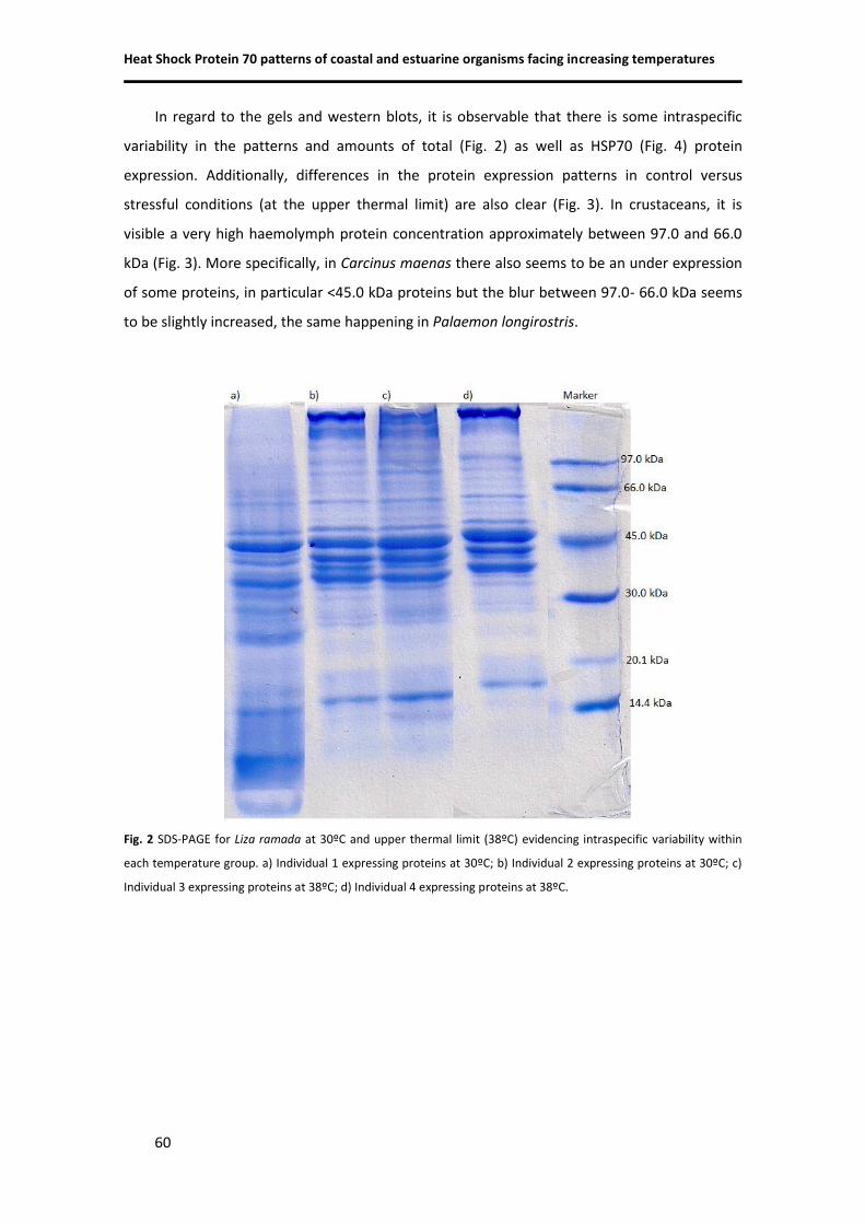
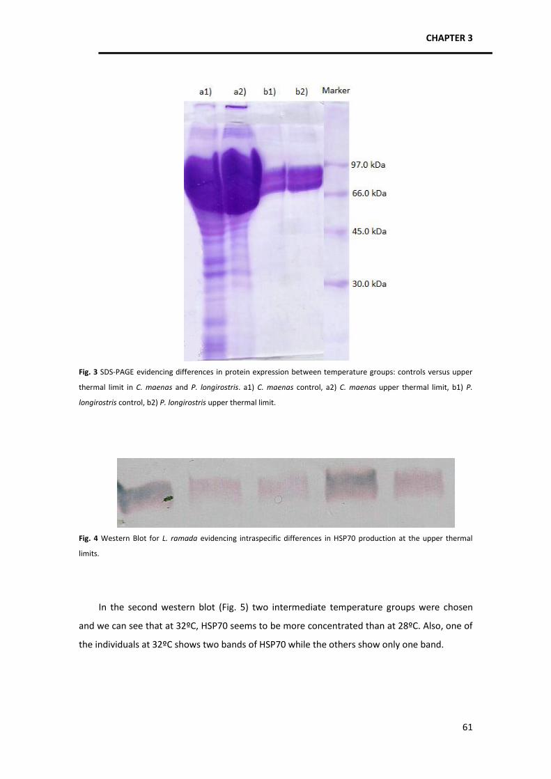
UNIVERSIDADE DE LISBOA FACULDADE DE CIÊNCIAS DEPARTAMENTO DE BIOLOGIA ANIMAL TEMPERATURE TOLERANCE AND POTENTIAL IMPACTS OF CLIMATE CHANGE ON MARINE AND ESTUARINE ORGANISMS Diana Sofia Gusmão Coito Madeira MESTRADO EM ECOLOGIA MARINHA 2011
Transcript
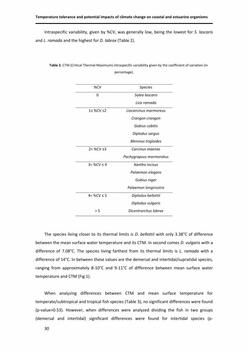
UNIVERSIDADE DE LISBOA
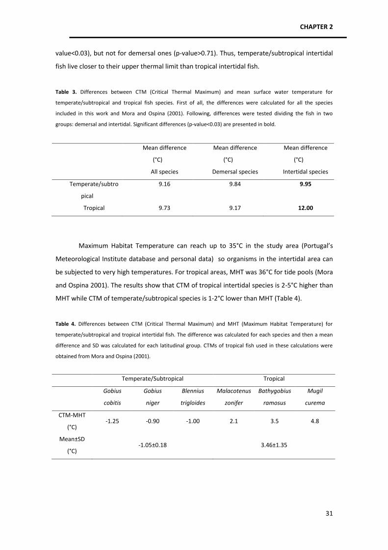
FACULDADE DE CIÊNCIAS
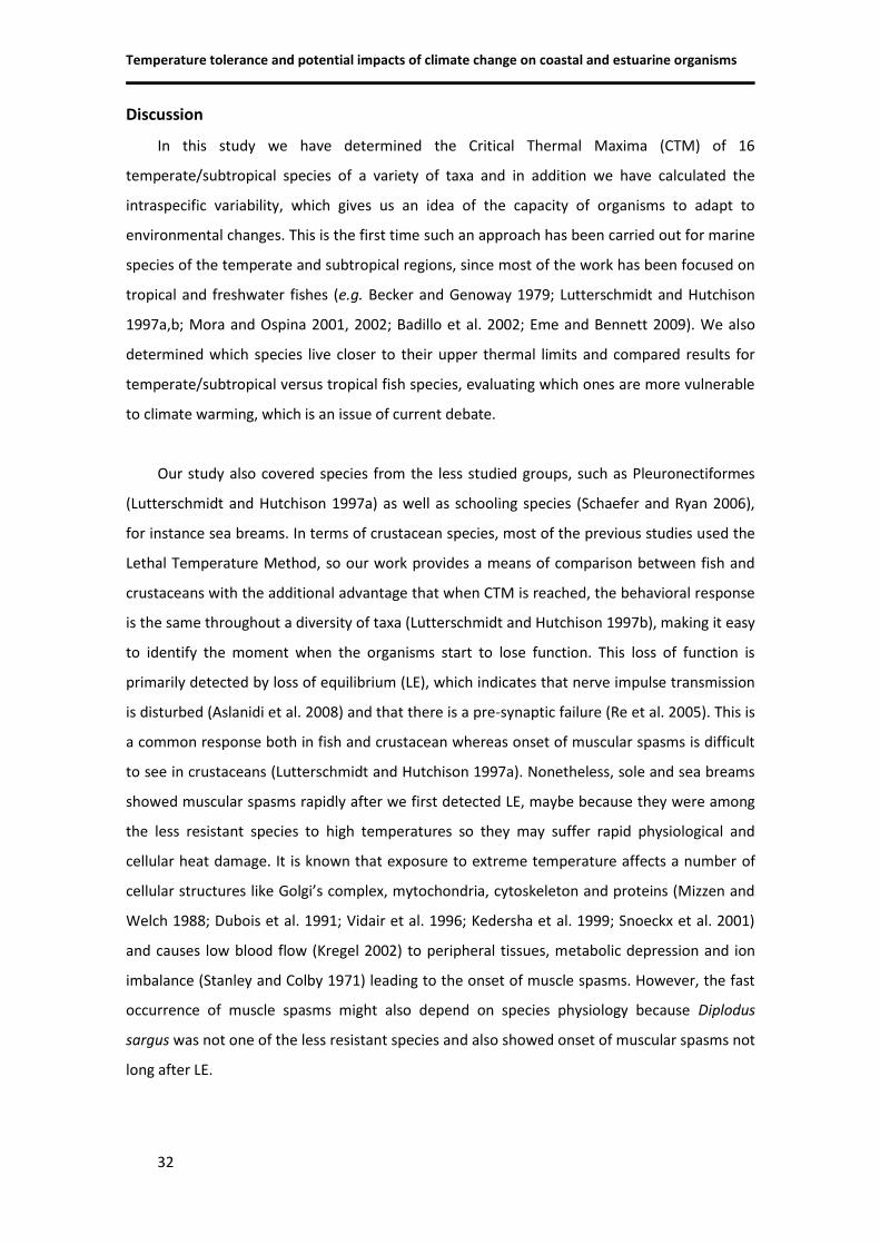
DEPARTAMENTO DE BIOLOGIA ANIMAL
TEMPERATURE TOLERANCE AND POTENTIAL IMPACTS OF
CLIMATE CHANGE ON MARINE AND ESTUARINE ORGANISMS
Diana Sofia Gusmão Coito Madeira
MESTRADO EM ECOLOGIA MARINHA 2011
UNIVERSIDADE DE LISBOA
FACULDADE DE CIÊNCIAS
DEPARTAMENTO DE BIOLOGIA ANIMAL
TEMPERATURE TOLERANCE AND POTENTIAL IMPACTS OF
CLIMATE CHANGE ON MARINE AND ESTUARINE ORGANISMS
Dissertação orientada pelos:
Professor Doutor Henrique Cabral
Doutora Catarina Vinagre
Diana Sofia Gusmão Coito Madeira
MESTRADO EM ECOLOGIA MARINHA 2011
i
Acknowledgements
I wish to express my gratitude to all people that guided, helped and contributed to this
work, in particular to:
Professor Doutor Henrique Cabral for kindly accepting me as his student and for believing
in me. Thank you for all your supervision, trust and support, kindness, friendship and
understanding. All of your knowledge and expertise was invaluable: it helped me grow as a
scientist and it made me take my professional skills one step further.
Doutora Catarina Vinagre for her unconditional guidance, supervision and enthusiasm.
Thank you for showing me the beauty of this work, for supporting me through the joyful and
harsh times, for giving me strength, for believing in my skills and for sharing your friendship
and companionship with me. This would not have been possible without everything you taught
me.
Doutor Mário Diniz for taking me into his guard at Faculdade de Ciências e Tecnologia da
Universidade Nova de Lisboa and for teaching me all about molecular techniques and
proteomics. His guidance, attention, friendship and support gave me the determination to
overcome hurdles along the way and keep the perseverance necessary to reach the finish line.
Thank you for all the opportunities you kindly offered me.
Professor Doutor Luís Narciso for his precious and joyful help during field and laboratory
work. Your experience and teaching helped me through this trial and your cheerful moods and
companionship surely made this journey much more fun.
All MSc, PhD and post-doc fellows at Faculdade de Ciências e Tecnologia, especially Joana
Lourenço, Marco Galésio, Ricardo Carreira, Gonçalo Vale, Pedro Costa and Íris Batalha, for
their warm welcome, sympathy and kindness. All your help unquestionably improved my work
and sharing ideas with you was enriching, both personally and professionally.
All team at Laboratório Marítimo da Guia, in particular Ana Pêgo and Doutor Rui Rosa for
all their understanding, encouragement and help in field work and aquaria maintenance.
ii
The crew of Albacora II and the team of Aquário Vasco da Gama, in particular Fátima Gil,
for their priceless help in the capture of the organisms and for providing us several species
when our field work was less successful. Without you this thesis would not have proceeded
according to plan.
Miguel Leal, who supported my work enthusiastically and gave me valuable advice
throughout this work, helping me out with tough decisions and doubts along the way. It was
really useful to have another friend and biologist’s eye on these matters.
To all my dearest friends, old ones from childhood and new ones from FCUL, who
encouraged me and showed me unreserved support. Your joyful company made me happy and
made me believe in myself when things did not go so well.
My family for everything they’ve done for me and everything they have taught me. To my
mother and father because they taught me to be hard working, to fight for what I want, to be
strong and patient. Your support, care and advice were really important. To my sister who
shares with me this passion for biology and gave me tones of advice, shared hard and
frustrating times with me and joked around to cheer me up when necessary.
At last, to Alexandre, who gave me his unconditional support, concern, care and love.
Thank you for your understanding, for giving me motivation, encouraging my work, cheering
me up and sharing your scientific expertise with me.
iii
Resumo
Este projecto tem como principais objectivos determinar as tolerâncias térmicas
superiores e os padrões de expressão das proteínas de choque térmico de várias espécies de
estuário e da costa Portuguesa com interesse comercial, com a finalidade de entender os
impactos da temperatura e das alterações climáticas sobre a fauna marinha. Este é um aspecto
particularmente importante pois sabe-se que a temperatura influencia processos bioquímicos,
fisiológicos, comportamentais e ecológicos, determinando assim parâmetros populacionais,
distribuição e abundância de espécies, estabilidade da teia trófica e potencialmente a futura
capacidade de exploração dos stocks de pesca. A variação espacio-temporal da temperatura
medeia os efeitos de todos os factores bióticos e abióticos e determina a diversidade de
adaptações dos organismos.
As principais questões relativas a alterações climáticas, e que têm sido alvo de grande
controvérsia, passam por entender como é que cada espécie reage às elevadas temperaturas,
como é que os aspectos ambientais e genéticos influenciam as respostas dos organismos e
quais os cenários faunísticos expectáveis tendo em conta a vulnerabilidade/resistência de cada
espécie. Como tal, o estudo da tolerância térmica é o primeiro passo para compreender esta
vulnerabilidade/resistência das espécies às alterações climáticas. O método escolhido para
estudar esta questão foi o Critical Thermal Maximum (CTM), em que os organismos são
expostos a um gradiente de temperatura com aumento de 1°C/h até atingirem o seu limite
térmico máximo. Este método permitiu ordenar as espécies em termos de vulnerabilidade a
temperaturas elevadas. Os resultados mostraram que espécies de diferentes taxa que vivem
em habitats semelhantes têm CTMs similares, uma vez que evoluíram em condições abióticas
semelhantes e potencialmente desenvolveram adaptações celulares e fisiológicas
semelhantes. O CTM é mais elevado para espécies típicas de ambientes quentes, instáveis e
muito variáveis e.g. intertidal/supratidal e para espécies migradoras, que têm de conseguir
atravessar inúmeras condições de temperatura ao longo dos seus movimentos para garantir o
sucesso reprodutivo. Relativamente às espécies de águas mais frias e com distribuição mais a
norte, o CTM foi mais baixo. Interespecificamente, o CTM foi mais variável em peixes do que
em caranguejos e camarões, possivelmente devido à grande capacidade locomotora dos
primeiros, que lhes permite colonizar inúmeros tipos de habitats. Os resultados permitiram
ainda concluir que a variabilidade intraespecífica é baixa e que, para espécies com uma larga
distribuição, não houve aclimatação ou adaptação local do limite térmico, o que pode indicar
pouca plasticidade nas respostas e pouca capacidade de adaptação a novas condições
iv
térmicas. De todas as espécies avaliadas, identificaram-se duas potencialmente vulneráveis às
alterações climáticas (Diplodus bellottii e D. vulgaris).
Um outro objectivo foi avaliar que espécies de peixes, temperadas/subtropicais ou
tropicais, é que vivem mais próximas do limite térmico, de forma a compreender quais serão
as mais vulneráveis ao aquecimento global. Concluiu-se que não existiam diferenças entre
espécies demersais mas que as espécies intertidais temperadas/subtropicais vivem mais
próximas do limite térmico uma vez que as temperaturas máximas do habitat podem
ultrapassar o seu CTM, enquanto que o CTM das espécies intertidais tropicais é 2-5°C mais
elevado do que a temperatura máxima do habitat.
Em resumo, nesta primeira parte do trabalho determinaram-se os CTMs de 16 espécies
com distribuição temperada/subtropical duma variedade de taxa (peixes, caranguejos e
camarões) e avaliaram-se diferenças inter e intraespecíficas. Foi a primeira vez que se fez uma
abordagem deste género para espécies marinhas com esta distribuição, visto que a maior
parte dos estudos tem sido focado em espécies tropicais. Assim, o presente trabalho fornece
resultados facilmente comparáveis com outros estudos, possibilitando uma avaliação da
vulnerabilidade das espécies de diferentes latitudes.
Na segunda parte do trabalho, a investigação foi direccionada para os mecanismos
celulares de defesa contra o stress térmico, com especial foco nas proteínas de choque
térmico (HSPs). Tendo em conta que a temperatura afecta os processos bioquímicos e provoca
stress proteotóxico através da desnaturação proteica e formação de agregados citotóxicos,
estas proteínas (chaperonas) são a componente de defesa que assegura a estabilização de
polipéptidos desnaturados e proteínas nascentes. Como tal, o objectivo foi determinar os
padrões de expressão da HSP de peso molecular 70 kDa, em várias espécies marinhas de
diferentes taxa, ao longo de um gradiente de temperatura e no limite térmico máximo (CTM).
Os métodos de análise proteica utilizados foram o ELISA (Enzyme Linked Immunosorbent
Assay), Western Blot e 1D SDS-PAGE (one-dimension sodium dodecyl sulfate polyacrylamide
gel electrophoresis). Foram identificadas quatro tendências nos perfis de resposta dos
organismos: aumento na produção de HSP70 à medida que a temperatura aumenta, seguido
de um decréscimo próximo dos limites térmicos (Liza ramada, Diplodus sargus, Pachygrapsus
marmoratus, Liocarcinus marmoreus); manutenção dos níveis de HSP70 ao longo de todo o
gradiente de temperatura (Diplodus vulgaris, Dicentrarchus labrax, Palaemon longirostris,
Palaemon elegans, Carcinus maenas); aumentos e decréscimos na produção de HSP70 ao
longo do gradiente de temperatura ( Gobius niger); aumento na produção de HSP70 ao longo
de todo o gradiente de temperatura (Crangon crangon). No geral, os padrões identificados são
independentes do taxon, CTM e tipo de habitat. No entanto, os resultados apontam para uma
v
relação entre a magnitude da expressão, as condições térmicas do habitat, o CTM e os limiares
de indução, uma vez que na maioria dos casos os organismos que habitam locais muito
quentes apresentaram maior quantidade de HSP70, limiares de indução mais elevados e maior
CTM. Relativamente a espécies de água mais fria, verificou-se que ou a expressão de HSP70
tem uma estreita amplitude no gradiente de temperatura ou que não existe sequer uma
produção induzida destas proteínas, indicando que são espécies potencialmente vulneráveis
ao aquecimento dos oceanos. Ainda assim, a magnitude da expressão e o tipo de padrão
apresentado estão muito relacionados com características específicas. Espécies congenéricas
foram comparadas de forma a testar as influências genéticas/filogenéticas e ambientais na
produção de HSPs. Os resultados mostraram que no género Diplodus parece existir uma
influência ambiental enquanto que no género Palaemon tudo aponta para uma influência
genética. Isto indica que poderá haver espécies com respostas mais plásticas e outras com
respostas geneticamente determinadas pelo que nestas questões é muito importante
considerar não só as condições ambientais mas também os múltiplos factores inerentes à
espécie, de forma a compreender as estratégias usadas para lidar com o stress. Verificou-se
que existe não só uma variabilidade interespecífica no tipo de resposta mas também uma
elevada variabilidade intraespecífica na quantidade de HSP70 produzida.
Concluindo, este projecto mostra que as espécies mais vulneráveis às temperaturas
elevadas e ao aquecimento glocal são espécies de águas frias e ambientes estáveis, espécies
sobre exploradas e espécies intertidais, que vivem próximo dos seus limites térmicos. Há que
ter também em conta outros factores nesta vulnerabilidade, tais como a idade da primeira
maturação, estratégia de reprodução (semelparidade ou iteroparidade) e capacidade de
adaptação dos organismos, que podem determinar se a população tem a capacidade de se
manter ou não. O estudo dos mecanismos de resistência à temperatura integra conhecimentos
de diversas áreas, pelo foi necessária uma abordagem multidisciplinar para desvendar
processos bioquímicos e celulares e avaliar os padrões dentro de um gradiente
ecologicamente relevante. Assim, este estudo contribui com informação importante para o
conhecimento de processos ecofisiológicos e pode ser relevante para a gestão dos recursos
marinhos, o que é um ponto essencial, especialmente para países com uma economia ligada
1986; Anderson et al., 1991; Bansal et al., 1991; Amici et al., 1992,
1993; Pratt, 1993; Schoeniger et al., 1994; Jacob et al., 1995;
Moseley, 1997; Nayeem et al., 1997; Snoeckx et al., 2001; Kregel, 2002; Cara et al. 1995; Gornati et al., 2004; Ali et al., 2006; Zhao et
al., 2010
Hsp90β Cytosol, nucleus,
endoplasmic reticulum
Constitutive Induced
Hsp94 x Protein folding Induced Hyperthermia; osmotic stress
CHAPTER 1 – General introduction
11
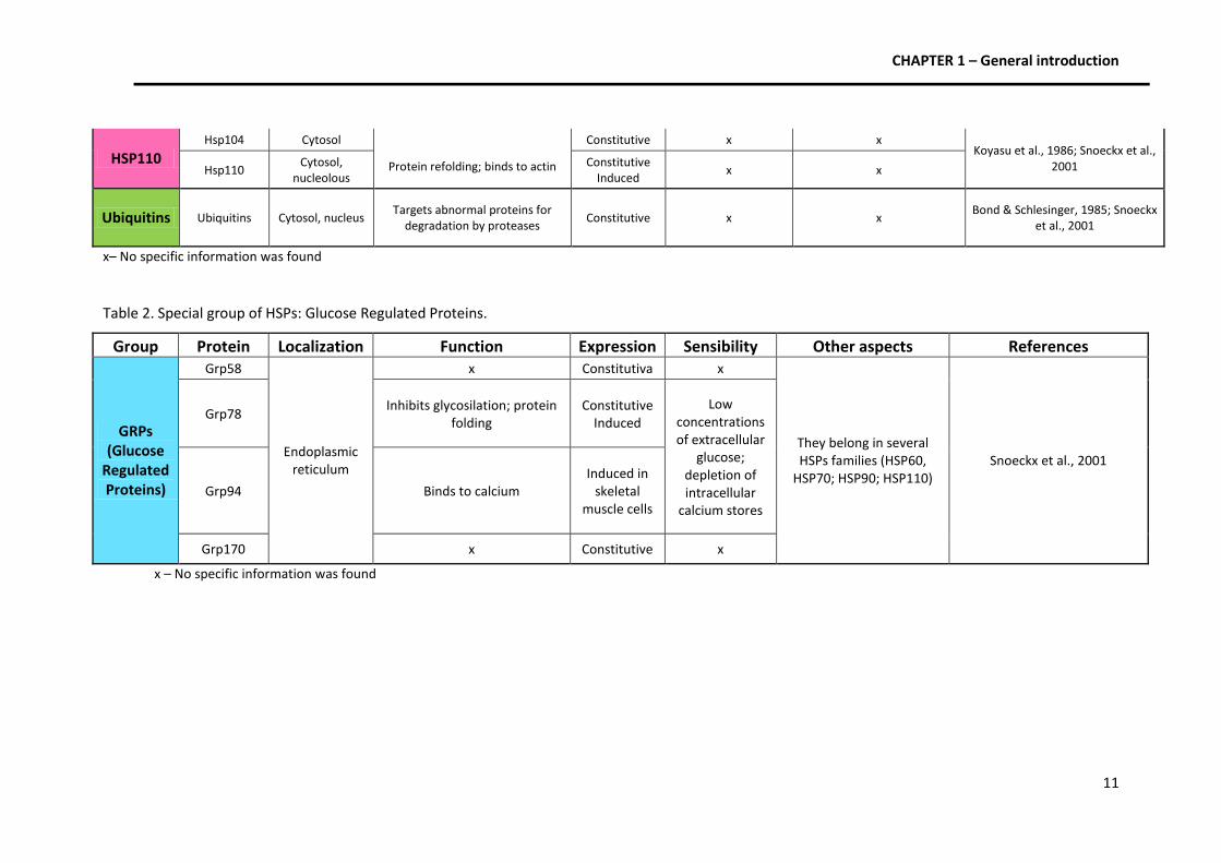
HSP110 Hsp104 Cytosol
Protein refolding; binds to actin
Constitutive x x Koyasu et al., 1986; Snoeckx et al.,
2001 Hsp110 Cytosol,
nucleolous Constitutive
Induced x x
Ubiquitins Ubiquitins Cytosol, nucleus Targets abnormal proteins for
degradation by proteases Constitutive x x
Bond & Schlesinger, 1985; Snoeckx et al., 2001
x– No specific information was found
Table 2. Special group of HSPs: Glucose Regulated Proteins.
Group Protein Localization Function Expression Sensibility Other aspects References
GRPs (Glucose
Regulated Proteins)
Grp58
Endoplasmic reticulum
x Constitutiva x
They belong in several HSPs families (HSP60,
HSP70; HSP90; HSP110) Snoeckx et al., 2001
Grp78 Inhibits glycosilation; protein
folding Constitutive
Induced
Low concentrations of extracellular
glucose; depletion of intracellular
calcium stores
Grp94 Binds to calcium Induced in
skeletal muscle cells
Grp170 x Constitutive x
x – No specific information was found
CHAPTER 1 – General introduction
12
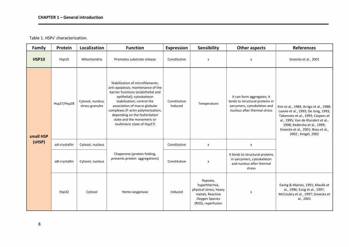
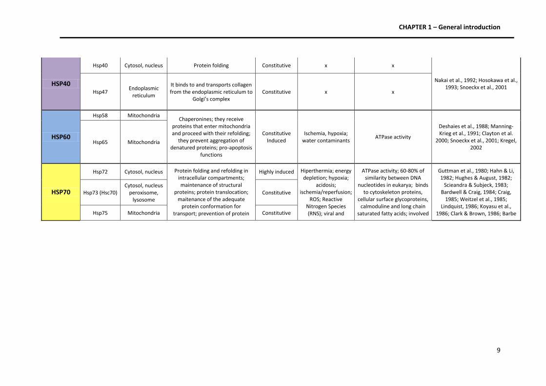
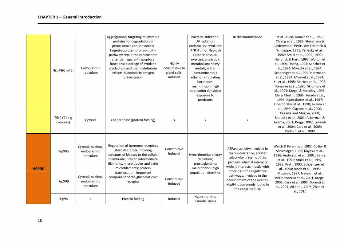
3.2. Physiological and ecological relevance of HSPs
Heat Shock Proteins might be considered indirect biochemical indicators of the degree of
damage and protein unfolding that is occurring in the cell (Hofmann 2005). Studies concerning
these proteins give us clues about the temperature at which species become thermally
stressed. HSP production is related to the past thermal history (Hofmann 2005) and the
thermal regime and its variability occurring in the habitat (Tomanek 2010), and may partially
explain the thermal limits of the species and their resistance/vulnerability to heat. They
highlight the eco-physiological complex patterns occurring in nature and contribute to our
understanding of the stress defense mechanisms and the species’ potential responses to
further climate forcing. This is of special interest considering that we need to understand
molecular and physiological traits and responses to be able to decode and explain not only
microhabitat scale patterns but also the occurring large-scale, biogeographic and ecological
patterns (Hofmann 2005).
Therefore, HSPs can have pleiotropic effects on organisms, interacting with multiple
systems in diverse ways, so the cellular response has impacts at several levels and it is
influenced by processes at levels of biological organization (Basu et al. 2002), modulating
ecology and evolution of the biological systems (Sørensen and Loeschcke 2007).
4. Aims and scopes of the dissertation
The general objectives of the research developed in the present dissertation were to
evaluate the tolerance to high temperatures and uncover the Heat Shock Protein 70 patterns
of expression in several marine taxa of commercial interest. The knowledge of thermal limits
and the cellular responses to stress provided a basis to relate several aspects of thermal
physiology to ecological patterns in order to understand the species’ vulnerability/resistance
to heat and the potential impacts of temperature and climate change on marine communities.
The objective was to assess these issues in a broad and multidisciplinary approach, relating
physiology, behavior and ecology.
More specifically, the objectives of this research are as follows:
1. To test, in the laboratory, the tolerance to high temperatures of several species of fish, crab
and shrimp from the Portuguese coast. The aim was to obtain a ranking in terms of
vulnerability through the method of Critical Thermal Maximum and discuss potential impacts
of sea warming in the distribution and abundance of the studied species;
CHAPTER 1 – General introduction
13
2. To investigate which species live closer to their upper thermal limits and compare the
results with available data for tropical species;
2. To compare inter and intraspecific variation in the thermal tolerance and HSP70 production;
3. To uncover patterns of HSP70 expression along a temperature gradient and at the upper
thermal limits;
4. To test the genetic influence on HSP70 production by analyzing whether congeneric species
have similar HSP70 expression patterns and thresholds of induction, maximal production and
shut-off;
5. To evaluate whether habitat features influence the species’ mechanisms of resistance;
6. To evaluate which species might be more vulnerable to climate change, particularly sea
warming.
This dissertation is presented in the form of two scientific articles (already submitted
to indexed scientific journals), the first one concerning thermal tolerances and the second one
concerning HSP70 expression patterns.
References
Ackerman PA, Iwama GK (2001) Physiological and Cellular Stress Responses of Juvenile Rainbow Trout to
Vibriosis. J Aquat Anim Health 13: 173-180. Agarraberes FA, Terlecky SR, Dice JF (1997) An intralysosomal hsp70 is required for a selective pathway of
lysosomal protein degradation. J Cell Biol 137: 825–834. Ali MM, Roe SM, Vaughan CK, Meyer P, Panaretou B, Piper PW, Prodromou C, Pearl LH (2006) Crystal
structure of an Hsp90-nucleotide-p23/Sba1 closed chaperone complex. Nature 440 (7087): 1013-1017. Amici C, Palamara AT, Santoro MG (1993) Induction of thermotolerance by prostaglandin A in human cells.
Exp Cell Res 207: 230–234. Amici C, Sistonen L, Santoro MG, Morimoto RI (1992) Antiproliferative prostaglandins activate heat shock
transcription factor. Proc Natl Acad Sci USA 89: 6227–6231. Anderson RL, Kraft PE, Bensaude O, Hahn GM (1991) Binding activity of glucocorticoid receptors after heat
shock. Exp Cell Res 197: 100–106. Angilleta MJ, Oufiero CE, Leaché AD (2006) Direct and indirect effects of environmental temperature on the
evolution of reproductive strategies: an information-theoretic approach. Am Nat 168(4): E123-E135. Angilletta MJ (2009) Looking for answers to questions about heat stress: researchers are getting warmer.
Funct Ecol 23: 231-232. Arrigo AP, Suhan JP, Welch WJ (1988) Dynamic changes in the structure and intracellular locale of the
mammalian low-molecularweight heat shock protein. Mol Cell Biol 8: 5059–5071. Badillo M, Alcaraz G, Chiappa-Carrara X (2002) Critical thermal maximum of the intertidal goby. International
Congress on the Biology of Fish. Vancouver, Canada.
Bansal GS, Norton PM, Latchman DS (1991) The 90-kDa heat shock protein protects mammalian cells from
thermal stress but not from viral infection. Exp Cell Res 195: 303–306. Barbe MF, Tytell M, Gower DJ, and Welch WJ (1988) Hyperthermia protects against light damage in the rat
retina. Science 241: 1817–1820. Bardwell JCA and Craig EA (1984) Major heat shock gene of Drosphila and Escherichia coli heat-inducible DNA
gene are homologous. Proc Natl Acad Sci USA 81: 848–852. Barton BA, Iwama GK (1991) Physiological changes in fish from stress in aquaculture with emphasis on the
response and effects of corticosteroids. Ann Rev Fish Dis 1: 3–26. Barton BA (1997) Stress in finfish: Past, present, and future—a historical perspective. In Iwama GK, Pickering
AD, Sumpter JP, Schreck CB (eds) Fish stress and health in aquaculture, pp.1-34. Cambridge University Press, Cambridge.
Barton BA (2002) Stress in Fishes: a diversity of responses with particular reference to changes in circulating
corticosteroids. Integ Comp Biol 42: 517–525. Basu N, Nakano T, Grau EG, Iwama GK (2001) The effects of cortisol on Heat Shock Protein 70 levels in two fish
species. Gen Comp Endocr 124(1): 97-105. Becker F, Craig E (1994) Heat shock proteins as molecular chaperones. Eur J Biochem 219: 11–23. Becker CD, Genoway RG (1979) Evaluation of the critical thermal maximum for determining thermal tolerance
of freshwater fish. Environ Biol Fish 4(3): 245, 256. Beckmann RP, Welch WJ, Mizzen LA (1990) Interaction of HSP70 with newly synthesized proteins: implication
for protein folding and assembly. Science 248: 850–854. Benjamin IJ, McMillan DR (1998) Stress (heat shock) proteins. Circ Res 83: 117–132. Bennett W, Currier RJ, Beitinger TL (1997) Cold tolerance and potential overwinter of red-bellied piranha,
Pygocentrus nattereri, in the United States. T Am Fish Soc 126(5): 841-849. Bennett WA, Judd FW (1992) Comparison of methods for determining low temperature tolerance:
experiments with pinfish, Lagodon rhomboides. Copeia 4: 1059–1065. Bond U, Schlesinger MJ (1985) Ubiquitin is a heat shock protein in chicken embryo fibroblasts. Mol Cell Biol 5:
949–956. Cabral HN, Costa MJ, Salgado JP (2001) Does the Tagus estuary fish community reflect environmental
changes? Clim Res 18: 119-126. Cara
JB, Aluru N, Moyano FJ, Vijayan MM (2005) Food-deprivation induces HSP70 and HSP90 protein
expression in larval gilthead sea bream and rainbow trout. Comp Biochem Phys B 142(4): 426-431. Caspers G-J, Leunissen JAM, De Jong WW (1995) The expanding small heat-shock protein family, and structure
predictions of the conserved “a-crystallin domain.” J Mol Evol 40: 238–248. Chi S-H, Mestril R (1996) Stable expression of a human HSP70 gene in a rat myogenic cell line confers
protection against endotoxin. Am J Physiol Cell Physiol 270: C1017–C1021. Chiang H-L, Terlecky SR, Plant CP, Dice JF (1989) A role for a 70 kilodalton heat shock protein in lysosomal
degradation of intracellular proteins. Science 246: 382–385. Clark BD, Brown IR (1986) A retinal heat shock protein is associated with elements of the cytoskeleton and
binds to calmodulin. Biochem Biophys Res Commun 139: 974–981. Clayton
ME, Steinmann R, Fent K (2000) Different expression patterns of heat shock proteins hsp 60 and hsp
70 in zebra mussels (Dreissena polymorpha) exposed to copper and tributyltin. Aquat Toxicol 47 (3-4): 213-226.
Collier NC, Schlesinger MJ (1986) The dynamic state of heat shock proteins in chicken embryo fibroblasts. J Cell Biol 103: 1495–1507.
Costa MJ, Vasconcelos R, Costa JL, Cabral HN (2007) River flow influence on the fish community of the Tagus
estuary (Portugal). Hydrobiologia 587: 113-123. Costa, MJ, Costa JL, Almeida PR, Assis CA (1994) Do eel grass beds and salt marsh borders act as preferential
nurseries and spawning grounds for fish? An example of the Mira estuary in Portugal. Ecol Eng 3 (2): 187-195. Craig EA (1985) The heat shock response. CRC Crit Rev Biochem 18: 239–280. Csermely P, Yahara I (2003) Heat shock proteins. In: Keri G, Toth I (eds) Molecular pathomechanisms and new
trends in drug research, Taylor & Francis Inc., London, pp 67-75. De Jong WW, Leunissen JA, Voorter CE (1993) Evolution of the a-crystallin/small heat shock protein family.
Mol Biol Evol 10: 103–126. Deshaies RJ, Koch BD, Werner-Washburne M, Craig EA, Schekman R (1988) A subfamily of stress proteins
facilitates translocation of secretory and mitochondrial precursor polypeptides. Nature 332: 800–805. Dubois MF, Hovanessian AG, Bensaude O (1991) Heat shock-induced denaturation of proteins. J Biol Chem
266: 9707–9711. Ellis RJ, Van der Vies SM (1991) Molecular chaperones. Annu Rev Biochem 60: 321–347. Ellis RJ (1990) The molecular chaperone concept. Sem Cell Biol 1: 1–9. Eme J, Bennett WA (2009) Critical thermal tolerance polygons of tropical marine fishes from Sulawesi,
Indonesia. J Therm Biol 34: 220-225. Essig DA, Borger DR, Jackson DA (1997) Induction of heme oxygenase-1 (HSP32) mRNA in skeletal muscle
following contractions. Am J Physiol Cell Physiol 272: C59–C67. Ewing JF, Maines MD (1991) Rapid induction of heme oxgenase-1 mRNA and protein by hyperthermia in rat
brain: heme oxygenase 2 is not a heat shock protein. Proc Natl Acad Sci USA 88: 5364–5368. Fink A (1999) Chaperone-mediated protein folding. Physiol Rev 79: 425–449. Flanagan SW, Ryan AJ, Gisolfi CV, Moseley PL (1995) Tissue- specific HSP70 response in animals undergoing
heat stress. Am J Physiol Regul Integr Comp Physiol 268: R28– R32. Frederich M, Pörtner HO (2000). Oxygen limitation of thermal tolerance defined by cardiac and ventilatory
performance in the spider crab Maja squinado. Am J Physiol 279: R1531–R1538. Gornati R, Papis E, Rimoldi S, Terova G, Saroglia M, Bernardini G (2004) Rearing density influences the
expression of stress related genes in seabass (Dicentrarchus labrax, L.). Gene 341: 111-118. Guttman SD, Glover CVC, Allis CD, Gorovsky MA (1980) Heat shock, deciliation and release from anoxia induce
the synthesis of the same polypeptides in starved T. pyriformis. Cell 22: 299–307. Hahn GM, Li GC (1982) Thermotolerance and heat shock proteins in mammalian cells. Radiat Res 92: 452–457. Hall DM, Xu L, Drake VJ, Oberley LW, Oberley TD, Moseley PL, and Kregel KC (2000) Aging reduces adaptive
capacity and stress protein expression in the liver after heat stress. J Appl Physiol 89: 749–759. Hartl FU, Martin J (1992) Protein folding in the cell. The role of molecular chaperones HSP70 and HSP60. Annu
Rev Biophys Biomol Struct 21: 293–322. Hartl FU (1996) Molecular chaperones in cellular protein folding. Nature 381: 571–580. Hendrick JP, Hartl F-U (1993) Molecular chaperone functions of heat shock proteins. Annu Rev Biochem 62:
349–384.
CHAPTER 1 – General introduction
16
Herrmann JM, Stuart RA, Craig EA, Neupert W (1994) Mitochondrial heat shock protein 70, a molecular
chaperone for proteins encoded by mitochondrial DNA. J Cell Biol 127: 893–902. Hightower LE (1991) Heat shock, stress proteins, chaperones, and proteotoxicity. Cell 66: 191–197. Hofmann GE, Todgham AE (2010) Living in the now: physiological mechanisms to tolerate a rapidly changing
environment. Annu Rev Physiol 72: 22.1–22.19. Hosokawa N, Takeshi S-I, Yokota H, Hirayoshi K, Nagata K (1993) Structure of the gene encoding the mouse
131-135. Hughes EN, August JT (1982) Co-precipitation of heat shock proteins with a cell surface glycoprotein. Proc Natl
Acad Sci USA 79: 2305–2309. IPCC (1997) The regional impacts of climate change: an assessment of vulnerability. Cambridge University
Press, UK, pp 517. IPCC (1997) The regional impacts of climate change: an assessment of vulnerability. Cambridge University
Press, UK. pp 517. IPCC (2001) Climate change 2001: impacts, adaptation and vulnerability. Contribution of Working Group II to
the Third Assessment Report of the Intergovernmental Panel on Climate Change. Cambridge University Press, Cambridge, United Kingdom and New York, USA.
IPCC (2002) Climate change and biodiversity: IPCC technical paper V. Geneva, Switzerland. pp 85.
IPCC (2007) Climate change 2007: impacts, adaptation and vulnerability. Contribution of working group II to the Fourth Assessment Report of the Intergovernmental Panel on Climate Change. Cambridge University Press, Cambridge, United Kingdom and New York, USA.
Iwama GK, Vijayan MM, Forsyth RB, Ackenrian PA (1999) Heat shock proteins and physiological stress in fish.
Am Zool 39: 901-909. Jacob U, Lilie H, Meyer I, Buchner J (1995) Transient interaction of HSP90 with early unfolding intermediates
of citrate synthase. J Biol Chem 270: 7288–7294. Jarvis LM (2007) Living on the edge: drugs targeting the protein Hsp90 push already unstable cancer cells to
the brink. Chem Eng News 85(9): 15-23. Kagawa N, Mugiya Y (2000) Exposure of goldfish (Carassius auratus) to bluegills (Lepomis macrochirus)
enhances expression of stress protein 70 mRNA in the brains and increases plasma cortisol levels. Zool Sci 17: 1061–1066.
Kedersha NL, Gupta M, Li W, Miller I, Anderson P (1999) RNA-binding Proteins TIA-1 and TIAR Link the
Phosphorylation of eIF-2 a to the Assembly of Mammalian Stress Granules. J Cell Biol 147 (7): 1431–1441. Kim YJ, Shuman J, Sette M, Przybyla A (1984) Nuclear localization and phosphorylation of three 25-kilodalton
rat stress proteins. Mol Cell Biol 4: 468–474. Kimball ME, Miller JM, Whitfield PE, Hare JA (2004) Thermal tolerance and potencial distribution of invasive
lionfish (Pterois volitans/miles complex) on the east coast of the United States. Mar Ecol Prog Ser 283: 269-278. Koyasu S, Nishida T, Kadowaki T, Matsuzaki F, Iida K, Harada F, Kasuga M, Sakai H, Yahara I (1986) Two
mammalian heat shock proteins, HSP90 and HSP100 are actin-binding proteins. Proc Natl Acad Sci USA 83: 8054–8055.
Kregel KC (2002) Heat shock proteins: modifying factors in physiological stress responses and acquired
thermotolerance. J Appl Physiol 92: 2177–2186.
CHAPTER 1 – General introduction
17
Kregel KC, Moseley PL (1996) Differential effects of exercise and heat stress on liver HSP70 accumulation with aging. J Appl Physiol 80: 262–277.
Lavoie J, Gingras-Bretan G, Tanguay RM, Landry J (1993) Induction of Chinese hamster HSP27 gene expression
in mouse cells confers tolerance to heat shock. HSP27 stabilization of the microfilament organization. J Biol Chem 268: 3420–3429.
Lindquist S, Craig EA (1988) The heat-shock proteins. Annu Rev Genet 22: 631–677. Lindquist S (1986) The heat shock response. Annu. Rev. Biochem. 55: 1151–1191. Löw-Friedrich I, Schoeppe W (1991) Effects of calcium channel blockers on stress protein synthesis in cardiac
myocytes. J Cardiovasc Pharmacol 17: 800–806. Lutterschmidt WI, Hutchison VH (1997) The critical thermal maximum: history and critique. Can J Zool 75(10):
1561-1574. Manning-Krieg UC, Scherer P, Schatz G (1991) Sequential action of mitochondrial chaperones in protein
import into the matrix. EMBO J 10: 3273–3280. Marber MS, Mestril R, Chi SH, Sayen R, Yellon YM, Dillman WH (1995)Overexpression of the rat inducible 70-
kDa heat stress protein in a transgenic mouse increases the resistance of the heart to ischemic injury. J Clin Invest 95: 1446–1456, 1995.
Maulik N, Sharma HS, Das DK (1996) Induction of the haem oxygenase gene expression during the reperfusion
of ischemic rat myocardium. J Mol Cell Cardiol 28: 1261–1270. McCoubry WKJ, Huang TJ, Maines MD (1997) Isolation and characterization of a cDNA from the rat brain that
encodes hemoprotein heme-oxygenase-3. Eur J Biochem 247: 725–732. Melzner F, Mark FC, Pörtner H (2007) Role of blood oxygen transport in thermal tolerance of the cuttlefish,
Sepia officinalis. Integr Comp Biol 47(4): 645-655. Miranda PMA, Coelho FES, Tomé AR, Valente MA (2002) 20th Century Portuguese Climate and Climate
Scenarios. Scenarios, Impacts and adaptation measures – SIAM Project. In: Santos FD, Forbes K, Moita R (eds) Climate change in Portugal. Gradiva, Lisboa, pp 27-83.
Mivechi NF, Koong AC, Giaccia AJ, Hahn GM (1994) Analysis of HSF-1 phosphorylation in A549 cells treated
with a variety of stresses. Int J Hyperthermia 10: 371–379. Moalic JM, Bauters C, Himbert D, et al. (1989) Phenylephrine, vasopressin and angiotensin II as determinants
of proto-oncogene and heat-shock protein gene expression in adult rat heart and aorta. J Hypertens 7: 195–201. Mora C, Ospina F (2001) Thermal tolerance and potential impact of sea warming on reef fishes from Gorgona
island (eastern Pacific Ocean). Mar Biol 139:765-769. Mora C, Maya MF (2006) Effect of the rate of temperature increase of the dynamic method on the heat
tolerance of fishes. J Therm Biol 31: 337-341. Mora C, Ospina AF (2002) Experimental effects of La Niña cold temperatures on the survival of reef fishes
from Gorgona Island (Tropical Eastern Pacific). Mar Biol 141: 789–793. Morimoto RI, Tissieres A, and Georgopoulos C (1994) Progress and perspectives on the biology of heat shock
proteins and molecular chaperones. In: Morimoto RI, Tissieres A, Georgopoulos C (eds) The Biology of Heat Shock Proteins and Molecular Chaperones, Cold Spring Harbor, NY: Cold Spring Harbor Laboratory Press.
Moseley PL (1997) Heat shock proteins and heat adaptation of the whole organism. J Appl Physiol 83: 1413–
1417. Munday PL, Cheal AJ, Graham NAJ, Meekan M, Pratchett MS, Sheaves M, Sweatman H, Wilson SK (2009)
Tropical Coastal Fish. A Marine Climate Change Impacts and Adaptation Report Card for Australia.
CHAPTER 1 – General introduction
18
Myrmel T, McCully JD, Malikin L, Krukenkamp IB, Levitsky S (1994) Heat shock protein 70 mRNA is induced by anaerobic metabolism in the heart. Circulation 90: II-299 –II-305.
Nakai A, Satoh M, Hirayoshi K, Nagata K (1992) Involvement of the stress protein HSP47 in procollagen
processing in the endoplasmic reticulum. J Cell Biol 117: 903–914. Nakicenovic N, Alcamo J, Davis G et al. (2000) Emission scenarios. A special report of working group III of the
IPCC. Cambridge University Press, Cambridge. Nayeem MA, Hess ML, Qian YZ, Loesser KE, Kukreja RC (1997) Delayed preconditioning of cultured adult rat
cardiac myocytes: role of 70- and 90-kDa heat stress proteins. Am J Physiol Heart Circ Physiol 273: H861–H868. Oberdörster E, Rittschof D, McClellan-Green P (1998) Induction of cytochrome P450 3A and heat shock
protein by tributyltin in blue crab, Callinectes sapidus. Aquat Toxicol 41 (1-2): 83-100. Ospina-Álvarez N, Piferrer F (2008) Temperature-dependent sex determination in fish revisited: prevalence, a
single sex-ratio response pattern, and possible effects of climate change. PLoS ONE 3(7): e2837. Padmini E, Geetha BV, Rani MU (2009) Pollution induced nitrative stress and heat shock protein 70
overexpression in fish liver mitochondria. Sci Total Environ 407(4): 1307-1317. Pörtner HO (2002) Climate variations and the physiological basis of temperature dependent biogeography:
systemic to molecular hierarchy of thermal tolerance in animals. Comp Biochem Physiol 132A:739–761. Pörtner HO, Knust R (2007) Climate change affects marine fishes through the oxygen limitation of thermal
tolerance. Science 315: 95-97. Pörtner HO, Mark FC, Bock C (2004) Oxygen limited thermal tolerance in fish? Answers obtained by nuclear
magnetic resonance techniques. Resp Physiol Neurobi 141: 243-260. Pratt WB (1993) The role of heat shock proteins in regulating the function, folding, and trafficking of the
glucocorticoid receptor. J Biol Chem 268: 21455–21458. Rajaguru S (2002) Critical thermal maximum of seven estuarine fishes. J Therm Biol 27(2): 125-128. Rajaguru S, Ramachandran S (2001) Temperature tolerance of some estuarine fishes. J Therm Biol 26: 41-45. Reis CS, Lemos R, Alagador D (2006) Fisheries. In: Santos FD, Miranda P (eds) Climate change in Portugal.
Scenarios, Impacts and adaptation measures – SIAM II Project. Gradiva, Lisboa, pp. 347-384. RI Perry, Cury P, Brander K, Jennings S, Möllmann C, Planque B (2010) Sensitivity of marine systems to climate
and fishing: Concepts, issues and management responses. J Marine Syst 79 (3-4): 427-435. Ritossa F (1962) A new puffing pattern induced by temperature shock and DNP in Drosophila. Experientia 18:
571–573. Roberts RJ, Agius C, Saliba C, Bossier P, Sung YY (2010) Heat shock proteins (chaperones) in fish and shellfish
and their potential role in relation to fish health: a review. J Fish Dis 33(10): 789 -801.
Roessig JM, Woodley CM, CechJr JJ, Hansen LJ (2004) Effects of global climate change on marine and estuarine
fishes and fisheries. Rev Fish Biol Fisher 14:251-275. Rosa R, Seibel BA (2008) Synergistic effects of climate-related variables suggest future physiological
impairment in a top oceanic predator. Proc Nat Acad Sci 105(52): 20776–20780. Rosenzweig C, Casassa G, Karoly DJ, Imeson A, Liu C, Menzel A, Rawlins S, Root TL, Seguin B, Tryjanowski P
(2007) Assessment of observed changes and responses in natural and managed systems. In: Parry ML, Canziani OF, Palutikof JP, van der Linden PJ, Hanson CE (eds) Climate Change 2007: Impacts, Adaptation and Vulnerability. Contribution of Working Group II to the Fourth Assessment Report of the Intergovernmental Panel on Climate Change, Cambridge University Press, Cambridge, UK, 79-131.
Sanchez C, Padilla R, Paciucci R, Zabala JC, Avila J (1994) Binding of heat-shock protein 70 (hsp70) to tubulin. Arch Biochem Biophys 310: 428–432.
Schoeniger LO, Audreonwi KA , Ott GR, et al. (1994) Induction of heat shock expression in postis chemic pig
liver depends on superoxide generation. Gastroenterology 106(1):177―184. Sciandra JJ, Subjeck JR (1983) The effect of glucose on protein synthesis and thermosensitivity in Chinese
hamster ovary cells. J Biol Chem 258: 12091–12093. Skidmore R, Gutierrez JA, Guerriero V Jr, and Kregel KC (1995) HSP70 induction during exercise and heat stress
in rats: role of internal temperature. Am J Physiol Regul Integr Comp Physiol 268: R92–R97. Snoeckx LEH, Cornelussen RN, van Nieuwenhoven FA, Reneman RS, van der Vusse GJ (2001) Heat Shock
Proteins and Cardiovascular Pathophysiology. Physiol Rev 81(4):1461-1485. Somero GN, DeVries AL (1967) Temperature tolerance of some Antarctic fishes. Science 156(3772): 257-258. Sørensen JG, Loeschcke V (2007) Studying stress responses in the post-genomic era: its ecological and
evolutionary role. J Biosci 32 447–456. Stevenson MA, Calderwood SK (1990) Members of the 70-kilodalton heat shock protein family contain a
highly conserved calmodulinbinding domain. Mol Cell Biol 10: 1234–1238. Takemoto L, Emmons T, Horwitz J (1993) The C-terminal region of aA-crystallin: involvement in protection
against heat-induced denaturation. Biochem J 292: 435–438. Tsang TC (1993) New model for 70 kDa heat-shock proteins’ potential mechanisms of function. FEBS Lett 323:
1–3. Terada K, Ueda I, Ohtsuka K, Oda T, Ichiyama A, Mori M (1996) The requirement of heat shock cognate 70
protein for mitochondrial import varies among precursor proteins and depends on precursor length. Mol Cell Biol 16: 6103–6109.
Terlecky SR, Chiang H-L, Olson TS, Dice JF (1992) Protein and peptide binding and stimulation of in vitro
lysosomal proteolysis by the 73 kDa heat shock cognate protein. J Biol Chem 267: 9202– 9209. Van de Klundert FAJM, Gijsen MLJ, Van den Ijsel PRLA, Snoeckx LHEH, De Jong WW (1998) aB-crystallin and
hsp25 in neonatal cardiac cells: differences in cellular localization under stress conditions. Eur J Cell Biol 75: 38–45. Vidair CA, Huang RN, Doxsey SJ (1996) Heat shock causes protein aggregation and reduces protein solubility at
the centrosome and other cytoplasmic locations. Int J Hypertherm 12: 681–695. Vijayan MM, Moon TW (1992) Acute handling stress alters hepatic glycogen metabolism in fooddeprived
rainbow trout (Oncorhynchus mykiss). Can J Fish Aquat Sci 49:2260-2266. Vijayan MM, Moon TW (1994) The stress response and the plasma disappearance of corticosteroid and
glucose in a marine teleost, the sea raven. Can J Zool 72:379-386. Vinagre C, Santos FD, Cabral H, Costa MJ (2011) Impact of climate warming upon the fish assemblages of the
Portuguese coast under different scenarios. Reg Environ Change. doi 10.1007/s10113-011-0215-z. Walsh SJ, Haney DC, Timmerman CM, Dorazio RM (1998) Physiological tolerances of juvenile robustred horse,
Moxostoma robustum: conservation implications for animperiled species. Environ Biol Fishes 51: 429–444. Walton PA, Wendland M, Subramani S, Rachubinski RA, Welch WJ (1994) Involvement of 70-kD heat-shock
proteins in peroxisomal import. J Cell Biol 125: 1037–1046. Weitzel G, Pilatus U, Rensing L (1985) Similar dose response of heat shock protein synthesis and intracellular
pH change in yeast. Exp Cell Res 159: 252–256. Welch WJ, Suhan JP (1986) Cellular and biochemical events in mammalian cells during and after recovery from
physiological stress. J Cell Biol 103: 2035–2052.
CHAPTER 1 – General introduction
20
Welch WJ, Feramisco JR (1984) Nuclear and nucleolar localization of the 72.000 dalton heat-shock protein in
heat-shocked mammalian cells. J Biol Chem 259: 4501–4513. Welch WJ (1992) Mammalian stress response: cell physiology, structure/ function of stress proteins, and
implications for medicine and disease. Physiol Rev 72: 1063–1081. Xu Q, Li D, Holbrook NJ, Udelsman R (1995) Acute hypertension induces heat shock protein 70 gene
expression in rat aorta. Circulation 92: 1223–1229. Zhao W, Chen L, Qin J, Wu P, Zhang F, Li E, Tang B (2011) MnHSP90 cDNA characterization and its expression
during the ovary development in oriental river prawn, Macrobrachium nipponense. Mol Biol Rep 38(2): 1399-1406.
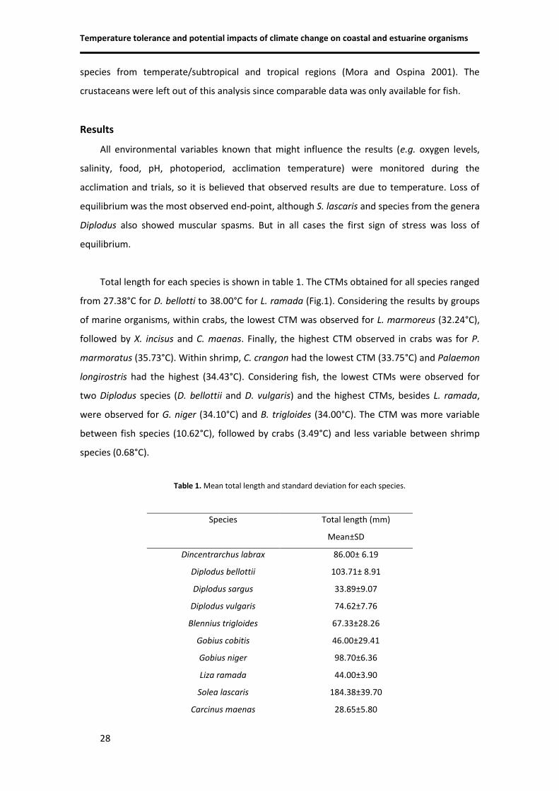
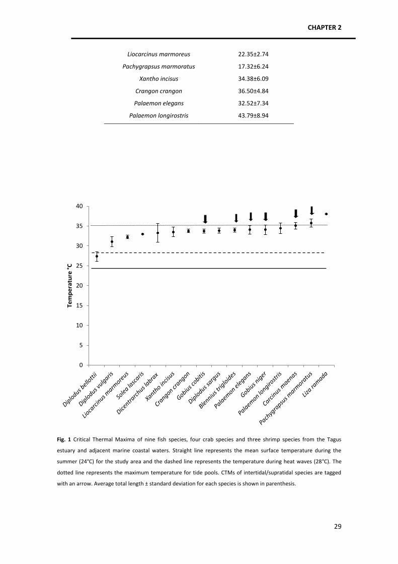
Temperature tolerance and potential impacts of climate change on coastal and estuarine organisms
34
should possess physiological and cellular adaptations that enable them to cope with such
extreme conditions. These adaptations lead to a higher CTM so organisms living in variable
environments have higher tolerances (e.g. Mora and Ospina 2001; Badillo et al. 2002; Schaefer
and Ryan 2006) than those coming from more stable environments, for example the subtidal
zone.
Pachygrapsus marmoratus was the crab species with the greatest upper thermal limit
certainly because it inhabits the most extreme environment: the supratidal zone, though it can
also occur in the intertidal zone (Ingle 1997; Flores and Paula 2001). The abiotic factors (mainly
temperature and insolation) occurring in the supratidal are crucial for littoral zonation of the
organisms living in that habitat, depending on how adapted they are to water loss and heat.
Our results, along with other studies (e.g. Davenport and MacAlister 1996; Davenport and
Davenport 2005) follow the perspective that organisms living higher in the shore are more
tolerant than those closer to low water, although other authors did not find such a connection
(e.g. Clarke et al. 2000).
Regarding the results for shrimp and fish species, a similar pattern occurs. Crangon
crangon has the lowest CTM among shrimps probably because it is a cold water species
(mainly from the Northeast Atlantic – see Hayward et al. 1996) and it is a demersal species
living in depths of 0-50m (Hayward and Ryland 1995), which means that it is exposed to lower
temperatures and lives in a more stable environment, although it can also occur in estuaries
(Boddeke 1989; data from Centro de Oceanografia) so it must be adapted to a certain degree
of environmental variability.
Palaemon longirostris and P. elegans inhabit the temperate and subtropical regions
(Alvarez 1968) and presented a higher CTM enabling them to live in warmer environments.
Palaemon longirostris is a catadromous migrating species (Cartaxana 1994; Barnes 1994; Paula
1998), which means it has to pass through several habitat types and thermal regimes to be
able to reproduce and keep the population numbers, so a high resistance for this species was
expected.
The linkage between thermal limits and aerobic scope (Pörtner and Knust 2007) is of
crucial importance in migrating species because fecundity and recruitment are connected to
aerobic scope (Farrell 2009). Organisms have a higher energy and oxygen demand due to an
additional swimming rhythm during the reproduction season (Cartaxana 1994) which means
CHAPTER 2
35
they need their full aerobic scope to carry out the migration and reproduce. According to
Farrell (2009), if warmer waters decrease aerobic scope, reproduction success might be
jeopardized, putting the population at risk. This is especially relevant considering that P.
longirostris has a one-year or two-years population cycle (Cartaxana 1994). Finally, with regard
to P. elegans, it lives in intertidal pools (Hayward et al. 1996) that have diel cycles of
temperature, oxygen availability, CO2 accumulation and pH fluctuation so it was also expected
to be one of the most heat resistant among shrimps.
At last, all fish species studied have a wide distributional range in the
temperate/subtropical region so besides species physiological performance, differences in
CTM should mainly come from habitat type, circulation and current patterns plus distributional
depth. In the case of sea breams, although they are found in estuaries as juveniles (Vinagre et
al. 2010), juveniles and adults also live in the continental shelf area so, in the Portuguese coast,
they are exposed to annual sea-surface temperatures that range from 15°C to 18°C (Levitus
and Boyer 1994) or even colder since adults can live in water depths down to approximately 50
m (D. sargus) to a 100 m (D. bellottii and D. vulgaris) (Bauchot and Hureau 1986). Besides, they
also suffer the influence of cold waters from coastal upwelling during the summer due to
northerly trade winds (Lemos and Sansó 2006). Knowing that D. bellottii is originally an African
species from the Mauritania upwelling system (Vinagre et al. in press), it was expected that its
upper thermal limit would not be one of the highest among the fish studied, even though the
African upwelling has waters that round a mean temperature of 20-25°C (Levitus and Boyer
1994), which are warmer than Portuguese waters. A similar situation occurs for D. vulgaris,
which has a discontinuous distribution, being absent from the warmer stretches of the African
coast (Bauchot and Hureau 1986). As for D. sargus, its wide distributional range is continuous
throughout the west coast of Africa indicating that it can live in tropical warm waters, so its
higher CTM than the other two Diplodus species was expected and is in accordance with these
species climatic envelopes. Vinagre et al. (in press) came to a similar conclusion. They found a
general trend of higher vulnerability to high temperatures in the species D. bellottii, unlike D.
sargus. According to the authors, the reason for this difference is related to a steep increase in
D. bellottii’s metabolism in high temperatures, a decrease in the aerobic scope and increased
mortality rate. On the contrary D. sargus maintained a low level of sensibility towards
increased temperatures, which is in accordance with our results.
Temperature tolerance and potential impacts of climate change on coastal and estuarine organisms
36
Similar intermediate values of CTM were found for S. lascaris and D. labrax when
comparing to other fish, possibly as a result of their demersal life and the depth at which they
can be found, which can reach 350m for soles and 100m for sea bass (Quéro et al. 1986;
Tortonese 1986; FAO 2011). Notwithstanding, they are frequently found in relatively shallow
coastal waters and estuaries so they are physiological capable of inhabiting warmer waters,
not only as adults but also as juveniles because sole and seabass use estuaries as nursery
grounds (Barnes 1994; Hayward and Ryland 1995; Vinagre et al. 2008, 2009).
Amongst the most resistant fish species were G. cobitis, B. trigloides and L. ramada, which
followed the pattern already described for crabs and shrimps. Gobius and Blennius are genera
inhabiting either estuaries, surf zone waters or the intertidal pools so they undergo wide
variations in abiotic variables and are exposed to very high temperatures, which they can cope
with due to their high upper thermal limit. The case of L. ramada is less obvious but similar to
P. longirostris since it is a migratory species that can live in coastal waters and inshore waters,
entering estuaries, lagoons and lower parts of the rivers (Sauriau et al. 1994; Hayward and
Ryland 1995) so it must be adapted to a diversity of thermal regimes where temperature can
vary considerably and reach elevated values.
Overall, when analyzing interspecific differences, we might initially expect a greater
disparity as the mean genetic distance increases but this would only be if ecological niches
were very dissimilar. Our results shed some light on this matter since it appears that species
from different taxa living in identical habitat types have similar upper thermal limits (Fig. 1)
because they evolved in similar abiotic conditions, probably developing similar physiological
and cellular adaptations. Another important result is that upper thermal limits were more
variable between fish than between crabs and shrimps. This is probably because fish have a
great locomotory capacity and colonize all kinds of habitats.
Intraspecific variability was generally low, which is in concordance with Mora and Ospina
(2001). According to Cuculescu et al. (1998), thermal tolerance is subject to phenotypic
alteration within a genetically fixed range. This phenotypic plasticity is dependent upon several
factors but thermal history of individuals and parental effects seem to be the most important
ones (Cossins and Bowler 1987; Schaefer and Ryan 2006), inducing irreversible changes to the
thermal tolerance (Schaefer and Ryan 2006). Then, variability found for each species relates
not only to specific genetic features but also the previous influence of environmental variables.
Nevertheless, if genetic variability is in general low, this might be a concern because species
CHAPTER 2
37
might not be able to adapt to climate changes (Mora and Ospina 2001). Our data shows that D.
labrax, D. bellotti and D. vulgaris appear to have the highest variability of response towards
temperature and thus possibly the highest genetic variability concerning the genes involved in
such a response.
There are various untested factors that can potentially influence species upper thermal
limits. These factors can be sex, reproductive state, nutritional condition, diseases and
parasites, inter-population variability, age and size (Cox 1974; Copeland et al. 1974; Hutchison
1976; Becker and Genoway 1979; Lutterschmidt and Hutchison 1997b) as well as thermal
history of individual animals (Schaefer and Ryan 2006) and also seasonal variation (Cuculescu
et al. 1998; Hopkin et al. 2006). Yet, there seems to exist a certain degree of controversy on
the influence of these factors since some authors found significant differences for instance
between different sized individuals (e.g. Cox 1974; Copeland et al. 1974; Peck et al. 2004,
2007, 2009) while others did not (e.g. Ospina and Mora 2004), nor for males versus females
(Badillo et al. 2002). Even though we limited the influence of some of these factors by, for
example, restricting sampling to the summer and testing individuals of approximately the
same size, this might have a disadvantage which is missing important intraspecific variability
patterns. Further studies should address this issue and CTM should be tested for different life
stages, different seasons and different habitats used by conspecifics, amongst other factors.
In this study it was confirmed that CTM of marine ectotherms is higher for species which
inhabit variable environments, like the intertidal or the supratidal and/or make reproduction
migrations that require the passage through different habitats with potentially very elevated
temperatures. Acclimation or local adaptation of species with a wide distribution was not
confirmed since C. maenas displayed the same CTM as found in the North Sea.
In order to know which species might be more vulnerable to temperature, we calculated
the difference between thermal limit and mean surface water temperature for each species.
Mora and Ospina (2001) conducted a similar study on tropical reef fish and concluded that the
CTM of the least tolerant species was 8ºC above the current mean sea temperature, while in
the present study the CTM of the least tolerant species, D. bellottii, is only 3°C above the
average summer estuarine temperature (it is in summer that this species juveniles occur in
estuaries). In fact, this species is already under stress during current heat waves, which makes
it clear that its presence in estuarine nursery areas (where juveniles are very abundant
Temperature tolerance and potential impacts of climate change on coastal and estuarine organisms
38
nowadays) is under threat by climate warming, as observed by Vinagre et al. (in press) through
experimental studies. In a climate warming perspective, if we add 2°C to the current
temperature attained by the waters during heat waves (28°C+2°C), it is clear that another
seabream, D. vulgaris, may also be under threat.
When we compared mean differences between CTM and mean water temperature for
temperate/subtropical and tropical fish, no significant differences were found considering all
species. When we evaluated demersal and intertidal species separately, demersal ones didn’t
show significant differences as well but intertidal species did. The intertidal
temperate/subtropical fish have an upper thermal limit on average 9.95°C above mean water
temperature (24°C) while tropical intertidal fish have an upper thermal limit on average 12°C
above mean water temperature (27°C). This result may indicate that temperate/subtropical
intertidal species might be more vulnerable to further increases in temperature.
In fact, one of the main findings of this study was the confirmation that organisms that
occur in thermally unstable environments, e.g. intertidal/supratidal habitats, live closer to their
thermal limits because maximum habitat temperatures may reach or exceed their CTM.
Comparing the results obtained for temperate/subtropical and tropical species, we can see
that the former species have CTMs on average 1.05°C under the maximum habitat
temperature while tropical species have CTMs on average 3.46°C above maximum habitat
temperatures. Therefore, maximum habitat temperatures in temperate/subtropical regions
may exceed the upper thermal limits of intertidal fish species, making them especially
vulnerable to high temperatures and climate warming. Additionally, since it is the extreme
values of environmental conditions that exert the most selective pressures (Lutterschmidt and
Hutchison 1997a), this result might indicate that temperate/subtropical intertidal fauna may
be exposed to strongest selection when it comes to temperature conditions.
This is indicative of a probable higher vulnerability of temperate/subtropical fish species,
and it is in concordance with the predictions that tropical fish might be affected through
indirect effects of climate change rather than direct ones (Mora and Ospina 2001). The
statement that tropical species live closer to their thermal limits is supported through the
studies of Jokiel and Coles (1977) and Sharp et al. (1997). However, these apply to corals, so in
order to understand climate impacts on marine communities, more species of various
taxonomic groups should be tested.
CHAPTER 2
39
Nonetheless, predicting the impacts of climate change on particular species is not a
simple task and requires in depth knowledge on several subjects from molecular biology,
physiology, ecology and evolution. Thus, realistic predictions will necessarily be the result of
multidisciplinary and integrative approaches. Nevertheless, since sea warming effects are
already clear throughout ecosystems and base studies are urgently needed, we consider this
work a necessary first step in the investigation of climate change impacts upon marine biota
and ecosystems.
Acknowledgements
Authors would like to thank everyone involved in the field work, maintenance of the experimental
tanks and in the feeding of the organisms. Authors would like to express their gratitude to “Aquário
Vasco da Gama” for the collaboration in the sampling and maintenance of organisms, in particular to Dr
Fátima Gil. This study had the support of the Portuguese Fundação para a Ciência e a Tecnologia (FCT)
through the funding of projects and the grant SFRH/BPD/34934/2007 awarded to C. Vinagre.
References
Alvarez RZ (1968) Crustáceos, decápodos ibéricos. Barcelona, pp 510. Ashanullah M, Newell RC (1977) Factors affecting the heart rate of the shore crab Carcinus maenas L. Comp
Biochem Physiol 39A:277–287. Aslanidi KB, Kharakoz DP, Chailakhyan LM (2008) Temperature shock and adaptation in fish. Dokl Biochem
Biophys 422:302-303. Badillo M, Alcaraz G, Chiappa-Carrara X (2002) Critical thermal maximum of the intertidal goby. International
Congress on the Biology of Fish. Vancouver, Canada. Barnes RSK (1994) The brackish water fauna of North-western Europe. Cambridge, UK, pp 287. Bauchot M-L, Hureau JC (1986) Sparidae. In: Whitehead PJP, Bauchot M-L, Hureau JC, Nielson J, Tortonese E
(eds) Fishes of the North-eastern Atlantic and the Mediterranean vol. II. UNESCO, Paris, pp 883-907. Becker CD, Genoway RG (1979) Evaluation of the critical thermal maximum for determining thermal tolerance
of freshwater fish Environ Biol Fish 4(3):245- 256. Beitinger TL, Bennett WA, McCauley RW (2000) Temperature tolerances of North American freshwater fishes
exposed to dynamic changes in temperature. Environ Biol Fish 58: 237–275. Boddeke R (1989) Management of the brown shrimp (Crangon crangon) stock in dutch coastal waters. In:
Caddy JF (ed) Marine invertebrate fisheries: their assessment and management. Netherlands, pp 752. Brey T (1995) Temperature and reproductive metabolism in macrobenthic population. Mar Ecol Prog Ser
125:87-93. Cartaxana A (1994) Distribution and migrations of the prawn Palaemon longirostris in the Mira river estuary
(Southwest Portugal). Estuaries 17(3):685-694. Christian KA, Tracy CR, Porter WP (1983) Seasonal shifts in body temperature and use of microhabitats by
Galapagos land iguanas (Conolophus pallidus). Ecology 64:463-468.
Temperature tolerance and potential impacts of climate change on coastal and estuarine organisms
40
Clarke AP, Mill PJ, Grahame J, McMahon RF (2000) Geographical variation in heat coma temperatures in Littorina species (Mollusca: Gastropoda). J Mar Biol Assoc UK 80:855–863.
Cossins AR, Bowler K (1987) Temperature biology of animals. Chapman and Hall, London. Cox DK (1974) Effects of three heating rates on the critical thermal maximum of bluegill.. In: Gibbons JW,
Sharitz R R (eds) Thermal Ecology, CONF- 730505, Nat. Tech. Inf. Sew. Springfield, VA, pp. 158-163. Cuculescu M, Hyde D, Bowler K (1998) Thermal tolerance of two species of marine crab, Cancer pagurus and
Carcinus maenas. J Therm Biol 23(2):107-110. Davenport J, Davenport JL (2005) Effects of shore height, wave exposure and geographical distance on
thermal niche width of intertidal fauna. Mar Ecol Prog Ser 292:41-50. Davenport J, MacAlister H (1996) Environmental conditions and physiological tolerances of intertidal fauna in
relation to shore zonation at Husvik, South Georgia. J Mar Biol Assoc UK 76:985–1002. Dent L, Lutterschmidt WI (2003) Comparative thermal physiology of two sympatric sunfishes (Centrarchidae:
Perciformes) with a discussion of microhabitat utilization. J Therm Biol 28:67-74. Dubois MF, Hovanessian AG, Bensaude O (1991) Heat shock-induced denaturation of proteins. J Biol Chem
266:9707–9711. Eme J, Bennett WA (2009) Critical thermal tolerance polygons of tropical marine fishes from Sulawesi,
Indonesia. J Therm Biol 34:220-225. FAO (2011) Fact Sheets. http://www.fao.org/fishery/species/2291/en. Acessed 8 July 2011. Farrell AP (2009) Environment, antecedents and climate change: lessons from the study of temperature
physiology and river migration of salmonids. J Exp Biol 212:3771-3780. Flores AVA, Paula J (2001) Intertidal distribution and species composition of brachyuran crabs at two rocky
shores in Central Portugal. Hydrobiologia 449:171-177. Frederich M, Pörtner HO (2000) Oxygen limitation of thermal tolerance defined by cardiac and ventilatory
performance in the spider crab Maja squinado. Am J Physiol 279:R1531–R1538. Freitas V, Cardoso J, Lika K, Peck MA, Campos J, Kooijman S, van der Veer HW (2010) Temperature tolerance
and energetic: a dynamic energy budget-based comparison of North Atlantic marine species. Phil Trans R Soc B 365:3553-3565.
Froese R, Pauly D (eds) 2011 FishBase. World Wide Web electronic publication. www.fishbase.org. Acessed 20
June 2011. Fry FE (1971) The effect of environmental factors on the physiology of fish. In: Hoar WS, Randall DJ (eds) Fish
Physiology vol 6, Environmental relations and behavior. New York, pp 1-98. Glynn PW (1988) El Niño-Southern Oscillation 1982-1983: near-shore population, community, and ecosystem
responses. Ann Rev Ecol Syst 19:309-345. Hayward PJ, Ryland JS (1995) Handbook of the marine fauna of North-west Europe. UK, 900 p. Hayward P, Nelson-Smith T, Shields C (1996) Sea shore of Britain and Europe. UK, 352 p. Hoegh-Guldberg O, Mumby PJ, Hooten AJ, Steneck RS, Greenfield P, Gomez E, Harvell CD, Sale PF, Edwards AJ,
Caldeira K., Knowlton N, Eakin CM, Iglesias-Prieto R, Muthiga N, Bradbury RH, Dubi A, Hatziolos ME (2007) Coral reefs under rapid climate change and ocean acidification. Science 318:1737-1742.
Hopkin RS, Qari S, Bowler K, Hyde D, Cuculescu M (2006) Seasonal thermal tolerance in marine Crustacea. J
Huey RB (1991) Physiological consequences of habitat selection. Am Nat 137, Supplement: Habitat Selection
S91-S115. Huey RB, Berrigan D (2001) Temperature, demography, and ectotherm fitness. Am Nat 158(2):204-210. Huey RB, Kingsolver JG (1993) Evolution to resistance to high-temperature in ectotherms. Am Nat 142:21–46. Hutchison V (1976) Factors influencing thermal tolerance of individual organisms. In: Esch GW, McFarlane R
(Eds.), Symposium Series of the National Technical Information Service. Springfiled, VA, pp 10–26. Ingle RW (1997) Crayfishes, lobsters, and crabs of Europe. London, UK, 281 p. IPCC (1997) The regional impacts of climate change: an assessment of vulnerability. Cambridge University
Press, UK, pp 517. IPCC (2007) Climate change 2007: impacts, adaptation and vulnerability. Contribution of working group II to
the Fourth Assessment Report of the Intergovernmental Panel on Climate Change. Cambridge University Press, Cambridge, United Kingdom and New York, USA.
Jokiel PL, Coles SL (1977) Effects of temperature on the mortality and growth of Hawaiian reef corals. Mar Biol 43:201–208.
Kedersha NL, Gupta M, Li W, Miller I, Anderson P (1999) RNA-binding Proteins TIA-1 and TIAR link the
phosphorylation of eIF-2 a to the assembly of mammalian stress granules. J Cell Biol 147 (7):1431–1441. Kennedy VS (1990) Anticipated effects of climate changes on estuarine and coastal fishes. Fisheries 15:16–24. Kregel KC (2002) Invited review: heat shock proteins: modifying factors in physiological stress responses and
acquired thermotolerance. J Appl Physiol 92:2177-2186. Kröncke I, Dippner JW, Heyen H, Zeiss B (1998) Long-term changes in macrofaunal communities off Norderney
(East Frisia, Germany) in relation to climate variability. Mar Ecol Prog Ser 167:25–36. Lemos RT, Sansó B (2006) Spatio-temporal variability of ocean temperature in the Portugal Current System. J
Geophys Res 111, C04010. Levitus S, Boyer TP (1994) Temperature. Vol. 4. World Ocean Atlas, 1994, NOAA Atlas NESDIS. Washington. Lutterschmidt WI, Hutchison VH (1997a) The critical thermal maximum: history and critique. Can J Zoolog
75(10):1561-1574. Lutterschmidt WI, Hutchison VH (1997b) The critical thermal maximum: data to support the onset of spasms
as the definitive endpoint. Can J Zoolog 75(10):1553-1560.
McFarlane GA, King JR, Beamish RJ (2000) Have there been recent changes in climate? Ask the fish. Prog Oceanogr 47(2-4):147-169.
Mizzen LA, Welch WJ (1988) Characterization of the thermotolerant cell. I. Effects on protein synthesis activity
and the regulation of heat-shock protein 70 expression. J Cell Biol 106:1105–1116. Mora C, Ospina F (2001) Thermal tolerance and potential impact of sea warming on reef fishes from Gorgona
island (eastern Pacific Ocean). Mar Biol 139:765-769.
Munday PL, Jones GP, Pratchett MS, Williams AJ (2008) Climate change and the future for coral reef fishes. Fish Fish 9:261-285.
Neill WH, Magnuson JJ (1974) Distributional ecology and behavioral thermoregulation of fishes in relation to
heated effluent from a power plant at Lake Monona, Wisconsin. T Am Fish Soc 103: 663-710. Neill WH, Magnuson JJ, Chipman G (1972) Behavioral thermoregulation by fishes: a new experimental
approach. Science 176:1443-1445.
Temperature tolerance and potential impacts of climate change on coastal and estuarine organisms
42
Officer CB, Biggs RB, Taft JL, Cronin LE, Tyler MA, Boynton WR (1984) Chesapeake Bay anoxia: origin, development and significance. Science 223:22–27.
Ospina AF, Mora C (2004) Effect of body size on reef fish tolerance to extreme low and high temperatures.
Environ Biol Fish 70(4):339-343. Palomares MLD, Pauly D (eds) (2011) SeaLifeBase. World Wide Web electronic publication.
www.sealifebase.org. Acessed 20 June 2011. Paula J (1998) Larval retention and dynamic of the prawns Palaemon longirostris H. Milne Edwards and
Crangon crangon Linnaeus (Decapoda, Caridae) in the Mira estuary, Portugal. Invertebrate Reproduction and Development, 33, (2-3): 221-228.
Peck LS, Clark MS, Morley SA, Massey A, Rossetti H (2009) Animal temperature limits and ecological relevance:
effects of size, activity and rates of change. Funct Ecol 23:249–257. Peck LS, Morley SA, Pörtner HO, Clark MS (2007) Thermal limits of burrowing capacity are linked to oxygen
availability and size in the Antarctic clam Laternula elliptica. Oecologia 154:479–484. Peck LS, Webb K, Bailey DM (2004) Extreme sensitivity of biological function to temperature in Antarctic
marine species. Funct Ecol 18: 625–630. Perry AL, Low PJ, Ellis JR, Reynolds JD (2005) Climate change and distribution shifts in marine fishes. Science
308:1912–1915. Porter WP (1989) New animal models and experiments for calculating growth potentials at different
elevations. Physiol Zool 62:286-313. Pörtner H, Bock C, Knust R, Lannig G, Lucassen M, Mark FC, Sartoris FJ. (2008). Cod and climate in a latitudinal
cline: physiological analyses of climate effects in marine fishes. Climate Res 37: 253-270. Pörtner HO, Knust R (2007) Climate change affects marine fishes through the oxygen limitation of thermal
tolerance. Science 315:95-97. Pörtner HO, Mark FC, Bock C (2004) Oxygen limited thermal tolerance in fish? Answers obtained by nuclear
magnetic resonance techniques. Resp Physiol Neurobi 141:243-260. Quéro JC, Desoutter M, Lagardère F (1986) Soleidae. In: Whitehead PJP, Bauchot M-L, Hureau JC, Nielson J,
Tortonese E (eds) Fishes of the North-eastern Atlantic and the Mediterranean. UNESCO, Paris, pp 1308-1324. Re AD, Diaz F, Sierra E, Rodríguez J, Perez E (2005) Effect of salinity and temperature on thermal tolerance of
brown shrimp Farfantepenaeus aztecus (Ives) (Crustacea, Penaeidae). J Therm Biol 30(8): 618-622. Rivadeneira MM, Fernández M (2005) Shifts in southern endpoints of distribution in rocky intertidal species
along the south-eastern Pacific coast. J Biogeogr 32:203–209. Roessig JM, Woodley CM, CechJr JJ, Hansen LJ (2004) Effects of global climate change on marine and estuarine
fishes and fisheries. Rev Fish Biol Fisher 14:251-275. Rosenzweig C, Casassa G, Karoly DJ, Imeson A, Liu C, Menzel A, Rawlins S, Root TL, Seguin B, Tryjanowski P
(2007) Assessment of observed changes and responses in natural and managed systems. In: Parry ML, Canziani OF, Palutikof JP, van der Linden PJ, Hanson CE (eds) Climate Change 2007: Impacts, Adaptation and Vulnerability. Contribution of Working Group II to the Fourth Assessment Report of the Intergovernmental Panel on Climate Change. Cambridge University Press, Cambridge, UK, 79-131.
Rozin PN, Meyer J (1961) Thermal reinforcement and thermoregulatory behavior in the goldfish, Carassius
auratus. Science 134: 942-943. Sagarin RD, Barry JP, Gilman SE, Baxter CH (1999) Climate related changes in an intertidal community over
short and long time scales. Ecol Monogr 69:465–490. Sauria P-G, Robin J-P, Marchand J (1994) Effects of the excessive organic enrichment of the Loire Estuary on
the downstream migratory patterns of the amphihaline grey mullet (Liza ramada) (Pisces: Mugilidae). In: Dyer KR, Orth RJ (eds) Changes in fluxes in estuaries: implications from science to management, 485 p.
Schaefer J, Ryan A (2006) Developmental plasticity in the thermal tolerance of zebrafish Danio rerio. J Fish
Biol 69:722–734. Sharp VA, Brown BE, Miller D (1997) Heat shock protein (hsp70) expression in the tropical reef coral
Goniopora djiboutiensis. J Therm Biol 22:11–20. Shaw RB, Bercaw BL (1962) Temperature and life span in poikilothermous animals. Nature 196: 454–457. Snoeckx LEH, Cornelussen RN, van Nieuwenhoven FA, Reneman RS, van der Vusse GJ (2001) Heat Shock
Proteins and Cardiovascular Pathophysiology. Physiol Rev 81(4):1461-1485. Somero GN (1969) Enzymatic mechanisms of temperature compensation: immediate and evolutionary effects
of temperature on enzymes of aquatic poikilotherms. Am Nat 103:517. Somero GN (2010) The physiology of climate change: how potentials of acclimatization and genetic adaptation
will determine ‘winners’ and ‘losers’. J Exp Biol 213:912-920. Sommer AM, Klein B, Pörtner HO (1997) Temperature induced anaerobiosis in two populations of the
polychaete worm Arenicola marina. J Comp Physiol B 167: 25–35. Southward AJ, Hawkins SJ, Burrows MT (1995) Seventy years’ observations of changes in distribution and
abundance of zooplankton and intertidal organisms in the western English Channel in relation to rising sea temperature. J Therm Biol 20:127–155.
Stanley JG, Colby PJ (1971) Effects of temperature on electrolyte balance and osmoregulation in the alewife
(Alosa pseudoharengus) in fresh and sea water. T Am Fish Soc 100(4):624 – 638. Tewksbury JJ, Huey RB, Deutsch CA (2008) Putting the heat on tropical animals. Science 320:1296-1297. Tortonese E (1986) Moronidae. In: Whitehead PJP, Bauchot M-L, Hureau J-C, Nielsen J, Tortonese E (eds)
Fishes of the North-eastern Atlantic and the Mediterranean. UNESCO, Paris. Vol. II, pp 793-796. Vidair CA, Huang RN, Doxsey SJ (1996) Heat shock causes protein aggregation and reduced protein solubility
at the centrosome and other cytoplasmic locations. Int J Hyperthermia 12: 681–695. Vinagre C, Narciso L, Cabral H, Costa MJ, Rosa R. In press Invasive species are not always the winners:
poleward range expansion of African fish is not favored by warming in European estuarine nurseries. J Fish Biol.
Vinagre C, Cabral HN, Costa MJ (2010) Relative importance of estuarine nurseries for species of the genus
Diplodus (Sparidae) along the Portuguese coast. Estuar Coast Shelf S 86: 197–202. Vinagre C, Duarte Santos F, Cabral HN, Costa MJ (2009) Impact of climate and hydrology on juvenile fish
recruitment towards estuarine nursery grounds in the context of climate change. Estuar Coast Shelf S 85:479-486. Vinagre C, Maia A, Reis-Santos P, Costa MJ, Cabral HN (2009) Small-scale distribution of Solea solea and Solea
senegalensis juveniles in the Tagus estuary (Portugal). Estuar Coast Shelf S 81(3): 296-300.
Vinagre C, Amara R, Maia A, Cabral HN (2008) Latitudinal comparison of spawning season and growth of 0-group sole, Solea solea (L.) Estuar Coast Shelf S 78(3): 521-528.
CHAPTER 3
CHAPTER 3
47
Heat Shock Protein 70 patterns of coastal and estuarine organisms facing
increasing temperatures (submitted to Molecular Ecology)
Abstract
Heat Shock Proteins are a crucial component of the cellular defense against proteotoxic
stress. This worked aimed to uncover HSP70 expression patterns in several marine species
along a temperature gradient and at the upper thermal limit. Congeneric species were
compared to test genetic versus environmental influences on HSP production. Exposure trials
were performed through the Critical Thermal Maximum (CTM) method and protein analysis
was performed using ELISA, Western Blot and SDS-PAGE. Several trends in HSP70 expression
profiles were identified in several species independently of taxa, CTM and habitat type.
Magnitude of expression seems to correlate with thermal conditions, with some exceptions. In
Diplodus genus HSP70 production seems to be influenced by thermal conditions while in
Palaemon genus it seems to be more genetically fixed. Cold and stable environment species
that lack an inducible Heat Shock Response or have a narrow range for HSP70 production may
(p>0.07) showed no significant differences among the temperature groups.
Heat Shock Protein 70 patterns of coastal and estuarine organisms facing increasing temperatures
56
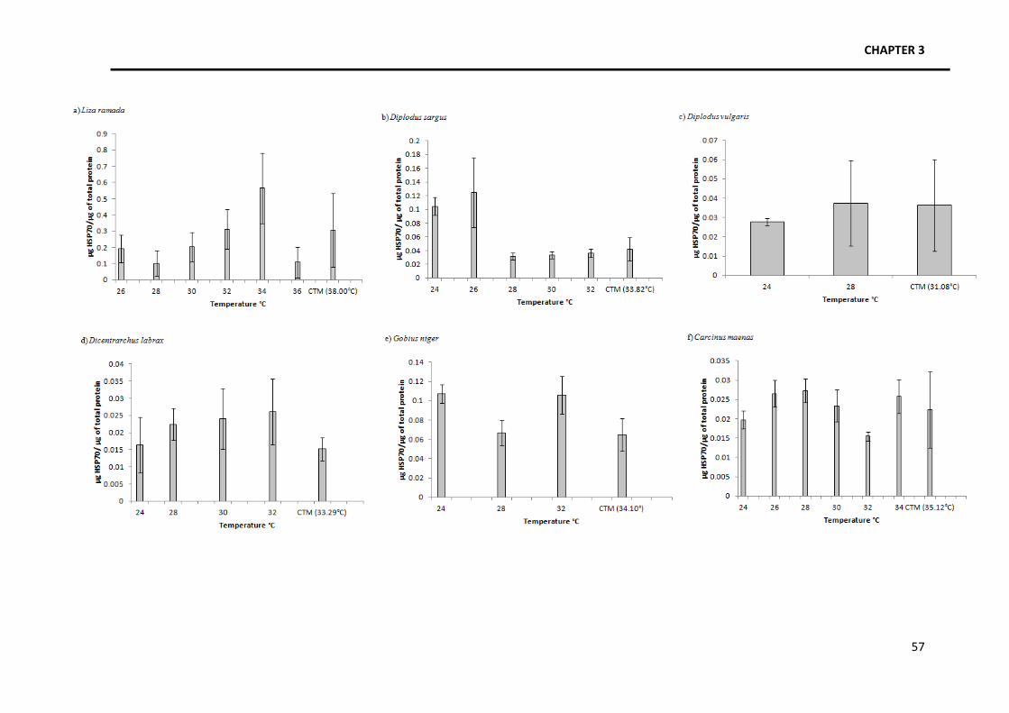
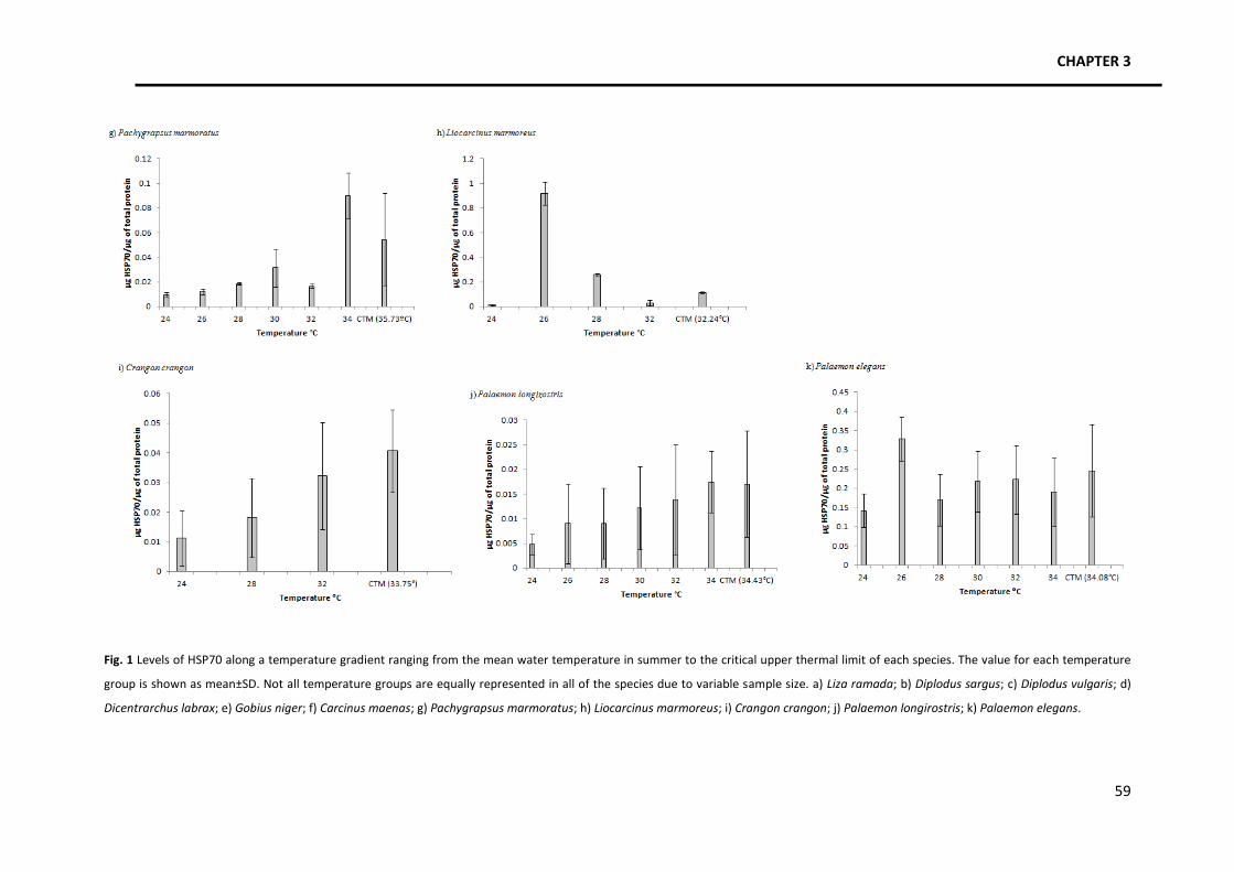
Focusing on the patterns of HSP70 expression (Fig. 1), four trends were identified. A
general trend was identified among various species in which there is an increase in the amount
of HSP70 as temperature increases, followed by a slight or steep decrease close to the thermal
limits. Species with this kind of response are Liza ramada, Diplodus sargus, Pachygrapsus
marmoratus and Liocarcinus marmoreus. Another pattern identified, in Gobius niger, is the
occurrence of several increases and decreases in the amount of HSP70 produced along the
temperature gradient, with a final decrease close to the thermal limits. The third identified
trend occurs in Crangon crangon and it consists in an increase in the amount of HSP along the
temperature gradient without a decrease close to the thermal limits. Finally, Diplodus vulgaris,
Dicentrarchus labrax, Palaemon longirostris, Palaemon elegans and Carcinus maenas seem to
maintain HSP70 levels constant along the temperature gradient.
CHAPTER 3
57
CHAPTER 3
59
Fig. 1 Levels of HSP70 along a temperature gradient ranging from the mean water temperature in summer to the critical upper thermal limit of each species. The value for each temperature
group is shown as mean±SD. Not all temperature groups are equally represented in all of the species due to variable sample size. a) Liza ramada; b) Diplodus sargus; c) Diplodus vulgaris; d)
Bachelet M, Adrie C, Polla BS (1998) Macrophages and heat shock proteins. Res Immunol 149: 727–732. Baker JR (1975) The humane killing of lobsters and crabs. The Humane Education Centre.
Baler R, Welch WJ, Voellmy R (1992) Heat shock gene regulation by nascent polypeptides and denatured
proteins: hsp70 as a potential autoregulatory factor. J Cell Biol 117: 1151–1159. Barnes RSK (1994) The brackish water fauna of North-western Europe. Cambridge, UK, pp 287. Barua D, Scott SA, Heckathorn A (2004) Acclimation of the temperature set-points of the heat-shock response.
J Therm Biol 29(3): 185-193.
Basu N, Nakano T, Grau EG and Iwama GK (2001) The effects of cortisol on Heat Shock Protein 70 Levels in two
fish species. Gen Comp Endocrinol 124 (1): 97-105. Basu N, Todgham AE, Ackerman PA, Bibeau MR, Nakano K, Schulte PM, Iwama GK (2002) Heat shock protein
genes and their functional significance in fish. Gene 295 (2, 7): 173-183. Bauchot M-L, Hureau JC (1986) Sparidae. In: Whitehead PJP, Bauchot M-L, Hureau JC, Nielson J, Tortonese E
(eds) Fishes of the North-eastern Atlantic and the Mediterranean vol. II. UNESCO, Paris, pp 883-907. Beg MU, Al-Subiai S, Beg KR, Butt SA, Al-Jandal N, Al-Hasan E, Al-Hussaini M (2010) Seasonal effect on Heat
Shock Proteins in fish from Kuwait Bay. Bull. Environ Contam Toxicol 84:91-95. Becker F, Craig E (1994) Heat shock proteins as molecular chaperones. Eur J Biochem 219: 11–23. Beckmann RP, WJ Welch, LA Mizzen (1990) Interaction of HSP70 with newly synthesized proteins: implication
for protein folding and assembly. Science 248: 850–854. Boddeke R (1989) Management of the brown shrimp (Crangon crangon) stock in dutch coastal waters. In:
Caddy JF (ed) Marine invertebrate fisheries: their assessment and management. Netherlands, pp 752.
Botton ML, Pogorzelska M, Smoral L, Shehata A, Hamilton MG (2006) Thermal biology of horseshoe crab embryos and larvae: A role for heat shock proteins. J Exp Mar Biol Ecol 336(1): 65-73.
Buckley BA , Hofmann GE (2002) Thermal acclimation changes DNA-binding activity of heat shock factor 1
(HSF1) in the goby, Gillichthys mirabilis: Implications for plasticity in the heat shock response in natural populations. J Exp Biol 205: 3231–3240.
Buckley BA, Hofmann GE (2004) Seasonal patterns and in vitro kinetics of HSF1 activation and Hsp70 mRNA
production in the goby, Gillichthys mirabilis. Physiol Biochem Zool 77: 570–581. Cara
JB, Aluru N, Moyano FJ, Vijayan MM (2005) Food-deprivation induces HSP70 and HSP90 protein
expression in larval gilthead sea bream and rainbow trout. Comp Biochem Physiol B 142(4): 426-431. Cartaxana A (1994) Distribution and migrations of the prawn Palaemon longirostris in the Mira river estuary
(Southwest Portugal). Estuaries 17(3):685-694. Chang ES (2005) Stressed-out lobsters: crustacean hyperglycemic hormone and stress proteins. Intedr Comp
Biol 45:43-50. Clayton
ME, Steinmann R, Fent K (2000) Different expression patterns of heat shock proteins hsp 60 and hsp
70 in zebra mussels (Dreissena polymorpha) exposed to copper and tributyltin. Aqua Toxicol 47 (3-4): 213-226. Currie S, Tufts BL, Moyes CD (1999) Influence of bioenergetic stress on heat shock protein gene expression in
nucleated red blood cells of fish. Am J Physiol Regul Integr Comp Physiol 276: 990-996.
Heat Shock Protein 70 patterns of coastal and estuarine organisms facing increasing temperatures
70
Currie S, Tufts BL (1997) Synthesis of stress protein 70 (hsp70) in rainbow trout (Oncorhynchus mykiss) red
blood cells. J. Exp. Biol. 200: 607–614. Deane EE, Woo NYS (2010) Advances and perspectives on the regulation and expression of piscine heat shock
proteins. Rev Fish Biol Fisheries. DOI 10.1007/s11160-010-9164-8. Dent L, Lutterschmidt WI (2003) Comparative thermal physiology of two sympatric sunfishes (Centrarchidae:
Perciformes) with a discussion of microhabitat utilization. J Therm Biol 28: 67-74. Dietz TJ, Somero GN (1992) The threshold induction temperature of the 90-kDa heat shock protein is subject
to acclimatization in eurythermal goby fishes (genus Gillichthys). Proc Nat Acad Sci USA 89: 3389-3393. Dietz TJ (1994) Acclimation of the threshold induction temperatures for 70-kDa and 90-kDa heat shock
proteins in the fish Gillichthys mirabilis. J Exp Biol 188:333–338. Dietz TJ, Somero GN (1993) Species- and tissue-specific synthesis patterns for heat-shock proteins HSP70 and
HSP90 in several marine teleost fishes. Physiol Zool 66: 863–880. Dubois MF, Hovanessian AG, Bensaude O (1991) Heat shock-induced denaturation of proteins. J Biol Chem
266: 9707–9711. Ellis RJ (1990) The molecular chaperone concept. Sem Cell Biol 1: 1–9. European Food Safety Authority, Scientific Panel on Animal Health and Welfare (2005) Aspects of the biology
and welfare of animals used for experimental and other scientific purposes. Fader SC, Yu Z, Spotila JR (1994) Seasonal variation in heat shock proteins (hsp70) in stream fish under natural
conditions. J Therm Biol 19:335–41. FAO (2011) Fact Sheets. http://www.fao.org/fishery/species/2291/en. Accessed 8 July 2011. Feder JH, Rossi JM, Solomon J, Solomon N, Lindquist S (1992) The consequences of expressing hsp70 in
Drosophila cells at normal temperatures. Genes Dev 6:1402–1413. Feder ME, Hofmann GE (1999) Heat-shock proteins, molecular chaperones and the stress response:
evolutionary and ecological physiology. Annu Rev Physiol 61: 243–282. Ferguson RA, Tufts BL, Boutilier RG (1989) Energy metabolism in trout red cells: consequences of adrenergic
stimulation in vivo and in vitro. J Exp Biol 143: 133–147. Fink A (1999) Chaperone-mediated protein folding. Physiol Rev 79: 425–449. Flores AVA, Paula J (2001) Intertidal distribution and species composition of brachyuran crabs at two rocky
shores in Central Portugal. Hydrobiologia 449:171-177. Forsyth RB, Candido EPM, Babich SL, Iwama GK (1997) Stress protein expression in coho salmon with bacterial
kidney disease. J Aquat Anim Health 9: 18-25.
Gaston KJ (2003) The structure and dynamics of geograhic ranges. Oxford University Press, Oxford. Glynn PW (1988) El Niño-Southern Oscillation 1982-1983: near-shore population, community, and ecosystem
responses. Ann Rev Ecol Syst 19:309-345. Gornati R, E Papis, S Rimoldi, G Terova, M Saroglia, G Bernardini. 2004. Rearing density influences the
expression of stress related genes in seabass (Dicentrarchus labrax, L.). Gene 341: 111-118. Hartl FU (1996) Molecular chaperones in cellular protein folding. Nature 381: 571–580. Hayward P, Nelson-Smith T, Shields C (1996) Sea shore of Britain and Europe. UK, 352 p. Hayward PJ, Ryland JS (1995) Handbook of the marine fauna of North-west Europe. UK, 900 p.
Hightower LE, Norris CE, DiIorio PJ, Fielding E (1999) Heat shock responses of closely related species of tropical and desert fish. Am Zool 39: 877–888.
Hightower LE (1991) Heat shock, stress proteins, chaperones, and proteotoxicity. Cell 66: 191–197. Hochachka PW, Somero GN (2002) Biochemical adaptation. Oxford University Press, New York. Hofmann GE (2005) Patterns of Hsp gene expression in ectothermic marine organisms on small to large
biogeographic scales. Integr. Comp Biol 45:247-255. Hofmann GE, Somero GN (1995) Evidence for protein damage at environmental temperatures: seasonal
changes in levels of ubiquitin conjugates and Hsp70 in the intertidal mussel Mytilus trossulus. J Exp Biol 198: 1509–1518.
Huey RB (1991) Physiological consequences of habitat selection. Am Nat 137, Supplement: Habitat Selection
S91-S115. Ingle RW (1997) Crayfishes, lobsters, and crabs of Europe. London, UK, 281 p. Ish-Horowicz D, Pinchin SM (1980) Genomic organization of the 87A7 and 87C1 heat-induced loci of
Drosophila melanogaster. J Mol Biol 142:231–45. Iwama GK, Vijayan MM, Forstyh RB, Ackerman PA (1999) Heat Shock Proteins and physiological stress in fish.
Amer Zool 39: 901-909. Iwama GK, Thomas PT, Forsyth RB, Vijayan MM (1998) Heat shock protein expression in fish. Rev Fish Biol Fish
8:35-56. Jacquier-Sarlin MR, Fuller K, Dinh-Xuan AT, Richard MJ, Polla BS (1994) Protective effects of hsp70 in
inflammation. Experientia 50: 1031–1038.
Johnston C, Jungalwalla P [n.d.] Aquatic Animal Welfare Guidelines: guidelines on welfare of fish and crustaceans in aquaculture and/or in live holding systems for human consumption. National Aquaculture Council of Australia. National Aquaculture Council Inc.
Kagawa N, Mugiya Y (2000) Exposure of goldfish (Carassius auratus) to bluegills (Lepomis macrochirus)
enhances expression of stress protein 70 mRNA in the brains and increases plasma cortisol levels. Zool Sci 17: 1061–1066.
Kedersha NL, Gupta M, Li W, Miller I, Anderson P (1999) RNA-binding Proteins TIA-1 and TIAR Link the
Phosphorylation of eIF-2 a to the Assembly of Mammalian Stress Granules. J Cell Biol 147 (7): 1431–1441.
Koban M, Yup AA, Agellon LB, Powers DA (1991) Molecular adaptation to environmental temperature: heat-shock response of the eurythermal teleost Fundulus heteroclitus. Mol Mar Biol Biotech 1: 1-17.
Kregel KC (2002) Heat shock proteins: modifying factors in physiological stress responses and acquired
thermotolerance. J Appl Physiol 92:2177-2186. Leigh-Brown AJ, Ish-Horowicz D (1981) Evolution of the 87A and 87C heat-shock loci in Drosophila. Nature
Lindquist S (1993) Autoregulation of the heat-shock response. In: Ilan J (ed) Translational Regulation of Gene
Expression 2, New York: Plenum, pp 279–320. Madeira D, Narciso L, Cabral H, Vinagre C In press Thermal tolerance and potential impacts of climate change
on coastal and estuarine organisms. Marine Biology.
Mestril R, Chi SH, Sayen MR, Dillmann WH (1994) Isolation of a novel inducible rat heat-shock protein (HSP70) gene and its expression during ischaemia/hypoxia and heat shock. Biochem J 3: 561–569.
Heat Shock Protein 70 patterns of coastal and estuarine organisms facing increasing temperatures
Misra S, Zafarullah M, Price-Haughey J, Gedamu L (1989) Analysis of stress-induced gene expression in fish cell
lines exposed to heavy metals and heat shock. Biochim Biophys Acta 1007: 325-333. MitoSciences Protocol. http://www.mitosciences.com/PDF/western.pdf.
Mizzen LA, Welch WJ (1988) Characterization of the thermotolerant cell. I. Effects on protein synthesis activity
and the regulation of heat-shock protein 70 expression. J Cell Biol 106: 1105–1116. Mora C, Ospina AF (2001) Tolerance to high temperatures and potential impact of sea warming on reef fishes
of Gorgona Island (tropical eastern Pacific). Mar Biol 139: 765-769. Morimoto RI, Tissieres A, Georgopoulos C (eds) (1990) The stress response, function of the proteins, and
perspectives. In: Stress Proteins in Biology and Medicine. Cold Spring Harbor Laboratory Press, Cold Spring Harbor, NY, pp. 1–36.
Moseley PL (1997) Heat shock proteins and heat adaptation of the whole organism. J Appl Physiol 83: 1413 1417.
Myrmel T, McCully JD, Malikin L, Krukenkamp IB, Levitsky S (1994) Heat shock protein 70 mRNA is induced by
anaerobic metabolism in the heart. Circulation 90: II-299 –II-305. Njemini, R. Demanet C, Mets T (2005) J Immunol Meth 306 (1-2): 176-182. doi:10.1016/j.jim.2005.08.012.
Parsell DA, Lindquist S (1993) The function of heat-shock proteins in stress tolerance: degradation and
reactivation of damaged proteins. Annu Rev Genet 27: 437-439. McClintock M (2009) Partnership for Interdisciplinary Studies of Coastal Oceans. http://www.piscoweb.org/research/science-by-discipline/species-performance/physiology. Accessed
12/09/2011. Paula J (1998) Larval retention and dynamic of the prawns Palaemon longirostris H. Milne Edwards and
Crangon crangon Linnaeus (Decapoda, Caridae) in the Mira estuary, Portugal. Invertebrate Reproduction and Development 33 (2-3): 221-228.
Polla BS, Bachelet M, Elia G, Santoro GM (1998) Stress proteins in inflammation. Ann N Y Acad Sci 851: 75–85. Pörtner HO, Knust R (2007) Climate change affects marine fishes through the oxygen limitation of thermal
tolerance. Science 315:95–97. Ramaglia V, Buck LT (2004) Time-dependent expression of heat shock proteins 70 and 90 in tissues of the
anoxic western painted turtle. J Exp Biol 207: 3775-3784. Reid SG, Bernier NJ, Perry SF (1998) The adrenergic stress response in fish: Control of catecholamine storage
and release. Comp Biochem Physiol 120C: 1–27. Renfro JL, Brown MA, Parker SL, Hightower LE (1993) Relationship of thermal and chemical tolerance to
transepithelial transport by cultured flounder renal epithelium. J Pharmacol Exp Therap 265: 992-1000.
Ryan JA, Hightower LE (1994) Evaluation of heavy-metal ion toxicity in fish cells using a combined stress protein and cytotoxicity assay. Environ Toxicol Chem 13:1231–40.
Sanders BM, Pascoe VM, Nakagawa PA Martin LS (1992). Persistence of the heat-shock response over time in
a common Mytilus mussel. Mol Mar Biol Biotech 1: 147–154. Sanford E (1999) Regulation of keystone predation by small changes in ocean temperature. Science 283:
Sauriau P-G, Robin J-P, Marchand J (1994) Effects of the excessive organic enrichment of the Loire Estuary on the downstream migratory patterns of the amphihaline grey mullet (Liza ramada) (Pisces: Mugilidae). In: Dyer KR, Orth RJ (eds) Changes in fluxes in estuaries: implications from science to management, 485 p.
Snoeckx LHEH, Cornelussen RN, van Nieuwenhoven FA, Reneman RS, van der Vusse GJ (2001) Heat shock
proteins and cardiovascular pathophysiology. Physiol Rev 81(4): 1461-1497. Somero GN (1995) Proteins and temperature. Ann Rev Physiol 57: 43–68.
Teranishi KS, Stillman JH (2007) A cDNA microarray analysis of the response to heat stress in hepatopancreas
tissue of the porcelain crab Petrolisthes cinctipes. Comp Bioch Physiol D 2(1): 53-62.
Tomanek L (2011) Environmental proteomics: changes in the proteome of marine organisms in response to environmental stress, pollutants, infection, symbiosis, and development. Annu Rev Mar Sc. 3: 14.1–14.27.
Tomanek L (2010) Variation in the heat shock response and its implication for predicting the effect of global
climate change on species’ biogeographical distribution ranges and metabolic costs. J Exp Biol 213:971-979. Tomanek L, Zuzow MJ (2010) The proteomic response of the mussel congeners Mytilus galloprovincialis and
M. trossulus to acute heat stress: implications for thermal tolerance and metabolic costs of thermal stress. J Exp Biol 213: 3559-3574.
Tomanek L, Somero GN (1999) Evolutionary and acclimation-induced variation in the heat-shock responses of
congeneric marine snails (genus Tegula) from different thermal habitats: implications for limits of thermotolerance and biogeography. J Exp Biol 202: 2925–2936.
Tomanek L, Somero GN (2002) Interspecific- and acclimation-induced variation in levels of heat-shock proteins
70 (hsp70) and 90 (hsp90) and heat-shock transcription factor-1 (HSF1) in congeneric marine snails (genus Tegula): implications for regulation of hsp gene expression. J Exp Biol 205: 677–685.
Tortonese E (1986) Moronidae. In: Whitehead PJP, Bauchot M-L, Hureau J-C, Nielsen J, Tortonese E (eds)
Fishes of the North-eastern Atlantic and the Mediterranean. UNESCO, Paris. Vol. II, pp 793-796.
Vidair CA, Huang RN, Doxsey SJ (1996) Heat shock causes protein aggregation and reduces protein solubility at the centrosome and other cytoplasmic locations. Int J Hypertherm 12: 681–695.
Vijayan MM, Pereira C, Forsyth RB, Kennedy CJ, Iwama GK (1997) Handling stress does not affect the
expression of hepatic heat shock protein 70 and conjugation enzymes in rainbow trout treated with beta-naphthoflavone. Life Sci 61: 117-127.
Vinagre C, Cabral HN, Costa MJ (2010) Relative importance of estuarine nurseries for species of the genus
Diplodus (Sparidae) along the Portuguese coast. Estuar Coast Shelf S 86: 197–202.
Welch WJ (1992) Mammalian stress response: cell physiology, structure/ function of stress proteins, and implications for medicine and disease. Physiol Rev 72: 1063–1081.
Williams JH, Farag AM, Stansbury MA, Young PA, Bergman HL, Petersen NS (1996) Accumulation of hsp 70 in
juvenile and adult rainbow trout gill exposed to metal-contaminated water and:or diet. Environ Toxicol Chem 15: 1324–1328.
Yamashita M, Yabu T, Ojima N (2010) Stress protein HSP70 in fish. Aqua-BioSci Monogr 3 (4): 111–141. Yu Z, Magee WE, Spotila JR (1994) Monoclonal antibody ELISA test indicates that large amounts of constitutive
HSP-70 are present in salamanders, turtle and fish. J Therm Biol 19: 41-53.
Zou Y, David J, Crowley, et al. (1998) Involvement of molecular chaper-ones in nucleotide excision repair. J Biol Chem 273: 12887―12892.