Page 1
HAL Id: hal-01768194https://hal-univ-rennes1.archives-ouvertes.fr/hal-01768194v2
Submitted on 27 Apr 2018
HAL is a multi-disciplinary open accessarchive for the deposit and dissemination of sci-entific research documents, whether they are pub-lished or not. The documents may come fromteaching and research institutions in France orabroad, or from public or private research centers.
L’archive ouverte pluridisciplinaire HAL, estdestinée au dépôt et à la diffusion de documentsscientifiques de niveau recherche, publiés ou non,émanant des établissements d’enseignement et derecherche français ou étrangers, des laboratoirespublics ou privés.
Template-Directed Synthesis of a Conjugated ZincPorphyrin Nanoball
Jonathan Cremers, Renée Haver, Michel Rickhaus, Juliane Gong, LudovicFavereau, Martin Peeks, Tim Claridge, Laura Herz, Harry Anderson
To cite this version:Jonathan Cremers, Renée Haver, Michel Rickhaus, Juliane Gong, Ludovic Favereau, et al.. Template-Directed Synthesis of a Conjugated Zinc Porphyrin Nanoball. Journal of the American ChemicalSociety, American Chemical Society, 2018, 140 (16), �10.1021/jacs.8b02552�. �hal-01768194v2�
Page 2
1
Template-Directed Synthesis of a Conjugated Zinc Porphyrin Nanoball
Jonathan Cremers,† Renee Haver,† Michel Rickhaus,† Juliane Q. Gong,‡ Ludovic Favereau,† Martin D.
Peeks,† Tim D. W. Claridge,† Laura M. Herz,‡ and Harry L. Anderson*,†
†Chemistry Research Laboratory, Department of Chemistry, University of Oxford, Oxford, OX1 3TA, United Kingdom ‡Clarendon Laboratory, Department of Physics, University of Oxford, Oxford, OX1 3PU, United Kingdom
Supporting Information Placeholder
ABSTRACT: We report the template-directed synthesis of a
π-conjugated 14-porphyrin nanoball. This structure consists of
two intersecting nanorings of 6 and 10 porphyrin units. Fluo-
rescence up-conversion spectroscopy experiments demonstrate
that electronic excitation delocalizes over the whole 3D π-
system in less than 0.3 ps, if the nanoball is bound to its tem-
plates, or over 2 ps if the nanoball is empty.
Spurred by the discovery of buckminsterfullerene,1 chemists
have sought rational strategies for the synthesis of 3D π-
conjugated geodesic cages. Stepwise chemical synthesis has
not yet been used to prepare any fullerenes, except C60 (ref. 2),
but several fullerene-like ball-shaped π-conjugated hydrocar-
bons have been synthesized recently.3 The high dimensionality
of these π-systems is expected to enhance their electronic
delocalization, compared with 1D or 2D molecular semicon-
ductors.4-6 Shape-persistent molecular cages are also in de-
mand for their gas adsorption properties, which mimic those of
zeolites.7 The best routes to molecular cages or capsules use
reversible reactions that allow error correction, such as metal
coordination8 or boronic ester condensation,9 but these reac-
tions do not produce π-conjugated connections. In principle,
reversible reactions such as imine formation,10 alkene metathe-
sis11 and alkyne metathesis12 could be used to construct π-
conjugated cages, but so far the cages made by these reactions
lack long-range conjugation.7,10-12 Template-directed coupling,
under kinetic control, is an alternative strategy for preparing
large macrocycles and cages.13 Here we show how simple
molecular templates can be used to synthesize the first fully π-
conjugated porphyrin ball b-P14 (Figure 1). This 14-porphyrin
prolate ellipsoidal cage consists of two perpendicular inter-
secting conjugation pathways, containing 6 and 10 porphyrin
units respectively. Structures of this type are valuable models
for photosynthetic light-harvesting systems.13-15 When all 14
porphyrin units in b-P14 are bound to templates, locking the
conformation, excited state energy delocalization occurs over
the whole system within 0.3 ps, whereas in the absence of the
templates excitation is distributed over the ball with a time
constant of about 2 ps.
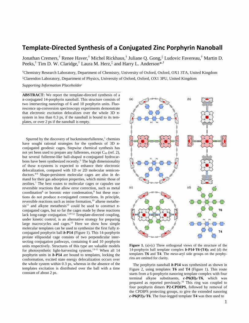
Figure 1. (a)-(c) Three orthogonal views of the structure of the
14-porphyrin ball template complex b-P14·T6·(T4)2 and (d) the
templates T6 and T4. The meso-aryl side groups on the porphy-
rins are omitted for clarity.
The porphyrin nanoball b-P14 was synthesized as shown in
Figure 2, using templates T6 and T4 (Figure 1). This route
starts from a 6-porphyrin nanoring template complex with four
terminal alkyne substituents, c-P6(H)4·T6, which was
prepared as reported previously.16 This ring was coupled to
four porphyrin dimers P2-CPDIPS, followed by removal of
the CPDIPS protecting groups, to give the extended nanoring
c-P6(P2)4·T6. The four-legged template T4 was then used to
Page 3
2
Figure 2. (a) Schematic synthetic route for b-P14. Reaction conditions: (i) P2-CPDIPS, Pd(PPh3)2Cl2, CuI, 1,4-benzoquinone, CHCl3/i-
Pr2NH, 41%; then TBAF, CH2Cl2, 97%; (ii) Pd(PPh3)2Cl2, CuI, 1,4-benzoquinone, CHCl3/i-Pr2NH, 51%; (iii) DABCO, size exclusion
chromatography, toluene/pyridine, 100%. (b) Chemical structures of c-P6-(H)4, P2-CPDIPS, c-P6(P2)4 and b-P14.
close the 10-porphyrin ring, yielding the bicyclic nanoball b-
P14·T6·(T4)2. The templates can readily be displaced from
this cage by high concentrations of a competing ligand;17
quinuclidine or DABCO remove both templates, giving b-P14,
whereas pyridine selectively removes T4, giving b-P14·T6.
Both b-P14 and b-P14·T6 were fully characterized (see SI).
Two types of aryl solubilizing groups were used in this molec-
ular design (Ar1 and Ar2, Figure 2) so as to confer high solu-
bility while avoiding excessive steric congestion.
Gel permeation chromatography (GPC) confirmed the puri-
ty of the nanoball and showed that its molecular weight is in
the expected range (ca. 20 kDa). The 1H NMR spectrum of b-
P14·T6·(T4)2 in CDCl3 at 298 K is consistent with the ex-
pected D2h gross symmetry and all the signals were assigned
by COSY and NOESY spectroscopy, except for unresolved
aliphatic multiplets in the 0.4–2.0 ppm region (see SI). This 1H
NMR spectrum shows that the nanoball consists of a mixture
of conformers with ethylhexyl chains pointing into and out of
the cavity; these rotamers are in slow exchange on the NMR
timescale, with ca. 40% of the alkoxy-chains pointing towards
the center of the ball. This ratio scarcely changes on removal
of the T4 templates. When all the templates are removed, the
porphyrins of the 6-ring rotate rapidly, simplifying the NMR
spectrum. Diffusion-ordered 1H NMR spectroscopy (DOSY)
experiments show that b-P14·T6·(T4)2 has a diffusion coeffi-
cient of 1.52 10–10 m2 s–1 (700 MHz, 298 K, CDCl3), which
corresponds to a hydrodynamic radius of about 27 Å, calculat-
ed using the Stokes-Einstein equation for a spherical molecule.
The UV-vis-NIR spectrum of b-P14·T6·(T4)2 (Figure 3a,
black curve) is essentially the sum of the absorption spectra of
its two component rings, as modeled by c-P6·T6 (the D6h ring
complex, with 3,5-bis(trihexylsilyl)phenyl substituents)18 and
c-P10·(T5)2 (where T5 is the version of T6 with five pyridyl
sites),16-20 although the absorption spectrum of b-P14·T6·(T4)2
is slightly red-shifted, demonstrating greater π-conjugation (SI
Figure S82). When b-P14·T6·(T4)2 is treated with quinu-
clidine to displace the templates, two distinct denaturation
processes are observed in the UV-vis-NIR titration (Figure 3):
At quinuclidine concentrations of 3–30 mM (Figure 3b), the
T4 units are displaced leading to disappearance of the sharp
peak at 883 nm;19-21 this peak is associated with the template-
bound conformation of the 10-porphyrin ring. At quinuclidine
concentrations of 30–300 mM (Figure 3c), the central T6 unit
is displaced causing disappearance of the distinctive three-
finger pattern in the Q band of the 6-porphyrin ring compo-
nent. All these spectral changes reflect the greater flexibility,
and wider distribution of porphyrin-porphyrin dihedral angles,
upon template removal.
Analysis of the denaturation binding isotherms13c,17,21 (Fig-
ure 3) reveals that the association constants for binding of T4
and T6 to b-P14 to form b-P14·T6·(T4)2 are (1.8 ± 0.2) × 1022
M–1 for T4 and (5.5 ± 1.2) × 1037 M–1 for T6, in toluene at 298
K. This analysis assumes that the denaturation processes for
each template (T4 and T6) are essentially all-or-nothing two
state equilibria (i.e. intermediate partially denatured species do
not build up to high concentrations); this assumption is sup-
ported by the isosbestic nature of the UV-vis-NIR titration and
the good fits of the curves to the calculated isotherm for a two-
state equilibrium (Figure 3b,c). The binding strength of T6 is
roughly an order of magnitude stronger in the ball than in a
comparable 6-ring system,17 which can be attributed to the
conformational preorganization provided by the 10-porphyrin
ring.
N N
NNZn
N N
NNZn
N N
NNZn
Ar2 Ar2
N
NN
NZn
N
NN
NZn
Ar1
Ar1
H HH HN N
NNZn
N
NN
NZn
N
NN
NZn
Ar1
Ar1
c-P6
T4 (ii)
c-P6(P2)4
P2-CPDIPS
(i)
22
(iii)
c-P6(H)4·T6 c-P6(P2)4·T6 c-P6(P2)4·T6·(T4)2 b-P14·T6·(T4)2 b-P14
N N
NNZn
N N
NNZn
N N
NNZn
Ar2
Ar2 Ar2
Ar2
N
NN
NZn
N
NN
NZn
Ar1
Ar1
b-P14
2
22 2
N N
NNZn
Ar2
H Si
CN
22
2
Ar1
Ar1
Ar2
Ar2 Ar2
Ar1
Ar1
Ar1
Ar1
P2-CPDIPS
(a)
(b)
O
Ar1 =
Ar2 =
Si(C6H13)3
Si(C6H13)3
Page 4
3
Figure 3. UV-vis-NIR denaturation titration of the b-
P14·T6·(T4)2 complex with quinuclidine (toluene, 298 K). (a)
Full titration, T4 is displaced first (black to green) followed by the
displacement of T6 (green to red). The two phases of the titration
are shown in (b) and (c), with the experimental (black circles) and
calculated isotherms (red lines) shown on the right.
The UV-vis-NIR absorption and fluorescence spectra of b-
P14·T6·(T4)2 and b-P14 are compared in Figure 4. The more
rigid conformation of the template-bound ball is reflected by
its sharper and more red-shifted absorption and emission spec-
tra.19-21 Fluorescence up-conversion spectroscopy experiments
reveal that the excited states delocalize across the two perpen-
dicular ring planes in the nanoball on an ultrafast timescale.
The fluorescence anisotropy dynamics of b-P14, with and
without templates, are shown in Figure 4c. The template com-
plex, b-P14·T6·(T4)2 (black points), exhibits a constant and
very low anisotropy ( = 0.02) over the time range of this
experiment (0–10 ps; time resolution 0.3 ps), showing that the
excited state delocalizes in three dimensions within 0.3 ps. In
contrast, b-P14·T6 (green points) and b-P14 (red points) show
a fast initial drop in anisotropy within the first 5 ps from =
0.1 towards ≈ 0. This fast depolarization resembles the ani-
sotropy decay in porphyrin nanorings with >24 porphyrin
units.15 The initial anisotropy of 0.1 suggests that upon excita-
tion, an exciton is delocalized over a full ring and that both
absorption and emission transition dipoles are polarized in the
ring plane. After ultrafast relaxation, the exciton localizes and
migrates rapidly around the entire porphyrin nanoball. Contri-
butions from emission components polarized in both planes
thus result in an anisotropy close to zero. Without the tem-
plates, exciton migration is slower, resulting in the observed
anisotropy decays, whereas in b-P14·T6·(T4)2 the anisotropy
decay is faster than the time resolution of the experiment. This
behavior is very different from that of the nanorings c-P6·T6,
c-P6 and c-P10, which exhibit anisotropies of near 0.1 (re-
maining constant during 10 ps after excitation), in agreement
with theoretical predictions for an excited state that is delocal-
ized over a 2D ring.15,16,18,20,22
Figure 4. UV-vis-NIR absorption (solid line) and fluorescence
(dashed line) spectra of (a) b-P14·T6·(T4)2 and (b) b-P14 (tolu-
ene, 298 K). The dip at 1130–1170 nm corresponds to solvent
absorption. (c) First order fits of the fluorescence anisotropy
decay. Solutions of b-P14·T6·(T4)2 (black), b-P14·T6 (green)
and b-P14 (red) were excited at 820 nm and emission was detect-
ed at 950 nm.
The prolate shape of b-P14·T6·(T4)2 and b-P14 is reminis-
cent of C70 fullerene. While C60 fullerene has complete polari-
zation memory loss with zero fluorescence anisotropy, C70
displays excitation-wavelength dependent anisotropy values
ranging between –0.2 and 0.1, because emission polarized in
the x-y plane is energetically favorable as a result of geomet-
rical deformation.23 However, b-P14·T6·(T4)2 and b-P14 do
not display any significant changes in anisotropy with excita-
tion wavelength in the range of 760–880 nm, probably because
the absorption features of their two constituent rings broadly
overlap and the emission may be polarized in both ring planes.
DFT calculations (B3LYP/6-31G*) indicate that the HOMO
of b-P14 is distributed over the entire π-system (Figure 5),
albeit with a higher density on the 6-ring and particularly the
tetraalkynylporphyrins. The LUMO and LUMO+1 are located
exclusively on the 6-ring and the 10-ring, respectively, with
nearly identical energies. Time-dependent DFT calculations
(B3LYP/6-31G*) were carried out to model the electronic
excited states of b-P14. Natural transition orbital plots (Tables
S4–S12) show that the two lowest energy singlet excited states
(S1 and S2) are mainly localized in the 6-porphyrin and 10-
porphyrin ring components, respectively. The transitions from
S0–S1 and S0–S2 are dipole-forbidden (f = 0), whereas transi-
tions to S3, S4 and S5 are allowed (f = 0.13, 5.12 and 0.83,
respectively) and these excited states are distributed over all
14 porphyrins. These calculations are in line with the observa-
Page 5
4
tion that excitation delocalizes between the two rings within
the ball on a timescale of less than 300 fs. The dimensions of
the calculated geometry of b-P14 are 52.7 Å, 27.6 Å and 23.9
Å (measured as the diameter along the D2h symmetry axes,
without including aryl side chains).
Figure 5. LUMO+1 (–3.22 eV), LUMO (–3.23 eV) and HOMO
(–4.66 eV) of b-P14 calculated at the B3LYP/6-31G* level of
theory with Grimme’s dispersion correction, GD3, shown at a
density isovalue of 0.008 a.u. together with their corresponding
energy levels. Aryl groups were replaced with hydrogen atoms to
simplify the calculations.
In summary, we have synthesized a fully π-conjugated
three-dimensional 14-porphyrin nanoball by a template di-
rected approach. UV-vis-NIR titrations show that the tem-
plates bound within the cavity can be removed successively by
the addition of a competing ligand. Fluorescence upconversion
spectroscopy reveals ultrafast electronic delocalization be-
tween the two perpendicular ring planes in the porphyrin ball.
The fluorescence anisotropy approaches zero, indicating that
excitation rapidly migrates between the two ring components
of the nanoball.
ASSOCIATED CONTENT
Supporting Information. Synthetic procedures, characterization
data, binding studies, NMR assignments, fluorescence spectros-
copy and calculated geometries. The Supporting Information is
available free of charge at http://pubs.acs.org.
AUTHOR INFORMATION
Corresponding Author
[email protected] .
Funding Sources
No competing financial interests have been declared.
ACKNOWLEDGMENT
We thank the ERC (320969), EPSRC and the Swiss National
Science Foundation (P2BSP2_168919) for funding, the EPSRC
UK National Mass Spectrometry Facility at Swansea University
for MALDI spectra and the University of Oxford Advanced Re-
search Computing facility (ARC,
http://dx.doi.org/10.5281/zenodo.22558) for support.
REFERENCES
(1) Kroto, H. W.; Heath, J. R.; O’Brien, S. C.; Curl, R. F.; Smalley, R.
E. Nature 1985, 318, 162.
(2) (a) Scott, L. T.; Boorum, M. M; McMahon, B. J.; Hagen, S.; Mack, J.; Blank, J.; Wegner, H.; de Meijere, A. Science 2002, 295, 1500. (b)
Kabdulov, M.; Jansen, M.; Amsharov, K. Yu. Chem. Eur. J. 2013, 19,
17262. (c) Greisch, J.-F.; Amsharov, K. Yu.; Weippert, J.; Weis, P.; Bottcher, A.; Kappes, M. M. J. Am. Chem. Soc. 2016, 138, 11254.
(3) (a) Matsui, K.; Segawa, Y.; Namikawa, T.; Kamada, K.; Itami, K.
Chem. Sci. 2013, 4, 84. (b) Kayahara, E.; Iwamoto, T.; Takaya, H.; Suzu-ki, T.; Fujitsuka, M.; Majima, T.; Yasuda, N.; Matsuyama, N.; Seki, S.;
Yamago, S. Nat. Commun. 2013, 4, 2694. (c) Matsui, K.; Segawa, Y.;
Itami, K. J. Am. Chem. Soc. 2014, 136, 16452. (d) Ikemoto, K.; Koba-yashi, R.; Sato, S.; Isobe, H. Angew. Chem. Int. Ed. 2017, 56, 6511.
(4) Gutzler, R.; Perepichka, D. F. J. Am. Chem. Soc. 2013, 135, 16585.
(5) Peeks, M. D.; Tait, C. E.; Neuhaus, P.; Fischer, G. M.; Hoffmann, M.; Haver, R.; Cnossen, A.; Harmer, J. R.; Timmel, C. R.; Anderson, H.
L. J. Am. Chem. Soc. 2017, 139, 10461. (6) Ball, M.; Zhong, Y.; Fowler, B.; Zhang, B.; Li, P.; Etkin, G.; Paley,
D. W.; Decatur, J.; Dalsania, A. K.; Li, H.; Xiao, S.; Ng, F.; Steigerwald,
M. L.; Nuckolls, C. J. Am. Chem. Soc. 2016, 138, 12861. (7) (a) Zhang, G.; Masteralerz, M. Chem. Soc. Rev. 2014, 43, 1934. (b)
Hasell, T.; Cooper, A. I. Nat. Rev. Mater. 2016, 1, 16053. (c) Santolini,
V.; Miklitz, M.; Berardo, E.; Jelfs, K. E. Nanoscale 2017, 9, 5280. (8) (a) Olenyuk, B.; Levin, M. D.; Whiteford, J. A.; Shield, J. E.; Stang,
P. J. J. Am. Chem. Soc. 1999, 121, 10434. (b) Sun, Q.-F.; Iwasa, J.; Oga-
wa, D.; Ishido, Y.; Sato, S.; Ozeki, T.; Sei, Y.; Yamaguchi, K.; Fujita, M. Science 2010, 1144.
(9) Zhang, G.; Presly, O.; White, F.; Oppel, I. M.; Mastalerz, M. An-
gew. Chem. Int. Ed. 2014, 53, 5126. (10) Rue, N. M.; Sun, J.; Warmuth, R. Isr. J. Chem. 2011, 51, 743.
(11) Zhu, B.; Chen, H.; Lin, W.; Ye, Y.; Wu, J.; Li, S. J. Am. Chem.
Soc. 2014, 136, 15126. (12) (a) Zhang, C.; Wang, Q.; Long, H.; Zhang, W. J. Am. Chem. Soc.
2011, 133, 20995. (b) Lee, S.; Yang, A.; Moneypenny, T. P., II; Moore, J.
S. J. Am. Chem. Soc. 2016, 138, 2182. (13) (a) O’Sullivan, M. C.; Sprafke, J. K.; Kondratuk, D. V.; Rinfray,
C.; Claridge, T. D.; Saywell, A.; Blunt, M. O.; O'Shea, J. N.; Beton, P. H.;
Malfois, M.; Anderson, H. L. Nature 2011, 469, 72. (b) Neuhaus, P.; Cnossen, A.; Gong, J. Q.; Herz, L. M.; Anderson, H. L. Angew. Chem. Int.
Ed. 2015, 54, 7344. (c) Favereau, L.; Cnossen, A.; Kelber, J. B.; Gong, J.
Q.; Oetterli, R. M.; Cremers, J.; Herz, L. M.; Anderson, H. L. J. Am. Chem. Soc. 2015, 137, 14256.
(14) (a) Wasielewski, M. R. Acc. Chem. Res. 2009, 42, 1910. (b) Ara-
tani, N.; Kim, D.; Osuka, A. Acc. Chem. Res. 2009, 42, 1922. (15) Parkinson, P.; Kondratuk, D. V.; Menelaou, C.; Gong, J. Q.; An-
derson, H. L.; Herz, L. M. J. Phys. Chem. Lett. 2014, 5, 4356.
(16) Gong, J. Q.; Favereau, L.; Anderson, H. L.; Herz, L. M. J. Phys. Chem. Lett. 2016, 7, 332.
(17) Hogben, H. J.; Sprafke, J. K.; Hoffmann, M.; Pawlicki, M.; An-
derson, H. L. J. Am. Chem. Soc. 2011, 133, 20962. (18) Sprafke, J. K.; Kondratuk, D. V.; Wykes, M.; Thompson, A. L.;
Hoffmann, M.; Drevinskas, R.; Chen, W.-H.; Yong, C. K.; Kärnbratt, J.;
Bullock, J. E.; Malfois, M.; Wasielewski, M. R.; Albinsson, B.; Herz, L. M.; Zigmantas, D.; Beljonne, D.; Anderson, H. L. J. Am. Chem. Soc.
2011, 133, 17262.
(19) Liu, S.; Kondratuk, D. V.; Rousseaux, S. A. L.; Gil-Ramírez, G.; O’Sullivan, M. C.; Cremers, J.; Claridge, T. D. W.; Anderson, H. L.
Angew. Chem. Int. Ed. 2015, 54, 5355.
(20) Gong, J. Q.; Parkinson, P.; Kondratuk, D. V.; Gil-Ramirez, G.; Anderson, H. L.; Herz, L. M. J. Phys. Chem. C 2015, 119, 6414.
(21) Cremers, J.; Richert, S.; Kondratuk, D. V.; Claridge, T. D. W.;
Timmel, C. R.; Anderson, H. L. Chem. Sci. 2016, 7, 6961.
Page 6
5
(22) Yong, C.-K.; Parkinson, P.; Kondratuk, D. V.; Chen, W.-H.;
Stannard, A.; Summerfield, A.; Sprafke, J. K.; O'Sullivan, M. C.; Beton, P. H.; Anderson, H. L.; Herz, L. M. Chem. Sci. 2015, 6, 181.
(23) Fedorov, A.; Berberan-Santos, M. N.; Lefèvre, J.-P.; Valeur, B.
Chem. Phys. Lett. 1997, 267, 467.


![Supplementary Information Section · 2016-03-21 · 1 Supplementary Information Section Meso-mono-[4-(1,4,7-triazacyclononanyl)]-tri(phenyl)]porphyrin and respective zinc(II)-complex:](https://static.documents.pub/doc/80x56/5ee2fbd8ad6a402d666d2349/supplementary-information-2016-03-21-1-supplementary-information-section-meso-mono-4-147-triazacyclononanyl-triphenylporphyrin.jpg)


