By Liu Fredrerick et al, year 2013.... I hope this will be a help for a reference :)
15
Epidemiology, Diagnosis, and Treatment of Temporomandibular Disorders Frederick Liu, DDS, MD*, Andrew Steinkeler, DMD, MD EPIDEMIOLOGY Temporomandibular disorders (TMD) are a broad group of clinical problems involving the masticatory musculature, the temporomandibular joint, surrounding bony and soft tissue components, and combinations of these problems. 1 Symptoms of TMD include decreased mandibular range of motion, pain in the muscles of mastication, temporo- mandibular joint (TMJ) pain, associated joint noise with function, generalized myofas- cial pain, and a functional limitation or deviation of the jaw opening. 1 The prevalence of TMD is thought to be greater than 5% of the population. 2 Lipton and colleagues 3 showed that about 6% to 12% of the population experience clinical symptoms of TMD. Patients with TMD symptoms present over a broad age range; however, there is a peak occurrence between 20 and 40 years of age. 4 Department of Oral and Maxillofacial Surgery, School of Dental Medicine, University of Pennsylvania, 3400 Spruce Street, Philadelphia, PA 19103, USA * Corresponding author. E-mail address: [email protected]KEYWORDS Temporomandibular disorders Epidemiology Diagnosis Treatment KEY POINTS Temporomandibular disorder (TMD) is a multifactorial disease process caused by muscle hyperfunction or parafunction, traumatic injuries, hormonal influences, and articular changes. Symptoms of TMD include decreased mandibular range of motion, muscle and joint pain, joint crepitus, and functional limitation or deviation of the jaw opening. Only after failure of noninvasive options should more invasive and nonreversible treat- ments be initiated. Treatment can be divided into noninvasive, minimally invasion, and invasive options. Temporomandibular joint replacement is reserved for severely damaged joints with end- stage disease that has failed all other more conservative treatment modalities. Dent Clin N Am 57 (2013) 465–479 http://dx.doi.org/10.1016/j.cden.2013.04.006 dental.theclinics.com 0011-8532/13/$ – see front matter Ó 2013 Elsevier Inc. All rights reserved.
Transcript
Epidemiology, Diagnosis, andTreatment of TemporomandibularDisorders
Frederick Liu, DDS, MD*, Andrew Steinkeler, DMD, MD
� Temporomandibular disorder (TMD) is a multifactorial disease process caused by musclehyperfunction or parafunction, traumatic injuries, hormonal influences, and articularchanges.
� Symptoms of TMD include decreased mandibular range of motion, muscle and joint pain,joint crepitus, and functional limitation or deviation of the jaw opening.
� Only after failure of noninvasive options should more invasive and nonreversible treat-ments be initiated.
� Treatment can be divided into noninvasive, minimally invasion, and invasive options.
� Temporomandibular joint replacement is reserved for severely damaged joints with end-stage disease that has failed all other more conservative treatment modalities.
EPIDEMIOLOGY
Temporomandibular disorders (TMD) are a broad group of clinical problems involvingthe masticatory musculature, the temporomandibular joint, surrounding bony and softtissue components, and combinations of these problems.1 Symptoms of TMD includedecreased mandibular range of motion, pain in the muscles of mastication, temporo-mandibular joint (TMJ) pain, associated joint noise with function, generalized myofas-cial pain, and a functional limitation or deviation of the jaw opening.1 The prevalence ofTMD is thought to be greater than 5% of the population.2 Lipton and colleagues3
showed that about 6% to 12% of the population experience clinical symptoms ofTMD. Patients with TMD symptoms present over a broad age range; however, thereis a peak occurrence between 20 and 40 years of age.4
Department of Oral and Maxillofacial Surgery, School of Dental Medicine, University ofPennsylvania, 3400 Spruce Street, Philadelphia, PA 19103, USA* Corresponding author.E-mail address: [email protected]
Dent Clin N Am 57 (2013) 465–479http://dx.doi.org/10.1016/j.cden.2013.04.006 dental.theclinics.com0011-8532/13/$ – see front matter � 2013 Elsevier Inc. All rights reserved.
TMD symptoms are more prevalent in women than men. Contrary to the knownincreased health risk in postmenopausal women of conditions such as heart diseaseand stroke, women tend to develop TMD during their premenopausal years.1 The rea-sons behind the sexual disequilibrium in TMD prevalence are not entirely clear, butsome have suggested a hormonal influence.5–7 In fact, both animal and human studieshave suggested that sex hormones may predispose to TMJ dysfunction and cartilag-inous breakdown.5–7 Elevated levels of estrogen have been found in patients withTMD.1 However, no definitive link between these hormones and causation of TMDhas been established.TMD is thought to be a multifactorial process secondary to muscle hyperfunction or
parafunction, traumatic injuries, hormonal influences, and articular changes within thejoint. Various investigators have found correlations between occlusion and TMJ symp-toms. Mohlin and Kopp8 showed an association between occlusal interferences andmyofascial pain and dysfunction. They found links between posterior crossbite withmuscular discomfort. Patients with deep bites, class II malocclusion, and anterioropen bites may also be predisposed to myofascial pain.9–12
DIAGNOSIS AND CLASSIFICATION
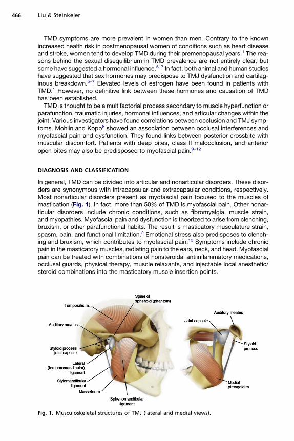
In general, TMD can be divided into articular and nonarticular disorders. These disor-ders are synonymous with intracapsular and extracapsular conditions, respectively.Most nonarticular disorders present as myofascial pain focused to the muscles ofmastication (Fig. 1). In fact, more than 50% of TMD is myofascial pain. Other nonar-ticular disorders include chronic conditions, such as fibromyalgia, muscle strain,and myopathies. Myofascial pain and dysfunction is theorized to arise from clenching,bruxism, or other parafunctional habits. The result is masticatory musculature strain,spasm, pain, and functional limitation.2 Emotional stress also predisposes to clench-ing and bruxism, which contributes to myofascial pain.13 Symptoms include chronicpain in the masticatory muscles, radiating pain to the ears, neck, and head. Myofascialpain can be treated with combinations of nonsteroidal antiinflammatory medications,occlusal guards, physical therapy, muscle relaxants, and injectable local anesthetic/steroid combinations into the masticatory muscle insertion points.
Fig. 1. Musculoskeletal structures of TMJ (lateral and medial views).
Treatment of Temporomandibular Disorders 467
Articular disorders (internal derangement) can be divided into inflammatory andnoninflammatory arthropathies. Inflammatory articular disorders include rheumato-logic processes, such as rheumatoid arthritis (RA), seronegative spondylopathies,such as ankylosing spondylitis, psoriatic arthritis, gout, and infectious arthritis. Nonin-flammatory articular disk disorders include osteoarthritis, joint damage from priortrauma or surgery, or other cartilage or bone disorders (Table 1). Mechanistically,articular disorders occur as a result of an altered balance of anabolic and catabolic cy-tokines. This cytokine imbalance creates an inflammatory milieu, which leads to oxida-tive stress, free radicals, and ultimately joint damage.2
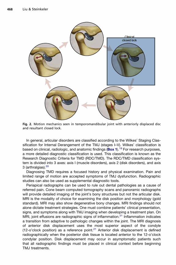
Internal derangement equates to changes in the disk-condyle relationship.14 Diskdisplacements are categorized as disk displacement with reduction or disk displace-ment without reduction (Fig. 2). The fibrocartilage disk is typically displaced anterome-dially but rarely may be displaced laterally or posteriorly.15–17 Anatomically, diskdisplacement with reduction is interference between the mandibular condyle withthe articular disk during jaw opening or closing. This interference may generate click-ing, popping, or crepitus in the joint, which can be associated with discomfort. Clickingalone, however, is not diagnostic of articular disk displacement. During disk displace-ment with reduction, the condyle meets the posterior aspect of the disk, which thenreduces to its proper position between the condyle and glenoid fossa.14,17 Articulardisk displacement is associated with TMD. One study found magnetic resonance im-aging (MRI) evidence of disk displacement in 84% of symptomatic patients with TMDversus 33% of asymptomatic patients.18 MRI findings, however, should not solelydictate treatment because disk displacement may occur in asymptomatic patients.Disk displacement without reduction results in a closed lock whereby the condylarmovement is physically blocked by the anteriorly displaced disk. Acute closed clockis associated with limited mandibular opening and severe pain.14,17 Physical examina-tion should include a general assessment of the head and neck, palpation of the masti-catory muscles, occlusal analysis, examination of the jaw opening and closing, andpalpation of the TMJ. Palpation of the muscles of mastication may elicit mild to severepain. Masseters are palpated with fingers positioned over the angle of the mandible.The temporalis muscles are palpated along the temple with the jaw relaxed andclenched. The pterygoid muscles are palpated intraorally along the medial aspect ofthe mandibular ramus between the tonsillar pillars.
Fig. 2. Motion mechanics seen in temporomandibular joint with anteriorly displaced discand resultant closed lock.
Liu & Steinkeler468
In general, articular disorders are classified according to the Wilkes’ Staging Clas-sification for Internal Derangement of the TMJ (stages I–V). Wilkes’ classification isbased on clinical, radiologic, and anatomic findings (Box 1).19 For research purposes,a more detailed diagnostic classification is used. This classification is known as theResearch Diagnostic Criteria for TMD (RDC/TMD). The RDC/TMD classification sys-tem is divided into 3 axes: axis I (muscle disorders), axis 2 (disk disorders), and axis3 (arthralgias).20
Diagnosing TMD requires a focused history and physical examination. Pain andlimited range of motion are accepted symptoms of TMJ dysfunction. Radiographicstudies can also be used as supplemental diagnostic tools.Periapical radiographs can be used to rule out dental pathologies as a cause of
referred pain. Cone beam computed tomography scans and panoramic radiographswill provide detailed imaging of the joint’s bony structures but not the articular disk.MRI is the modality of choice for examining the disk position and morphology (goldstandard). MRI may also show degenerative bony changes. MRI findings should notalone dictate treatment strategies. One must combine patients’ clinical presentation,signs, and symptoms along with TMJ imaging when developing a treatment plan. OnMRI, joint effusions are radiographic signs of inflammation.21 Inflammation indicatesa transition from adaptive to pathologic changes within the joint. The MRI diagnosisof anterior disk displacement uses the most superior aspect of the condyle(12-o’clock position) as a reference point.21 Anterior disk displacement is definedradiographically when the posterior disk tissue is located anterior to the 12-o’clockcondylar position. Disk displacement may occur in asymptomatic patients suchthat all radiographic findings must be placed in clinical context before beginningTMJ treatments.
Box 1
Wilkes’ staging for internal derangement of the TMJ
I. Early stage
A. Clinical presentation: no pain or decreased range of motion, possible clicking
B. Radiographic presentation: disk anteriorly positioned, normal bony contours
C. Anatomic correlation: anterior displacement, normal anatomic form of bone, and disk
II. Early/intermediate stage
A. Clinical presentation: episodes of pain, opening clicks, intermittent locking
B. Radiographic presentation: anterior disk displacement, thickened posterior disk, bonycontours normal
C. Anatomic correlation: early disk deformity, anterior displacement, normal bonycontours
III. Intermediate stage
A. Clinical presentation: many painful episodes, intermittent closed locking, multiplefunctional symptoms, decreased range of motion
B. Radiographic presentation: anterior disk displacement with disk deformity
C. Anatomic correlation: marked disk displacement and deformity, normal bony contours
IV. Intermediate/late stage
A. Clinical presentation: increased pain relative to earlier stages
B. Radiographic presentation: bony changes, such as flattened eminence, condylardeformity, osteosclerotic changes
C. Anatomic correlation: adhesions of disk, bony changes, evidence of osteoarthritis,osteophytes, no disk perforations
V. Late stage
A. Clinical presentation: episodic or continuous pain, crepitus, limited range of motion atall times, constant functional difficulties
B. Radiographic presentation: disk perforations, gross deformities of bony structures andcartilage, progressive arthritic changes
C. Anatomic correlation: gross hard and soft tissue changes, perforations, adhesions,subcortical cysts
Adapted from Bronstein S, Merrill B. Disorders of the TMJ. Oral and Maxillofacial SurgeryClinics North America. Philadelphia: Saunders; 1989.
Treatment of Temporomandibular Disorders 469
TREATMENT
The treatment of TMJ osteoarthrosis and internal derangement can be divided into 3broad categories: noninvasive, minimally invasive, and invasive management. Thespecific management plan can vary depending on the specific diagnosis and severityof TMJ disorder; however, the underlying principles of treatment apply universally.
1. Multidisciplinary approach involving multiple specialties, including general den-tistry, oral medicine, orofacial pain, orthodontics, oral surgery, physical therapy,and psychiatry may be necessary to fully address the problem from all angles.
2. There is progression of treatment only after failure of more conservative modalities.The least invasive and most reversible treatments should be tried first. Only after a
Liu & Steinkeler470
failure to alter the disease process and clinical symptoms should more invasive andoften nonreversible treatments be initiated.
Goals of treatment1. Decreasing joint pain2. Increasing joint function and opening3. Preventing further joint damage4. Improving overall quality of life and reducing disease-related morbidities
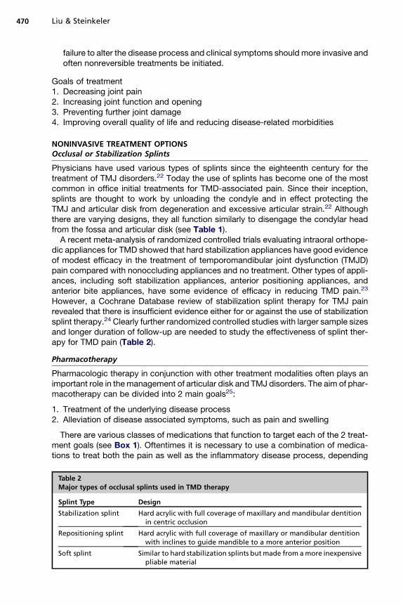
NONINVASIVE TREATMENT OPTIONSOcclusal or Stabilization Splints
Physicians have used various types of splints since the eighteenth century for thetreatment of TMJ disorders.22 Today the use of splints has become one of the mostcommon in office initial treatments for TMD-associated pain. Since their inception,splints are thought to work by unloading the condyle and in effect protecting theTMJ and articular disk from degeneration and excessive articular strain.22 Althoughthere are varying designs, they all function similarly to disengage the condylar headfrom the fossa and articular disk (see Table 1).A recent meta-analysis of randomized controlled trials evaluating intraoral orthope-
dic appliances for TMD showed that hard stabilization appliances have good evidenceof modest efficacy in the treatment of temporomandibular joint dysfunction (TMJD)pain compared with nonoccluding appliances and no treatment. Other types of appli-ances, including soft stabilization appliances, anterior positioning appliances, andanterior bite appliances, have some evidence of efficacy in reducing TMD pain.23
However, a Cochrane Database review of stabilization splint therapy for TMJ painrevealed that there is insufficient evidence either for or against the use of stabilizationsplint therapy.24 Clearly further randomized controlled studies with larger sample sizesand longer duration of follow-up are needed to study the effectiveness of splint ther-apy for TMD pain (Table 2).
Pharmacotherapy
Pharmacologic therapy in conjunction with other treatment modalities often plays animportant role in the management of articular disk and TMJ disorders. The aim of phar-macotherapy can be divided into 2 main goals25:
1. Treatment of the underlying disease process2. Alleviation of disease associated symptoms, such as pain and swelling
There are various classes of medications that function to target each of the 2 treat-ment goals (see Box 1). Oftentimes it is necessary to use a combination of medica-tions to treat both the pain as well as the inflammatory disease process, depending
Table 2Major types of occlusal splints used in TMD therapy
Splint Type Design
Stabilization splint Hard acrylic with full coverage of maxillary and mandibular dentitionin centric occlusion
Repositioning splint Hard acrylic with full coverage of maxillary or mandibular dentitionwith inclines to guide mandible to a more anterior position
Soft splint Similar to hard stabilization splints butmade from amore inexpensivepliable material
Treatment of Temporomandibular Disorders 471
on the severity of disease. However, care must be taken to avoid the prolonged use ofcertain medications, in particular analgesics, to prevent drug tolerance and depen-dency. The health provider’s ultimate goal should be symptomatic relief for a periodof time in the hopes that this will break the disease cycle and lead to permanentimprovement.Despite the frequent use of pharmacologic agents, numerous review articles have
shown insufficient evidence to support or not support the effectiveness of pharmaco-logic interventions for pain in patients with TMJ disorders.26,27 There is an obviousneed for further randomized controlled trials to study the effectiveness of pharmaco-logic interventions to treat pain associated with TMD (Table 3).
Physical Therapy
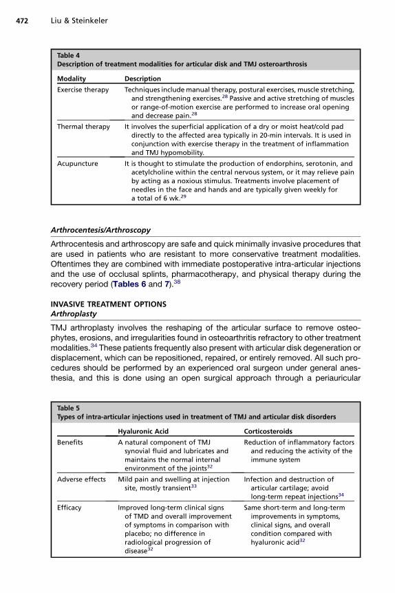
Physical therapy is commonly used in the outpatient setting to relieve musculoskeletalpain, reduce inflammation, and restore oral motor function. Physical therapy plays anadjunctive role in virtually all TMJ disorders treatment regimens. Various physical ther-apy modalities are available to the outpatient health provider (see Table 2). Althoughthe evidence is weak, there are numerous systematic review articles that support theefficacy of exercise therapy, thermal therapy, and acupuncture to reduce symptoms,such as pain, swelling, and TMJ hypomobility (Table 4).29–31
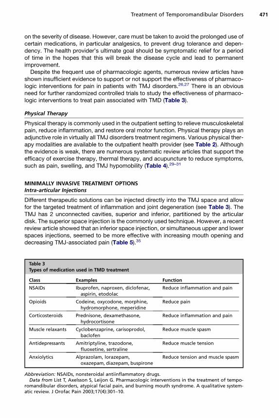
Different therapeutic solutions can be injected directly into the TMJ space and allowfor the targeted treatment of inflammation and joint degeneration (see Table 3). TheTMJ has 2 unconnected cavities, superior and inferior, partitioned by the articulardisk. The superior space injection is the commonly used technique. However, a recentreview article showed that an inferior space injection, or simultaneous upper and lowerspaces injections, seemed to be more effective with increasing mouth opening anddecreasing TMJ-associated pain (Table 5).35
Abbreviation: NSAIDs, nonsteroidal antiinflammatory drugs.Data from List T, Axelsson S, Leijon G. Pharmacologic interventions in the treatment of tempo-
romandibular disorders, atypical facial pain, and burning mouth syndrome. A qualitative system-atic review. J Orofac Pain 2003;17(4):301–10.
Table 4Description of treatment modalities for articular disk and TMJ osteroarthrosis
Modality Description
Exercise therapy Techniques include manual therapy, postural exercises, muscle stretching,and strengthening exercises.28 Passive and active stretching of musclesor range-of-motion exercise are performed to increase oral openingand decrease pain.28
Thermal therapy It involves the superficial application of a dry or moist heat/cold paddirectly to the affected area typically in 20-min intervals. It is used inconjunction with exercise therapy in the treatment of inflammationand TMJ hypomobility.
Acupuncture It is thought to stimulate the production of endorphins, serotonin, andacetylcholine within the central nervous system, or it may relieve painby acting as a noxious stimulus. Treatments involve placement ofneedles in the face and hands and are typically given weekly fora total of 6 wk.29
Liu & Steinkeler472
Arthrocentesis/Arthroscopy
Arthrocentesis and arthroscopy are safe and quick minimally invasive procedures thatare used in patients who are resistant to more conservative treatment modalities.Oftentimes they are combined with immediate postoperative intra-articular injectionsand the use of occlusal splints, pharmacotherapy, and physical therapy during therecovery period (Tables 6 and 7).38
INVASIVE TREATMENT OPTIONSArthroplasty
TMJ arthroplasty involves the reshaping of the articular surface to remove osteo-phytes, erosions, and irregularities found in osteoarthritis refractory to other treatmentmodalities.34 These patients frequently also present with articular disk degeneration ordisplacement, which can be repositioned, repaired, or entirely removed. All such pro-cedures should be performed by an experienced oral surgeon under general anes-thesia, and this is done using an open surgical approach through a periauricular
Table 5Types of intra-articular injections used in treatment of TMJ and articular disk disorders
Hyaluronic Acid Corticosteroids
Benefits A natural component of TMJsynovial fluid and lubricates andmaintains the normal internalenvironment of the joints32
Reduction of inflammatory factorsand reducing the activity of theimmune system
Adverse effects Mild pain and swelling at injectionsite, mostly transient33
Infection and destruction ofarticular cartilage; avoidlong-term repeat injections34
Efficacy Improved long-term clinical signsof TMD and overall improvementof symptoms in comparison withplacebo; no difference inradiological progression ofdisease32
Same short-term and long-termimprovements in symptoms,clinical signs, and overallcondition compared withhyaluronic acid32
Table 6Arthrocentesis for TMD treatment
Arthrocentesis
Description Saline lavage of the superior joint space, hydraulic pressure andmanipulation to release adhesions, and elimination of intra-articularinflammatory mediators (Fig. 3)36,37; less invasive than arthroscopy andcan be done in outpatient setting with local anesthesia andintravenous sedation
Indication � Limited opening with anteriorly displaced articular disk withoutreduction
� Chronic pain with good range of movement and displaced articulardisk with reduction
� Degenerative osteoarthritis
Contraindications � TMJ with bony or fibrous ankylosis� Extracapsular source of pain� Patients who have not undergone noninvasive treatment modalities
Efficacy Recently reported 83.5% treatment success rate in patients with internalderangement and osteoarthritis (as defined as an improvement inmaximum jaw opening and a reduction in pain level and mandibulardysfunction)38
Data from Fonseca RJ. Oral and maxillofacial surgery. Chicago: Saunders; 2000.
Treatment of Temporomandibular Disorders 473
skin incision (Fig. 5). Complications are rare but can include wound infection, facialnerve injury, permanent occlusal changes, relapsing joint pain, and life-threateningvascular injuries.42 As with all TMJ-related surgeries, early postoperative physicaltherapy and range-of-motion exercises are vital to achieving long-term functionalimprovements.
� Disk repositioning: Reposition the disk back to its normal anatomic position inpatients with internal derangement. This procedure is most effective in disksthat are normal appearing (white, firm, shiny) with minimal displacement.
� Disk repair: Small disk perforations can be repaired with a tension-free primaryclosure.
� Discectomy alone: Removal of the articular disk is indicated in patients withsevere disk perforation, complete loss of disk elasticity, and who are persistentlysymptomatic even after disk repositioning.43 Although studies have shown there
Fig. 3. Minimally invasive treatment of TMD: Arthrocentesis.
Table 7Arthroscopy for TMD treatment
Arthroscopy
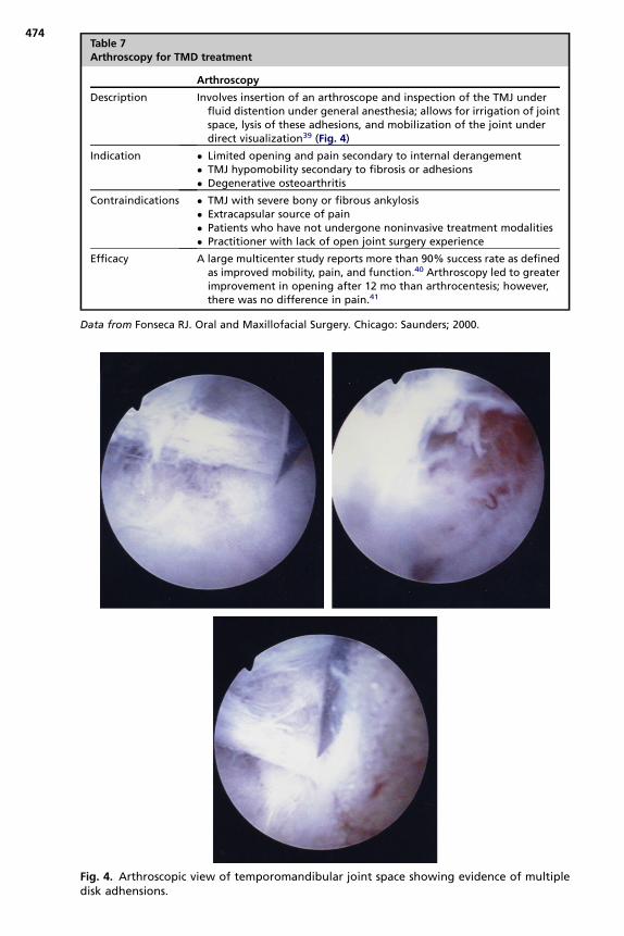
Description Involves insertion of an arthroscope and inspection of the TMJ underfluid distention under general anesthesia; allows for irrigation of jointspace, lysis of these adhesions, and mobilization of the joint underdirect visualization39 (Fig. 4)
Indication � Limited opening and pain secondary to internal derangement� TMJ hypomobility secondary to fibrosis or adhesions� Degenerative osteoarthritis
Contraindications � TMJ with severe bony or fibrous ankylosis� Extracapsular source of pain� Patients who have not undergone noninvasive treatment modalities� Practitioner with lack of open joint surgery experience
Efficacy A large multicenter study reports more than 90% success rate as definedas improved mobility, pain, and function.40 Arthroscopy led to greaterimprovement in opening after 12 mo than arthrocentesis; however,there was no difference in pain.41
Data from Fonseca RJ. Oral and Maxillofacial Surgery. Chicago: Saunders; 2000.
Fig. 4. Arthroscopic view of temporomandibular joint space showing evidence of multipledisk adhensions.
474
Fig. 5. Open surgical approach made through outlined periauricular and endural skinincision.
Treatment of Temporomandibular Disorders 475
is generally an improvement in pain and maximal mouth opening following thesurgical removal of the disk, patients also exhibited signs of fibrous adhesions,narrowing of joint space, and osteophyte formation on MRI.44–46
� Discectomy with graft replacement: The placement of a graft is thought to pro-tect the joint from further degeneration and prevent the formation of fibrous ad-hesions. The use of autogenous sources, such as temporalis flaps, auricularcartilage, and dermal grafts, results in superior clinical outcomes comparedwith alloplastic grafts.42 Studies have showed that autogenous grafts actuallydid not prevent remodeling of the joint but may help to reduce the onset of crep-itus resulting from discectomy alone.47,48 However, it was shown that discec-tomy with dermis graft replacement does result in a statistically significantimprovement in pain, chewing, and general health (Box 2).
Total Joint Replacement
TMJ replacement is intended primarily at restoration of form and function, and anypain relief gained is only a secondary benefit.47 The need for TMJ replacement typi-cally indicates severely damaged joints with end-stage disease that has failed all othermore conservative treatment modalities. Autogenous costochondral bone grafts havebeen frequently used in TMJ reconstruction in the past because of its gross anatomicsimilarity to the mandibular condyle, ease of adaptation to the recipient site, and itsdemonstrated growth potential in juveniles.49 However, because of potentialharvest-site morbidity and failure during the transplantation process or from functional
Box 2
Criteria for successful TMJ disk surgery
1. Mild, brief pain of no concern to patients
2. Vertical range of motion greater than 35mm and lateral range of motion greater than 6mm
3. Ability to tolerate a regular diet
4. Stabilization of any degenerative radiological changes
5. Absence of symptoms for at least 2 years
6. Absence of significant surgical complications
Data from Holmlund AB. Surgery for TMJ internal derangement. Evaluation of treatment andcriteria for success. Int J Oral Maxillofac Surg 1993;22:75–7.
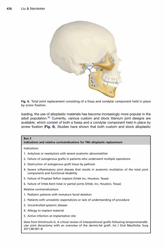
Fig. 6. Total joint replacement consisting of a fossa and condylar component held in placeby screw fixation.
Liu & Steinkeler476
loading, the use of alloplastic materials has become increasingly more popular in theadult population.42 Currently, various custom and stock titanium joint designs areavailable, which consist of both a fossa and a condylar component held in place byscrew fixation (Fig. 6). Studies have shown that both custom and stock alloplastic
Box 3
Indications and relative contraindications for TMJ alloplastic replacement
Indications
1. Ankylosis or reankylosis with severe anatomic abnormalities
2. Failure of autogenous grafts in patients who underwent multiple operations
3. Destruction of autogenous graft tissue by pathosis
4. Severe inflammatory joint disease that results in anatomic mutilation of the total jointcomponents and functional disability
5. Failure of Proplast-Teflon implant (Vitek Inc, Houston, Texas)
6. Failure of Vitek-Kent total or partial joints (Vitek, Inc, Houston, Texas)
Relative contraindications
1. Pediatric patients with immature facial skeleton
2. Patients with unrealistic expectations or lack of understanding of procedure
3. Uncontrolled systemic disease
4. Allergy to implant material
5. Active infection at implantation site
Data from Dimitroulis G. A critical review of interpositional grafts following temporomandib-ular joint discectomy with an overview of the dermis-fat graft. Int J Oral Maxillofac Surg2011;40:561–8.
Treatment of Temporomandibular Disorders 477
TMJ replacements resulted in statistically significant improvement in pain level, jawfunction, and incisal opening (Box 3).50,51
REFERENCES
1. Wadhwa S, Kapila S. TMJ Disorders: future innovations in diagnostics and ther-apeutics. J Dent Educ 2008;72(8):930–47.
2. Ghali GE, Miloro M, Waite PD, et al, editors. Peterson’s principles of oral andmaxillofacial surgery. 3rd edition. Shelton (CT): Pmph USA; 2012.
3. Lipton JA, Ship JA, Larach-Robinson D. Estimated prevalence and distributionof reported orofacial pain in the United States. J Am Dent Assoc 1993;124:115–21.
4. Manfredini D, Guarda-Nardini L, Winocur E, et al. Research diagnostic criteriafor temporomandibular disorders: a systematic review of axis I epidemiologicfindings. Oral Surg Oral Med Oral Pathol Oral Radiol Endod 2011;112:453–62.
5. Abubaker AO, Raslan WF, Sotereanos GC. Estrogen and progesterone recep-tors in temporomandibular joint discs of symptomatic and asymptomatic per-sons: a preliminary study. J Oral Maxillofac Surg 1993;51:1096–100.
6. Milam SB, Aufdemorte TB, Sheridan PJ, et al. Sexual dimorphism in the distribu-tion of estrogen receptors in the temporomandibular joint complex of thebaboon. Oral Surg Oral Med Oral Pathol 1987;64:527–32.
7. Aufdemorte TB, Van Sickels JE, Dolwick MF, et al. Estrogen receptors in thetemporomandibular joint of the baboon (Papio cynocephalus): an autoradio-graphic study. Oral Surg Oral Med Oral Pathol 1986;61:307–14.
8. Mohlin B, Kopp S. A clinical study on the relationship between malocclusion,occlusal interferences, and mandibular pain and dysfunction. Swed Dent J1978;2:103.
9. Perry H. Relationship of the occlusion to temporomandibular joint dysfunction. Aretrospective study. J Prosthet Dent 1977;39:420.
10. Upton L, Scott R, Haywood J. Major maxillomandibular malrelations and tempo-romandibular joint pain-dysfunction. J Prosthet Dent 1984;51:686.
11. Williamson E. Temporomandibular dysfunction in pretreatment adolescencepatients. Am J Orthod 1977;72:429.
12. Riolo M, Brandt D, Tenhaue T. Association between occlusal characteristics andsigns and symptoms of TMJ dysfunction in children and young adults. Am JOrthod Dentofacial Orthop 1987;92:467.
13. Rasmussen O. Description of population and progress of symptoms in a longi-tudinal study of temporomandibular joint arthropathy. Scand J Dent Res 1981;89:196–203.
14. McNeill C. Temporomandibular disorders: guidelines for classification, assess-ment, and management. The American Academy of orofacial pain. Chicago:Quintessence; 1993.
15. Blankestijn J, Boering G. Posterior dislocation of the temporomandibular disc.Int J Oral Surg 1985;14:437–43.
16. Liedberg J, Westesson P, Kurita K. Side-ways and rotational displacementof the temporomandibular joint disk: diagnosis by arthrography and correla-tion to cryosectional morphology. Oral Surg Oral Med Oral Pathol 1990;69:757–63.
17. Sanders B. Management of internal derangements of the temporomandibularjoint. Semin Orthod 1995;4(1):244–57.
18. Tallents RH, Katzberg RW, Murphy W, et al. Magnetic resonance imaging find-ings in asymptomatic volunteers and symptomatic patients with temporoman-dibular disorders. J Prosthet Dent 1996;75:529.
19. Wilkes C. Internal derangement of the temporomandibular joint. In: Clark G,Sanders B, Bertolami C, editors. Advances in diagnostic and surgical arthros-copy of the temporomandibular joint. Philadelphia: Sanders; 1993.
20. Dworkin SF, Leresche L. Research diagnostic criteria for temporomandibulardisorders: review, criteria, examinations and specifications, critique. J Cranio-mandib Disord 1992;6:301–55.
21. Shaefer J, Riley C, Caruso P, et al. Analysis of criteria for MRI diagnosis of TMJdisc displacement and arthralgia. Int J Dent 2012;2012:283163.
22. Klasser GD, Greene CS. Oral appliances in the management of temporoman-dibular disorders. Oral Surg Oral Med Oral Pathol Oral Radiol Endod 2009;107(2):212–23.
23. Fricton J, Look JO, Wright E, et al. Systematic review and meta-analysis ofrandomized controlled trials evaluating intraoral orthopedic appliances fortemporomandibular disorders. J Orofac Pain 2010;24(3):237–54.
25. Dionne RA. Pharmacologic treatments for temporomandibular disorders. OralSurg Oral Med Oral Pathol Oral Radiol Endod 1997;83(1):134–42.
26. Mujakperuo HR, Watson M, Morrison R, et al. Pharmacological interventions forpain in patients with temporomandibular disorders. Cochrane Database SystRev 2010;(10):CD004715.
27. List T, Axelsson S, Leijon G. Pharmacologic interventions in the treatment oftemporomandibular disorders, atypical facial pain, and burning mouth syn-drome. A qualitative systematic review. J Orofac Pain 2003;17(4):301–10.
28. Fricton JR. Management of masticatory myofascial pain. Semin Orthod 1995;1(4):229–43.
29. Rosted P. Practical recommendations for the use of acupuncture in the treat-ment of temporomandibular disorders based on the outcome of publishedcontrolled studies. Oral Dis 2001;7(2):109–15.
30. McNeely ML, Armijo Olivo S, Magee DJ. A systematic review of the effective-ness of physical therapy interventions for temporomandibular disorders. PhysTher 2006;86(5):710–25.
31. Cho SH, Whang WW. Acupuncture for temporomandibular disorders: a system-atic review. J Orofac Pain 2010;24(2):152–62.
32. Shi Z, Guo C, Awad M. Hyaluronate for temporomandibular joint disorders[review]. Cochrane Database Syst Rev 2003;(1):CD002970.
33. Bertolami CN, Gay T, Clark GT, et al. Use of sodium hyaluronate in treatingtemporomandibular joint disorders: a randomized, double blind, placebocontrolled clinical trial. J Oral Maxillofac Surg 1993;51:232–42.
34. Tanaka E, Detamore MS, Mercuri LG. Degenerative disorders of the tempo-romandibular joint: etiology, diagnosis, and treatment. J Dent Res 2008;87:296.
35. Li C, Zhang Y, Lv J, et al. Inferior or double joint spaces injection versus superiorjoint space injection for temporomandibular disorders: a systematic review andmeta-analysis. J Oral Maxillofac Surg 2012;70(1):37–44.
36. Guo C, Shi Z, Revington P. Arthrocentesis and lavage for treating temporoman-dibular joint disorders. Cochrane Database Syst Rev 2009;(4):CD004973.
37. Nitzan DW. Arthrocentesis for management of severe closed lock of the tempo-romandibular joint. Oral Maxillofac Surg Clin North Am 1994;6:245–57.
38. Monje-Gil F, Nitzan D, Gonzalez-Garcia R. Temporomandibular joint arthrocent-esis. Review of the literature. Med Oral Patol Oral Cir Bucal 2012;17(4):e575–81.
39. Indresano AT. Surgical arthroscopy as the preferred treatment for internalderangements of the temporomandibular joint. J Oral Maxillofac Surg 2001;59(3):308–12.
40. McCain JP, Sanders B, Koslin MG, et al. Temporomandibular joint arthroscopy: a6-year multicenter retrospective study of 4,831 joints. J Oral Maxillofac Surg1992;50(9):926–30.
41. Rigon M, Pereira LM, Bortoluzzi MC, et al. Arthroscopy for temporomandibulardisorders. Cochrane Database Syst Rev 2011;(5):CD006385.
42. Fonseca RJ. Oral and maxillofacial surgery. Chicago: Saunders; 2000. Print.43. Lanz AB. Discitis mandibularis. Zentralbl Chir 1909;36:289–91.44. Westesson PL, Cohen JM, Tallents RH. Magnetic-resonance-imaging of
temporomandibular-joint after surgical-treatment of internal derangement. OralSurg Oral Med Oral Pathol 1991;71:407–11.
45. Hansson LG, Eriksson L, Westesson PL. Magnetic-resonance evaluation aftertemporomandibular-joint discectomy. Oral Surg Oral Med Oral Pathol 1992;74:801–10.
46. Miloro M, Henriksen B. Discectomy as the primary surgical option for internalderangement of the temporomandibular joint. J Oral Maxillofac Surg 2010;68:782–9.
47. Dimitroulis G. A critical review of interpositional grafts following temporomandib-ular joint discectomy with an overview of the dermis-fat graft. Int J Oral Maxillo-fac Surg 2011;40:561–8.