28
of Diagnostic of Diagnostic Imaging Imaging Procedures - Procedures - Update Update Dr Keith Foord Consultant Radiologist, East Sussex Hospitals
| Date post: | 01-Jan-2016 |
| Category: |
Documents |
| Upload: | fay-spencer |
| View: | 215 times |
| Download: | 1 times |

Terminology for Terminology for representation of representation of
Diagnostic Imaging Diagnostic Imaging Procedures - UpdateProcedures - Update
Dr Keith FoordConsultant Radiologist, East Sussex Hospitals

A national system of RIS coding A national system of RIS coding and descriptors ?and descriptors ?
Relates to needs of request/entry systems within ICRS – pre-RIS - SNOMED match to record request to Spine
Consistency and uniqueness in requesting terminology – pre-RIS and within RIS
Consistency in activity measurement - RIS Consistency in clinical coding of events – RIS - SNOMED match But must be as intuitive and easy to use as possible Should have national acceptance For accurate communication of results data between hospitals –
post RIS results reporting, cluster stores and national spine - SNOMED match to ‘performed examination code’ to Spine
For ‘Payment by results’ – accurate records of same patient activity – national tariffs - SNOMED match / accurate HRGs
DICOM Structured Reporting

NHS Costings Code BookNHS Costings Code Book

DescriptorsDescriptors
Descriptors need to be UNIQUE in NCRS
FOOT LEFT not uniqueWhen a user searches all of the examinations available for ‘Foot Left’the search may return:
FOOT LEFT, FOOT LEFT Swab, FOOT LEFT Physiotherapy, FOOT LEFT Dressing,etc., etc.
But XR FOOT LEFT is unique
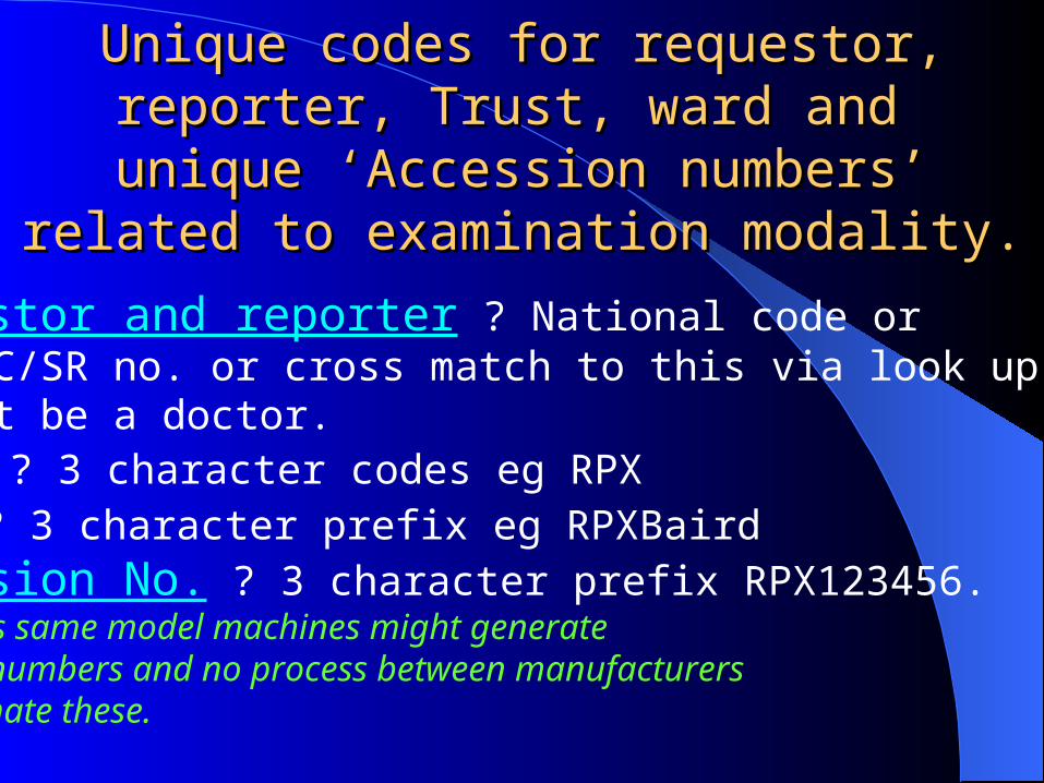
Unique codes for requestor, reporter, Trust, Unique codes for requestor, reporter, Trust, ward and unique ‘Accession numbers’ ward and unique ‘Accession numbers’
related to examination modality.related to examination modality.
Requestor and reporter ? National code or GMC/GDC/SR no. or cross match to this via look up table.May not be a doctor.Trust ? 3 character codes eg RPXWard ? 3 character prefix eg RPXBairdAccession No. ? 3 character prefix RPX123456.Needed as same model machines might generate identical numbers and no process between manufacturersto coordinate these.

Radiology Short CodesRadiology Short Codes
Used in RIS as shortcuts
For bookingsFor internal communications within RadiologyTo help group proceduresFor internal management / audit / activity
For common use need a structure, ideallyshort (max. 6 letters/digits) and logical
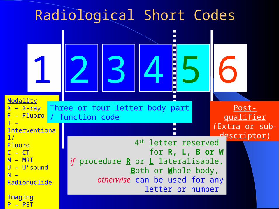
1 2 3 4 5 6ModalityX – X-rayF – FluoroI – Interventional/FluoroC – CTM – MRIU – U’soundN – Radionuclide ImagingP – PETE- EndoscopyZ- Image analysis or review
Post-qualifier
(Extra or sub-descriptor)
4th letter reserved for R, L, B or W
if procedure R or L lateralisable,Both or Whole body,
otherwise can be used for anyletter or number
Three or four letter body part/ function code
Radiological Short Codes
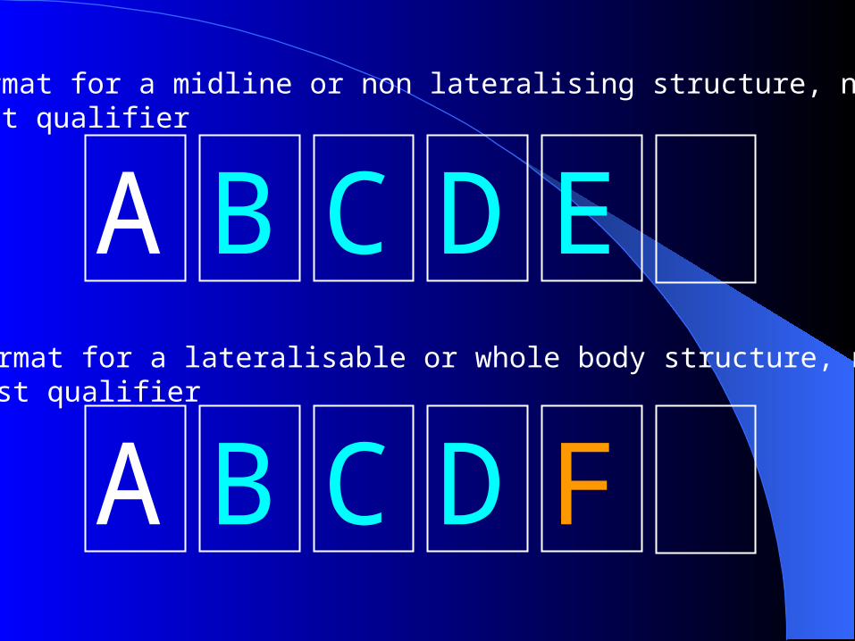
A B C D E
A B C D F
Format for a midline or non lateralising structure, nopost qualifier
Format for a lateralisable or whole body structure, no post qualifier

A B C D E G
A B C D F G
Format for a midline or non lateralising structure, with a post qualifier
Format for a lateralisable or whole body structure, with a post qualifier

Extra qualifiersExtra qualifiers (6(6thth letter/number = letter/number = G)) A Ablation B Biopsy (Core or FNA) D Drainage or Aspiration of fluid E Embolisation I Insertion of device J inJection - as an objective of the procedure, not as part of the preliminary to this objective M Mobile - for any modality, but particularly for 'portable' plain films and use of mobile image
intensifiers O tOmography in its wider sense. O may be added to any plain film examination to define planar
tomography - or postcoordinated P Plasty - as in angioPlasty or dacrocystoPlasty - ie balloon dilatation R for Radiotherapy planning S Stent T Use of intraThecal contrast X eXtraction - eg in retrieval of intravascular foreign bodies or removal of temporary IVC filter 1 First part of study 2 Second part of study 3 Third part of study 4 Fourth visit etc. – 5,6,7,8,9 – 10th = 0
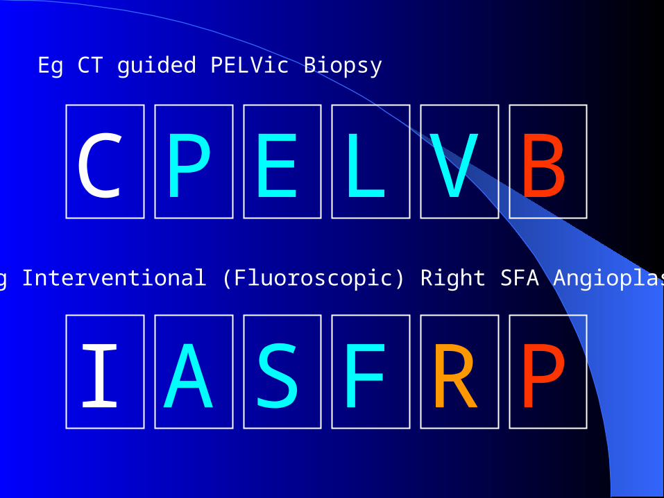
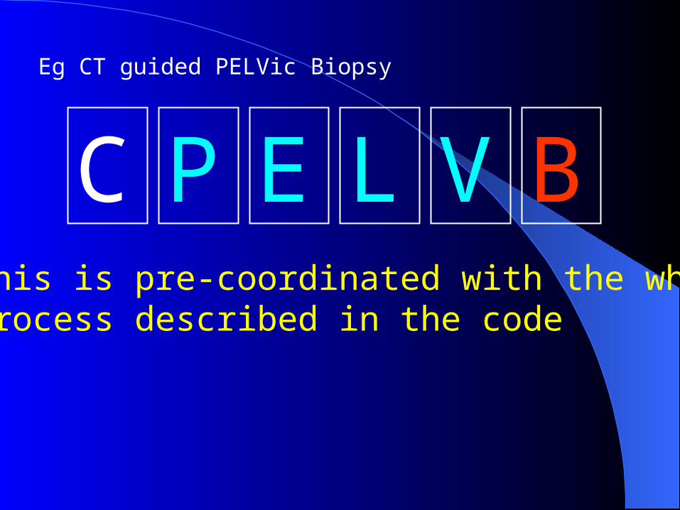
C P E L V B
I A S F R P
Eg CT guided PELVic Biopsy
Eg Interventional (Fluoroscopic) Right SFA Angioplasty

Pre and Post Co-ordination (1)Pre and Post Co-ordination (1) In order to group procedures many RIS systems lack
the ability to post co-ordinate procedures together under one accession number.
Particular examples are for 'both' plain film exams eg 'both ankles' and in CT where examinations often combine e.g. CT Chest, Abdomen, Pelvis.
Pre co-ordination or grouping of these procedures is therefore required in advance.
Pre co-ordination should not be used in RIS-PACS systems capable of full post co-ordination as with these individual procedure codes will be automatically or manually grouped prior to archiving and reporting
C P E L V B
Eg CT guided PELVic Biopsy
This is pre-coordinated with the wholeprocess described in the code

Pre and Post Co-ordination (2)Pre and Post Co-ordination (2) In modern RIS systems post co-ordination can be applied to
group related procedures together. All RIS systems supplied via LSPs should do this.
Some procedure codes such as 'U/S biopsy' by themselves do not define precisely what has happened although it would define the activity of “Performing a biopsy under ultrasound control and the consumables/activity associated with this.”
Such codes need post co-ordinating with the relevant body part to fully inform activity statistics
Similarly separate CT body part examinations can be post co-ordinated together to enable the multiple examinations to be reported together as one report.
The advantage is a more sophisticated approach to audit, activity measurement and stocktaking

Eg CT guided PELVis Biopsy
C P E L V
C B I O P BPLUSPLUS
Are POST coordinated and describe both processes which arethen reported as one. CT biopsy cost structures do not need to be built into multiple codes
C P E L V B
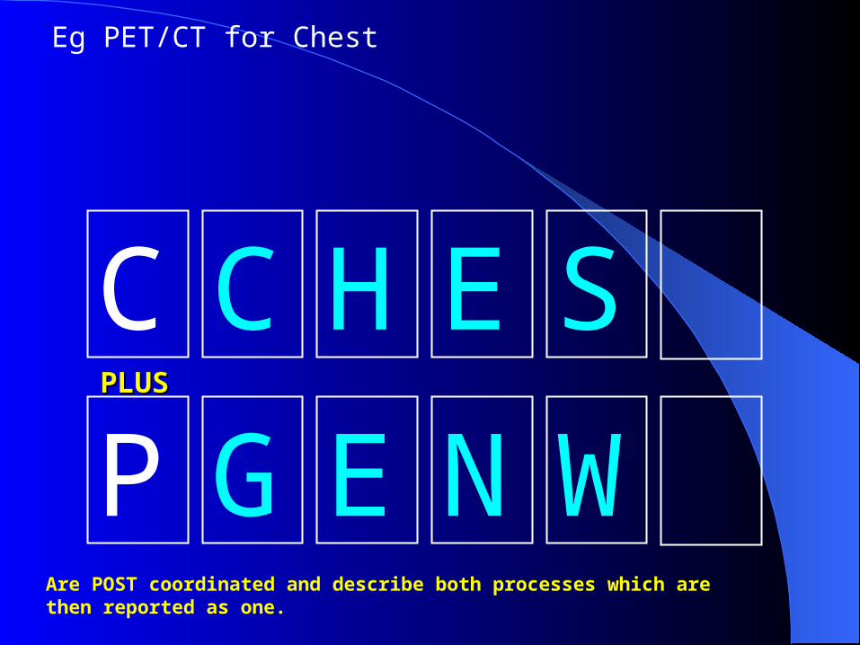
Eg PET/CT for Chest
C C H E S
P G E N WPLUSPLUS
Are POST coordinated and describe both processes which arethen reported as one.

SNOMED CTDescriptors and CodesSNOMED CT
Descriptors and Codes
SNOMED CTDescriptors and Codes
Full list ofRIS Codes & Descriptors+ Synonyms
PostCoordinating
RIS singledescriptors
List ofOrderableProcedures
NCRS‘Reporting’
Module
SPINE
HL7
SNOMED CT
HL7
NCRS‘Order’ Entry
RIS
NACSLocation &
People codes

Sub-Descriptors / CodesSub-Descriptors / Codes REQUESTING Layer
(1st order) Right Oblique QR Left Oblique QL Right Lateral LR Left Lateral LL Weight Bearing WB Standing ST Axial AX AP20o 20 Judet’s JU Stryker’s SY Etc…
IN RADIOLOGY (RIS) Layer (2nd order)
Same list + Supine SU Prone PR Decubitus DE Complex Oblique QC Angled Oblique
22,30,45 Frog laterals FR May need to combine
together or with 1st order list eg DELR

NPfIT and Descriptors/CodesNPfIT and Descriptors/Codes
Southern Cluster – IDX – GE PACS- Kodak CR - HSS CRIS London Cluster-IDX- Philips PACS-Philips(Fuji)CR-? RIS NE Cluster- iSOFT- Agfa PACS-Agfa CR-? RIS EEM Cluster- iSOFT- Agfa PACS -Agfa CR -? RIS NWWM Cluster- iSOFT- ComMedica PACSComMedica PACS –Kodak CR-
Kodak RIS Has RCR endorsement SNOMED CT can be integrated-matched
SNOMED CTSNOMED CT
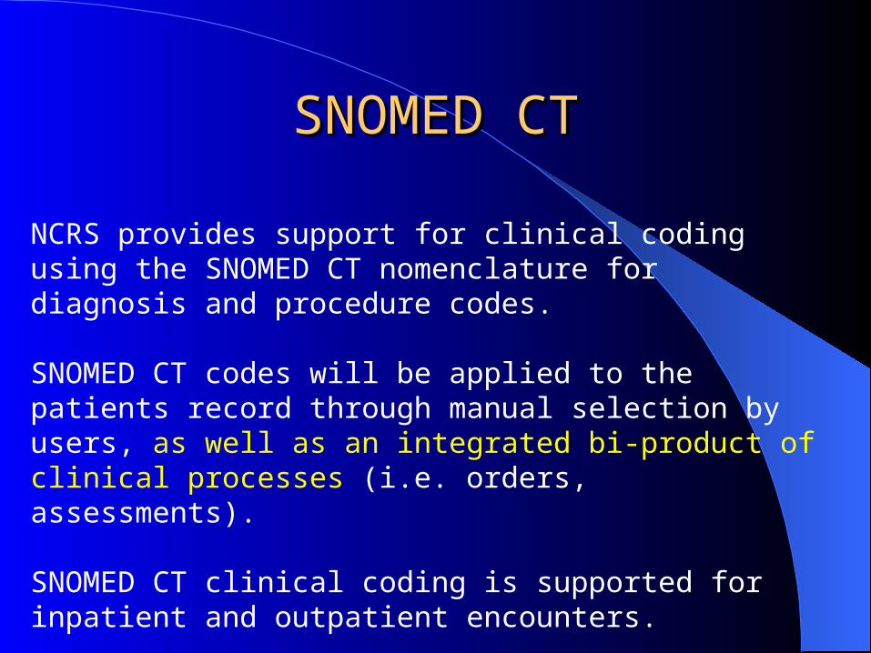
NCRS provides support for clinical coding using the SNOMED CT nomenclature for diagnosis and procedure codes.
SNOMED CT codes will be applied to the patients record through manual selection by users, as well as an integrated bi-product of clinical processes (i.e. orders, assessments).
SNOMED CT clinical coding is supported for inpatient and outpatient encounters.

SNOMED CTSNOMED CT
At the end of an episode / encounter of care, SNOMED CT codes are recorded in NCRS via the Discharge Summary / Encounter diagnosis and procedure codes. The SNOMED codes recorded in NCRS are sent to the 3M clinical encoder where clinical coding is completed in SNOMED CT, ICD10, Read, and OPCS4.
Codes will be transferred back to NCRS and will update, not replace, the patient diagnosis and procedure codes. A full audit trail is available.

SNOMED CTSNOMED CTWithin NCRS P1R2, users will have the ability to manually record SNOMED CT codes within the following areas:§ Discharge Summary / Encounter § Problems / Provisional Diagnoses
Within NCRS P1R2, SNOMED CT codes will be recorded against the patients record, as a bi-product of clinical processes, in the following clinical areas: § Assessments§ Findings / Flowsheets
§ Orders (viz. the code for the request)
§ Results (viz. the code for the procedure(s) performed, not the radiological diagnosis or report which will be transferred via HL7 messaging)

OrdersOrders and and ResultsResultsin Radiologyin Radiology
SNOMED CT Order codes can be derived from Order/Entry systems, but will be MUCH MORE ACCURATE if derived from the accepted and if required modified final RIS procedure entry with SNOMED CT matching.
SNOMED CT Results codes from Radiology are a dilemma. This does not apply to ‘Procedure performed’ , but to a provisional radiological diagnosis which may be a list of differential diagnoses which could be entered by a reporter (ie manually). Unlikely to happen given pressures of work!
The use of DICOM structured reporting may give the possibility of automatically constructing radiological diagnosis codes from the structured report

Incorporated into the report are captured images of key findings (which can be exploded to full screen presentation), structured diagnosis information, recorded audio, the ability to sort findings by anatomy or priority, to view prior findings associated with the corresponding patient and hyperlinks to related information.
DICOM SR – is an ‘envelope’, but within this useful structure is available.
User decides how much structure to use and controls with templates the type of content, if it is mandatory or optional and modes of expression
Structured reporting

Link Features to Description
New nodulesuperimposedwith rightfourth rib
Free air
10% Pneumothorax
Cavitation
Structured reporting

David Clunie
Development Director, Imaging Products
ComView Corporation – Paper at SPIE, 2001
Structured reporting

David Clunie
Development Director, Imaging Products
ComView Corporation – Paper at SPIE, 2001
Structured reporting


