Purification and Preliminary Characterization of Tetraheme Cytochrome and Adenylylsulf te Reductase from the Peptidolytic Sulfate-Reducing Bacterium Desulfovibrio aminophilus DSM 12254 Alejandro L6pez-Cort6s *, Sergey Bursakov , Angelo Figueiredo , Anders E.Thapper , Smilja Todorovic , Jos J.G. Moura , Bernard Ollivier3, Isabel Moura and Guy Fauque Centro de lnvestigaciones Biol6gicas del Noroeste, (CIBNOR). Mar Bermejo 195, Playa Palo Santa Rita, La Paz, Baja California Sur 23090, Mdxico. 2REQUIMTE/CQFB, Departamento de Quimica, Faculdade de Cincias e Tecnologia da Universidade Nova de Lisboa, 2829-516 Monte de Caparica, Portugal. 3Laboratoire de Microbiologie IRD, UR 101, IFR-BAIM, ESIL UniversitOs de Provence et de la Mditerrane, Campus de Luminy, Case 925, 13288 Marseille Cedex 09, France. ABSTRACT Two proteins were purified and preliminarily characterized from the soluble extract of cells (310 g, wet weight) of the aminolytic and peptidolytic sulfate-reducing bacterium Desulfovibrio (D.) aminophilus DSM 12254. The iron-sulfur flavoenzyme adenylylsulfate (adenosine 5"-phosphosulfate, APS) reductase, a key enzyme in the microbial dissimilatory sulfate reduction, has been purified in three chromatographic steps (DEAE-Biogel A, Source 15, and Superdex 200 columns). It contains two different subunits with molecular masses of 75 and 18 kDa. The fraction after the last purification step had a purity index (A278 ,m/A388 nm) of 5.34, which was used for further EPR spectroscopic studies. The D. aminophilus APS reductase is very similar to the homologous enzymes isolated from D. gigas and D. desulfuricans ATCC 27774. A tetraheme cytochrome c3 (His-heme iron-His) has been purified in three chromatographic steps (DEAE- Biogel A, Source 15, and Biogel-HTP columns) and preliminarily characterized. It has a purity index ([A.s.s3 nm" A570 nm]rcd/m280nm ox) of 2.9 and a molecular mass of around 15 kDa, and its spectroscopic characterization (NMR and EPR) has been carried out. This hemoprotein presents similarities with the tetraheme cytochrome c3 from Desulfomicrobium (Des.) norvegicurn (NMR spectra, and N-terminal amino acid sequence). Corresponding author: Alejandro L6pez-Cort6s. Phone: (612) 123-84-25 Fax: (612) 125-36-25 e-mail: ._hgpczOA(i.cibno r. mx 81
Transcript
Purification and Preliminary Characterization ofTetraheme Cytochrome and Adenylylsulf te
Reductase from the Peptidolytic Sulfate-ReducingBacterium Desulfovibrio aminophilus DSM 12254
Alejandro L6pez-Cort6s *, Sergey Bursakov, Angelo Figueiredo, Anders E.Thapper, SmiljaTodorovic, Jos J.G. Moura, Bernard Ollivier3, Isabel Moura and Guy Fauque
Centro de lnvestigaciones Biol6gicas del Noroeste, (CIBNOR). Mar Bermejo 195, Playa PaloSanta Rita, La Paz, Baja California Sur 23090, Mdxico.
2REQUIMTE/CQFB, Departamento de Quimica, Faculdade de Cincias e Tecnologia daUniversidade Nova de Lisboa, 2829-516 Monte de Caparica, Portugal.
3Laboratoire de Microbiologie IRD, UR 101, IFR-BAIM, ESIL UniversitOs de Provence et de la
Mditerrane, Campus de Luminy, Case 925, 13288 Marseille Cedex 09, France.
ABSTRACT
Two proteins were purified and preliminarily characterized from the soluble extract of cells (310 g, wet
weight) of the aminolytic and peptidolytic sulfate-reducing bacterium Desulfovibrio (D.) aminophilus DSM
12254. The iron-sulfur flavoenzyme adenylylsulfate (adenosine 5"-phosphosulfate, APS) reductase, a key
enzyme in the microbial dissimilatory sulfate reduction, has been purified in three chromatographic steps
(DEAE-Biogel A, Source 15, and Superdex 200 columns). It contains two different subunits with molecular
masses of 75 and 18 kDa. The fraction after the last purification step had a purity index (A278 ,m/A388 nm) of
5.34, which was used for further EPR spectroscopic studies. The D. aminophilus APS reductase is verysimilar to the homologous enzymes isolated from D. gigas and D. desulfuricans ATCC 27774. A tetraheme
cytochrome c3 (His-heme iron-His) has been purified in three chromatographic steps (DEAE- Biogel A,Source 15, and Biogel-HTP columns) and preliminarily characterized. It has a purity index ([A.s.s3 nm"
A570 nm]rcd/m280nm ox) of 2.9 and a molecular mass of around 15 kDa, and its spectroscopic characterization
(NMR and EPR) has been carried out. This hemoprotein presents similarities with the tetraheme cytochrome
c3 from Desulfomicrobium (Des.) norvegicurn (NMR spectra, and N-terminal amino acid sequence).
Desulfomicrobium (Des.), Deutsche Sammlung von Mikroorganismen (DSM), Laboratoire de Chimie
Bacterienne, (LCB), Centre National de la Recherche Scientifique (CNRS), basic local alignment search tool
(BLAST), D6partement Soutien et Formation des Communaut6s Scientifiques du Sud (DSF), Institut de
Recherche pour le Developpement, (IRD).
INTRODUCTION
Sulfate-reducing bacteria constitute a group of anaerobic prokaryotes sharing the capacity to carry out
dissimilatory sulfate reduction to sulfide as a major component of their bioenergetics processes [1-3], and
contain a complex and diversified electron carrier system [4, 5]. D. aminophilus DSM 12254 is a mesophilic
strain of sulfate-reducing bacterium isolated from an anaerobic sludge of a dairy wastewater treatment plantin Santa Fe de Bogota, Colombia [6]. This strain uses a wider range of energy substrates than reported for
most Desulfovibrio species [1 ]. It presents, in particular, an important property of fermenting or oxidizing
proteinaceous compounds, such as amino acids and peptides. This sulfate-reducing strain is also able to
disproportionate sulfite and thiosulfate, suggesting that it plays a major role in regulating electron flow in the
dissimilatory sulfur cycle [6]. We report here purification and preliminary characterization of two proteins
involved in the respiratory system of D. aminophilus: one electron carrier, the tetraheme cytochrome c3, and
one enzyme, the adenylylsulfate reductase.
EXPERIMENTAL
Bacterial strain and growth conditions
D. aminophilus DSM 12254 was grown at 37C in a lactate/sulfate medium under anaerobic conditions in
the Unit6 de Fermentation, Laboratoire de Chimie Bacterienne (LCB), Centre National de la Recherche
Scientifique (CNRS), in Marseille, France, and cells were harvested as previously described [7].
Preparation of the soluble fraction
The cells (310 g, wet weight) were suspended in 10 mM Tris-HC1 buffer, pH 7.6, then ruptured bypassing twice through a French press. The extract was centrifuged for 1 h at 15,000 g and the supernatant(crude cell extract.) was centrifuged for 40 min at 26,000 g to separate the membrane (pellet) from the
soluble extract.
82
A. Lopez-Cortes et al. Bioinorganic Chemistry and Applications
Proteins purification
The soluble fraction was then loaded onto a DEAE Bio-Gel A column (Bio-Rad, 44 x 4.5 cm)equilibrated with 10 mM Tris-HC1 buffer, pH 7.6. A gradient of 11 10mM Tris-HC1 pH 7.6 and 11 500 mM
Tris-HC1 pH 7.6 was set up. Five major proteins were eluted from the column: tetraheme cytochrome c3, two
molybdenum iron-sulfur-containing proteins (one with aldehyde oxidoreductase activity), bisulfite reductase
of the desulfoviridin-type, and adenylylsulfate reductase. Two of these proteins (tetraheme cytochrome c3
and APS reductase) were completely purified in two supplementary purification steps. The tetrahemiC
cytochrome c. fraction, after concentration on a Diaflo apparatus using a YM-10 membrane, was then
applied to a Pharmacia Biotech ion exchange column Source 15 (32 x 2.6 cm) equilibrated with 10 mM Tris-
HC1 pH 7.6 and eluted with 10 mM Tris-HC1 pH 7.6 to 10 mM Tris-HC1 pH 7.6 and 500 mM NaC1. After
concentration (Diaflo YM-10 membrane), the tetraheme cytochrome c3 was finally passed over a Biogel-HTP
column (45 x 1.6 cm) equilibrated with 10 mM Tris-HC1 pH 7.6 and eluted with a continuous gradient of
sodium phosphate buffer pH 7.6 (250 ml 10 mM/250 ml 250 mM). After concentration (Diaflo YM-30
membrane), the APS reductase fraction eluted from the DEAE-Biogel A column was applied to a Source 15
column (Pharmacia Biotech, 32 x 2.6 cm) equilibrated with 10 mM Tris-HC1 buffer pH 7.6 and eluted with
10 mM Tris HC1 pH 7.6 to 10 mM Tris-HC1 + 500 mM NaC1. APS reductase after concentration (DiafloYM-30 membrane) was finally passed over a Superdex 200 column (Amersham Biosciences, 67 x 2.6 cm)equilibrated with 50 mM Tris-HC1 pH 7.6 and 300 mM NaC1.
Molecular mass and purity determination
Subunit composition, molecular mass, and purity of proteins were determined by denaturing PAGE, using
as running buffer Tris (0.025M)-glycine (0.192M), SDS (0.1%) pH 8.3. Low molecular weight kit markers
for SDS electrophoresis (Pharmacia Biotech 17-0446-01) were used for the calibration of APS reductase and
tetraheme cytochrome c3. The protein standards with approximate molecular weights were: phosphorylase b,
alfa lactalbumin 14.4 kDa. The gels were stained for protein by coomassie blue 0.5%.
Ultraviolet (UV)-visible spectroscopy
UV-visible absorption spectra were recorded on a Shimadzu UV-2101 PC split beam spectrophotometer
using 1-cm quartz cells.
Electron paramagnetic resonance (EPR) spectroscopy
EPR spectra were recorded on a X-band Bruker EMX spectrometer equipped with a dual-mode cavity
(Model ER 4116DM). Samples were cooled with helium gas using a continuous-flow cryostat (OxfordInstruments, UK).
83
Vol. 3, Nos. 1-2, 2005 Purification and Prelhninary Characterization ofTetrahemeCytochrome c
Nuclear magnetic resonance (NMR) spectroscopy
The NMR spectra were taken for a 0.8-1.0 mM tetraheme cytochrome c3 sample in DeO. The NMR
spectra of D. aminophilus tetraheme cytochrome c3 in the oxidized state were recorded on a 400 MHz Bruker
ARX-400 spectrometer equipped with an inverse detection 5-mm probe and a variable temperature unit
Bruker B-VT 2000. The 1D-NMR spectra were measured in oxidized state at 317K, 313K, 308K, 303K,
and 283.2K (pH 7.6) with a spectral width of 40.3kHz and a transmitter power level of 1.0 dB. All
experiments were obtained with water pre-saturation and chemical shift and presented in ppm relative to the
internal standard 2,2-dimethyl-2-silapentane-5-sulfonate (DSS).
N-terminal amino acid sequencing of tetraheme cytochrome c3
N-terminal amino acid sequence of D. aminophilus tetraheme cytochrome c3 was determined by
automated Edman degradation in a protein sequencer (Applied Biosystem model 477) coupled to an analyzer
(Applied Biosystem model 120) following the manufacturer’s instructions, using 100 pmol of tetraheme
cytochrome c3.
RESULTS AND DISCUSSION
Adenylylsulfate (APS) reductase
Although the first reports on APS reductase were published in the sixties, its three-dimensional structure
was published only recently [8]. Comparison of physicochemical and spectroscopical properties of APS
reductases isolated from several Desulfovibrio species show great similarity and high degree of homology
[9]. Only recently has APS reductase been isolated from Archaeoglobus fulgidus and found to be a
heterodimer with one subunit (75 kDa, 1FAD) and one subunit (18 kDa, 2 [4Fe-4S]) [10]. APS reductase has
been purified in three chromatographic steps from the soluble fraction of D. aminophilus and preliminarilycharacterized. The UV-visible spectrum of APS reductase in the native form shows a broad maximum around
392 nm with shoulders at 445 and 475 nm and a protein absorption peak at 278 nm (unpublished results). Theoverall visible spectrum indicates the presence of a flavin group and iron sulfur centers. APS reductase is a
heterodimer with one subunit (75 kDa, containing FAD) and one subunit (18 kDa, containing 214Fe-4S]centers) (Fig. 1).
EPR spectroscopic studies were carried out at different temperatures with the D. aminophilus APS
reductase in the native state (Fig. 2-A) in the presence of natural substrates (AMP and sulfite) (Fig. 2B) and
in th.e reduced form (Fig. 2C and 2D). Temperature dependence studies helped separate resonances
originating in different species. In the native state (Fig. 2A), two clusters of APS reductase are in the [4Fe-4S]2/ oxidized state, with four iron-atoms as Fea’5+, giving total spin of S 0. Nevertheless, the EPR spectrumshows a signal spread around g 2.00. The spectral shape and g value of the signal indicate that the broad
resonance accounts for only 0.1-0.25 spins/mol, which can be attributed to the residual [3Fe-4S] cluster [9].This signal is in D. aminbphilus, superimposed with the FAD radical (g 2.0048). As seen in Fig. 2, the
84
A. Lopez-Cortes et al. Bioinorganic Chemistry and Applications
94 kDa67 kDa
43 kDa
30 kDa
2
20.1 kDa
14.4 kDa
Fig. l :" Denaturing SDS-PAGE 12.5%. Lane 1: profile of low molecular-weight markers. Lane 2: a
heterodimer APS from D. aminophilus reductase with one subunit, around 75 kDa, containing FAD,and another subunit visible, around 18 kDa, from D. aminophilus.
former resonance can be detected up to -30K and the latter up to 45K. The short reduction of the protein
with Na2S204 (at 15 sec, pH 9.5) (Fig. 2C), as well as the addition of the substrates AMP and Na2SO3 (Fig. 2-
B), gives rise to a rhombic signal (gl 2.084, g2 1.94 and g3 1.90) that was attributed to the reduced
[4Fe-4S] Center I, S 1/2. In addition to the iron-sulfur cluster resonances, there was a g 2.0048 signal
originating from the FAD radical and the residual resonance of the native APS reductase. The studies of
temperature and power dependence of the spectra indicate that the FAD radical is present up to 100K (datanot shown), while the Center cluster can be seen up to 45K. Apparently, the long reduction with Na2S204(Fig. 2D) did not result in a fully reduced APS reductase after several attempts to fully reduce the sample.
After more than 30 min of reduction with dithionite (pH 9.5), both [4Fe-4S] clusters should be reduced, each
having total spin S 1/2, but the EPR spectrum of APS reductase most likely originates from only one
reduced [4Fe-4S]. It is not clear at this point whether this can be related to some specific characteristic of
APS reductase in D. aminophilus.
APS reductase is a major cytoplasmic enzyme constituting 2 to 3% of soluble proteins in sulfate reducers
of the genus Desulfovibrio [4, 5]. It is a nonheme iron flavoprotein which has also been found in several
genera of sulfate-reducing bacteria: Desulfobacter, Desulfotomaculum, Desulfosarcina, Desulfococcus,
Desulfobulbus, Thermodesulfobacterium, and Archaeoglobus [4, 9, 10, 11 ]. APS reductase from eight species
and strains of Desulfovibrio and one from Archaeoglobus were purified and their biochemical and
85
Yol. 3, Nos. 1-2, 2005
A
3000
8*K
10*g
/’’- 33"K45"K
3200 3400 3600 3800
Magnetic Field (Gauss)
Purification and Preliminary Characterization ofTetrahemeCS’tochrome c3
B
120K
!8"K
("-22"K
28"K35"K
____....__ 450K
3000 3200 3400 3600 3800
Magnetic Field (Gauss)
C
45"Ki = i
3000 3200 3400 3600 3800
Magnetic Field
D
25"K
3000 3200 3400 3600 3800
Magnetic Field (Gauss)
Fig. 2: Temperature dependece of EPR spectra of APS reductase from D. aminophilus with: (A) Native
enzyme, signal from residual [3Fe-4S] cluster. (B) native APS reductase incubated with substrates
AMP and SO3"2. (C) native APS reductase with Na2S20, ~15 sec (only Center is reduced). (D)APS reductase with Na2S20, > 30 min (both [4Fe-4S] clusters are reduced). The spectrometer
amplitude: 8 G; modulation frequency: 100 kHz; field center: 3400 G; sweep width: 1000 G.
spectroscopic properties determined. They present a high degree of homology in their physicochemical
characteristics and their visible and EPR spectra [9]. APS reductases isolated from Desulfovibrio species are
proteins containing one FAD per molecule and eight iron atoms arranged in two [4Fe-4S] clusters (Centerand Center II). They have a monomeric molecular mass ranging between 150 and 180 kDa and possess two
different subunits with molecular masses of around 20 and 70 kDa [9]. The reaction of sulfite with APS
reductase results in the formation of a FAD-sulfite adduct causing the bleaching of the FAD and the
86
A. Lopez-Cortes et aL Bioinorganic Chemistry andApplications
appearance of a maximum at 320 nm, corresponding to the reaction of sulfite at the N-5 position of the
isoalloxazine ring of FAD. The subsequent addition of AMP results in a decrease in absorbance at 320 nm,
partial reduction of iron-sulfur centers, and the formation of APS. A common feature of all APS reductases
from Desulfovibrio species is the perturbation of the EPR spectral features of Center after its reaction with
AMP and sulfite, as well as its high redox potential (0 to -50 mV) when compared with other [4Fe-4S]clusters. Center II is a [4Fe-4S] cluster with a redox potential lower than -400 mV [9]. APS reductase in
sulfate-reducing bacteria is an enzyme highly conserved in terms of its composition at the active site as well
as its physiological properties.
Tetrahemic cytochrome c3
A tetrahemic cytochrome (,’3 (His-heme iron-His) has been purified in three chromatographic steps from
the D. aminophilus soluble extract. It has a purity index ([A553 nm-A570 nm]rc,/Aes0 ox) equal to 2.90.
Denaturing SDS-PAGE corroborated the purity of this protein (Fig. 3). The UV-visible spectrum of the
oxidized tetrahemic cytochrome c3 exhibits a broad absorption band around 531 nm (beta band), a Soret peak
(gamma band) with a maximum at 410 nm, another broad band at 350 nm (delta band), and a protein peak at
280 nm (Fig. 4). The tetraheme cytochrome c3 is not reduced by sodium ascorbate, but is fully reduced by
94 kDa67 kDa
43 kDa
30 kDa
Fig. 3:
20.1 kDa
Denaturing SDS-PAGE 15%. Lane 1: profile of low molecular-weight markers. Lane 2: tetrahcmic
cytochrome c3 (5 tl). Lane 3: tetrahemic cytochrome c3 (2 tl) from D. aminophilus; singlc band
around 15 kDa/subunit interpreted as a pure protein.
amplitude" 10 G; modulation frequency: 100 kHz; field center" 3400 G; sweep width 4000 G.
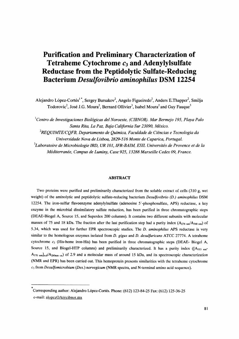
sodium dithionite, showing absorption maxima at 553 nm (alpha band), 523 nm (beta band), and a Soret peak
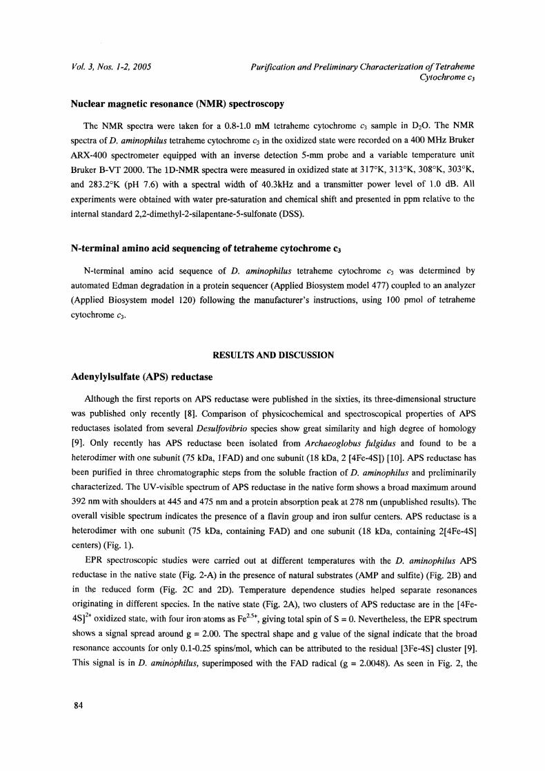
at 418 nm (gamma band) (Fig. 4). Figure 5 shows the EPR spectrum of the D. aminophilus ferritetraheme
cytochrome c3 recorded at 10K. The spectrum shows a prominent feature at g 2.920 in the g max region
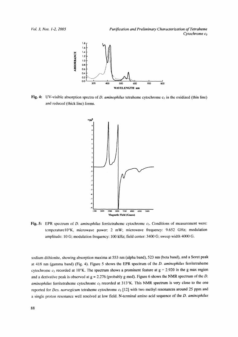
and a derivative peak is observed at g 2.276 (probably g med). Figure 6 shows the NMR spectrum of the D.
aminophilus ferritetraheme cytochrome ca recorded at 313K. This NMR spectrum is very close to the one
reported for Des. norvegicum tetraheme cytochrome c3 [12] with two methyl resonances around 25 ppm and
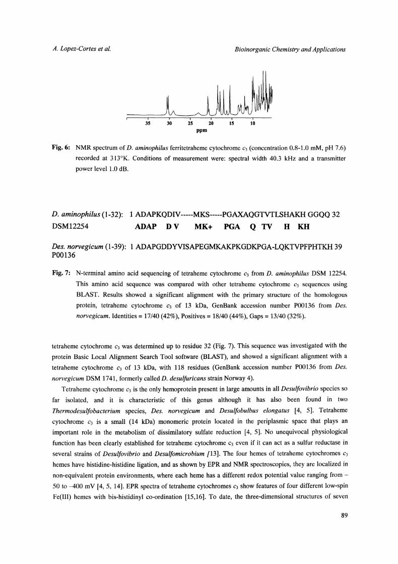
a single proton resonance well resolved at low field. N-terminal amino acid sequence of the D. aminophilus
88
A. Lopez-Cortes et al. Bioinorganic Chemistry and Applications
30 15 10
ppm
Fig. 6: NMR spectrum of D. aminophilus ferritetraheme cytochrome c3 (concentration 0.8-1.0 mM, pH 7.6)recorded at 313K. Conditions of measurement were: spectral width 40.3 kHz and a transmitter
tetraheme cytochrome c3 was determined up to residue 32 (Fig. 7). This sequence was investigated with the
protein Basic Local Alignment Search Tool software (BLAST), and showed a significant alignment with a
tetraheme cytochrome c3 of 13 kDa, with 118 residues (GenBank accession number P00136 from Des.
norvegicum DSM 1741, formerly called D. desulfuricans strain Norway 4).Tetraheme cytochrome c3 is the only hemoprotein present in large amounts in all Desulfovibrio species so
far isolated, and it is characteristic of this genus although it has also been found in two
Thermodesulfobacterium species, Des. norvegicum and Desulfobulbus elongatus [4, 5]. Tetraheme
cytochrome c3 is a small (14 kDa) monomeric protein located in the periplasmic space that plays an
important role in the metabolism of dissimilatory sulfate reduction [4, 5]. No unequivocal physiological
function has been clearly established for tetraheme cytochrome c3 even if it can act as a sulfur reductase in
several strains of Desulfovibrio and Desulfomicrobium /13]. The four hemes of tetraheme cytochromes c3
hemes have histidine-histidine ligation, and as shown by EPR and NMR spectroscopies, they are localized in
non-equivalent protein environments, where each heme has a different redox potential value ranging from-
50 to -400 mV [4, 5, 14]. EPR spectra of tetraheme cytochromes c3 show features of four different low-spin
Fe(III) hemes with bis-histidinyl co-ordination [15,16]. To date, the three-dimensional structures of seven
89
Vol. 3, Nos. 1-2, 2005 Purification and Preliminary Characterization ofTetrahemeCytochrome cj
tetraheme cytochrome c3s have been determined by X-ray diffraction [16-19]. The most striking
characteristics of the three-dimensional structures of the tetraheme cytochromes c3 are the compact
organization of the four hemes with a relatively high degree of solvent exposure. Despite the rather low
homology among the amino-acid sequences of tetraheme cytochromes c3 (lowest homology of 20%), no
significant differences in the overall structure and spatial arrangement of the four hemes have been observed
[16-19].Here, we described the purification and preliminary characterization of two key proteins involved in the
dissimilatory sulfate reduction pathway of D. aminophilus. We have shown that, according to the UV-visible
and EPR spectra, D. aminophilus APS reductase is very close to the homologous enzymes isolated from D.
gigas and D. desulfuricans ATCC 27774. D. aminophilus tetraheme cytochrome c3 presents more homology
with the homologous protein present in Des. norvegicum (N-termimd amino acid sequence and NMR
spectra).
ACKNOWLEDGEMENTS
We are indebted to R. Toci and M. Bauzan for growing the bacteria used in this study. A. L6pez-Cort6s
received financial support from the D6partement Soutien et Formation des Communaut6s Scientifiques du
Sud (DSF)-Institut de Recherche pour le D6veloppement (IRD), France, during his postdoctoral stay in
Marseille, France. Editorial staff at CIBNOR improved the English text.
REFERENCES
1. F. Widdel, In: A.J.B. Zehnder (Ed), Biology ofAnaerobic Microorganisms, John Wiley & Sons, Inc.,
New York, 469 (1988)G. D. Fauque, In: L.L. Barton (Ed.), Biotechnology Handbooks, Volume 8, Sulfate-Reducing Bacteria,
Plenum Press, New York and London, 217 (1995)G. Fauque and B. Ollivier, In: A. T. Bull (Ed), Microbial Diversity and Bioprospecting, ASM Press,
Washington, D.C., 169 (2004)4. J. LeGall and G. Fauque, In: A.J.B. Zehnder (Ed), Biology ofAnaerobic Microorganisms, John Wiley &
Sons, Inc., New York, 587 (1988)5. G. Fauque, J. LeGall and L.L. Barton, In: J.M. Shively and L.L. Barton (Ed.), Variations in Autotrophic
Life, Academic Press Limited, London, 271 (1991)6. S. Baena, M.L. Fardeau, M. Labat, B. Ollivier, J.L. Garcia and B.K.C. Patel, System. Appl. Microbiol,
21,498 (1998)7. J. LeGall, G. Mazza and N. Dragoni, Biochim. Biophys. Acta, 99, 385 (1965)8. G. Fritz, A. Roth, A. Schiffer, T. Buchert, G. Bourenkov, H.D. Bartunik, H. Huber, K.O Stetter, P.M.H.
Kroneck, and U. Erlmer, Proc. Nat. Acad. Sci, 99, 1836 (2002)9. J. Lampreia, A.S. Pereira and J.J.G. Moura, Methods in Enzymol, 243, 241 (1994)
G. Fritz, T. Buchert, H. Huber, K.O. Stetter and P.M.H. Kroneck, FEBS Lett, 473, 63 (2000)10.
90
A. Lopez-Cortes et aL Bioinorganic Chemistry and Applications
11. J. Lampreia, G. Fauque, N. Speich, C. Dahl, I. Moura, H.G. Truper and J.J.G. Moura, Biochem.
Biophys. Res. Commun., 181,342 (1991)12. I. Moura, A.V. Xavier, J.J.G. Moura, G. Fauque, J. LeGall, G.R. Moore and B.H. Huynh, Rev. Port.
Quire., 27, 212 (1985)13. G.D. Fauque, Methods in Enzymol. 243, 353 (1994)14. I.B. Coutinho and A.V. Xavier, Methods in Enzymol. 243, 119 (1994)15. I. Moura, G. Fauque, J. LeGall, A.V. Xavier and J.J.G. Moura, Eur. J. Biochem., 162, 547 (1987)16. O. Einsle, S Foerster, K. Mann, G. Fritz, A. Messerschmidt and P.M.H. Kroneck, Eur. J. Biochem., 268,
3028(2001)17. Y. Higuchi, H Akutsu and N Yasuoka, Biochimie, 76, 537 (1994)18. J. Morais, P.N. Palma, C. Frazao, J. Caldeira, J. LeGall, I. Moura, J.J.G. Moura and M.A. Carrondo,
Biochemistry, 34, 12830 (1995)19. S. Norager, P. Legrand, L. Pieulle, C. Hatchikian and M. Roth, J. Mol. Biol., 290, 881 (1999)